ADHD Signs - Signs Of ADHD - ADHD Symptoms - Symptoms Of ADHD - Myths

SIGNS AND SYMPTOMS OF RESPIRATORY SYSTEM DISEASES
LECTURE IN INTERNAL MEDICINE PROPAEDEUTICS
M. Yabluchansky, L. Bogun, L.Martymianova, O. Bychkova, N. Lysenko, N. Makienko
V.N. Karazin National University Medical School’ Internal Medicine Dept.
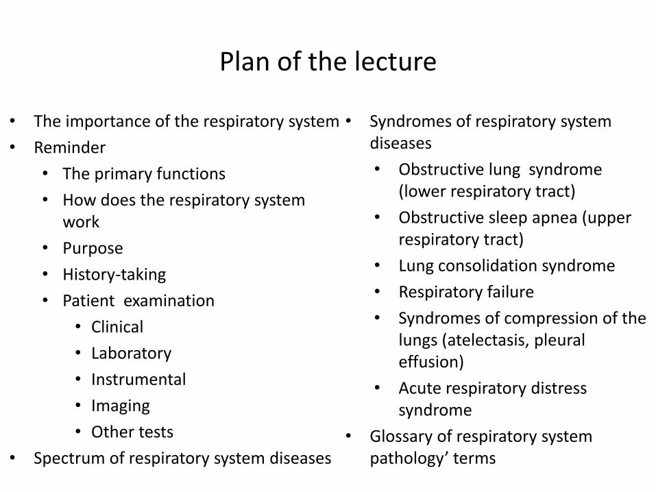
Plan of the lecture
• The importance of the respiratory system
• Reminder
• The primary functions
• How does the respiratory system work
• Purpose
• History-taking
• Patient examination
• Clinical
• Laboratory
• Instrumental
• Imaging
• Other tests
• Spectrum of respiratory system diseases
• Syndromes of respiratory system diseases
• Obstructive lung syndrome (lower respiratory tract)
• Obstructive sleep apnea (upper respiratory tract)
• Lung consolidation syndrome
• Respiratory failure
• Syndromes of compression of the lungs (atelectasis, pleural effusion)
• Acute respiratory distress syndrome
• Glossary of respiratory system pathology’ terms
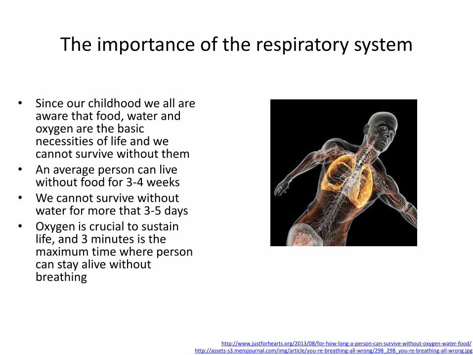
The importance of the respiratory system
• Since our childhood we all are aware that food, water and oxygen are the basic necessities of life and we cannot survive without them
• An average person can live without food for 3-4 weeks
• We cannot survive without water for more that 3-5 days
• Oxygen is crucial to sustain life, and 3 minutes is the maximum time where person can stay alive without breathing
http://www.justforhearts.org/2013/08/for-how-long-a-person-can-survive-without-oxygen-water-food/ http://assets-s3.mensjournal.com/img/article/you-re-breathing-all-wrong/298_298_you-re-breathing-all-wrong.jpg
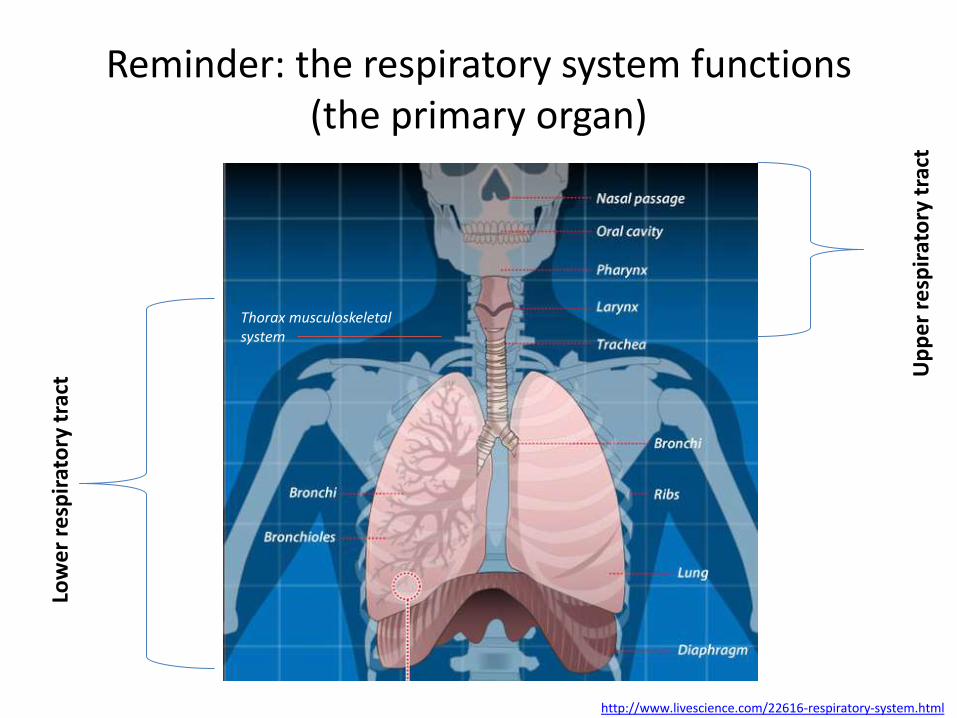
Reminder: the respiratory system functions (the primary organ)
http://www.livescience.com/22616-respiratory-system.html
Thorax musculoskeletal system
Low
er
resp
irat
ory
tra
ct U
pp
er
resp
irat
ory
tra
ct

Reminder: the respiratory system functions (gas exchange process)
http://www.livescience.com/22616-respiratory-system.html
Thorax musculoskeletal system
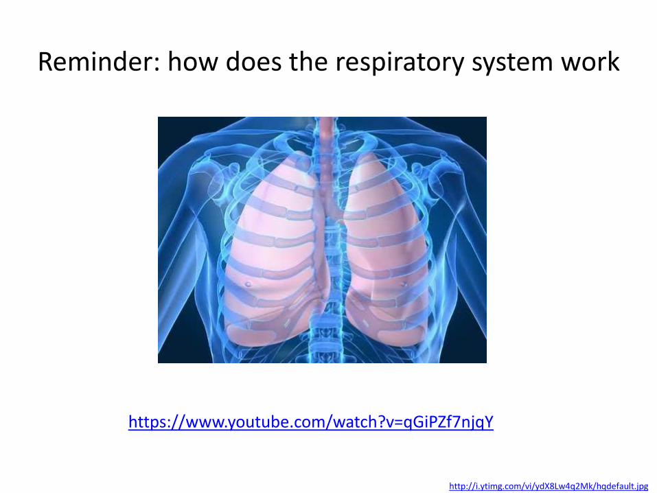
Reminder: how does the respiratory system work
https://www.youtube.com/watch?v=qGiPZf7njqY
http://i.ytimg.com/vi/ydX8Lw4q2Mk/hqdefault.jpg

Reminder: the respiratory system functions
• Gas exchange
• Immune functions
• Metabolic functions
• Endocrine functions
• Vocalization
• Temperature control
• Clearing the air (coughing and sneezing)
https://en.wikipedia.org/wiki/Respiratory_system https://aidanews.files.wordpress.com/2013/11/28thapr_2012_1.jpg

Reminder: purpose
• General evaluation of health
• Diagnosis of disease or disorders of the respiratory system
• Diagnosis of other systemic diseases that affect respiratory system functions
• Monitoring of patients with respiratory system diseases
http://www.americannursetoday.com/wp-content/uploads/2014/10/AMNT-Nov14-CNE-624x295.jpg
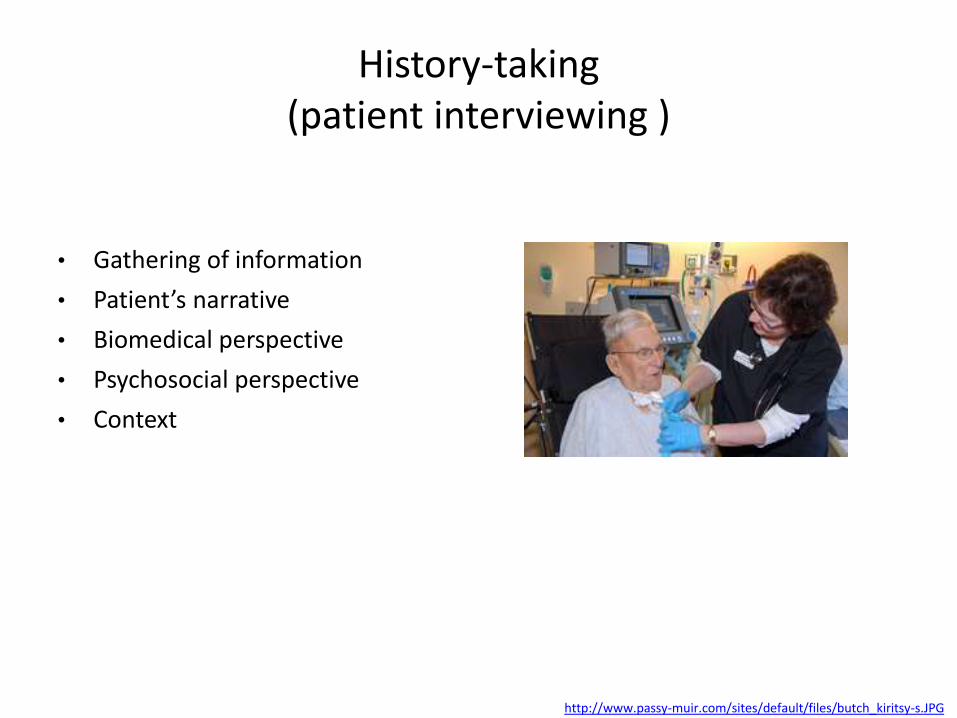
History-taking (patient interviewing )
• Gathering of information
• Patient’s narrative
• Biomedical perspective
• Psychosocial perspective
• Context
http://www.passy-muir.com/sites/default/files/butch_kiritsy-s.JPG

History-taking (patient interviewing )
• The history can often establish whether symptoms of dyspnea, chest pain, wheezing, stridor, hemoptysis, and cough are likely to be pulmonary in origin
• When more than one symptom occurs concurrently, the history should focus on which symptom is primary and whether constitutional symptoms, such as fever, weight loss, and night sweats, are also present
• Other important information includes:
– Occupational and environmental exposures
• Family history, travel history, and contact history
• Previous illnesses
• Use of prescription, OTC, or illicit drugs
• Previous test results (e.g., tuberculin skin test, chest x-rays)
http://www.healthunit.org/infectious/tb/tbimages/TB-Reading-3.jpg http://www.merckmanuals.com/professional/pulmonary-disorders/approach-to-the-pulmonary-patient/evaluation-of-the-pulmonary-patient
Tuberculin skin test
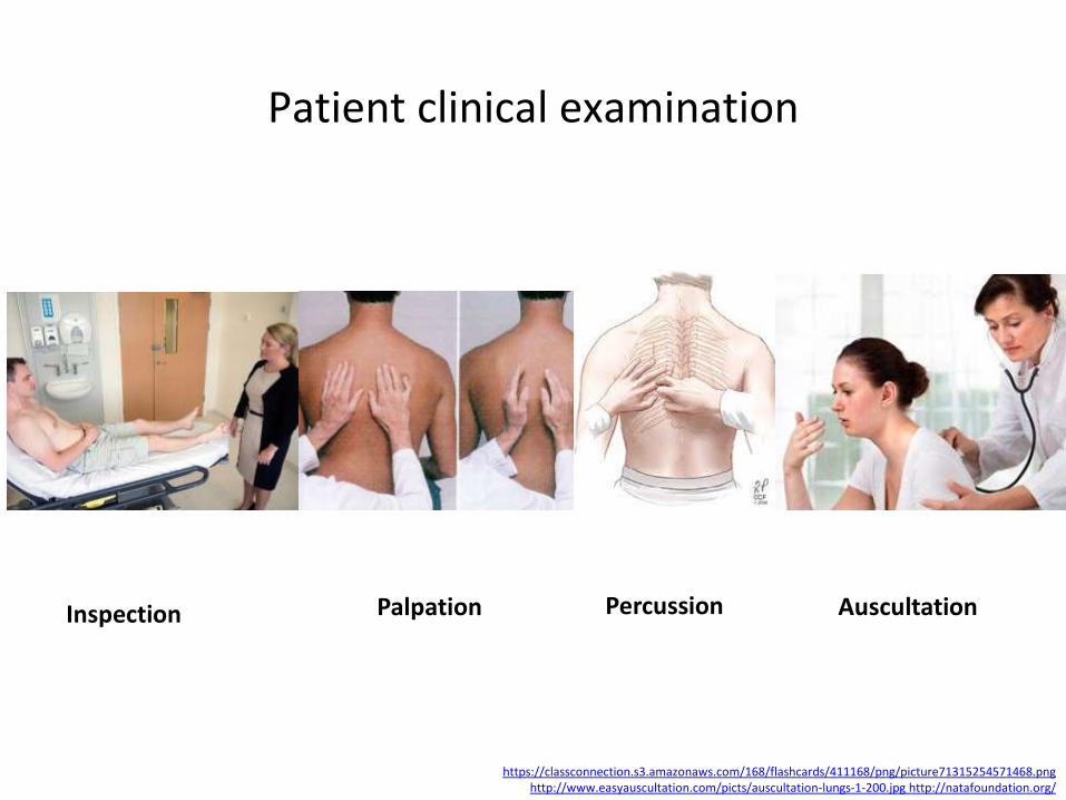
Patient clinical examination
https://classconnection.s3.amazonaws.com/168/flashcards/411168/png/picture71315254571468.png http://www.easyauscultation.com/picts/auscultation-lungs-1-200.jpg http://natafoundation.org/
Palpation Percussion Auscultation Inspection
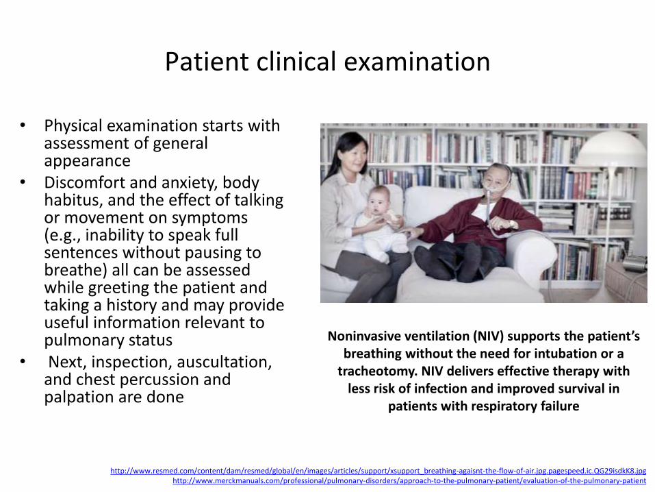
Patient clinical examination
• Physical examination starts with assessment of general appearance
• Discomfort and anxiety, body habitus, and the effect of talking or movement on symptoms (e.g., inability to speak full sentences without pausing to breathe) all can be assessed while greeting the patient and taking a history and may provide useful information relevant to pulmonary status
• Next, inspection, auscultation, and chest percussion and palpation are done
http://www.resmed.com/content/dam/resmed/global/en/images/articles/support/xsupport_breathing-agaisnt-the-flow-of-air.jpg.pagespeed.ic.QG29isdkK8.jpg http://www.merckmanuals.com/professional/pulmonary-disorders/approach-to-the-pulmonary-patient/evaluation-of-the-pulmonary-patient
Noninvasive ventilation (NIV) supports the patient’s breathing without the need for intubation or a
tracheotomy. NIV delivers effective therapy with less risk of infection and improved survival in
patients with respiratory failure

Patient clinical examination: inspection
• Signs of respiratory difficulty and hypoxemia (e.g., restlessness, tachypnea, cyanosis, accessory respiratory muscle use)
• Signs of possible chronic pulmonary disease (e.g., clubbing,, pedal edema)
• Chest wall deformities • Abnormal breathing
patterns (e.g., Cheyne-Stokes respiration, Kussmaul respirations)
• Jugular venous distention
http://www.merckmanuals.com/professional/pulmonary-disorders/approach-to-the-pulmonary-patient/evaluation-of-the-pulmonary-patient
The ratio of the anteroposterior diameter of the finger at the nail bed (a–b) to that at the distal
interphalangeal joint (c–d) is a simple measurement of finger clubbing. It can be
obtained readily and reproducibly with calipers. If the ratio is > 1, clubbing is present. Finger
clubbing is also characterized by loss of the normal angle at the nail bed.
Patient clinical examination: Cheyne-Stokes respiration
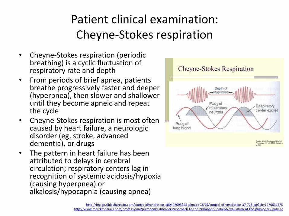
• Cheyne-Stokes respiration (periodic breathing) is a cyclic fluctuation of respiratory rate and depth
• From periods of brief apnea, patients breathe progressively faster and deeper (hyperpnea), then slower and shallower until they become apneic and repeat the cycle
• Cheyne-Stokes respiration is most often caused by heart failure, a neurologic disorder (eg, stroke, advanced dementia), or drugs
• The pattern in heart failure has been attributed to delays in cerebral circulation; respiratory centers lag in recognition of systemic acidosis/hypoxia (causing hyperpnea) or alkalosis/hypocapnia (causing apnea)
http://image.slidesharecdn.com/controlofventilation-100407095845-phpapp02/95/control-of-ventilation-37-728.jpg?cb=1270634375 http://www.merckmanuals.com/professional/pulmonary-disorders/approach-to-the-pulmonary-patient/evaluation-of-the-pulmonary-patient
Patient clinical examination: Kussmaul respirations
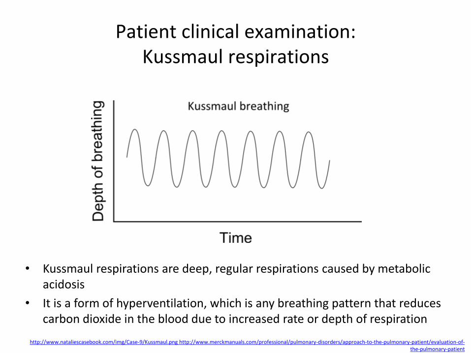
• Kussmaul respirations are deep, regular respirations caused by metabolic acidosis
• It is a form of hyperventilation, which is any breathing pattern that reduces carbon dioxide in the blood due to increased rate or depth of respiration
http://www.nataliescasebook.com/img/Case-9/Kussmaul.png http://www.merckmanuals.com/professional/pulmonary-disorders/approach-to-the-pulmonary-patient/evaluation-of-the-pulmonary-patient
Patient clinical examination: jugular venous distention
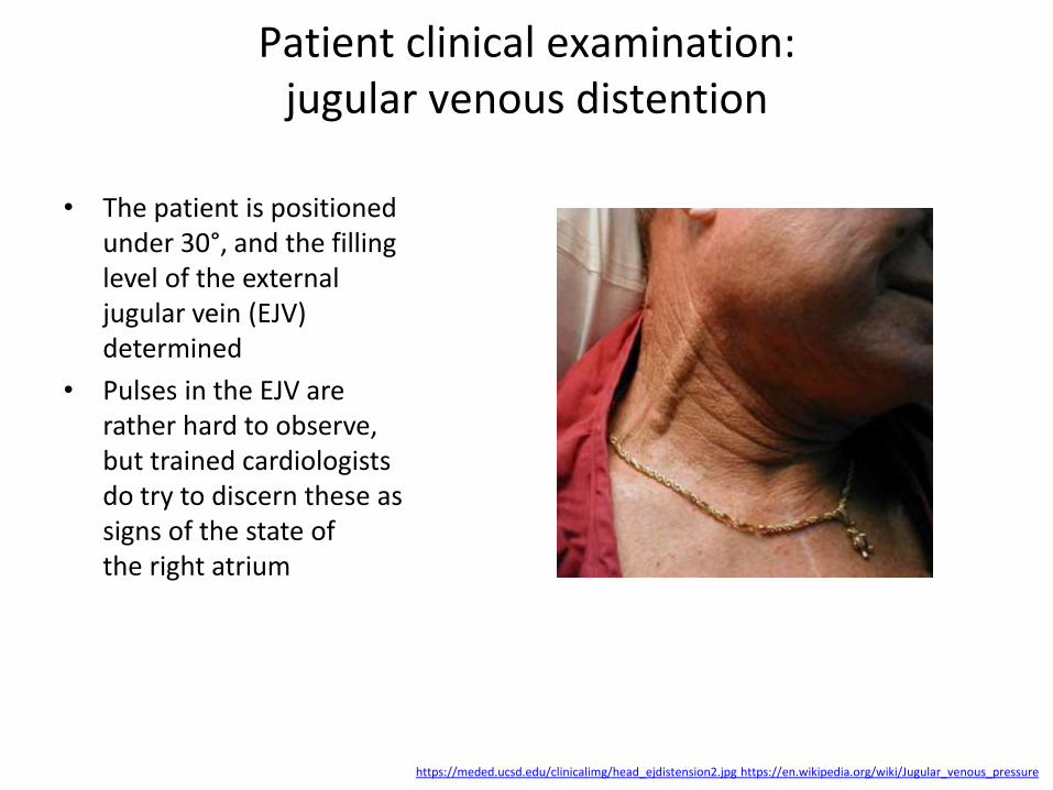
• The patient is positioned
under 30°, and the filling level of the external jugular vein (EJV) determined
• Pulses in the EJV are rather hard to observe, but trained cardiologists do try to discern these as signs of the state of the right atrium
https://meded.ucsd.edu/clinicalimg/head_ejdistension2.jpg https://en.wikipedia.org/wiki/Jugular_venous_pressure

Patient clinical examination: palpation and percussion
• Palpation includes tactile fremitus (vibration of the chest wall felt while a patient is speaking); it is decreased in pleural effusion and pneumothorax and increased in pulmonary consolidation (e.g., lobar pneumonias); point tenderness on palpation may signal underlying rib fracture or pleural inflammation
• Percussion is the primary physical maneuver used to detect the presence and level of pleural effusion: finding areas of dullness during percussion signifies underlying fluid or, less commonly, consolidation
• In cor pulmonale, a right ventricular impulse at the left lower sternal border may become evident and may be increased in amplitude and duration (right ventricular heave)
http://intranet.tdmu.edu.ua/data/kafedra/internal/distance/classes_stud/English/1course/Heath%20Assessment%20Practicum/Health%20Assessment%20Practicum/17.%20Respiratory%20System%20Assesment.files/image004.jpg http://www.merckmanuals.com/professional/pulmonary-disorders/approach-to-the-pulmonary-patient/evaluation-of-the-pulmonary-patient
Patient clinical examination: lung auscultation
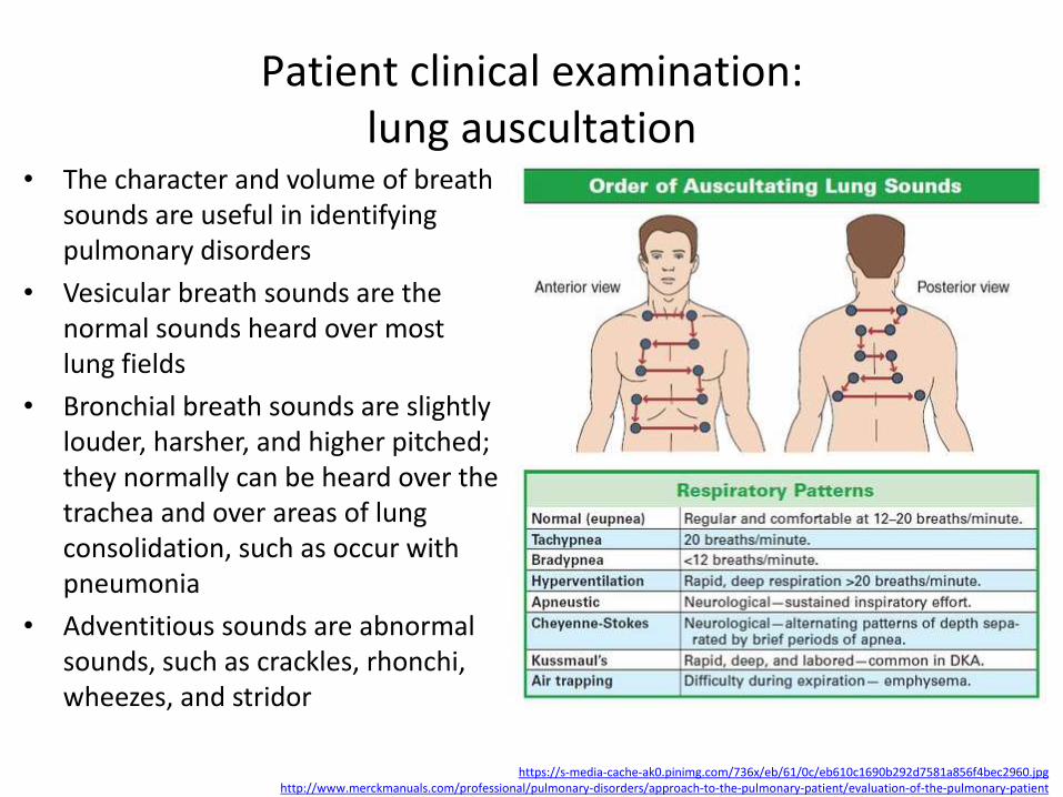
• The character and volume of breath sounds are useful in identifying pulmonary disorders
• Vesicular breath sounds are the normal sounds heard over most lung fields
• Bronchial breath sounds are slightly louder, harsher, and higher pitched; they normally can be heard over the trachea and over areas of lung consolidation, such as occur with pneumonia
• Adventitious sounds are abnormal sounds, such as crackles, rhonchi, wheezes, and stridor
https://s-media-cache-ak0.pinimg.com/736x/eb/61/0c/eb610c1690b292d7581a856f4bec2960.jpg http://www.merckmanuals.com/professional/pulmonary-disorders/approach-to-the-pulmonary-patient/evaluation-of-the-pulmonary-patient
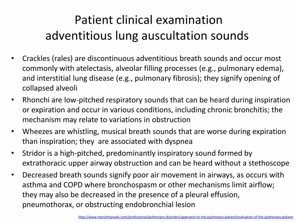
Patient clinical examination adventitious lung auscultation sounds
• Crackles (rales) are discontinuous adventitious breath sounds and occur most commonly with atelectasis, alveolar filling processes (e.g., pulmonary edema), and interstitial lung disease (e.g., pulmonary fibrosis); they signify opening of collapsed alveoli
• Rhonchi are low-pitched respiratory sounds that can be heard during inspiration or expiration and occur in various conditions, including chronic bronchitis; the mechanism may relate to variations in obstruction
• Wheezes are whistling, musical breath sounds that are worse during expiration than inspiration; they are associated with dyspnea
• Stridor is a high-pitched, predominantly inspiratory sound formed by extrathoracic upper airway obstruction and can be heard without a stethoscope
• Decreased breath sounds signify poor air movement in airways, as occurs with asthma and COPD where bronchospasm or other mechanisms limit airflow; they may also be decreased in the presence of a pleural effusion, pneumothorax, or obstructing endobronchial lesion
http://www.merckmanuals.com/professional/pulmonary-disorders/approach-to-the-pulmonary-patient/evaluation-of-the-pulmonary-patient
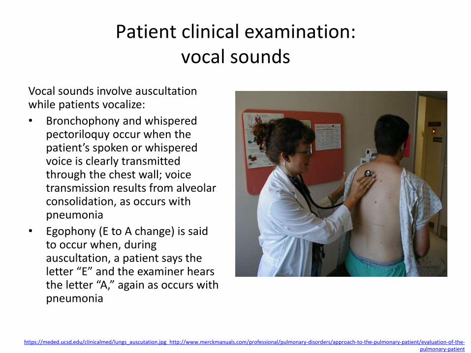
Patient clinical examination: vocal sounds
Vocal sounds involve auscultation while patients vocalize:
• Bronchophony and whispered pectoriloquy occur when the patient’s spoken or whispered voice is clearly transmitted through the chest wall; voice transmission results from alveolar consolidation, as occurs with pneumonia
• Egophony (E to A change) is said to occur when, during auscultation, a patient says the letter “E” and the examiner hears the letter “A,” again as occurs with pneumonia
https://meded.ucsd.edu/clinicalmed/lungs_auscutation.jpg http://www.merckmanuals.com/professional/pulmonary-disorders/approach-to-the-pulmonary-patient/evaluation-of-the-pulmonary-patient
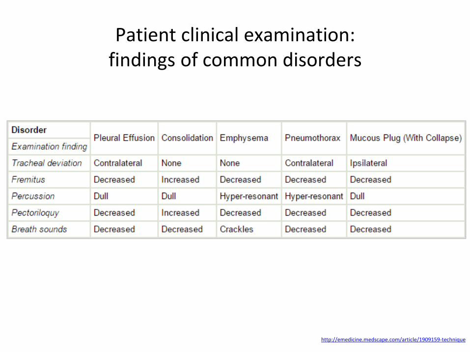
Patient clinical examination: findings of common disorders
http://emedicine.medscape.com/article/1909159-technique
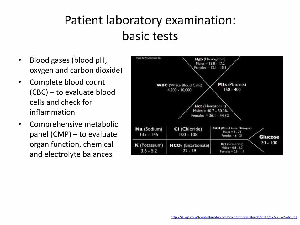
Patient laboratory examination: basic tests
• Blood gases (blood pH, oxygen and carbon dioxide)
• Complete blood count (CBC) – to evaluate blood cells and check for inflammation
• Comprehensive metabolic panel (CMP) – to evaluate organ function, chemical and electrolyte balances
http://i1.wp.com/leonardonoto.com/wp-content/uploads/2013/07/c767d9a61.jpg
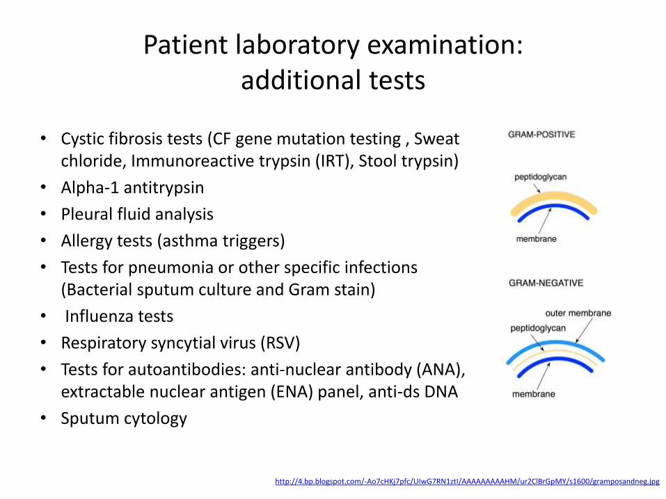
Patient laboratory examination: additional tests
• Cystic fibrosis tests (CF gene mutation testing , Sweat chloride, Immunoreactive trypsin (IRT), Stool trypsin)
• Alpha-1 antitrypsin
• Pleural fluid analysis
• Allergy tests (asthma triggers)
• Tests for pneumonia or other specific infections (Bacterial sputum culture and Gram stain)
• Influenza tests
• Respiratory syncytial virus (RSV)
• Tests for autoantibodies: anti-nuclear antibody (ANA), extractable nuclear antigen (ENA) panel, anti-ds DNA
• Sputum cytology
http://4.bp.blogspot.com/-Ao7cHKj7pfc/UIwG7RN1ztI/AAAAAAAAAHM/ur2ClBrGpMY/s1600/gramposandneg.jpg

Patient instrumental examination
• Spirometry (to evaluate narrowed or obstructed airways)
• Oximetry (measures the oxygen saturation of the blood)
• Exercise stress test on a stationary bike or treadmill
• Air flow with a peak flow meter (measures the rate of exhalation at home)
• Lung volume (the quantity of air a person takes into their lungs and how much is left in the lungs after exhalation)
• Diffusing capacity measurement (the transfer of oxygen from the lung air sacs to the bloodstream)
https://classconnection.s3.amazonaws.com/168/flashcards/411168/png/picture71315254571468.png http://www.easyauscultation.com/picts/auscultation-lungs-1-200.jpg http://natafoundation.org/ http://www.berktree.com/assets/images 1801.jpg

Patient instrumental examination: imaging tests
• Chest x-ray – to look at lung structure and chest cavity
• CT (computed tomography) scan – a more detailed evaluation of lung structure
• MRI (Magnetic resonance imaging) – detailed pictures of organs and vessels in the chest
• Ultrasound – used to detect fluid between the pleural membranes
• Nuclear lung scanning – used to help detect pulmonary embolism and, rarely, to evaluate the effectiveness of lung cancer treatment
• Positron emission tomography (PET) scans – used to help diagnose lung cancer
https://classconnection.s3.amazonaws.com/168/flashcards/411168/png/picture71315254571468.png http://www.easyauscultation.com/picts/auscultation-lungs-1-200.jpg http://natafoundation.org/ http://www.siemens.com/press/pool/de/pressebilder

Patient instrumental examination: other tests
• Electrocardiogram (EKG, ECG) – to look at heart rhythm, to determine if heart disease may be affecting breathing
• Sleep studies – usually performed at special sleep centers to help determine whether a person is breathing normally during sleep
https://classconnection.s3.amazonaws.com/168/flashcards/411168/png/picture71315254571468.png http://www.easyauscultation.com/picts/auscultation-lungs-1-200.jpg http://natafoundation.org/ https://www.getsleepnj.com/images/sleep-stages.gif

Spectrum of the respiratory system diseases
• Obstructive conditions (e.g., emphysema, bronchitis, asthma attacks)
• Restrictive conditions (e.g., fibrosis, sarcoidosis, alveolar damage, pleural effusion)
• Vascular diseases (e.g., pulmonary edema, pulmonary embolism, pulmonary hypertension)
• Infectious, environmental and other "diseases" (e.g., pneumonia, tuberculosis, asbestosis, particulate pollutants)
The respiratory tract is constantly exposed to microbes due to the extensive surface area,
which is why the respiratory system includes many mechanisms to defend itself and prevent
pathogens from entering the body
http://www.disabled-world.com/health/respiratory/ http://www.tobaccolabels.ca/
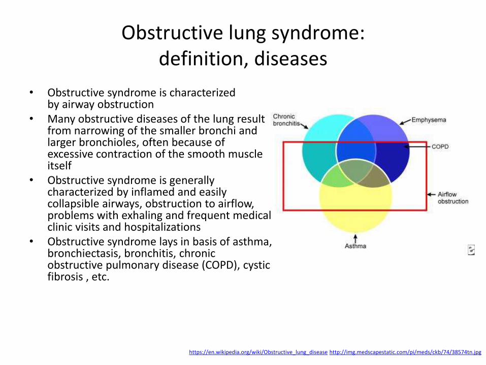
Obstructive lung syndrome: definition, diseases
• Obstructive syndrome is characterized by airway obstruction
• Many obstructive diseases of the lung result from narrowing of the smaller bronchi and larger bronchioles, often because of excessive contraction of the smooth muscle itself
• Obstructive syndrome is generally characterized by inflamed and easily collapsible airways, obstruction to airflow, problems with exhaling and frequent medical clinic visits and hospitalizations
• Obstructive syndrome lays in basis of asthma, bronchiectasis, bronchitis, chronic obstructive pulmonary disease (COPD), cystic fibrosis , etc.
https://en.wikipedia.org/wiki/Obstructive_lung_disease http://img.medscapestatic.com/pi/meds/ckb/74/38574tn.jpg
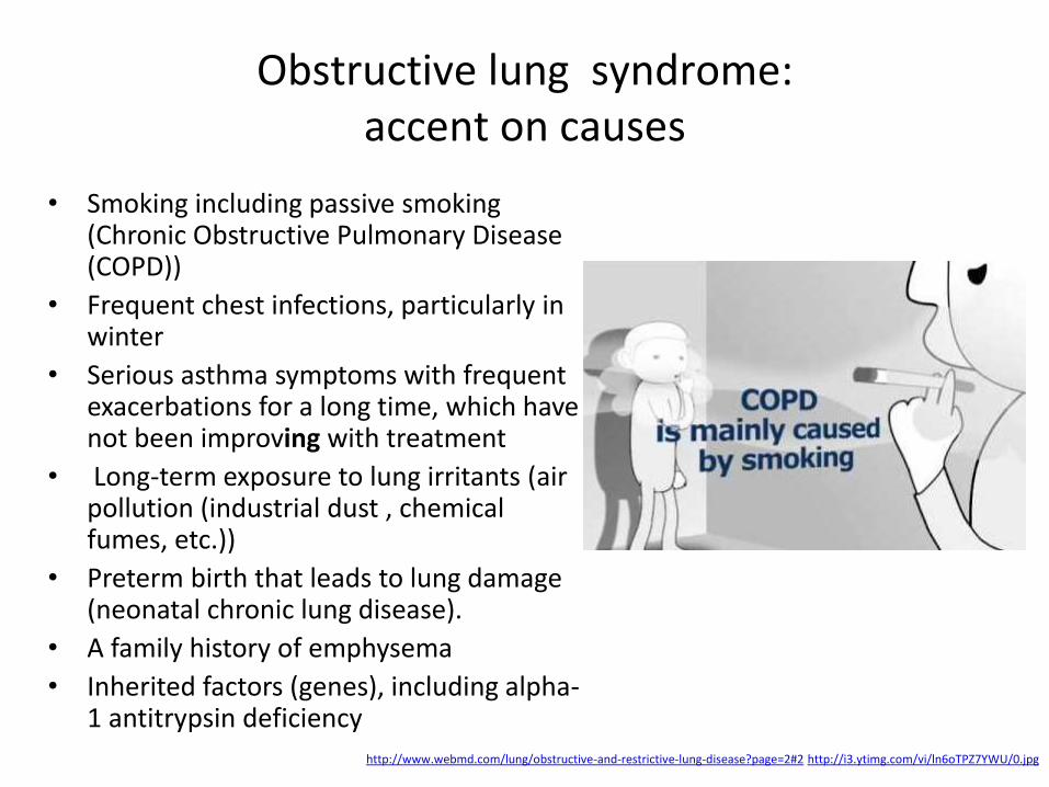
Obstructive lung syndrome: accent on causes
• Smoking including passive smoking (Chronic Obstructive Pulmonary Disease (COPD))
• Frequent chest infections, particularly in winter
• Serious asthma symptoms with frequent exacerbations for a long time, which have not been improving with treatment
• Long-term exposure to lung irritants (air pollution (industrial dust , chemical fumes, etc.))
• Preterm birth that leads to lung damage (neonatal chronic lung disease).
• A family history of emphysema
• Inherited factors (genes), including alpha-1 antitrypsin deficiency
http://www.webmd.com/lung/obstructive-and-restrictive-lung-disease?page=2#2 http://i3.ytimg.com/vi/ln6oTPZ7YWU/0.jpg

Obstructive lung syndrome (OSL): proportional Venn diagram of OLS in the United States (NHANES III surveys from 1988 to 1994)
http://journal.publications.chestnet.org/data/Journals/CHEST/21997/cb0635683005.jpeg
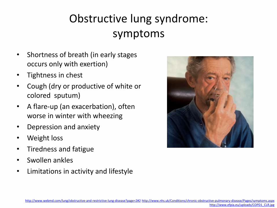
Obstructive lung syndrome: symptoms
• Shortness of breath (in early stages occurs only with exertion)
• Tightness in chest
• Cough (dry or productive of white or colored sputum)
• A flare-up (an exacerbation), often worse in winter with wheezing
• Depression and anxiety
• Weight loss
• Tiredness and fatigue
• Swollen ankles
• Limitations in activity and lifestyle
http://www.webmd.com/lung/obstructive-and-restrictive-lung-disease?page=2#2 http://www.nhs.uk/Conditions/chronic-obstructive-pulmonary-disease/Pages/symptoms.aspx http://www.efpia.eu/uploads/COPD1_CLR.jpg

Obstructive lung syndrome: conditions and nature
https://en.wikipedia.org/wiki/Obstructive_lung_disease
Condition Main site Major changes Causes Symptoms
Chronic bronchitis Bronchus
Hyperplasia and
hypersecretion of
mucus glands
Tobacco smoking and
air pollutants Productive cough
Bronchiectasis Bronchus Dilation and scarring of
airways
Persistent severe
infections
Cough, purulent
sputum and fever
Asthma Bronchus
Smooth
muscle hyperplasia,
excessive mucus,
inflammation, constriction
Immunologic
or idiopathic
Episodic
wheezing, cough
and dyspnea
Bronchiolitis (subgroup of
chronic bronchitis) Bronchiole
Inflammatory scarring and
bronchiole obliteration
Tobacco smoking and
air pollutants Cough, dyspnea

Obstructive lung syndrome: spot the difference
• In asthma the bronchial tubes (airways) are hyperresponsive and usually triggered by breathing in things in the air such as dust, pollen, etc. with recurring episodes of wheezing, breathlessness, chest tightness, and coughing, particularly at night or in the early morning
• Bronchiectasis refers to the abnormal, irreversible dilatation of the bronchi caused by destructive and inflammatory changes in the airway walls
• Chronic obstructive pulmonary disease (COPD) is characterized by airflow limitation that is not fully reversible
http://www.meddean.luc.edu/lumen/MedEd/elective/pulmonary/bronchiectasis/bronchi_f.htm https://en.wikipedia.org/wiki/Obstructive_lung_disease

Obstructive lung syndrome: spot the difference
http://vaticancities.com/image/551e2d0f7f9ef.jpg
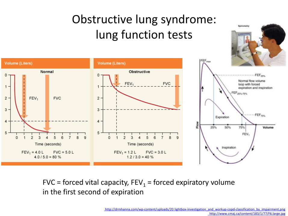
Obstructive lung syndrome: lung function tests
http://drmhanna.com/wp-content/uploads/20 lightbox-investigation_and_workup-copd-classification_by_impairment.png http://www.cmaj.ca/content/183/1/77/F6.large.jpg
FVC = forced vital capacity, FEV1 = forced expiratory volume in the first second of expiration

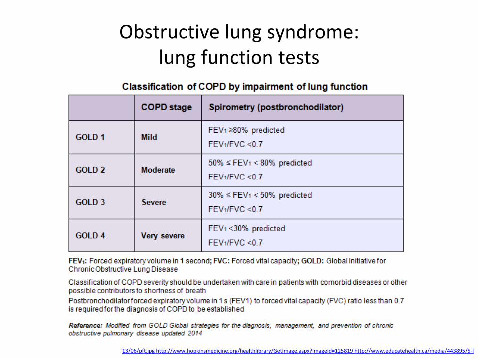
Obstructive lung syndrome: lung function tests
The obstructive defect is reversible because at least one of the two measurements (FVC or FEV1) increased by at least 0.2 L and by at least 12%. (FEF25%–75% = forced expiratory flow at 25% to 75% of FVC; FEV1 = forced expiratory volume in one second; FVC = forced vital capacity; LLN = lower limit of normal)
http://www.aafp.org/afp/2014/0301/afp20140301p359-f4.gif
Obstructive lung syndrome: lung function tests
13/06/pft.jpg http://www.hopkinsmedicine.org/healthlibrary/GetImage.aspx?ImageId=125819 http://www.educatehealth.ca/media/443895/5-l

Obstructive lung syndrome: spirometric measures in asthma, COPD and ACOS
http://www.goldcopd.org/uploads/users/files/AsthmaCOPDOverlap.pdf
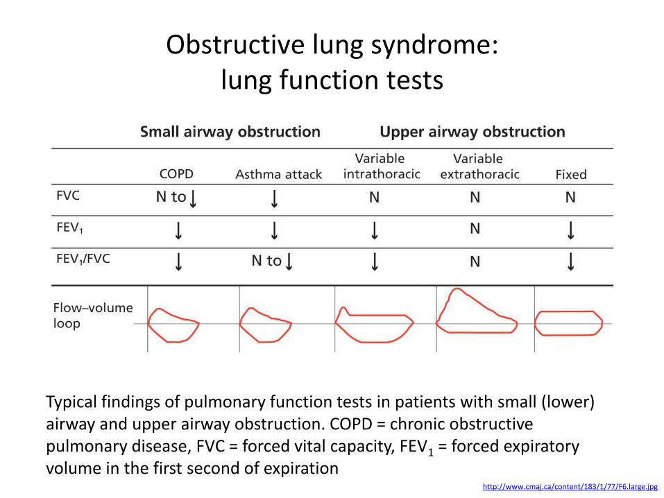
Obstructive lung syndrome: lung function tests
http://www.cmaj.ca/content/183/1/77/F6.large.jpg
Typical findings of pulmonary function tests in patients with small (lower) airway and upper airway obstruction. COPD = chronic obstructive pulmonary disease, FVC = forced vital capacity, FEV1 = forced expiratory volume in the first second of expiration

Obstructive lung syndrome: lung function decline in smokers and nonsmokers
http://www.cmaj.ca/content/183/1/77/F6.large.jpg
Smokers who are susceptible to lung injury experience an increase in the rate of age-related loss in FEV1 compared with nonsmokers (red, green, and blue lines) After lung function declines to threshold levels, clinical symptoms develop (black dotted lines) When a smoker stops smoking, the rate of FEV1 loss again approximates to that of a nonsmoker (blue dotted line)
FEV1 = forced expiratory volume in one second

Obstructive lung syndrome: chest X-ray
• A chest X-ray may show signs of obstructive lung syndromes and can be used to help exclude other serious conditions (including lung cancer)
• The X-ray may show:
– Flattening of the diaphragm, the large muscle that separates the lungs and heart from the abdominal cavity
– Increased size of the chest, as measured from front to back
– A long narrow heart
– Abnormal air collections within the lung (focal bullae)
• A normal chest X-ray does not mean patient do not have COPD
https://emcow.files.wordpress.com/2013/10/copd-and-ptx.jpg?w=540&h=444 http://www.webmd.com/lung/copd/chest-x-rays-for-chronic-obstructive-pulmonary-disease-copd http://www.webmd.com/lung/obstructive-and-restrictive-lung-disease?page=2#2
The X-ray demonstrates a pneumothorax on the left side in which a chest tube was placed for reexpansion. On the right side the patient has multiple large apical bullae which are also at risk of rupture.

Obstructive lung syndrome: arterial blood gas test
• Arterial blood gas analysis is used to measure the pH and the partial pressures of oxygen and carbon dioxide in arterial blood
• Interpretation of an arterial blood gas result should not be done without considering the clinical findings
• Factors relating to sampling technique, specimen processing and environment may also influence the results
https://labtestsonline.org/understanding/analytes/blood-gases/tab/test/ http://www.australianprescriber.com/magazine/33/4/124/9

Obstructive lung syndrome: oximetry
• The test measures the oxygen saturation in the blood
• The test can be useful in finding out whether oxygen treatment is needed, but it provides less information than the arterial blood gas test
http://www.copd-alert.com/OximetryPG.pdf http://www.webmd.com/lung/obstructive-and-restrictive-lung-disease?page=2#2

Obstructive lung syndrome: electrocardiogram
ECG changes occur in obstructive lung syndromes due to:
• The presence of hyperexpanded emphysematous lungs within the chest
• The long-term effects of hypoxic pulmonary vasoconstriction upon the right side of the heart, causing pulmonary hypertension and subsequent right atrial and right ventricular hypertrophy (i.e. cor pulmonale)
http://lifeinthefastlane.com/ecg-library/copd/
The ECG demonstrates many of the features of chronic pulmonary disease: Rightward QRS axis (+90 degrees) Peaked P waves in the inferior leads > 2.5 mm (P pulmonale) with a rightward P-wave axis (inverted in aVL) Clockwise rotation of the heart with a delayed R/S transition point (transitional lead = V5) Absent R waves in the right precordial leads (SV1-SV2-SV3 pattern) Low voltages in the left-sided leads (I, aVL, V5-6)
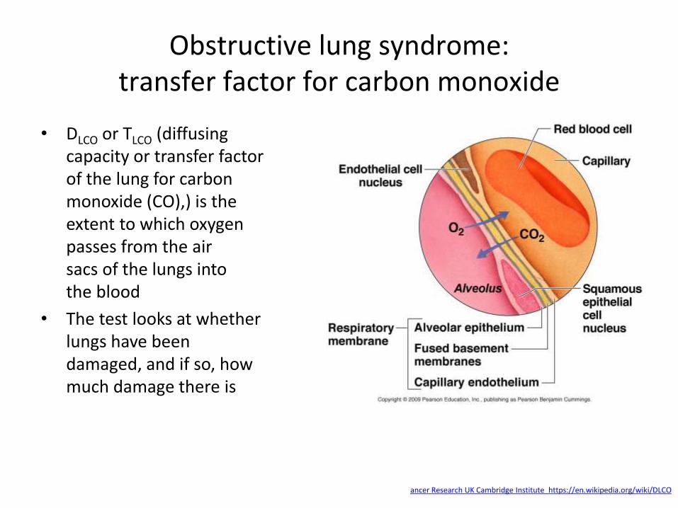
Obstructive lung syndrome: transfer factor for carbon monoxide
• DLCO or TLCO (diffusing capacity or transfer factor of the lung for carbon monoxide (CO),) is the extent to which oxygen passes from the air sacs of the lungs into the blood
• The test looks at whether lungs have been damaged, and if so, how much damage there is
ancer Research UK Cambridge Institute https://en.wikipedia.org/wiki/DLCO

Obstructive lung syndrome: tests rarely done
• Alpha-1 antitrypsin (AAT) test for recognizing emphysema
• A CT scan for detailed picture of the lungs
http://www.webmd.com/lung/obstructive-and-restrictive-lung-disease?page=2#2
Computed tomography of the lung showing emphysema and bullae in the lower lung lobes of a subject with type ZZ alpha-1-antitrypsin deficiency

Obstructive lung syndrome: specialized investigations sometimes used in
distinguishing asthma and COPD
http://www.goldcopd.org/uploads/users/files/AsthmaCOPDOverlap.pdf
Obstructive lung syndrome: regular checkups
• Spirometry
• Arterial blood gas test
• X-rays or ECGs
http://i.dailymail.co.uk/i/pix/2014/02/05/article-2552340-1B046ECC00000578-941_634x649.jpg http://www.webmd.com/lung/copd/tc/chronic-obstructive-pulmonary-disease-copd-exams-and-tests
I quit smoking 30 years ago, not soon enough, I have COPD

Obstructive sleep apnea: definition, causes
• Obstructive sleep apnea (OSA) is caused by obstruction of the upper airway
• OSA is characterized by repetitive pauses (apneas ) in breathing during sleep, which typically last 20 to 40 seconds despite the effort to breathe.
• OSA is usually associated with a reduction in blood oxygen saturation
• OSA is commonly accompanied with snoring
• The main causes of OSA are old age, temporary or permanent brain injury, decreased muscle tone, excess soft tissue around the airway (common with obese patients), something physical in the throat or mouth/jaw shape
http://www.mayoclinic.org/diseases-conditions/obstructive-sleep-apnea/basics/definition/con-20027941 https://en.wikipedia.org/wiki/Obstructive_sleep_apnea http://ptsddiary.com/wp-content/uploads/2012/10/Screen-shot-2012-10-08-at-1.32.59-PM.png

Obstructive sleep apnea: symptoms
• Excessive daytime sleepiness
• Loud snoring
• Episodes of breathing cessation in sleep
• Abrupt awakenings by shortness of breath
• Awakening with a dry mouth or sore throat
• Awakening with chest pain
• Morning headache
• Difficulty concentrating during the day
• Experiencing mood changes
• Difficulty staying asleep
• High blood pressure
http://www.mayoclinic.org/diseases-conditions/obstructive-sleep-apnea/basics/definition/con-20027941 http://www.parmardmd.com/images/Symptoms-of-Sleep-Apnea-Slide.jpg

Obstructive sleep apnea: diagnosis
• Nocturnal polysomnography - records brain wave changes, eye movements, leg movements, blood oxygen levels, muscle tone, heart rhythms and respiration during sleep
• Oximetry
• Epworth sleepiness scale - to measure the patient's level of daytime sleepiness
• The three ratings for OSA:
• Mild - 5-14 episodes of apnea or hypopnea per hour
• Moderate - 15 to 30 episodes of apnea or hypopnea per hour
• Severe - over 30 episodes of apnea or hypopnea per hour
http://www.dentist-charlotte-north-carolina-nc.com/images/sleep-study-1.jpg http://www.medicalnewstoday.com/articles/178633.php

Obstructive sleep apnea: nocturnal polysomnography
• 30-second epoch of a polysomnographic recording in the 13 channels muscular tension (EMG), eye movements (EOG), bioelectrical brain function (EEG), heart rate (ECG), breathing (flow, sum, upper and lower effort), snoring (Trach), body position (BodyPos) and oxygen saturation (SPO2) are recorded
• During the first 10 seconds an obstructive apnea (cessation of breathing) is clearly visible as a flat line in the flow channel
http://www.schlaflabor-saletu.at/tl_files/Schlaflabor_Saletu/Schlaflabor_Abb/polysomnogramm.gif
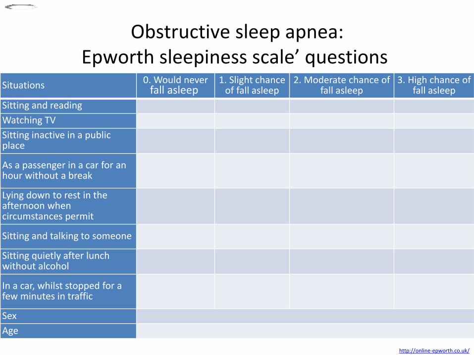
Obstructive sleep apnea: Epworth sleepiness scale’ questions
http://online-epworth.co.uk/
Situations 0. Would never
fall asleep 1. Slight chance
of fall asleep 2. Moderate chance of
fall asleep 3. High chance of
fall asleep
Sitting and reading
Watching TV
Sitting inactive in a public place
As a passenger in a car for an hour without a break
Lying down to rest in the afternoon when circumstances permit
Sitting and talking to someone
Sitting quietly after lunch without alcohol
In a car, whilst stopped for a few minutes in traffic
Sex
Age
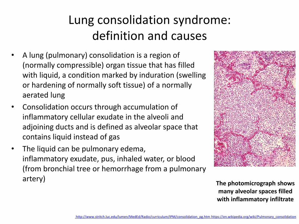
Lung consolidation syndrome: definition and causes
• A lung (pulmonary) consolidation is a region of (normally compressible) organ tissue that has filled with liquid, a condition marked by induration (swelling or hardening of normally soft tissue) of a normally aerated lung
• Consolidation occurs through accumulation of inflammatory cellular exudate in the alveoli and adjoining ducts and is defined as alveolar space that contains liquid instead of gas
• The liquid can be pulmonary edema, inflammatory exudate, pus, inhaled water, or blood (from bronchial tree or hemorrhage from a pulmonary artery)
http://www.stritch.luc.edu/lumen/MedEd/Radio/curriculum/IPM/consolidation_pg.htm https://en.wikipedia.org/wiki/Pulmonary_consolidation
The photomicrograph shows many alveolar spaces filled with inflammatory infiltrate
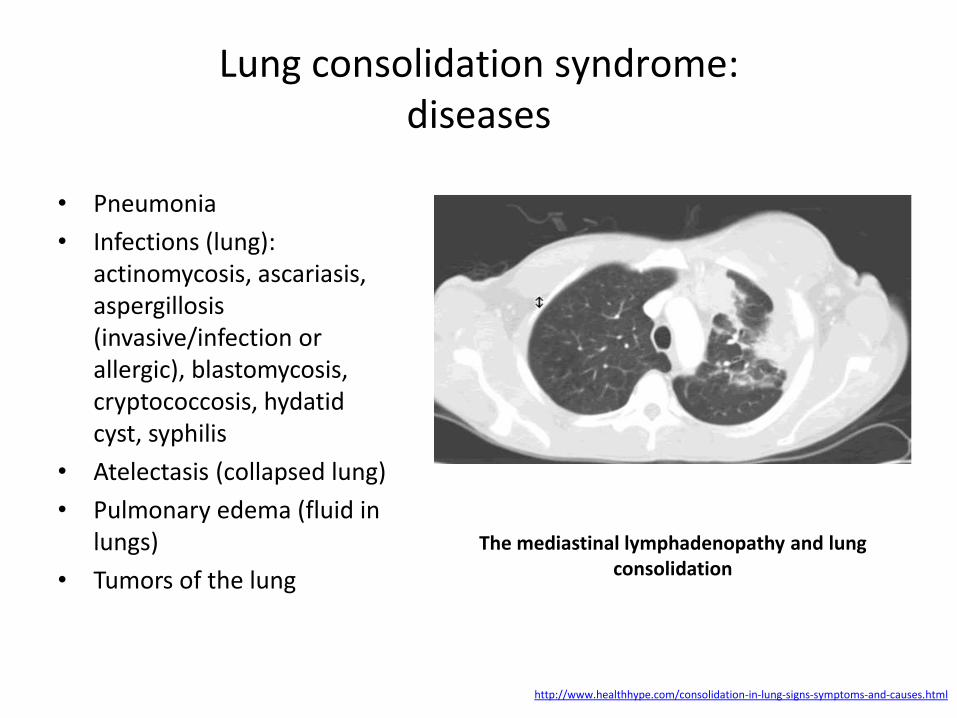
Lung consolidation syndrome: diseases
• Pneumonia
• Infections (lung): actinomycosis, ascariasis, aspergillosis (invasive/infection or allergic), blastomycosis, cryptococcosis, hydatid cyst, syphilis
• Atelectasis (collapsed lung)
• Pulmonary edema (fluid in lungs)
• Tumors of the lung
http://www.healthhype.com/consolidation-in-lung-signs-symptoms-and-causes.html
The mediastinal lymphadenopathy and lung consolidation

Lung consolidation syndrome: symptoms
• Dyspnea which is dependent on the extent of consolidation
• Abnormal breathing sounds
• Coughing
• Pallor acrocyanosis
• Percussion : dull note
• Palpation : tactile fremitus
• Vocal resonance
• Bronchial breathing and egophony (it is said to occur when, during auscultation, a patient says the letter “E” and the examiner hears the letter “A”)
• Pleural friction rub
• Unilateral reduction in chest expansion
http://openi.nlm.nih.gov/imgs/512/176/2647177/2647177_kjr-10-21-g003.png http://www.healthhype.com/consolidation-in-lung-signs-symptoms-and-causes.html
High-resolution CT scan at level of lower lung zones shows extensive "crazy-
paving" pattern involving both lower lobes, lingula and middle lobe, in
association with areas of air-space consolidation

Lung consolidation syndrome: X-ray features
• Opacity of the affected area, lobule or lobe
• Loss of clarity of the heart border, diaphragm and or verterbal bodies (thoracic vertebrae)
• Patchy consolidation may be seen with bronchopenumonia while confluent consolidation seen in lobar pneumonia
• Cavitation, bulging interlobular fissures and pleural effusion may also be evident
http://www.mayoclinic.org/~/media/kcms/gbs/patient%20consumer/images/2013/08/26/10/01/ds00135_im00621_pnuesmal_gif.ashx http://www.healthhype.com/consolidation-in-lung-signs-symptoms-and-causes.html
The chest X-ray shows an area of lung inflammation indicating the presence of
pneumonia

Lung consolidation syndrome: X-ray patterns of consolidation
• Consolidation may be complete or incomplete
• The distribution of the consolidation can vary widely
• A consolidation could be described as “patchy”, “homogenous”, or generalized”
• A consolidation may be described as focal or by the lobe or segment of lobe affected
http://www.wikiradiography.net/page/Patterns+of+Consolidation
There is abnormal opacity on the right (arrowed). There is also loss of clarity of the right heart border known as silhouette sign

Lung consolidation syndrome: Right Upper Lobe (RUL) consolidation
http://www.wikiradiography.net/page/Patterns+of+Consolidation

Lung consolidation syndrome: Right Middle Lobe (RUL) consolidation
http://www.wikiradiography.net/page/Patterns+of+Consolidation

Lung consolidation syndrome: Right Lower Lobe (RUL) consolidation
http://www.wikiradiography.net/page/Patterns+of+Consolidation

Lung consolidation syndrome: Left Upper Lobe (RUL) consolidation
http://www.wikiradiography.net/page/Patterns+of+Consolidation

Lung consolidation syndrome: Left Lower Lobe (RUL) consolidation
http://www.wikiradiography.net/page/Patterns+of+Consolidation
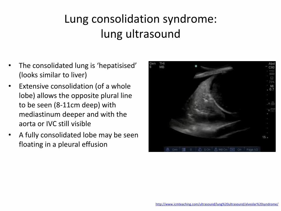
Lung consolidation syndrome: lung ultrasound
• The consolidated lung is ‘hepatisised’ (looks similar to liver)
• Extensive consolidation (of a whole lobe) allows the opposite plural line to be seen (8-11cm deep) with mediastinum deeper and with the aorta or IVC still visible
• A fully consolidated lobe may be seen floating in a pleural effusion
http://www.icmteaching.com/ultrasound/lung%20ultrasound/alveolar%20syndrome/

Respiratory failure: definition and types
• Respiratory failure occurs when the respiratory system fails in oxygenation and/or carbon dioxide (CO2) elimination.
• It may be acute (develops over minutes to hours) or chronic (develops over several weeks-months (clinical markers include polycythemia and cor pulmonale))
• Types:
I - Hypoxemic (PaO2 is less than 60 mm Hg (8 kPa) with a normal or low PaCO2) is caused by ventilation-perfusion mismatch
II - Hypercapnic (PaCO2 is more than 50 mm Hg (6.5 kPa) and indicates inadequate alveolar ventilation)
https://gardenrain.files.wordpress.com/2009/05/respiratory-impairment.gif http://patient.info/doctor/Respiratory-Failure.htm

Respiratory failure: causes
Type I
• Chronic obstructive pulmonary disease (COPD)
• Pneumonia
• Pulmonary oedema
• Pulmonary fibrosis
• Asthma
• Pneumothorax
• Pulmonary embolism
• Pulmonary hypertension
• Cyanotic congenital heart disease
• Bronchiectasis
• Acute respiratory distress syndrome
• Kyphoscoliosis
• Obesity
Type II
• COPD • Severe asthma • Drug overdose, poisoning • Myasthenia gravis • Polyneuropathy • Poliomyelitis • Muscle disorders • Head injuries • Neck injuries • Obesity • Pulmonary oedema • Adult respiratory distress
syndrome • Hypothyroidism
https://gardenrain.files.wordpress.com/2009/05/respiratory-impairment.gif http://patient.info/doctor/Respiratory-Failure.htm

Respiratory failure: causes
http://www.nhlbi.nih.gov/sites/www.nhlbi.nih.gov/files/images_287
• Conditions that affect the nerves and muscles that control breathing (examples include muscular dystrophy, amyotrophic lateral sclerosis (ALS), spinal cord injuries, and stroke)
• Damage to the tissues and ribs around the lungs
• Problems with the spine, such as scoliosis (a curve in the spine)
• Drug or alcohol overdose (an overdose affects the area of the brain that controls breathing)
• Lung diseases and conditions, such as chronic obstructive pulmonary disease, pneumonia, acute respiratory distress syndrome (ARDS), pulmonary embolism, and cystic fibrosis
• Acute lung injuries (e.g., inhaling harmful fumes or smoke)
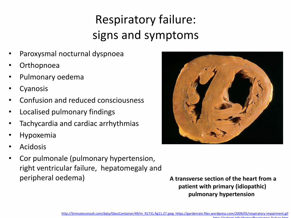
Respiratory failure: signs and symptoms
• Paroxysmal nocturnal dyspnoea
• Orthopnoea
• Pulmonary oedema
• Cyanosis
• Confusion and reduced consciousness
• Localised pulmonary findings
• Tachycardia and cardiac arrhythmias
• Hypoxemia
• Acidosis
• Cor pulmonale (pulmonary hypertension, right ventricular failure, hepatomegaly and peripheral oedema)
http://5minuteconsult.com/data/GbosContainer/49/m_91731.fig11.27.jpeg https://gardenrain.files.wordpress.com/2009/05/respiratory-impairment.gif http://patient.info/doctor/Respiratory-Failure.htm
A transverse section of the heart from a patient with primary (idiopathic)
pulmonary hypertension

Respiratory failure: diagnostic tests
• Pulmonary function tests (spirometry, arterial blood gas test, etc.)
• Chest X-ray
• Full Blood Count (anemia contributes to hypoxia, polycythemia contributes to chronic hypoxemic respiratory failure)
• Renal and liver function tests (may provide clues to the etiology or identify complications associated with respiratory failure)
• Serum creatine kinase and troponin I (to help exclude recent myocardial infarction)
• Thyroid Function Test (hypothyroid chronic hypercapnic respiratory failure)
• Echocardiography (cardiac cause of acute respiratory failure)
• ECG (cardiovascular cause, dysrhythmias resulting from severe hypoxaemia or acidosis)
• Right heart catheterisation (if there is uncertainty about cardiac function)
• Pulmonary capillary wedge pressure (distinguishing cardiogenic from noncardiogenic edema)
http://patient.info/doctor/Respiratory-Failure.htm
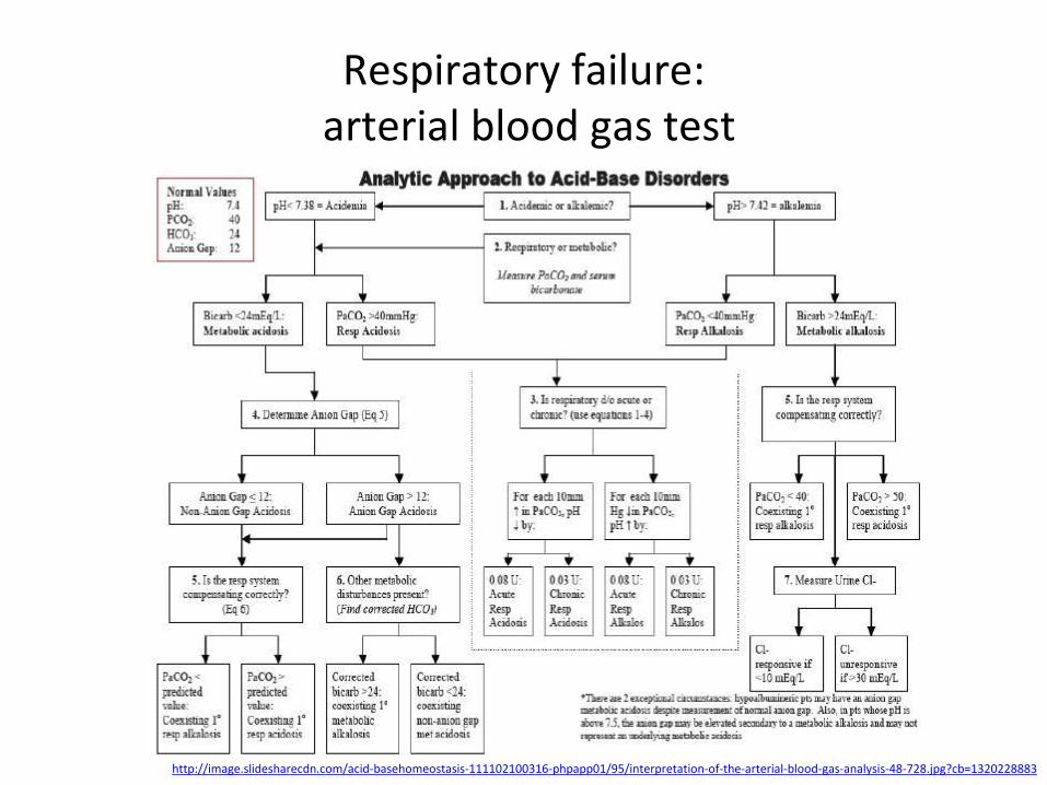
Respiratory failure: arterial blood gas test
http://image.slidesharecdn.com/acid-basehomeostasis-111102100316-phpapp01/95/interpretation-of-the-arterial-blood-gas-analysis-48-728.jpg?cb=1320228883
,,,
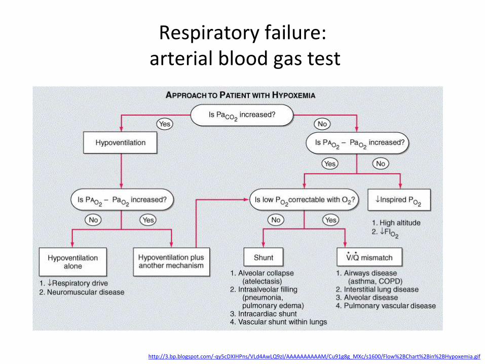
Respiratory failure: arterial blood gas test
http://3.bp.blogspot.com/-qy5cDXIHPns/VLd4AwLQ9zI/AAAAAAAAAAM/Cu91g8g_MXc/s1600/Flow%2BChart%2Bin%2BHypoxemia.gif
,,,
The lung compression syndrome: atelectasis (definition and types)
• Atelectasis is defined as the collapse of part or all of the lungs; when this occurs, for whatever reason, fresh air does not reach the tiniest of airways, and oxygen and carbon dioxide can’t be exchanged; this, in turn, can lead to decreased levels of oxygen being delivered to the organs and tissues of the body (hypoxia)
• Atelectasis may be acute, occurring suddenly over a matter of minutes, or chronic, developing over a period of days to weeks
• Atelectasis may be the result of a blocked airway (obstructive) or of pressure from outside the lung (nonobstructive)
• Almost everyone who has surgery has some atelectasis from anesthesia
• Atelectasis is particularly prominent after heart bypass surgery
http://lungcancer.about.com/od/Respiratory-Symptoms/a/Atelectasis.htm http://www.mayoclinic.org/diseases-conditions/atelectasis/basics/causes/con-20034847
The lung compression syndrome: atelectasis (mechanisms)
1. Obstruction: blockage of an airway, either from inside (by a foreign body that is aspirated, or a mucous plug), or the outside (e.g., by a lung cancer pressing on the airway),
2. Compression: compression of the airways in the lungs can be caused by fluid or air surrounding the lungs (as in a pleural effusion or a pneumothorax); by enlargement or an aneurysm of the heart; by tumors such as cancers metastatic to the lungs, lymphomas, or enlarged lymph nodes; or by abdominal distention which causes pressure on the lungs
3. Adhesion: when the surfactant is lacking, the lungs lose surface tension and can collapse; this is the cause of respiratory distress in newborns and can also occur in adults with adult respiratory distress syndrome (ARDS), smoke inhalation, and kidney failure
4. Hypoventilation: failure to take deep breaths can result in collapse of part of the lungs during surgery, especially with general anesthesia, and when breathing is shallow due to pain (such as with rib fractures)
http://lungcancer.about.com/od/Respiratory-Symptoms/a/Atelectasis.htm

The lung compression syndrome: atelectasis (obstructive atelectasis causes)
• Mucus plug after accumulation of mucus in airways, often occurring during and after surgery, in children, people with cystic fibrosis and during severe asthma attacks
• Foreign body is common in children who have inhaled an object, such as a peanut or small toy part, into their lungs
• Narrowing of major airways from disease (chronic infections, including fungal infections, tuberculosis and other diseases)
• Tumor in a major airway
• Blood clot after significant bleeding into the lungs that can't be coughed out
http://www.radiologyassistant.nl/data/bin/w440/a50d998498e4df_11b-rll-atelectasis.jpg http://www.mayoclinic.org/diseases-conditions/atelectasis/basics/causes/con-20034847
Lower lobe atelectasis

The lung compression syndrome: atelectasis (nonobstructive atelectasis causes)
• Injury (chest trauma)
• Pleural effusion
• Pneumonia
• Pneumothorax
• Scarring of lung tissue
• Tumor
https://en.wikipedia.org/wiki/Pleural_effusion http://www.virtualmedstudent.com/images/pneumothorax_xray_marked.jpg http://dvirtualdoctor.hubpages.com/ http://www.mayoclinic.org/diseases-conditions/atelectasis/basics/causes/con-20034847
Obstructive atelectasis of the
left upper lob
Pleural effusion

The lung compression syndrome: atelectasis (symptoms)
• Atelectasis may have few or no symptoms if it develops slowly or involves only a small portion of the lungs
• Conversely, if the condition affects a large portion of the lungs, or develops rapidly, symptoms may be dramatic and may even progress to shock
• Common symptoms include:
– Shortness of breath – a sensation of breathlessness is the most common symptom
– Coughing – this cough is often described as “hacking” and is most often non-productive, meaning that no mucous is coughed up
– Pleurisy – chest pain that is sharp and worsens with a deep breath or coughing (pleuritic chest pain) may occur
– Fever – at one time, it was thought that fever was a sign
http://lungcancer.about.com/od/Respiratory-Symptoms/a/Atelectasis.htm

The lung compression syndrome: atelectasis (diagnosis)
• Physical exam: findings may include quiet or absent breath sounds
• Chest x-ray: the trachea and heart may be deviated towards the side of the chest where a lung is partially collapsed; the diaphragm may also be elevated on the side of the collapse
• Chest CT scan: may further define an area of possible atelectasis and to look for other causes of obstruction, such as tumors or enlarged lymph nodes
• Bronchoscopy: may be used to determine the cause of a bronchial obstruction
• Blood gases or oximetry: may be done to determine how much atelectasis is interfering with the ability to get oxygen to your tissues
• Other tests may be ordered depending upon the condition; for example, a bloodwork to evaluate kidney function
http://lungcancer.about.com/od/Respiratory-Symptoms/a/Atelectasis.htm

The lung compression syndrome: atelectasis (bronchoscopy)
A peanut in the left main bronchus
http://img.medscape.com/pi/features/slideshow-slide/pediatric-ENT-surgery/fig15.jpg

The lung compression syndrome: pleural effusion (definition and types)
• Pleural effusion is excess fluid that accumulates in the pleural cavity, the fluid-filled space that surrounds the lungs
• The fluid excess can impair breathing by limiting the expansion of the lungs (>500 ml)
• Various kinds of pleural effusion, depending on the nature of the fluid and what caused its entry into the pleural space, are hydrothorax (serous fluid), hemothorax (blood), urinothorax (urine), chylothorax (chyle), or pyothorax (pus)
http://images.radiopaedia.org/images/4431478/8e49879424380b75830066020b6d35.jpg https://en.wikipedia.org/wiki/Pleural_effusion
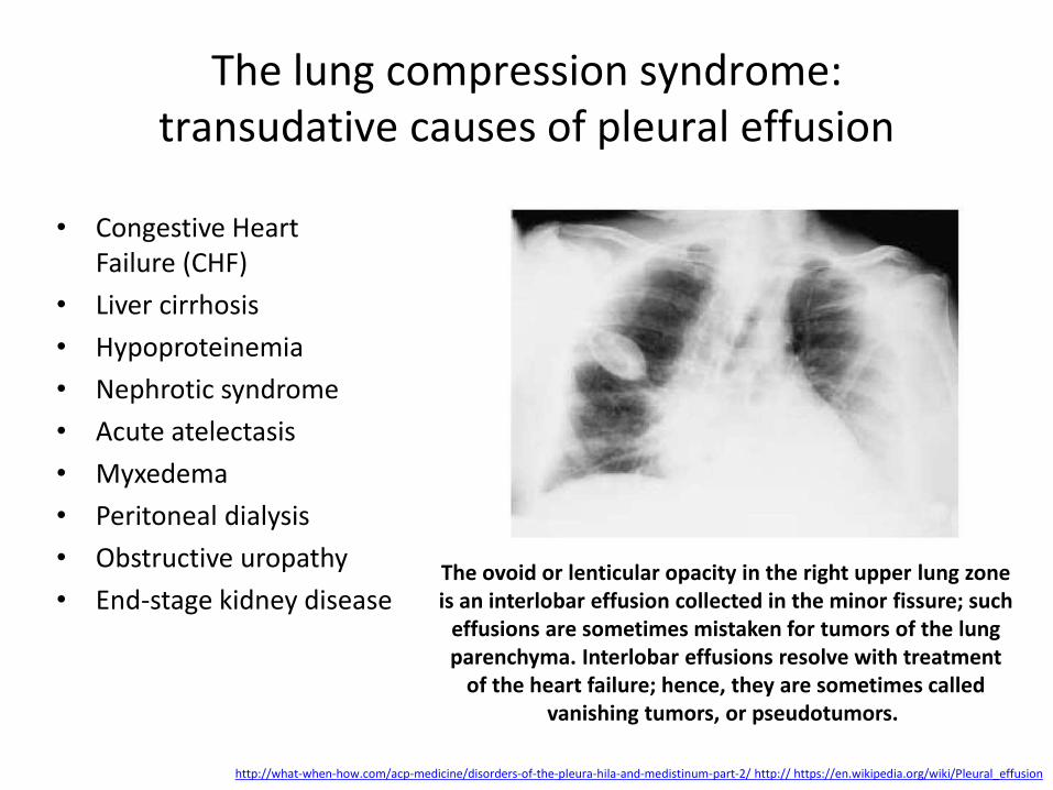
The lung compression syndrome: transudative causes of pleural effusion
• Congestive Heart Failure (CHF)
• Liver cirrhosis
• Hypoproteinemia
• Nephrotic syndrome
• Acute atelectasis
• Myxedema
• Peritoneal dialysis
• Obstructive uropathy
• End-stage kidney disease
http://what-when-how.com/acp-medicine/disorders-of-the-pleura-hila-and-medistinum-part-2/ http:// https://en.wikipedia.org/wiki/Pleural_effusion
The ovoid or lenticular opacity in the right upper lung zone is an interlobar effusion collected in the minor fissure; such
effusions are sometimes mistaken for tumors of the lung parenchyma. Interlobar effusions resolve with treatment
of the heart failure; hence, they are sometimes called vanishing tumors, or pseudotumors.

The lung compression syndrome: exudative causes of pleural effusion
• Pneumonia
• Cancer
• Pulmonary embolism
• Kidney disease
• Inflammatory disease
https://www.med-ed.virginia.edu/courses/rad/peds/chest_images/PA_CXR_LLL_with_pleural_fluid.jpg http://my.clevelandclinic.org/health/diseases_conditions/pleural-effusion
A left lower lobe consolidation, representing pneumonia. The meniscus in the left costophrenic angle
indicating a parapneumonic left pleural effusion.
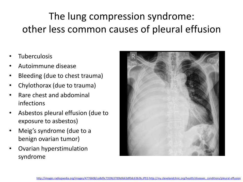
The lung compression syndrome: other less common causes of pleural effusion
• Tuberculosis
• Autoimmune disease
• Bleeding (due to chest trauma)
• Chylothorax (due to trauma)
• Rare chest and abdominal infections
• Asbestos pleural effusion (due to exposure to asbestos)
• Meig’s syndrome (due to a benign ovarian tumor)
• Ovarian hyperstimulation syndrome
http://images.radiopaedia.org/images/4776608/ca8d9c7359b3700b0b63df0dc63b3b.JPEG http://my.clevelandclinic.org/health/diseases_conditions/pleural-effusion
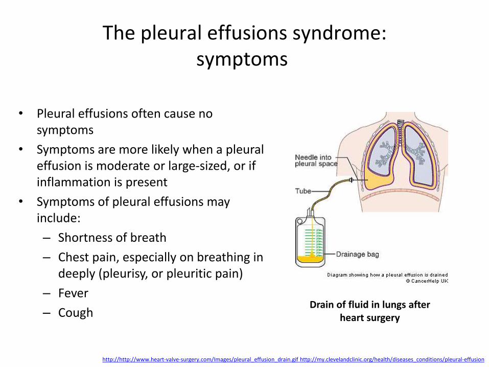
The pleural effusions syndrome: symptoms
• Pleural effusions often cause no symptoms
• Symptoms are more likely when a pleural effusion is moderate or large-sized, or if inflammation is present
• Symptoms of pleural effusions may include:
– Shortness of breath
– Chest pain, especially on breathing in deeply (pleurisy, or pleuritic pain)
– Fever
– Cough
http://http://www.heart-valve-surgery.com/Images/pleural_effusion_drain.gif http://my.clevelandclinic.org/health/diseases_conditions/pleural-effusion
Drain of fluid in lungs after heart surgery
The pleural effusions syndrome: diagnosis
• Pleural effusion is usually diagnosed on the basis of medical history and physical exam, and confirmed by chest x-ray
• Once accumulated fluid is more than 300 ml, there are usually detectable clinical signs in the patient, such as decreased movement of the chest on the affected side, stony dullness to percussion over the fluid, diminished breath sounds on the affected side, decreased vocal resonance and fremitus (though this is an inconsistent and unreliable sign), and pleural friction rub
• Above the effusion, where the lung is compressed, there may be bronchial breathing and egophony
• A large effusion there may cause tracheal deviation away from the effusion
https://en.wikipedia.org/wiki/Pleural_effusion
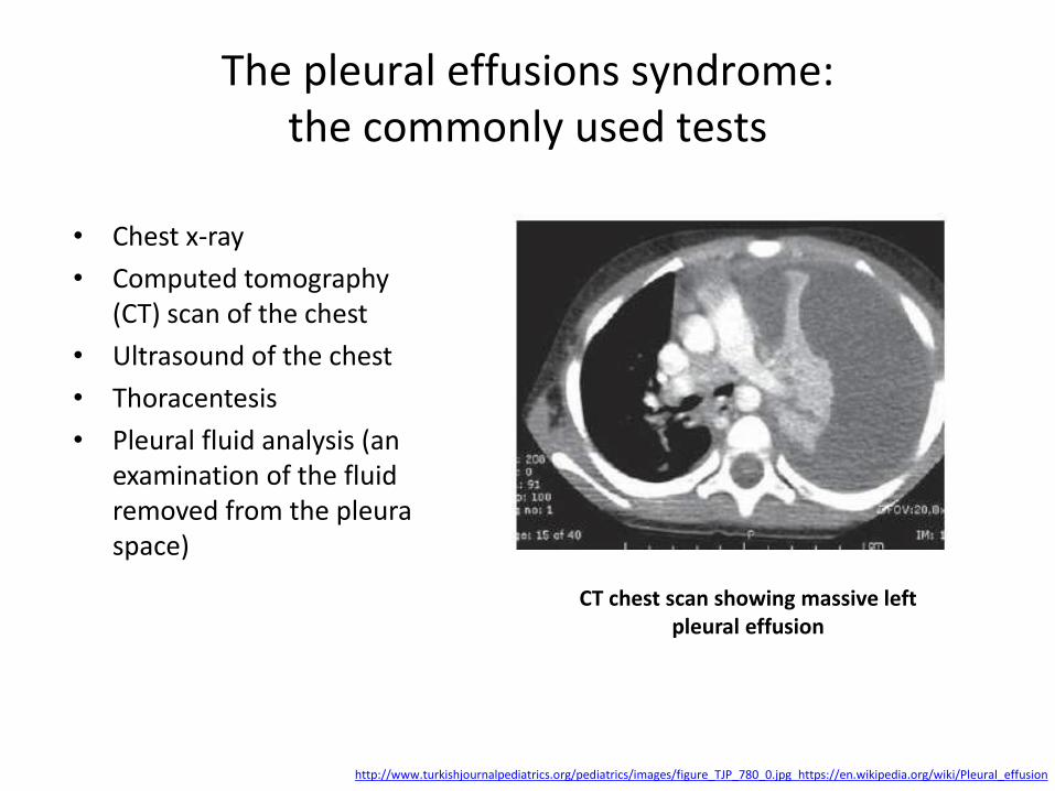
The pleural effusions syndrome:
the commonly used tests
• Chest x-ray
• Computed tomography (CT) scan of the chest
• Ultrasound of the chest
• Thoracentesis
• Pleural fluid analysis (an examination of the fluid removed from the pleura space)
http://www.turkishjournalpediatrics.org/pediatrics/images/figure_TJP_780_0.jpg https://en.wikipedia.org/wiki/Pleural_effusion
CT chest scan showing massive left pleural effusion
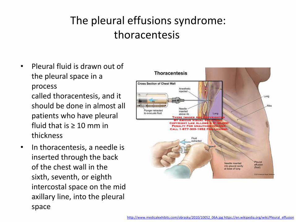
The pleural effusions syndrome:
thoracentesis
• Pleural fluid is drawn out of the pleural space in a process called thoracentesis, and it should be done in almost all patients who have pleural fluid that is ≥ 10 mm in thickness
• In thoracentesis, a needle is inserted through the back of the chest wall in the sixth, seventh, or eighth intercostal space on the mid axillary line, into the pleural space
http://www.medicalexhibits.com/obrasky/2010/10052_06A.jpg https://en.wikipedia.org/wiki/Pleural_effusion
The pleural effusions syndrome:
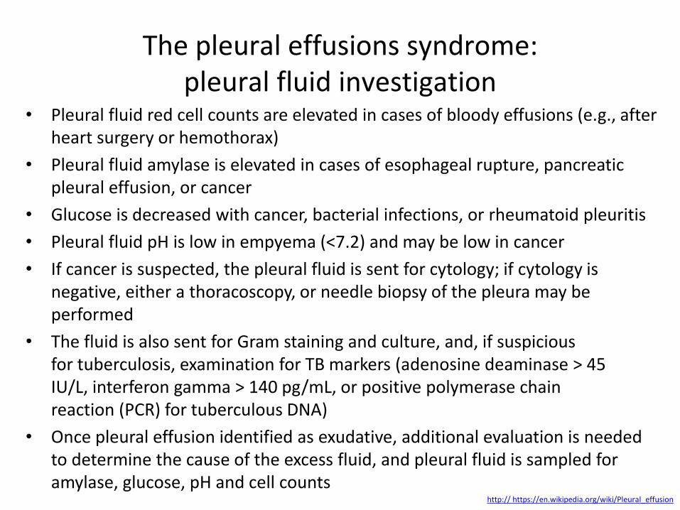
pleural fluid investigation • Pleural fluid red cell counts are elevated in cases of bloody effusions (e.g., after
heart surgery or hemothorax)
• Pleural fluid amylase is elevated in cases of esophageal rupture, pancreatic pleural effusion, or cancer
• Glucose is decreased with cancer, bacterial infections, or rheumatoid pleuritis
• Pleural fluid pH is low in empyema (<7.2) and may be low in cancer
• If cancer is suspected, the pleural fluid is sent for cytology; if cytology is negative, either a thoracoscopy, or needle biopsy of the pleura may be performed
• The fluid is also sent for Gram staining and culture, and, if suspicious for tuberculosis, examination for TB markers (adenosine deaminase > 45 IU/L, interferon gamma > 140 pg/mL, or positive polymerase chain reaction (PCR) for tuberculous DNA)
• Once pleural effusion identified as exudative, additional evaluation is needed to determine the cause of the excess fluid, and pleural fluid is sampled for amylase, glucose, pH and cell counts
http:// https://en.wikipedia.org/wiki/Pleural_effusion

The pleural effusions syndrome: light's criteria transudate vs. exudate
http:// https://en.wikipedia.org/wiki/Pleural_effusion
Parameters Transudate Exudate
↑ hydrostatic pressure,
↓ colloid osmotic pressure
Inflammation-increased
pascular permeability Main causes
Appearance Clear Cloudy
Specific gravity < 1.012 > 1.020
Protein content < 2.5 g/dL > 2.9 g/dL
fluid protein/serum protein < 0.5 > 0.5
Difference of albumin content with blood
albumin > 1.2 g/dL < 1.2 g/dL
fluid LDH upper limit for serum < 0.6 or < 2⁄3 > 0.6 or > 2⁄3
Cholesterol content < 45 mg/dL > 45 mg/dL

Acute respiratory distress syndrome (ARDS): definition and causes
• Acute respiratory distress syndrome (respiratory distress syndrome (RDS), acute lung injury, adult respiratory distress syndrome, shock lung) is a severe, life-threatening medical condition characterized by widespread inflammation in the lungs
• Common causes of ARDS include sepsis, pneumonia, trauma, multiple blood transfusions, babesiosis, lung contusion, aspiration of stomach contents, and drug abuse or overdose
• Other causes of ARDS include burns, pancreatitis, near drowning, or the inhalation of chemical irritants such as smoke, phosgene, or chlorine gas
• Some cases of ARDS are linked to large volumes of fluid used during post-trauma resuscitation
• The syndrome has a high mortality between 20 and 50%
http://3.bp.blogspot.com/_https://en.wikipedia.org/wiki/Acute_respiratory_distress_syndrome
Acute respiratory distress syndrome (ARDS): mechanisms
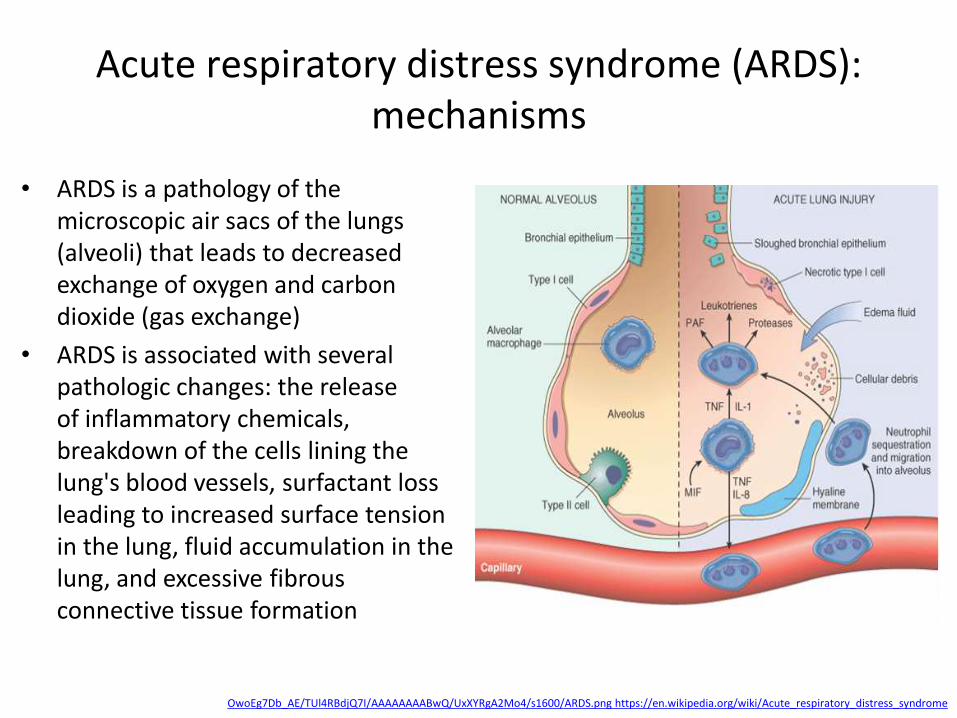
• ARDS is a pathology of the microscopic air sacs of the lungs (alveoli) that leads to decreased exchange of oxygen and carbon dioxide (gas exchange)
• ARDS is associated with several pathologic changes: the release of inflammatory chemicals, breakdown of the cells lining the lung's blood vessels, surfactant loss leading to increased surface tension in the lung, fluid accumulation in the lung, and excessive fibrous connective tissue formation
OwoEg7Db_AE/TUl4RBdjQ7I/AAAAAAAABwQ/UxXYRgA2Mo4/s1600/ARDS.png https://en.wikipedia.org/wiki/Acute_respiratory_distress_syndrome
Acute respiratory distress syndrome (ARDS): signs and symptoms
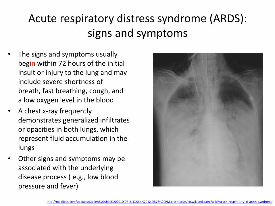
• The signs and symptoms usually begin within 72 hours of the initial insult or injury to the lung and may include severe shortness of breath, fast breathing, cough, and a low oxygen level in the blood
• A chest x-ray frequently demonstrates generalized infiltrates or opacities in both lungs, which represent fluid accumulation in the lungs
• Other signs and symptoms may be associated with the underlying disease process ( e.g., low blood pressure and fever)
http://medlibes.com/uploads/Screen%20shot%202010-07-21%20at%2012.26.23%20PM.png https://en.wikipedia.org/wiki/Acute_respiratory_distress_syndrome

Acute respiratory distress syndrome (ARDS): diagnosis
The "Berlin criteria" of 2012 proposed by the European Society of Intensive Care Medicine, endorsed by the American Thoracic Society and the Society of Critical Care Medicine:
• Acute onset
• Bilateral infiltrates on chest radiograph sparing costophrenic angles
• Pulmonary artery wedge pressure < 18 mmHg (obtained by pulmonary artery catheterization), if this information is available; if unavailable, then lack of clinical evidence of left atrial hypertension
• if PaO2:FiO2 < 300 mmHg (40 kPa) acute lung injury (ALI) is considered to be present
• if PaO2:FiO2 < 200 mmHg (26.7 kPa) acute respiratory distress syndrome (ARDS) is considered to be present
http:// www.ersj.org.uk/content/20/4/1017/F2.large.jpg https://en.wikipedia.org/wiki/Acute_respiratory_distress_syndrome
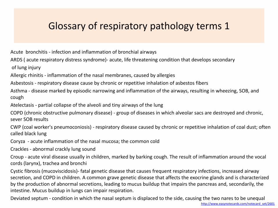
Glossary of respiratory pathology terms 1
Acute bronchitis - infection and inflammation of bronchial airways
ARDS ( acute respiratory distress syndrome)- acute, life threatening condition that develops secondary
of lung injury
Allergic rhinitis - inflammation of the nasal membranes, caused by allergies
Asbestosis - respiratory disease cause by chronic or repetitive inhalation of asbestos fibers
Asthma - disease marked by episodic narrowing and inflammation of the airways, resulting in wheezing, SOB, and cough
Atelectasis - partial collapse of the alveoli and tiny airways of the lung
COPD (chronic obstructive pulmonary disease) - group of diseases in which alveolar sacs are destroyed and chronic, sever SOB results
CWP (coal worker's pneumoconiosis) - respiratory disease caused by chronic or repetitive inhalation of coal dust; often called black lung
Coryza - acute inflammation of the nasal mucosa; the common cold
Crackles - abnormal crackly lung sound
Croup - acute viral disease usually in children, marked by barking cough. The result of inflammation around the vocal cords (larynx), trachea and bronchi
Cystic fibrosis (mucoviscidosis)- fatal genetic disease that causes frequent respiratory infections, increased airway secretion, and COPD in children. A common grave genetic disease that affects the exocrine glands and is characterized by the production of abnormal secretions, leading to mucus buildup that impairs the pancreas and, secondarily, the intestine. Mucus buildup in lungs can impair respiration.
Deviated septum - condition in which the nasal septum is displaced to the side, causing the two nares to be unequal http://www.easynotecards.com/notecard_set/2601

Glossary of respiratory pathology’ terms 2
Emphysema - abnormal increase in the size of air spaces distal to the terminal bronchiole and destruction of the alveolar walls
Empyema - collection of infected fluid (pus) between the two pleural membranes that line the lungs
Epistaxis - episode of bleeding from the nose; commonly known as a nosebleed
Hemoptysis - coughing up blood from the respiratory tract
Hemothorax - blood or bloody fluid collected within the intrapleural space, causing lung compression and respiratory distress
Histoplasmosis - systemic respiratory disease caused by a fungus found in soil contaminated with bird droppings
Hypercapnia - chronic retention of CO2, causing mental cloudiness and lethargy
Influenza - common, contagious, acute viral respiratory illness
Laryngitis - condition of inflammation of the larynx
Legionellosis - bacterial lung infection caused by the bacterium Legionella pneumophila
Nasal polyps - rounded tissue growths on the nasal or sinal mucosa
OSA (obstuctive sleep apnea) - dysfunctional breathing that occurs when the upper airway is intermittently blocked during sleep
Orthopnea - labored breathing that occurs when lying flat and improves when sitting up
Pharyngitis - inflammation of the pharynx; commonly called sore throat
http://www.easynotecards.com/notecard_set/2601
Glossary of respiratory pathology’ terms 3
Pleural effusion - excessive collection of fluid in the intrapleural space
Pleurisy - condition in which the pleurae become inflamed, causing sharp inspiratory chest pain
Pneumoconiosis - any disease of the respiratory tract caused by chronic or repetitive inhalation of dust particles
Pneumonia - bacterial or viral infection of the lungs
Pneumothorax - air collects in the intrapleural space; categorized as open, closed, spontaneous, or tension, and commonly called collapsed lung
Pulmonary embolism (PE) - sudden obstruction of a pulmonary blood vessel by debris, blood clots, or other matter
Pulmonary tuberculosis (TB) - infection caused by the Mycobacterium tuberculosis
Rhonchi - gurgling sound heard in the lungs with a stethoscope
Silicosis - respiratory disease caused by chronic or repetitive inhalation of silica dust
Sinusitis - inflammation of the lining of the sinus cavities
Stridor - high-pitched upper airway sound heard without a stethoscope
Upper respiratory infection (URI- infection and inflammation of the upper-airway structures, usually caused by a virus; often called a common cold
Wheeze - somewhat musical sound heard in the lungs, usually with a stethoscope, caused by partial airway obstruction
http://www.easynotecards.com/notecard_set/2601