Signaling Pathways in Liver Diseases - download.e … · 4 Signaling Pathways in Liver Diseases...
30
Signaling Pathways in Liver Diseases
Transcript of Signaling Pathways in Liver Diseases - download.e … · 4 Signaling Pathways in Liver Diseases...
Signaling Pathways in Liver Diseases

Jean-Francois DufourPierre-Alain Clavien (Eds.)
Signaling Pathways in Liver Diseases
Second edition

ISBN: 978-3-642-00149-9 e-ISBN: 978-3-642-00150-5
DOI: 10.1007/978-3-642-00150-5
Springer Heidelberg Dordrecht London New York
Library of Congress Control Number: 2009931701
© Springer-Verlag Berlin Heidelberg 2010
This work is subject to copyright. All rights are reserved, whether the whole or part of the material is concerned, specifically the rights of translation, reprinting, reuse of illustrations, recitation, broadcasting, reproduction on microfilm or in any other way, and storage in data banks. Duplication of this publication or parts thereof is permitted only under the provisions of the German Copyright Law of September 9, 1965, in its current version, and permission for use must always be obtained from Springer. Violations are liable to prosecution under the German Copyright Law.
The use of general descriptive names, registered names, trademarks, etc. in this publication does not imply, even in the absence of a specific statement, that such names are exempt from the relevant protective laws and regulations and therefore free for general use.
Product liability: The publishers cannot guarantee the accuracy of any information about dosage and appli-cation contained in this book. In every individual case the user must check such information by consulting the relevant literature.
Cover design: eStudio Calamar, Figueres/Berlin
Printed on acid-free paper
Springer is part of Springer Science+Business Media (www.springer.com)
Prof. Jean-François DufourUniversity of BernInstitute of Clinical Pharmacology and Visceral ResearchMurtenstrasse 353010 [email protected]
Prof. Dr. Pierre-Alain ClavienSwiss HPB (Hepato-Pancreatico Biliary) CenterUniversity Hospital of ZurichDepartment of Visceral and Transplant SurgeryRämistrasse 1008091 [email protected]
v
Recent advances in diagnostic and therapeutic approaches to liver disease have car-ried hepatology to new frontiers. The increasing frequency with which steatotic and cirrhotic livers undergo complex curative treatment strategies is a challenge to liver surgeons and hepatologists, who need to understand the molecular mechanisms at play in these situations. Comprehension of the signaling pathways participating in liver regeneration, hepatocellular apoptosis, and ischemia/reperfusion injury is essen-tial. This book serves as a source of information to facilitate the reading of the litera-ture and the planning of trials. Translational medicine implies knowledge of the molecular targets of novel therapeutic strategies. It is our goal to stimulate research that leads to exchanges between the laboratory, the clinical ward, and the operating room. Such a comprehensive insight including molecular and cellular events will pave the way for improvement of pharmacological and surgical interventions in com-plex liver disease.
Bern, Switzerland Jean-Francois DufourZurich, Switzerland Pierre-Alain Clavien
Preface
vii
Part I The Cell Types and the Matrix . . . . . . . . . . . . . . . . . . . . . . . . . . . . . 1
1 Hepatocytes. . . . . . . . . . . . . . . . . . . . . . . . . . . . . . . . . . . . . . . . . . . . . . . . 3Giuliano Ramadori and Pierluigi Ramadori
2 Signaling Pathways in Biliary Epithelial Cells . . . . . . . . . . . . . . . . . . . 25M. Fatima Leite, Viviane A. Andrade, and Michael H. Nathanson
3 Stellate Cells . . . . . . . . . . . . . . . . . . . . . . . . . . . . . . . . . . . . . . . . . . . . . . . 41Fabio Marra, Sara Galastri, Sara Aleffi, and Massimo Pinzani
4 Signaling Pathways in Liver Diseases Kupffer Cells . . . . . . . . . . . . . . 69Christian J. Steib and Alexander L. Gerbes
5 Hepatic Sinusoidal Endothelial Cells . . . . . . . . . . . . . . . . . . . . . . . . . . . 79Robert C. Huebert and Vijay H. Shah
6 Extracellular Matrix . . . . . . . . . . . . . . . . . . . . . . . . . . . . . . . . . . . . . . . . 93Scott L. Friedman
7 Platelets: A New Cell Type in Liver Physiology . . . . . . . . . . . . . . . . . . 105Mickael Lesurtel and Pierre-Alain Clavien
8 Immune Cell Communication and Signaling Systems in Liver Disease . . . . . . . . . . . . . . . . . . . . . . . . . . . . . . . . . . . . . . . . . . . . 117Ricky H. Bhogal and Simon C. Afford
Part II The Pathways . . . . . . . . . . . . . . . . . . . . . . . . . . . . . . . . . . . . . . . . . . 147
9 Toll-Like Receptors . . . . . . . . . . . . . . . . . . . . . . . . . . . . . . . . . . . . . . . . . 149Gyongyi Szabo and Pranoti Mandrekar
10 TNF/TNF Receptors . . . . . . . . . . . . . . . . . . . . . . . . . . . . . . . . . . . . . . . . 161Jörn M. Schattenberg and Mark J. Czaja
11 Fas/FasL . . . . . . . . . . . . . . . . . . . . . . . . . . . . . . . . . . . . . . . . . . . . . . . . . . 179Maria Eugenia Guicciardi and Gregory J. Gores
Contents

viii Contents
12 Interferon Signaling. . . . . . . . . . . . . . . . . . . . . . . . . . . . . . . . . . . . . . . . . 189Markus H. Heim
13 NF-kB . . . . . . . . . . . . . . . . . . . . . . . . . . . . . . . . . . . . . . . . . . . . . . . . . . . . 201Tom Luedde and Christian Trautwein
14 JNKs in liver diseases . . . . . . . . . . . . . . . . . . . . . . . . . . . . . . . . . . . . . . . 215R. Schwabe
15 Insulin Pathway . . . . . . . . . . . . . . . . . . . . . . . . . . . . . . . . . . . . . . . . . . . . 229Miran Kim and Jack R. Wands
16 Role of PKB/Akt in Liver Diseases . . . . . . . . . . . . . . . . . . . . . . . . . . . . . 243Elena Zhuravleva, Oliver Tschopp, and Brian A. Hemmings
17 Targeting mTOR Signaling Pathways in Liver Diseases . . . . . . . . . . . 261Hala E. Thomas and Sara C. Kozma
18 AMP-Activated Protein Kinase in Liver . . . . . . . . . . . . . . . . . . . . . . . . 275Louis Hue, Laurent Bultot, and Mark H. Rider
19 ER Stress Signaling in Hepatic Injury . . . . . . . . . . . . . . . . . . . . . . . . . . 287Cheng Ji and Neil Kaplowitz
20 PPARa, A Key Regulator of Hepatic Energy Homeostasis in Health and Disease. . . . . . . . . . . . . . . . . . . . . . . . . . . . . . . . . . . . . . . . 305Nicolas Leuenberger and Walter Wahli
21 Bile Acids and Their Receptors . . . . . . . . . . . . . . . . . . . . . . . . . . . . . . . 317Thierry Claudel and Michael Trauner
22 Signaling Pathways in Liver Diseases: PXR and CAR. . . . . . . . . . . . . 333Catherine A.M. Stedman, Michael Downes, and Christopher Liddle
23 p53 . . . . . . . . . . . . . . . . . . . . . . . . . . . . . . . . . . . . . . . . . . . . . . . . . . . . . . . 345Wen-Wei Tsai and Michelle Craig Barton
24 The MYC Network and Cancer . . . . . . . . . . . . . . . . . . . . . . . . . . . . . . . 359Snorri S. Thorgeirsson and Valentina M. Factor
25 The WNT/b-Catenin Pathway . . . . . . . . . . . . . . . . . . . . . . . . . . . . . . . . 367Satdarshan P. S. Monga
26 Sonic Hedgehog Pathway . . . . . . . . . . . . . . . . . . . . . . . . . . . . . . . . . . . . 393Alessia Omenetti and Anna Mae Diehl

Contents ix
27 Hypoxia-Inducible Factor-1 Signaling System . . . . . . . . . . . . . . . . . . . 403Deborah Stroka and Daniel Candinas
28 VEGF Signaling . . . . . . . . . . . . . . . . . . . . . . . . . . . . . . . . . . . . . . . . . . . . 421David Semela and Jean-François Dufour
29 Apoptosis and Mitochondria. . . . . . . . . . . . . . . . . . . . . . . . . . . . . . . . . . 439Jose C. Fernández-Checa and Carmen Garcia-Ruiz
30 Calcium Signaling . . . . . . . . . . . . . . . . . . . . . . . . . . . . . . . . . . . . . . . . . . 455Thierry Tordjmann
31 HBV Signaling . . . . . . . . . . . . . . . . . . . . . . . . . . . . . . . . . . . . . . . . . . . . . 465Massimo Levrero and Laura Belloni
32 Hepatitis C Virus and Insulin Signaling . . . . . . . . . . . . . . . . . . . . . . . . 483Francesco Negro and Sophie Clément
33 MicroRNAs . . . . . . . . . . . . . . . . . . . . . . . . . . . . . . . . . . . . . . . . . . . . . . . . 493Onpan Cheung and Arun J. Sanyal
34 Hepatic Clocks . . . . . . . . . . . . . . . . . . . . . . . . . . . . . . . . . . . . . . . . . . . . . 501Ueli Schibler, Gad Asher, Camille Saini, Jörg Morf, and Hans Reinke
Answers . . . . . . . . . . . . . . . . . . . . . . . . . . . . . . . . . . . . . . . . . . . . . . . . . . . . . . . 513
Index . . . . . . . . . . . . . . . . . . . . . . . . . . . . . . . . . . . . . . . . . . . . . . . . . . . . . . . . . 515

xi
Simon C . Afford The Liver Research Group, School of Infection and Immunity, College of Medicine and Dentistry, University of Birmingham, UK
Sara Aleffi Dipartimento di Medicina Interna, Università degli Studi di Firenze, Firenze, Italy
Viviane A . Andrade Department of Biochemistry and Immunology, UFMG, Belo Horizonte, Brazil
Gad Asher Department of Molecular Biology and National Center of Competence in Research “Frontiers in Genetics”, Sciences III, University of Geneva, 30, Quai Ernest Ansermet, CH-1211, Geneva-4, Switzerland
Michelle Craig Barton Department of Biochemistry and Molecular Biology, University of Texas M.D. Anderson Cancer Center, 1515 Holcombe Blvd., Houston, TX 77030, USA
Laura Belloni Rome Oncogenomic Center, CRS-Regina Elena Cancer Center, Rome, Italy
Ricky H . Bhogal The Liver Research Group, School of Infection and Immunity, College of Medicine and Dentistry, University of Birmingham, UK
Daniel Candinas Clinic of Visceral Surgery and Medicine, Inselspital, University of Bern, Switzerland
Thierry Claudel Laboratory of Experimental and Molecular Hepatology, Division of Gastroenterology and Hepatology, Department of Medicine, Medical University, Graz, Austria
Pierre-Alain Clavien Swiss HPB Center, Department of Visceral and Transplant Surgery, University Hospital of Zurich, Rämistrasse 100, 8091 Zürich, Switzerland
Onpan Cheung Department of Internal Medicine, Division of Gastroenterology, Hepatology and Nutrition, Virginia Commonwealth University Medical Center, Richmond, VA 23298, USA
Sophie Clément Division of Clinical Pathology, University Hospitals, Geneva, Switzerland
Mark J . Czaja Marion Bessin Liver Research Center, Albert Einstein College of Medicine, 1300 Morris Park Avenue, Bronx, NY 10461, USA
Contributors

xii Contributors
Anna Mae Diehl Department of Medicine, Division of Gastroenterology, Duke University Medical Center, GSRBI, 595 LaSalle Street, Suite 1073, Box 3256, Durham, NC 27710, USA
Michael Downes Howard Hughes Medical Institute and Gene Expression Laboratory, The Salk Institute for Biological Studies, 10010 Torrey Pines Road, La Jolla, CA 92037, USA
Jean-François Dufour Institute of Clinical Pharmacology and Visceral Research, University of Bern, Murtenstrasse 35, 3010 Bern, Switzerland
Valentina M . Factor Laboratory of Experimental Carcinogenesis, Center for Cancer Research, National Cancer Institute, National Institutes of Health, 37 Convent Drive, Bethesda, MD 20892, USA
Jose C . Fernández-Checa Liver Unit and Centro de Investigaciones Biomédicas Esther Koplowitz, IMDiM, Hospital Clínic i Provincial and CIBEREHD, IDIBAPS, C/Villarroel 170, 08036-Barcelona, Spain
Scott L . Friedman Division of Liver Diseases, Mount Sinai School of Medicine, 1425 Madison Avenue, Box 1123, New York, NY 10029-6574, USA
Sara Galastri Dipartimento di Medicina Interna, Università degli Studi di Firenze, Firenze, Italy
Carmen Garcia-Ruiz Department of Cell Death and Proliferation, Instituto Investigaciones Biomédicas de Barcelona, Consejo Superior de Investigaciones Científicas, 08036 B arcelona, Spain
Alexander L . Gerbes Department of Medicine II, Ludwig-Maximilians-University, Klinikum Großhadern, Marchioninistraβe 15, 81377 Munich, Germany
Gregory J . Gores Mayo Clinic College of Medicine, 200 First Street SW, Rochester, MN 55905, USA
Maria Eugenia Guicciardi Miles and Shirley Fiterman Center for Digestive Diseases, Division of Gastroenterology and Hepatology, Mayo Clinic College of Medicine, 200 First Street SW, Rochester, MN 55905, USA
Markus H . Heim Division of Gastroenterology and Hepatology, University Hospital, Basel, 4031 Basel, Switzerland
Brian A . Hemmings Friedrich Miescher Institute for Biomedical Research, Maulbeerstrasse 66, 4058 Basel, Switzerland
Louis Hue Hormone and Metabolic Research Unit, Université Catholique de Louvain, and de Duve Institute Avenue, Hippocrate 75 UCL 7529, B-1200 Brussels, Belgium
Robert C . Huebert GI Research Unit, Guggenheim 10, Mayo Clinic, 200 First Street SW, Rochester, MN 55905, USA
Cheng Ji Keck School of Medicine, University of Southern California, 2011 Zonal Avenue, HMR 101, Los Angeles, CA 90033, USA
Contributors xiii
Neil Kaplowitz Department of Medicine, USC–UCLA Research Center for Alcoholic, and Pancreatic Diseases, and USC Research Center for Liver Diseases, Keck School of Medicine, University of Southern California, Los Angeles, CA, USA
Miran Kim The Liver Research Center, Alpert Medical School of Brown University, Providence, RI 02903, USA
Sara C . Kozma Genome Research Institute, 2180 E. Galbraith Road, University of Cincinnati, Cincinnati, OH 45237, USA
M . Fatima Leite Department of Physiology and Biophysics, UFMG, Belo Horizonte, Brazil
Mickael Lesurtel Swiss HPB (Hepato-Pancreatico- Biliary) Center, Department of Surgery, University Hospital of Zurich, Zurich, Switzerland
Nicolas Leuenberger Center for Integrative Genomics and National Research Center Frontiers in Genetics, University of Lausanne, Switzerland
Massimo Levrero Dipartimento di Medicina Interna, Sapienza Universita’ di Roma, Policlinico Umberto I, Viale del Policlinico 155, 0061 Rome, Italy
Christopher Liddle Department of Clinical Pharmacology, Storr Liver Unit, Westmead Millennium Institute and University of Sydney, Westmead Hospital, Westmead NSW 2145, Australia
Tom Luedde Medical Department III, University Hospital RWTH Aachen, Pauwelsstraβe 30, 52074 Aachen, Germany
Pranoti Mandrekar Department of Medicine, University of Massachusetts Medical School, Worcester, MA 01605, USA
Fabio Marra Dipartimento di Medicina Interna, Università di Firenze, Viale Morgagni, 85 50134 Firenze, Italy
Satdarshan P .S . Monga Division of Experimental Pathology, University of Pittsburgh, School of Medicine, Pittsburgh, PA 15261, USA
Jörg Morf Department of Molecular Biology and National Center of Competence in Research “Frontiers in Genetics”, Sciences III, University of Geneva, Quai Ernest Ansermet 30, 1211 Geneva-4, Switzerland
Michael H . Nathanson Department of Medicine, Yale University School of Medicine, 1 Gilbert Street, New Haven, CT 06520-8019, USA
Francesco Negro Departments of Internal Medicine and Pathology and Immunology, University of Geneva Medical Center, 1 Rue Michel-Servet, 1205 Geneva, Switzerland
Alessia Omenetti Department of Medicine, Division of Gastroenterology, Duke University Medical Center, Durham, NC, USA
Massimo Pinzani Dipartimento di Medicina Interna, Università degli Studi di Firenze, Firenze, Italy

xiv Contributors
Giuliano Ramadori Department of Internal Medicine, Section of Gastroenterology and Endocrinology, Georg-August-University Göttingen, Robert-Koch-Straβe 40, 37075 Göttingen, Germany
Pierluigi Ramadori Department of Internal Medicine, Section of Gastroenterology and Endocrinology, Georg-August-University Göttingen, Robert-Koch-Straβe 40, 37075 Göttingen, Germany
Hans Reinke Department of Molecular Biology and National Center of Competence in Research “Frontiers in Genetics”, Sciences III, University of Geneva, Quai Ernest Ansermet 30, 1211 Geneva-4, Switzerland
Mark H . Rider Université Catholique de Louvain, de Duve Institute, Brussels, Belgium
Arun Sanyal Department of Internal Medicine, Division of Gastroenterology, Hepatology and Nutrition, Virginia Commonwealth University Medical Center, Richmond, VA 23298, USA
Jörn M . Schattenberg I. Department of Medicine, Johannes Gutenberg University, 5501 Mainz, Germany
Ueli Schibler Department of Molecular Biology and National Center of Competence in Research “Frontiers in Genetics”, Sciences III, University of Geneva, Quai Ernest Ansermet 30, 1211, Geneva-4, Switzerland
Robert Schwabe Department of Medicine, Columbia University, Russ Berrie Pavilion, Room 415, 1150 St. Nicholas Avenue, New York, NY 10032, USA
David Semela Division of Gastroenterology and Hepatology, University Hospital Basel, Basel, Switzerland
Vijay Shah GI Research Unit, Guggenheim 10, Mayo Clinic, 200 First Street SW, Rochester, MN 55905, USA
Catherine A .M . Stedman Department of Gastroenterology, Christchurch Hospital and University of Otago, Christchurch, Private Bag 4710, Christchurch, New Zealand
Christian Steib Department of Medicine II, Ludwig-Maximilians-University, Klinikum Großhadern, Munich, Germany
Deborah Stroka Visceral Surgery Research Laboratory, Department of Clinical Research, University of Bern, Murtenstrasse 35, 3010 Bern, Switzerland
Gyongyi Szabo Department of Medicine, University of Massachusetts Medical School, 364 Plantation Street, Worcester, MA 01605, USA
Hala E . Thomas Genome Research Institute, 2180 E. Galbraith Road, University of Cincinnati, Cincinnati, OH 45237, USA
Snorri S . Thorgeirsson Laboratory of Experimental Carcinogenesis, Center for Cancer Research, National Cancer Institute, National Institutes of Health, 37 Convent Drive, Bethesda, MD 20892, USA

Contributors xv
Thierry Tordjmann INSERM U757, Université Paris Sud, Bât. 443, 91405, Orsay, France
Michael Trauner Laboratory of Experimental and Molecular Hepatology, Division of Gastroenterology and Hepatology, Department of Internal Medicine, Medical University, Graz, Stiftingalstrasse 24, 8010 Graz, Austria
Christian Trautwein Medical Department III, University Hospital RWTH Aachen, Pauwelsstraβe 30, 52074 Aachen, Germany
Wen-Wei Tsai Department of Biochemistry and Molecular Biology, Program in Genes and Development, Graduate School of Biomedical Sciences, Center for Cancer Epigenetics, University of Texas M.D. Anderson Cancer Center, Houston, TX 77030, USA
Oliver Tschopp Clinic of Endocrinology, Diabetes and Clinical Nutrition, University Clinic Zurich, Rämistrasse 100, 8091 Zurich, Switzerland
Walter Wahli Center for Integrative Genomics, National Research Center Frontiers in Genetics, University of Lausanne, Genopode Building, 1015 Lausanne, Switzerland
Jack R . Wands The Liver Research Center, Alpert Medical School of Brown University, 55 Claverick Street, 4th Floor, Providence RI 02903, USA
Elena Zhuravleva Friedrich Miescher Institute for Biomedical Research, Maulbeerstrasse 66, 4058 Basel, Switzerland

Part
The Cell Types and the MatrixI
J.-F. Dufour, P.-A. Clavien (eds.), Signaling Pathways in Liver Diseases, 3 DOI: 10.1007/978-3-642-00150-5_1, © Springer-Verlag Berlin Heidelberg 2010
Introduction
The liver is the largest organ of the body. Its weight (1.5–1.8 kg) represents about 2% of the total human body weight. The anatomical location is of course linked to its function. The liver function is comparable to that of the stomach, intestine, pancreas, and kidney together. In fact, all nutrients resulting from the diges-tion of the food are taken up by the intestine and then by the liver. Furthermore, the liver is responsible for the synthesis of most of the serum proteins and by this means for the oncotic pressure and the retention of water within the vessels. The liver stores nutrients and the energy derived from the oxidation of the nutrients.
However, the liver is not only a power plant but also a cleaning device. In fact, the direct relationship with the intestine is not without danger. The large intestine despite the reabsorption of water contains an enormous number of bacteria and an enormous amount of their products. The bacteria and their products can reach the venous blood and the liver sinusoid where they are taken up and digested.
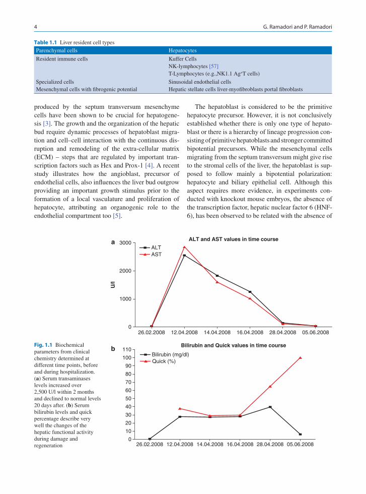
Although the liver is made of several cell popula-tions (Table 1.1), the most abundant cell type by mass and number is the hepatocyte. The human liver is made of 80 × 109 hepatocytes. To understand best the func-tions of the hepatocytes, it is useful to look at the labo-ratory findings of a 60-year old lady who developed jaundice followed by loss of appetite and was admitted
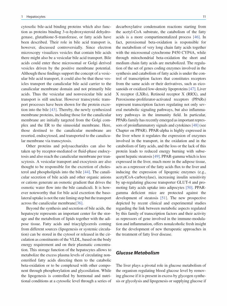
to the university clinic because of reduced liver func-tion. The following parameters describe her clinical situation well: serum bilirubin levels 27.7 mg/dl (nor-mal <2), tromboplastin time 29% (normal >60%), SGPT of 2,850 U/l, and SGOT 2,553 U/l (normal <40 U/l for both) (Fig. 1.1). Further, microscopical analysis of liver biopsy showed massive areas of necro-sis with increased deposition of connective tissue and presence of regenerative nodules with proliferating hepatocytes. An immunostaining for the proliferation marker Ki67 revealed that all the intact hepatocytes were in a proliferative phase. As can be seen from the follow-up study, regeneration succeeded and liver function recovered almost fully, 2 months later. The case is paradigmatic for the consequence of the lack of sufficient functional liver cell mass and the enormous capacity of hepatocytes to regenerate (Fig. 1.2).
Hepatocyte Development
Recent studies on different mammalian species indi-cate that during embryonic development the hepatic genes are induced in a segment of the ventral endo-derm through the activation of specific transcription factors (Foxa2, GATA-4, C/EBPb, Nf-1) forming a complex able to bind the chromatin upstream liver spe-cific genes such as albumin [109]. The subsequent cell migration and organogenesis depend tightly on the orchestration of inductive signals between epithelial cells, mesenchymal cells, and endothelial cells. Experimental stimulation of in vitro embryo cultures suggests that fibroblast growth factor (Fgf) signaling from the cardiogenic mesoderm induces the liver bud-ding in the ventral foregut endoderm [2]. Moreover, Bone morphogenetics proteins (Bmp-2 and Bmp-4)
Hepatocytes
Giuliano Ramadori and Pierluigi Ramadori
1
G. Ramadori ()Department of Internal Medicine, Section of Gastroenterology and Endocrinology, Georg-August-University Göttingen, Robert-Koch-Strabe 40, 37075 Göttingen, Germanye-mail: [email protected]
4 G. Ramadori and P. Ramadori
produced by the septum transversum mesenchyme cells have been shown to be crucial for hepatogene-sis [3]. The growth and the organization of the hepatic bud require dynamic processes of hepatoblast migra-tion and cell–cell interaction with the continuous dis-ruption and remodeling of the extra-cellular matrix (ECM) – steps that are regulated by important tran-scription factors such as Hex and Prox-1 [4]. A recent study illustrates how the angioblast, precursor of endothelial cells, also influences the liver bud outgrow providing an important growth stimulus prior to the formation of a local vasculature and proliferation of hepatocyte, attributing an organogenic role to the endothelial compartment too [5].
The hepatoblast is considered to be the primitive hepatocyte precursor. However, it is not conclusively established whether there is only one type of hepato-blast or there is a hierarchy of lineage progression con-sisting of primitive hepatoblasts and stronger committed bipotential precursors. While the mesenchymal cells migrating from the septum transversum might give rise to the stromal cells of the liver, the hepatoblast is sup-posed to follow mainly a bipotential polarization: hepatocyte and biliary epithelial cell. Although this aspect requires more evidence, in experiments con-ducted with knockout mouse embryos, the absence of the transcription factor, hepatic nuclear factor 6 (HNF-6), has been observed to be related with the absence of
Parenchymal cells Hepatocytes
Resident immune cells Kuffer CellsNK-lymphocytes [57]T-Lymphocytes (e.g.,NK1.1 Ag+T cells)
Specialized cells Sinusoidal endothelial cellsMesenchymal cells with fibrogenic potential Hepatic stellate cells liver-myofibroblasts portal fibroblasts
Table 1.1 Liver resident cell types
Fig. 1.1 Biochemical parameters from clinical chemistry determined at different time points, before and during hospitalization. (a) Serum transaminases levels increased over 2,500 U/l within 2 months and declined to normal levels 20 days after. (b) Serum bilirubin levels and quick percentage describe very well the changes of the hepatic functional activity during damage and regeneration
26.02.2008 12.04.2008 14.04.2008 16.04.2008 28.04.2008 05.06.20080
1000
2000
3000ALTAST
U/l
ALT and AST values in time course
26.02.2008 12.04.2008 14.04.2008 16.04.2008 28.04.2008 05.06.20080
10
20
30
40
50
60
70
80
90
100
110Bilirubin (mg/dl)Quick (%)
Bilirubin and Quick values in time course
a
b

1 Hepatocytes 5
the gall bladder and an excess of the biliary cell popu-lation, suggesting a regulatory role of this protein in the hepatoblast- biliary shift [6]. A similar morphologi-cal picture was observed in conditionally homozygous hepatoblast for the transcription factor HNF-1b [7]. An important contribution to the biliary cells differentia-tion seems to be afforded by the Notch pathway as well. Mutations in the Notch pathway are in fact related in humans with the Alagille syndrome, a disease char-acterized by a reduced number of intrahepatic ducts [8]. A solid experimental evidence is given by observa-tions on knockout Jag1 or Notch-2 mice, showing an analog phenotype at the birth; a more detailed analysis suggests an involvement of this pathway in the bile duct morphogenesis and remodeling rather than in the hepatoblast fate differentiation [9]. In humans, the pro-genitor cells are immunoreactive for cytokeratins 8, 18, 19, and 14. Most of the progen-itor cells develop to adult hepatocytes while losing cytokeratin 14 and 19; the latter is no longer detectable in human hepatoblasts at 20-week gestation [10, 11]. Using this marker together with the transcription factor Prox-1, as an hepatoblast marker, we detected CK-19+/Prox-1 small cells in cell cultures obtained from fetal rat liver at ED 14 [12], which indicate a possible different develop-mental pathway for biliary cells. The synthesis of alpha-fetoprotein (AFP) has the characteristic of the hepatoblast, which begins in the human liver as early as day 29. Although it is known that albumin gene expression starts later and increases in parallel with the decrease of AFP-gene expression which almost com-pletely stops at birth, several reposts claim that albu-min gene expression begins as early as AFP expression [13, 14]. Analysis with the use of more sensible meth-ods such as in situ hybridization and biosynthetic label-ing on rat embryos revealed that the albumin gene expression, together with its protein synthesis and the complete enzymatic secretory apparatus, is present and functional at the very early stages of liver develop-ment and in the ventral foregut endodermal cells, com-parable with that of adult hepatocytes. Furthermore, although the ratio of albumin- and AFP-expressing cells to proliferating cells increases during embryonic stage, at the time of liver formation, both the genes and proteins are expressed in hepatoblast together with fibrinogen. In brief, during embryonic and fetal stages, 50% of the liver cells express secretory functions; while in the embryonic stage the liver growth depends mainly on cell proliferation, in the successive step, that
is, the fetal stage, the increase of cellular volume plays a more important role than cell division [15].
During fetal life the cell plates are three to five cells thick. At birth the plates are two cells thick and one cell-thick plate of the adult human liver is reached at 5 years of age. During fetal life hepatocytes exhibit considerable DNA-synthesis and replication. Two hours after birth DNA-synthesis rates are elevated with »18% of the hepatocytes incorporating 3H-thymidine into DNA. Three weeks later only 9% of the hepato-cytes show evidence of DNA-synthesis. This activity declines continuously after birth until at 6 weeks of age only few hepatocytes (0.1–0.4%) show evidence of DNA synthesis [16].
Within the first 3 weeks after birth liver mass increases together with hepatocellular DNA-synthesis but no increase of mitosis in hepatocytes can be observed. This may mean that increased metabolic requirements induce a hypertrophy of the hepatocytes characterized by DNA-synthesis and enlargement of the hepatocytes and consequently of liver growth. A similar phenomenon can be observed in the adult animal after a certain period of fasting [17, 18].
In the fetal liver, the DNA-synthesis is quite strong and only mononuclear diploid cells are observed with the one third of the nuclei being in the S-phase in the suckling phase; in young adult animals, the DNA-synthesis strongly decreases and in the young adult, the number of diploid nuclei decreases to »50%, the most being polyploidy (44% tetraploid) [19].
The hepatocyte polyploidy parallels increasing cell size and cytoplasmatic complexity correspond to the adaption of the cell to the increasing metabolic demand of the adult status. In the early life diurnal variability of the hepatic DNA-synthesis can be observed. This phenomenon is linked to feeding; in fact, reversal of food intake schedules alters the profiles of DNA-synthesis. In lower animals, food intake induces poly-ploidy in most gut cells.
Hepatocyte Structure and Renewal
The hepatocyte belongs to the largest cells of the body. It has a size of 20–30 mm with a volume of 11,000 mm3 (information can vary between 10 and 60,000 mm3). The size however can vary quite strongly depending on age, location, and therefore on the blood

6 G. Ramadori and P. Ramadori
flow and metabolic load. The hepatocyte is an unbe-lievably complex system which has to fulfill several complex functions at the same time. These different functions are accomplished by means of very effec-tively functioning structures and organelles. A hepa-tocyte can be compared with the picture of the “Potsdamer Platz” in Berlin at the peak time of recon-struction or with the “big dig” in Boston where one could have difficulties to believe that a perfect syn-chronism between machine and human activity, give rise to a well organized end product. Hepatocytes are long-lived cells with little turnover in the absence of cell loss. Some people believe that liver tissue renews entirely approximately once a year and approximately one mitotic hepatocyte could be identified per 20,000 hepatocytes throughout the liver acinus [20]. For many years the hepatocytes response to the liver injury, in terms of a reparative proliferation through a compensatory hyperplasia, was based on a great num-ber of studies of the liver after partial hepatectomy. This great capacity to divide in response to injury clearly emerged in contrast to a very limited pool of adult hepatocytes that lost the capacity to proliferate and differentiate. Even if there is no confirmed evi-dence of stem-cell involvement in normal hepatocytes turnover, in recent years the body of experimental data suggesting that hematopoietic cells could repre-sent a possible source for replacement of hepatocytes in adult livers has been increasing [21]. On the other hand, the very low efficiency observed after cell trans-plantation, the possible errors in the interpretation of the results in terms of cell-fusion against cells ampli-fication, low specificity of the methods and the proper characterization of distinct cell populations, give rise to a conspicuous number of questions that still requires concrete answers [22].
The hepatocyte is polyedric and possesses 5–12 facettes. Of these 1–3 are in contact with the sinusoidal blood whereas 4–9 are in contact with the biliary pool of the neighborhood-cell.
Plasmamembrane
The hepatocyte is a polarized cell possessing three dif-ferent specialized membrane domains (a) the basolat-eral or sinusoidal domain, (b) the canalicular domain, and (c) the lateral domain.
The Basolateral or Sinusoidal Domain
Basolateral or sinusoidal domain faces the sinusoids and the perisinusoidal space of Disse. This domain is also called the vascular pole of the hepatocyte and consti-tutes 70% of total cell surface. It presents itself by 25–50 microvilli/mm, each of them measuring 0.5 mm in length and 0.1 mm in diameter. However, they are not uniformly distributed as there are clusters of thinner and longer microvilli on concavities existing on the basolateral domain that face the concavities on the surface of the opposite hepatocyte, that also contains these long, slen-der microvilli. The microvilli pervade the space of Disse and protrude the fenestrae of the sinusoidal endothelial cells into the sinusoids. For this reason they are thought to play a pivotal role in maintaining the integrity of the space of Disse [23]. However endo and exocytosis is the major function of the basolateral domain. For this rea-son the basolateral domain shows indentations or pits. Some of them represent exocytosis by secretory vesiculi, whereas others represent the so-called coated pits which are involved in receptor- mediated endocytosis. The Na+/K+-adenosine triphosphate (ATPase) ion pump, the Na+/H+-exchanger as well as the Na+:HCO
3 – cotrans-
porter are located at the basolateral domain maintaining a substantial ion gradient and trans-membrane potential necessary for driving these transports across the cell membrane [24, 25].
The Canalicular or Apical Domain
Canalicular or apical domain is also called the biliary pole of the hepatocyte. This domain constitutes 15% of the total hepatocyte surface and forms the bile canali-culus along with the canalicular domain of the oppo-site hepatocyte. The bile canaliculus is a half tubule cut into the hepatocyte surface. Laterally it is restricted by a smooth surface with junctional complexes. The diameter of the bile canaliculi changes with the site in the organ. In acinar zone 3, it is 0.5–1 mm and in acinar zone 1, it is 1–2.5 mm [23]. The canaliculus contains microvilli that are more abundant at the edges of the half tubulus. In the cytoplasm around the canaliculus there is also a network of contractile microfilaments driving the caliber of the canaliculus thereby regulat-ing the bile flow. In the canalicular domain the apical bile acid transporter, organic ion transporters and

1 Hepatocytes 7
P-glycoproteins are located being responsible for the primary triphosphate (ATP)-dependent transport of organic components [26, 27].
The Lateral Domain
The lateral domain of the hepatocyte ranges from the edge of the canalicular domain to the edge of the sinu-soidal domain, representing about 15% of the total cell surface. The border between the lateral and the canali-cular domains are represented by junctional complexes that include (a) tight junctions, (b) gap junctions, and (c) desmosomes.
Tight Junctions, Gap Junctions, and Desmosomes
The tight junctions represent the barrier between the canaliculus and the rest of the intercellular space. They are composed of belt-like zones made up of three to five parallel strands, whereby the cohesiveness of the tight junctions depends on the number of strands. The gap junctions are patches in close approximation with adjacent membranes which are separated by a gap of 2–4 nm. The gap is bridged by trans-membrane protein particles which protrude from the membrane and contain a central pore. Two of those protrusions from opposite cells, both containing a central pore serve as a channel of intercellular communication [28]. In addition, the lateral surface also contains the so-called “press-stud” or “snap-fastener” types of intercellular junctions that consist of membrane pro-trusions that interact with membrane indentations on the opposite cell.
Organelles
As noticed above the hepatocyte is one of the most metabolically active cell type of the body. This already suggests that it contains a large amount of organelles. The most abundant are the endocytoplas-matic reticulum (ER), the mitochondria, lysosomes, and the peroxisomes (Fig. 1.3).
Endoplasmic Reticulum
The ER represents 15% of total cell volume, however, its surface area is about 35-fold that of the cell mem-brane. The ER represents a complex system of mem-brane bound channels. Two different types of ER could be distinguished according to its appearance on electron microscopy – the rough ER (RER), so called because of its association with ribosomes that give a rough appear-ance and the smooth ER (SER) which consists of smooth membrane-bound channels and is less in num-ber when compared to the RER. The relative amount of the two types in the hepatocyte is not constant but varies with location of the hepatocyte in the acinus and its physiologic state; e.g., the surface area of the ER in zone three is twice that in zone one. The RER appears as parallel profiles of flattened cisternae distributed ran-domly in the cytoplasm. It communicates on the one hand with the nuclear envelope, on the other hand with the SER. The SER in turn communicates with the RER and the Golgi complex but not with the nuclear enve-lope and consists of anastomosing and interlinked chan-nels. It is noteworthy that neither the RER nor the SER communicates with the plasma membrane. The synthe-sis of proteins takes place in the polyribosomes that are attached to the RER. From here they finally reach the cisternae of the RER and are transported to the SER and finally to the Golgi complex. In the Golgi complex they are packed into the secretory Golgi vesicles. Many functions of the hepatocyte are accredited to the ER: synthesis of secretory proteins, synthesis of structural membrane proteins, metabolism of fatty acids, phos-pholipids and triglycerides, production and metabolism of cholesterol, xenobiotic metabolism, ascorbic acid synthesis and heme degradation. All this functions are localized to specialized domains of the ER. Structural proteins can also be synthesized by free ribosomes, especially during liver development and regeneration. While the RER is mainly involved in protein synthesis, in the SER, there are enzymes needed for drug metabo-lism, cholesterol biosynthesis, and conversion of cho-lesterol to bile acids. For example the well known cytochrome P450 enzyme system is located in the ER. When this system is induced, this leads to hypertrophy of the SER whose histological correlate is a “ground glass” appearance of the cytoplasm. Another very important enzyme, the Glucose-6-phosphatase is also associated with the ER. During glycogenesis and gly-cogenolysis the SER proliferates. As noticed above

8 G. Ramadori and P. Ramadori
there is a close connection between the RER and the mitochondria and these complexes are important for synthesis of membranes and heme. Heme itself is an important component of the cytochromes [29, 30].
Golgi Complex
Two to four percent of the total cell volume is made by the Golgi complex which is located nearby the bile canaliculus and the nucleus. It is made up of about 50 interconnected complexes, each of them being com-posed of 3–5 parallel cisternae with associated vesicles and lysosomes. The surface of the cisternae can be divided into the cis-surface (convex surface) and the trans-surface (concave surface). The cis-surface faces
the ER and the vesicles from the ER transport proteins from the ER to this surface. The trans-surface is associ-ated with vesicles containing osmophilic spheres cor-responding to very low-density lipoproteins (VLDL) demonstrating that the Golgi complex of the hepato-cytes is important for VLDL synthesis. In addition the Golgi complex is responsible for terminal glycosyla-tion of secretory protein and recycling of membrane glycoprotein receptors [25, 31, 32].
Mitochondria
The mitochondria make up 13–20% of total hepatocyte cytoplasmic volume. About 1,000 mitochondria can be found in a hepatocyte. They are the power plant of
Fig. 1.2 Liver sections stained with the Goldner staining (con-nective tissue) (sections are a gift from Prof. L. Fuzesi, depart-ment of Pathology, University Hospital of Goettingen). (a) It is possible to appreciate collagen and reticular fibers disposition in the area of necrosis (blue stain). (b) Presence of areas of
proliferative hepatocytes and of necrotic areas with connective tissue deposition (arrowheads). (c) Detail of a regenerative nodule of proliferating hepatocytes (H/E). (d) Part of the same liver biopsy immuno-stained with the anti-Ki67-antibody, showing a massive number of cells in a proliferative phase
ba
dc

1 Hepatocytes 9
the hepatocyte generating the energy required for the metabolic functions of the hepatocyte e.g., fatty acid oxidation. They are able to change their shape, have the possibility to fuse and are able to move in the cytoplasm which is associated with microtubules. They consist of an outer and an inner membrane. The outer membrane has no enzymatic activity but contains a transport pro-tein called porin. Porin is able to form channels that are permeable for proteins of <2 kDa. The inner membrane is highly convoluted and folded to the so called cristae that are closely associated with matrix granules. The inner membrane contains the enzymes of the respira-tory chain, responsible for oxidative phosphorylation and generation of ATP. The matrix of the mitochondria contains a small circular DNA, ribosomes, and enzymes of the citric acid cycle and urea cycle and beta oxida-tion of fatty acids. In addition, calcium stores are found that form electron-dense granules. While the mitochon-drial DNA encodes the mitochondrial proteins synthe-sized by the mitochondrial ribosomes, most of the mitochondrial proteins are imported from the nucleus-derived transcription. Interestingly, the mitochondria are able to self-replicate and the half life is about 10 days [33–36].
Lysosomes
The primary lysosomes are enveloped by a single mem-brane. On their inner side, they contain enzymes such as acid hydolases, acid phosphatase, aryl sulphatase, esterase, and b-glucuronidase. By these means they serve as storage granules and sequester enzymes pro-duced by the ER and packed by the Golgi. The so called secondary lysosomes are formed when primary lyso-somes fuse with other membrane bound vesicles con-taining endogenous cellular material or exogenous material destined for degradation. These are called autophagic vesicles. The secondary lysosomes are mainly located near the Golgi complex and the canalic-ular domain. In electron microscopy, they appear as the so called peribiliary dense bodies. The secondary lyso-somes vary in size and number and contain a variety of materials. During starvation, regeneration and develop-ment they are particularly numerous. For example lyso-somes accumulate lipofuscin, ferritin in iron overload states, copper in case of Wilson’s disease and glycogen during glycogenolysis. Coated endocytic vesicles, the so
called endosomes, are derived from receptor- mediated endocytosis (after the internalization of receptor-ligand complexes on the basolateral domain). Such ligands are insulin, low-density lipoproteins, transferrin, immuno-globulins (IgA), and asialoglycoproteins. The ligands themselves may be processed before exocytosis, degraded (in autophagic vesicles made by fusion with lysosomes), or transported across the cell along micro-tubules. The latter process is called transcytosis.
Peroxysomes
The peroxisomes are small, ovoid membrane bound granula of 0.1–0.2 mm diameter. Each hepatocyte con-tains 300–600 of them. They contain oxidases and cata-lases that are responsible for 20% of the total oxygen consumption of the hepatocyte. The oxygen is used to oxidize numerous substrates and to produce energy. The hydrogen peroxide formed during this process is hydrolyzed by the peroxisomal catalyze. Ethanol is also metabolized in the liver by the peroxisomal cata-lase. The importance of the peroxisomes becomes obvious by their proliferation during liver development and liver regeneration (e.g., recovery from partial hepa-tectomy and various liver diseases) as well as after administration of salicylate and clofibrate [1, 33, 37].
Nucleus/Polyploidy
The onset of polyploidy in the liver is known for quite a long time and has been studied widely in hepatocytes. While most hepatocytes (see above) contain a single nucleus, up to 40–50% contain two and more nuclei. Multinucleated hepatocytes are thought to develop in response to completion of DNA synthesis and mitosis; however, failure of cell division of cytokinesis would normally generate daughter cells containing single nuclei. It has also been hypothesized that polyploidy cells originate from the fusion of multinucleated cells, especially because studies have shown that the preva-lence of multinucleated cells declines in response to simultaneous induction of hepatic polyploidy. However, this has not been validated until now and other mechanisms resulting in loss of multinucleated cells have not yet been excluded e.g., cells containing

10 G. Ramadori and P. Ramadori
two nuclei could divide into two mononuclear cells or undergo apoptosis [17, 38]. Another concept considers the fate of mammalian cells when mitotic progression is perturbated like in case of disruption of the centrom-ere assembly. In this situation cells are able to continue to synthesize DNA and generate additional comple-ments of chromosomes in the same nucleus with loss of cytokinesis ability [39]. The post natal liver growth is mainly due to hypertrophy, during which hepato-cytes accumulate increased amounts of DNA, probably as an adaptive response to increasing metabolic require-ments [40]. During postnatal liver growth, hepatocytes undergo polyploidization. Analysis of DNA content in the nuclei of hepatocytes demonstrated that DNA syn-thesis was prominent in the fetal liver with almost 1/3 of all nuclei being in the S-phase.This decreases fast after birth with fewer than 5% being in the S- phase. In young adults DNA synthesis was even less and only 1% of nuclei were in the S-phase. In contrast the liver of fetal and suckling animals contained only diploid cells whereas prevalence of diploid nuclei in young adults declined to 53% with remainder showing pro-gression toward polyploidy.
The ploidy class here is predominantly tetraploid (44%) [41]. Until now little is known about the func-tion and fate of polyploidy hepatocytes. However some suggestion might be allowed. When diseased hepato-cytes are replaced rapidly by proliferation of hepato-cytes following self-limited liver injury, full recovery is expected, even though some cells undergo polyploidy. On the other hand, perpetuation of polyploidizing stim-uli in the liver would lead to excessive appearance of polyploidy cells in the liver, which might be associated with accelerated rates of apoptosis and impaired capac-ity for organ restoration by proliferation in polyploidy cells. This could lead to two consequences. First, when proliferation of hepatocytes fails, liver failure will occur. Second, the emergence of cell clones resistant to ongoing disease processes or with genetic instabilities such as in chronic liver hepatitis, alcoholic liver dis-ease, or genetic hemochromatosis, etc. could be associ-ated with the development of liver cancer.
Physiology
The liver exerts many functions that comprise storage, metabolism, production, and secretion. Besides the functions of the hepatocytes mentioned above, the
processing of absorbed nutrients and xenobiotics as well as the bile acid synthesis and bile formation, the hepatocytes are also responsible for maintenance of glucose, amino acid, ammonia, and bicarbonate homeo-stasis in the body, the synthesis of most plasma pro-teins, the storage and processing of signal molecules, and last but not least participate in the acute phase reac-tion (Fig. 1.3). At this point it is mandatory to keep in mind that the well organized physiological function of the hepatocyte largely depends on the proper function of hepatocyte nuclear factors (HNF) and their receptors [42] on one side but also on the proper function of the excretory apparatus.
Lipid/Lipoprotein, Cholesterol, and Bile Metabolism
The liver plays a central role in lipoprotein metabo-lism. The hepatocytes, like the parenchymal cells of many other tissues possess the LDL-receptor (low density lipoprotein receptor) which is capable to take up cholesterol. In addition hepatocytes possess another receptor that binds and internalizes apo-E containing lipoproteins. Cholesterol is the basis for bile acid syn-thesis or is directly shifted through the cell and secreted into the bile. While the formation of primary bile acids such as cholic and chenodeoxcholic acids takes place exclusively in the liver, the formation of secondary bile acids (deoxycholic acid, lithocholic acid) occurs by metabolism by the intestinal bacteria in which the pri-mary bile acids are conjugated with amino acids, sul-fate or glucuronic acid. This leads to a decrease in toxicity, relieved biliary excretion and an increase in water solubility. The rate controlling step in bile acid synthesis is the microsomal P450 enzyme cholesterol-7a-hydroxylase [36]. This as well as the hydroxym-ethylglutaryl-CoA reductase (HMGCoA reductase), which is the rate limiting enzyme of the hepatocyte cholesterol synthesis, underlie a feedback inhibition by bile acids at the levels of transcription and activity.
The bile acids undergo an entero-hepatic circula-tion so that only small amounts of bile salts excreted by the liver derive from a de novo synthesis. Therefore an efficient uptake mechanism is required, realized by transporters described earlier in this chapter. Little is known about the intracellular transport of bile acids from the sinusoidal to the canalicular region in the hepatocyte. While diffusion may be involved, three
1 Hepatocytes 11
cytosolic bile-acid binding proteins which also func-tion as proteins binding 3-a-hydroxysteroid dehydro-genase, glutathione-S-transferase, or fatty acids have been described. Their role in bile acid transport is, however, discussed controversially. Since electron microscopy visualizes vesicles that contain bile acids there might also be a vesicular bile acid transport. Bile acids could enter these microsomal or Golgi derived vesicles driven by the positive membrane potential. Although these findings support the concept of a vesic-ular bile acid transport, it could also be that these ves-icles transport the canalicular bile acid carrier to the canalicular membrane domain and not primarily bile acids. Thus the vesicular and nonvesicular bile acid transport is still unclear. However transcytotic trans-port processes have been shown for the protein excre-tion into the bile [43]. Thereby, the newly synthesized membrane proteins, including those for the canalicular membrane are initially targeted from the Golgi com-plex and the ER to the sinusoidal membrane. Here, those destined to the canalicular membrane are resorted, endocytosed, and transported to the canalicu-lar membrane via transcytosis.
Other proteins and polysaccharides can also be taken up by receptor-mediated or fluid-phase endocy-tosis and also reach the canalicular membrane per tran-scytosis. A vesicular transport and exocytosis are also thought to be responsible for the excretion of choles-terol and phospholipids into the bile [44]. The canali-cular secretion of bile acids and other organic anions or cations generate an osmotic gradient that drives the osmotic water flow into the bile canaliculi. It is how-ever noteworthy that for bile acid excretion the baso-lateral uptake is not the rate liming step but the transport across the canalicular membrane[36].
Beyond the synthesis and secretion of bile acids, the hepatocyte represents an important center for the stor-age and the metabolism of lipids together with the adi-pose tissue. Fatty acids and triacylglycerols coming from different sources (lipogenesis or systemic circula-tion) can be stored in the cytosol or released in the cir-culation as constituents of the VLDL, based on the body energy requirement and on their plasmatic concentra-tion. This storage function of the hepatocytes allows to metabolize the excess plasma levels of circulating non-esterified fatty acids directing them to the catabolic beta-oxidation or to be complexed with other compo-nent through phosphorylation and glycosilation. While the lipogenesis is controlled by hormonal and nutri-tional conditions at a cytosolic level through a series of
decarboxylative condensation reactions starting from the acetyl-CoA substrate, the catabolism of the fatty acids is a more compartimentalized process [46]. In fact, peroxisomal beta-oxidation is responsible for the metabolism of very long chain fatty acids together with the microsomal cytochrome P450 CYP4A, while through mitochondrial beta-oxidation the short and medium chain fatty acids are metabolized. The regula-tion of the set of genes coding enzymes involved in the synthesis and catabolism of fatty acids is under the con-trol of transcription factors that constitutes receptors from the same acids or their derivatives, such as eico-sanoids or oxidized low-density lipoproteins [47]. Liver X receptor (LXRs), Retinoid receptor X (RRX), and Peroxisome-proliferator-activated receptors (PPARs) represent transcription factors regulating not only sev-eral metabolic signaling pathways, but also inflamma-tory pathways in the immunity field. In particular, PPARs family has recently emerged as important repres-sors of proinflammatory signals and cytokines [48] (see Chapter on PPAR). PPAR-alpha is highly expressed in the liver where it regulates the expression of enzymes involved in the transport, in the oxidation and in the catabolism of fatty acids, and the loss or the lack of this protein leads to reduced energy burning with subse-quent hepatic steatosis [49]. PPAR-gamma which is less expressed in the liver, much more in the adipose tissue, acts as a repressor of the fatty acids flux to the liver and inducing the expression of lipogenic enzymes (e.g., acetylCoA-carboxylase), increasing insulin sensitivity by up-regulating glucose transporter GLUT-4 and pro-moting fatty acids uptake into adipocytes [50]. PPAR-gamma deficient mice are protected against the development of steatosis [51]. The new prospective depicted by recent clinical and experimental studies regarding the link between metabolic aspects regulated by this family of transcription factors and their activity as repressors of gene involved in the immune-modula-tion and inflammation, offers nonalcoholic fresh insight for the development of new therapeutic approaches in the treatment of fatty liver disease.
Glucose Metabolism
The liver plays a pivotal role in glucose metabolism of the organism regulating blood glucose level by remov-ing glucose if it is present in excess by glycogen synthe-sis or glycolysis and lipogenesis or supplying glucose if

12 G. Ramadori and P. Ramadori
it is needed by glycogenolysis or gluconeogenesis (Fig. 1.4). The glucose uptake and release across the hepatocellular plasma membrane is guaranteed by a bidirectional transporter, the Glut-2 transporter. On the other hand it has to be noticed that the plasma mem-brane transport of glucose is not the major regulatory site of glucose metabolism. Several factors are capable to control the reversible switch between glycogenoly-sis/gluconeogenesis and glycogen synthesis/ glycolysis such as substrate concentrations, hormone levels (insu-lin above all), hepatic nerves, the hepatocellular hydra-tation, and zonal hepatocyte heterogeneity [52, 53]. The glycogen synthesis and glycolysis are predominantly
regulated by the portal glucose concentration with insu-lin (about 80% of endogenously secreted and 50% of intravenously infused insulin is cleared in the hepato-cytes [54]) and parasympathic nerves being auxiliary factors). Glycogenolysis and gluconeogenesis, on the other hand, are initiated by glucagons and sympathic nerves but inhibited by high portal glucose concentra-tion. Three substrate cycles, the glucose/ glucose-6-P cycle, the fructose-6-P/fructose-1,6-bisphosphate cycle and the pyruvate/ phosphoenolpyrophate cycle are involved in regulating the hepatic gluconeogenesis/ gly-colysis. The nature of these enzymes allows a rapid switch of flux through the pathways determining
Fig. 1.3 Hepatocyte metabolism (modified from Drenckhahn, 1994). SER smooth endocytoplasmic reticulum; RER rough endocytoplasmic reticulum; Nu nucleus (the nuclei in the chart are small because of lack of space); GC Golgi complex; Po peroxisomes; VLDL very low lipoproteins; Li lipid droplets;
Lf lipofuscin. It is possible to note how all the physiological functions of the hepatocyte are regulated by a fine network of transcriptional regulators and their receptors with hepatocyte nuclear factors (HNF) acting as master regulators

1 Hepatocytes 13
whether the liver becomes a producer or consumer of glucose [55]. The gene transcription of these gluconeo-genic enzymes is controlled by hormones, mainly insu-lin, glucagon, and glucocorticoids. While insulin acts as an inhibitor of the gluconeogenesis by suppressing the expression of PEPCK and G6Pase, glucagon, and glu-cocorticoids stimulate hepatic glucose production by inducing the same genes.
In the case of diabetes type II, reduced uptake of insulin by the hepatocyte may be one of the causes of hyperinsulinemia and hyperglycemia. Furthermore, clinical reports show that those patients develop a liver fat content 54% higher compared to the nondiabetic subjects and an insulin clearance 24% lower, indicating an inverse association between hepatic fat accumula-tion and insulin sensitivity, independent of intra-abdominal fat [56]. In humans, the hepatocytes are
considered to be also the main source of insulin-like factor binding protein 1 (IGFBP-1), and insulin is sup-posed to be its main regulator. The analysis of liver fat content, through magnetic resonance spectroscopy together with the serum levels of IGFBP-1, was able to confirm the inverse relationship between insulin sensi-tivity and liver fat accumulation even in nondiabetic subjects [57]. In the context of the metabolic syndrome, characterized by NAFLD, obesity and the development of diabetes type II, the development of an inflammatory condition could be responsible for the activation in the hepatocytes of pathways of signals that trigger the induction of the family of inhibitory proteins, SOCS. It has been recently described that a member of these pro-teins (SOCS-3) might reduce insulin signaling through inhibition of the insulin receptor [58] (Fig. 1.5). The hepatic insulin resistance linked to the accumulation of
Fig. 1.4 The liver is a central organ of insulin and glucose physi-ology. More than 70% of the insulin reaching the liver from the pancreas it is taken up by the hepatocytes, as the case of the glu-cose absorbed in the intestine is metabolized or stored in the cells (thick arrows). Only a part of the insulin and glucose reach the
systemic circulation (dashed arrow). This amount might increase if the functional capacity of the liver is reduced (by reduction of the number of hepatocytes or by reduction of the functional capa-city of the single hepatocyte)
pancreas
adipocytes
muscle
brain
liver
Glucose
�dipo�ines
���
�lycerol
�minoacids�actate
�euralmediators
Insulin
intestine
�nsulin
�lucose
�nsulin�lucose
14 G. Ramadori and P. Ramadori
body fat is a central component in the physiopathology of the pluri-metabolic syndrome.
Amino Acid and Ammonia Metabolism
The liver also participates in the amino acid homeosta-sis of the body. An increased portal amino acid load leads to swelling of the hepatocytes and consecutively to an increase of the amino acid flux across the plasma membrane. This stimulates the amino acid breakdown and utilization for protein synthesis as well as glyco-gen synthesis and simultaneously inhibits amino acid generation from proteolysis. However, it also has to be noted that hepatic amino acid extraction is not equipo-tent for all amino acids. The highest amino acid extrac-tion is observed for the gluconeogenic aminoacids alanine, serine, and threonine. Also most essential amino acids are extracted by the liver. On the other hand branched amino acids such as leucine, valine, and isoleucine which are only used for protein synthesis are not catabolized. The portal blood contains high concentrations of ammonia derived from the genera-tion by the intestinal mucosa from glutamine and from intestinal microorganisms. However, ammonia is also produced by the hepatocytes during processing of amino acids. The detoxification of ammonia occurs by both the liver-specific urea synthesis and the glutamine synthesis. Failure of the hepatocellular function leads to increased serum levels of the neurotoxin ammonia and thereby to encephalopathy and neuropsychiatric disorder.
A spillover of two enzymes into the blood involved in amino acid metabolism is the maker for hepatocel-lular necrosis. There are the AST (aspartate amin-otransferase) and the ALT (alanine aminotransferase). Since AST has also a mitochondrial isoform, it is expressed in relevant quantities in muscles and is therefore less specific for liver damage than ALT.
Protein Synthesis
Apart from immunoglobulins the liver provides the most circulating plasma proteins which are synthe-sized by the hepatocytes, except von Willebrand factor which is produced by endothelial cells [59], IGFBP3
[60] and probably C1q which are synthesized by mac-rophages [61]. Among these serum proteins are albu-min, factors of the coagulation system (Factor I (fibrinogen), Factor II (prothrombin), Factor V, Factor VI, Factor IX, Factor X) as well as compounds of the complement cascade [62–64]. The major protein secreted by the hepatocytes is albumin (50% of the secreted proteins!), whose main function is to regulate the fluid homeostasis within the vessels. The indi-vidual hepatocyte however is not specialized on pro-duction of specific plasma protein. In fact it is able to synthesize the whole spectrum of proteins in compa-rable amounts. However, in vivo for some plasma pro-teins, a heterogeneity of production dependant on the zonal location of the hepatocyte could be observed e.g., for albumin. The albumin production by the hepa-tocytes is especially high. 12 g per day are produced in man and this amount could be increased up to fourfold upon albumin loss. Except for albumin and C-reactive protein all proteins produced by the hepatocytes are glycoproteins. On the other hand specific receptors for galactose- or mannose-terminated glycoproteins allow the hepatocytes to take part in the clearance of the gly-coproteins [65].
The hepatocytes secrete proteins constitutively, meaning that they continuously produce and secrete proteins and contain no stores for the synthesized pro-teins. Therefore the rate of protein secretion depends on the protein synthesis. However, as the transport time of the synthesized proteins from the ER to the Golgi com-plex as well as the retention of the proteins in the Golgi complex differ, the secretion nevertheless is different for the diverse proteins. Examples for fast secreted proteins are albumin (albumin is secreted so fast that it is difficult to find it also in the intracellular space), fibronectin, or a1-protease inhibitor; examples for slow secreted pro-teins are fibrinogen or transferrin. However, protein synthesis of the hepatocytes underlies a certain control by amino acid concentration, cell swelling growth and thyroid hormones, glucagons, and vasopressin.
Acute Phase Response
The liver is the site of synthesis of acute phase pro-teins. They comprise a heterogeneous group of plasma protein concentrations which rapidly change upon tis-sue injury in extrahepatic sites. The synthesis of these

1 Hepatocytes 15
proteins in the liver is mediated by cytokines (acute phase mediators) such as interleukin-6 (IL6), interleu-kin-1 (IL-1), tumor necrosis factor alpha (TNF-a), and ultimately interferon-gamma (IFN-g) [66], which are secreted by inflammatory cells at sites of injury. Among these IL-6 seems to be the most important regulator of inflammation. Its secretion by monocytes, macrophages and endothelial cells underlies the con-trol of IL-1, TNF-a, endotoxin, or microorganisms (e.g., viruses). IL-6 possesses a specific receptor on the hepatocyte surface leading to an increased transcrip-tion of genes encoding for acute phase proteins. However it is also noteworthy that the presence of glu-cocorticoids is necessary for the cytokine induced syn-thesis of acute phase proteins. The increased synthesis of acute phase proteins leads to increased plasma lev-els that range from 1.5-fold (ceruloplasmin, comple-ment C3) to up to several hundred fold (C-reactive protein (CRP), serum amyloid A) and also their effects which is of a large scale. Proteins e.g., such as a1-antitrypsin and a1-antichymotrypsin are protease inhibitors thus restricting the protease activity of enzymes of inflammatory cells. Others are immuno-modulators (e.g. a1-acid glycoprotein) or take part in the clearance of xenobiotic material and microorgam-nisms (e.g. CRP, serum amyloid A) or in host defence (e.g. proteins of the complement system) [54, 72, 74]. Acute phase mediators not only influence the gene expression of secretory proteins (IGFBP-1, IGFBP-4, etc.) but also of intracellular proteins belonging to the defense apparatus of the hepatocytes, such as HO-1 [69]. Recently, it has been demonstrated that hepato-cytes can control the changes occurring in the bone marrow during the acute phase by synthesizing cytok-ines such as IL-8 [66], a chemo-attractive mediator for polymorphonucleate granulocytes, thrombospondin, and thrombopoietin (TPO) involved in the control of platelet production [70].
Iron Metabolism
The liver is the central organ of iron metabolism. It receives the iron contained in the heme of the erythro-cytes which are eliminated by the Kupffer cells. The hepatocyte represents the major iron-storing cells of the body. It expresses both classic transferrin recep-tors (TfR1 and TfR2) and is thought to possess ferritin
receptors. Regulation of TfR1 is highly iron depen-dent but its role in liver iron uptake is unclear and this receptor is expressed in only small amounts on hepa-tocytes. The TfR2 however is highly liver specific. It occurs in two forms. As it is missing the iron-respon-sive elements (IRS) the expression of the a form is not regulated by iron. The b form is widely expressed at low level and may be a secreted protein. Upon bind-ing the Tf-TfR-complex is internalized into endo-somes. Here the Fe is then released from Tf by a reduction of endosomal pH. Once released the Fe is transported through the endosomal membrane and into the cell via Nramp2 or the divalent metal trans-porter 1 (DMT1). From this point Fe can be used for a variety of metabolic processes or stored within the protein ferritin.
However excessive iron uptake e.g., in all nontrans-fusion iron overload diseases is due to the uptake of non-Tf-bound iron by the hepatocyte. Following this, the deposition of iron in the liver occurs in any case in which the iron binding capacity of Tf is exceeded.
However, little is known of this transport system, e.g., it is unclear whether it recognizes Fe2+ and/or Fe3+. On the other hand the non-Tf-iron transport system seems to be always active and is not down- regulated by iron as it is for TfR1 [71, 72].
Hepatocytes express all the proteins involved in the iron metabolism described so far. They synthesize and secrete hepcidin, which is considered to be responsi-ble for the changes in the uptake of iron in the intesti-nal cells and in macrophages. It has been shown that hepcidin behaves like a positive acute phase protein, while hemojuvelin, another key protein in the regula-tion of iron metabolism, results to be down-regulated [73].
Hepatocyte as an Endocrine Cell
In response to stimulation with the growth hormone (GH), hepatocytes can synthesize insulin-like growth factor-1 (IGF-1), a hypoglycaemic hormone inducing GLUT-4 membrane expression stimulating glucose uptake in the muscle. IGF-1 as well as IGF-1 binding protein production, which is important for maintain-ing IGF-I in the circulation, is controlled by the coop-eration of multiple cytokine pathways (e.g., MAPK-pathway) and cytokines including IL-6,
16 G. Ramadori and P. Ramadori
insulin, TGF-a, and TGF-b [60, 74]. However, although it has long been suggested that circulating IGF-1 is responsible for body growth, newer studies clearly indicate that locally produced IGF-1 is involved in growth but only to a much lesser extent than the circulating IGF-1 [75]. On the other hand lack of the IGF acid labile subunit which is also produced by the hepatocytes and regulated by GH may be responsible for growth impairment [76]. Above that a recent pub-lication suggests that the hepatocyte derived IGF-I has an important role in maintaining a fine balance between GH and insulin to promote normal carbohy-drate and lipid metabolism [77]. In addition hepato-cyte IGF-1 production not alone but in concert with hepatocyte growth factor (HGF) may be an important factor in progression of hepatocellular carcinoma (HCC) [78].
During embryonic development the liver represents the major center of erythropoiesis and the most impor-tant producer of erythropoietin (EPO) [79]. After birth, the kidney replaces the hepatic production of the eryth-ropoietic hormone, whereas the hepatocytes take part to EPO synthesis during anemic or hypoxic conditions [80]. Furthermore hormones (e.g., GH, thyroid hor-mone) stimulation can induce an increase of EPO gene expression in the hepatocytes [81].
Transport-Systems
The hepatocellular transport systems can be divided into basolateral and canalicular transport systems (for an overview see Fig. 1.6).
The main basolateral transport systems comprise of the Na+ dependent bile salt uptake, the Na+ indepen-dent uptake of amphipathic substrates and the basolat-eral efflux pumps.
Na+ Dependent Bile Salt Uptake
Studies on rat liver and hepatocytes as well as on basolateral plasma membrane vesicles indicated that more than 80% of taurocholate uptake and less than 50% of cholate uptake into hepatocytes are sodium-dependent. While unconjugated bile salts are uncharged molecules that can transverse membranes by passive nonionic diffusion, conjugation with glycine, or taurine
requires the presence of a specific carrier protein for hepatocellular uptake. The main uptake system for conjugated bile salts in mammalian liver is called the Na+-taurocholate cotransporting polypeptide (NTCP), which is exclusively expressed at the basolateral domain of the hepatocyte. The only nonbile salt sub-strates that are transported by NTCP are selected sul-fated steroid conjugates such as estrone-3-sulfate and dehydroepiandrosterone sulfate (DHEA). NTCP is structurally related to the intestinal bile salt transporter (IBAT), which also mediates Na+ dependent uptake of bile salts; it is not only expressed in the ileum but also in the kidney and in cholangiocytes [82]. The study of the molecular mechanisms regulating components of NTCP is important to throw light on the physio-pathology of several liver diseases, in which NTCP expression has been shown to be altered, such as preg-nancy or cholestatic diseases (e.g., primary biliary cir-rhosis). High levels of bile acids have been observed to suppress its gene expression, as also endotoxin and proinflammatory IL-1b [83], while glucocorticoids and PPAR-g ligands trigger to the activation of NCTP gene [84].
Na+ Independent Hepatic Uptake of Amphipatic Substrates: The Organic Anion Transporting Polypeptide Family (OATP)
While uptake of conjugated bile salts into the liver is largely a Na+-dependent process mediated by the NTCP, numerous other endogenous, or xenobiotic compounds including nonbile salt organic anions and drugs are cleared from the sinusoidal blood by carrier-mediated uptake into the hepatocyte.
Following hepatocellular uptake, many of these compounds are biotransformed in two phases. Phase I is mediated by cytochrome P450 enzymes and pre-pares the drug for conjugation by creating polar groups. Phase II conjugates drugs with glucuronate, sulfate, glycin, or methyl group thereby representing a detoxifi-cation step. These conjugates can then be excreted into the bile or the urine. The Na+ independent hepatocellu-lar uptake of bile salts and nonbile salt amphipathic compounds cannot be attributed to a single transport protein. In fact it is guaranteed by a family of transport proteins called the OATP. So far three members of the OATP family have been identified in rat hepatocytes called OATP1, OATP2, and OATP4. OATP1 is localized

1 Hepatocytes 17
at the basolateral membrane of the hepatocytes and the apical membranes of the kidney proximal tubular cells and the choroids plexus epithelial cells. Several lines of evidence suggest that OATP1 is a “multispecific bile acid transporter”. OATP1 has been shown to mediate heptocellular uptake of bile salts, bromosulphophatha-lin (BSP), conjugated steroids, thyroid hormones, leu-kotriene C4, bilirubin monoglucuronide, ouabain, ochratoxin A, the anionic magnetic resonance imaging agent gadoxetate, the angiotensin-converting enzyme inhibitors enalapril and temocaprilat, the HMG-CoA reductase inhibitor pravastatin and even oligopeptides including the thrombin inhibitor CRC-220, and opioid receptor antagonists. The driving force for OATP medi-ated substrate transport is yet not fully understood although it has been shown that OATP1 can mediate bidirectional transport of BSP and anion exchange of taurocholate/HCO3−. An important driving force for the OATP1 dependent organic anion uptake may be the counter transport of reduced glutathione (GSH). OATP2 is located at the basolateral membrane of the hepato-cytes and has also been detected in the retina, in endothelial cells of the blood brain barrier and at the basolateral plasma membrane of the choroids plexus epithelial cells. OATP2 is a close homologue of OATP1 and transports bile salts, cardiac glycosides (e.g., digoxin) and cyclic peptides. The most important dif-ference between OATP1 and OATP2 is their acinar location in the liver. While OATP1 is distributed homog-enously within the liver acinus, OATP2 is predomi-nously expressed in the perivenous hepatocytes excluding the innermost two cell layers around the cen-tral vein. Interestingly, drugs such as phenobarbital or digoxin and thyroxin lead to a significant increase of OATP2 expression even in the innermost layer of the central vein. OATP4 can also mediate NA+-independent uptake of bile salts. It transports numerous organic anions including taurocholate, BSP, conjugated ste-roids, and thyroid hormones. In human liver at least four OATPs have been identified called OATP-A, OATP-B, OATPC, and OATP8. OATP-C and OATP8 are exclusively expressed on the basolateral domain of the hepatocyte and are 80% homologous. The closest homologue in the rat liver is OATP4 which is 64 and 66%. Therefore the substrate specificity of the human OATP-C and OATP8 and that of the rat OATP4 are very similar. Transport substrates of OATP-C are taurocholate, bilirubin monoglucuronide, DHEAS, pravastatin, and BSP.
OATP8 has a similar substrate specificity but addi-tionally transports digoxin and cholecystokinin. OATP-B is strongly expressed in the human liver and additionally expressed in spleen, placenta, lung, kid-ney, heart, ovary, small intestine, and brain. OATP-B has a limited substrate specificity for the organic anions BSP, estrone-3-sulfate, and DHEAS when compared to OATP-C and OATP8. OATP-A is expressed in rela-tively low levels in human hepatocytes. Although it was originally isolated from the human liver it is pre-dominantly expressed in human cerebral endothelial cells. OATP-A transports bile salts, BSP, estrone-3-sulfate DHEAS, opioid receptor antagonists, antihista-mics and amphipathic organic cations. Thus in contrast to the preference of OATP-B, OATP-C, and OATP-8 for organic anions, OATP-A additionally transports amphipathic organic cations indicating that they might mediate substrate uptake independently from their charge. Taken together, the OATP family of transport-ers plays a central role in organic anion and drug clear-ance of the hepatocyte [82, 85]. Although so far no specific diseases result from an impaired function of the OATP transporters, alterations of their activity might interfere with the biotransformation or catabo-lism of certain drugs, modifying their therapeutic effects. The genetic control of these transporters has been shown to depend on the activity of the hepatic nuclear factors HNF-1a and HNF-4a [86].
Na+ Independent Hepatic Uptake of Hydrophilic Organic Cations and Anions: The Organic Ion Transporter Family (OAT/OCT)
In addition to the NCTP and the OATPs a third fam-ily mediates the substrate uptake called the organic anion transporter family. This family comprises the organic anion transporter (OAT), the organic cation transporter (OCT) and the organic cation transporter novel type (OCTN)/ carnitine transporter families. Whereas Oat1 is expressed only in rat kidney, Oat2 is expressed exclusively and Oat3 predominantly in rat liver. In human liver only Oat2 has been found so far. Oat2 mediates the sodium independent transport of a ketoglutarate and salicylates, whereas Oat3 transports para aminhippurate (PAH), estrone-3-sulfate, and the cationic compound cimetidine.
The organic cation transporter called OCT1 is expressed in rat kidney, small intestine enterocytes

18 G. Ramadori and P. Ramadori
and the basolateral domain of the hepatocytes. In humans hOCT1 is exclusively expressed in the liver and mediates the hepatic clearance of type I cations such as dopamine, choline, tetraethylammonium, or N-methylnicotinamide [87–89].
Basolateral Efflux Pumps
The basolateral membrane of the hepatocyte also pos-sesses members of the multidrug resistance protein family (MRP) belonging to the superfamily of ATP-binding cassette (ABC) transporters. MRP1 mediates ATPdependent efflux of glutathion S-conjugates, leu-kotriene C4, steroid conjugates, or bile salt conju-gates. MRP1 is normally expressed at low levels in hepatocytes. MRP3 mediates the basolateral efflux of the organic anions estradiol-17-b-D-glucuronide and S(2,4-dinitrophenyl) GSH, the anticancer drugs meth-otrexate and etoposide and even monovalent bile salts. MRP5 is suggested to be an anion transporter; how-ever, its expression in adult liver is very low. MRP6 is localized at the lateral membrane of the hepatocytes and transports the cyclic pentapeptide and endothelin antagonist BQ-123 [90–92].
The canalicular transport systems manage one of the major functions of the liver, the bile salt excretion, the excretion of nonbile salt organic anions and the copper excretion. The importance of this function becomes obvious from the view of the endoscopist who finds the green color of the bile to be predominant in the many parts of the gut.
Bile Salt Excretion
The canalicular excretion is the rate limiting step in the secretion of bile salts from blood into the bile. Whereas bile salt concentrations in the hepatocyte are in the micromolar range canalicular bile salt concentrations are 1,000-fold higher, which requires active transport across the canalicular hepatocyte membrane. The char-acterization of ATP-dependent taurocholate transport in canalicular membrane vesicles indicated the presence of a specific carrier system for monovalent bile salts.
The main transport system that mediates excretion of monovalent bile salts is the so called “bile salt export pump” (BSEP). The sequence is more homologous with the MDR family than with the MRPs. BSEP is
expressed on the surface of canalicular microvilli as indicated by electron microscopic studies. Rat BSEP mediates the ATPdependent transport of taurocholate, glycocholate, taurochenodeoxycholate, and taurour-sodeoxcholate. The human BSEP gene locus has been identified as the positional candidate for progressive familial intrahepatic cholestasis type 2 (PFIC2), a pro-gressive liver disease characterized by low biliary bile salt concentrations. In PFIC2, BSEP is absent from the canalicular membrane and biliary salt concentrations are less than 1% of normal [93, 94].
Interestingly, a recent work with a tissue-specific selection of the transcription factor FOXA2 in hepato-cyte has been shown to lead to cholestasis, exacerbated with a cholic acid supplemented diet [95]. Accumulation of hepatic bile acids in those mice was explained by the authors by lowered hepatic mRNA and protein lev-els of several transporters of bile acids and their conju-gates, including Mrp2, Mrp4, Oatp2, and Mrp3, but the levels of expression of Bsep were unchanged. The identification of FOXA2 as an important regulator of the main bile acids transporters could represent a start-ing point for the development of new therapeutic strategies.
Excretion of Nonbile Salt Organic Anions
The excretion of nonbile salt organic anions into the bile is mediated by the canalicular multidrug resistance pro-tein 2 (MRP2). Both rat and human MRP2 are expressed predominantly in the liver with exclusive localization in the canalicular membrane. The spectrum of organic anions is qualitatively similar to that of MRP1. It includes glutathion conjugates, glucuronides (conjugated biliru-bin), leukotriene C4 and divalent bile salts, but not mon-ovalent bile salts. A role for MRP2 in the canalicular excretion of reduced GSH, a major driving force for the maintenance of bile salt-independent bile flow, has also been demonstrated. In addition it is suggested that vari-ous structurally and functionally unrelated xenobiotics e.g., robenecid, glibenclamide, rifampicin inhibit excre-tion of organic anion excretion [92, 93].
Phospholipid Excretion
The major lipid that is excreted together with choles-terol is phosphatidylcholine. The constant delivery of

1 Hepatocytes 19
phosphatidylcholine from the inner to the outer hemileaflet of the canalicular membrane is mediated by several ATP-dependent and ATP-independent phos-phatidylcholine floppases. The ATP dependent flip-pase is a multidrug resistance glycoprotein (MDR) called Mdr2 in rodents or MDR3 in humans. Mdr2 and MDR3 are highly expressed in the canalicular mem-brane of the hepatocytes [96, 97].
Copper Excretion
The liver is the central organ of copper homeostasis and has great capacity to store and excrete this metal. The degree of copper excretion is directly propor-tional to the size of the hepatic copper pool, suggest-ing that hepatocytes can sense the copper status in the cytoplasm and then regulates copper excretion into the bile. The biliary excretion of heavy metals such as copper is an important detoxifying mecha-nism of the liver. Copper excretion is mediated by a copper transporting P-type ATPase called ATP7B, predominantly expressed in the liver. It is localized to the trans-Golgi network where it mediates the incorporation of cop-per into cuproenzymes such as coeruloplasmin. An isoform of ATP7B is located in the mitochondria which might be a reason for the abnormalities of the mitochondria morphology in Wilson´s disease. The ATP7B was also found in the so called late endosomes. Copper incorporated into late endosomes is probably transported to lysosomes and subsequently excreted into bile by a process known as biliary lysosomal excretion. Copper is probably taken up into the hepatocytes via the copper transporters hCTR1 and hCTR2. As the copper con-centration of the hepatocyte in-creases, ATP7B redis-tributes from the trans Golgi network to a cytoplasmic vesicular compartment and to pericanalicular vacu-oles. After copper depletion, ATP7B returns to the trans-Golgi network. By these means, copper can induce redistribution of its own transporter from the trans-Golgi network to the apical membrane, where it may mediate biliary copper excretion. Copper-induced redistribution of ATP7B may provide a mechanism to preserve copper when it is scarce and to prevent copper toxicity when levels become too
high [98, 99].
Multiple Choice Questions
1. What is correct?(a) The canalicular membrane contains several
ATP-dependent transporters(b) Conjugated bile salts are taken up by the baso-
lateral NTCP transporter(c) The apical transporter MRP2 excretes conju-
gated bilirubin into the bile(d) Tight junctions are essential to seal the bile
canaliculus(e) All the above statements are correct
2. Hepatocytes synthesize(a) Von Willebrand factor(b) Immunoglobulins(c) Complement C1q(d) Factor V(e) Growth hormone
Summary
Hepatocytes are arranged in plates between ›the portal space and the central vein.Hepatocytes are polarized cells with an apical ›membrane facing the bile canaliculus and a basal side facing the sinusoid.Hepatocytes in adults are frequently polyploid. ›Hepatocytes play a central role in lipid, cho- ›lesterol, and glucose metabolism.Hepatocytes are responsive to acute phase sig- ›nals and massively increase the synthesis of specific proteins.Hepatocytes regulates iron metabolism and ›secrete hepcidin.Hepatocytes are not only targets for hormones ›such as insulin and glucagons, but synthesize hormones such as IGF-1 and, during the fetal life, EPO.Hepatocytes are equipped with numerous ›transporters.Hepatocytes excrete into the bile conjugated bile ›salts, conjugated bilirubin and phospholipids.Hepatocytes excrete into the bile copper. ›