Sialolithremovalinthesubmandibularregionusingsurgicaldiode ...drorionhaas.com/Artigos/2018/sialolito...
7
CASE REPORT Sialolith removal in the submandibular region using surgical diode laser: report of two cases and literature review Orion Luiz Haas Jr 1 & Neimar Scolari 1 & Lucas da Silva Meirelles 1 & André Xavier Favoretto 1 & Rogério Belle de Oliveira 1 Received: 1 June 2017 /Accepted: 9 January 2018 # Springer-Verlag GmbH Germany, part of Springer Nature 2018 Abstract Purpose Sialolithiasis is defined as the presence of one or more calcified structures within the duct of a major or minor salivary gland. It occurs as a result of deposition of calcium salts around an accumulation of organic debris in the duct lumen. The main signs and symptoms are edema and bacterial infection with abscess formation. Methods This study aimed to report two cases of submandibular sialolithiasis treated surgically with diode laser and conduct a review of the literature by means of a systematic search. In the two cases, the calculi were located in the distal part of the submandibular duct and could be palpated intraorally. Surgery was performed in an outpatient setting under local anesthesia. A linear incision was made in the floor of the mouth, in the region of the opening of Wharton’ s duct, to expose and remove the calculi. Laser cutting was performed using a diode laser module coupled to a 400-μm optical fiber emitting at a wavelength of 980 nm (infrared), 2.5 W output power, and in continuous pulse mode. Results The use of diode laser is a safe and minimally invasive option for this type of procedure. Conclusion Offering advantages such as enhanced coagulation properties and high-quality incision, absence of bleeding, low risk of nerve damage, and few comorbidities. Keywords Sialolithiasis . Salivary glands . Diode lasers Introduction Sialolithiasis is defined as the presence of one or more oval or round calcified structures (referred to as salivary stones or calculi) within the duct of a major or minor salivary gland. Obstruction of the salivary duct can cause transient swelling of the concerned gland at mealtimes and bacterial infection [1]. Most salivary gland inflammatory diseases, as well as most sialolithiasis cases, occur in the submandibular gland [2]. The development of submandibular calculi is believed to result from accumulation of organic material within the duct, followed by deposition of inorganic substances, both derived from salivary fluid [3], associated with the length and tortuous nature of the path of the submandibular duct around the mylohyoid muscle [4]. Approximately 40% of all submandib- ular calculi are located in the distal part of the duct and can be removed by surgical procedures performed under local anes- thesia. For calculi located in the proximal part of the duct or within the submandibular gland, sialoadenectomy has been the treatment of choice [5]. Recent surgical techniques using lasers have been employed in the treatment of sialolithiasis, including the use of pulsed-dye laser beam for the fragmentation of salivary calculi [ 6 ], CO 2 laser treatment [ 7 ], and the use of Erbium:YAG laser for endoscopic lithotripsy of salivary cal- culi [8]. Another type of laser used in the oral cavity is the diode laser, which has several advantages including enhanced coagulation properties and the quality of the incision effected by the equipment, thus benefiting postoperative rehabilitation [9]. The aim of this study was to report two cases of subman- dibular sialolithiasis treated surgically with diode laser through an intraoral approach and conduct a review of the Electronic supplementary material The online version of this article (https://doi.org/10.1007/s10006-018-0674-1) contains supplementary material, which is available to authorized users. * Lucas da Silva Meirelles [email protected] 1 Department of Oral and Maxillofacial Surgery, Pontificial Catholic University of Rio Grande do Sul, PUC/RS, Av. Ipiranga, n.6681, Building 6, Porto Alegre, Rio Grande do Sul 91530-001, Brazil Oral and Maxillofacial Surgery https://doi.org/10.1007/s10006-018-0674-1
-
Upload
phunghuong -
Category
Documents
-
view
213 -
download
0
Transcript of Sialolithremovalinthesubmandibularregionusingsurgicaldiode ...drorionhaas.com/Artigos/2018/sialolito...

CASE REPORT
Sialolith removal in the submandibular region using surgical diode laser:report of two cases and literature review
Orion Luiz Haas Jr1 & Neimar Scolari1 & Lucas da Silva Meirelles1 & André Xavier Favoretto1& Rogério Belle de Oliveira1
Received: 1 June 2017 /Accepted: 9 January 2018# Springer-Verlag GmbH Germany, part of Springer Nature 2018
AbstractPurpose Sialolithiasis is defined as the presence of one or more calcified structures within the duct of a major or minor salivarygland. It occurs as a result of deposition of calcium salts around an accumulation of organic debris in the duct lumen. The mainsigns and symptoms are edema and bacterial infection with abscess formation.Methods This study aimed to report two cases of submandibular sialolithiasis treated surgically with diode laser and conduct areview of the literature by means of a systematic search. In the two cases, the calculi were located in the distal part of thesubmandibular duct and could be palpated intraorally. Surgery was performed in an outpatient setting under local anesthesia. Alinear incision was made in the floor of the mouth, in the region of the opening of Wharton’s duct, to expose and remove thecalculi. Laser cutting was performed using a diode laser module coupled to a 400-μm optical fiber emitting at a wavelength of980 nm (infrared), 2.5 W output power, and in continuous pulse mode.Results The use of diode laser is a safe and minimally invasive option for this type of procedure.Conclusion Offering advantages such as enhanced coagulation properties and high-quality incision, absence of bleeding, low riskof nerve damage, and few comorbidities.
Keywords Sialolithiasis . Salivary glands . Diode lasers
Introduction
Sialolithiasis is defined as the presence of one or more oval orround calcified structures (referred to as salivary stones orcalculi) within the duct of a major or minor salivary gland.Obstruction of the salivary duct can cause transient swellingof the concerned gland at mealtimes and bacterial infection[1]. Most salivary gland inflammatory diseases, as well asmost sialolithiasis cases, occur in the submandibular gland [2].
The development of submandibular calculi is believed toresult from accumulation of organic material within the duct,
followed by deposition of inorganic substances, both derivedfrom salivary fluid [3], associated with the length and tortuousnature of the path of the submandibular duct around themylohyoid muscle [4]. Approximately 40% of all submandib-ular calculi are located in the distal part of the duct and can beremoved by surgical procedures performed under local anes-thesia. For calculi located in the proximal part of the duct orwithin the submandibular gland, sialoadenectomy has beenthe treatment of choice [5].
Recent surgical techniques using lasers have beenemployed in the treatment of sialolithiasis, including the useof pulsed-dye laser beam for the fragmentation of salivarycalculi [6], CO2 laser treatment [7], and the use ofErbium:YAG laser for endoscopic lithotripsy of salivary cal-culi [8]. Another type of laser used in the oral cavity is thediode laser, which has several advantages including enhancedcoagulation properties and the quality of the incision effectedby the equipment, thus benefiting postoperative rehabilitation[9].
The aim of this study was to report two cases of subman-dibular sialolithiasis treated surgically with diode laserthrough an intraoral approach and conduct a review of the
Electronic supplementary material The online version of this article(https://doi.org/10.1007/s10006-018-0674-1) contains supplementarymaterial, which is available to authorized users.
* Lucas da Silva [email protected]
1 Department of Oral and Maxillofacial Surgery, Pontificial CatholicUniversity of Rio Grande do Sul, PUC/RS, Av. Ipiranga, n.6681,Building 6, Porto Alegre, Rio Grande do Sul 91530-001, Brazil
Oral and Maxillofacial Surgeryhttps://doi.org/10.1007/s10006-018-0674-1
literature by a systematic search for relevant articles on the useof lasers for the surgical removal of sialoliths from the salivaryglands.
Case report
Between July 2013 and June 2014, two patients were referredto the Oral and Maxillofacial Surgery and Trauma Service atPontifícia Universidade Católica do Rio Grande do Sul(PUCRS), Brazil, due to the presence of sialolithiasis in themandibular region. A preliminary diagnosis of submandibularsialolithiasis was made after clinical examination associatedwith imaging tests, including mandibular occlusal radiographand cone-beam computed tomography (CBCT). The presentstudy was performed under the principles of the Declaration ofHelsinki and ethical approval by Pontificial CatholicUniversity of Rio Grande do Sul (CEP 12/02890). Writteninformed consent was obtained from the patients for publica-tion of this case series.
Surgical technique
In both patients, local anesthetic infiltration was performed inthe sublingual region near the salivary calculus with injectionof 4% articaine, associated with 1:100,000 epinephrine to ef-fect local vasoconstriction. Only one 1.8-mL cartridge of an-esthetic solution was required for nerve block.
Surgical treatment was performed the same surgery andwith a diode laser module (DC-International LLC, ModelDenLase-980/7, China) coupled to a 400-μm optical fiberemitting at a wavelength of 980 nm (infrared), 2.5 W outputpower, and in continuous pulse mode. No cooling techniquewas used on irradiated tissues.
Intraoral access was obtained by making a linear incisionalong the path of Wharton’s duct in the floor of the mouthposterior to the sublingual caruncle. Tissues were dissectedwith blunt forceps and cuts were made with use of the diodelaser. In the two cases, the calculi were located in the distalpart of the submandibular duct and could be palpatedintraorally. No suturing was performed and wounds healedby secondary intention. No intraoperative complications werereported.
The following drugs were prescribed postoperatively: oralamoxicillin (500 mg) every 8 h for 7 days and oral acetamin-ophen (750 mg) every 6 h for 3 days.
Both patients were evaluated on days 7, 14, and 30after surgery and were found to be asymptomatic. Thepatients remain under regular follow-up every 6 monthsat PUCRS Oral and Maxillofacial Surgery and TraumaService.
Case 1
A 33-year-old man presented with fever, edema and pain inthe right sublingual region for 6 days. He reported having usedanalgesics and anti-inflammatory drugs for a short period oftime to alleviate symptoms. He had no significant medical orfamily history.
Intraoral palpation revealed a firm mobile mass in the rightsublingual region (Fig. 1a). A mandibular occlusal radiographshowed a round radiopaque area located in the sublingualregion. The CBCT scan showed two calcifications within theduct of the submandibular gland, compatible with the diagno-sis of sialolithiasis (Fig. 1b, c, d, e, f).
An additional blood test and abdominal ultrasound wereordered to investigate other possible sites of calcification,which was not observed.
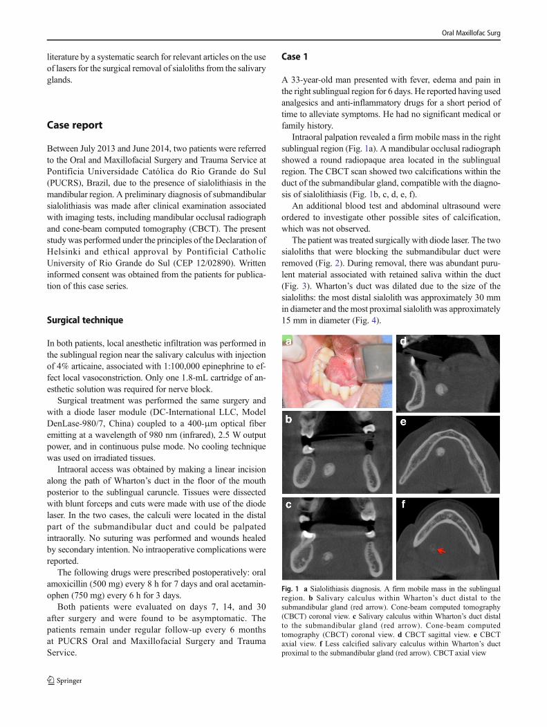
The patient was treated surgically with diode laser. The twosialoliths that were blocking the submandibular duct wereremoved (Fig. 2). During removal, there was abundant puru-lent material associated with retained saliva within the duct(Fig. 3). Wharton’s duct was dilated due to the size of thesialoliths: the most distal sialolith was approximately 30 mmin diameter and the most proximal sialolith was approximately15 mm in diameter (Fig. 4).
Fig. 1 a Sialolithiasis diagnosis. A firm mobile mass in the sublingualregion. b Salivary calculus within Wharton’s duct distal to thesubmandibular gland (red arrow). Cone-beam computed tomography(CBCT) coronal view. c Salivary calculus within Wharton’s duct distalto the submandibular gland (red arrow). Cone-beam computedtomography (CBCT) coronal view. d CBCT sagittal view. e CBCTaxial view. f Less calcified salivary calculus within Wharton’s ductproximal to the submandibular gland (red arrow). CBCT axial view
Oral Maxillofac Surg

After 7 days, the patient was reexamined and reported nodiscomfort on the region and had no postoperative complica-tions. The surgical wound was completely healed within14 days. There was no recurrence of sialolithiasis at 36-month follow-up.
Case 2
A 93-year-old woman, taking simvastatin, enalapril, and aspi-rin, presented with clinical signs of infection in the right sub-lingual region and reported pain when eating for at least5 days. During history taking, the patient reported that shehad undergone an intervention for treatment of sialolithiasisin the same region 15 years earlier. Preoperative blood testswere ordered and showed no abnormalities.
A mandibular occlusal radiograph showed a radiopaquearea located in the right side of the floor of the mouth, com-patible with the clinical diagnosis of recurrent submandibularsialolithiasis. The patient was treated surgically with diodelaser. Three calculi of approximately 4, 8, and 3 mm diametereach were removed, as shown in the video of the surgicaltechnique (Video 1).
After 7 days, the patient reported no postoperative compli-cations. The surgical wound was completely healed within14 days, and there was no recurrence of sialolithiasis at 24-month follow-up.
Review of the literature
A systematic literature search was conducted independentlyby two authors (OLHJ and NS) using MEDLINE (accessedvia PubMed) and EMBASE electronic databases and GoogleScholar. The authors also hand searched the reference list ofall selected articles to identify additional potentially relevantstudies. No limits were applied for language or year of publi-cation. Clinical trials, case series, and case reports describingthe surgical technique and type of laser used to surgicallyremove sialoliths from the ducts of salivary glands were eli-gible for inclusion in this review. The last electronic searchwas conducted on August 26, 2016.
The following search strategy using Medical SubjectHeadings (MeSH) was applied to MEDLINE/PubMed:(BSalivary Gland Calculi^ or BCalculi, Salivary Gland^ orBCalculus, Salivary Gland^ or BGland Calculi, Salivary^ or
Fig. 2 a Outpatient surgical procedure. Surgical approach with a diodelaser. Incision and cauterization of the tissue. b Salivary duct lumen withthe sialolith located most distal to the submandibular gland
Fig. 3 a Removal of the most distal salivary calculus with drainage ofpurulent material and salivary fluid. b Removal of the salivary calculuslocated most proximal to the submandibular gland
Oral Maxillofac Surg

BGland Calculus, Salivary^ or BSalivary Gland Calculus^ orBSialoliths^ or BSialolith^ or BSalivary Gland Stones^ orBGland Stone, Salivary^ or BGland Stones, Salivary^ orBSalivary Gland Stone^ or BStone, Salivary Gland^ orBStones, Salivary Gland^ or BSalivary Duct Calculi^ orBSialolithiasis^ or BCalculi, Salivary Duct^ or BCalculus,Salivary Duct^ or BDuct Calculi, Salivary^ or BDuctCalculus, Salivary^ or BSalivary Duct Calculus^ or BParotidDuct Calculi^ or BSubmandibular Duct Calculi^ or BSalivaryDuct Stones^ or BDuct Stone, Salivary^ or BDuct Stones,Salivary^ or BStone, Salivary Duct^ or BStones, SalivaryDuct^ or BSialolithiasis, Ductal^ or BSialolithiases, Ductal^)and (BLaser Therapy^ or BLaser Therapies^ or BTherapies,Laser^ or BTherapy, Laser^ or BVaporization, Laser^ orBLaser Vaporization^ or BLaser Ablation^ or BAblation,Laser^ or BLaser Tissue Ablation^ or BTissue Ablation,Laser^ or BPulsed Laser Tissue Ablation^ or BLaserPhotoablation of Tissue^ or BNonablative Laser Treatment^or BLaser Treatment, Nonablative^ or BLaser Treatments,Nonablative^ or BNonablative Laser Treatments^ or BLaserScalpel^ or BLaser Scalpels^ or BScalpel, Laser^ or
BScalpels, Laser^ or BLaser Knives^ or BKnife, Laser^ orBKnives, Laser^ or BLaser Knife^ or BLaser Knife^ orBKnife, Laser^ or BKnifes, Laser^ or BLaser Knifes^ orBLaser Surgery^ or BLaser Surgeries^ or BSurgeries, Laser^or BSurgery, Laser^). The search returned 39 articles, of whichfive studies [7, 9–12] met the inclusion criteria.
The EMBASE database was searched using Emtree termsas follows: ‘sialolithiasis’/syn and ‘laser surgery’/syn. Thesearch returned 12 articles. Of these, five were selected, butonly one study [13] was included because the other four hadalready been retrieved from theMEDLINE/PubMed database.
Google Scholar was searched using the main MeSH termsas follows: (BSalivary Gland Calculi^ or BSalivary DuctCalculi^) and BLaser Therapy.^ The search returned 68 arti-cles, but only one study [14], which had not been previouslyretrieved from MEDLINE/PubMed and EMBASE databases,was included.
No additional studies were obtained by checking the refer-ence lists.
Therefore, a total of seven studies without language restric-tion were ultimately included in this review of the literature(Table 1).
Discussion
In this study, we conducted a review of the literature by meansof a systematic search using the main databases rather than asystematic review for which we would need to formulate aresearch question based on the PICOS criteria, analyze thestudies with increased methodological rigor, and assess thequality of the included studies [14]. Our goal was to standard-ize the search criteria to allow the search strategies to bereproduced by the readers who are interested in the use oflasers for the surgical removal of sialoliths from the salivaryglands and to provide the current authors with a broader sci-entific basis to prepare the manuscript. We believe that thisgoal was achieved as the two independent reviewers obtainedthe same search results (seven included articles). As no lan-guage restriction was applied, an article not written in Englishwas identified and included [12]. Also, articles retrieved fromdifferent databases were included (MEDLINE/PubMed = 5studies [1, 10–13], EMBASE = 1 study [15] and GoogleScholar = 1 study [16]).
In addition to providing a more comprehensive literaturereview, the systematic search allowed us to extend the com-parison of the retrieved studies to the two cases reported here.This approach also made it clear the lack of prospective clin-ical trials and the small number of case series (five studies [7,10, 12, 13, 15]) on the topic. From this point of view, thisstudy is unique in that it is the first to provide a reproducibleliterature review and an organized summary of clinical data onthe use of lasers in the surgical treatment of sialolithiasis.
Fig. 4 a Wharton’s duct lumen without postoperative suture to preventranula formation and drainage of salivary fluid. b Removed salivarycalculi
Oral Maxillofac Surg
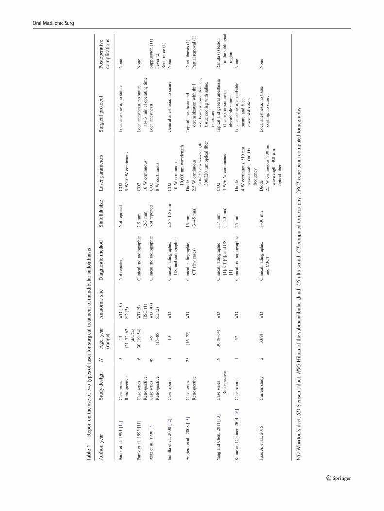
Table1
Reporto
ntheuseof
twotypesof
laserforsurgicaltreatm
ento
fmandibularsialolith
iasis
Author,year
Study
design
NAge,year
(range)
Anatomicsite
Diagnostic
method
Sialolithsize
Laser
parameters
Surgicalprotocol
Postoperative
complications
Barak
etal.,1991
[10]
Caseseries
Retrospective
1344
(21–72)62
(46–74)
WD(10)
SD(3)
Not
reported
Not
reported
CO2
5W/10W
continuous
Localanesthesia,nosuture
None
Barak
etal.,1993
[11]
Caseseries
Retrospective
630
(19–54)
WD(5)
HSG
(1)
Clin
icalandradiographic
2.5mm
(2-3
mm)
CO2
10W
continuous
Localanesthesia,nosuture,
±14.3min
ofoperatingtim
e
None
Azazet
al.,1996
[7]
Caseseries
Retrospective
4945
(15–85)
WD(47)
SD(2)
Clinicalandradiographic
Not
reported
CO2
8W
continuous
Localanesthesia
Suppuration(11)
Fever(2)
Recurrence(1)
Buhillaet
al.,2000
[12]
Casereport
113
WD
Clin
ical,radiographic,
US,and
sialographic
2.5×1.5mm
CO2
10W
continuous,
10,600
nmwavelength
Generalanesthesia,nosuture
None
Angiero
etal.,2008
[15]
Caseseries
Retrospective
25(16–72)
WD
Clinical,radiographic,
CT(few
cases)
15mm
(3–45mm)
Diode
2.5W
continuous,
810/830nm
wavelength,
300/320μm
opticalfiber
Topicalanesthesiaand
desensitizationwith
thel
aser
beam
atsomedistance,
tissuecoolingwith
saline,
nosuture
Ductfibrosis(1)
Partialrem
oval(1)
YangandChen,2011
[13]
Caseseries
Retrospective
1930
(8–54)
WD
Clinical,radiographic
[1],CT[6],andUS
[1]
3.7mm
(1–20mm)
CO2
4W/6
Wcontinuous
Topicaland
generalanesthesia
(1case),no
sutureor
absorbablesuture
Ranula(1)lesion
inthesublingual
region
Kilinç
andÇetiner,2014[16]
Casereport
157
WD
Clin
icalandradiographic
25mm
Diode
4W
continuous,810
nm
wavelength,1000
Hz
frequency
Localanesthesia,absorbable
suture,and
duct
marsupialization
None
HaasJr.etal.,2015
Current
study
233/93
WD
Clinical,radiographic,
andCBCT
3–30
mm
Diode
2.5W
continuous,980
nm
wavelength,400μm
opticalfiber
Localanesthesia,notissue
cooling,no
suture
None
WDWharton’sduct,SDStensen’sduct,H
SGHilu
mof
thesubm
andibulargland,
USultrasound.C
Tcomputedtomography.CBCTcone-beam
computedtomography
Oral Maxillofac Surg

Clinically, a total of 114 patients operated on with laser forsurgical removal of sialoliths from the salivary glands havebeen reported in the literature. Combining our two cases withthose in the literature, a total of 116 cases have been described.Wharton’s duct was the anatomic site most commonly affect-ed by salivary calculi—109 cases (95%), including the twocases reported here. Patient age at diagnosis varied widely,from 8 years [13] to 85 years [7]. However, the oldest patientwas the one described as case 2 in the present report, who was93 years old at diagnosis. The patient was on oral anticoagu-lants, which demonstrated the safety of making an incisionwith a diode laser in a highly vascularized anatomic site withimportant varicosities compatible with advanced age.
The diagnosis of sialolithiasis is primarily clinical, associ-ated with panoramic and/or occlusal radiograph [7, 11–13, 15,16]—only Barak et al. (1991) [12] did not report the use ofpreoperative diagnostic tests. The signs and symptoms of sal-ivary flow obstruction are well defined, including transientlocal edema formation and pain before and during meals, withprogressive postprandial remission; also, chronic recurrentduct obliteration can cause inflammation and infection [17,18]. All these features have been confirmed in the literature.However, some studies have demonstrated the need to ordermore specific imaging tests, such as computed tomography[13, 15], ultrasound [12, 13, 18], and contrast sialography[12, 19], because non-palpable calculi commonly have false-negative results on radiographs [13, 15]. This peculiarity wasalso observed in case 1 reported here, in which, at first, therewas a radiographic diagnosis of a single sialolith, but, as thepatient had an important sublingual edema, we felt the need toorder a CBCT scan for better surgical planning. Based onthese images, the second sialolith, located more proximal tothe submandibular gland, was diagnosed at a less advancedstage of calcification, which had made it not visible on theradiograph.
Diagnostic confirmation of this type of disease requiressurgical intervention as treatment. Traditionally, surgery isregarded as a challenge that is dependent on the anatomicconditions of the submandibular region and the systemic con-di t ions of the pat ient . Thus, techniques such assialoadenectomy [18–20] are considered more invasive andcan put many important structures at risk, especially the(motor) marginal mandibular and hypoglossal nerves and the(sensory) lingual nerve. Another complicating factor is theformation of a hypertrophic scar at the site [11]. However,the best treatment option for cases in which the intraductaldisease is located in the distal part of the duct is a minimallyinvasive surgical intervention using a laser, as reported in thepresent study. This technique is relatively simple, requiresonly local anesthesia and can be performed in an outpatientsetting by an intraoral approach, since there is decreasedbleeding due to hemostasis provided by laser treatment, whichreduces the intervention time and operative morbidity [21].
The studies included in this review [7, 10–13, 15, 16] re-port on the use of two types of laser for surgical treatment ofmandibular sialolithiasis (Table 1): two studies used the diodelaser [14, 16] and five used the CO2 laser [7, 10–13]. Themaindifference between these two types of laser lies in the fact thatCO2 has an absorption peak close to that of water [22], work-ing best on tissues containing large amounts of water, whilethe diode laser has peak emissions at wavelengths where thelight is mainly absorbed by hemoglobin and melanin [16].Therefore, the diode laser was adopted in the present studybecause it is indicated for procedures in patients with coagu-lation disorders, such as the patient described as case 2 here,without the need to discontinue the anticoagulant therapy be-fore surgery [22]. In addition, the diode laser can be used insurgical procedures involving soft tissues of the oral and max-illofacial region, such as in tumor removal, frenectomy, exci-sion of gingival hyperplasia, vestibuloplasty, removal of hem-angiomas, adenomas and fibromas, and peri-implant surgery[23]. Its only contraindication is the impossibility ofperforming histopathologic examination [24], which is notrequired in cases of sialolithiasis.
None of the 116 reported cases required suturing at the endof surgery, which reduces and/or eliminates the risk of post-operative sublingual ranula formation, thus allowing propersalivary drainage without the need for marsupialization orplacement of drains. Furthermore, the effects of laserphotobiomodulation on the tissue lead to increased productionof collagenase, an enzyme that is potentially effective in thetreatment of wounds because of its ability to destroy the col-lagen cells that form the necrotic tissue of the wound [25].Thus, by using laser, new cells can be formed and proliferatemore easily, leading to enhanced wound-healing quality. Ininfected tissues and in cases of suppuration, such as in case1 reported here, the laser incision, which is self-sterilizing,produces a thin surface layer of collagen that serves as anBimpermeable dressing^, protecting the tissue against irritationcaused by oral fluids [26]. This also avoids recurrent and/orpersistent infections when there is concomitant patient coop-eration for oral hygiene maintenance of the operated site.
All the aforementioned features allow postoperative reha-bilitation with minimal swelling, bleeding, infection, and pain,obviating the need for many drugs [27]. The choice of drugtreatment in the present study was due to the systemic involve-ment observed during history taking and on complementarytests. However, traditionally, patients undergoing intraoral la-ser treatment receive only analgesics and anti-inflammatorydrugs after surgery.
In view of the foregoing, we believe that surgical treatmentwith diode laser is superior to conventional surgical treatmentdue to the safety and minimal invasiveness of the procedure,resulting in significantly reduced morbidity. Nevertheless,some limitations need to be considered, such as increasedoperative time and the high cost of equipment.
Oral Maxillofac Surg
Conclusion
The results obtained with the use diode laser and reported inthe present study show that this device is extremely useful as atool to access the salivary gland for the removal of calculilocated in the distal part of Wharton’s duct. Its use improvespostoperative patient comfort while minimizing complica-tions in highly vascularized sites, such as the floor of themouth. None of the laser-treated patients had intraoperativeor postoperative complications.
Compliance with ethical standards
Conflict of interest The authors declare that they have no conflict ofinterest.
Ethical approval The present study was performed under the principlesof the Declaration of Helsinki and ethical approval by Pontificial CatholicUniversity of Rio Grande do Sul (CEP 12/02890).
Informed consent Informed consent was obtained from the patients forpublication of this case series.
References
1. Siddiqui SJ (2002) Sialolithiasis: an unusually large submandibularsalivary stone. Br Dent J 193(2):89–91. https://doi.org/10.1038/sj.bdj.4801491
2. Sialadenitis BR (1995) Sialolithiasis: diagnosis and management.Oral Maxillofac Surg Clin North Am 7:479–502
3. Mandel ID (1963) Histochemical and biochemical aspects of cal-culus formation. J Am Soc Periodont 1:43
4. Lustmann J, Regev E, Melamed Z (1990) Sialolithiasis. A surveyon 245 patients and a review of the literature. Int J Oral MaxillofacSurg 19:135–138
5. Gallo O, Berloco P, Bruschini L (2001) Sialadenectomy. In:McGurk M, Renehan AG (eds) Controversies in the managementof salivary gland disease. Oxford University Press, Oxford, pp297–303
6. Ito H, Baba S (1996) Pulsed dye laser lithotripsy of submandibulargland salivary calculus. J Laryngol Otol 110(10):942–946
7. Azaz B, Regev E, Casap N, Chicin R (1996) Sialolithectomy donewith a CO2 laser: clinical and scintigraphic results. J OralMaxillofac Surg 54(6):685–688. https://doi.org/10.1016/S0278-2391(96)90681-3
8. Raif J, Vardi M, Nahlieli O, Gannot I (2006) An Er:YAG laserendoscopic fiber delivery system for lithotripsy of salivary stones.Lasers Surg Med 38(6):580–587. https://doi.org/10.1002/lsm.20344
9. Sexton J, O’Hare D (1993) Simplified treatment of vascular lesionsusing the argon laser. J Oral Maxillofac Surg 51(1):12–16. https://doi.org/10.1016/S0278-2391(10)80380-5
10. Barak S, Horowitz I, Katz J, Kaplan I (1991) Experiences of theCO2 laser in the surgical treatment of intraoral salivary gland pa-thology. J Clin Laser Med Surg 9(4):295–299
11. Barak S, Katz J, Mintz S (1993) Use of the carbon dioxide laser tolocate small sialoliths. J Oral Maxillofac Surg 51(4):379–381.https://doi.org/10.1016/S0278-2391(10)80349-0
12. Alvarez-Buhilla LP, Blanco-Bruned JL, Torres-Piedra C, Alfonso-Sánchez L (2000) CO2 laser treatment of sialolithiasis. An EspPediatr 53(1):62–63
13. Yang SW, Chen TA (2011) Transoral carbon dioxide lasersialolithectomy with topical anaesthesia. A simple, effective, andminimally invasive method. Int J Oral Maxillofac Surg 40(2):169–172. https://doi.org/10.1016/j.ijom.2010.09.020
14. Liberati A, Altman DG, Tetzlaff J, Mulrow C, Gøtzsche PC,Ioannidis JP, Clarke M, Devereaux PJ, Kleijnen J, Moher D(2009) The PRISMA statement for reporting systematic reviewsandmeta-analyses of studies that evaluate health care interventions:explanation and elaboration. J Clin Epidemiol 62(10):e1–34.https://doi.org/10.1016/j.jclinepi.2009.06.006
15. Angiero F, Benedicenti S, Romanos GE, Crippa R (2008)Sialolithiasis of the submandibular salivary gland treated with the810- to 830-nm diode laser. Photomed Laser Surg 26(6):517–521.https://doi.org/10.1089/pho.2007.2226
16. Kılınç Y, Çetiner S (2014) Surgical removal of a giant sialolith bydiode laser. Open J Stomato 4(10):484–488. https://doi.org/10.4236/ojst.2014.410065
17. Mandel L, Witek EL (2001) Chronic parotitis diagnosis and treat-ment. J Am Dent Assoc 132:1707
18. Eggers G, Chilla R (2005) Ultrasound guided lithotripsy of salivarycalculi using an electromagnetic lithotriptor. Int J Oral MaxillofacSurg 34(8):890–894. https://doi.org/10.1016/j.ijom.2005.04.012
19. Chu DW, Chow TL, Lim BH, Kwok SP (2003) Endoscopic man-agement of submandibular sialolithiasis. Surg Endosc 17(6):876–879. https://doi.org/10.1007/s00464-002-8563-x
20. Baurmash HD (2004) Submandibular salivary stones: current man-agement modalities. J Oral Maxillofac Surg 62(3):369–378. https://doi.org/10.1016/j.joms.2003.05.011
21. Hald J, Andreassen UK (1994) Submandibular gland excision:short- and long-term complications. ORL J OtorhinolaryngolRelat Spec 56(2):87–91. https://doi.org/10.1159/000276616
22. Romeo U, Del Vecchio A, Russo C, Palaia G, Gaimari G, Arnabat-Dominguez J, España AJ (2013) Laser treatment of 13 benign oralvascular lesions by three different surgical techniques. Med OralPatol Oral Cir Bucal 18(2):e279–e284
23. Romanos G, Nentwig GH (1999) Diode laser (980nm) in oral andmaxillofacial surgical procedures: clinical observations based onclinical applications. J Clin Laser Med Surg 17(5):193–197
24. Derowe A, Landsberg R, Leonov Y, Katzir A, Ophir D (1998)Subjective comparison of ND:YAG diode and CO2 lasers for en-doscopically guided inferior turbinate reduction surgery. Am JRh i n o l 1 2 ( 3 ) : 2 0 9 –2 12 . h t t p s : / / d o i . o r g / 1 0 . 2 5 0 0 /105065898781390145
25. Suzuki R, Takakuda K (2016) Wound healing efficacy of a 660-nmdiode laser in a rat incisional wound model. Lasers Med Sci 31(8):1683–1689. https://doi.org/10.1007/s10103-016-2038-0
26. Gontiya G, Bhatnagar S, Mohandas U, Galgali SR (2011) Laser-assisted gingivectomy in pediatric patients: a novel alternative treat-ment. J Indian Soc Pedod Prev Dent 29(3):264–269. https://doi.org/10.4103/0970-4388.85839
27. Boj JR, Poirier C, Hernandez M, Espasa E, Espanya A (2011) Caseseries: laser treatments for soft tissue problems in children. EurArch Paediatr Dent 12(2):113–117. https://doi.org/10.1007/BF03262790
Oral Maxillofac Surg