Shoulder dystocia rabi
61
Shoulder Dystocia Dr. Rabinarayan Satapathy Dr. Rabinarayan Satapathy Asst. Professor Asst. Professor Dept. of Obst.& Gynae Dept. of Obst.& Gynae S.C.B. Medical College,Cuttack S.C.B. Medical College,Cuttack
-
Upload
rabi-satpathy -
Category
Health & Medicine
-
view
242 -
download
3
Transcript of Shoulder dystocia rabi

Shoulder Dystocia
Dr. Rabinarayan SatapathyDr. Rabinarayan SatapathyAsst. ProfessorAsst. ProfessorDept. of Obst.& GynaeDept. of Obst.& GynaeS.C.B. Medical College,CuttackS.C.B. Medical College,Cuttack


Definition
• “…a delivery that requires additional obstetric maneuvers following failure of gentle downward traction on the fetal head to effect delivery of the shoulders.”
• ACOG, Practice Bulletin 40 (November 2002)

Definition
• “Prolonged head-to-body expulsion time”
• Objectively defined as 60 seconds
• Deliveries with head-to-body interval of > 60 seconds more commonly have higher birth weight, shoulder dystocia, and low 1 minute Apgar scores
– Beall et al 1998; Spong et al 1995

Functional Definition
• A delivery in which the shoulders do not follow the head as usual, but rather are delayed in delivering or require the use of ancillary obstetric maneuvers to effect delivery.
• The anterior shoulder may be impacted behind the symphysis pubis, or (less commonly) the posterior shoulder behind the sacral promontory

Shoulder dystocia
• Shoulder dystocia can be one of the most frightening emergencies in the delivery room
• Although many factors have been associated with shoulder dystocia, most cases occur with no warning
• Defined as a delivery in which additional maneuvers are required to deliver the fetus after normal gentle downward traction has failed– Shoulder dystocia occurs when the fetal
anterior shoulder impacts against the maternal symphysis


Epidemiology
• The overall incidence of shoulder dystocia varies based on fetal weight– 0.6-1.4%: 2500g(5lb,8oz) to 4000g(8lb,13oz)– 5-9%: 4000g to 4500g(9lb,14oz), born to
mothers without diabetes• 3969g (8lb,12oz) – our patient
• Shoulder dystocia occurs with equal frequency in primigravid and multigravid women– More common in infants born to women with
diabetes• The single most common risk factor for
shoulder dystocia is the use of a vacuum extractor or forceps during delivery
• However, most cases occur in fetuses of normal birth weight and are unanticipated, limiting the clinical usefulness of risk-factor identification
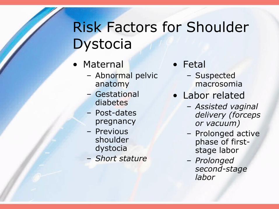
Risk Factors for Shoulder Dystocia• Maternal
– Abnormal pelvic anatomy
– Gestational diabetes
– Post-dates pregnancy
– Previous shoulder dystocia
– Short stature
• Fetal– Suspected
macrosomia
• Labor related – Assisted vaginal
delivery (forceps or vacuum)
– Prolonged active phase of first-stage labor
– Prolonged second-stage labor
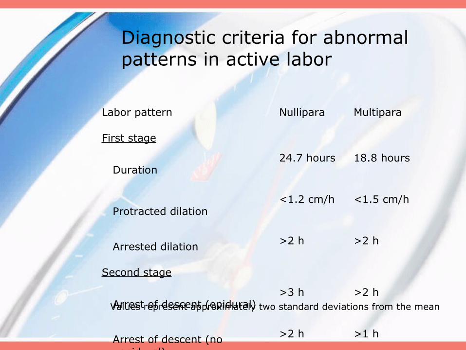
Labor pattern Nullipara Multipara
First stage
Duration24.7 hours 18.8 hours
Protracted dilation<1.2 cm/h <1.5 cm/h
Arrested dilation >2 h >2 h
Second stage
Arrest of descent (epidural)>3 h >2 h
Arrest of descent (no epidural)
>2 h >1 h
Values represent approximately two standard deviations from the mean
Diagnostic criteria for abnormal patterns in active labor


Complications of Shoulder Dystocia*• Maternal
– Postpartum hemorrhage (11%)
– Rectovaginal fistula – Symphyseal
separation • With or without
transient femoral neuropathy
– 3rd or 4th degree (3.8%) episiotomy or tear
– Uterine rupture
• Fetal– Brachial plexus palsy
(4-15%)†– Clavicle fracture– Fetal hypoxia
• With or without permanent neurologic damage
– Fracture of the humerus
– Fetal death
†Nearly all palsies resolve within six to 12 months, with fewer than 10% resulting in permanent injury
*These rates remain constant, independent of operator experience

HELPERR pneumonic
• H – Help– Call for additional assistance
• E – Evaluate for episotomy• L – Legs (McRobert’s Maneuver)• P – Pressure (suprapubic)• E – Enter the vagina• R – Remove the posterior arm• R – Roll the patient
– To hands and knees

HELPERR pneumonic (cont.)• Although there is no indication that
any one of these techniques is superior to another, together they effectively relieve the impacted shoulder
• The order of the steps is not as important as the fact that they each be employed efficiently and appropriately
• Persistence in any one ineffective maneuver should be avoided
• Clinical judgment always should guide the progression of procedures used

Clinical Management
• Step One: Recognize the presence of a shoulder dystocia
• Step Two: Be sure enough help is present– Nursing– Obstetrics– Pediatrics– Anesthesiology

Clinical Management
• Step Three: Apply primary maneuvers– Mc Roberts maneuver– Oblique suprapubic pressure
• Step Four: Apply secondary maneuvers; no prescribed order– Rubin; Woods screw; Posterior
arm; All-fours; Clavicular fracture

Clinical Management
• Step Five (concurrent):– Repeat steps three and four
(different operator?)– Consider if an episiotomy is needed
(intentional 4th degree?)
• Step Six: Apply final (heroic) maneuvers– Zavanelli; symphysiotomy

H - Help
• This refers to activating the pre-arranged protocol or requesting the appropriate personnel to respond with necessary equipment to the labor and delivery unit
• If standard levels of traction do not relieve the shoulder dystocia, the physician must move quickly to other maneuvers while asking for help and notifying the family
• A critical step in addressing the emergency management of shoulder dystocia is ensuring that all involved hospital personnel are familiar with their roles and responsibilities

E - Evaluate for episotomy
• Episiotomy should be considered when a shoulder dystocia is encountered, although because the primary problem is a bony impaction, episiotomy by itself will not release the impaction
• Episiotomy does provide additional room for the physician's hand when internal rotation maneuvers are required
• Given the success of the McRobert’s maneuver and suprapubic pressure in relieving a large percentage of cases of shoulder dystocia, performing an episiotomy can wait until later in the sequence

L – Legs (McRobert’s maneuver)• The simplicity of the McRobert’s maneuver and its
proven effectiveness make it an ideal first step in the management of shoulder dystocia
• This procedure results in a cephalad rotation of the symphysis pubis and a flattening of the sacral promontory – These motions push the posterior shoulder over the
sacral promontory, allowing it to fall into the hollow of the sacrum, and rotate the symphysis over the impacted shoulder
• When this maneuver is successful, the fetus should be delivered with normal traction
• The McRobert’s maneuver alone is believed to relieve more than 40% of all shoulder dystocias and, when combined with suprapubic pressure, resolves more than 50% of shoulder dystocias


P – Pressure (suprapubic)
• When applying suprapubic pressure, an assistant's hand should be placed on top of the mother's abdomen over the fetal anterior shoulder, applying pressure in a compression/relaxation cycle analogous to cardiopulmonary resuscitation, so that the shoulder will adduct and pass under the symphysis
• Pressure should be applied from the side of the mother, with the heel of the assistant's hand moving in a downward and lateral motion on the posterior aspect of the fetal impacted shoulder
• Initially, the pressure can be continuous, but if delivery is not accomplished, a rocking motion is recommended to dislodge the shoulder from behind the pubic symphysis
• Fundal pressure is never appropriate and only serves to worsen the impaction, potentially injuring the fetus or mother


E – Enter maneuvers
• Rotation maneuvers may require episiotomy to gain posterior vaginal space for the physician’s hand
• The Rubin II maneuver consists of inserting the fingers of one hand vaginally behind the posterior aspect of the anterior shoulder of the fetus and rotating the shoulder toward the fetal chest
• This motion will adduct the fetal shoulder girdle, reducing its diameter
• The McRobert’s maneuver also can be applied during this maneuver and may facilitate its success

E – Enter maneuvers (cont.)• If the Rubin II maneuver is unsuccessful, the
Woods corkscrew maneuver may be attempted• The physician places at least two fingers on the
anterior aspect of the fetal posterior shoulder, applying gentle upward pressure around the circumference of the arc in the same direction as with the Rubin II maneuver
• The Rubin II and Woods corkscrew maneuvers may be combined to increase torque forces by using two fingers behind the fetal anterior shoulder and two fingers in front of the fetal posterior shoulder
• Procedurally, this step often is difficult because of limited space for the physician's hand
• Downward traction should be continued during these rotational maneuvers, simulating the rotation of a screw being removed
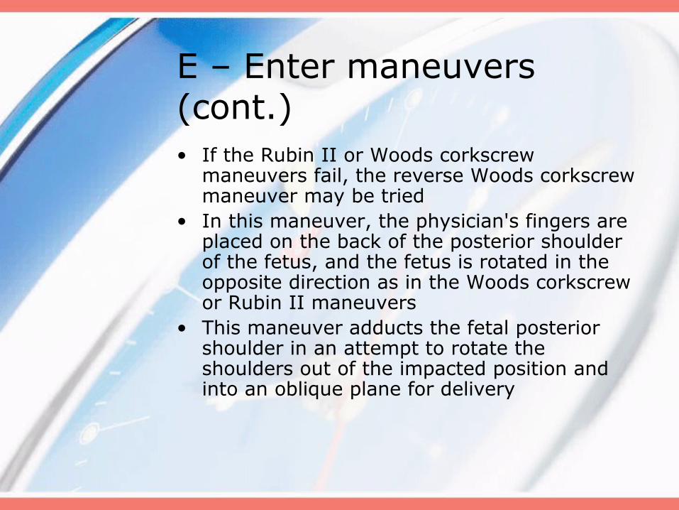
E – Enter maneuvers (cont.)• If the Rubin II or Woods corkscrew
maneuvers fail, the reverse Woods corkscrew maneuver may be tried
• In this maneuver, the physician's fingers are placed on the back of the posterior shoulder of the fetus, and the fetus is rotated in the opposite direction as in the Woods corkscrew or Rubin II maneuvers
• This maneuver adducts the fetal posterior shoulder in an attempt to rotate the shoulders out of the impacted position and into an oblique plane for delivery

Rubin II At vaginal examination apply pressure as indicated. If shoulders move into the oblique diameter, attempt delivery
Rubin II + Woods corkscrew maneuver If unsuccessful, add the Woods corkscrew maneuver and continue rotation in the same direction. Use both hands and apply pressure as indicated. If shoulders now move into the oblique, attempt delivery. If this is unsuccessful, continue rotation 180 degrees and deliver
Reverse Woods corkscrew maneuver
If the last maneuver is unsuccessful, change to reverse Woods corkscrew maneuver. Slide fingers down to back of posterior shoulder and attempt 180-degree rotation in the opposite direction

R – Remove the posterior arm• Removal of the posterior arm involves placing the
physician's hand in the vagina and locating the fetal arm, which sometimes is displaced behind the fetus and must be nudged anteriorly– The physician's hand, wrist, and forearm may need
to enter the vagina, necessitating an episiotomy or extension
• The fetal elbow is then flexed, and the forearm is delivered in a sweeping motion over the anterior chest wall of the fetus
• The upper arm should never be grasped and pulled directly, because this step may result in a fracture of the humerus
• The posterior hand, followed by the arm and shoulder, will be reduced, facilitating delivery of the infant
• The anterior shoulder will then fall under the symphysis and deliver

R – Roll the patient
• Rolling the patient onto her hands and knees, known as the all-fours or Gaskin maneuver, is a safe, rapid, and effective technique for the reduction of shoulder dystocia
• Once the patient is repositioned, the physician provides gentle downward traction to deliver the posterior shoulder with the aid of gravity
• The all-fours position is compatible with all intravaginal manipulations for shoulder dystocia, which can then be reattempted in this new position
• All-fours positioning may be disorienting to physicians who are unfamiliar with attending a delivery in this position
• Performing a few “normal” deliveries in this position before encountering a case of shoulder dystocia may prepare physicians for more emergent situations

Maneuvers of last resort
• If the maneuvers described in HELPERR are unsuccessful, several techniques have been described as “last-resort” maneuvers
• Once the infant is delivered, quick assessment and employment of resuscitation efforts, if necessary, are vital

Maneuvers of last resort (cont.)• Deliberate clavicle fracture
– Direct upward pressure on the mid-portion of the fetal clavicle; reduces the shoulder-to-shoulder distance
• Zavanelli maneuver– Cephalic replacement followed by cesarean
delivery; involves rotating the fetal head into a direct occiput anterior position, then flexing and pushing the vertex back into the birth canal, while holding continuous upward pressure until cesarean delivery is accomplished
– An operating team, anesthesiologist, and physicians capable of performing a cesarean delivery must be present, and this maneuver should never be attempted if a nuchal cord previously has been clamped and cut
Maneuvers of last resort (cont.)• General anesthesia
– Musculoskeletal or uterine relaxation with halothane (Fluothane) or another general anesthetic may bring about enough uterine relaxation to affect delivery
– Oral or intravenous nitroglycerin may be used as an alternative to general anesthesia
• Abdominal surgery with hysterotomy– General anesthesia is induced and cesarean
incision performed, after which the surgeon rotates the infant transabdominally through the hysterotomy incision, allowing the shoulders to rotate, much like a Woods corkscrew maneuver
– Vaginal extraction is then accomplished by another physician

Documentation
• Documentation of the management of shoulder dystocia should concentrate on the maneuvers performed and the duration of the event
• Terms such as mild, moderate, or severe shoulder dystocia offer little information about the situation or care encountered
• Other team members assisting the delivery should be listed, as well as cord pH, if obtained
• Specific notation regarding which arm was impacted against the pubis should be made in the event that subsequent nerve palsy develops
• The delivery should be reviewed with the parents, and the management and prognosis for any infant palsy should be explained
Steps One and Two
• Personnel (continued)– Anesthesiology– Obstetrics
• Attending to supervise and step in as needed
• 2 residents at minimum– Ideally 2 at perineum– One to assist with maneuvers
(suprapubic pressure) away from perineum

Step Three – Primary Maneuvers• McRoberts maneuver
– Patient positioned with hips at edge of the broken-down birthing bed
– Both hips are sharply flexed with knees remaining flexed (“knees to shoulders”)
– Ideally performed by staff, not family, to assure it is adequately performed
– No benefit to “prophylactic” McRoberts

McRoberts Maneuver

McRoberts Maneuver
• This maneuver assists delivery by:– Straightening maternal lumbar
lordosis– Rotates symphysis superiorly and
anteriorly– Improving angle between pelvic
inlet and direction of maximal expulsive force
– Elevates anterior shoulder allowing posterior shoulder to descend
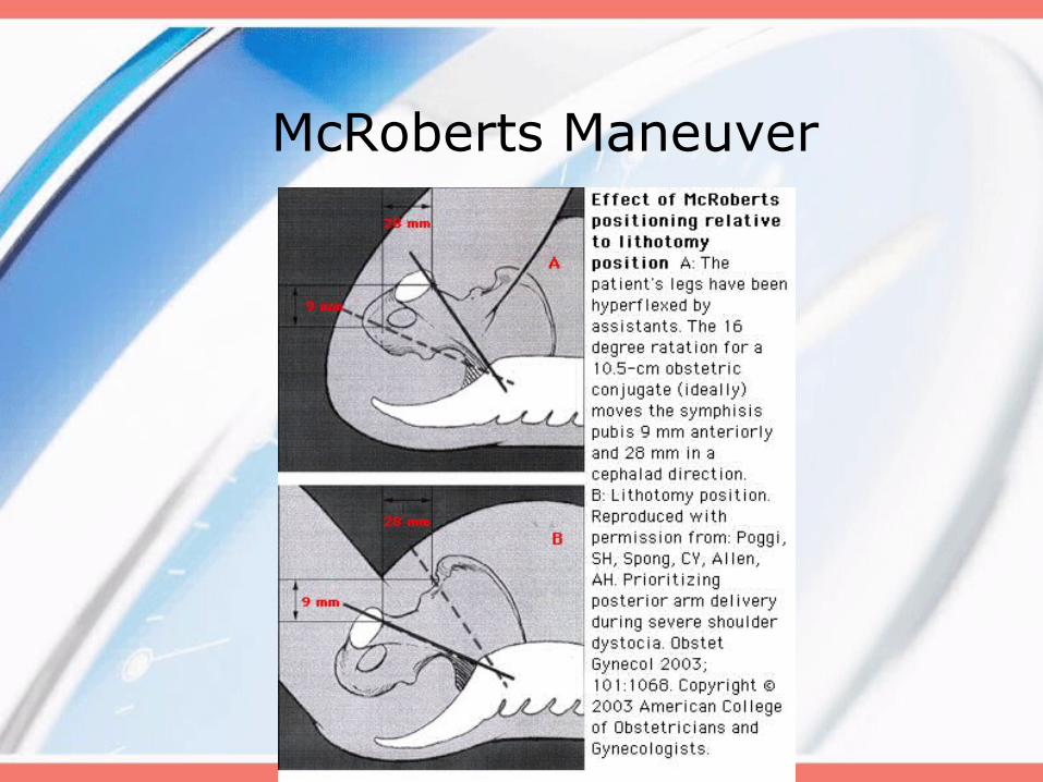
McRoberts Maneuver

Oblique suprapubic pressure• Usually applied in concert with
McRoberts maneuver• Directed downward and laterally
in order to effect rotation of the fetal anterior shoulder under the symphysis
• Should be applied from the fetal posterior
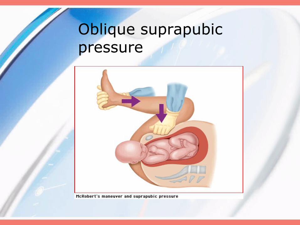
Oblique suprapubic pressure

Step Four – Secondary Maneuvers• There is no conclusive evidence
that one maneuver is superior to another
• In each patient, the operator must decide which maneuver will be most effective
• This is a good time to decide about an episiotomy – is there room to get your hand in?
• Time to initiate perinatal code (4-2012)

Woods screw maneuver
• Apply pressure on the clavicle to effect rotation of the shoulders out of the vertical orientation
• As fetus rotates, anterior shoulder should pass under symphysis
• May be a good choice for a right-handed operator when the fetal occiput is oriented to the maternal right

Woods screw maneuver

Woods screw maneuver
• Potential complication:– Fetal clavicular fracture IN
DIRECTION OF APEX OF LUNG

Rubin’s maneuver
• Apply pressure to the fetal scapula to effect rotation of the shoulders out of the vertical orientation
• As fetus rotates, anterior shoulder should pass under symphysis
• May be a good first choice for a right-handed operator when the fetal occiput is directed to the maternal left

Rubin’s maneuver
• May result in need for less traction and less brachial plexus strain than McRoberts maneuver
– Gurewitsch, 2005

Delivery of Posterior Arm
• The operator inserts a hand into the vagina and locates the posterior arm.
• The operator applies pressure in the antecubital fossa to flex the elbow across the chest
• The operator grasps the forearm or hand and pulls it out of the vagina

Delivery of Posterior Arm
• The anterior shoulder should pass under the symphysis
• Rotation maneuvers (Woods or Rubin’s) can be applied if needed
• This maneuver will tend to be more difficult with one’s non-dominant hand

Delivery of Posterior Arm

Delivery of Posterior Arm
• Potential complications– Fracture of humerus– Fracture of clavicle

Gaskin All Fours Maneuver• Attributed to midwife Ina May
Gaskin• An option for a patient without
anesthesia• Traction is applied in the
opposite direction (still toward the floor, but now directed towards delivery of the posterior shoulder first)

Intentional clavicular fracture• Apply pressure over mid-clavicle
in a vector AWAY from the lung• May be difficult to perform• If successful, may reduce the
diameter of the shoulder girdle• Potential complication:
– Lung injury

Still not out?!What now???

Step Five – Regroup and Repeat• Considerations:• Time passed so far?• Episiotomy?• Different operator?• Make OR preparations!

Step Six – Final Steps
• Zavanelli maneuver (cephalic replacement)– Relax uterus with terbutaline– Rotate head back to OA (“reverse
restitution”)– Flex neck– Upward pressure– To OR

Step Six – Final Steps
• Symphysiotomy– Not commonly done when
cesarean is available– Last ditch effort
• Insert Foley catheter• Use vaginal hand to laterally displace
urethra to avoid injury• Incise symphysis through mons pubis

Do not:
• Panic• Apply any more lateral traction
than would be applied in an uncomplicated delivery
• Apply fundal pressure – may worsen the shoulder impaction or even rupture the uterus
• Cut a nuchal cord until after the shoulders are released

Do:
• Remain calm• Communicate well
– Mark time of head delivery– Consider calling out time in one
minute increments
• Call for help• Document clearly and legibly

Do:
• Be sure to “debrief” as a team after the delivery is completed– Opportunity to analyze situation
and critique team performance– Opportunity to be sure
documentation is consistent– Who did what becomes very
important
• Send cord gases

Do:
• Review with the family exactly what happened and answer questions – soon after delivery, but probably not immediately
• Follow the baby’s course in the nursery
• Notify Risk Management

References
• Shoulder Dystocia (Practice Bulletin 40). American College of Obstetricians and Gynecologists. November 2002.
• Rodis, JF. Management of fetal macrosomia and shoulder dystocia. Up to date, v 14.1; last updated October 12, 2005.
• Brachial Plexus. Wikipedia, the online encyclopedia. http://en.wikipedia.org/wiki/Brachial_plexus Accessed March 21, 2006.
• Beall, MH, et al. Objective definition of shoulder dystocia: a prospective evaluation. Am J Obstet Gynecol 1998;179:934.
• Spong CY, et al. An objective definition of shoulder dystocia: prolonged head-to-body interval and/or the use of ancillary obstetric maneuvers. Obstet Gynecol 1995;86:433
• Gurewitsch ED et al. Comparing McRoberts’ and Rubin’s maneuvers for initial management of shoulder dystocia: an objective evaluation. Am J Obstet Gynecol 2005;192:153.