
Isolating the impact of visual perception on dyslexics’ reading ability
DORIS DUKE MEDICAL STUDENTS’ JOURNAL Volume III, 2003-2004
Short-Term Memory in Dyslexics: an fMRI Study
Stephanie Holler Howard Mentor:Dr. Joy Hirsch A. Study Purpose and Rationale
Overview Functional MRI allows the localization of human mental operations related to specific
activities by mapping changes in brain hemodynamics. When brain neuronal activity increases, blood flow to the area of activity increases proportionally. The oxygen consumption of that area, however, does not increase at the same magnitude (Roy, 1890, Plum, 1968, Posner, 1969, Fox 1985.) Thus, areas of heightened neuronal activity are associated with decreased levels of deoxyhemoglobin, (Fox, et al 1985.) Because deoxyhemoglobin is paramagnetic, it is used as an endogenous contrast agent in the T2* weighted MRI signal, (Ogawa, et al, 1990 a and b, 1993, Belliveau, et al, 1990, 1991, Tank, et al, 1992.) Areas of high neuronal activity, and therefore increased blood flow and lower deoxyhemoglobin levels, can then be observed with resolution of about 1.5 x 1.5 mm.
Functional imaging has helped decipher the differences in neural activity between dyslexic and non-impaired readers. A 1999 Positron Emission Tomography study examining neural activation during reading in dyslexic adults found that dyslexics show less activation in the posterior parts of the brain, specifically the left posterior inferior temporal cortex, the left and midline cerebellum, the left thalamus, and the medial extrastriate cortex, but recruit more anterior areas, specifically Broca’s area, than controls (Brunswick, et al 1999.) Similar results were found using fMRI; dyslexics showed decreased activity in Wenickes’s area, the angular gyrus, and the striate cortex, while they showed a relative overactivation in the inferior frontal gyrus, (Shaywitz, et al 1998.)
Dyslexia is believed to be due to deficits in phonological awareness and phonological decoding skills (Shaywitz, 2003.) Dyslexic children appear to have difficulty building a full mental phonemic dictionary. When they learn to read, children recover spoken segments from the orthographic form and overlay them as they sound out words, (Mody, 2003.) If children have an incomplete or poorly developed phonemic dictionary they struggle when attempting to relate text to familiar phonemic sounds, thereby making reading an arduous and time-consuming task (Mody, 2003, Shaywitz, 2003.) While various small deficiencies likely combine to comprise the overall syndrome of dyslexia, our experiment hopes to use fMRI to compare brain activity during short-term memory tasks in dyslexic and non-impaired readers.
Dyslexic readers consistently perform worse than normal readers on short-term verbal memory tasks associated with speech, (Hansen & Bowery, 1994, Jorm, 1983, Brady, 1983.) Dyslexic’s have no trouble, however, with non-linguistic short-term memory tasks like picture, nonsense figure, or character recall, (Gould and Glencross, 1990, Katz, 1981,) This linguistic short-term memory difficulty may hinder the development of the phonemic dictionary in dyslexic children, and hence may contribute greatly to the reading problems of dyslexia (Mody, 2003.)
a. Specific Aims We plan to compare brain activity in dyslexic adults and normal readers during short-
term memory tasks. The tasks will first measure short-term memory of pictures, a situation in which dyslexics typically show no delay, and where we believe we will see same brain activity for both dyslexics and controls. Then we will test short-term memory of letters, a situation where we know dyslexics typically perform poorly compared to normal readers. We expect to see different intensities of brain activation, with dyslexic subjects having more blood flow to the area and hence stronger signals due to the fact the task is more difficult for them. We also believe
Columbia University College of Physicians and Surgeons
20
DORIS DUKE MEDICAL STUDENTS’ JOURNAL Volume III, 2003-2004
dyslexic readers may recruit other areas of the brain not typically used for short-term memory, and will be able to see if this recruitment occurs. The next run of the test will examine the short-term memory of words, enabling us to see differences in brain activity between dyslexics and normal readers as the linguistic tasks increase in difficulty. Finally, we will test the short-term memory of pseudowords, which will allow us to determine if familiarity with the word changes the way the brain attempts to store information in short-term memory.
b. Significance A more thorough understanding of short-term memory function in people with dyslexia
will allow us to better understand the more general differences in neuronal activity we see during reading tasks. This study will enable us to see what sort of memory tasks elicit differences in neuronal activity, and exactly what those differences are. We will better be able to isolate what brain areas dyslexic people are using for verbal memory as they read. As we gather more information regarding how people with dyslexia process text, we will be able to design more suitable strategies for helping these people learn to read.
B. Study Design and Statistical Analysis
a. Study Groups The study will have two groups of participants: a group of dyslexic patients and gender
and educational level matched controls. We did not match for age as we believe educational level matching will allow us to better match for reading level. Furthermore, as only participants aged 18-25 will be allowed to participate in the study, we do not feel that differences in age within this range should result in differences in brain activation. Each participant will be required to take the performance portion of the Weschler Adult Intelligence Scale test, and only those participants scoring within one standard deviation of the mean or above will be allowed to participate in the study. Participants will also take the word identification and word attack portions of the Woodcock Reading Mastery test. Dyslexic subjects must score below the 25th percentile on this test, while controls must score in the 39th percentile or above. No subjects with history of hearing or visual impairment, brain injury, or neurological or severe emotional disorders will be included in the study.
There will be approximately 20 people in the control group, and 10 in the dyslexic group. Every two control participants will be matched by gender and education level with a dyslexic participant. These numbers were derived using sample size equations assuming a power of 90%, with an alpha of .05, while requiring an effect size of 7%, with a standard deviation of 5%.
This study will be of the same design as the fMRI studies currently ongoing in the fMRI lab, under the IRB protocol, “Functional Mapping of the Human Brian Using MRI: Development of Techniques and Methods,” written by Dr. Joy Hirsch. We will obtain both “baseline” scans, when the subject is doing nothing but looking at a crosshair, and “active” scans, when the subject is performing a memory task. Each of the four memory tasks will compose a separate run. The four different types of runs will test either short-term memory of pictures, letters, words, or pseudowords. It takes approximately 40 seconds to obtain ten images of the entire brain. In each of the four types of runs there will be two 40 second stimulus epochs, so that we basically scan the entire brain during each run twice, flanked by baseline (resting) epochs.
Average signal level during each run will be compared to average signal level at baseline using independent t-tests (Winer, 1971.) The fundamental unit of analysis is the single voxel where signal intensities are compared over all acquired images. A control period is imbedded in the image acquisition sequence with an initial and recovery baseline. Voxels will be sorted into two categories, activated and non-activated, based on how they compare to the control period, which consists of the subject’s initial and recovery non-task baseline scans. If both the initial and recovery baselines are statistically different from the task’s signal mean then the pixel is identified as activated. Otherwise the pixel is non-activated and excluded from further consideration. If a voxel is identified as activated it is assigned a color code based on the
Columbia University College of Physicians and Surgeons
21

DORIS DUKE MEDICAL STUDENTS’ JOURNAL Volume III, 2003-2004
certainty of its activation (p level) and the polarity of activation (more active or less active than the baseline scans.)
We assume that multiple sources can lead to voxel activation: chance, motion (due to the pulsatile movements of the brain, gross movements of the head, and the flow of CSF,) and the BOLD (Blood Oxygen Level Dependent) signal (Bandettini, et al 1993.) To isolate the voxels with activations due to the BOLD response, we additionally assume that the anatomical “motion and chance” voxels tend to vary over repeated epochs. Corresponding anatomical slices from repeated epochs will be registered based on anatomic features, and those color coded voxels that were coincident on both epochs are stored, all others are excluded from further consideration. Each of the four types of runs will contain two epochs. Since the same slices will be taken during each of the epochs, the anatomies of similar epochs are easily registered and coincidences in voxels that occur over the two time frames are identified. Only those voxels identified as coincident amongst the two epochs will be used in group analysis.
Areas of significant activation from each individual then will be grouped by the individual’s reading ability. All data from dyslexic participants will be compared to the compiled data from normal readers for each run using t-tests. Various image registration and “warping” techniques will be applied to compare images within the database, to ensure appropriate anatomical areas of various individuals’ brains are being compared. Data will be analyzed and displayed using either the program SPM or fMRI. Results will be displayed in 2 and 3-D formats both individually and over the entire sample of patients and normal volunteers.
All estimates of error rates (false positive decisions) are based on empirical determinations from images acquired on copper-sulfate phantoms. A copper sulfate ball is scanned, and regional flow monitored. As this ball should have no regional flow, any flow registered by the scanner will be considered a false positive result and the scanner will be recalibrated.
C. Study Procedures
a. Imaging Procedures Images will be acquired on a 1.5 T magnetic resonance scanner (General Electric Twin
Speed) using the echo planar or equivalent option and a typical T2* weighted gradient echo sequence such as (TE =60 ms, TR= 4,000ms, flip angle=60 degrees.) A 40 x 20 cm field of view imaged on a 256 x 128 grid yielding an in-plane resolution of 1.56 x 1.56 mm and slice thickness of 5 mm or less are typical. On each run simultaneous images will be acquired on multiple contiguous slices chosen to cover the entire brain. Slice positions are based on a conventional “scout” image obtained prior to the run usually using the clinical T1 sequence or a SPGRS sequence. Slice positions will be maintained as constant as possible from run to run in the case of repeated scans. The experiment will be run in a grouped manner to take advantage of the coincidence options for image processing. Approximately thirty-three images will be acquired during each typical run, thirteen in the initial baseline period, of which the first three are discarded, ten in the active period, and ten in the final baseline period.
b. Experimental Procedures Prior to entering the scanner room all subjects will have given written consent to
participate in the research. We plan to use the same non-patient volunteer consent form as is currently being used in the fMRI lab under Dr. Joy Hirsch’s Study Protocol #14371, (see attached sheet.) Investigators in the fMRI lab who are medical cleared, have been certified by the Good Clinical Practice, and have completed HIPPA certification are Dr. Joy Hirsch. Stephanie Holler Howard, Dr. Kathy Kessler, Dr. Bharathi Batchu, Steve Dashnaw, and Jennifer Wydra. Only these investigators will be permitted to consent patients.
Participants will sign a HIPPA Clinical Research Authorization form, and complete an IQ test, the relevant portions of the Woodcock-Johnson test, and a handedness questionnaire. Finally, they will be screened for safety factors (see forms questionnaires in appendix.) The list of
Columbia University College of Physicians and Surgeons
22

DORIS DUKE MEDICAL STUDENTS’ JOURNAL Volume III, 2003-2004
contraindications will be reviewed with the subject by the consenting investigator and also by the MR technician who is responsible for the safety of subjects while in the scanner. The subjects will also take a short N-BACK practice test on a computer ensuring that they understand how to perform the test. We believe this initial screening and consenting will take approximately one hour.
Next, the subject will enter the scanner room. In addition to safety, every effort is made to assure subject comfort and support. The subject will be positioned on the platform of the scanner in the same manner as in a standard clinical scan using a standard coil and cushions to stabilize the head. Earplugs are provided to reduce scanner noise. The subject will wear goggles that correct for vision as no glasses are permitted in the scanner.
Conventional MR images (T1) will be acquired at the start of each session to locate plane lines and anatomical features, enabling us to ensure proper subject placement. Next the subject will identify ten letters aloud which will be presented on a screen at the same size as the letters presented during the active part of the test. This identification task will ensure that subjects are able to see the screen well, and that appropriate corrections for visual acuity have been made.
When the actual recorded scanning begins the subject will be instructed to focus on a crosshair for an initial baseline period of fifty-two seconds. Since each image of the brain takes four seconds to acquire, we will obtain thirteen images in this initial baseline time. The first three images are typically discarded, and thus we will have ten good comparison images, which will span the entire brain. Then the N-BACK test will run for eighty seconds, allowing us twenty images, or two full brain scans. After the N-BACK test is completed, the subject will again focus on the crosshair for a final forty seconds, supplying us with our final ten comparison images.
We plan to study short-term memory using the 2-BACK test of the N-BACK test format. N-BACK was chosen because it is easy to use and produces a clear reproducible fMRI signal, with percent changes in neuronal activation from baseline to task ranging approximately 7-10% (from pilot data done by Dr. Joy Hirsch.) The N-BACK test consists of multiple images presented in rapid succession. The subject will look for repeated images. In the 2-BACK test subjects press a button on a hand-held push button box when they see an image that is the repeat of the second to last image shown. For example, on a picture test, a subject may be presented with the pictures of a cat, then another cat, then a bottle, then a car, then a bottle, then a house, then a car, then a house. The subject would press the button when he sees the second bottle and again when he sees the second house since those images are repeats of pictures presented two pictures previously. There will be a 2-BACK run for images consisting of pictures, then another run for letters, then a separate run for words, then finally a run for pseudowords. Each run will have an active period of 80 seconds (hence producing 20 activated brain images) with stimuli being presented every second. The entire brain can be scanned in 40 seconds, therefore in each run’s active period the brain will be scanned twice. Performance will be monitored using a standard push-button technique, enabling us to determine the percentage of correct responses for each individual for each run. Rest periods, during which subjects will simply stare at a crosshair, will surround the active period of each run and provide initial and recovery comparison data for each individual on each task. The total time taken to bring the subject into the room, place him into the scanner, take anatomical scans, and perform the four 160 second runs will take approximately forty minutes.
c. After-Scan Interview Following the imaging session the investigator (or designated co-worker) will ask the
subject to report their experience. These discussions are aimed at confirming that stimuli were seen as expected and that responses were provided according to instructions. Any questions about the procedure will be answered.
D. Study Drugs
Columbia University College of Physicians and Surgeons
23
DORIS DUKE MEDICAL STUDENTS’ JOURNAL Volume III, 2003-2004
(none)
E. Medical Devices All equipment is FDA approved for clinical MR scanning and commercially available.
The scanner used in this experiment is used only for research purposes, five days a week in the fMRI center. The scanner has been regularly and safely used in this location since last spring.
F. Study Questionnaires
All subjects are asked to fill out the Biographical Information and Edinburgh Handedness
inventory prior to the scanning procedure (see appendix.) This is a standard 12-question form that is intended to document hand dominance and serves to collect biographical information such as gender, age, and ethnic information as recommended by Federal Guidelines. All subjects are also required to fill out the Screening Questionnaire (appendix) to confirm eligibility for scanning based on safety-related factors.
G. Study Subjects
a. Eligibility Criteria (Normal Readers) Volunteers will be recruited from a pool of students, interested colleagues, and
investigators involved in current projects. The eligibility criteria for normal volunteers include all individuals between the ages of 18 and 45 who are without neurological complaint and who are not currently diagnosed or under treatment for a neurological or seizure disorder. Subjects must have no history of brain injury, must be at least within one standard deviation of the mean on the Weschler IQ test, and must score above the 39th percentile on the relevant portions of the Woodcock-Johnson test. All participants must have no significant hearing or visual impairments. All safety criteria (as determined by the attached screening questionnaire) must be met and any woman who suspects she may be pregnant or is confirmed pregnant will not be scanned. In accordance with all Federal guidelines women and minorities are expected to be represented according to their distributions in the subject population.
b. Eligibility Criteria (Dyslexic Readers) The eligibility criteria for dyslexic volunteers include all individuals between the ages of
18 and 45 who are without neurological complaint and who are not currently diagnosed or under treatment for a neurological or seizure disorder. Subjects must have no history of brain injury, must be at least within one standard deviation of the mean on the Weschler IQ test, and must score below the 25th percentile on the relevant portions of the Woodcock-Johnson test. All participants must have no significant hearing or visual impairments. All safety criteria (as determined by the attached screening questionnaire) must be met and any woman who suspects she may be pregnant or is confirmed pregnant will not be scanned. In accordance with all Federal guidelines women and minorities are expected to be represented according to their distributions in the subject population.
H. Recruitment of Subjects
a. Healthy Volunteers Laboratory practice is to keep a list of those people who contact the lab to volunteer for a
functional study. Word-of-mouth and the educational benefit fills the need for most volunteers. Volunteers are called from the list on an “as needed” basis. In cases where the volunteer waiting list is not sufficient the attached recruitment ad is enclosed for IRB approval. One group of
Columbia University College of Physicians and Surgeons
24

DORIS DUKE MEDICAL STUDENTS’ JOURNAL Volume III, 2003-2004
employees who will not be recruited are employees in the functional MRI Center at Columbia or any person who is directly employed by any principal investigator. Although employees may be subjects if they wish, their participation is in no way related to their employment and their participation is not solicited.
b. Dyslexic Volunteers Volunteers will be recruited by posting flyers at local libraries, clinics and at meetings for
dyslexic groups in the New York metropolitan area. The New York branch of the International Dyslexic Association has agreed to allow our flyer to appear in their newsletter, and to distribute flyers from their office and at their meetings pending IRB approval. Once the study is approved they have also agreed to send flyers to their mailing list of approximately three hundred adults who have contacted the center in the past six months with inquires about dyslexia. A copy of the recruitment flyer and the letter we propose to send to these individuals is enclosed for IRB approval. This flyer may also be posted on dyslexic support group websites, on the internet, and in other dyslexic organizations’ bulletins.
c. Informed Consent The key elements of the informed consent procedure that will be explained to the subjects
are: 1,) the research status of the study 2.) the prospect of physical and psychological risk and the provisions for it 3.) the lack of guarantee of benefit from participation 4.) the confidentiality of the subjects’ results 5.) the voluntary nature of the study 6.) the lack of consequence to medical care in the decision to consent or refuse to
participate 7. the freedom to withdraw from the study at any time. 8.) the possibility of an incidental finding based on the anatomical scan
I. Confidentiality of Study Data
Research records are confidential. Participants’ names or any other personally
identifying information will not be used in reports or publications resulting from the study. The Food and Drug Administration or other authorized agencies may inspect the records. All records related to involvement in this study will be coded to insure privacy. J. Potential Conflict of Interest
No investigator in this study has any financial or proprietary interest in a procedure under
investigation or any potential financial benefit from results of this study.
K. Location of the Study The MR scanner is the (1.5 T) GE Twin Speed located in the basement of the
Neurological Institute as part of the new functional MRI center. All required image analysis capabilities will be available on the dedicated fMRI analysis computer in the laboratory.
L. Potential Risks
There are no known risks associated with procedures used in this study. It is possible that
some subjects might experience minor distress by the confined and noisy conditions in the scanner. This possibility will be minimized by ear protective devices, a mirror on the head coil that enables a view outside the scanner, and experienced technicians who will monitor all subjects
Columbia University College of Physicians and Surgeons
25
DORIS DUKE MEDICAL STUDENTS’ JOURNAL Volume III, 2003-2004
for distress and reassure patients when necessary. In the event that a patient becomes anxious during a scan, the study will be halted. Patients will be able to communicate with the investigators at all times using the intercom system should they wish to request that a study be terminated or have concerns or questions during the procedure. The subject is in full view of the operator at all times.
In the case of healthy volunteers, the probability of an incidental finding that might lead to the diagnosis of an unknown abnormality is greater than zero. All subjects will be alerted to this possibility during the consent process. All subjects will be provided copies of their anatomical scans and advised to see a physician for further evaluation if they have concerns.
M. Potential Benefits
Participants will be contributing to the study of human brain mapping. We offer subjects
the opportunity to review results with the investigators in an effort to provide an educational advantage. Volunteers are also provided copies of their anatomical scans for reference. Other benefits include the contribution to understanding the functional organization of the human brain. Although we hope that this research study will be of benefit to the subjects and that it will help others, we cannot say that it will help any individual directly. However, participation will provide information that will increase our knowledge of how the human brain is functionally organized, and will thereby contribute to a better understanding of the brain, as well as contribute to the development of novel applications for clinical diagnosis and treatment.
N. Alternative Therapies
We are not providing a specific therapy, and are merely gathering information on which
to base future therapy, hence we have no alternative therapies.
O. Compensation to Subjects If a subject is injured as a result of participation in this research study, emergency care,
hospitalization, and outpatient care will be made available by the hospital and billed to the patient or subject as medical expenses. No money will be provided by the hospital as compensation for a research-related injury.
P. Cost to the Subjects
(none)
Q. Minors as Research Subjects (none)
R. Radiation or Radioactive Substance (none)
S. Literature Cited Bandettini, P., Jesmanowicz, A., Wong, E., Hyde, J. (1993) Processing strategies for
timecourse data sets in functional MRI of the human brain. Magn Reson Med, 30 161-173.
Columbia University College of Physicians and Surgeons
26

DORIS DUKE MEDICAL STUDENTS’ JOURNAL Volume III, 2003-2004
Belliveau, J., Rosen, B., Kantor, H., Rzedzian, R., Kennedy, D., McKinstry, R., Vevea, J., Cohen, M., Pykett, I., & Brady, T. (1990) Functional cerebral imaging by susceptibility-contrast NMR, Magn Reson Med, 14, 538-546.
Belliveau, J., Kennedy D., McKinstry, R., Buchbinder, B., Weisskoff, R., Cohen, M.,
Vevea, J., Brady, T., Rosen, B. (1991) Functional cerebral imaging by susceptibility-contrast NMR, Magn Reson Med, 14, 538-546.
Brady, S., Shankweiler, D., Mann, V. (1983) Speech perception and memory coding in
relation to reading ability. Journal of Experimental Child Psychology, 35, 345-367. Brunswick, N., McCrory, E., Price, C., Frith, C., Frith, U. Explicit and implicit
processing of words and pseudowords by adult developmental dyslexics, Brain, 122 (10) 1901-1917.
Fox , P.T., & Raichle, M.E. (1985) Stimulus rate determines regional brain blood flow in
the striate cortex, Annals of Neurology, 17(3), 303-305. Gould, J., Glencross, D. (1990) Do children with a specific reading disability have a
general serial-ordering deficit? Neuropsychologia, 28, 271-278. Hansen, J., Bowery, J. (1994) Phonological analysis skills, verbal working memory and
reading ability in second grade children. Child Development, 54, 938-950. Jorm, A. (1983) Specific reading retardation and working memory: A review. British
Journal of Psychology, 74, 311-342. Katz, R., Shankweiler, R., Liberman, I. (1981) Memory for item order and phonetic
recoding in the beginning reader. Journal of Experimental Child Psychology, 32, 474-484. Mody, M. (2003) Phonological basis in reading disability: A review and analysis of the
evidence, Reading and Writing: And Interdisciplinary Journal 16, 21-39. Ogawa, S., Lee, T.M., Nayak, A.S., & Glynn, P. (1990) Oxygenation-sensitive contrast in
magnetic resonance image of rodent brain at high magnetic fields, Magn Reson Med, 14, 68-78. Ogawa, S., Lee, T.M., Nayak, A.S., & Glynn, P. (1990) Brain magnetic resonance
imaging with contrast dependent on blood oxygenation, Proc Natl Acad Sci USA, 87, 9868-9872. Plum, F., Posner, J., Troy, B. (1968) Arch. Neurol., 18, 1-13. Posner, J., Plum, F., Posznak, A. (1969) Arch. Neurol., 20, 388-396. Roy, C., Sherrington, C. (1890) J. Physiol. (London) 11, 85-108 Shaywitz, B., Shaywitz, S., Pugh, K., Mencl, W., Fulbright, R., Skudlarski, P., Constable,
T., Marchoine, K., Fletcher, J., Lyon, G., Gore, J. (2002) Disruption of posterior brain systems for reading in children with developmental dyslexia, Biological Psychiatry, 52 (2), 101-110.
Shaywitz, S. Overcoming Dyslexia a New and Complete Science Based Program for
Reading Problems at Any Level, Alfred Knopf, New York, New York (2003.)
Columbia University College of Physicians and Surgeons
27

DORIS DUKE MEDICAL STUDENTS’ JOURNAL Volume III, 2003-2004
Shaywitz, S., Shaywitz, B., Pugh, K., Fulbright, K., Constable, T., Mencl, W.,
Shankweiler, D., Liberman,A., Skudlarski, P., Fletcher., J., Katz, L., Marchione, K., Lacadie, C., Gatenby, C., Gore, J. (1998) Functional disruption in the organization of the brain for reading in dyslexia, Proc. Natl. Acad. Sci. USA, 95 (5): 2636-2641.
Tank, D., Ogawa, S., Ugurbil, K., Mapping the brain with MRI, Brain Imaging, 2 (10),
525-528. Winer, B. (1971) Statistical Principles in experimental design. In McGraw-Hill Book Co.
Columbia University College of Physicians and Surgeons
28

DORIS DUKE MEDICAL STUDENTS’ JOURNAL Volume III, 2003-2004
Statement of Consent Columbia-Presbyterian Medical Center Study Protocol #14371 Dr. Joy Hirsch
Consent to Participate in a Research Study
TITLE: THE FUNCIIONAL M[APPING OF BRAIN USING MRI: DEVELOPMENT OF TECHNIQUES AND ME`FHODS
PURPOSE: To identify the locations of cortical areas that are active during the performance of specific tasks including sensory and motor systems, and
methods and language and cognitive functions. STATEMENT OF INVESTIGATOR OBTAINING RVORNEED CONSENT I have discussed this study with (Investigator's Name) to my satisfaction. I understand
that my participation is voluntary and that I can withdraw from the study at any time without prejudice. I have read the above and agree to enter this research study as a o patient at Columbia-Presbyterian Medical Center, o non-patient volunteer. Signing this form does not waive any of my legal rights.
I have been informed that if I believe that I have sustained injury as a result of
participating in a research study. I may contact the Principal Investigator, Dr. Joy Hirsch at (212) 342-0299 or the Institutional Review Board at (212) 305-5883, so that I can review the matter and identify the medical resources which may be available to me.
I understand that:
a) The Presbyterian Hospital will furnish that emergency medical care determined to be necessary by the medical staff of this hospital;
b) I will be responsible for the cost of such care, either personally or through my medical insurance or other form of medical coverage;
c) No monetary compensation for wages lost as a result of injury will be paid to me by the Columbia-Presbyterian Medical Center, and;
d) I will receive a copy of this consent form. Signatures: Participant: ______________________________________ Date: ________________
Investigator Eliciting Consent: __________________________ Date: _______________ The solicitation of subjects into this study has been approved by the Columbia
Presbyterian Medical Center Institutional Review Board.
- 1
Columbia-Presbyterian Medical Center Study Protocol # 14371 Dr. Joy Hirsch
Consent to Participate in a Research Study
Columbia University College of Physicians and Surgeons
29

DORIS DUKE MEDICAL STUDENTS’ JOURNAL Volume III, 2003-2004
INFORMED CONSENT FOR CLINICAL RESEARCH: NON-PATIENT VOLUNTEER The purpose of this consent form is to provide you with the information you need to consider in deciding whether to participate in this research study. STUDY TITLE: FUNCTIONAL MAPPING OF THE HUMAN BRAIN USING MRI: DEVELOPMENT OF TECHNIQUES AND METHODS
STUDY PURPOSE:
If you decide to volunteer, you will be participating in a functional Magnetic Resonance experiment designed to measure the locations of the neural pathways involved in mediating specific types of sensory motor and cognitive information in the brain. Before participating in the experiment, we recommend that you observe a similar experiment. This provides an opportunity for you to familiarize yourself with the procedure and the equipment. We consider the volunteer as a member of the research team and strive to develop.the opportunities for learning. If you participate you are welcome to join our subsequent data analysis meetings and discussions about the study that you have participated in. The research coordinator will provide you with schedule details.
STUDY PROCEDURES:
The magnetic resonance scanner (GE, 1.5 Tesla, Twin Speed) is a standard chnical scanner and is operated by trained technicians. An experiment usually runs about an hour. A single imaging run is usually about 2.5 minutes, and we run 5 to 10 runs in a session. In between imaging runs, members of the research team can accompany you in the scanner room You will be provided with head phones and an intercom that allows two way communication with the operators at all times, a miffor allows you to look outside the scanner, and you will be in full view of the operator through the shielded window at all times.
STUDY RISKS:
Although the imaging process is noninvasive. and associated with minimal risk, it is possible that the confined conditions in the scanner may be a little uncomfortable. Should you become uncomfortable, you can stop the experiment at any time.. An investigator trained in functional testing and experienced in ffAR1 is available to provide reassurance and encouragement at all times. It is possible (but unlikely) that the anatomical scan will reveal an incidental finding. You will be provided copies of your scans for medical follow-up if you choose to do so. The scans will not be read by a physician as part of this study.
STUDY BENEFITS:
Your participation will provide information which will increase our knowledge of how the human brain is functionally organized and will contribute toward the development of new medical applications. Benefits to you include the educational experience and familiarity with research methods for brain imaging.
ALTERNATES:
(does not apply for volunteers)
COSTS: Participation in this study will not involve any financial costs to you.
Columbia University College of Physicians and Surgeons
30

DORIS DUKE MEDICAL STUDENTS’ JOURNAL Volume III, 2003-2004
COMPENSATION: There is no monetary compensation for participation in this study.
COMWENSATION FOR INJURY: If you are injured as a result of your participation in this research study, emergency care,
hospitalization, and outpatient care will be made available by the hospital and billed to you as a medical expenses. No money will be provided by the hospital as compensation for a research-related injury.
CONFUDENTIALITY:
Your research and records are confidential. Your name or any other personally identifying reformation will not be used in reports or publications resulting from the study. All records related to your involvement in this study will be coded to insure privacy.
PARTICIPATION IS VOLUNTARY:
Your participation in this study is voluntary. You can withdraw from the study at any tirne, and such a decision will not affect your medical care at Columbia-Presbyterian Medical Center, now or in the future, your academic standing, or any aspect of your employment. Signing this form does not waive any of your legal rights. Any new findings which may affect your willingness to continue in the study will be communicated to you.
QUESTIONS:
If you have any questions, please ask, and we will do our best to answer them. If you have additional questions in the future, you can reach Dr. Joy Hirsch at (212) 305 - )OM. If you wish to have additional information on your rights as a research subject, you can call the Institutional Review Board at (212) 305-5883.
POTENTIAL CONFLICT OF INTEREST:
No investigator in this study has any financial or proprietary interest in a procedure under investigation or any potential financial benefit from results of this study.
Columbia University College of Physicians and Surgeons
31

DORIS DUKE MEDICAL STUDENTS’ JOURNAL Volume III, 2003-2004
Form A
HIPAA Clinical Research Authorization
AUTHORIZATION TO USE AND DISCLOSE HEALTH INFORMATION Functional mapping of human brain using MRI- Development of techniques and
methods I agree to, permit the fMRI Research Center, my doctors, and my other health care
providers (together "Providers7), and Dr. Joy Hirsch and her staff (together “Researchers"),to use and disclose health infon-nation about me as described below.
1. The health Information that may be used and disclosed Includes:
• all information collected during the research described in the Informed Consent Form for the Functional mapping of human brain using MRI: Development of techniques and methods (0the Research"); and
• health inforrnation in mv nvxkal records that is relevant to the Research. 2. The Providers-may disclose health Infort-nation In my medical records to: • the Researchers; • the sponsor of the Research, and Its agents and contractors (together “Sponsor");
and • representatives of government agencies, review ' board ' s, and other persons who
watch over the. safety, effectiveness, and conduct of research.
3. The Researchers may use and sham my health Information: among themselves and with odw
participating researchers to conduct the Research; and as permitted by Me Informed Consent Form.
4. The Sponsor may use and share my health information as permitted by the Informed Consent
Form. 6. Once my health Information has been disdosed to a third party, federal privacy laws may no
longer protect it from further disclosure. 6. Please note that: You do not have to sign this Authorization, but if you do not, you may not participate in the Research.
You may change your mind and revoke (take back) this Authorization at any time and for
any reason. To revoke this Authorization, you must write to Privacy Officer, Columbia University Health Sciences, 601 West 16e Street, Apt 22, New York, N.Y. 10021. However, it you revoke this Authorization, you will notbe allowed to continue taking part in the Research. Also, even N you revoke this Authorization, the Researchers and the Sponsor may continue to use and disclose the information they have already collected as pen-nitted by the Informed Consent Form.
Columbia University College of Physicians and Surgeons
32
DORIS DUKE MEDICAL STUDENTS’ JOURNAL Volume III, 2003-2004
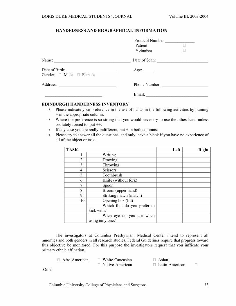
HANDEDNESS AND BIOGRAPHICAL INFORMATION
Protocol Number ______________ Patient Volunteer
Name: ____________________________________ Date of Scan: ________________________
Date of Birth: ________________________ Age: _____ Gender: Male Female Address: ___________________________ Phone Number: ______________________ ___________________________ Email: _____________________________
EDINBURGH HANDEDNESS INVENTORY
∗ Please indicate your preference in the use of hands in the following activities by puming + in the appropriate column.
∗ Where the preference is so strong that you would never try to use the othex hand unless bsolutely forced to, put ++.
∗ If any case you are really indifferent, put + in both columns. ∗ Please try to answer all the questions, and only leave a blank if you have no experience of
all of the object or task.
TASK Left Right1 Writing 2 Drawing 3 Throwing 4 Scissors 5 Toothbrush 6 Knife (without fork) 7 Spoon 8 Broom (upper hand) 9 Striking match (match) 10 Opening box (lid) Which foot do you prefer to
kick with?
Wich eye do you use when using only one?
The investigators at Columbia Presbywian. Medical Center intend to represent all
mnonties and both genders in all research studies. Federal Guidelines require that progress toward flus objective be monitored. For this purpose the investigators request that you infficate your primary ethnic affihation.
Afro-American White-Caucasian Asian Native-American Latin-American
Other
Columbia University College of Physicians and Surgeons
33
DORIS DUKE MEDICAL STUDENTS’ JOURNAL Volume III, 2003-2004
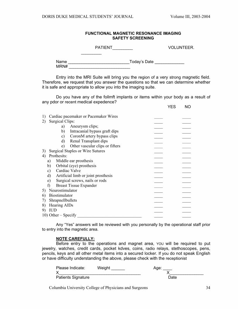
FUNCTIONAL MAGNETIC RESONANCE IMAGING SAFETY SCREENING
PATIENT_________ VOLUNTEER.
_________ Name ___________________________Today’s Date _____________ MRN# ___________________________
Entry into the MRI Suite will bring you the region of a very strong magnetic field. Therefore, we request that you answer the questions so that we can determine whether it is safe and appropriate to allow you into the imaging suite.
Do you have any of the follmft implants or items within your body as a result of
any pdor or recent medical expedence? YES NO
1) Cardiac pacemaker or Pacemaker Wires ____ ____ 2) Surgical Clips: ____ ____
a) Aneurysm clips; ____ ____ b) Intracanial bypass graft dips ____ ____ c) CoronM artery bypass clips ____ ____ d) Renal Transplant dips ____ ____ e) Other vascular clips or fifters ____ ____
3) Surgical Staples or Wire Sutures ____ ____ 4) Prothesits: ____ ____
a) Middle ear prosthesis ____ ____ b) Orbital (eye) prosthesis ____ ____ c) Cardiac Valve ____ ____ d) Artificial limb or joint prosthesis ____ ____ e) Surgical screws, nails or rods ____ ____ f) Breast Tissue Expander ____ ____
5) Neurostimulator ____ ____ 6) Biostimulator ____ ____ 7) Shrapnellbullets ____ ____ 8) Hearing AIDs ____ ____ 9) IUD ____ ____ 10) Other – Specify ______________________________ ____ ____
Any “Yes” answers will be reviewed with you personally by the operational staff prior
to entry into the magnetic area. NOTE CAREFULLY: Before entry to the operations and magnet area, YOU will be required to put
jewelry, watches, credit cards, pocket kdves, coins, radio relays, stethoscopes, pens, pencils, keys and all other metal items into a secured locker. If you do not speak English or have difficulty understanding the above, please check with the receptionist
Please Indicate: Weight ______ Age: ____ X____________________________________ X_______________ Patients Signature Date
Columbia University College of Physicians and Surgeons
34

DORIS DUKE MEDICAL STUDENTS’ JOURNAL Volume III, 2003-2004
COLUMBIA UNIVERSITY COLLEGE OF PHYSICIANS & SURGEONS
DEPARTMENT OF RADIOLOGY
CENTER FOR, NEUROBIOLOGY AND BEHAVIOR, fMRI RESEARCH CENTER
Dear Sir or Madam, We are currently conducting a research study at the Columbia University fMRI Research
Center examining the role of short-term memory in reading in people with dyslexia. We are recruiting adults with dyslexia to participate in the study. Participation involves coming to the fMR1 lab at Columbia University's Neurological Institute, taking an IQ and reading test, and then participating in a functional MR1 scan of your brain. A functional MRI scan differs from a normal MRI in that we are able to detect changes in blood flow to different parts of your brain while you perform memory tasks. These scans allow us to determine what parts of your brain are used in memory. We will compare the Scans of dyslexic readers to those of non-impaired readers to determine if dyslexic readers use different areas of their brain for memory.
The initial testing and fMRI scanning would takc approximately two hours, and we are
quite flexible in scheduling. The fMRI scanning is extremely safe and, involves no radiation exposure, and is completely non-invasive (no needles or medications are required.) This study has been approved by the Institutional Review Board of Columbia University. After participating in the study you will receive free anatomical MR1 images of your brain. There is no cost for participating in the study and participants are free to withdraw from the study at any time.
We were given your name by the New York Branch of the International Dyslexia
AssociationThe New York Branch of the International Dyslexia Association, Inc-(NYB-IDA) is making this research study known to you but is not endorsing or supporting this research project. Your participation in the study is'independent of your association with the NYB-1DA and they are not responsible for your activities related to the study.
If you would be interested in learning more about our study or would like to set up an
appointment with our investigators please call (212) 342-0299. Sincerely,.
710 West 168th Street Neurological Institute B1 NewYork, NY 10032
212-342-0299 Fax 212-342-0855 [email protected] [email protected] www.fmri.org
Columbia University College of Physicians and Surgeons
35
DORIS DUKE MEDICAL STUDENTS’ JOURNAL Volume III, 2003-2004
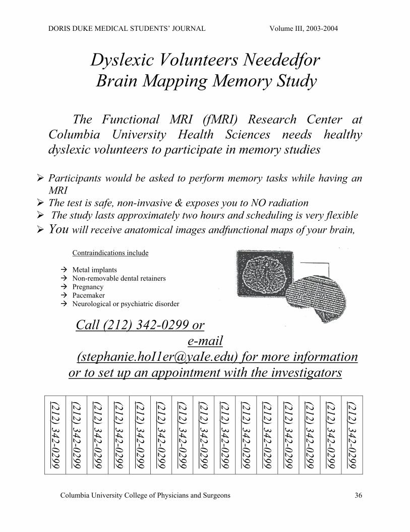
Dyslexic Volunteers Neededfor Brain Mapping Memory Study
The Functional MRI (fMRI) Research Center at
Columbia University Health Sciences needs healthy dyslexic volunteers to participate in memory studies
Participants would be asked to perform memory tasks while having an MRI The test is safe, non-invasive & exposes you to NO radiation The study lasts approximately two hours and scheduling is very flexible You will receive anatomical images andfunctional maps of your brain,
Contraindications include
Metal implants Non-removable dental retainers Pregnancy Pacemaker Neurological or psychiatric disorder
Call (212) 342-0299 or e-mail
([email protected]) for more information or to set up an appointment with the investigators
(212) 342-0299
(212) 342-0299
(212) 342-0299
(212) 342-0299
(212) 342-0299
(212) 342-0299
(212) 342-0299
(212) 342-0299
(212) 342-0299
(212) 342-0299
(212) 342-0299
(212) 342-0299
(212) 342-0299
(212) 342-0299
(212) 342-0299
Columbia University College of Physicians and Surgeons
36
DORIS DUKE MEDICAL STUDENTS’ JOURNAL Volume III, 2003-2004
Columbia University College of Physicians and Surgeons
37
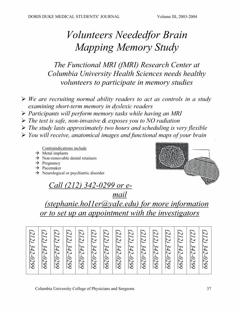
Volunteers Neededfor Brain Mapping Memory Study
The Functional MRI (fMRI) Research Center at
Columbia University Health Sciences needs healthy volunteers to participate in memory studies
We are recruiting normal ability readers to act as controls in a study examining short-term memory in dyslexic readers Participants will perform memory tasks while having an MRI The test is safe, non-invasive & exposes you to NO radiation The study lasts approximately two hours and scheduling is very flexible You will receive, anatomical images and functional maps of your brain
Contraindications include Metal implants Non-removable dental retainers Pregnancy Pacemaker Neurological or psychiatric disorder
Call (212) 342-0299 or e-mail
([email protected]) for more information or to set up an appointment with the investigators
(212) 342-0299
(212) 342-0299
(212) 342-0299
(212) 342-0299
(212) 342-0299
(212) 342-0299
(212) 342-0299
(212) 342-0299
(212) 342-0299
(212) 342-0299
(212) 342-0299
(212) 342-0299
(212) 342-0299
(212) 342-0299
(212) 342-0299


