
Shock Mahdi Alemrajabi Colorectal surgeon Faculty member of Iran university of medical sciences...
94
Shock Mahdi Alemrajabi Colorectal surgeon Faculty member of Iran university of medical sciences Firoozghar general hospital www.alemrajabi.ir
-
Upload
dinah-garrison -
Category
Documents
-
view
213 -
download
0
Transcript of Shock Mahdi Alemrajabi Colorectal surgeon Faculty member of Iran university of medical sciences...
Shock
Mahdi AlemrajabiColorectal surgeon
Faculty member of Iran university of medical sciencesFiroozghar general hospital
www.alemrajabi.ir

• "Shock is the manifestation of the rude unhinging of the machinery of life.“
• Samuel V. Gross, 1872

Historical background
• Integral to our understanding of shock is the appreciation that our bodies attempt to maintain a state of homeostasis.
• Claude Bernard suggested in the mid-nineteenth century that the organism attempts to maintain constancy in the internal environment against external forces that attempt to disrupt the milieu interieur.
•
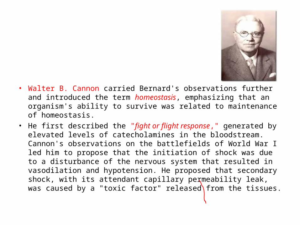
• Walter B. Cannon carried Bernard's observations further and introduced the term homeostasis, emphasizing that an organism's ability to survive was related to maintenance of homeostasis.
• He first described the "fight or flight response," generated by elevated levels of catecholamines in the bloodstream. Cannon's observations on the battlefields of World War I led him to propose that the initiation of shock was due to a disturbance of the nervous system that resulted in vasodilation and hypotension. He proposed that secondary shock, with its attendant capillary permeability leak, was caused by a "toxic factor" released from the tissues.

1899-1934
•In a series of critical experiments, Alfred Blalock documented that
the shock state in hemorrhage was associated with reduced cardiac output due to volume loss, not a "toxic factor.“• In 1934, Blalock proposed four categories of shock: hypovolemic, vasogenic, cardiogenic, and neurogenic.

• In 1947, Wiggers developed a sustainable, irreversible model of hemorrhagic shock based on uptake of shed blood into a reservoir to maintain a set level of hypotension.
•

• G. Tom Shires added further understanding of hemorrhagic shock with a series of clinical studies demonstrating that a large extracellular fluid deficit, greater than could be attributed to vascular refilling alone, occurred in severe hemorrhagic shock.
• The phenomenon of fluid redistribution after major trauma involving blood loss was termed third spacing and described the translocation of intravascular volume into the peritoneum, bowel, burned tissues, or crush injury sites.
Objectives
Definition Approach to the hypotensive
patient Types Specific treatments
Definition of Shock
• Inadequate oxygen delivery to meet metabolic demands
• Results in global tissue hypoperfusion and metabolic acidosis
• Shock can occur with a normal blood pressure and hypotension can occur without shock
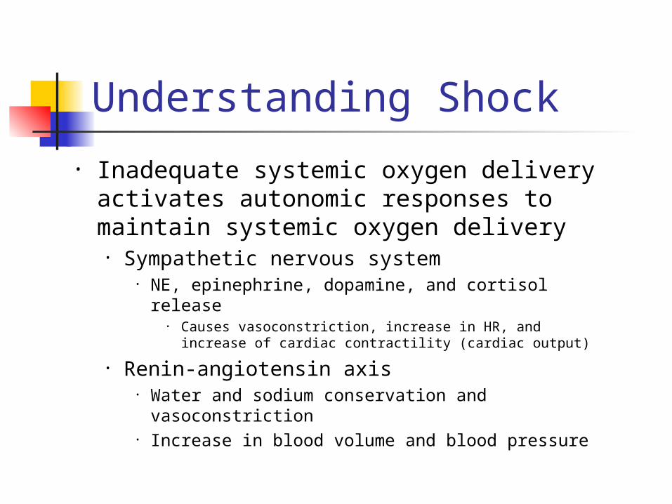
Understanding Shock
• Inadequate systemic oxygen delivery activates autonomic responses to maintain systemic oxygen delivery
• Sympathetic nervous system• NE, epinephrine, dopamine, and cortisol release
• Causes vasoconstriction, increase in HR, and increase of cardiac contractility (cardiac output)
• Renin-angiotensin axis• Water and sodium conservation and
vasoconstriction• Increase in blood volume and blood pressure

Understanding Shock• Cellular responses to decreased systemic
oxygen delivery• ATP depletion → ion pump dysfunction• Cellular edema• Hydrolysis of cellular membranes and
cellular death• Goal is to maintain cerebral and cardiac
perfusion• Vasoconstriction of splanchnic,
musculoskeletal, and renal blood flow• Leads to systemic metabolic lactic acidosis
that overcomes the body’s compensatory mechanisms

Global Tissue Hypoxia
• Endothelial inflammation and disruption
• Inability of O2 delivery to meet demand
• Result: • Lactic acidosis• Cardiovascular insufficiency• Increased metabolic demands
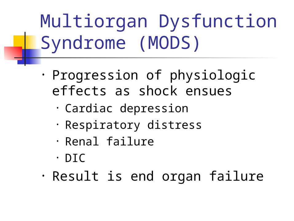
Multiorgan DysfunctionSyndrome (MODS)
• Progression of physiologic effects as shock ensues• Cardiac depression• Respiratory distress• Renal failure• DIC
• Result is end organ failure

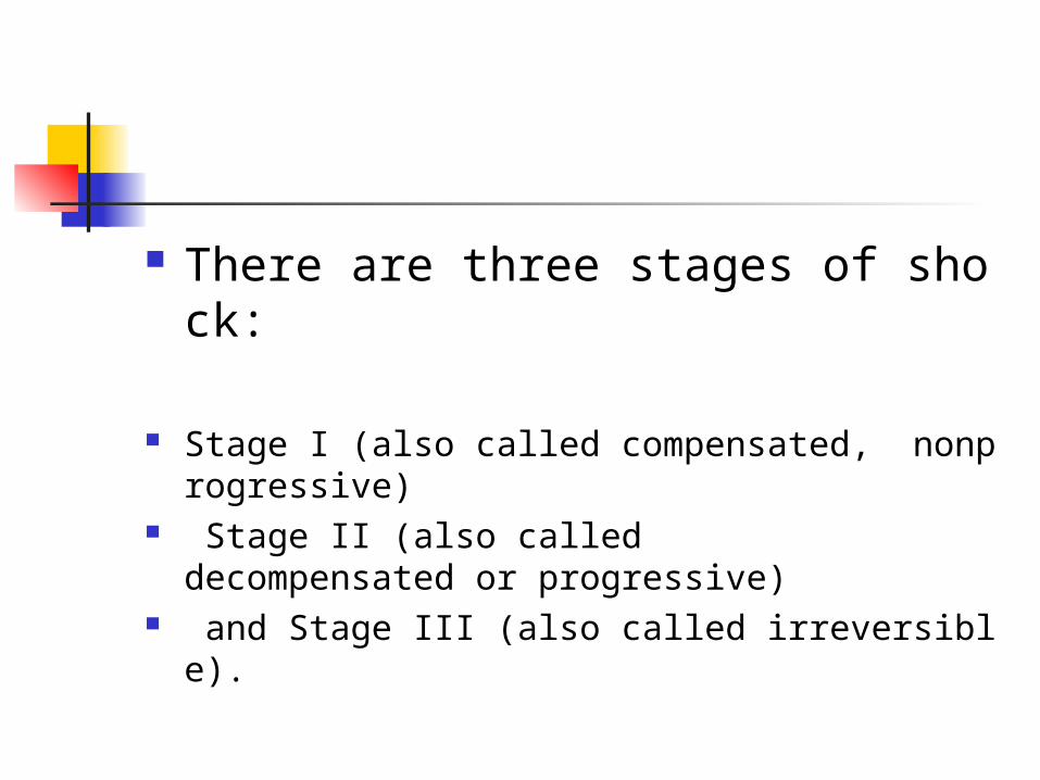
There are three stages of shock:
Stage I (also called compensated, nonprogressive)
Stage II (also called decompensated or progressive)
and Stage III (also called irreversible).

Stage 1 low blood flow (perfusion) is first detected
a number of systems are activated in order to maintain/restore perfusion.
All this serves to maximize blood flow to the most important organs and systems in the body.
The patient in this stage of shock has very few symptoms
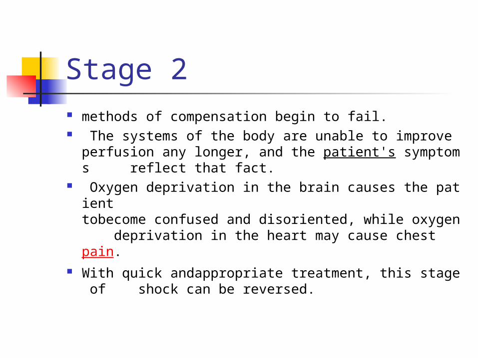
Stage 2 methods of compensation begin to fail. The systems of the body are unable to improve
perfusion any longer, and the patient's symptoms reflect that fact.
Oxygen deprivation in the brain causes the patient tobecome confused and disoriented, while oxygen deprivation in the heart may cause chest pain.
With quick andappropriate treatment, this stage of shock can be reversed.
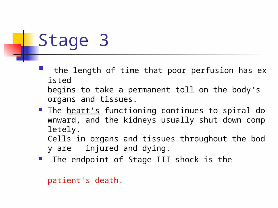
Stage 3
the length of time that poor perfusion has existed begins to take a permanent toll on the body's organs and tissues.
The heart's functioning continues to spiral downward, and the kidneys usually shut down completely. Cells in organs and tissues throughout the body are injured and dying.
The endpoint of Stage III shock is the patient's death.

• ABCs• Cardiorespiratory monitor• Pulse oximetry• Supplemental oxygen • IV access• ABG, labs• Foley catheter• Vital signs including rectal
temperature
Approach to the Patient in Shock
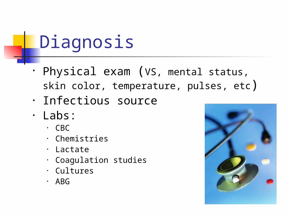
Diagnosis• Physical exam (VS, mental status, skin
color, temperature, pulses, etc)• Infectious source• Labs:
• CBC• Chemistries• Lactate• Coagulation studies• Cultures• ABG
Further Evaluation
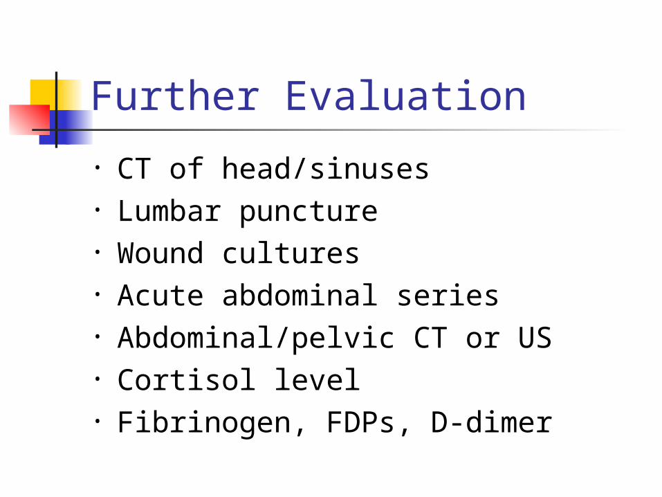
• CT of head/sinuses• Lumbar puncture• Wound cultures• Acute abdominal series• Abdominal/pelvic CT or US• Cortisol level• Fibrinogen, FDPs, D-dimer
Approach to the Patient in Shock
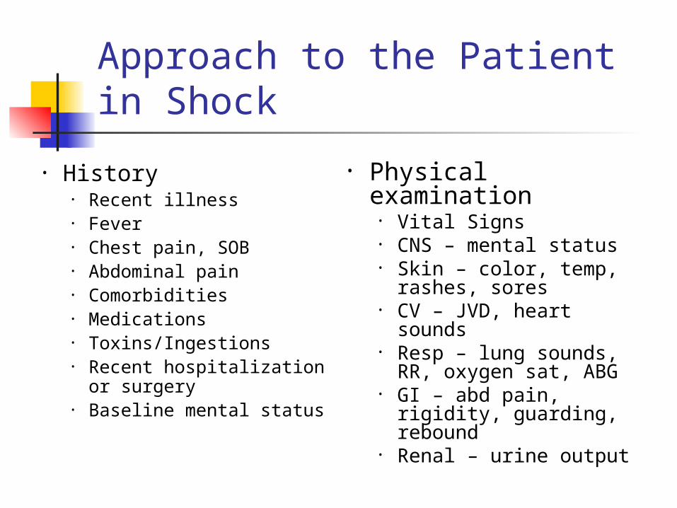
• History• Recent illness• Fever• Chest pain, SOB• Abdominal pain• Comorbidities• Medications• Toxins/Ingestions• Recent hospitalization
or surgery• Baseline mental status
• Physical examination
• Vital Signs• CNS – mental status• Skin – color, temp,
rashes, sores• CV – JVD, heart
sounds• Resp – lung sounds,
RR, oxygen sat, ABG• GI – abd pain, rigidity,
guarding, rebound• Renal – urine output
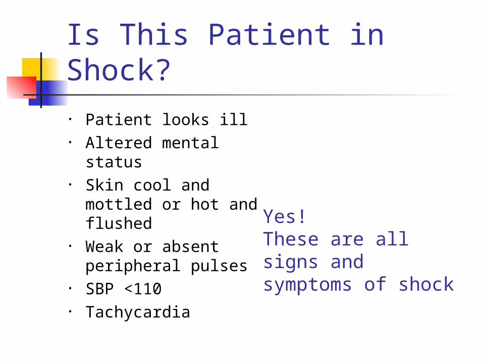
Is This Patient in Shock?• Patient looks ill• Altered mental
status• Skin cool and
mottled or hot and flushed
• Weak or absent peripheral pulses
• SBP <110• Tachycardia
Yes! These are all signs and symptoms of shock
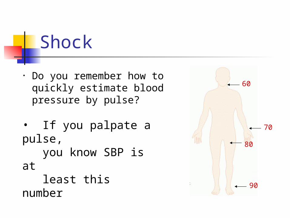
Shock
• Do you remember how to quickly estimate blood pressure by pulse?
60
80
70
90
• If you palpate a pulse, you know SBP is at least this number

Goals of Treatment
• ABCDE• Airway• control work of Breathing• optimize Circulation• assure adequate oxygen Delivery• achieve End points of resuscitation

Airway• Determine need for intubation but
remember: intubation can worsen hypotension
• Sedatives can lower blood pressure• Positive pressure ventilation decreases
preload • May need volume resuscitation prior to
intubation to avoid hemodynamic collapse
Control Work of Breathing
• Respiratory muscles consume a significant amount of oxygen
• Tachypnea can contribute to lactic acidosis
• Mechanical ventilation and sedation decrease WOB and improves survival

Optimizing Circulation
• Isotonic crystalloids• Titrated to:
• CVP 8-12 mm Hg • Urine output 0.5 ml/kg/hr (30 ml/hr) • Improving heart rate
• May require 4-6 L of fluids• No outcome benefit from colloids

Maintaining Oxygen Delivery
• Decrease oxygen demands• Provide analgesia and anxiolytics to relax
muscles and avoid shivering• Maintain arterial oxygen
saturation/content• Give supplemental oxygen• Maintain Hemoglobin > 10 g/dL,(8 g/dl)
• Serial lactate levels or central venous oxygen saturations to assess tissue oxygen extraction

End Points of Resuscitation
• Goal of resuscitation is to maximize survival and minimize morbidity
• Use objective hemodynamic and physiologic values to guide therapy
• Goal directed approach• Urine output > 0.5 mL/kg/hr• CVP 8-12 mmHg• MAP 65 to 90 mmHg• Central venous oxygen concentration >
70%

Persistent Hypotension• Inadequate volume
resuscitation• Pneumothorax• Cardiac tamponade• Hidden bleeding• Adrenal insufficiency• Medication allergy
Practically Speaking….
• Keep one eye on these patients• Frequent vitals signs:
• Monitor success of therapies• Watch for decompensated shock
• Let your nurses know that these patients are sick!
Types of Shock
• Hypovolemic• Septic• Cardiogenic • Anaphylactic• Neurogenic • Obstructive• traumatic

What Type of Shock is This?
• 68 yo M with hx of HTN and DM presents to the ER with abrupt onset of diffuse abdominal pain with radiation to his low back. The pt is hypotensive, tachycardic, afebrile, with cool but dry skin
Types of Shock• Hypovolemic• Septic• Cardiogenic • Anaphylactic• Neurogenic • Obstructive
Hypovolemic Shock

Hypovolemic Shock
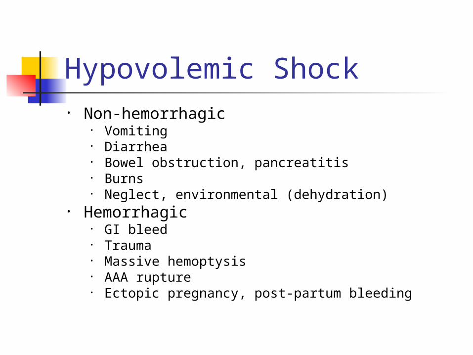
• Non-hemorrhagic • Vomiting• Diarrhea• Bowel obstruction, pancreatitis• Burns • Neglect, environmental (dehydration)
• Hemorrhagic • GI bleed• Trauma• Massive hemoptysis• AAA rupture• Ectopic pregnancy, post-partum bleeding
Hypovolemic Shock

Hypovolemic Shock• ABCs• Establish 2 large bore IVs or a central line• Crystalloids
• Normal Saline or Lactate Ringers• Up to 3 liters
• PRBCs• O negative or cross matched
• Control any bleeding• Arrange definitive treatment

Evaluation of Hypovolemic Shock
• CBC• ABG/lactate• Electrolytes• BUN, Creatinine• Coagulation studies• Type and cross-
match
• As indicated• CXR• Pelvic x-ray• Abd/pelvis CT• Chest CT• GI endoscopy• Bronchoscopy• Vascular radiology

Infusion Rates Access Gravity Pressure
18 g peripheral IV 50 mL/min 150 mL/min
16 g peripheral IV 100 mL/min 225 mL/min
14 g peripheral IV 150 mL/min 275 mL/min
8.5 Fr CV cordis 200 mL/min 450 mL/min
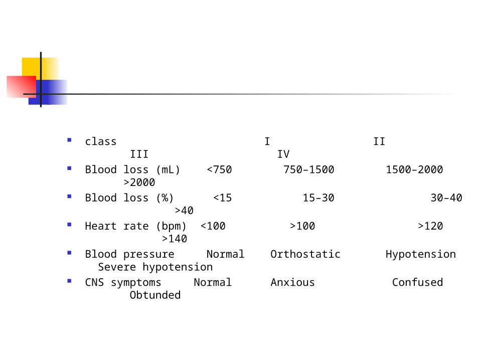
class I II III IV Blood loss (mL) <750 750–1500 1500–2000 >2000 Blood loss (%) <15 15–30 30–40 >40 Heart rate (bpm) <100 >100 >120 >140 Blood pressure Normal Orthostatic Hypotension Severe
hypotension CNS symptoms Normal Anxious Confused Obtunded

What Type of Shock is This?
• An 81 yo F resident of a nursing home presents to the ED with altered mental status. She is febrile to 39.4, hypotensive with a widened pulse pressure, tachycardic, with warm extremities
Types of Shock• Hypovolemic• Septic• Cardiogenic • Anaphylactic• Neurogenic • Obstructive
Septic
Septic Shock
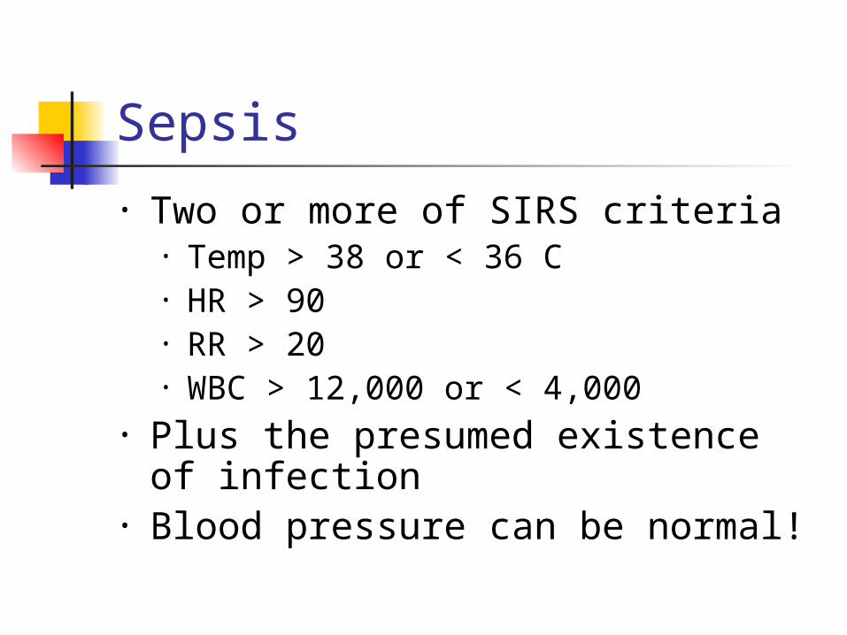
Sepsis
• Two or more of SIRS criteria• Temp > 38 or < 36 C• HR > 90• RR > 20• WBC > 12,000 or < 4,000
• Plus the presumed existence of infection
• Blood pressure can be normal!

Septic Shock
• Sepsis (remember definition?)• Plus refractory hypotension
• After bolus of 20-40 mL/Kg patient still has one of the following:
• SBP < 90 mm Hg • MAP < 65 mm Hg • Decrease of 40 mm Hg from
baseline
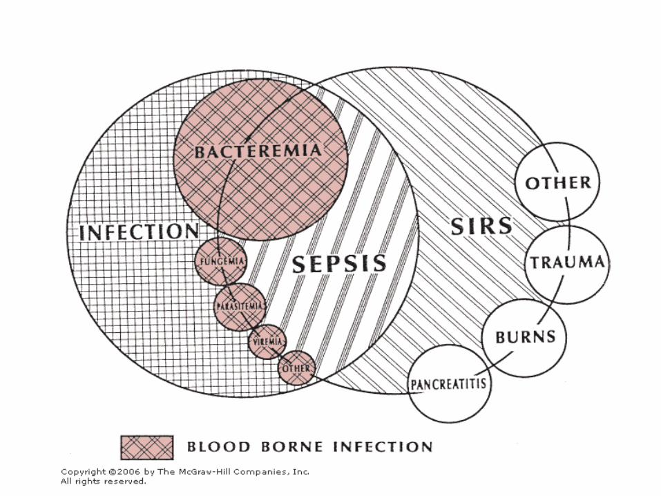
Sepsis

Pathogenesis of Sepsis
Nguyen H et al. Severe Sepsis and Septic-Shock: Review of the Literature and Emergency Department Management Guidelines. Ann Emerg Med. 2006;42:28-54.
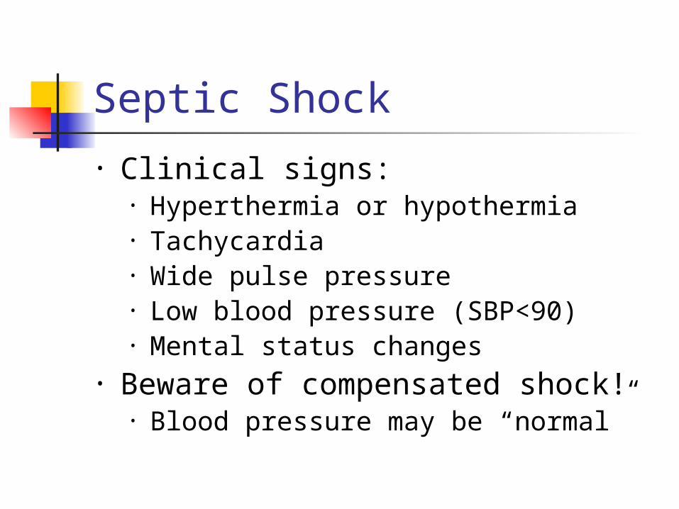
Septic Shock
• Clinical signs:• Hyperthermia or hypothermia• Tachycardia• Wide pulse pressure• Low blood pressure (SBP<90)• Mental status changes
• Beware of compensated shock!• Blood pressure may be “normal”
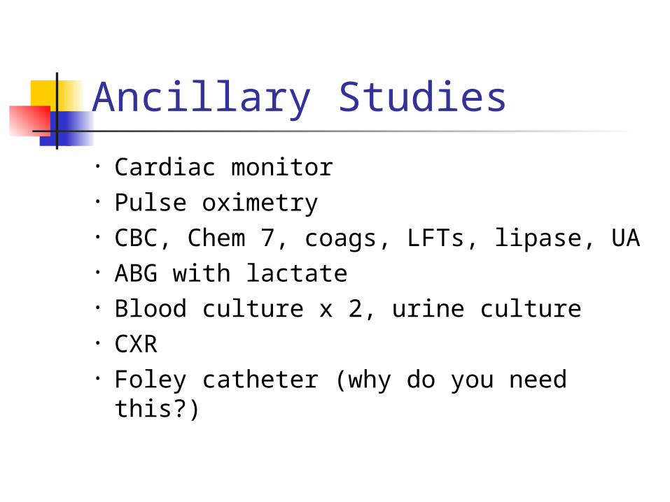
Ancillary Studies
• Cardiac monitor• Pulse oximetry• CBC, Chem 7, coags, LFTs, lipase, UA• ABG with lactate• Blood culture x 2, urine culture• CXR• Foley catheter (why do you need
this?)

Treatment of Septic Shock
• 2 large bore IVs• NS IVF bolus- 1-2 L wide open (if no
contraindications) • Supplemental oxygen• Empiric antibiotics, based on
suspected source, as soon as possible

Treatment of Sepsis• Antibiotics- Survival correlates with how
quickly the correct drug was given• Cover gram positive and gram negative
bacteria• Add additional coverage as indicated
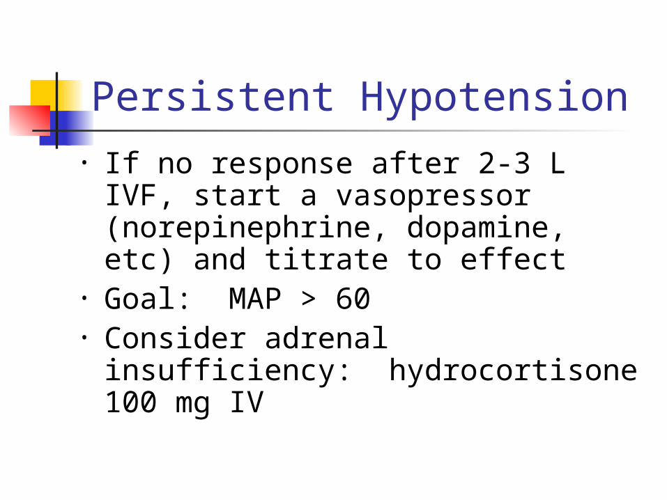
Persistent Hypotension • If no response after 2-3 L IVF, start
a vasopressor (norepinephrine, dopamine, etc) and titrate to effect
• Goal: MAP > 60• Consider adrenal insufficiency:
hydrocortisone 100 mg IV
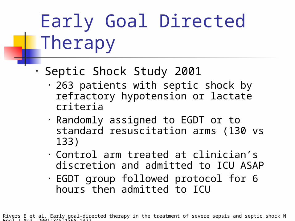
Early Goal Directed Therapy
• Septic Shock Study 2001• 263 patients with septic shock by
refractory hypotension or lactate criteria• Randomly assigned to EGDT or to
standard resuscitation arms (130 vs 133)• Control arm treated at clinician’s
discretion and admitted to ICU ASAP• EGDT group followed protocol for 6 hours
then admitted to ICU
Rivers E et al. Early goal-directed therapy in the treatment of severe sepsis and septic shock N Engl J Med. 2001:345:1368-1377.
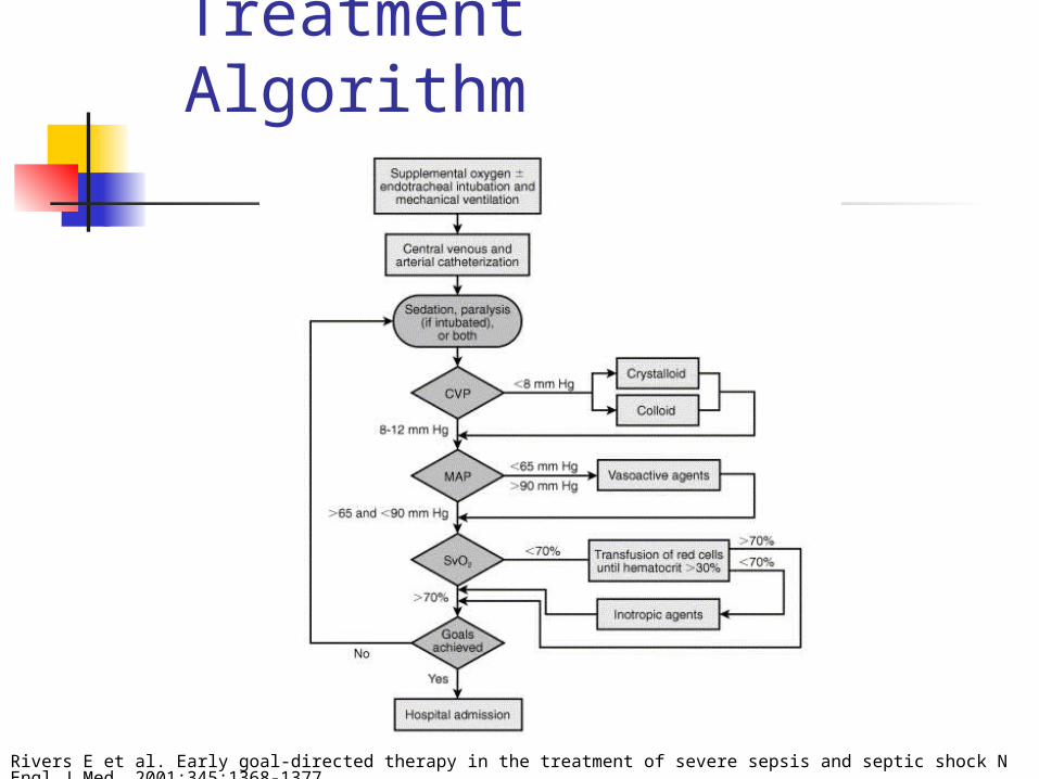
Treatment Algorithm
Rivers E et al. Early goal-directed therapy in the treatment of severe sepsis and septic shock N Engl J Med. 2001:345:1368-1377.
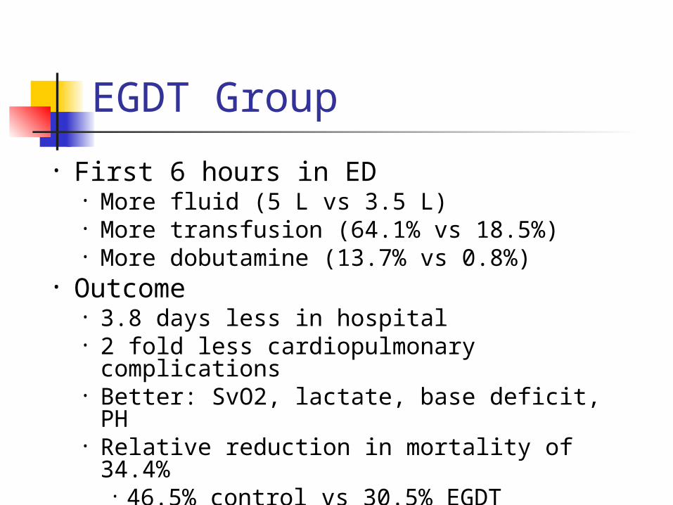
EGDT Group
• First 6 hours in ED• More fluid (5 L vs 3.5 L)• More transfusion (64.1% vs 18.5%)• More dobutamine (13.7% vs 0.8%)
• Outcome• 3.8 days less in hospital• 2 fold less cardiopulmonary
complications• Better: SvO2, lactate, base deficit, PH• Relative reduction in mortality of
34.4%• 46.5% control vs 30.5% EGDT

What Type of Shock is This?• A 55 yo M with hx of
HTN, DM presents with “crushing” substernal CP, diaphoresis, hypotension, tachycardia and cool, clammy extremities
Types of Shock• Hypovolemic• Septic• Cardiogenic • Anaphylactic• Neurogenic • Obstructive
Cardiogenic
Cardiogenic Shock

Cardiogenic Shock
• Signs:• Cool, mottled skin• Tachypnea • Hypotension• Altered mental
status• Narrowed pulse
pressure• Rales, murmur
• Defined as:• SBP < 90 mmHg• CI < 2.2 L/m/m2
• PCWP > 15 mmHg

Etiologies
• What are some causes of cardiogenic shock?
• AMI• Sepsis• Myocarditis• Myocardial contusion• Aortic or mitral stenosis, HCM• Acute aortic insufficiency

Pathophysiology of Cardiogenic Shock
• Often after ischemia, loss of LV function• Lose 40% of LV clinical shock ensues
• CO reduction = lactic acidosis, hypoxia• Stroke volume is reduced
• Tachycardia develops as compensation • Ischemia and infarction worsens

Ancillary Tests
• EKG• CXR• CBC, Chem 10, cardiac enzymes,
coagulation studies• Echocardiogram

Treatment of Cardiogenic Shock
• Goals- Airway stability and improving myocardial pump function
• Cardiac monitor, pulse oximetry• Supplemental oxygen, IV access• Intubation will decrease preload and
result in hypotension • Be prepared to give fluid bolus
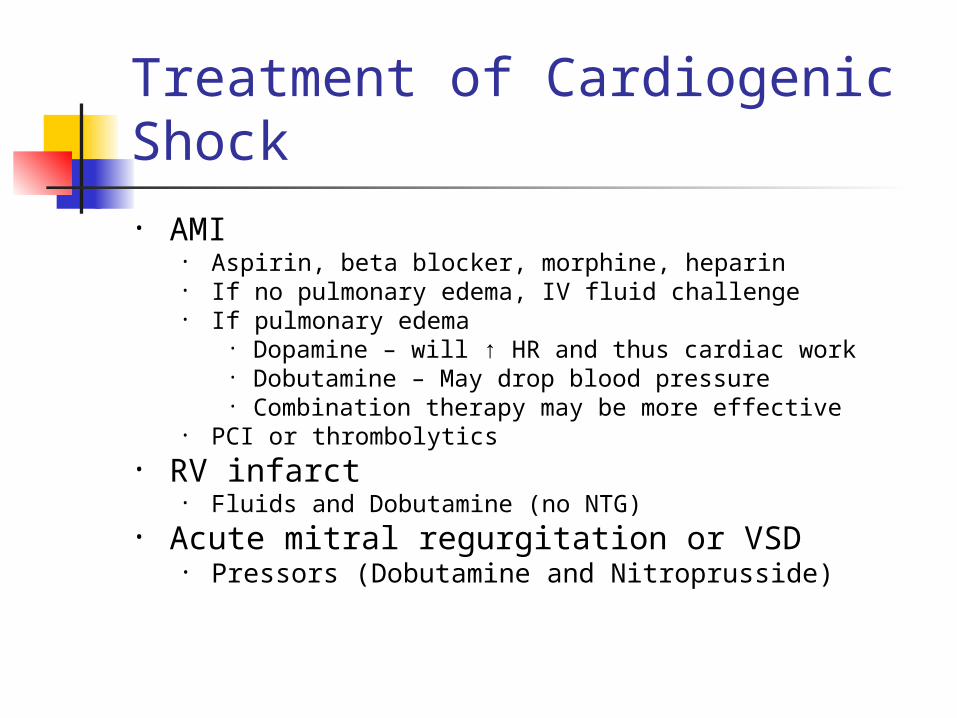
Treatment of Cardiogenic Shock• AMI
• Aspirin, beta blocker, morphine, heparin• If no pulmonary edema, IV fluid challenge• If pulmonary edema
• Dopamine – will ↑ HR and thus cardiac work• Dobutamine – May drop blood pressure• Combination therapy may be more effective
• PCI or thrombolytics• RV infarct
• Fluids and Dobutamine (no NTG)• Acute mitral regurgitation or VSD
• Pressors (Dobutamine and Nitroprusside)
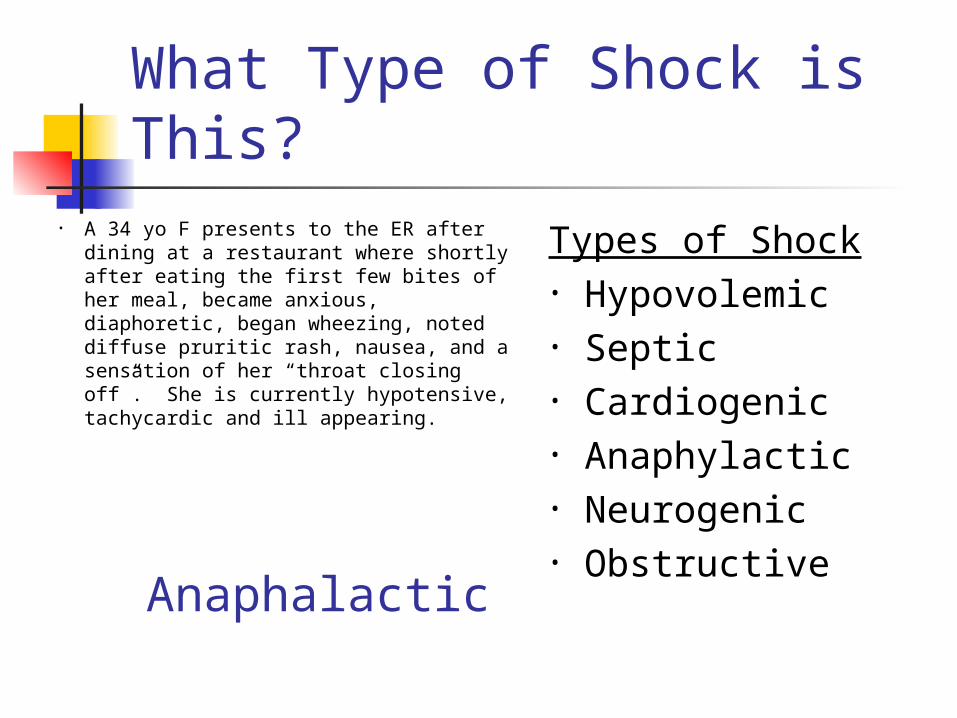
What Type of Shock is This?
• A 34 yo F presents to the ER after dining at a restaurant where shortly after eating the first few bites of her meal, became anxious, diaphoretic, began wheezing, noted diffuse pruritic rash, nausea, and a sensation of her “throat closing off”. She is currently hypotensive, tachycardic and ill appearing.
Types of Shock• Hypovolemic• Septic• Cardiogenic • Anaphylactic• Neurogenic • Obstructive
Anaphalactic

Anaphalactic Shock

Anaphylactic Shock
• Anaphylaxis – a severe systemic hypersensitivity reaction characterized by multisystem involvement • IgE mediated
• Anaphylactoid reaction – clinically indistinguishable from anaphylaxis, do not require a sensitizing exposure• Not IgE mediated

• What are some symptoms of anaphylaxis?
Anaphylactic Shock
• First- Pruritus, flushing, urticaria appear
•Next- Throat fullness, anxiety, chest tightness, shortness of breath and lightheadedness
•Finally- Altered mental status, respiratory distress and circulatory collapse
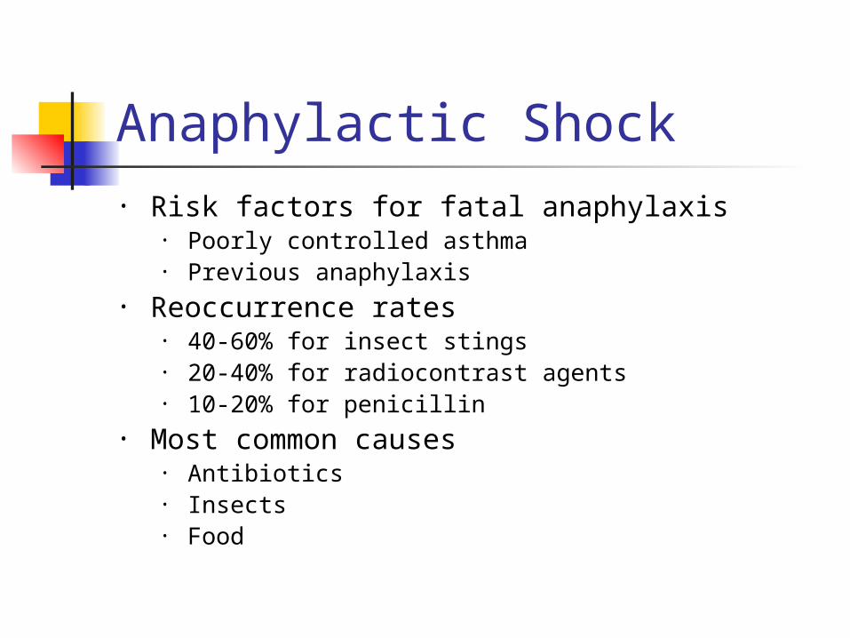
• Risk factors for fatal anaphylaxis • Poorly controlled asthma • Previous anaphylaxis
• Reoccurrence rates• 40-60% for insect stings• 20-40% for radiocontrast agents• 10-20% for penicillin
• Most common causes• Antibiotics• Insects• Food
Anaphylactic Shock
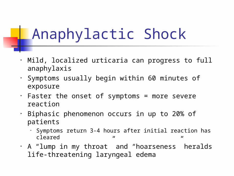
• Mild, localized urticaria can progress to full anaphylaxis
• Symptoms usually begin within 60 minutes of exposure
• Faster the onset of symptoms = more severe reaction
• Biphasic phenomenon occurs in up to 20% of patients
• Symptoms return 3-4 hours after initial reaction has cleared
• A “lump in my throat” and “hoarseness” heralds life-threatening laryngeal edema
Anaphylactic Shock

Anaphylactic Shock- Diagnosis
• Clinical diagnosis• Defined by airway compromise,
hypotension, or involvement of cutaneous, respiratory, or GI systems
• Look for exposure to drug, food, or insect
• Labs have no role

• ABC’s• Angioedema and respiratory compromise
require immediate intubation• IV, cardiac monitor, pulse oximetry• IVFs, oxygen• Epinephrine• Second line
• Corticosteriods• H1 and H2 blockers
Anaphylactic Shock- Treatment

• Epinephrine• 0.3 mg IM of 1:1000 (epi-pen) • Repeat every 5-10 min as needed• Caution with patients taking beta blockers- can
cause severe hypertension due to unopposed alpha stimulation
• For CV collapse, 1 mg IV of 1:10,000• If refractory, start IV drip
Anaphylactic Shock- Treatment

• Corticosteroids• Methylprednisolone 125 mg IV • Prednisone 60 mg PO
• Antihistamines• H1 blocker- Diphenhydramine 25-50 mg IV• H2 blocker- Ranitidine 50 mg IV
• Bronchodilators• Albuterol nebulizer• Atrovent nebulizer• Magnesium sulfate 2 g IV over 20 minutes
• Glucagon• For patients taking beta blockers and with refractory hypotension• 1 mg IV q5 minutes until hypotension resolves
Anaphylactic Shock - Treatment

• All patients who receive epinephrine should be observed for 4-6 hours
• If symptom free, discharge home• If on beta blockers or h/o severe
reaction in past, consider admission
Anaphylactic Shock - Disposition
What Type of Shock is This?
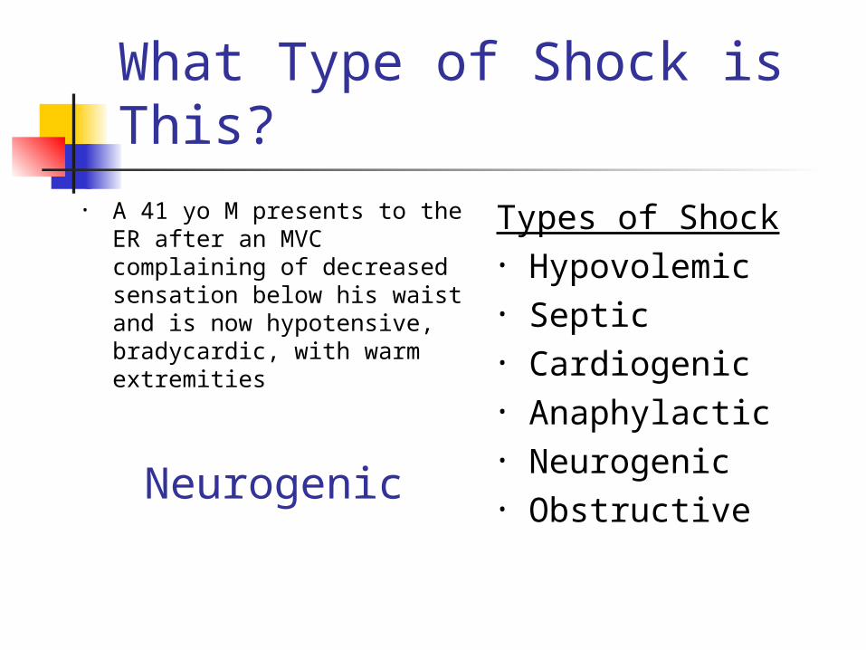
• A 41 yo M presents to the ER after an MVC complaining of decreased sensation below his waist and is now hypotensive, bradycardic, with warm extremities
Types of Shock• Hypovolemic• Septic• Cardiogenic • Anaphylactic• Neurogenic • Obstructive
Neurogenic

Neurogenic Shock

Neurogenic Shock • Occurs after acute spinal cord injury• Sympathetic outflow is disrupted
leaving unopposed vagal tone• Results in hypotension and bradycardia• Spinal shock- temporary loss of spinal
reflex activity below a total or near total spinal cord injury (not the same as neurogenic shock, the terms are not interchangeable)

• Loss of sympathetic tone results in warm and dry skin
• Shock usually lasts from 1 to 3 weeks
• Any injury above T1 can disrupt the entire sympathetic system• Higher injuries = worse paralysis
Neurogenic Shock
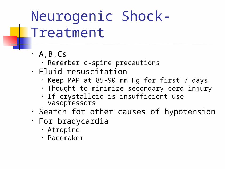
• A,B,Cs• Remember c-spine precautions
• Fluid resuscitation• Keep MAP at 85-90 mm Hg for first 7 days• Thought to minimize secondary cord injury• If crystalloid is insufficient use vasopressors
• Search for other causes of hypotension• For bradycardia
• Atropine• Pacemaker
Neurogenic Shock- Treatment

Neurogenic Shock- Treatment
• Methylprednisolone• Used only for blunt spinal cord injury• High dose therapy for 23 hours• Must be started within 8 hours• Controversial- Risk for infection, GI
bleed
What Type of Shock is This?
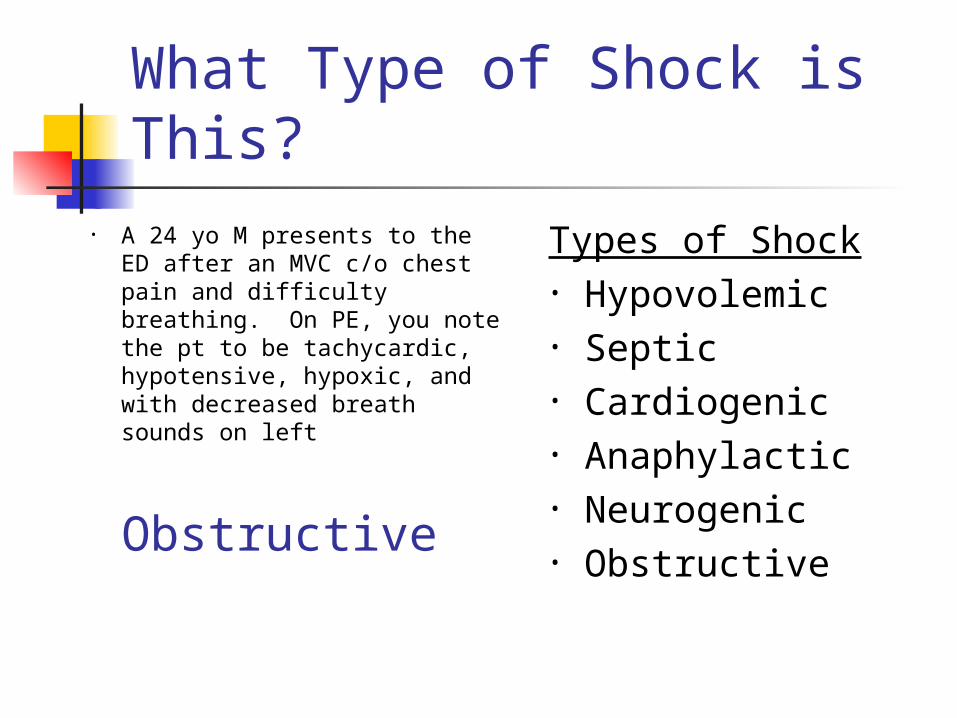
• A 24 yo M presents to the ED after an MVC c/o chest pain and difficulty breathing. On PE, you note the pt to be tachycardic, hypotensive, hypoxic, and with decreased breath sounds on left
Types of Shock• Hypovolemic• Septic• Cardiogenic • Anaphylactic• Neurogenic • Obstructive
Obstructive
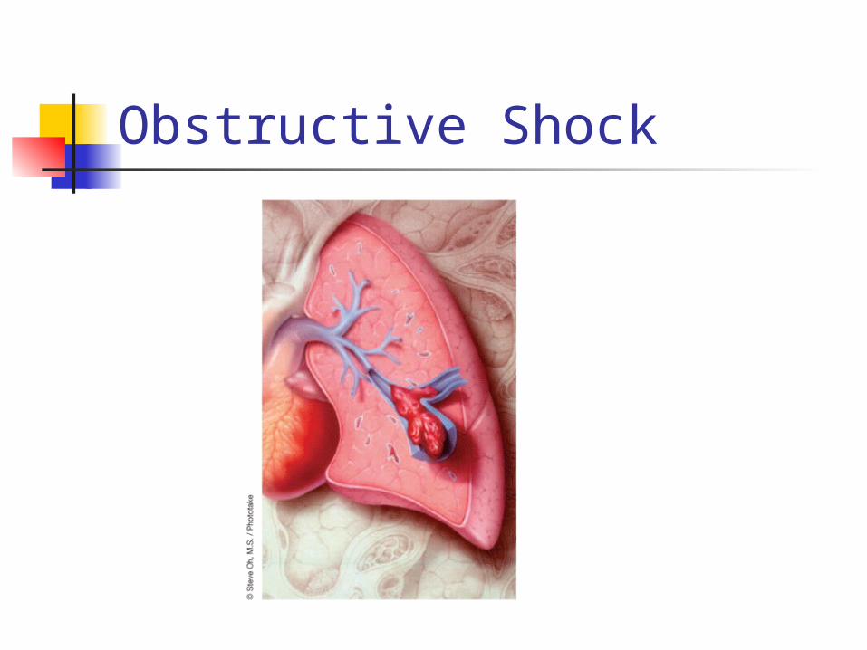
Obstructive Shock
Obstructive Shock
• Tension pneumothorax• Air trapped in pleural space with 1 way
valve, air/pressure builds up• Mediastinum shifted impeding venous
return• Chest pain, SOB, decreased breath
sounds• No tests needed!• Rx: Needle decompression, chest tube
Obstructive Shock
• Cardiac tamponade• Blood in pericardial sac prevents
venous return to and contraction of heart
• Related to trauma, pericarditis, MI• Beck’s triad: hypotension, muffled
heart sounds, JVD• Diagnosis: large heart CXR, echo• Rx: Pericardiocentisis

Obstructive Shock
• Pulmonary embolism• Virscow triad: hypercoaguable,
venous injury, venostasis• Signs: Tachypnea, tachycardia,
hypoxia• Low risk: D-dimer• Higher risk: CT chest or VQ scan• Rx: Heparin, consider thrombolytics
Obstructive Shock
• Aortic stenosis• Resistance to systolic ejection causes
decreased cardiac function• Chest pain with syncope• Systolic ejection murmur• Diagnosed with echo• Vasodilators (NTG) will drop pressure!• Rx: Valve surgery

Shock Types & PhysiologyShock CVP/PCWP CO SVRI
Hemorrhagic
Septic
Cardiogenic
Neurogenic
Hypoadrenal
Anaphylactic

Shock Types & PhysiologyShock CVP/PCWP CO SVRI
Hemorrhagic ↓ ↓ ↑
Septic
Cardiogenic
Neurogenic
Hypoadrenal
Anaphylactic
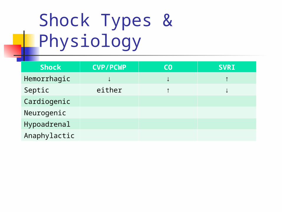
Shock Types & PhysiologyShock CVP/PCWP CO SVRI
Hemorrhagic ↓ ↓ ↑
Septic either ↑ ↓
Cardiogenic
Neurogenic
Hypoadrenal
Anaphylactic

Shock Types & PhysiologyShock CVP/PCWP CO SVRI
Hemorrhagic ↓ ↓ ↑
Septic either ↑ ↓
Cardiogenic ↑ ↓ ↑
Neurogenic
Hypoadrenal
Anaphylactic

Shock Types & PhysiologyShock CVP/PCWP CO SVRI
Hemorrhagic ↓ ↓ ↑
Septic either ↑ ↓
Cardiogenic ↑ ↓ ↑
Neurogenic ↓ ↓ ↓
Hypoadrenal
Anaphylactic

Shock Types & PhysiologyShock CVP/PCWP CO SVRI
Hemorrhagic ↓ ↓ ↑
Septic either ↑ ↓
Cardiogenic ↑ ↓ ↑
Neurogenic ↓ ↓ ↓
Hypoadrenal either ↓ ↓
Anaphylactic
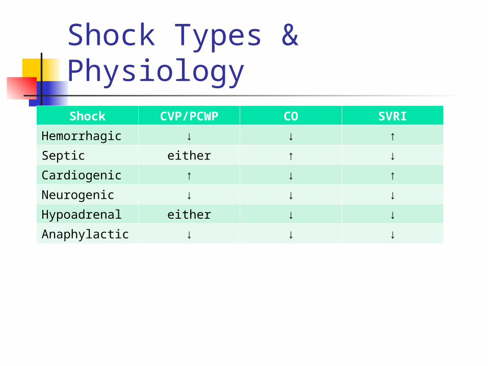
Shock Types & PhysiologyShock CVP/PCWP CO SVRI
Hemorrhagic ↓ ↓ ↑
Septic either ↑ ↓
Cardiogenic ↑ ↓ ↑
Neurogenic ↓ ↓ ↓
Hypoadrenal either ↓ ↓
Anaphylactic ↓ ↓ ↓
Shock: Take Home Points
Shock = inadequate tissue perfusion Types of shock: hypovolemic, septic,
cardiogenic, neurogenic, anaphylactic Signs of shock: altered MS, tachycardia,
hypotension, tachypnea, low UOP Always start with ABCs Resuscitation begins with fluid