SHOCK IN THE NEONATES · syndrome ETIOLOGY DISTRIBUTIVE SHOCK. CARDIOGENIC Cardiac Dysfunction...
37
SHOCK IN THE NEONATES Dr. Bouasengniyom Phrasitthideth NICU-Mahosot Hospital @ 2018
Transcript of SHOCK IN THE NEONATES · syndrome ETIOLOGY DISTRIBUTIVE SHOCK. CARDIOGENIC Cardiac Dysfunction...
SHOCK IN THE NEONATES
Dr. Bouasengniyom Phrasitthideth
NICU-Mahosot Hospital @ 2018

DEFINITION• For cells to survive and function, they need
oxygen.• Inadequate tissue perfusion and oxygen delivery
to vital organs Shock• Shock presents before hypotension• Hypotension represent uncompensated shock

Acess Circulation and Cardiovascular status• Cardiac Output (CO)
CO = HR x SV• Oxygen Content in the Arterial Blood (CaO2)
CaO2 = 1.36 x Hgb x SaO2 + (0.0031 x PaO2)• Oxygen Delivery (DO2)
DO2 = CO x CaO2
Systemic vascular resistance (SVR) is the resistance that the left ventricle must overcome to pump blood through the systemic circulation
Blood pressure (BP) BP = CO x SVR
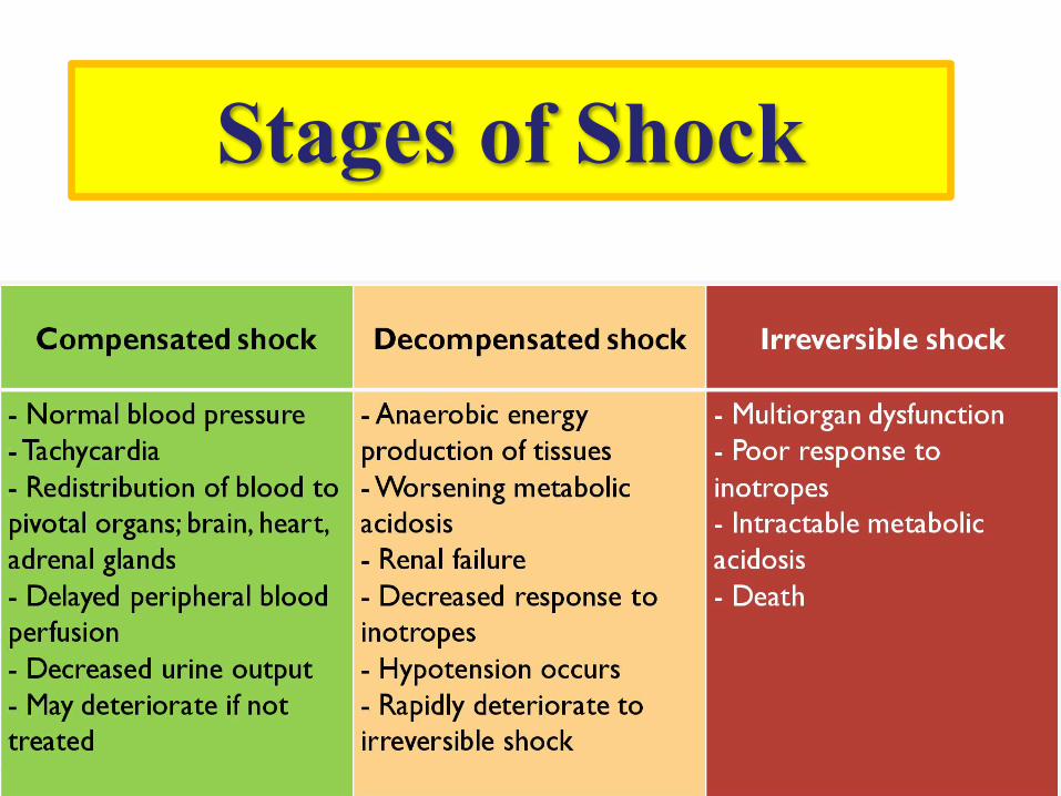
Stages of Shock
PATHOGENESIS
Shock, a state of cellular and tissue hypoxia, is due to reduced oxygen delivery, increased oxygen consumption, and/or inadequate oxygen utilization .
Cellular hypoxia results in a switch to anaerobic metabolism and accumulation of lactic acid. Increasing levels of lactic acid causes metabolic acidosis, which interferes with cell and organ function and, if not addressed, cell death.

Blood Pressure- 3 Main Causes of Shock
• Hypovolemia --> Hypovolemic Shock
• Heart Failure --> Cardiogenic Shock
• Infection --> Septic Shock


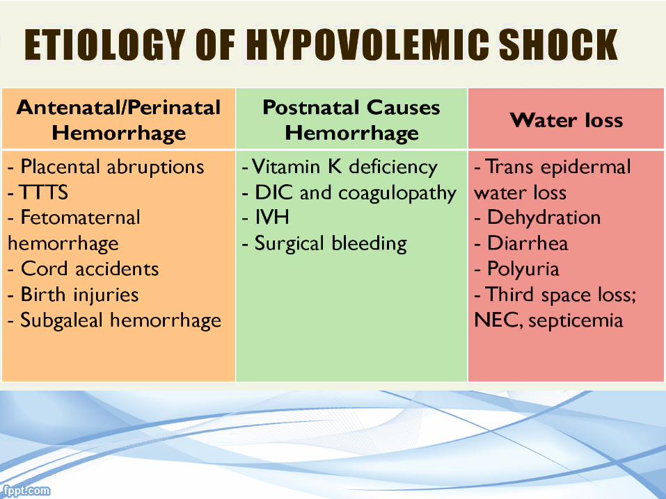
• Hypovolemic − Due to insufficient circulating blood volume, resulting in a reduction in cardiac output (CO) (reduced preload) or reduced oxygen delivery
Hypovolemic

Distributive − Severely decreased systemic vascular resistance (SVR) due to impairment of vascular tone, which results in maldistribution of blood within the microcirculation and regional and global hypoperfusion.
DISTRIBUTIVE

• Sepsis
• Adrenal insufficiency
• include hydrops fetalis and toxic shock syndrome
ETIOLOGY DISTRIBUTIVE SHOCK
CARDIOGENIC
Cardiac Dysfunction (Pump Failure) or Arrhythmia, Causing a decrease in CO primarily due to deceased stroke volume (SV) (eg, impaired contractility). In cases of complete heart block, the significant reduction in heart rate (HR) leads to inadequate CO.
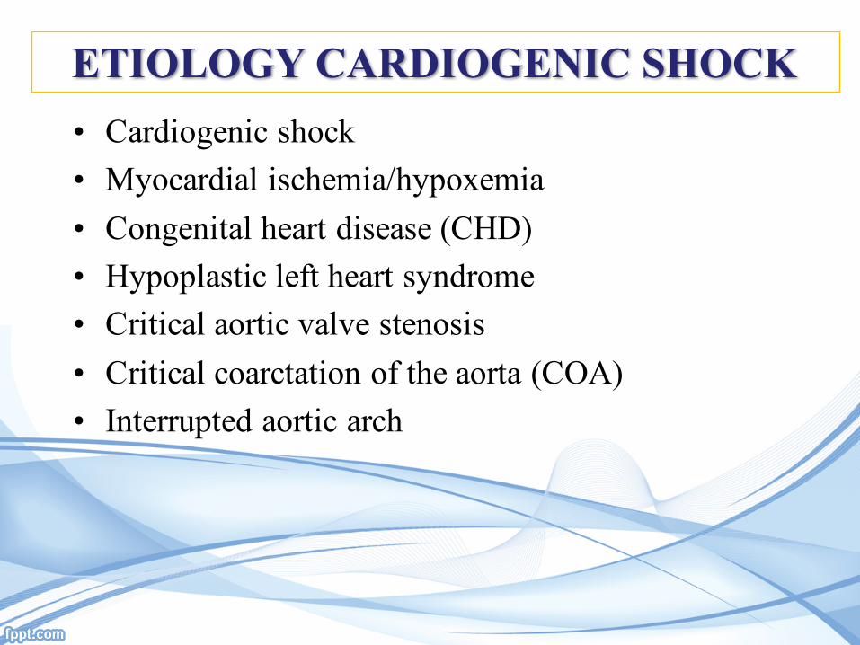
• Cardiogenic shock • Myocardial ischemia/hypoxemia• Congenital heart disease (CHD)• Hypoplastic left heart syndrome• Critical aortic valve stenosis• Critical coarctation of the aorta (COA) • Interrupted aortic arch
ETIOLOGY CARDIOGENIC SHOCK

Etiology Cardiogenic Shock (Cont….)• Cardiac arrhythmias• Complete congenital heart block• Supraventricular tachycardia.• Ventricular tachycardia• Myocarditisusually due to viral infection, most
commonly coxsackievirus.• Congenital cardiomyopathythat typically presents
with hydrops fetalis.
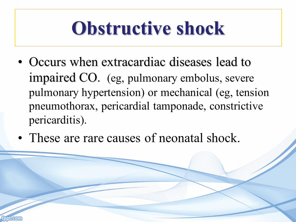
Obstructive shock • Occurs when extracardiac diseases lead to
impaired CO. (eg, pulmonary embolus, severe pulmonary hypertension) or mechanical (eg, tension pneumothorax, pericardial tamponade, constrictive pericarditis).
• These are rare causes of neonatal shock.
Multifactorial shock
It is important to identify the multiple etiologies in order to guide treatment decision.

Vital Signs• Abnormal heart rate• Hypotension• Abnormal body temperature• Decreased peripheral perfusion• Cool extremities, acrocyanosis, and pallor• Delayed capillary refill >4 seconds• Neurologic neurologic changes vary from lethargy
(including poor feeding) to irritability

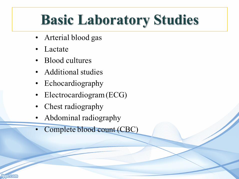
Basic Laboratory Studies• Arterial blood gas • Lactate• Blood cultures• Additional studies• Echocardiography• Electrocardiogram (ECG)• Chest radiography• Abdominal radiography• Complete blood count (CBC)

Laboratory Findings• Anemia due to blood loss • Prolonged PT/INR and PTT tests• Glucose levels may be elevated or decreased during neonatal
shock. • Hyperkalemiadue to tissue injury and cell death• Serum bilirubin levels and liver enzymes may be elevated due to
hepatic injury and dysfunction• Serum creatinine and BUN may be elevated due to renal injury and
dysfunction.

Management
1. Airway/breathing
2. Vascular access
3. Fluid resuscitation
THERAPEUTIC INTERVENTIONS
• Fluid resuscitation — Fluid resuscitation aims to improve preload, thereby increasing stroke volume (SV) and cardiac output (CO).
• Type of solution — Several studies suggest that isotonic crystalloid solutions (eg, normal saline or Ringer's lactate) are preferable to colloid solutions (eg, albumin). Normal saline is the most commonly administered isotonic fluid in neonates and is the solution of choice
THERAPEUTIC INTERVENTIONS• Volume and Rate — Infants with hypovolemic shock, as well as
many infants with distributive shock, require aggressive fluid resuscitation. In these patients, we administer 10 mL/kg per bolus of normal saline infused over 10 to 15 minutes.
• Therapy is Repeated, as needed, up to four times if there is no clinical evidence of improvement and there are no signs of fluid overload (rales or hepatomegaly).
• Additional therapies, such as transfusion of blood products in neonates with shock due to hemorrhage
THERAPEUTIC INTERVENTIONS (Cont…)
• Fluid Resuscitation may be harmful for neonates with cardiogenic shock or those who have compensated shock with certain comorbidities(eg, prematurity, acute renal failure, intrinsic
respiratory diseases).
THERAPEUTIC INTERVENTIONS
•Patients with obstructive shock may also require volume expanders to improve cardiac preload, but therapy should focus on emergent correction of the underlying cause
(eg, needle or chest tube thoracostomy for tension pneumothorax or pericardiocentesis for cardiac tamponade).

Transfusion
•Transfusion — For neonates with shock due to acute blood loss, red blood cell (RBC) transfusion can be lifesaving

Antibiotic Therapy• The empiric antibiotic regimen should include agents active
against group B streptococcal and other organisms that cause neonatal sepsis (eg, Escherichia coli).
• The combination of ampicillinand gentamicin or ampicillin and a third generation cephalosporin (eg, cefotaxime, if available) are potential regimens that provide empiric coverage for these organisms until culture results are available.
Vasoactive agents
• Vasopressor and inotropic agents are used to support neonates with distributive shock who have not improved with initial fluid resuscitation and those with cardiogenic shock
• Commonly used vasoactive agents include dopamine, epinephrine, dobutamine, and milrinone.

• Dopamine is the most commonly used agent in neonates, based on longer clinical experience and familiarity with its use.
• Epinephrine is often beneficial for the management of shock in older patients, but its pharmacologic properties are less well understood in neonates.
• Dobutamine is typically the first inotrope administered for cardiogenic neonatal shock.

Dopamine• For infants with distributive shock who do not respond
adequately to fluid resuscitation, dopamine is infused beginning at a rate of 5 mcg/kgper min with titration up to a maximum of 15 mcg/kgper min based on the infant's clinical response.
• Dopamine can also be used for the management of cardiogenic shock.
Dopamine• The hemodynamic effects of dopamine are dose-dependent:
• Low dosage: 1 to 5 mcg/kg/minute, increased renal blood flow and urine output
• Intermediate dosage: 5 to 15 mcg/kg/minute, increased renal blood flow, heart rate, cardiac contractility, cardiac output, and blood pressure
• High dosage: >15 mcg/kg/minute, alpha-adrenergic effects begin to predominate, vasoconstriction, increased blood pressure
•

Dobutamine
• Dobutamine increases CO via improved myocardial contractility and increased HR. Since it can also decrease SVR and cause hypotension, it is useful for patients with decreased myocardial function due to cardiogenic shock who are normotensiv
Dobutamine
• Dobutamine infusion begins at a rate of 5 mcg/kg per min with titration up to a maximum of 20 mcg/kg per min based on the infant's clinical response

Epinephrine
• Epinephrine increases myocardial contractility and is a potent vasoconstrictor. It is sometimes used for distributive shock that is refractory to dopamine or as a first-line vasoactive therapy for cardiogenic shock.

Epinephrine
• Epinephrine is typically started at a rate of 0.1 mcg/kg per min and titrated up to a maximum of 1 mcg/kg per min based on the infant's clinical response.

Thank Youk