Shigella - Journal of Bacteriologyjb.asm.org/content/38/4/355.full.pdfA. C. THAYSEN the daughter...
12
PULLULOMYXA BOTRYTIS N. SP. A. C. THAYSEN Microbiological Section, Chemical Research Laboratory of the Department of Scientific and Industrial Research, Teddington, England Received for publication January 31, 1939 A brief note was published some years ago by W. T. Morgan of the Lister Institute, London, and the writer (1932) describing the isolation from decayed woody tissues of a micro-organism which decomposes the specific polysaccharides of Shigella dysenteriae, Shiga; Shigella dysenteriae, Flexner Y; Pneumococcus Type II; and the tubercle bacillus. At the time a study of the relationship of this organism was not proceeded with as it was felt that its morphological peculiari- ties deserved closer attention than it was then possible to devote ;o them. The organism was provisionally referred to as a Myxo- coccus, a designation which was based solely on its microscopic appearance. Strained in vivo with a dilute solution of methylene blue it resembles the coccoid stage of many myxococci as depicted by Krzemieniewsey (1928), and incidentally of Spirochaeta cytophaga as described by Hutchinson and Clayton (1919), an organism which Kremienewska (1930) states approaches the myxococci in its characters. Figure 1 shows the polysaccharide-decomposing organism stained in vivo with dilute methylene blue. Its apparently thick-walled coccoid cells contain deeply staining plasmatic inclusions which, in many cases, appear to adhere to one side of the cell wall. The cells measure between 2.5 and 3 1u in diameter; a few may exceed 3 gu and some, not including those attached to larger cells, may be no more than 2 gu in diameter. In the pro- duction of smaller coccoid cells by normally sized spheres the organism differs strikingly from the coccoid stage of the myxo- cocci. These attached smaller coccoid cells can be seen to be 355 on April 16, 2019 by guest http://jb.asm.org/ Downloaded from
Transcript of Shigella - Journal of Bacteriologyjb.asm.org/content/38/4/355.full.pdfA. C. THAYSEN the daughter...
PULLULOMYXA BOTRYTIS N. SP.
A. C. THAYSEN
Microbiological Section, Chemical Research Laboratory of the Department ofScientific and Industrial Research, Teddington, England
Received for publication January 31, 1939
A brief note was published some years ago by W. T. Morgan ofthe Lister Institute, London, and the writer (1932) describing theisolation from decayed woody tissues of a micro-organism whichdecomposes the specific polysaccharides of Shigella dysenteriae,Shiga; Shigella dysenteriae, Flexner Y; Pneumococcus Type II;and the tubercle bacillus.At the time a study of the relationship of this organism was
not proceeded with as it was felt that its morphological peculiari-ties deserved closer attention than it was then possible to devote;o them. The organism was provisionally referred to as a Myxo-coccus, a designation which was based solely on its microscopicappearance. Strained in vivo with a dilute solution of methyleneblue it resembles the coccoid stage of many myxococci as depictedby Krzemieniewsey (1928), and incidentally of Spirochaetacytophaga as described by Hutchinson and Clayton (1919), anorganism which Kremienewska (1930) states approaches themyxococci in its characters.
Figure 1 shows the polysaccharide-decomposing organismstained in vivo with dilute methylene blue. Its apparentlythick-walled coccoid cells contain deeply staining plasmaticinclusions which, in many cases, appear to adhere to one side ofthe cell wall. The cells measure between 2.5 and 3 1u in diameter;a few may exceed 3 gu and some, not including those attached tolarger cells, may be no more than 2 gu in diameter. In the pro-duction of smaller coccoid cells by normally sized spheres theorganism differs strikingly from the coccoid stage of the myxo-cocci. These attached smaller coccoid cells can be seen to be
355
on April 16, 2019 by guest
http://jb.asm.org/
Dow
nloaded from

A. C. THAYSEN
the daughter cells of the normally sized spheres, arising from thelatter by a process of budding. There is therefore only one cellform in the life cycle of the polysaccharide-splitting organism.Among the myxococci, on the other hand, the coccoid stage
represents the resting stage in the life cycle; on germination itgives rise to rod-shaped cells. These rods are the active or re-productive stage of the life cycle. Only when reproductionceases does a coccoid stage again appear among the myxococci
atAim t i9<
* .~Ofs
FIG. 1. THREE-DAY-OLD CULTURE OF PULLULOMIYXA BOTRYTIS, STAINED IN VIVOWITH MIETHYLENE BLUE. X 2550 APPROX.
and this through the contraction of rodshaped cells into spheres,never as a result of the budding of coccoids.
It is true that in other groups of myxobacteria the two stagesin the life cycle are morphologically more uniform than is thecase among the myxococci. But in these cases the cell form isinvariably rod shaped, both during the resting stage and thereproductive stage. No case has so far been described in whichan organism belonging to the Myxobacteriales has shown a com-plete absence of rod-formed cells and a mode of reproductionreminiscent of budding.
356
on April 16, 2019 by guest
http://jb.asm.org/
Dow
nloaded from
PULLULOMYXA BOTRYTIS N. SP.
It is for this reason that the writer has felt compelled to refrainfrom incorporating the organism here described among theMyxobacteriales.The observation that reproduction in the polysaccharide-
splitting organism takes place by budding naturally led to acomparison of this process as it occurs here with the buddingprocess among the yeasts in order to see whether the new organ-ism could be incorporated among the latter. But here again thewriter was unable to find convincing evidence for doing so. In
w.}sHe Now
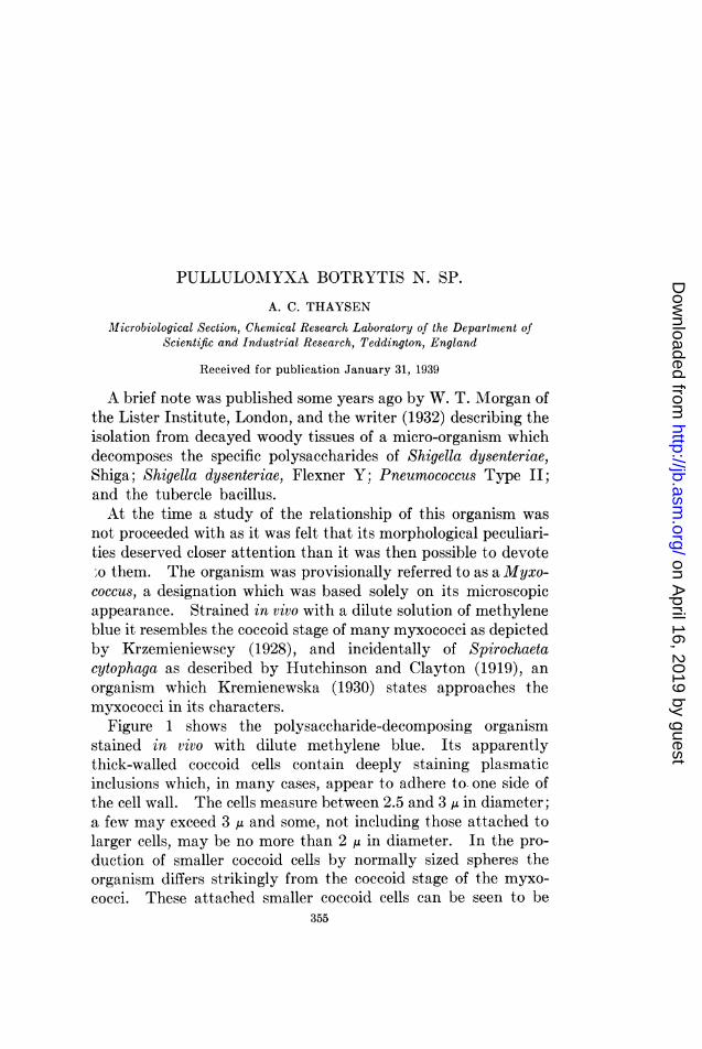
FIG. 2. TWAENTY-FOUR-HOUR CULTURE OF SACCHAROMYCES ELIPSOIDEUS, STAINEDWITH FEULGEN'S STAIN. X 950
fact the observations which he made led him to conclude thatvery considerable differences exist between the process of buddingin the two cases, notably in the behaviour of the thymonucleicsubstances.Among the yeasts, reproduction by budding proceeds, as shown
in figure 2, by the formation of a daughter cell which invariablyattains a considerable size before part of the thymonucleicsubstances of the mother cell proceeds towards it, and eventuallyenters it through the narrow connection between the two cells.
357
on April 16, 2019 by guest
http://jb.asm.org/
Dow
nloaded from
A. C. THAYSEN
The process of transfer of thymonucleic substances from themother cell to the daughter cell is comparatively simple. Theoriginal spherical form of this material will stretch to produce ashorter or longer thread depending on the distance it is removedfrom the connecting passage between mother and daughter cell.Part of this thread will squeeze through the passage and form asphere in the daughter cell, whilst the remainder, still in themother cell, will again contract and form a sphere. The partitionof the thymonucleic substances in the yeast cell thus correspondsto an amitotic nuclear division. The staining method adopted to
FI. 3t FIG.p 4
I * H
jp *,¢
FIG. 3 FIG. 4
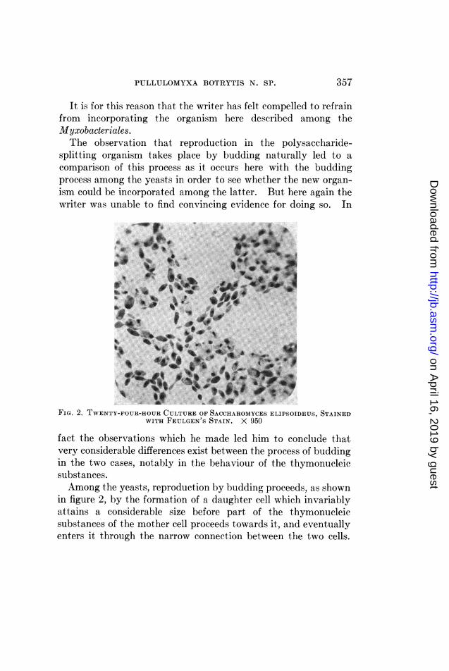
FIG. 3. SEVENTY-DAY-OLD CULTURE OF PULLULONIYXA BOTRYTIS, STAINED WITHFEULGEN'S STAIN. X 2550 APPROX.
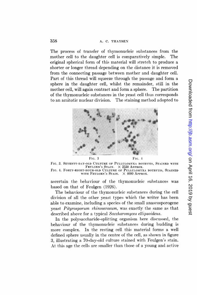
FIG. 4. FORTY-EIGHT-HOUR-OLD CULTURE OF PULLULOMYXA BOTRYTIS, STAINEDWITH FEULGEN'S STAIN. X 4000 APPROX.
ascertain the behaviour of the thymonucleic substances wasbased on that of Feulgen (1926).The behaviour of the thymonucleic substances during the cell
division of all the other yeast types which the writer has beenable to examine, including a species of the small anascosporogeneyeast Pityrosporum rhinoserosumn, was exactly the same as thatdescribed above for a typical Saccharomyces ellipsoideus.
In the polysaccharide-splitting organism here discussed, thebehaviour of the thymonucleic substances during budding ismore complex. In the resting cell this material forms a welldefined sphere usually in the centre of the cell, as shown in figure3, illustrating a 70-day-old culture stained with Feulgen's stain.At this age the cells are smaller than those of a young and active
358
on April 16, 2019 by guest
http://jb.asm.org/
Dow
nloaded from
PULLULOMYXA BOTRYTIS N. SP.
culture. When growth commences, and before any extraneoussigns of budding are noticeable, the sphere of thymonucleicsubstances elongates to form an equatorial band in the cell.Eventually this band may stretch further and become U shaped.In other cases the band of thymonucleic substances is representedby two parallel placed rods. These early stages can be observedin figures 4, 5, 6 and 7.
It has not been possible to ascertain with certainty the detailsof the further subdivision of the thymonucleic substances asrevealed by Feulgen's stain. A closer scrutiny of figures 4 and5 will show cells with two and three separate globules of thesesubstances, indicating perhaps a further subdivision of the twoabove-described parallel rods, or possibly of the original U shaped
0*l 4 Ab .
* .
. V
FIn. 5 FIG. 6 FIG. 7FIGS. 5, 6 AND 7. FIVE-DAY-OLD CULTURES OF PULLULOMYXA BOTRYTIS, STAINED
WITH FEULGEN'S STAIN. X 4000 APPROX.
band. In some cases a subdivision of the substances into 4separate units has been observed, and it cannot be excluded thatthe stage of three or of two separate globules may represent a sub-sequent fusion of a previously larger number of units. Through-out the period of subdivision of the thymonucleic substances thecell has remained globular without excrescences. However,when the stage of the existence of two or three definite globulesof thymonucleic substances has been reached the cell can be seento acquire a slightly egg-shaped form with one granule of thy-monucleic substances eventually occupying the pointed end ofthe cell. Here the bud finally arises.The mode of separation of the daughter cell from the mother
cell in the polysaccharide-splitting organism also differs from that
359
on April 16, 2019 by guest
http://jb.asm.org/
Dow
nloaded from

A. C. THAYSEN
of the yeasts, or at any rate from that of the genus Saccharomycesin which, according to Guilliermond (1920) and Lindner (1930), atransverse cellwall is formed between mother and daughter cellprior to the separation of the two cells. In the polysaccharide-decomposing organism on the other hand the two cells draw awayfrom each other leaving a connecting filament between themwhich eventually breaks (see fig. 1), and may remain attached toone or both cells for some time as a short stalk. Where thedaughter cell has produced a daughter cell prior to separationfrom its mother cell a short chain of three individuals may beformed. Such structures are strikingly similar to those describedby Baker (1933) under the name of "giant cocci." Baker foundsuch giant cocci in large numbers in cavities of the cellulosiccontent of the intestine of the guinea pig, where he associatedthem with the breakdown of hemicelluloses and possibly ofcellulose.The data recorded in the previous pages have convinced the
writer that the polysaccharide-decomposing organism differsfundamentally from the yeasts and that, in fact, it is impossibleon the available information to group it with any existing typeof microorganism, including that of the myxococci to which, asalready mentioned, early investigation had indicated that it mightbelong. Under the circumstances, he has thought it desirableto regard it as a new species, the name of which should indicatethe most characteristic property of the organism, that of repro-duction by budding, and to leave it to future investigations todecide its true position among the known groups of micro-organisms, if it should be found of sufficient interest to deservefuture attention.At the suggestion of Dr. Ramsbottom of the British Museum
the writer proposes that the organism be known as Pullulomyxabotrytis n. sp.
For the further characterization of Pullulomyxa botrytis thefollowing data have been compiled.
Motility. The organism is non-motile.Staining properties. The ordinary aniline dyes are readily
absorbed. A young culture of 2 to 3 days growth is gram posi-
360
on April 16, 2019 by guest
http://jb.asm.org/
Dow
nloaded from
PULLULOMYXA BOTRYTIS N. SP.
tive, the stain giving good pictures of the cellular contents and ofthe connecting filaments between adjacent, directly related cells.A 20-day-old culture is still essentially gram positive though someof the cells will be found to take the counter stain.
Growth on agar media. No growth can be obtained on ordinarystandard agar media. Even on the specially devised agar me-dium, growth is not visible during the first one or two days atthe optimum temperature.
After 6 days incubation at 30 to 370C. the surface colonies haveattained a size of from 50 to 100 ,u with an average of 80 ,/. Bothsurface and embedded colonies are circular, with a smooth edgeand a slightly granular interior. They are greyish white, trans-parent and moist. It is noticeable that the size of the coloniesis larger on plates with a larger number of colonies than on thosecontaining a few only. On older plates this difference is reversedand after 14 days the sparsely seeded plates may show coloniesof a diameter of from 0.4 to 1.2 mm., while those on thicklyseeded plates fail to exceed 0.4 mm. Older colonies are no longertransparent but retain the original moist appearance. Theircolour is no longer greyish white, but faintly brownish grey.
Growth on gelatin. Growth is not easily obtained since thetemperature of incubation is well below the optimum of theorganism. After 17 days incubation no growth could be observedon the inoculated plates. Nevertheless, gelatin is very slowlyliquefied by the organism. This was shown by adding a thicksuspension of living cells to a tube of ordinary gelatin and in-cubating the latter at 370C. for 17 days. By then the gelatin,when cooled, could not be made to solidify though control tubeswithout an addition of the organism did so.
Growth in liquid media. In the most favourable medium,described subsequently, visible growth may be noticeable at370C. within 24 hours. The clear liquid has become very faintlyturbid and slightly bleached. Turbidity and bleaching increaseslowly during the subsequent period of incubation until, after 8days at 370C., the medium is markedly turbid with a noticeablegreyish sediment. When growth in liquid media takes place inshallow layers it is more rapid, and marked turbidity and sedi-
361
on April 16, 2019 by guest
http://jb.asm.org/
Dow
nloaded from
A. C. THAYSEN
mentation is already noticeable after 24 hours incubation at370C. The more rapid development causes the cells to accumu-late in clusters which may be sufficiently large within the first48 hours of growth to be visible with a hand lens. The rapidgrowth in shallow layers confirms the aerobic nature of the or-ganism. Even with the addition of suitable carbohydrates,growth is not possible under strictly anaerobic conditions.
Temperature range of growth. Initial growth, during the first24 hours of incubation is very similar in extent throughout therange tested, between 18 and 400C. Subsequently, however,little progress in growth is made in cultures kept at the formertemperature and even at 250C. The range from 30 to 370C.appears to be suitable for a normal growth rate, and there is littleto choose between the two extremes. The higher temperature of40'C. has not been found suitable for the maintenance of cultures.
Lethal temperatures. A young culture kept for 30 minutes at52CC. was found capable of propagating when subcultured intofresh medium and incubated at 370C. An identical culturekept for the same period at 55CC. failed to do so.pH requirements of the organism. The most abundant growth
is observed when the standard medium is adjusted to a hydrogen-ion-concentration range of between pH 7.0 and 7.6. This growthcan be maintained even at pH 8.0, but no growth was noticeableat a pH of 8.6. On the acid side of the neutral point, a certainamount of growth could be induced in the standard medium at apH of 6.5 but little if any at 6.0 and below.
Composition of standard medium. When the organism wasfirst discovered it was seen under the microscope in a mediumcomposed of ammonium sulphate 0.01 per cent, dipotassiumhydrogen phosphate 0.02 per cent and water. To this had beenadded 0.001 per cent of the specific polysaccharide of Shigelladysenteriae, Shiga. The rate of growth of the organism was veryslow in this medium and it was found that the addition of a cer-tain amount of an extract made from fresh rabbits' droppingsgreatly increased the rate of growth and facilitated the eventualisolation of the organism.
All subsequent work, and the cultivation of PuUulomyxa
362
on April 16, 2019 by guest
http://jb.asm.org/
Dow
nloaded from
PULLULOMYXA BOTRYTIS N. SP.
botrytis, has been done in a medium the composition of which isbased on the above observation.The following procedure is adopted in the preparation of the
standard medium: 10 gr. of fresh rabbit pellets are left over nightat room temperature in 1 litre of tap water. The extract ob-tained is poured off, and rejected. It is replaced by a furtherlitre of tap water, with which the pellets are given a short boil.The second extract thus obtained is filtered and to the clear liquidare added 2 g. of ammonium sulphate, 4 g. of dipotassium hy-drogen phosphate, and 4 g. of a carbohydrate such as fructose orxylose. The volume of extract is finally made up to 2 litres andsterilized. When solid media are required, 1.5 per cent of agaragar are dissolved in the standard medium prior to its beingsterilised. The sterilization has usually been done fractionallyon three successive days with one hour's steaming daily.On or in the above medium, Pullulomyxa botrytis grows more
abundantly than on any other tried. This, however, is notmeant to imply that the growth of the organism, even under themost favourable conditions, is really copious, but merely that it ismarkedly richer than on the ordinary bacteriological culturemedia, which in most cases are unsuitable.
Utilisation of carbohydrates. In the preliminary note whichappeared in Nature (1933) it was mentioned that Pullulomyxabotrytis destroyed the specific properties of a number of bacterialpolysaccharides. This was thought to indicate that the sac-charolytic enzyme complex of the organism would be able tofunction on a wide range of carbohydrates, since it is doubtfulwhether the organism could have met with polysaccharides ofpathogenic bacteria in its natural habitats. It was thought ofinterest therefore, to test the action of Pullulomyxa botrytis ona wide range of carbohydrates, including several polysaccharides.Of these, cellulose was found to remain unaffected. Gum
arabic, however, and xylan as well as a polysaccharide isolatedby Campbell (1935) from oak sapwood were suitable sources ofcarbohydrate. Starch and inulin did not promote growth.Among the disaccharides tested, cellobiose, maltose, sucrose andlactose could be utilized, the latter less readily than the three
363
on April 16, 2019 by guest
http://jb.asm.org/
Dow
nloaded from

A. C. THAYSEN
former. Of the monosaccharides, fructose appeared more suit-able than glucose. In fact it is questionable whether glucosecan readily be utilised. The two pentoses tested, xylose andarabinose, were both suitable. Growth for prolonged periods onany of these carbohydrates did not in the least weaken the actionof the organism on the specific bacterial polysaccharides whichit was originally found capable of destroying.
It is of interest to note that Pullulomyxa botrytis is capable ofutilising not only hemicelluloses which occur widespread in nature,but also an intermediate product of hydrolysis of cellulose. Itwill be remembered that attention was drawn above to thesimilarity in morphology between this organism and the "giantcocci" observed by Baker (1933) in the coecum of certain herbiv-erous animals. From a physiological point of view, therefore,a similarity also exists between the two types.The action of Pullulomyxa botrytis on the various carbohy-
drates mentioned as suitable for growth did not lead to the pro-duction of visible quantities of acid or gas. It was decided,therefore, to study this action in the Barcroft manometer, and todetermine the oxygen uptake of an active suspension of theorganism on some of the carbohydrates which had been previouslytested for their suitability as energy suppliers.
Using a suspension in saline of a 10-day old culture grown onxylose agar, xylose showed an oxygen uptake of 28 pl within 4hours and a gas evolution of 52 ul, assuming this gas to have beencarbon dioxide. On the assumption of the evolved gas beinghydrogen the oxygen uptake was 80 gl within the first 4 hoursand the gas evolution 52 pl as before.The same suspension of the organism gave with fructose an
oxygen uptake of 40 Al assuming the gas evolved during the re-action to have been hydrogen. In the case of this particularcarbohydrate no carbon dioxide can have been evolved.
Glucose was unable to show an oxygen uptake by the sus-pension, even when the experiment was continued for 24 hours.A considerable amount of work was devoted to the isolation
of the enzyme system of Pullulomyxa botrytis which causes thedestruction of the polysaccharides of bacterial toxines. These
364
on April 16, 2019 by guest
http://jb.asm.org/
Dow
nloaded from

PULLULOMYXA BOTRYTIS N. SP.
efforts, however, were completely unsuccessful. Neither byautolysis, nor by freezing and disintegration of the living cellscould the responsible enzymes be separated in active form. Ithas not been possible, therefore, to study the effect on infectedanimals of an injection of the polysaccharide-splitting enzyme ofPullulomyxa botrytis.
CONCLUSIONS
A description is given of the cytological, morphological andphysiological characters of an organism which was isolated fromdecaying vegetable debris and which previous work had shownwas capable of destroying the specific polysaccharides of certainbacterial toxins.The cytological study has shown that the organism in question,
for which the name Pullulomyxa botrytis n. sp. is suggested,represents a type which, to the writer's knowledge, has neverbefore been described in the literature.
Its propagation proceeds by budding, but the cytologicalchanges involved are far more complex than those met with inthe yeasts.
Details are given of the morphological and cultural character-istics of Pullulomyxa botrytis n. sp. and a reference is made to thework which has been done on the isolation of the enzyme systemby which the specific polysaccharide of the Shiga dysentry toxinmust be assumed to be destroyed by the organism.
The writer wishes to place on record his thanks to Dr. Rams-bottom of the British Museum for his advice on nomenclature;to Mr. W. G. Campbell of the Forest Products Research Labora-tory of the Department of Scientific and Industrial Research,for the supply of a sample of oak softwood polysaccharide; andto Mr. K. R. Butlin of his staff, for the determination of therespiratory properties of Pullulomyxa botrytis.The work described above was carried out as part of the pro-
gramme of the Chemistry Research Board, and is published bypermission of the Department of Scientific and Industrial Re-search, London, England.
365
on April 16, 2019 by guest
http://jb.asm.org/
Dow
nloaded from
366 A. C. THAYSEN
REFERENCESBAKER, F. 1933 Studies in the microbiology of organisms associated with the
disintegration of vegetable remains, etc. Zentr. Bakt. Parasitenk.,Abt. II, 88, 17-42.
CAMPBELL, W. S. 1935 Wood Hemicelluloses. Nature, 136, 299.FEULGEN, R. 1926 Die NuclearfArbung. Abderhalden Handbuch der biol.
Arbeitsmethoden, 5, 1055-1074.GUILLIERMOND, A., ANDTANNER, F. W. 1920 The Yeasts. New York, J. Wiley
and Sons, Inc.HUTCHINSON, H. B., AND CLAYTON, J. 1919 On the decomposition of cellulose
by an aerobic organism (Spirochaeta cytophaga n. sp.). J. Agr. Sci.,9, 143-173.
KRZEMIENIEWSCY, H., AND S. 1928 Zur Morphologie der Myxobakterienzelle.Acta Soc. Botan. Poloniae, 5, 46-90.
KREMIENIEWSKA, H. 1930 Le cycle 6vulotif de Spirochaeta cytophaga Hutchin-son and Clayton. Acta Soc. Botan. Poloniae, 7, 507-519.
LINDNER, P. 1930 Mikroskopische und biologischen Betriebkontrolle in derGarungsgewerben, Berlin, 422. Paul Parey.
on April 16, 2019 by guest
http://jb.asm.org/
Dow
nloaded from