
Shharraa MMM.. hMaarrsshaalll,, M..SS.
52
1 Shara M. Marshall, M.S.
Transcript of Shharraa MMM.. hMaarrsshaalll,, M..SS.
1
SShhaarraa MM.. MMaarrsshhaallll,, MM..SS..

2
ANATOMICAL REGIONS, POSITIONS AND TERMINOLOGY
Overview
Anatomy:
Physiology:
Function:
Levels of Structural Organization
Chemical:
Cellular:
Tissue:
Organ:
Organ system:
Organismal:
Survival Needs
1.Nutrients
•
•
2.Oxygen
•
3.Water
•
•
4.Normal body temperature
•
5.Appropriate atmospheric pressure
•
Homeostasis
Definition:
Components of a Control Mechanism (contain at least the following three components)
1.Receptor (sensor)
•
•
2.Control center
•
•
•

3
3.Effector
•
•
•
Negative Feedback
Example: Regulation of body temperature
Positive Feedback
Example:
• Enhancement of labor contractions by oxytocin
Anatomical Position
Purpose:
Standard anatomical body position:
•
•
•
Directional Terms
Superior (cranial) -
Inferior (caudal) -
Ventral (anterior) -
Dorsal (posterior)-
Medial-
Lateral-
Intermediate-
Proximal –
Distal -
Superficial-
Deep -
4
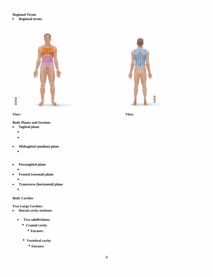
Regional Terms
Regional terms:
View: View:
Body Planes and Sections
Sagittal plane
•
•
Midsagittal (median) plane
•
Parasagittal plane
•
Frontal (coronal) plane
•
Transverse (horizontal) plane
•
Body Cavities
Two Large Cavities:
Dorsal cavity encloses:
Two subdivisions:
• Cranial cavity
• Encases:
• Vertebral cavity
• Encases:

5
Ventral cavity
• Houses:
• Two subdivisions (separated by diaphragm):
• •
Thoracic cavity subdivisions:
• Two pleural cavities
• Each one houses:
• Mediastinum
• Contains:
• Also contains:
• Pericardial cavity
• Encloses:
Abdominopelvic cavity subdivisions:
• Abdominal cavity
• Contains:
• Pelvic cavity
• Contains:
Abdominopelvic Regions
Nine divisions:
BASIC CHEMISTRY
Matter and Composition of Matter
Definition:

6
Matter is composed of elements -
Atoms-
Atomic Structure
Neutrons
•
• Mass =
Protons
•
• Mass =
Electrons
•
• Mass =
• Equal in number to:
Energy
Definition:
Types of energy:
• Kinetic:
• Potential:
• Electrical:
Identifying Elements
Atoms of different elements contain different numbers of protons
• Compare hydrogen, helium and lithium
Atomic number =
Mass number =
•
• Isotopes =
Atoms of Elements can Combine Chemically with other atoms to form Molecules and Compounds
• Molecule: (e.g., H2 or C6H12O6)
• Compound: (e.g., C6H12O6)

7
Chemical Bonds
Octet rule:
Chemically Inert Elements
Chemically Reactive Elements
TYPES OF CHEMICAL BONDS
Ionic Bonds
Ions are formed by:
• Anions (– charge):
• Cations (+ charge):
Attraction of opposite charges results in:
Covalent Bonds
Formed by sharing of two or more valence shell electrons
Sharing of electrons may be equal or unequal
• Equal sharing produces:
• CO2
Unequal sharing by atoms with different electron-attracting abilities produces:
• H2O
Hydrogen Bonds
Attractive force between electropositive hydrogen of one molecule and an electronegative atom of another molecule
TYPES OF CHEMICAL REACTIONS
Synthesis Reactions
A + B AB
•
•
•

8
Decomposition Reactions
AB A + B
•
•
•
•
CLASSES OF COMPOUNDS
Inorganic compounds
• Do not contain: (ex’s. water, salts, and many acids and bases)
Organic compounds
•Contain: (ex’s. carbohydrates, fats, proteins, nucleic acids)
Water
Salts
Ionic compounds that dissociate in water
Acids
Acids:
• HCl H+ + Cl
–
Bases Bases:
• NaOH Na+ + OH
–
Acid-Base Concentration
Acid solutions contain higher concentrations of H+
• As [H+] increases:
Basic solutions contain higher concentrations of OH–
• As [H+] decreases (or as [OH
–] increases):
pH =
Neutral solutions:
• pH =
•
Acidic solutions
• pH =
•
•
Basic solutions

9
• pH=
•
For the following organic compounds, they are formed by a synthesis type of chemical reaction and broken down by a chemical
reaction called hydrolysis.
Carbohydrates
Sugars and starches whose building blocks =
Three classes
• Monosaccharides-
• Disaccharides-
• Polysaccharides-
Functions
• Primary role:
Lipids
Main types:
• Triglycerides
• Phospholipids
• Steroids
Triglycerides
Defined as:
Building blocks =
Main functions
•
•
•
Phospholipids
Similar to triglycerides:
• Building blocks =
“Head” and “tail” regions:
Important in:

10
Steroids
Steroids—
Ex’s. -Cholesterol, vitamin D, steroid hormones, and bile salts
Proteins
Building blocks =
After amino acids are linked together:
This folding process results in four different levels of protein structure:
Protein Denaturation
Definition:
A denatured protein is:
Enzymes
Biological catalysts
• Function:
Nucleic Acids
DNA and RNA
•
Building blocks =
Deoxyribonucleic Acid (DNA)
Four bases:
• adenine (A), guanine (G), cytosine (C), and thymine (T)
11
Ribonucleic Acid (RNA)
Four bases:
• adenine (A), guanine (G), cytosine (C), and uracil (U)
Three varieties of RNA carry out the DNA orders for protein synthesis
•
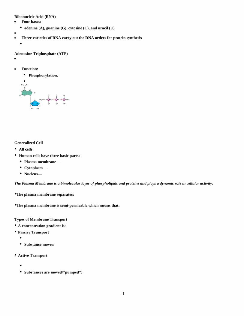
Adenosine Triphosphate (ATP)
Function:
• Phosphorylation:
•
Generalized Cell
• All cells:
• Human cells have three basic parts:
• Plasma membrane—
• Cytoplasm—
• Nucleus—
The Plasma Membrane is a bimolecular layer of phospholipids and proteins and plays a dynamic role in cellular activity:
•The plasma membrane separates:
•The plasma membrane is semi-permeable which means that:
Types of Membrane Transport
• A concentration gradient is:
• Passive Transport
•
• Substance moves:
• Active Transport
•
• Substances are moved/”pumped”:
12
Passive Transport
• What determines whether or not a substance can passively cross the plasma membrane?
1.
2.
Three Types of Passive Transport Across Cellular membranes
• Simple diffusion
• Facilitated diffusion
• Osmosis
Passive Transport: Simple Diffusion
• What types of substances use simple diffusion to cross the plasma membrane?
•
Passive Transport: Facilitated Diffusion
• What types of substances use facilitated diffusion to cross the plasma membrane?
•
• Can pass through:
Passive Transport: Osmosis
•
• Water diffuses through plasma membranes:
• Mainly through channels called aquaporins (AQPs)
Importance of Osmosis
•When osmosis occurs:
•A change in cell volume:
Tonicity
•Defined as:
•Isotonic:
•Hypertonic:
•Hypotonic:

13
Active Transport
Defined as:
The Sodium-potassium pump (Na+-K
+ ATPase) is a specific example of active transport
o
o
Other Cellular Organelles:
Membranous structures
o Nucleus with chromatin-
o Mitochondria –
o Endoplasmic Reticulum (ER) (rough and smooth) –
o Golgi Apparatus-
o Lysosomes-
Non-membranous structures
o Centrioles –
o Cytoskeleton -
Extensions of the plasma membrane
o Cilia are –
o Flagella are –
o Microvilli are -
The Cell Cycle
• Includes:
• Interphase
•
•Three Subphases:
A B C
14
•Gap 1 (G1)-
•Synthesis Phase (S phase)-
•Gap 1 (G2)-
•Cell Division (mitotic phase or mitosis)
•Includes four sub phases of mitosis (PMAT) and cytokinesis
Mitosis
Purpose:
Does not occur in:
During the S-phase of Interphase DNA is Replicated
• Helicase:
• DNA polymerase:
• End result:
•This process is called:
•After DNA has been replicated:
Mitosis and Cytokinesis
Mitosis—four stages of nuclear division:
• Prophase -
• Metaphase -
• Anaphase -
• Telophase -
• Cytokinesis -
There are four tissue types in the body
• Epithelial tissue
• Connective tissue
• Muscle tissue
• Nerve tissue

15
Characteristics of Epithelial Tissue
1.Cells have polarity—
•
•
2.Are composed of closely packed cells
•
3.Avascular
4.High rate of regeneration
Classification of Epithelia
• Ask two questions:
1. How many layers?
1 =
>1 =
• What type of cell?
• • •
• (If stratified, name according to apical layer of cells)
Overview of Epithelial Tissues
Epithelia: Simple Squamous
* Description:
* Function:
* Location:

16
Epithelia: Simple Cuboidal
* Description:
* Function:
* Location:
Epithelia: Simple Columnar
* Description:
* Function:
* Location:
Epithelia: Pseudostratified Ciliated Columnar Epithelium
* Description:
* Function:
* Location:
Epithelia: Stratified Squamous
* Description:
* Function:
* Location:
Epithelia: Stratified Cuboidal and Columnar Epithelium
* Description:
* Function:
* Location:
Epithelia: Transitional Epithelium
* Description:
* Function:
* Location:
Glandular Epithelia
Gland:
Two major types:
o Endocrine glands-
o
o Exocrine glands-
17
Connective Tissue
• Most abundant and widely distributed tissue type
Flow Chart:
Characteristics of Connective Tissue
• Connective tissues have:
*
* Cells separated by nonliving extracellular matrix (ground substance and fibers)
Extracellular Matrix
• Ground substance
• Components
•
• Fibers (three types)
• Collagen
•
• Elastic
•
• Reticular
•
•Cells of Connective Tissue
•“blasts” = Mitotically active and secretory cells
•“cytes” = Mature cells
•Fibroblasts in :
•Chondroblasts and chondrocytes in:
•Osteoblasts and osteocytes in:
•Hematopoietic stem cells in:
Overview of Connective Tissues
• Connective Tissues: Osseous
* Description:
* Function:
* Location:

18
• Connective Tissues: Hyaline Cartilage
* Description:
* Function:
* Location:
• Connective Tissues: Elastic Cartilage
* Description:
* Function:
* Location:
• Connective Tissues: Fibrocartilage Cartilage
* Description:
* Function:
* Location:
• Connective Tissues: Dense Regular Tissue
* Description:
* Function:
* Location:
• Connective Tissues: Dense Irregular Tissue
* Description:
* Function:
* Location:
• • Connective Tissues: Areolar Connective Tissue
* Description:
* Function:
* Location:
• Connective Tissues: Adipose Connective Tissue
* Description:
* Function:
* Location:
• Connective Tissues: Reticular Connective Tissue
* Description:
* Function:
* Location:

19
• Connective Tissues: Blood
* Description:
* Function:
* Location:
Muscle Tissue (Three Types)
• Skeletal Muscle
* Description:
* Function:
* Location:
• Cardiac Muscle
* Description:
* Function:
* Location:
• Smooth muscle
* Description:
* Function:
* Location:
Nervous Tissue
* Description:
* Function:
* Location:
Steps in Wound Repair
• Inflammation
• Organization and restored blood supply
• Regeneration and fibrosis

20
SKIN AND BODY MEMBRANES
Epithelial Membranes
• The cutaneous membrane is the:
• Mucous membranes
• Mucosae
• (e.g., digestive and respiratory tracts)
• Serous Membranes
• Paired membranes that line:
• Parietal layer -
• Visceral layer –
• Serous Membranes are named based on their location:
•Parietal and visceral pleura surround:
•Parietal and visceral pericardium surround:
•Parietal and visceral peritoneum surround:
Skin (Integument)
• Consists of three major regions
1. Epidermis—
2. Dermis—
3. Hypodermis (superficial fascia)—
•
Epidermis
•
• Cells of epidermis
• Keratinocytes—
• Melanocytes
•
Layers of the Epidermis: Stratum Basale
•
• Also called stratum germinativum:
• Cells travel from basal layer to surface
• Takes 25–45 days
Layers of the Epidermis: Stratum Spinosum
• •
Layers of the Epidermis: Stratum Granulosum
• •
21
Layers of the Epidermis: Stratum Lucidum
• • •
Layers of the Epidermis: Stratum Corneum
• • • Functions
•
•
Dermis
• Made up of Two layers:
• Papillary layer
• Reticular layer
Layers of the Dermis: Papillary Layer
• Papillary layer
• Composed of:
• Contains dermal papillae which may have:
• • •
Layers of the Dermis: Reticular Layer
• Reticular layer
•Composed of:
•
Skin Color
• Three pigments contribute to skin color:
1. Melanin
• •
2. Carotene
• 3. Hemoglobin
•

22
Appendages of the Skin
• Derived from the epidermis
•
•
•
•
Sweat Glands
• Two main types of sweat (sudoriferous) glands
1. Eccrine sweat glands— abundant on palms, soles, and forehead
• Sweat:
• •
2. Apocrine sweat glands—confined to:
• Sebum:
•
•
Sebaceous (Oil) Glands
• • • • Sebum
•
•
•
Hair
• Functions
•
•
• Consists of:
Hair Follicle
• Two-layered wall consisting of:
• Hair bulb:
• Hair follicle receptor (root hair plexus):
•

23
• Arrector pili
•
•
Structure of a Nail
• •Structures of the nail:
Functions of the Integumentary System
1.Protection—
• Chemical
• • Physical/mechanical barriers
• •
• Biological barriers
• 2.Body temperature regulation
•
•
3.Cutaneous sensations
•
4.Metabolic functions
•
•
5.Blood reservoir—
6.Excretion—
Skin Cancer
• Three major types:
• Basal cell carcinoma
• Squamous cell carcinoma
• Melanoma
Basal Cell Carcinoma
•
•Appearance:
•
•

24
Squamous Cell Carcinoma
•
• Appearance:
•
Melanoma
•
• Appearance:
•
•
Melanoma
• Characteristics (ABCDE rule)
A: Asymmetry-
B: Border-
C: Color-
D: Diameter-
E: Evolution-
Partial-Thickness Burns
• First degree
•
• • Second degree
•
•
Full-Thickness Burns
• Third degree
•
• • •
25
THE SKELETAL SYSTEM
Bones: An Overview
Functions of Bones
• Support
•
• Protection
•
• Movement
•
• Storage
•
• Blood cell formation (hematopoiesis) in:
•
Classification of Bones by Shape
• Long bones
•
•
• Short bones
•
•
• Flat bones
•
• Examples:
• Irregular bones
•
• Examples:
Bone Markings (Refer to Table 5.1)
Bone Markings: Projections
• Sites of muscle and ligament attachment
• Tuberosity—rounded projection
• Crest—narrow, prominent ridge
• Trochanter—large, blunt, irregular surface
• Tubercle—small rounded projection
• Epicondyle—raised area above a condyle
• Spine—sharp, slender projection
• Process—any bony prominence

26
• Projections that help to form joints
• Head - bony expansion carried on a narrow neck
• Facet - smooth, nearly flat articular surface
• Condyle - rounded articular projection
• Ramus -armlike bar
Bone Markings: Openings
• Meatus -canal-like passageway
• Sinus - cavity within a bone
• Fossa -shallow, basinlike depression
• Groove- furrow
• Fissure -farrow, slitlike opening
• Foramen -round or oval opening through a bone
Bone Textures
• Compact bone
•
•
• Spongy bone
•
Membranes of Bones
Periosteum (fibrous connective tissue)
o Surrounds:
o Contains:
o Secured to compact bone by:
Endosteum (areolar CT)
o
Structure of a Long Bone
• Diaphysis (shaft)
•
• Epiphyses
•
•
•
•

27
Structure of Short, Irregular, and Flat Bones
• “think of a stiffened spongy bone sandwich”
Microscopic Anatomy of Bone: Compact Bone
• Haversian system, or osteon—structural unit
• Lamellae
• •
• Central (Haversian) canal
•
• Perforating (Volkmann’s) canals
•
•
• Lacunae—
• Canaliculi—
Microscopic Anatomy of Bone: Spongy Bone
•
•
•
Chemical Composition of Bone: Organic
• Bone cells: osteoblasts, osteocytes, osteoclasts
• Osteoid—organic bone matrix secreted by osteoblasts
•
•
Chemical Composition of Bone: Inorganic
• Hydroxyapatites (mineral salts)
•
•
•
Bone Formation, Growth and Remodeling
• Ossification—
• Stages
• Bone formation—

28
• Postnatal bone growth—
• Bone remodeling and repair—
Growth in Length of Long Bones
• Interstitial growth:
•
Growth in Width of All Bones
• Appositional growth:
•
Control of Remodeling
• What controls continual remodeling of bone?
•
•
Hormonal Control of Blood Ca2+
• Primarily controlled by parathyroid hormone (PTH)
Blood Ca2+
levels
__________________________
PTH stimulates osteoclasts to degrade bone matrix and release Ca2+
____________________________________
Response to Mechanical Stress
• Curved bones are thickest where they are most likely to buckle
• Trabeculae form along lines of stress
• Large, bony projections occur where heavy, active muscles attach
Stages in the Healing of a Bone Fracture
1.Hematoma forms
•
•
2. The break is splinted by a fibrocartilage callus
•
•
•
•

29
3.Bony callus formation
•
•
4.Bone remodeling
•
•
Homeostatic Imbalances: Read about Osteoporosis and understand its risk factors, symptoms and characteristics.
The Skeleton
Two Major Divisions of the Skeletal System:
The Axial Skeleton Includes Three Regions:
o
o
o
The Skull
Two sets of bones
1. Cranial bones (8 cranial bones)
o Enclose the brain in the cranial cavity
o Calvaria:
o Cranial base:
2. Facial bones (14 Facial bones)
o Cavities for:
o Provide sties of attachment for:
o All skull bones are joined by:
Frontal Bone
Parietal Bones and Major Associated Sutures
•
• Four sutures mark the articulations of parietal bones with frontal, occipital, and temporal bones:
1. Coronal suture—
2. Sagittal suture—
3. Lambdoid suture—
4. Squamous (squamosal) sutures—
Cranial Bones (8) Facial Bones (14)

30
Occipital Bone
Contains:
Temporal Bones
Inferior to parietal bones
External acoustic canal:
Zygomatic process:
Mastoid process:
Styloid process:
Mandibular fossa:
Sphenoid Bone
Three pairs of processes:
Contains the:
Ethmoid Bone
Superior part of :
Contains:
Mandible
Temporomandibular joint:
Mandibular condyle articulates w:
Maxillary Bones
Zygomatic Bones
Nasal Bones and Lacrimal Bones
Nasal bones
Form:
Lacrimal bones

31
Lacrimal fossa houses:
Palatine Bones and Vomer
• Palatine bones
• Vomer
Inferior Nasal Conchae
Form part of lateral walls of nasal cavity
Orbits
Encase:
Sites of attachment for:
Formed by:
Hyoid Bone
Site of attachment for:
Vertebral Column
•
•
• Flexible curved structure containing 26 irregular bones (vertebrae)
Cervical vertebrae (7)—vertebrae of:
Thoracic vertebrae (12)—vertebrae of:
Lumbar vertebrae (5)—vertebra of:
Sacrum—bone inferior to:
Coccyx—terminus of vertebral column
Vertebral Column: Curvatures
• Increase the resilience and flexibility of the spine
Primary curvatures
Secondary curvatures
Read about Abnormal spine curvatures (Scoliosis, Kyphosis, Lordosis)

32
General Structure of Vertebrae
• Body or centrum
• Vertebral arch
Formed from:
• Vertebral foramen
Seven processes per vertebra:
Spinous process—
Transverse processes (2)—
Superior articular processes (2)—
Inferior articular processes (2)—
Cervical Vertebrae
C1 to C7:
C3 to C7 share the following features
Transverse foramen in:
C1 (atlas) and C2 (axis) have unique features
Atlas (C1)
Consists of:
Superior articular facets articulate with:
Axis (C2)
Thoracic Vertebrae
T1 to T12
Lumbar Vertebrae
L1 to L5
Sacrum and Coccyx
• Sacrum
o
o Forms:
o Contains the sacral canal for:
o Articulates with:
• Coccyx
o
o

33
o Articulates superiorly with:
Thoracic (Rib) Cage
• Composed of
Thoracic vertebrae
Sternum
Ribs and their costal cartilages
Sternum
Three fused bones
o Manubrium
o Body
o Xiphoid process
Ribs and Their Attachments
12 pairs
o All attach:
Pairs 1 through 7
o
o Attach:
Pairs 8 through 10
o False ribs
o
Pairs 11-12
o
o
The Appendicular Skeleton consists of the bones of the upper and lower limbs and their girdles (pectoral and pelvic)
Pectoral Girdle (Shoulder Girdle)
Clavicles and the scapulae
o Attach:
o Provide:
Clavicles
Acromial (lateral) end:
Sternal (medial) end:
Scapulae (Shoulder Blades)

34
Three fossa are the site of origin for the rotator cuff muscles:
The Upper Limb
Arm
o
Forearm
o
Hand
o 8 carpal bones in the wrist
o 5 metacarpal bones in the palm
o 14 phalanges in the fingers
Humerus
Head of Humerus Articulates with:
Contains:
Articulates inferiorly with the radius and ulna via the:
Bones of the Forearm
• Ulna
o
o Contains:
• Radius
o
o Contains:
Hand: Carpus
Eight bones in two rows
Only scaphoid and lunate:
Hand: Metacarpus and Phalanges
Metacarpus
o
Phalanges
o Each finger (digit), except the thumb, has three phalanges-
o The thumb:
Pelvic (Hip) Girdle
• Consists of:
Attaches:
Transmits:

35
• Each coxal bone consists of three fused bones: ilium, ischium, and pubis
o Iliac crest, sacroiliac joint
Ischium-
o
Pubis
o Pubic symphysis, pubic arch
Comparison of Male and Female Pelves
Female pelvis
o Adapted for:
o True pelvis:
o Cavity of the true pelvis is:
Male pelvis
o Adapted for:
o Cavity of the true pelvis is:
The Lower Limb
Three segments of the lower limb
o Thigh:
o Leg:
o Foot: 7 tarsal bones in the ankle, 5 metatarsal bones in the metatarsus, and 14 phalanges in the toes
Femur
Femoral head articulates:
Contains:
Bones of the Leg
• Tibia
o
o Articulates with:
o Contains:
• Fibula
o
o Articulates with:
o Contains:

36
Foot: Tarsals
o
o
Foot: Metatarsals and Phalanges
• Metatarsals:
o
• Phalanges
o Each digit (except the hallux) has three phalanges –
o The great toe:
JOINTS
Joints (Articulations)
• Articulation—
• Functions of joints:
•
•
Joints can be classified Functionally and Structurally
Functional Classification of Joints
• • Three functional classifications:
• Synarthroses—
• Amphiarthroses—
• Diarthroses—
Structural Classification of Joints
• Based on:
• Three structural classifications:
• Fibrous (As a general rule, these are immovable)
• Cartilaginous (Both movable and immovable, most are amphiarthroses)
• Synovial (All are freely movable)
Fibrous Joints
• Bones joined by:
•
• Three types: Sutures, Syndesmoses, Gomphoses

37
Fibrous Joints: Sutures
• • • •
Fibrous Joints: Syndesmoses
• • • Examples:
• Interosseous membrane btwn. Radius/ulna and btwn. Tibia/fibula (distal tibiofibular joint)
Cartilaginous Joints
• • Examples, Symphyses (pubic symphysis, intervertebral discs)
Synovial Joints
• •
Four Distinguishing features:
1.Articular Cartilage-
2.Articular Capsule-
3. Joint cavity-
• Encloses synovial fluid
4. Reinforcing Ligaments
•
Synovial Joints: Friction-Reducing Structures
• Bursae:
•
• Contain:
• Commonly act as “ball bearings” where ligaments, muscles, skin, tendons, or bones rub together
• Tendon sheath:
•

38
Classification of Synovial Joints
• Six types, based on shape of articular surfaces:
• Plane
• Hinge
• Pivot
• Condyloid
• Saddle
• Ball and socket
Plane Joints
• • • Example(s):
Hinge Joints
• • • Example(s)
Pivot Joints
• • • Example(s)
Condyloid Joints
• • • Example(s)
Saddle Joints
• • • Example(s)
Ball-and-Socket Joints
• • • Example(s)

39
THE MUSCULAR SYSTEM
Three Types of Muscle Tissue
1.Skeletal muscle tissue:
•
•
•
•
2.Cardiac muscle tissue:
•
•
•
3.Smooth muscle tissue:
•
•
•
Muscle Functions
1. 2. 3. 4.
Skeletal Muscle
• Each muscle is served by one artery, one nerve, and one or more veins
• Connective tissue sheaths of skeletal muscle:
• Epimysium:
• Perimysium:
• Endomysium:
Skeletal Muscle: Attachments
• Muscles attach to bone by an origin and insertion
• Origin-
• Insertion-
•
Microscopic Anatomy of a Skeletal Muscle Fiber
• • •

40
Myofibrils
• • • Exhibit striations:
Sarcomere
• • •
Regions of a Sarcomere
• A band (Dark Band)-
• H zone:
• M line:
• I band (Light Band)-
• Z disc:
Structure of a Thick Filament
• Composed of many myosin proteins
• A single myosin protein contains:
• A tail
• A head which can:
•
•
Structure of Thin Filament
•
•
Other Skeletal Muscle Organelles
Sarcoplasmic Reticulum (SR)
Network of:
Stores:
T (transverse) Tubules:
Continuous with:
Penetrate cells interior at each:
o T tubules:

41
Sliding Filament Theory
• In the relaxed state:
• During contraction:
• As H zones shorten and disappear:
The Neuromuscular Junction
• Defined as:
• Axons of motor neurons:
• Each axon:
• Each axon ending forms:
• Axon terminal and muscle fiber are:
• Synaptic vesicles within axon terminal contain:
• Junctional folds of the sarcolemma contain:
Events at the Neuromuscular Junction
1. A nerve impulse:
2. Ca2+:
3. Ca2+ entry causes:
4. Ach diffuses:
5. Ach binding:
6. Na+:
7. Once threshold is reached:
The Action Potential
•The AP is:
•Repolarization:
Destruction of Acetylcholine
• ACh effects are quickly terminated by:
• Prevents:

42
Role of Calcium (Ca2+
) in Contraction
• At low intracellular Ca2+
concentration:
•
•
•
• At higher intracellular Ca2+
concentrations:
•
•
•
•
Cross Bridge Cycle
• Continues as long as:
• Cross bridge formation:
• Power stroke:
• Cross bridge detachment:
• “Cocking” of the myosin head:

43
Motor Unit: The Nerve-Muscle Functional Unit
• Motor unit =
Graded Muscle Responses
Defined:
1. Changing:
2. Changing:
Response to Change in Stimulus Frequency
• A single stimulus results in a single contractile response called a:
• Increase frequency of stimulus
• Ca2+
release stimulates further contraction
• Further increase in stimulus frequency
• If stimuli are given quickly enough,
Muscle Metabolism: Energy for Contraction
• • • ATP is regenerated by:
• Direct phosphorylation of ADP by creatine phosphate (CP)
• Anaerobic pathway
• Aerobic respiration
•Direct phosphorylation of ADP by creatine phosphate (CP)
• CP is:
• When ATP stores are depleted:
• Products are:
• Provides energy for:
•Anaerobic Pathway
• Under intense muscle activity or when oxygen delivery is impaired:
• Begins just like aerobic pathway (glycolysis) but pyruvic acid is:
• Products are:
• Provides energy for:

44
•Aerobic Pathway
• Produces:
• Fuels:
• Products are:
• Provides energy for:
Muscle Fatigue
• Defined:
• Results from:
•
•
•
Skeletal Muscles: Functional Groups
Antagonists
o
Synergists
o
Naming Skeletal Muscles
Location—bone or body region associated with the muscle
Shape—e.g., deltoid muscle (deltoid = triangle)
Relative size—e.g., maximus (largest), minimus (smallest), longus (long)
Direction of fibers or fascicles—e.g., rectus (fibers run straight), transversus, and oblique (fibers run at angles to an
imaginary defined axis)
Naming Skeletal Muscles
Number of origins—e.g., biceps (2 origins) and triceps (3 origins)
Location of attachments—named according to point of origin or insertion
Action—e.g., flexor or extensor, muscles that flex or extend, respectively
Muscles of Facial Expression
Epicranius (Frontal belly and Occipital belly)
o
o
Orbicularis Oculi
o
Orbicularis Oris
o
Buccinator
o
Zygomaticus
o
Platysma
o
Muscles of Mastication
Temporalis
o Prime movers of jaw closure
Masseter
o
45
Muscles of the Neck
Sternocleidomastoid
o
Anterior Trunk Muscles
Pectoralis Major
o
Rectus Abdominis
o
External and Internal obliques
o
Transversus Abdominis
o
Posterior Trunk Muscles
Trapezius
o
Latissimus Dorsi
o
Erector Spinae
o
Arm Muscles
Anterior Flexor muscles
o Brachialis, Biceps brachii, Brachioradialis
Posterior Extensor muscles
o Triceps brachii
Shoulder Muscles
Deltoid
o
Rotator cuff Muscles
o Supraspinatus
o Infraspinatus
o Subscapularis
o Teres Minor
Muscles of the Forearm
Actions:
Most anterior muscles are :
Most posterior muscles are :

46
Hip/Thigh Muscles
Iliopsoas and Sartorius
o
Gluteus Maximus
o
o
Gluteus Medius
o
Adductor Muscles
o
o
Hamstrings (Biceps Femoris, Semitendinosus, Semimembranosus)
o
o
Quadriceps Group (Vastus Medialis, Lateralis, intermedius and rectus femoris)
o
o
Muscles of the Anterior Compartment of the Lower Leg
Tibialis anterior and Extensor digitorum longus
o
Muscles of the Lateral Compartment of the Lower Leg
Fibularis longus
o
Muscles of the Posterior Compartment of the Lower Leg
Gastrocnemius and Soleus
o
o
SPECIAL SENSES – THE EYE
The Eye and Vision
• • •

47
Eyebrows
• Function in
•
•
Eyelids
• • Palpebral fissure—
• Medial and Lateral Canthi-
• Caruncle—
• Tarsal plates—
• Levator palpebrae superioris—
• Eyelashes
•
• Lubricating glands associated with the eyelids
• Tarsal glands-
Conjunctiva
• Mucous membranes of the eye
• Palpebral conjunctiva:
• Bulbar conjunctiva:
Lacrimal Apparatus
• Consists of:
• Releases: Lacrimal secretion (tears)
• The solution also contains:
• Blinking spreads the tears towards the:
Extrinsic Eye Muscles
• The movement of the eye is controlled by six muscles on the external surface of each eye:
• Superior rectus:
• Inferior rectus:
•
• Lateral rectus:
• Medial rectus:
• Inferior oblique:
• Superior oblique:
• The innervations to each muscle can be remembered by the following equation: (LR6SO4)O3
• Which means:
• Lateral rectus:
48
• Superior oblique:
• All others:
Structure of the Eyeball
• The eyeball is composed of three layers
• Fibrous layer
• Vascular layer
• Sensory layer
Fibrous Layer
• • Two regions: sclera and cornea
1.Sclera
•
2. Cornea:
•
Vascular Layer
• Middle layer
• Three regions: choroid, ciliary body, and iris
1. Choroid region
• • •
2. Ciliary body
•
•
3. Iris
• The anterior colored part of the eye
• Pupil—
• Close vision and bright light—
• Distant vision and dim light—
Sensory Layer: Retina
• Delicate two-layered membrane
• Pigmented layer –
• Neural layer -
The Retina
• Ganglion cell axons
•
•
49
• Optic disc (blind spot)
•
•
Photoreceptors
• Rods
• More numerous at:
• Operate in:
• Provide:
• Cones
• Found in:
• Operate in:
• Provide:
Cones
• There are three types of cones named for the colors of light absorbed:
• Intermediate hues are perceived by:
• Color blindness is due to:
Internal Chambers and Fluids
• The lens and ciliary zonule separate the anterior and posterior segments
• Posterior segment contains vitreous humor that:
•
• Anterior segment is composed of two chambers
• Anterior chamber—
• Posterior chamber—
• Anterior segment contains aqueous humor
•
• Supplies:
• Glaucoma:
Lens
•
•
• Lens fibers—
• Lens becomes denser, more convex, and less elastic with age
• Cataracts (clouding of lens) occur as a consequence of aging, diabetes mellitus, heavy smoking, and frequent exposure to
intense sunlight

50
Light
• Our eyes respond to:
• Light:
• Rods and Cones:
Refraction and Lenses
• Refraction
•
• Light passing through a convex lens:
• The image formed at the focal point:
Focusing Light on the Retina
• Pathway of light entering the eye: cornea, aqueous humor, lens, vitreous humor, neural layer of retina, photoreceptors
• Light is refracted
•
•
•
• Change in lens curvature allows for fine focusing of an image
Focusing for Distant Vision
• Light rays from distant objects are:
• Ciliary muscles are:
• Lens is:
Focusing for Close Vision
• Light from a close object diverges as it approaches the eye; requires that the eye make active adjustments
• Close vision requires
• Accommodation—
• Near point of vision is determined by:
• Presbyopia—
• Constriction—
• Convergence—
Problems of Refraction
• Myopia (nearsightedness)—
• Hyperopia (farsightedness)—
Functional Anatomy of Photoreceptors
• Rods and cones
• Outer segment of each contains visual pigments -
• Once light is absorbed by the visual pigments:
The Visual Pathway from the optic nerve to the occipital lobe of brain

51
SPECIAL SENSES- THE EAR
The Ear
• Three parts of the ear
1. External (outer) ear
2. Middle ear (tympanic cavity)
3. Internal (inner) ear
The Ear: Hearing and Balance
• External and middle ear are involved with:
• Internal ear (labyrinth) functions in both:
External Ear
• The auricle (pinna) is composed of:
•
•
• External acoustic meatus (auditory canal)
•
• Tympanic membrane (eardrum)
•
•
•
Middle Ear
• Pharyngotympanic (auditory) tube—connects the middle ear to the nasopharynx
•
Ear Ossicles
• Three small bones in tympanic cavity: the malleus, incus, and stapes
• Transmit :
• Tensor tympani and stapedius muscles :
Internal Ear
• Bony labyrinth
•
• Three parts:
• Filled with:
•
• Filled with:
Vestibule
•
• Contains two membranous sacs (saccule and utricle) that:
•
•

52
Semicircular Canals
• Three canals that:
• Membranous semicircular ducts:
• Ampulla (swelling) of each canal houses:
• Receptors respond to:
Equilibrium
The Cochlea
• A spiral, conical, bony chamber
•
• Contains the cochlear duct:
Sound and the Cochlea
Transmission of Sound to the Internal Ear
•
•
•
•
• Deafness
• Hearing loss can be temporary or permanent
• Common causes:
•
• Conduction deafness
• Can be caused by:
•
•
•
•
• Nerve Deafness
• Can be caused by:
•
•
•
•


![الكلمات المكنونة العربية...˘ ˘ˇ ˜MMMOV^bS F MMMˇ‘$ MMM#$ ˛MMM˛ˇaU]] ^S RS TR MMMZ_WMMMXSS R^UVW M MMX ˜YMMMZ W MMM[" VMMMˇ\$]SS SSRRRRRR]U R ^ U ]MMM#N](https://static.fdocuments.net/doc/165x107/5e5bbc4ad53cd05c95188155/f-f-oemmmovbs-f-mmma.jpg)
