SHERRIN A/P NOI - eprints.utm.myeprints.utm.my/id/eprint/79032/1/SherrinNoiMFBME2016.pdf ·...
33
INVESTIGATION OF USING POTENTIOSTATS AND MICROFLUIDIC DEVICES FOR CONTINUOUS IONS DETECTION SHERRIN A/P NOI A thesis submitted in fulfilment of the requirements for the award of the degree of Master of Engineering (Biomedical) Faculty of Biosciences and Medical Engineering Universiti Teknologi Malaysia AUGUST 2016
Transcript of SHERRIN A/P NOI - eprints.utm.myeprints.utm.my/id/eprint/79032/1/SherrinNoiMFBME2016.pdf ·...

INVESTIGATION OF USING POTENTIOSTATS AND MICROFLUIDIC DEVICES FOR CONTINUOUS IONS DETECTION
SHERRIN A/P NOI
A thesis submitted in fulfilment of the requirements for the award of the degree of
Master of Engineering (Biomedical)
Faculty of Biosciences and Medical Engineering Universiti Teknologi Malaysia
AUGUST 2016

To my beloved family
iv
ACKNOWLEDGEMENT
My heartiest gratitude, first and foremost is credited to my supervisor, Dr. Fauzan Khairi Che Harun, who granted me this golden opportunity to work under his supervision. His research support and guidance have been the most significant factor in completing this thesis.
My sincere acknowledgement is also delivered to our research group members, Dr. Leow Pei Ling and Dr. Chee Pei Song for my system integration and research publication.
In addition, I would like to acknowledge Dr. Dedy Hermawan Bagus Wicaksono for allowing me to use the equipments in his laboratory. Special thank is delivered to Mr. Lam Chee Leong who assist me in equipment usage in the laboratory.
More importantly, I would like to express my gratitude to Prof. Dr. Abdull Rahim Mohd Yusoff for his excellent assistance in the electrochemistry knowledge.
Additionally, I would like to thank to Ministry of Higher Education of Malaysia for sponsoring this research study through the Master scholarship. Finally, I would like to express my gratitude to my family for the support and encouragement given.
V
Abstract
In the light of the importance of the bedside patient monitoring system, a miniaturized, flexible, versatile, disposable and cost effective bedside patient continuous monitoring system is essential. Therefore, this research addresses the development of a cost effective and miniaturize continuous monitoring system. For electrochemical analysis, three potentiostats were used: EmStat, CheapStat and in house UTMStat. For lab-on-chip system, two models were proposed and their electrochemistry and pumping characteristics were studied. The 2 layers detection zone was developed through fused filament technology and replication moulding technique with a screen printed electrode attached together. It achieved the maximum flow rate of 0.30405 ml/min with resonance frequency of 20 Hz in micropump reverse direction. With the maximum frequency, the highest oxidation peak current of 15.86176 |iA in cyclic voltammetry measurement was achieved by 10 mM ferrocyanide ions at potential 0.25 V. The monolithic microfluidic device was developed through sticker masks fabrication and replication moulding technique with two screen printed electrodes attached beneath the inlets and the outlets of the micropump. It achieved the maximum flow rate of 0.19693 ml/min with resonance frequency of 10 Hz in micropump forward direction. With the maximum frequency, the highest oxidation peak current of 28.32518 |xA in cyclic voltammetry measurement was achieved by 10 mM ferrocyanide ions at potential 0.32 V. Additionally, the electrochemical investigation was extended by measuring the cyclic voltammetry measurements of chloride ions from a mixture by using EmStat and CheapStat. The highest oxidation peak was observed at 61.26875 |jA and 1.04400 |±A by using EmStat and CheapStat respectively at potential 0.13 V. Specifically, the monolithic microfluidic device is well integrated in lab-on-chip system with the advantage of miniaturize with the dimensions of 41 mm x 26 mm, cost effective by using sticker masks fabrication and replication moulding technique, disposability since it is inexpensive and meant for biomedical analysis, flexibility where it can be used for other ions detection just by changing the screen printed electrode and can measure the data during pumping. This research successfully provides an alternative approach for continuous monitoring of ferrocyanide and chloride ions detection via cyclic voltammetry and amperometry measurements.
vi
ABSTRAK
Mengambil kira kepentingan sistem pemantauan sisi katil pesakit, sistem pemantauan pesakit secara berterusan yang bersaiz kecil, fleksibel, serba boleh, boleh guna dan kos efektif adalah penting. Oleh itu, kajian ini menunjukkan pembangunan sistem pemantauan secara berterusan yang murah dan bersaiz kecil. Untuk analisis elektrokimia, tiga potentiostats telah digunakan: EmStat, CheapStat dan “in house” UTMStat. Untuk sistem “makmal dalam cip”, dua model telah dicadangkan dan ciri-ciri elektrokimia dan mengepam telah dikaji. 2 lapisan zon pengesanan telah dibangunkan melalui teknologi “fused filament” dan teknik acuan replikasi dengan satu elektrod skrin bercetak yang disertakan. Peranti ini mencapai kadar aliran tahap maksimum pada 0.30405 ml/min dengan frekuensi resonans 20 Hz dalam arah berlawanan. Menggunakan frekuensi maximum, pengoksidaan puncak arus yang tertinggi sebanyak 15.86176 |iA dalam pengukuran voltammetri berkitar dicapai oleh 10 mM ion “ferrocyanide” pada potensi 0.25 V. Peranti “microfluidic” monolitik dibangunkan melalui fabrikasi “sticker mask” dan teknik acuan replikasi dengan dua elektrod skrin bercetak yang disertakan di bawah salur masuk dan salur keluar micropump itu. Peranti ini mencapai kadar aliran tahap maksimum pada 0.19693 ml/min dengan frekuensi resonans 10 Hz dalam arah ke hadapan. Menggunakan frekuensi maximum, pengoksidaan puncak arus yang tertinggi sebanyak 28.32518 |jA dalam pengukuran voltammetri berkitar dicapai oleh 10 mM ion “ferrocyanide” pada potensi 0.32 V. Selain itu, siasatan elektrokimia dilanjutkan dengan pengukuran voltammetri berkitar ion klorida daripada campuran dengan menggunakan EmStat dan CheapStat. Pengoksidaan puncak arus yang tertinggi diperhatikan di 61.26875 jiA dan 1.04400 |jA dengan menggunakan EmStat dan CheapStat masing-masing pada potensi 0.13 V. Secara khusus, peranti “microfluidic” monolitik yang dibentangkan adalah disepadukan dalam sistem “makmal dalam cip” dengan kelebihan bersaiz kecil dengan dimensi 41 mm x 26 mm, kos efektif dengan menggunakan fabrikasi “sticker mask” dan teknik acuan replikasi, “pakai buang” kerana ia adalah murah dan bertujuan untuk analisis bioperubatan, fleksibiliti di mana ia boleh digunakan untuk pengesanan ion-ion lain hanya dengan menukar elektrod skrin bercetak dan dapat mengukur data semasa mengepam. Kajian ini telah berjaya menyediakan satu pendekatan altematif untuk pemantauan ion “ferrocyanide” dan klorida secara berterusan melalui ukuran voltammetri berkitar dan amperometri.

Contents
CHAPTER TITLE PAGE
DECLARATION iiDEDICATION iiiACKNOWLEDGEMENT ivABSTRACT vABSTRAK viCONTENTS viiLIST OF TABLES ixLIST OF FIGURES xiLIST OF ABBREVIATIONS xivLIST OF SYMBOLS xvi
1 INTRODUCTION 11.1 Research Background 11.2 Problem Statement 21.3 Research Objectives and Scope of the Thesis 31.4 Thesis Outline 3
2 LITERATURE REVIEW 42.1 Applications of Continuous Monitoring 42.2 Ions Detection 82.3 Electrochemical Techniques 102.4 Lab on Chip (LOC) 14
3 RESEARCH METHODOLOGY 173.1 Development of Potentiostat 173.2 Development of 2 Layers Detection Zone 24
3.2.1 Device Layout and Design 253.2.2 Materials 263.2.3 Method Development and Fabrication 26
3.2.3.1 Fused Filament Fabrication 263.2.3.2 REM and Bonding Process 27
3.2.4 Experiment Setup 293.3 Development of Monolithic Microfluidic 30
3.3.1 Device Layout and Design 323.3.2 Materials 333.3.3 Method Development and Fabrication 33
3.3.3.1 Sticker Mask Fabrication 343.3.3.2 REM and Bonding Process 35
3.3.4 Experiment Setup 403.4 Summary 41
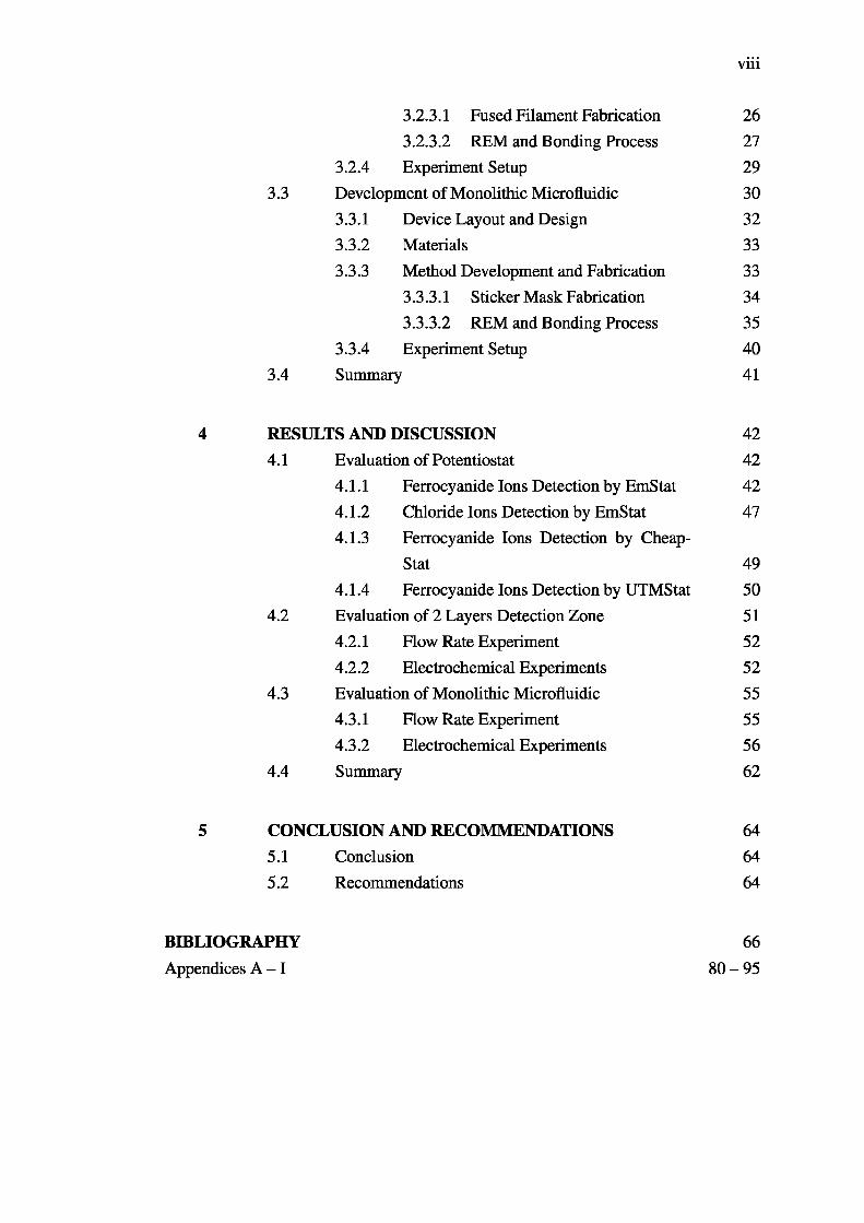
4 RESULTS AND DISCUSSION 424.1 Evaluation of Potentiostat 42
4.1.1 Ferrocyanide Ions Detection by EmStat 424.1.2 Chloride Ions Detection by EmStat 474.1.3 Ferrocyanide Ions Detection by Cheap
Stat 494.1.4 Ferrocyanide Ions Detection by UTMStat 50
4.2 Evaluation of 2 Layers Detection Zone 514.2.1 How Rate Experiment 524.2.2 Electrochemical Experiments 52
4.3 Evaluation of Monolithic Microfluidic 554.3.1 Flow Rate Experiment 554.3.2 Electrochemical Experiments 56
4.4 Summary 62
5 CONCLUSION AND RECOMMENDATIONS 645.1 Conclusion 645.2 Recommendations 64
BIBLIOGRAPHY 66Appendices A - 1 80-95
viii
ix
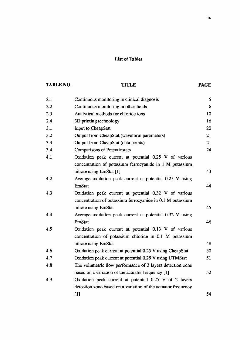
List of Tables
TABLE NO. TITLE PAGE
2.1 Continuous monitoring in clinical diagnosis 52.2 Continuous monitoring in other fields 62.3 Analytical methods for chloride ions 102.4 3D printing technology 163.1 Input to CheapStat 203.2 Output from CheapStat (waveform parameters) 213.3 Output from CheapStat (data points) 213.4 Comparisons of Potentiostats 244.1 Oxidation peak current at potential 0.25 V of various
concentration of potassium ferrocyanide in 1 M potassiumnitrate using EmStat [1] 43
4.2 Average oxidation peak current at potential 0.25 V usingEmStat 44
4.3 Oxidation peak current at potential 0.32 V of various concentration of potassium ferrocyanide in 0.1 M potassiumnitrate using EmStat 45
4.4 Average oxidation peak current at potential 0.32 V usingEmStat 46
4.5 Oxidation peak current at potential 0.13 V of various concentration of potassium chloride in 0.1 M potassiumnitrate using EmStat 48
4.6 Oxidation peak current at potential 0.25 V using CheapStat 504.7 Oxidation peak current at potential 0.25 V using UTMStat 514.8 The volumetric flow performance of 2 layers detection zone
based on a variation of the actuator frequency [1] 524.9 Oxidation peak current at potential 0.25 V of 2 layers
detection zone based on a variation of the actuator frequency[1] 54

4.10
4.11
4.12
4.13
4.14
4.15
Oxidation peak current at potential 0.25 V of 2 layers detection zone based on a variation concentrations range at the maximum flow rate [1] 55The volumetric flow performance of monolithic microfluidic based on a variation of the actuator frequency 55Oxidation peak current at potential 0.32 V of monolithic microfluidic based on a variation of the actuator frequency 57Oxidation peak current at potential 0.32 V of monolithic microfluidic based on a variation concentrations range at the maximum flow rate 58Average oxidation peak current at potential 0.32 V of monolithic microfluidic based on a variation concentrations range at the maximum flow rate 59Oxidation peak current at potential 0.13 V of monolithic microfluidic based on a variation concentrations range of chloride ions at the maximum flow rate 61

xi
List of Figures
FIGURE NO. TITLE PAGE
2.1 CV for ferricyanide/ferrocyanide couple detection [2] 132.2 The oxidative stripping of silver nanoparticles from a glassy
carbon electrode in 0.1 M sodium nitrate spiked with different amounts of potassium chloride (KC1) at a scan rate of 0.05 V/s. Green: no KC1; black: 2 mM KC1; pink: 8 mM KC1; orange: 16 mM KC1; blue: 32 mM KC1. [3] 14
3.1 EmStat by PalmSens 173.2 GUI of EmStat 183.3 The CheapStat, an inexpensive, “do-it-yourself’ potentiostat
[4] 183.4 GUI of CheapStat 193.5 Input from user 223.6 BMP file for cyclic voltammetry-based monitoring of
potassium ferrocyanide 233.7 0.5 mm thickness mould 253.8 3.0 mm thickness mould 253.9 Length and width of 0.5 mm and 3.0 mm thickness moulds 253.10 3.0 mm and 0.5 mm Poly Lactic Acid (PLA) moulds 273.11 Replica moulding (REM) of the PDMS structure: (a) PDMS
mixture in a desiccator; (b) vacuumed in desiccators after being poured into the moulds; (c) moulds covered by glasscovers [1] 27
3.12 0.5 mm and 3.0 mm thickness of PDMS 283.13 3D view of the 2 layers detection zone 293.14 Experimental setup for measurements of the flow rate and the
electrochemical performances, simultaneously [1] 293.15 SLA technology: (a) SLA based moulds; (b) SLA based
microfluidic 303.16 2 cm gap monolithic microfluidic 313.17 Design and dimension for microchannel, R: radius 32

3.183.193.203.213.223.233.243.253.263.27
4.1
4.2
4.3
4.4
4.5
4.6
4.7
4.8
4.9
4.10
X ll
Silhouette SD Digital Craft Cutter 33Vinyl sticker masks 34Silicone transfer film adhesive 34Templates for PDMS replica in the REM procedure 35Replica moulding (REM) of the PDMS structure 36Dimensions for: (a) upper; (b) middle; (c) bottom layers 373 PDMS layers: (a) upper; (b) middle; (c) bottom layers 38Bonded middle-bottom layer 38The upper layer with a thin PDMS-based membrane 39The monolithic microfluidic device: (a) actual device; (b) 3D view 40Background-subtracted cyclic voltammetry-based monitoring of potassium ferrocyanide in 1 M potassium nitrate using EmStat [1] 43Amperometry-based monitoring of potassium ferrocyanide in1 M potassium nitrate using EmStat 44Background-subtracted cyclic voltammetry-based monitoring of potassium ferrocyanide in 0.1 M potassium nitrate using EmStat 45Amperometry-based monitoring of potassium ferrocyanide in 0.1 M potassium nitrate using EmStat 46Cyclic voltammetry-based monitoring of potassium chloride in 0.1 M potassium nitrate using EmStat 47Cyclic voltammetry-based monitoring of potassium chloride in 0.1 M potassium nitrate using EmStat from +0.00 V to +0.20 V 48Background-subtracted cyclic voltammetry-based monitoring of potassium ferrocyanide in 1 M potassium nitrate using CheapStat 49Background-subtracted cyclic voltammetry-based monitoring of potassium ferrocyanide in 1 M potassium nitrate using UTMStat 51Background-subtracted cyclic voltammetry-based monitoring of 10 mM potassium ferrocyanide in 1 M potassium nitrate at various flow rates [1] 53Background-subtracted cyclic voltammetry-based monitoring of potassium ferrocyanide in 1 M potassium nitrate at the maximum frequency [1] 54

xiii
4.11 Background-subtracted cyclic voltammetry-based monitoringof 10 mM potassium ferrocyanide in 0.1 M potassium nitrate 57
4.12 Background-subtracted cyclic voltammetry-based monitoringof potassium ferrocyanide in 0.1 M potassium nitrate at the maximum frequency 58
4.13 Amperometry-based monitoring of potassium ferrocyanide in0.1 M potassium nitrate at the maximum frequency 59
4.14 Cyclic voltammetry-based monitoring of potassium chloridein a mixture at the maximum frequency by using EmStat 60
4.15 Cyclic voltammetry-based monitoring of potassium chloridein a mixture at the maximum frequency by using CheapStat 61
5.1 8 working electrodes and 1 common reference electrode 65A.l UTMStat block diagram 80E.l Read the waveform parameters 91F.l Get current values 92G.l Get potential values 93H. 1 Store potential-current pair into CSV and BMP files 94

LIST OF ABBREVIATIONS
LOC - Lab on a chip
PDMS - Poly(dimethylsiloxane)
SLA - Stereolithography
WE - Working electrode
CE - Counter electrode
RE - Reference electrode
CCS - Carbon capture and storage
PVC - Polyvinyl chloride
3D - 3 dimensional
DNA - Deoxyribonucleic acid
RNA - Ribonucleic acid
CMOS - Complementary metal-oxide semiconductor
BiCMOS - Bipolar and CMOS
NI - National instruments
SPE - Screen printed electrode
EFF - Fused filament fabrication
FDM - Fused deposition modeling
UV - Ultraviolet
SLS - Selective laser sintering
REM - Replica moulding
3DP - 3D printing
ESI-MS - Electrospray ionisation mass spectrometer
PLA - Polylactic acid
ABS - Acrylonitrile butadiene styrene
USB - Universal serial bus
GUI - Graphical user interface
SMD - Surface mount device

XV
PCB - Printed circuit board
PC - Personal computer
MSB - Most significant bit
LSB - Least significant bit
BMP - Bitmap
ISR - Interrupt service routine
CSV - Comma separated values
AC 1 .W2.RS - SPE with platinum WE and CE and silver RE
GCODE - G programming language
AC1.W3.R1 - SPE with silver WE, platinum CE and Ag/AgCl RE
DXF - Drawing interchange format
NHE - Normal hydrogen electrode
SBF - Simulated body fluid
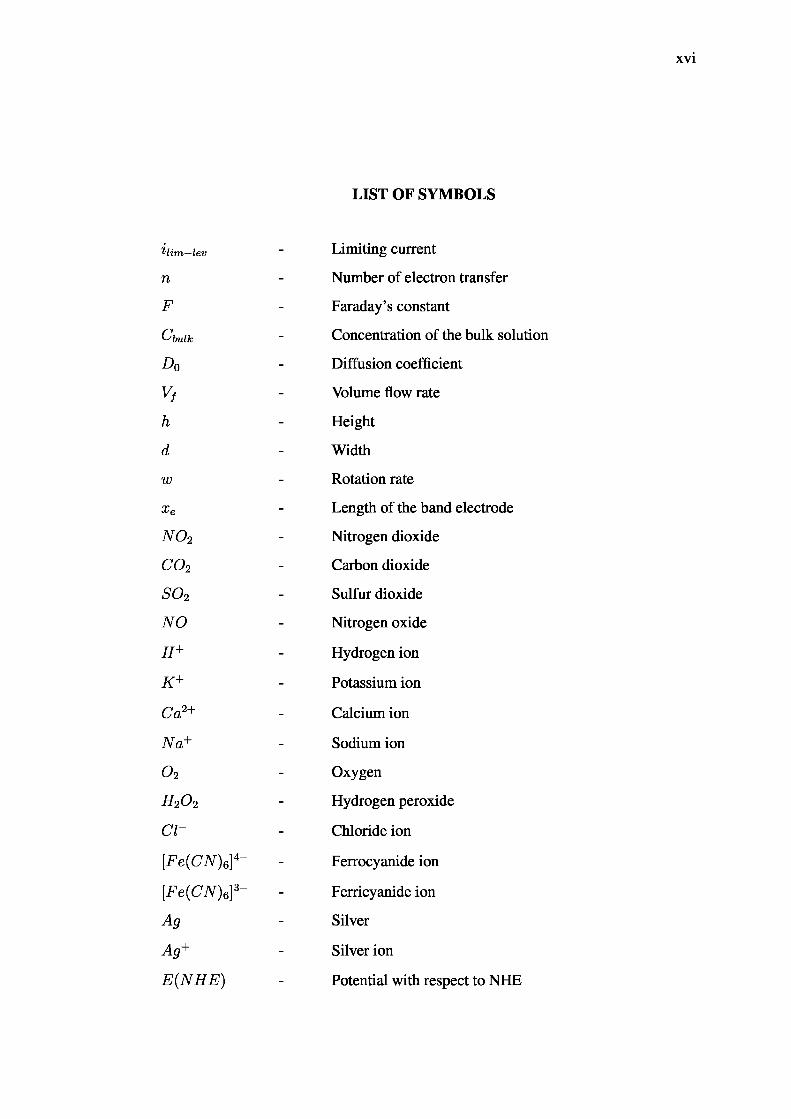
LIST OF SYMBOLS
Him—lev - Limiting current
n - Number of electron transfer
F - Faraday’s constant
Cbuik - Concentration of the bulk solution
D0 - Diffusion coefficient
Vf - Volume flow rate
h - Height
d - Width
w - Rotation rate
xe - Length of the band electrode
N 0 2 - Nitrogen dioxide
C 0 2 - Carbon dioxide
S 0 2 - Sulfur dioxide
NO - Nitrogen oxide
H + - Hydrogen ion
K + - Potassium ion
Ca2+ - Calcium ion
Na+ - Sodium ion
0 2 - Oxygen
H20 2 - Hydrogen peroxide
Cl~ - Chloride ion
[Fe(CN)6\4~ - Ferrocyanide ion
[Fe(CN)e]3~ - Fenicyanide ion
Ag - Silver
Ag+ - Silver ion
E (N H E ) - Potential with respect to NHE

E(Ag /AgCl) -
e~
Potential with respect to Ag/AgCl
Electron
Chapter 1
INTRODUCTION
1.1 Research Background
Monitoring chemicals and biomarkers is essential to predict patients’ critical conditions during surgical procedures and following these in intensive care units. Monitoring biomarkers provides information of any abnormalities occurring in metabolic pathways, and hence offer further understanding or can potentially be an early diagnosis for a number of illnesses. For example, hypoxia can be detected by a decrease in glucose and an increase in lactate, typically of a lack of oxygen during the biochemical pathways.
Biomarkers are typically detected in human fluids such as blood, serum, urine or cerebral spinal fluid and interstitial space. This brings a challenge when monitoring biomarkers through the conventional method by using an expensive and large piece of equipment. With this equipment, it is hard to provide health care for the rural population due to its low socio-economic income [5], transportation and distance challenges. In addition, current ions analyzer is normally located in the central laboratories which requires specialized technicians to operate on the machine, offers delayed diagnosis and not disposable. Besides, during the process, mix-ups sometimes occur when samples are sent to laboratories.
Therefore, there are numerous efforts in developing an affordable miniaturized bedside patient continuous monitoring system. One of the approaches is through lab on chip (LOC) device due to its unique ability in micro-scale sample handling, mixing, separation, detection, user friendly and inexpensive [6-9]
Many reported continuous monitoring system, nevertheless, utilizes external regulated pressure source or by syringe pumps or even through manual pipetting. This

2
approach has constrained the purpose of portability of LOC. Consequently, to mitigate such drawbacks, the integration of the micropump and the reaction zone is crucial.
Potentiometric system has always been related to electrochemical detection method [10] in LOC. The combination of electrochemical cell and a potentiostat circuit forms a potentiostatic system. Potentiostat is a feedback control system [11] which adjusts the voltage across the WE (working electrode) - CE (counter electrode) pair to maintain the preset potential between the WE and RE (reference electrode) of an electrochemical cell [12],
Besides clinical diagnosis [13-22], continuous monitoring system has also been applied in water quality control [23-27], pharmaceutical product [28-32], corrosion quality control [33-37], environmental emission [38-42] and other fields [43—47].
1.2 Problem Statement
In view of the importance of the bedside patient monitoring system, a miniaturized continuous monitoring system with flexibility, versatility and disposable is much needed. An external pressure source is utilized in most of the applications of the current continuous monitoring system to deliver the sample to the detection zone [2,13,14]. But, there are few drawbacks in this approach which has restricted the system’s mobility due to the additional manual procedures or exterior components for measurement setup are needed. Moreover, the losses because of the friction and shear stress of the tubing wall might be introduced by the connection between the pressure source and the electrochemical detection zone [1].
In addition, as stated in [4], current method is expensive where commercially available laboratory potentiostats sold for more than a thousand dollars. Besides, current medical practice is still relying on the expensive and large piece of equipment which generally located in the central laboratories that require specialized technicians, offers delayed diagnosis and not disposable. In a lab on chip design, disposability is a main aspect that should be highlighted to confirm that the sample is unpolluted as the continuous patient monitoring system is meant for biomedical analysis. To eliminate the sterilizing procedure and to confirm the hygiene condition, the device needs to be disposed.
3
1.3 Research Objectives and Scope of the Thesis
The primary objective of this project is to develop an in channel electrochemical detection for continuous flow lab on chip application. The specific goals can be farther expressed as:
1. To develop electrochemical detection system for continuous monitoring of ions.
2. To evaluate the performance of the developed device in a continuous monitoring system.
To accomplish these objectives, two different models are proposed and their electrochemistry and pumping characteristics are studied. The first model is to demonstrate the effect of pinching right above the electrochemical detection zone. The electrochemical detection zone is developed in two different thickness of poly (dimethylsiloxane) (PDMS) polymer layers with fused filament technology and replication moulding technique with a screen printed electrode attached together.
Structurally, the inlet and outlet of the micropump were modified based on the electrode surface where the screen printed electrodes were place beneath. To ease the fabrication technique and to have smoother mould surface, the electrochemical detection zone approaches sticker mask fabrication and as mentioned above replication moulding technique with two screens printed electrodes attached together. This new design of the electrochemical detection zone is introduced in the second model.
1.4 Thesis Outline
A review of the continuous monitoring system development is given in Chapter2. Then in Chapter 3, the methods in developing of potentiostat, 2 layers detection zone and monolithic microfluidic are described. In Chapter 4, the examinations of potentiostat, continuous monitoring systems for 2 layers detection zone and monolithic microfluidic are discussed. Finally, an outlook on future project development is concluded in Chapter 5.

Bibliography
1. S. Noi, P. S. Chee, F. K. Che Harun, P. L. Leow, and A. Aziz, “Integration of Electrochemical Detection into Micropumps for Continuous Monitoring System,” in Proceedings of the 10th Asian Control Conference 2015 (ASCC
2015), pp. 1658-1662, 2015.
2. D. Grieshaber, J. Voros, T. Zambelli, V. Ball, P. Schaaf, J.-C. Voegel, and F. Boulmedais, “Swelling and Contraction of Ferrocyanide-Containing Polyelectrolyte Multilayers upon Application of an Electric Potential,” Langmuir : the ACS journal of surfaces and colloids, vol. 24, pp. 13668- 13676, Dec. 2008.
3. H. S. Toh, C. Batchelor-mcauley, K. Tschulik, and R. G. Compton, “Electrochemical detection of chloride levels in sweat using silver nanoparticles: a basis for the preliminary screening for cystic fibrosis,” The
Analyst, vol. 138, no. 15, pp. 4292-7, 2013.
4. A. A. Rowe, A. J. Bonham, R. J. White, M. P. Zimmer, R. J. Yadgar,T. M. Hobza, J. W. Honea, I. Ben-Yaacov, and K. W. Plaxco, “CheapStat: an open-source, "do-it-yourself potentiostat for analytical and educational applications,” PloS one, vol. 6, p. e23783, Jan. 2011.
5. H. Yadav, “A review of maternal mortality in Malaysia,” International e-
Joumal of Science, Medicine & Education (IeJSME), vol. 6, pp. 142-151, 2012.
6. B. H. Weigl, R. L. Bardell, and C. R. Cabrera, “Lab-on-a-chip for drugdevelopment,” Advanced drug delivery reviews, vol. 55, no. 3, pp. 349-377, 2003.
7. A. J. Tudos, G. J. Besselink, and R. B. Schasfoort, “Trends in miniaturizedtotal analysis systems for point-of-care testing in clinical chemistry,” Lab on
a chip, vol. 1, no. 2, pp. 83-95, 2001.
8. J. Gardeniers and A. Van den Berg, “Lab-on-a-chip systems for biomedical and environmental monitoring,” Analytical and bioanalytical chemistry,
vol. 378, no. 7, pp. 1700-1703, 2004.

67
9. A. Oedit, P. Vulto, R. Ramautar, P. W. Lindenburg, and T. Hankemeier, “Lab-on-a-Chip hyphenation with mass spectrometry: strategies for bioanalytical applications,” Current opinion in biotechnology, vol. 31, pp. 79-85, Feb. 2015.
10. G. S. Wilson and M. a. Johnson, “In-vivo electrochemistry: What can we learn about living systems?” Chemical Reviews, vol. 108, pp. 2462-2481, July 2008.
11. P. M. Levine, P. Gong, R. Levicky, and K. L. Shepard, “Active CMOS sensor array for electrochemical biomolecular detection,” in IEEE Journal of Solid-
State Circuits, vol. 43, pp. 1859-1871, 2008.
12. A. J. Bard, L. R. Faulkner, J. Leddy, and C. G. Zoski, Electrochemical
methods: fundamentals and applications, vol. 2. Wiley New York, 1980.
13. L. Tmkova, V. Adam, J. Hubalek, P. Babula, and R. Kizek, “Amperometric Sensor for Detection of Chloride Ions,” Sensors, vol. 8, pp. 5619-5636, Sept. 2008.
14. R. Dutt-Ballerstadt, C. Evans, A. P. Pillai, and A. Gowda, “A label-free fiberoptic Turbidity Affinity Sensor (TAS) for continuous glucose monitoring,” Biosensors & bioelectronics, vol. 61, pp. 280-284, Nov. 2014.
15. I. Rodrfguez-Ruiz, E. Masvidal-Codina, T. N. Ackermann, and A. Llobera, “Photonic lab-on-chip (PhLOC) for enzyme-catalyzed reactions in continuous flow,” Microfluidics and Nanofluidics, vol. 18, no. 5-6, pp. 1277-1286, 2015.
16. M. Hassan and M. Mashor, “A portable continuous blood pressuremonitoring kit,” Business, Engineering and Industrial Applications (ISBEIA),
2011 IEEE Symposium on, pp. 503-507, 2011.
17. X. Munoz-Berbel, R. Escude-Pujol, N. Vigues, M. Cortina-Puig, C. Garcfa-Aljaro, J. Mas, and F. X. Munoz, “Real time automatic system for the impedimetric monitoring of bacterial growth,” Analytical Letters, vol. 44, no. 16, pp. 2571-2581,2011.
18. R. Ferrigno and P. Pittet, “Combining microfluidics and electrochemicaldetection,” Engineering in Medicine and Biology Society, 2009. EMBC 2009.
Annual International Conference of the IEEE, pp. 4144—4146, 2009.
19. A. Nilghaz, D. H. Wicaksono, D. Gustiono, F. A. A. Majid, E. Supriyanto,and M. R. A. Kadir, “Flexible microfluidic cloth-based analytical devices using a low-cost wax patterning technique,” Lab on a Chip, vol. 12, no. 1, pp. 209-218, 2012.
68
20. I. B. Tahirbegi, M. Mir, S. Schostek, M. Schurr, and J. Samitier, “in vivo ischemia monitoring array for endoscopic surgery,” Biosensors &
bioelectronics, vol. 61, pp. 124-130, Nov. 2014.
21. G. Vretzakis, B. Papaziogas, E. Matsaridou, G. Vasiliadou, G. Papadopoulos, C. Patsialas, and F. Kostopoulou, “Continuous monitoring of arterial blood gases and pH during intraoperative rapid blood administration using a Paratrend sensor,” Vox sanguinis, 2000.
22. W. Jin; L. Wu; Y. Song; J. Jiang; X. Zhu et al., “Continuous intra-arterialblood pH monitoring by a fiber-optic fluorosensor,” in IEEE transactions on
bio-medical engineering, vol. 58, pp. 1232-1238, 2011.
23. M. R. Awual and M. M. Hasan, “A novel fine-tuning mesoporous adsorbentfor simultaneous lead(II) detection and removal from wastewater,” Sensors
and Actuators B: Chemical, vol. 202, pp. 395-403, Oct. 2014.
24. A. Calvo-Lopez, E. Arasa-Puig, M. Puyol, J. M. Casalta, and J. Alonso-Chamarro, “Biparametric potentiometric analytical microsystem for nitrate and potassium monitoring in water recycling processes for manned space missions,” Analytica chimica acta, vol. 804, pp. 190-196, Dec. 2013.
25. C. Guijarro, K. Fuchs, U. Bohm, E. Stiitz, and S. Wolfl, “Simultaneousdetection of multiple bioactive pollutants using a multiparametric biochip for water quality monitoring,” Biosensors & bioelectronics, vol. 72, pp. 71-79, Apr. 2015.
26. K. S. Tew, M.-Y. Leu, J.-T. Wang, C.-M. Chang, C.-C. Chen, and P.-J. Meng, “A continuous, real-time water quality monitoring system for the coreil reef ecosystems of Nanwan Bay, Southern Taiwan,” Marine pollution bulletin,
vol. 85, pp. 641-7, Aug. 2014.
27. K. Murphy, T. Sullivan, B. Heery, and F. Regan, “Data analysis from a low-cost optical sensor for continuous marine monitoring,” Sensors and Actuators
B: Chemical, vol. 214, pp. 211-217, July 2015.
28. M. K. Abd El-Rahman and M. Y. Salem, “Ion selective electrode (in-lineanalyzer) versus UV-spectroscopy (at-line analyzer); which strategy offers more opportunities for real time monitoring of the degradation kinetics of pyridostigmine bromide,” Sensors and Actuators B: Chemical, vol. 220, pp. 255-262, Dec. 2015.
29. M. K. Abd El-Rahman, H. E. Zaazaa, N. Badr ElDin, and A. A.Moustafa, “Novel strategy for online monitoring of the degradation kinetics of propantheline bromide via a calixarene-based ion-selective electrode,”
69
Talanta, vol. 132, pp. 52-58, Jan. 2015.
30. D. Markl, G. Hannesschlager, S. Sacher, M. Leitner, and J. G. Khinast, “Optical coherence tomography as a novel tool for in-line monitoring of a pharmaceutical film-coating process,” European journal of pharmaceutical
sciences : official journal of the European Federation for Pharmaceutical
Sciences, vol. 55, pp. 58-67, May 2014.
31. M. C. Sarraguga, P. R. S. Ribeiro, A. O. Santos, M. C. D. Silva, and J. a. A. Lopes, “A PAT approach for the on-line monitoring of pharmaceutical cocrystals formation with near infrared spectroscopy,” International Journal of
Pharmaceutics, vol. 471, pp. 478—484, Aug. 2014.
32. R. Laitinen, J. Lahtinen, P. Silfsten, E. Vartiainen, P. Jarho, and J. Ketolainen, “An optical method for continuous monitoring of the dissolution rate of pharmaceutical powders.” Journal of pharmaceutical and biomedical
analysis, vol. 52, pp. 181-9, June 2010.
33. W.-J. Park, H.-S. Lee, S.-H. Joh, and H.-S. Lee, “Monitoring method for the chloride ion penetration in mortar by a thin-film sensor reacting to chloride ion,” Construction and Building Materials, vol. 53, pp. 403-410, Feb. 2014.
34. P. Romano, P. S. Brito, and L. Rodrigues, “Monitoring of the degradation of concrete structures in environments containing chloride ions,” Construction
and Building Materials, vol. 47, pp. 827-832, Oct. 2013.
35. L. Han and S. Song, “A measurement system based on electrochemical frequency modulation technique for monitoring the early corrosion of mild steel in seawater,” Corrosion Science, vol. 50, pp. 1551-1557, 2008.
36. J. M. Gandia-Romero, R. Bataller, P. Monzon, I. Campos, E. G. Breijo, M. Valcuende, and J. Soto, “Characterization of embeddable potentiometric thick-film sensors for monitoring chloride penetration in concrete,” Sensors
and Actuators B: Chemical, July 2015.
37. A. Brenna, F. Bolzoni, S. Beretta, and M. Ormellese, “Long-term chloride-induced corrosion monitoring of reinforced concrete coated with commercial polymer-modified mortar and polymeric coatings,” Construction
and Building Materials, vol. 48, pp. 734—744, Nov. 2013.
38. A. Fort, C. Lotti, M. Mugnaini, R. Palombari, S. Rocchi, and V. Vignoli, “A two electrode C-NiO Nafion amperometric sensor for N02 detection,” Microelectronics Journal, vol. 40, no. 9, pp. 1308-1312, 2009.
39. M. S. Bufaroosha, M. A. Alnaqbi, M. H. Al-Marzouqi, and S. A. Marzouk, “Portable dual-channel gas analyzer for continuous monitoring of carbon

70
dioxide in gas streams,” Microchemical Journal, vol. 110, pp. 185-191, Sept. 2013.
40. K. Shitashima, Y. Maeda, and A. Sakamoto, “Detection and monitoring ofleaked C02 through sediment, water column and atmosphere in a sub-seabed CCS experiment,” International Journal of Greenhouse Gas Control, vol. 38, pp. 135-142, July 2015.
41. M. Kaykhaii and I. Ciucanu, “Membrane in tandem with a helical sorbent trap as continuous sampling technique of the polyvinyl chloride thermo-oxidative degradation products for their on-line gas chromatographic monitoring,” Analytica ChimicaActa, vol. 491, pp. 163-171, Sept. 2003.
42. C. Kern, J. Sutton, T. Elias, L. Lee, K. Kamibayashi, L. Antolik, andC. Werner, “An automated S02 camera system for continuous, real-time monitoring of gas emissions from Kilauea Volcano’s summit Overlook Crater,” Journal of Volcanology and Geothermal Research, vol. 300, pp. 81-94, July 2015.
43. R. Sekar and R Prasad, “Monitoring chloride and nitrite ions in 2, 4- dichlorofluorobenzene plant effluent by CE,” Chromatographia, vol. 69, pp. 1097-1100, Feb. 2009.
44. W.-P. Cheng, Y.-J. Hsieh, R.-F. Yu, Y.-W. Huang, S.-Y. Wu, and S.-M. Chen,“Characterizing polyaluminum chloride (PAC1) coagulation floe using an online continuous turbidity monitoring system,” Journal of the Taiwan Institute
of Chemical Engineers, vol. 41, pp. 547-552, Sept. 2010.
45. E. Ivorra, S. V. Amat, A. J. Sanchez, J. M. Barat, and R. Grau, “Continuousmonitoring of bread dough fermentation using a 3D vision Structured Light technique,” Journal of Food Engineering, vol. 130, pp. 8-13, June 2014.
46. J. Silva, M. Azenha, A. Gomes Correia, and J. Granja, “Continuousmonitoring of sand-cement stiffness starting from layer compaction with a resonant frequency-based method: Issues on mould geometry and sampling,” Soils and Foundations, vol. 54, pp. 56-66, Feb. 2014.
47. A. Saisi, C. Gentile, and M. Guidobaldi, “Post-earthquake continuousdynamic monitoring of the Gabbia Tower in Mantua, Italy,” Construction
and Building Materials, vol. 81, pp. 101-112, Apr. 2015.
48. C. D. Saudek, “Continuous Blood Glucose Monitoring: A Review andPreview,” Proceedings of the Annual Symposium on Computer Application
in Medical Care, 1983.
49. N. Roveri, A. Carcaterra, and A. Sestieri, “Real-time monitoring of railway

71
infrastructures using fibre Bragg grating sensors,” Mechanical Systems and
Signal Processing, vol. 60-61, pp. 14-28, Aug. 2015.
50. E. Braunwald, “Biomarkers in heart failure,” New England Journal of
Medicine, vol. 358, no. 20, pp. 2148-2159, 2008.
51. B. D. Malhotra and A. Chaubey, “Biosensors for clinical diagnostics industry,” Sensors and Actuators B: Chemical, vol. 91, no. 1, pp. 117-127,2003.
52. D. Eiferman, R. A. Perez-Tamayo, K. Abe, E. Okum, and R. Higgins, “Real-time monitoring of cardiac metabolism using biosensors shows myocardial protection during ischemia-reperfusion injury with glucose- insulin-potassium administration,” Surgery, vol. 142, no. 2, pp. 150-155,2007.
53. D. S. Eiferman, L. Nguyen, and R. A. Perez-Tamayo, “Real-time myocardial glucose measurement using biosensors,” ASAIO Journal, vol. 54, no. 1, pp. 120-123, 2008.
54. J. H. Chua, R.-E. Chee, A. Agarwal, S. M. Wong, and G.-J. Zhang, “Label- free electrical detection of cardiac biomarker with complementary metal- oxide semiconductor-compatible silicon nanowire sensor arrays,” Analytical
chemistry, vol. 81, no. 15, pp. 6266-6271, 2009.
55. T. Kokubo and H. Takadama, “How useful is SBF in predicting in vivo bone bioactivity?,” Biomaterials, vol. 27, pp. 2907-15, May 2006.
56. M. Montemor, J. Alves, A. Simoes, J. Fernandes, Z. Lourengo, A. Costa,A. Appleton, and M. Ferreira, “Multiprobe chloride sensor for in situ monitoring of reinforced concrete structures,” Cement and Concrete
Composites, vol. 28, no. 3, pp. 233-236, 2006.
57. K. G. Kumar, K. S. John, et al., “A chloride ion-selective potentiometricsensor based on a polymeric schiff base complex,” Indian journal of chemical
technology, vol. 13, no. 1, pp. 13-16, 2006.
58. N. Kocherginsky and Z. Wang, “Polyaniline membrane based potentiometricsensor for ascorbic acid, other redox active species and chloride,” Journal of
Electroanalytical Chemistry, vol. 611, no. 1, pp. 162-168, 2007.
59. B. Schazmann, N. Alhashimy, and D. Diamond, “Chloride selective calix[4] arene optical sensor combining urea functionality with pyrene excimer transduction,” Journal of the American Chemical Society, vol. 128, no. 26, pp. 8607-8614, 2006.

72
60. R. Sundaram, K. Hariprasad, et al., “Synthesis of chloride ion-selective potentiometric sensor based on coordination polymer complex,” Indian
Journal of Chemical Technology, vol. 14, no. 5, pp. 451-458, 2007.
61. C. Xu, Y. Qin, and E. Bakker, “Optical chloride sensor based on [9]mercuracarborand-3 with massively expanded measuring range,” Talanta,
vol. 63, no. 1, pp. 180-184, 2004.
62. W. Zhang, E. Rozniecka, E. Malinowska, R Parzuchowski, and M. E.Meyerhoff, “Optical chloride sensor based on dimer-monomer equilibriumof indium (iii) octaethylporphyrin in polymeric film,” Analytical chemistry,
vol. 74, no. 17, pp. 4548^557, 2002.
63. J. N. Babu, V. Bhalla, M. Kumar, R. Mahajan, and R. K. Puri, “A chlorideselective sensor based on a calix [4] arene possessing a urea moiety,” Tetrahedron Letters, vol. 49, no. 17, pp. 2772-2775, 2008.
64. A. M. Pimenta, A. N. Araujo, M. C. B. Montenegro, C. Pasquini, J. J. Rohwedder, and I. M. Raimundo, “Chloride-selective membrane electrodes and optodes based on an indium (iii) porphyrin for the determination of chloride in a sequential injection analysis system,” Journal of pharmaceutical
and biomedical analysis, vol. 36, no. 1, pp. 49-55, 2004.
65. J. Junsomboon and J. Jakmunee, “Determination of chloride in admixtures and aggregates for cement by a simple flow injection potentiometric system,” Talanta, vol. 76, no. 2, pp. 365-368, 2008.
66. M. Philippi, H. S. dos Santos, A. O. Martins, C. M. Azevedo, andM. Pires, “Alternative spectrophotometric method for standardization of chlorite aqueous solutions,” Analytica chimica acta, vol. 585, no. 2, pp. 361- 365, 2007.
67. H. Cao and D. H. Wu, “Rapid and sensitive determination of trace chloride ion in drinks using resonance light scattering technique,” Journal
of Analytical Methods in Chemistry, vol. 2008, 2008.
68. R. Wu and X. Shao, “[application of near-infrared spectra in thedetermination of water soluble chloride ion in plant samples].,” Guang pu
xue yu guang pufen xi= Guang pu, vol. 26, no. 4, pp. 617-619, 2006.
69. R. B. Mesquita, S. M. Fernandes, and A. O. Rangel, “Turbidimetricdetermination of chloride in different types of water using a single sequential injection analysis system,” journal of Environmental monitoring, vol. 4, no. 3, pp. 458-461, 2002.
70. T. Deng, M. Li, C. Zhao, and J. Qin, “Characteristic investigation of a

73
static micro polymerase chain reaction chip based on in situ electrochemical detection,” Micro & Nano Letters, IET, vol. 7, no. 12, pp. 1226-1229, 2012.
71. M. Paeschke, F. Dietrich, A. Uhlig, and R. Hintsche, “Voltammetric multichannel measurements using silicon fabricated microelectrode arrays,” Electroanalysis, vol. 8, no. 10, pp. 891-898, 1996.
72. I. Ramfos, N. Vassiliadis, S. Blionas, K. Efstathiou, A. Fragoso, C. K. O’Sullivan, and A. Birbas, “A compact hybrid-multiplexed potentiostat for real-time electrochemical biosensing applications,” Biosensors and
Bioelectronics, vol. 47, pp. 482-489, 2013.
73. S. P. Kounaves, “Voltammetric techniques,” 1997.
74. J. Huntley, and V. Maikov, “Amperometric Probes or DPD Analyzers: Which Is Best For On-Line Chlorine Monitoring?” Internet:http://www.hach.com/asset-get.download-en.jsa?id=7639984804, 2009. Retrieved November 05, 2014, from http://www.hach.com/asset-get.download- en.jsa?id=7639984804.
75. J. C. Fidler, W. R. Penrose, and J. P. Bobis, “A potentiostat based on a voltage-controlled current source for use with amperometric gas sensors,” IEEE Transactions on Instrumentation and Measurement, vol. 41, no. 2, pp. 308-310,1992.
76. M. Vergani, M. Carminati, G. Ferrari, E. Landini, C. Caviglia, A. Heiskanen, C. Comminges, K. Zor, D. Sabourin, M. Dufva, M. Dimaki, R. Raiteri, U. Wollenberger, J. Emneus, and M. Sampietro, “Multichannel Bipotentiostat Integrated with a Microfluidic Platform for Electrochemical Real- Time Monitoring of Cell Cultures,” IEEE transactions on biomedical circuits and
systems (Accepted for publication), vol. 6, no. 5, pp. 498-507, 2012.
77. C.-Y. H. C.-Y. Huang, Y.-C. W. Y.-C. Wang, H.-C. C. H.-C. Chen, and K.-C. H. K.-C. Ho, “Design of a portable potentiostat for electrochemical sensors,” Proceedings of the 2004 Intelligent Sensors, Sensor Networks and
Information Processing Conference, 2004., pp. 331-336, 2004.
78. A. B. Islam, M. R. Haider, A. Atla, S. K. Islam, R. Croce, S. Vaddiraju, F. Papadimitrakopoulos, and F. Jain, “A potentiostat circuit for multiple implantable electrochemical sensors,” in ICECE 2010 - 6th International
Conference on Electrical and Computer Engineering, no. December, pp. 314-317, 2010.
79. H. L. H. Li, X. L. X. Luo, C. L. C. Liu, L. J. L. Jiang, D. C. D. Cui, X. C. X. Cai, and Q. Y. Q. Yang, “Multi-channel electrochemical detection system

74
based on Lab VIEW,” International Conference on Information Acquisition,
2004. Proceedings., pp. 224—227, 2004.
80. M. A. Tapsak, J. G. Houseknecht, and P. V. Goode, “A low cost- computer-controlled and powered multichannel potentiostat for general use in development of inexpensive electrochemical sensors,” Instrumentation
Science and Technology, vol. 35, no. 6, pp. 589-598, 2007.
81. F. J. Sun and J. Wang, “Development and application of virtual potentiostat on electrochemical corrosion measurement,” Wuli Huaxue Xuebao/ Acta
Physico - Chimica Sinica, vol. 28, no. 3, pp. 615-622, 2012.
82. M. Duwe and T. Chen, “Low power integrated potentiostat design for/i electrodes with improved accuracy,” in 2011 IEEE 54th International
Midwest Symposium on Circuits and Systems (MWSCAS), pp. 1-4, IEEE, 2011.
83. S. Hwang and S. Sonkusale, “CMOS VLSI potentiostat for portableenvironmental sensing applications,” IEEE Sensors Journal, vol. 10, no. 4, pp. 820-821, 2010.
84. S. S. Ghoreishizadeh, I. Taurino, S. Carrara, and G. De Micheli, “A currentmode potentiostat for multi-target detection tested with different lactate biosensors,” in 2012 IEEE Biomedical Circuits and Systems Conference:
Intelligent Biomedical Electronics and Systems for Better Life and Better
Environment, BioCAS 2012 - Conference Publications, pp. 128-131, 2012.
85. M. H. Nazari and R. Genov, “A fully differential CMOS potentiostat,” inProceedings - IEEE International Symposium on Circuits and Systems, vol. 9,
pp. 2177-2180, 2009.
86. S. Martin, F. Gebara, T. Strong, and R. Brown, “A low-voltage, chemical sensor interface for systems-on-chip: the fully-differential potentiostat,” 2004 IEEE International Symposium on Circuits and Systems (IEEE Cat.
No.04CH37512), vol. 4, pp. IV - 892-5, 2004.
87. A. Gore, S. Chakrabartty, S. Pal, and E. C. Alocilja, “A multichannel femtoampere-sensitivity potentiostat array for biosensing applications,” IEEE Transactions on Circuits and Systems I: Regular Papers, vol. 53, no. 11, pp. 2357-2363, 2006.
88. S. Ayers, K. D. Gillis, M. Lindau, and B. A. Minch, “Design of a CMOS potentiostat circuit for electrochemical detector arrays,” IEEE Transactions
on Circuits and Systems I: Regular Papers, vol. 54, no. 4, pp. 736-744, 2007.
89. P. A. Boutet and S. Manen, “Low power CMOS potentiostat for three
75
electrodes amperometric chemical sensor,” in 2011 Faible Tension Faible
Consommation, FTFC 2011, pp. 15-18, 2011.
90. R. Genov, M. Stanacevic, M. Naware, G. Cauwenberghs, and N. V. Thakor, “16-Channel integrated potentiostat for distributed neurochemical sensing,” IEEE Transactions on Circuits and Systems I: Regular Papers, vol. 53, no. 11, pp. 2371-2376, 2006.
91. Z. Shihong, G. Yong, W. Lifeng, R Wei, W. Guohui, and D. Yinyu, “Design and realization of dc-dc converter life prediction system based on labview.,” Journal of Convergence Information Technology, vol. 6, no. 11, 2011.
92. M. Samer, C. Loebsin, K. von Bobrutzki, M. Fiedler, C. Ammon, W. Berg, R Sanftleben, and R. Brunsch, “A computer program for monitoring and controlling ultrasonic anemometers for aerodynamic measurements in animal buildings,” Computers and Electronics in Agriculture, vol. 79, pp. 1-12, Oct. 2011.
93. X. Zhang, D. Zhang, R Lu, C. Bai, and R Xiao, “Monitoring the nitrification and identifying the endpoint of ammonium oxidation by using a novel system of titrim etry Water Science and Technology, vol. 64, no. 11, pp. 2246-2252, 2011.
94. L. Yi, Z. Feng, L. Gang, and W. Kai, “The real-time monitor system based on Lab VIEW,” Computer Science and Network Technology (ICCSNT), 2011
International Conference, vol. 2, pp. 848-851, 2011.
95. A. Chouder, S. Silvestre, B. Taghezouit, and E. Karatepe, “Monitoring, modelling and simulation of PV systems using Lab VIEW,” Solar Energy,
vol. 91, pp. 337-349, May 2013.
96. A. Yazidi, H. Henao, G.-A. Capolino, F. Betin, and F. Filippetti, “A Web-Based Remote Laboratory for Monitoring and Diagnosis of AC Electrical Machines,” IEEE Transactions on Industrial Electronics, vol. 58, no. 10, pp. 4950-4959, 2011.
97. L. V. Ausdeln, “A personal computer-based monitoring and control systemfor electron accelerators,” Particle Accelerator Conference, 2001. PAC 2001.
Proceedings of the 2001, vol. 2, pp. 828-830, 2001.
98. N. Sahgal, “Monitoring and analysis of lung sounds remotely,” International
Journal ofCOPD, vol. 6, no. 1, pp. 407-412, 2011.
99. A. Odon and Z. Krawiecki, “LabVIEW application for computer simulationof the conversion technique of dual-slope analog-to-digital converter,” Measurement: Journal of the International Measurement Confederation,

76
vol. 44, no. 8, pp. 1406-1411, 2011.
100. A. Bozatzidis, A. G. Anastopoulos, and T. Laopoulos, “Automated data acquisition setup for interfacial tension and capacitance measurements at Hg- solution contacts under Lab VIEW control,” Electroanalysis, vol. 19, no. 16, pp. 1711-1718,2007.
101. W. Lijun, X. Long, and Z. Jianbin, “Multi-channel peripheral data acquisition system based on labview and scm,” Journal of Convergence Information
Technology, vol. 7, no. 22, 2012.
102. I. A. Rojas-Olmedo, R. Lopez-Callejas, A. de la Piedad-Beneitez, R. Valencia-Alvarado, R. Pena Eguiluz, A. Mercado-Cabrera, S. R. Barocio, A. E. Munoz Castro, and B. G. Rodnguez-Mendez, “An automated system for DC and RF plasma characterization by guard double electric probes,” Surface and Coatings Technology, vol. 205, no. SUPPL. 2, pp. S397-S401, 2011.
103. R. S. Nicholson and I. Shain, “Theory of stationary electrode polarography. single scan and cyclic methods applied to reversible, irreversible, and kinetic systems.,” Analytical Chemistry, vol. 36, no. 4, pp. 706-723, 1964.
104. S. W. Feldberg, “Digital simulation: a general method for solvingelectrochemical diffusion-kinetic problems,” Electroanalytical chemistry,
vol. 3, pp. 199-296, 1969.
105. D. K. Gosser, Cyclic voltammetry: simulation and analysis of reaction
mechanisms, vol. 43. VCH New York, 1993.
106. W. Heineman and J. Beebe, Chemistry experiments for instrumental methods.
John Wiley & Sons: New York, 1984.
107. A. Howard, “Chemical instrumentation: A systematic approach,” A Wiley
Interscience Publication, vol. 1070, 1989.
108. R. N. Adams, “Electrochemistry at solid electrodes,” 1969.
109. E. Ghafar-Zadeh and M. Sawan, “A hybrid microfluidic/CMOS capacitive sensor dedicated to lab-on-chip applications,” IEEE Transactions on
Biomedical Circuits and Systems, vol. 1, no. 4, pp. 270-277, 2007.
110. R. Pai, J. Roussel, T.J., M. Crain, D. Jackson, J. Conklin, R. Baldwin, R. Keynton, J. Naber, and K. Walsh, “Integrated electrochemical detection for lab on a chip analytical microsystems,” Proceedings of the Fourteenth
Biennial University/Government/Industry Microelectronics Symposium (Cat.
No.01CH37197), pp. 167-170, 2001.

77
111. M. M. A. Mohamed, A. A. Youssef, Y. H. Ghallab, and W. Badawy, “On the design of digital control for lab-on-chip systems,” in Proceedings - IEEE
International Symposium on Circuits and Systems, pp. 1016-1019, 2009.
112. K. Chakrabarty, “Towards fault-tolerant digital microfluidic lab-on-chip: Defects, fault modeling, testing, and reconfiguration,” in 2008IEEE-BIOCAS
Biomedical Circuits and Systems Conference, BIOCAS 2008, pp. 329-332,2008.
113. J. M. Moreno, C. Aracil, and J. M. Quero, “Low cost fluid microextractor for lab-on-chip,” in Proceedings of the 2009 Spanish Conference on Electron
Devices, CDE’09, pp. 274-277, 2009.
114. A. T. Giannitsis and M. Min, “Usage of microfluidic lab-on-chips in biomedicine,” in BEC 2010 - 2010 12th Biennial Baltic Electronics
Conference, Proceedings of the 12th Biennial Baltic Electronics Conference,
pp. 249-252, 2010.
115. R. Bogue, “3D printing: the dawn of a new era in manufacturing?” Assembly
Automation, vol. 33, no. 4, pp. 307-311, 2013.
116. A. Kantaros and D. Karalekas, “Fiber Bragg grating based investigation of residual strains in ABS parts fabricated by fused deposition modeling process,” Materials & Design, vol. 50, pp. 44—50, Sept. 2013.
117. S. H. Jang, S. T. Oh, I. H. Lee, H.-C. Kim, and H. Y. Cho, “3- Dimensional Circuit Device Fabrication Process Using Stereolithography and Direct Writing,” International Journal of Precision Engineering and
Manufacturing, vol. 16, no. 7, pp. 1361-1367, 2015.
118. Y. Du, H. Liu, J. Shuang, J. Wang, J. Ma, and S. Zhang, “Microsphere-based selective laser sintering for building macroporous bone scaffolds with controlled microstructure and excellent biocompatibility,” Colloids and
Surfaces B: Biointerfaces, vol. 135, pp. 81-89, Nov. 2015.
119. 3DPrinting.com, “What is 3D printing? How does 3D printing work?” Internet: http://3dprinting.com/what-is-3d-printing, 2014. Retrieved August02, 2015, from http://3dprinting.com/what-is-3d-printing.
120. D. Roberson, D. Espalin, and R. Wicker, “3D printer selection: A decisionmaking evaluation and ranking model,” Virtual and Physical Prototyping,
vol. 8, no. 3, pp. 201-212, 2013.
121. R. Samuel, C. M. Thacker, A. V. Maricq, and B. K. Gale, “Simple and cost-effective fabrication of microvalve arrays in PDMS using laser cut molds with application to C. elegans manipulation in microfluidics,” Journal of

78
Micromechanics and Microengineering, vol. 24, p. 105007, Sept. 2014.
122. P. S. Chee, R. Abdul Rahim, R. Arsat, U. Hashim, and P. L. Leow, “Bidirectional flow micropump based on dynamic rectification,” Sensors and
Actuators A: Physical, vol. 204, pp. 107-113, Dec. 2013.
123. J. C. McDonald and G. M. Whitesides, “Poly (dimethylsiloxane) as a material for fabricating microfluidic devices,” Accounts of chemical research, vol. 35, no. 7, pp. 491-499, 2002.
124. A. Goyanes, A. B. M. Buanz, A. W. Basit, and S. Gaisford, “Fused- filament 3D printing (3DP) for fabrication of tablets,” International journal
of pharmaceutics, vol. 476, pp. 88-92, Dec. 2014.
125. J. Skowyra, K. Pietrzak, and M. A. Alhnan, “Fabrication of extended-release patient-tailored prednisolone tablets via fused deposition modelling (FDM) 3D printing,” European journal of pharmaceutical sciences : official journal
of the European Federation for Pharmaceutical Sciences, vol. 68, pp. 11-7, Feb. 2015.
126. J. S. Mathieson, M. H. Rosnes, V. Sans, P. J. Kitson, and L. Cronin, “Continuous parallel ESI-MS analysis of reactions carried out in a bespoke 3D printed device,” Beilstein Journal of Nanotechnology, vol. 4, pp. 285-291, Jan. 2013.
127. P. J. Kitson, M. H. Rosnes, V. Sans, V. Dragone, and L. Cronin, “Configurable 3D-Printed millifluidic and microfluidic ’lab on a chip’ reactionware devices,” vol. 12, p. 3267, Oct. 2012.
128. J. D. Wardyn, C. Sanderson, L. E. Swan, and M. Stagi, “Low cost production of 3D-printed devices and electrostimulation chambers for the culture of primary neurons,” Journal of neuroscience methods, vol. 251, pp. 17-23, May 2015.
129. X. Wang, X. Cai, Q. Guo, T. Zhang, B. Kobe, and J. Yang, “I3DP, a robust3D printing approach enabling genetic post-printing surface modification,” Chemical Communications, vol. 49, pp. 10064—10066, Oct. 2013.
130. A. Anwar, “Periodic degassing of PDMS to create a perfect bubble-freesample.” Internet: http://blogs.rsc.org/chipsandtips/2013/10/02/periodic-degassing-of-pdms-to-create-a-perfect-bubble-free-sample,2013. Retrieved November 25, 2014, fromhttp://blogs.rsc.org/chipsandtips/2013/10/02/periodic-degassing-of-pdms-to- create-a-perfect-bubble-free-sample.
131. A. J. Esswein, Y. Surendranath, S. Y. Reece, and D. G. Nocera, “Highly

79
active cobalt phosphate and borate based oxygen evolving catalysts operating in neutral and natural waters,” Energy & Environmental Science, vol. 4, no. 2, pp. 499-504, 2011.


![EMM 331 Solid Mechanics [Mekanik Pepejal] - eprints.usm.myeprints.usm.my/30568/2/EMM_331_SOLID_MECHANICS_DEC_2015.pdf · Takrifkan pengerasan berkitar bagi suatu bahan di bawah keadan](https://static.fdocuments.net/doc/165x107/5c9273af09d3f293558b6434/emm-331-solid-mechanics-mekanik-pepejal-takrifkan-pengerasan-berkitar-bagi.jpg)