Immunoelectrophoresis (IEP), Serum Protein Electrophoresis (SPE) & Immunofixation (IFX)
Shelley Godley, MS4 October 15th, 2008

Background Common presentations Diagnosis/workup Staging SBP, EMP Studies
Discrete, solitary mass of neoplastic plasma cells
Two subtypes of solitary plasmacytoma Solitary Plasmacytoma of Bone Extramedullary Plasmacytoma
< 5% of plasma cell neoplasms 1-2% of all US cancers are plasma cell tumors
M>F (3-4:1) Median age at dx: 50 – 55 years old
About 10 years younger than MM Most common in the axial skeleton
Especially thoracic spine (13/46 in one study)
Bone pain – most common presentation Pathologic fractures
Vertebral -> severe back pain, spasms, cord compression
Rib, clavicular -> pleuritic pain

Labs CBC w/diff Calcium Creatinine Beta-2 microglobulin, LDH, CRP reflective of tumor
burden SPEP/UPEP, immunofixation Bone marrow aspirate and bx
Skeletal survey CXR MRI
Assess presence of multiple lesions Detect early associated MM Moulopolous et al. Magnetic resonance imaging in
the staging of solitary plamacytoma of bone. J clin oncol 1993.
Single bone lesion with biopsy-proven clonal plasma cells
Rule out MM Normal bone marrow (<10% plasma cells) Otherwise negative skeletal survey Absence of anemia, hypercalcemia, or renal
impairment Low/no M ptn on SPEP, UPEP and immunofixation MRI
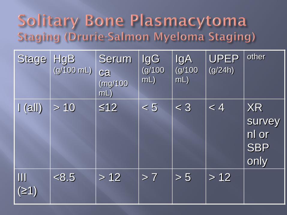
Stage HgB (g/100 mL)
Serum ca (mg/100 mL)
IgG (g/100 mL)
IgA (g/100 mL)
UPEP (g/24h)
other
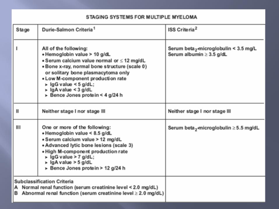
I (all) > 10 ≤12 < 5 < 3 < 4 XR survey nl or SBP only
III (≥1)
<8.5 > 12 > 7 > 5 > 12
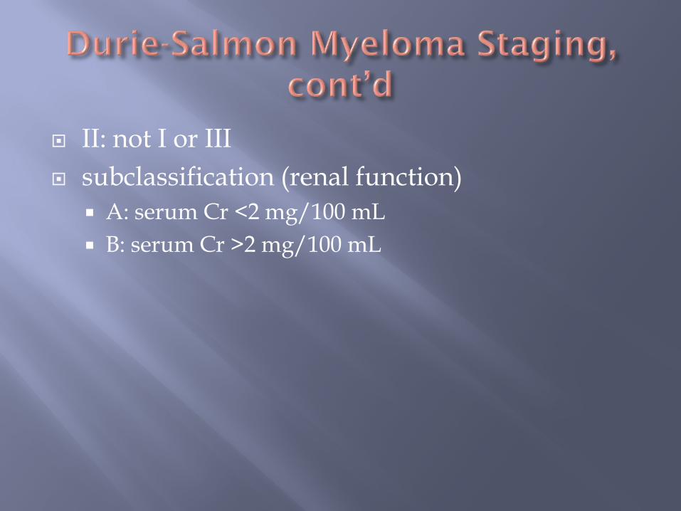
II: not I or III subclassification (renal function)
A: serum Cr <2 mg/100 mL B: serum Cr >2 mg/100 mL

I: serum beta-microglobulin <3.5 mg/L, serum albumin ≥ 3.5 g/dL MS: 62 mos
II: not one or three MS: 44 mos
III: serum beta-microglobulin ≥ 5.5 mg/L MS: 29 mos
50-60% progress to MM Median time to progression is 2-3 years Median overall survival is 10 yrs Some postulate it is part of the MGUS – MM
continuum

Tumors >4-5 cm ≥ 60 years old High M protein levels (1g/dL) Persistence of M protein after treatment Spine lesions
Definitive XRT ≥ 45 Gy Involved bone with 2-3 cm margin Chemotherapy and surgery usually not
indicated Chemo reserved for systemic disease Orthopedic surgery may be indicated
LC 80-90%, better if <5 cm
Labs (every 3-6 months for 1 year then annually)
Measure frequently early on to confirm radiosensitivity of tumor SPEP/UPEP, immunofixation CBC Calcium Creatinine
Skeletal survey or MRI Every 6 months for 1 year, then as clinically
indicated

M. Dimopoulos, et al. Curability of Solitary Bone Plasmacytoma. JCO. 1992
Retrospective study 45 patients with SBP Minimum dose 30 Gy
Single institution - MDACC
45 patients with SBP treated with XRT from 1966-91
Minimum dose of 30 Gy Diagnosis
Bone marrow (<5% plasma cells), skeletal survey, SPEP/UPEP, quantitative Igs
93% had preserved uninvolved Igs
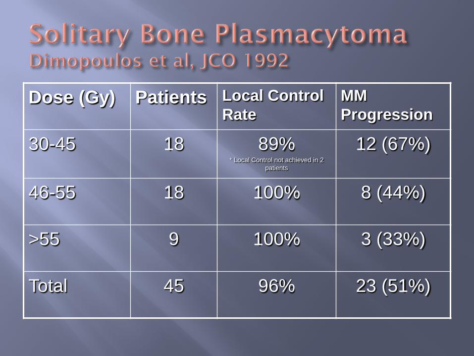
Dose (Gy) Patients Local Control Rate
MM Progression
30-45 18 89% * Local Control not achieved in 2
patients
12 (67%)
46-55 18 100% 8 (44%)
>55 9 100% 3 (33%)
Total 45 96% 23 (51%)
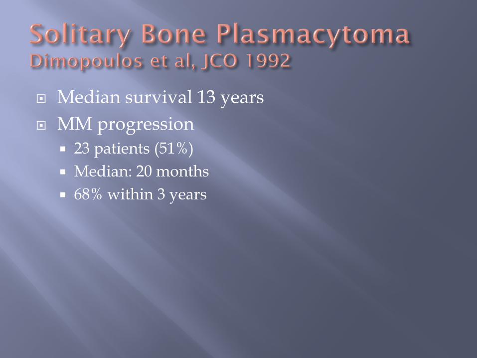
Median survival 13 years MM progression
23 patients (51%) Median: 20 months 68% within 3 years
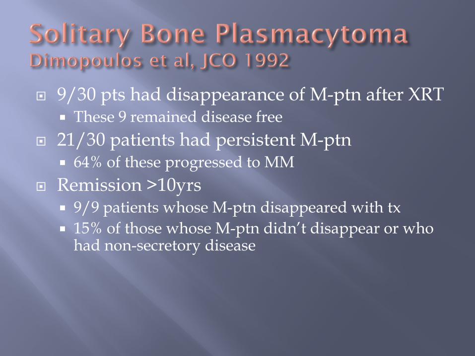
9/30 pts had disappearance of M-ptn after XRT These 9 remained disease free
21/30 patients had persistent M-ptn 64% of these progressed to MM
Remission >10yrs 9/9 patients whose M-ptn disappeared with tx 15% of those whose M-ptn didn’t disappear or who
had non-secretory disease
Conclusions In SBP patients, involved field XRT + disappearance
of M-protein predicted long-term disease free survival
Nonsecretory disease and persistent myeloma protein after treatment were adverse prognostic factors
Liebross, et al. Solitary Bone Plasmacytoma: outcome and prognostic factors following radiotherapy. IJROBP 1998
Retrospective 57 patients with SBP treated with XRT at
MDACC 1965 – 1996
LC achieved in 55/57 (96%) of patients Median time to MM progression: 1.8 years
Dimopoulus: 20 mos Median survival: 11 years
11 patients had disappearance of M-ptn following XRT All had ≤1.0 g/dL initially (total: 22) 2 relapsed (at 4 and 12 years)
17/30 (57%) with a persistent protein peak relapsed
Patients with thoracolumbar disease 7/8 patients diagnosed with plain films alone
developed MM 1/7 diagnosed with MRI of the spine + plain films
developed MM
Conclusion Precise staging including MRI of the spine is
necessary for patient selection for definitive XRT Disappearance of M-protein following XRT
represents a high likelihood of cure
Plasmacytoma outside of bone marrow Often associated with IgA monoclonal ptn Predominantly in the upper respiratory tract
3-4% of plasma cell malignancies Median age of diagnosis 60 yo M>F About 85% in upper aerodigestive tract (UAD)
GI tract 2nd MC May also involve lung, bladder, thyroid, testis,
ovary, tonsil
Upper respiratory tract Epistaxis Increased nasal discharge Obstruction
Other sites produce localized symptoms
Biopsy-proven extramedullary monoclonal plasma cell tumor
No evidence of bone destruction or occult disease elsewhere
10-40% progress in 10 years Much less common that SBP patients
10 year OS 40-90% LC ≥ 80%

Definitive XRT 45-50 Gy/4-5 weeks Consider covering primary draining nodes
Surgery may be considered for GI disease Alexiou et al showed no difference between surgery,
XRT, or combined modality tx for sites other than H&N
Adjuvant chemo doesn’t lower relapse rate or increase DFS
Alexiou, et al. Extramedullary Plasmacytoma: Tumor Occurrence and Therapeutic Concepts. Cancer. 1999
Literature search MEDLINE, Index Medicus, Deutsches Institut fur
Medizinische Dokumentation und Information, reference lists from respective publications
Reviewed 400+ publications between 1905-97
869 patients with EMP treated with surgery,
XRT, or combined modality tx 82.2% in UAD, 43.8% in nasal cavity or
paranasal sinuses
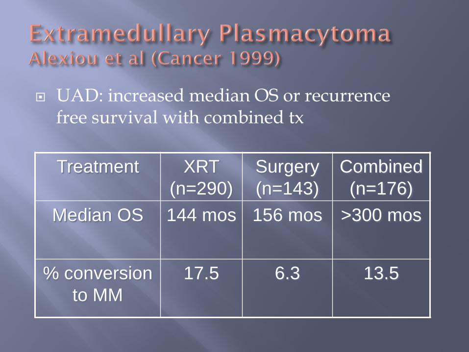
UAD: increased median OS or recurrence free survival with combined tx Treatment XRT
(n=290) Surgery (n=143)
Combined (n=176)
Median OS 144 mos 156 mos >300 mos
% conversion to MM
17.5 6.3 13.5
Non-UAD: no survival difference between tx
arms Low rate of LN involvement: 7.6% in UAD,
2.6% non-UAD
Conclusions Surgery alone gives the best results in EMP if
resectability is good if not feasible, combined therapy is indicated
Bachar et al. Solitary Extramedullary Plasmacytoma of the Head and Neck – Long-term Outcome Analysis of 68 Cases. Head and Neck. 2008
Retrospective review at PMH 1960 – 2000 (68 patients) Median follow up 8 years
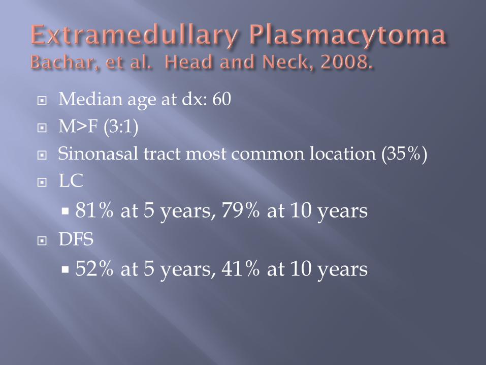
Median age at dx: 60 M>F (3:1) Sinonasal tract most common location (35%) LC
81% at 5 years, 79% at 10 years DFS
52% at 5 years, 41% at 10 years
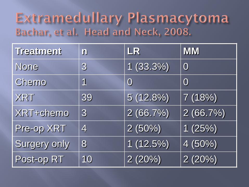
Treatment n LR MM None 3 1 (33.3%) 0 Chemo 1 0 0 XRT 39 5 (12.8%) 7 (18%) XRT+chemo 3 2 (66.7%) 2 (66.7%) Pre-op XRT 4 2 (50%) 1 (25%) Surgery only 8 1 (12.5%) 4 (50%) Post-op RT 10 2 (20%) 2 (20%)
Conclusions XRT is the treatment of choice for EMP Surgery is reserved for large tumors and extensive
bone destruction
Plasmacytoma is a rare disease Treatment study consists mainly of
retrospective reviews Based on retrospective data, XRT is the
treatment of choice in SBP For EMP, there is conflicting data. However,
based on the radiosensitivity of plasma tumors, XRT is accepted as the treatment of choice
Moulopoulos, LA, Dimopoulos, MA, Weber, D, et al. Magnetic resonance imaging in the staging of solitary plasmacytoma of bone. J Clin Oncol 1993; 11:1311.
Liebross, R, Ha, C, Cox, J, et al. Solitary Bone Plasmacytoma: Outcome and Prognostic Factors Following Radiotherapy. IJROBP 1998; 41(5): 1063-7
Ozsahin, M, Tsang, R, Poortmans, P, et al. Outcomes and Patterns of Failure in Solitrary Plasmacytoma: A MultiCenter Rare Cancer Network Study of 258 Patients. IJROBP 2006; 64(1): 210-7.
Dimopoulos, MA, Goldstein, J, Fuller, L, et al. Curability of solitary bone plasmacytoma. J Clin Oncol 1992; 10:587.
Alexiou, C, Kau, RJ, Dietzfelbinger, H, et al. Extramedullary plasmacytoma: Tumor occurrence and therapeutic concepts. Cancer 1999; 85:2305.
Bachar, G, Goldstein, D, Brown, D, et al. Solitary Extramedullary Plasmacytoma of the Heand and Neck – Long-Term Outcome Analysis of 68 Cases. Head & Neck 2008. 1012-1019.