Shear-thinning Nanocomposite Hydrogels for the Treatment...
40
1 Shear-thinning Nanocomposite Hydrogels for the Treatment of Hemorrhage Akhilesh K. Gaharwar, Reginald K. Avery, Alexander Assmann, Arghya Paul, Gareth H. McKinley, Ali Khademhosseini*, Bradley D. Olsen* *Prof. B. D. Olsen, Dr. A. K. Gaharwar Department of Chemical Engineering, Massachusetts Institute of Technology, Cambridge, MA 02139 (USA) Email: ([email protected]) *Prof. A. Khademhosseini, Dr. A. K. Gaharwar, Dr. A. Assmann, Dr. A. Paul Wyss Institute for Biologically Inspired Engineering, Harvard University, Boston, MA 02115 (USA) Email: ([email protected]) Prof. A. Khademhosseini, Dr. A. K. Gaharwar, R. K. Avery, Dr. A. Assmann, Dr. A. Paul Center for Biomedical Engineering, Department of Medicine, Brigham and Women’s Hospital, Harvard Medical School, Cambridge, MA 02139 (USA) Prof. A. Khademhosseini, Dr. A. K. Gaharwar, Dr. A. Assmann, Dr. A. Paul Harvard-MIT Division of Health Sciences and Technology, Massachusetts Institute of Technology, Cambridge, MA 02139 (USA). Dr. A. K. Gaharwar David H. Koch Institute for Integrative Cancer Research, Massachusetts Institute of Technology, Cambridge, MA 02139 (USA) R. K. Avery Department of Biological Engineering, Massachusetts Institute of Technology, Cambridge, MA 02139 (USA) Prof. G. H. McKinley Department of Mechanical Engineering, Massachusetts Institute of Technology, Cambridge, MA 02139 (USA) Dr. A. Assmann Department of Cardiovascular Surgery, Heinrich Heine University, Medical Faculty, 40225 Duesseldorf (Germany)
Transcript of Shear-thinning Nanocomposite Hydrogels for the Treatment...

1
Shear-thinning Nanocomposite Hydrogels for the Treatment of Hemorrhage
Akhilesh K. Gaharwar, Reginald K. Avery, Alexander Assmann, Arghya Paul, Gareth H.
McKinley, Ali Khademhosseini*, Bradley D. Olsen*
*Prof. B. D. Olsen, Dr. A. K. Gaharwar
Department of Chemical Engineering, Massachusetts Institute of Technology, Cambridge, MA
02139 (USA)
Email: ([email protected])
*Prof. A. Khademhosseini, Dr. A. K. Gaharwar, Dr. A. Assmann, Dr. A. Paul
Wyss Institute for Biologically Inspired Engineering, Harvard University,
Boston, MA 02115 (USA)
Email: ([email protected])
Prof. A. Khademhosseini, Dr. A. K. Gaharwar, R. K. Avery, Dr. A. Assmann, Dr. A. Paul
Center for Biomedical Engineering, Department of Medicine, Brigham and Women’s Hospital,
Harvard Medical School, Cambridge, MA 02139 (USA)
Prof. A. Khademhosseini, Dr. A. K. Gaharwar, Dr. A. Assmann, Dr. A. Paul
Harvard-MIT Division of Health Sciences and Technology, Massachusetts Institute of
Technology, Cambridge, MA 02139 (USA).
Dr. A. K. Gaharwar
David H. Koch Institute for Integrative Cancer Research, Massachusetts Institute of Technology,
Cambridge, MA 02139 (USA)
R. K. Avery
Department of Biological Engineering, Massachusetts Institute of Technology, Cambridge, MA
02139 (USA)
Prof. G. H. McKinley
Department of Mechanical Engineering, Massachusetts Institute of Technology, Cambridge, MA
02139 (USA)
Dr. A. Assmann
Department of Cardiovascular Surgery, Heinrich Heine University, Medical Faculty, 40225
Duesseldorf (Germany)

2
Shear-thinning Nanocomposite Hydrogels for the Treatment of Hemorrhage
Internal hemorrhaging is a leading cause of death after traumatic injury on the battlefield.
Although several surgical approaches such as the use of fibrin glue and tissue adhesive
have been commercialized to achieve hemostasis, these approaches are difficult to employ
on the battlefield and cannot be used for incompressible wounds. Here, we present shear-
thinning nanocomposite hydrogels composed of synthetic silicate nanoplatelets and gelatin
as injectable hemostatic agents. These materials are demonstrated to decrease in vitro
blood clotting times by 77 %, and shown to form stable clot-gel systems. In vivo tests
indicated that the nanocomposites are biocompatible and capable of promoting hemostasis
in an otherwise lethal liver laceration. The combination of injectability, rapid mechanical
recovery, physiological stability, and the ability to promote coagulation result in a new
hemostat for treating incompressible wounds in out-of-hospital, emergency conditions.
A range of hemostats have been reported that decrease the time required to establish
hemostasis by dehydrating the injury site,1, 2 concentrating clotting factors,2, 3 delivering clotting
agents (such as thrombin and fibrinogen)1, 2, 4 or forming a physical barrier against bleeding,5 to
the injured site to promote clotting. However, most of these hemostats are suitable for external
wounds,6, 7 where hemostatic agents and external pressure can be applied simultaneously.
Recently, thrombin and fibrinogen-based injectable solutions have been developed, but these
injectable solution-based hemostats pose the risk of introducing strong coagulation activators
into the circulatory system.8, 9 Therefore, there is a need for new hemostatic biomaterials that are
injectable into a wound, mechanically stable, and induce rapid and local hemostasis.

3
One approach to developing hemostatic agents for traumatic injuries is to engineer
injectable biomaterials that can be introduced into a wound site, forming a physiologically stable
artificial matrix and promoting the natural clotting cascade. Specifically, the biomaterial should
flow with minimal applied pressure during injection, providing a method of application that
avoids additional patient trauma. However, once in the wound, the material should solidify to
prevent biomaterial loss to unaffected areas. Shear-thinning hydrogels can satisfy these
requirements and have been developed from a wide variety of material platforms.10, 11
Multiple approaches have been developed to incorporate functional materials into
hemostats to enhance their therapeutic properties. These include commercial products such as
QuikClotTM that incorporate kaolin, a crystalline mineral that functions as an absorbent and
coagulation activator.12 Floseal® uses gelatin and thrombin to promote clotting in an injectable
form.8 An emerging approach to integrate functionality into hydrogel networks focuses on
incorporating nanoparticles.13, 14, 15, 16, 17 Nanomaterials have been shown to interact with blood to
promote clotting via mechanisms such as platelet activation, dehydration of the plasma, delivery
of coagulating factors, formation of physical barriers, or the activation of clotting factors.1, 18, 19,
20, 21, 22
Highly charged nanoparticles, such as synthetic silicate nanoplatelets, have been shown
to induce blood coagulation by concentrating clotting factors.4 Synthetic silicates are charged
disks, 20-30 nm in diameter and ~1 nm in thickness.23 Due to the anisotropic distribution of their
surface charge, positive along the edge and negative on the top and bottom surfaces, the
nanoplatelets can form self-assembled structures which can dynamically form and break,
creating shear thinning gels when in aqueous media.24 Recently, synthetic silicate nanoplatelets
have been used as osteogenic agents,23 drug delivery agents,25 tissue engineered scaffolds,26, 27,

4
28, 29 solid hemostat products,4, 30, 31 additives in cosmetic creams,32, 33 and rheological
modifiers.34 Synthetic silicates such as Laponite are shown to degrade into non-toxic components
(Na+, Mg2+, Si(OH)4, Li+) in physiological conditions.35 Moreover, these silicates are found to be
cytocompatible with human stem cells and animal cells as reported in previous literature.23, 26, 28
Natural and synthetic polymers have been shown to interact with synthetic silicates
through physical interactions, forming physically crosslinked networks.26, 28, 29, 36, 37 Earlier
studies on silicate-gelatin interactions show that polyampholytic gelatin, containing positive and
negative regions, strongly interacts with the oppositely charged areas of the synthetic silicate
nanoplatelets.38 Gelatin is denatured collagen, and mimics components of the native extracellular
matrix (ECM) in structure and chemical composition.39 Gelatin’s hygroscopic property also
allow for absorption of body fluids and is proposed for a range of tissue engineering applications.
Physical mixtures of gelatin and silicate nanoplatelets were used to formulate the
nanocomposite hydrogels for this study. Briefly, the silicate nanoplatelets were exfoliated in
ultrapure (Milli-Q) water using a vortexer to enhance the surface area available for interaction
with gelatin. Next, a gelatin stock, heated to liquefy the solution, was vigorously mixed with the
exfoliated silicate at room temperature (Figure 1a). Vigorous agitation was necessary to prevent
clumping of the nanoplatelets during gelation; however, the nanoplatelets are stably dispersed
after the gel has set.24 Nanocomposite hydrogels were fabricated with solid concentrations of 3,
6, and 9 wt% and gelatin:nanoplatelet ratios from 0:1 to 1:0 and labeled as xNCy (“x” represents
the total solid weight percent and “y” is percent of the total solid weight percent that is
nanoplatelet) (Supplementary Table 1). Initially the gelatin solution was a viscous liquid (at 37
oC), but once silicate nanoplatelets were mixed with gelatin, the solution gelled within a minute.

5
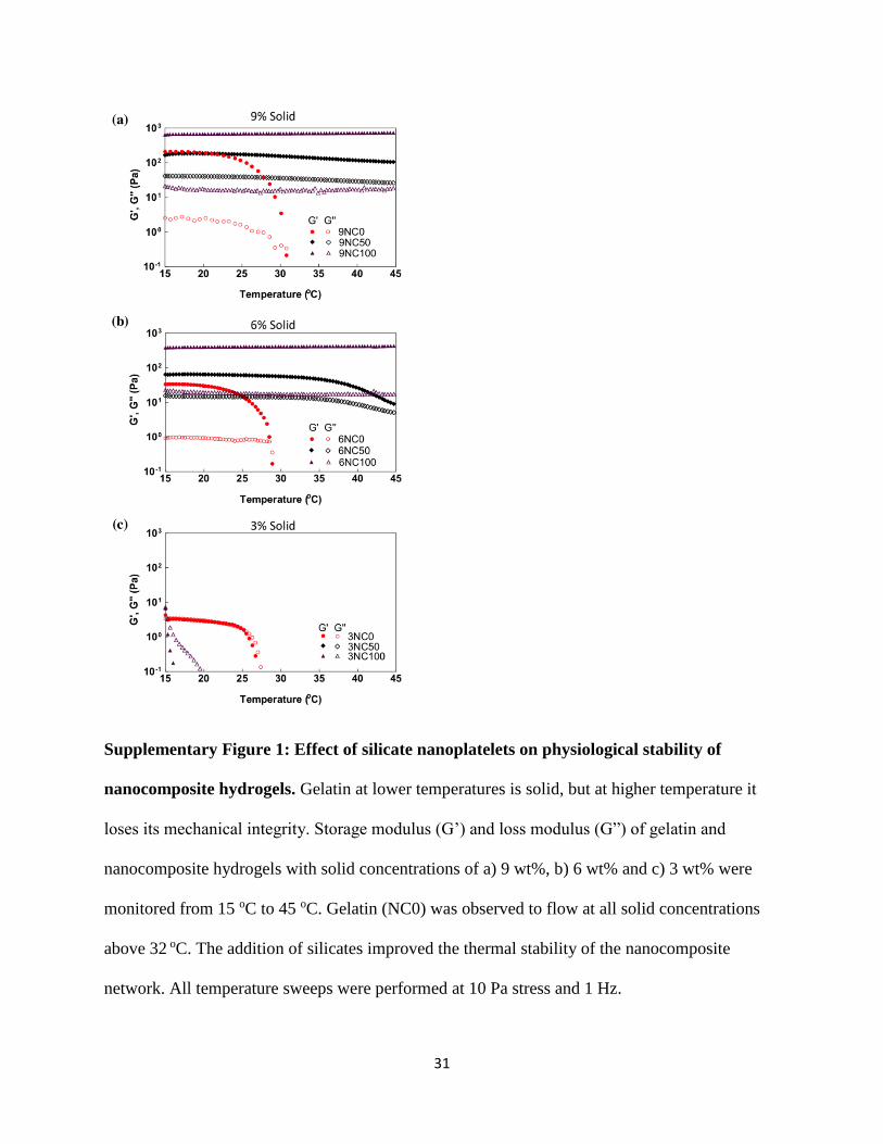
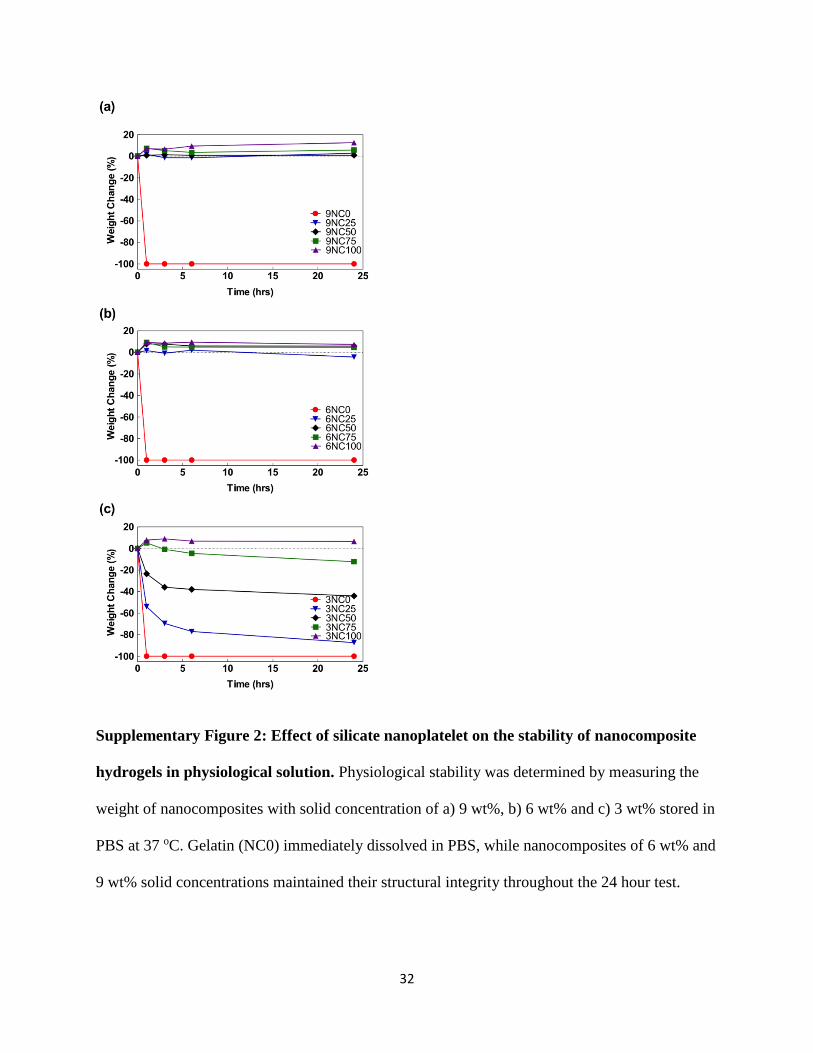
When mixed with gelatin, silicate nanoplatelets improved the thermal stability of the
hydrogel. Thermal stability of the nanocomposite formulations was determined by oscillatory
shear rheology over a temperature range from 15 °C to 45 °C, mimicking common
environmental and physiological temperatures that the nanocomposite may be exposed during
sample preparation and in vivo application. 9NC0 had a gel-sol transition temperature of 32 °C
(Supplementary Figure 1), consistent with the literature and too low for application as a
hemostat.40 However, the addition of silicate nanoplatelets to gelatin improved the thermal
stability, increasing the sol-gel transition to above 45 °C for solids concentrations of 6 wt% or
greater. In contrast, 3 wt% solids nanocomposites were not solid within the experimentally
observed temperature range. For treatment of hemorrhage, thermal stability is necessary once the
nanocomposite is injected so that it can remain at the wound site without flowing into adjacent
areas. Similarly, physiological stability was observed for all 9 wt% nanocomposites, 6NC50,
6NC75, and 6NC100 (Supplementary Figure 2). All gelatin samples, quickly dissolved in PBS
at 37 °C while the aforementioned samples had no observed weight loss over 24 hours. For this
reason, only nanocomposite hydrogels with more than 6 wt% solids were characterized further.
Zeta potential measurements suggest that electrostatic interactions between nanoplatelets
and gelatin contributed to the observed increase in the thermal stability (Figure 1b). Solutions
of silicate nanoplatelets possessed a zeta potential of -39 mV, whereas gelatin solutions had a
zeta potential of 10 mV. Because the two components had opposite charges, electrostatic
interactions between silicate and gelatin were expected. This was also supported by earlier
findings which showed that strong interactions between montmorillonite (another type of silicate
clay) and gelatin can function to increase the sol-gel transition temperature of the composite.41
6
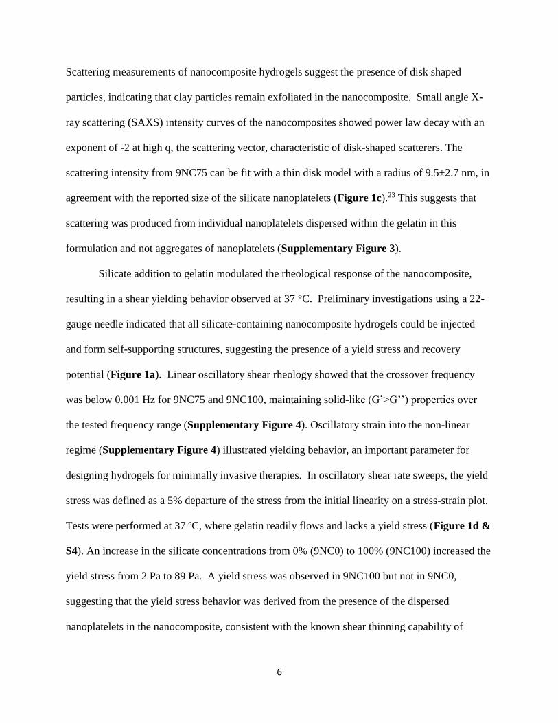
Scattering measurements of nanocomposite hydrogels suggest the presence of disk shaped
particles, indicating that clay particles remain exfoliated in the nanocomposite. Small angle X-
ray scattering (SAXS) intensity curves of the nanocomposites showed power law decay with an
exponent of -2 at high q, the scattering vector, characteristic of disk-shaped scatterers. The
scattering intensity from 9NC75 can be fit with a thin disk model with a radius of 9.5±2.7 nm, in
agreement with the reported size of the silicate nanoplatelets (Figure 1c).23 This suggests that
scattering was produced from individual nanoplatelets dispersed within the gelatin in this
formulation and not aggregates of nanoplatelets (Supplementary Figure 3).
Silicate addition to gelatin modulated the rheological response of the nanocomposite,
resulting in a shear yielding behavior observed at 37 °C. Preliminary investigations using a 22-
gauge needle indicated that all silicate-containing nanocomposite hydrogels could be injected
and form self-supporting structures, suggesting the presence of a yield stress and recovery
potential (Figure 1a). Linear oscillatory shear rheology showed that the crossover frequency
was below 0.001 Hz for 9NC75 and 9NC100, maintaining solid-like (G’>G’’) properties over
the tested frequency range (Supplementary Figure 4). Oscillatory strain into the non-linear
regime (Supplementary Figure 4) illustrated yielding behavior, an important parameter for
designing hydrogels for minimally invasive therapies. In oscillatory shear rate sweeps, the yield
stress was defined as a 5% departure of the stress from the initial linearity on a stress-strain plot.
Tests were performed at 37 ºC, where gelatin readily flows and lacks a yield stress (Figure 1d &
S4). An increase in the silicate concentrations from 0% (9NC0) to 100% (9NC100) increased the
yield stress from 2 Pa to 89 Pa. A yield stress was observed in 9NC100 but not in 9NC0,
suggesting that the yield stress behavior was derived from the presence of the dispersed
nanoplatelets in the nanocomposite, consistent with the known shear thinning capability of

7
nanoplatelets.40 Because increasing concentration of gelatin reduces the yield stress, it eases
delivery of the nanocomposite by injection.
Recovery of the elastic gel strength in less than 10 seconds was observed in
nanocomposites for nanoplatelet loadings greater than 50% (9NC50, 9NC75, and 9NC100).
Such rapid self-healing after the removal of stress can prevent material flow after application to a
wound site. This provides a significant advantage over self-assembling peptides, which risk
being washed away because they have relatively long self-healing times after the deformation of
physically crosslinked networks.5 Figure 1e shows four cycles of high (100%) to low (1%)
oscillatory strain amplitudes and the resulting nanocomposite moduli. These results indicate
rapid recovery of the storage modulus after repeated application of high oscillatory strain
amplitudes, suggesting rapid recovery of the physically crosslinked networks. After four cycles
of high and low oscillatory strain, the 9NC50 modulus observed during large amplitude strain
oscillations was 80% lower than the initial modulus. At higher silicate loading (9NC75 and
9NC100), the moduli were 33% and 29% lower compared to the initial values. Extended
monitoring (Supplementary Figure 5) indicated that after 30 seconds the moduli reached
asymptotic values, indicating completion of the healing process.
The incorporation of silicate nanoplatelets into gelatin led to a decrease in the observed
clotting time in vitro. The hemostatic ability of nanocomposite hydrogels was evaluated by
monitoring the clotting time of whole blood in contact with the nanocomposite surfaces in 96-
well plates. Under normal conditions, human blood coagulates in 5-6 minutes.4, 42 Similar
clotting times (5.2±0.5 minutes) for whole blood were observed in control wells containing
neither gelatin nor nanocomposite (Figure 2a and 2b). A slight color change was observed
9NC0 by 5 minutes. This was attributed to the tamponade ability of gelatin.43 Gelatin is

8
hygroscopic and could absorb the fluid components of whole blood but could not stimulate clot
formation within 5 minutes. The addition of nanoplatelets to gelatin reduced blood clotting time
in a dose-dependent manner. Figure 2a and 2b demonstrate the decreased clotting time for
higher nanoplatelet concentrations. 9NC25, 9NC50 and 9NC75 reduced the clotting time by
32%, 54%, and 77%, respectively when compared to the control (blood in uncoated wells). The
representative images of wells at select time points, shown in Figure 2a, clearly highlight the
presence of a clot earlier in nanocomposites with higher nanoplatelet loadings. This was
attributed to the strong negative charge of the synthetic silicate nanoplatelet that can facilitate
concentration of clotting factors near the nanocomposite surface. Prior studies have shown a
decrease in clotting time due to the addition of negatively charged particles to polymer
hydrogels.4, 12 Preliminary in vitro studies indicate that the nanocomposites induced minimal
cytotoxic effect or inflammatory response (Supplementary Figure 6), with gelatin-containing
nanocomposites having higher cell viability and lower inflammatory response than NC100 gels,
supporting the potential application of these materials as biocompatible hemostatic gels.
Decreased clotting times of blood in contact with nanocomposites were also observed
through rheological measurements. Small amplitude oscillatory time sweeps of blood in contact
with gelatin or gelatin-silicate nanocomposites were performed to evaluate clotting kinetics
(Figure 2c). Clotting induced an increase in the elastic modulus of the gelatin-clot system from
10-1 to 104 Pa (Figure 2c). Whole blood in contact with gelatin clotted in 5-7 minutes, which is
consistent with our earlier clotting data and the literature.44 When gelatin was replaced with a
silicate-gelatin nanocomposite, the clotting transition occurred even prior to the initiation of
measurements, reflecting a large reduction in the clotting time. Comparing the decrease in
clotting time observed with nanocomposites to other reported hemostatic products, the
9
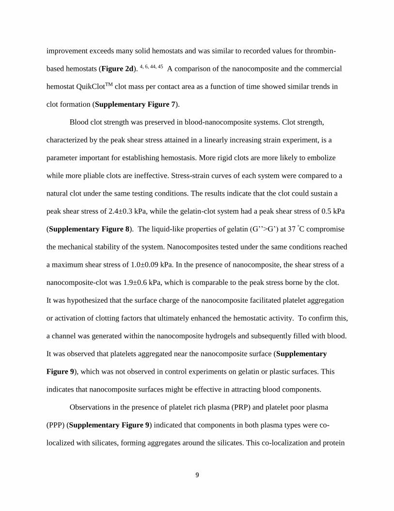
improvement exceeds many solid hemostats and was similar to recorded values for thrombin-
based hemostats (Figure 2d). 4, 6, 44, 45 A comparison of the nanocomposite and the commercial
hemostat QuikClotTM clot mass per contact area as a function of time showed similar trends in
clot formation (Supplementary Figure 7).
Blood clot strength was preserved in blood-nanocomposite systems. Clot strength,
characterized by the peak shear stress attained in a linearly increasing strain experiment, is a
parameter important for establishing hemostasis. More rigid clots are more likely to embolize
while more pliable clots are ineffective. Stress-strain curves of each system were compared to a
natural clot under the same testing conditions. The results indicate that the clot could sustain a
peak shear stress of 2.4±0.3 kPa, while the gelatin-clot system had a peak shear stress of 0.5 kPa
(Supplementary Figure 8). The liquid-like properties of gelatin (G’’>G’) at 37 ºC compromise
the mechanical stability of the system. Nanocomposites tested under the same conditions reached
a maximum shear stress of 1.0±0.09 kPa. In the presence of nanocomposite, the shear stress of a
nanocomposite-clot was 1.9±0.6 kPa, which is comparable to the peak stress borne by the clot.
It was hypothesized that the surface charge of the nanocomposite facilitated platelet aggregation
or activation of clotting factors that ultimately enhanced the hemostatic activity. To confirm this,
a channel was generated within the nanocomposite hydrogels and subsequently filled with blood.
It was observed that platelets aggregated near the nanocomposite surface (Supplementary
Figure 9), which was not observed in control experiments on gelatin or plastic surfaces. This
indicates that nanocomposite surfaces might be effective in attracting blood components.
Observations in the presence of platelet rich plasma (PRP) and platelet poor plasma
(PPP) (Supplementary Figure 9) indicated that components in both plasma types were co-
localized with silicates, forming aggregates around the silicates. This co-localization and protein

10
adsorption to the nanocomposite surfaces could be driven by electrostatic or hydrophobic
interactions, which have been shown to determine protein adsorption to biomaterial surfaces.46, 47
The first row of images in Supplementary Figure 9 shows a uniform fluorescence from a
silicate nanoplatelet solution. The second row (whole blood) shows blood cells with a minimal
fluorescent signature. The mixture of labelled 9NC100 and PPP showed co-localization of the
plasma components with the silicate nanoplatelets (third row). The same was observed for the
nanoplatelets and PRP (fourth row). The uniform fluorescence of the nanoplatelets is disrupted
by the presence of blood components, suggesting a change in the interactions between
nanoplatelets. The co-localization of silicates and blood components is thought to originate from
plasma proteins and blood cells interacting with the charged surfaces of silicate nanoplatelets,
increasing blood component concentrations surrounding the nanocomposite. Earlier studies have
shown that charge interactions can initiate the coagulation cascade, such as the interactions of
GPIb-V receptors (negative) with platelets and von Willebrand factor (positive) with collagen.20,
48, 49 The attractive properties of the silicates could serve to accelerate hemostasis, with similar
protein–nanocomposite interactions observed in other hemostats.45
In vivo biocompatibility of nanocomposite hydrogels was investigated by dorsal
subcutaneous injection in rats via 1 cm incisions. 9NC75 (200 µl) or QuikClotTM samples (200
µl) were injected or implanted, respectively, in subcutaneous pockets (n=16) as shown in Figure
3a. All animals survived the follow-up period of 28 days without any signs of physical
impairment or systemic inflammation, and exhibited regular somatic growth. After 3 days, both
implant materials (9NC75 and QuikClotTM) could be easily detected in the subcutaneous pockets.
At day 28, the QuikClotTM particles appeared macroscopically unchanged and 9NC75 was
integrated in the surrounding tissue. Hematoxylin & Eosin (H&E) staining confirmed these

11
observations indicating that 9NC75 was predominantly degraded within 28 days after injection,
while QuikClotTM did not undergo degradation (Figure 3b).50 Both implants induced an acute
locally restricted inflammatory reaction in the host, including cellular infiltration. This process
turned into chronic inflammation of the surrounding tissue, whereas the inflammatory response
against QuikClotTM was substantially stronger when compared to 9NC75, resulting in a higher
density and larger area of predominantly mononuclear cellular infiltrates. Moreover, in the
QuikClotTM group, dense fibrous tissue formation was detected around the implants at day 28,
indicating fibrous capsule formation (Figure 3b).
In order to investigate the potential of the nanocomposite to stop otherwise lethal
bleeding, a standardized liver bleeding model was applied (Supplementary Figure 10). In the
liver bleeding experiments (n=12 rats), median laparotomy was performed, and the central liver
lobe was exposed. After draping the surrounding situs with filter paper for blood collection, a
circular liver laceration (1 cm diameter) with standardized shape and size was created.
Immediately after the injury, 9NC75 or QuikClotTM was applied on the site of lesion. The
principal lethality of this bleeding model was assessed in control animals (n=5) without
application of a hemostat. Both 9NC75 (n=5) and QuikClotTM (n=2) were effective in stopping
relevant hemorrhage within seconds and prevented hypovolemic conditions. A log rank analysis
of the early post-interventional survival data revealed significant improvement by the
nanocomposite (logrank (Mantel-Cox) test: p=0.007 versus control; hazard ratio 11.5 with 95%
CI 1.93-68.9) (Figure 3c). The total blood loss after 5 and 10 minutes was significantly
decreased by the application of 9NC75 (p<0.001 versus control) (Figure 3d). The small amount
of applied nanocomposite (200 µl) was more than sufficient, since the superficial parts of the
nanocomposite were not soaked with blood, and thorough removal of this excess material did not

12
cause re-bleeding (Figure 3e). All hemostat-treated liver bleeding animals survived the complete
follow-up period of 28 days without secondary hemorrhage. At explantation, no remnants of the
nanocomposite were observed and the liver presented an intact surface, while QuikClotTM was
still present, accompanied by soft tissue adhesion to the site of lesion. These results show that the
nanocomposite gel offers a strong hemostatic potential for in vivo applications and is suitable to
stop lethal bleeding.
In conclusion, nanocomposite hydrogels containing synthetic silicate disks and gelatin
form injectable biomaterials that can promote in vitro and in vivo coagulation. The addition of
silicate nanoplatelets to gelatin significantly improved the physiological stability, injectability,
hemostatic performance, and nanocomposite-clot strength. Due to these unique features, the
newly developed silicate based gelatin nanocomposite can be used as an injectable hemostat to
treat incompressible wounds.

13
Methods
Materials: Synthetic silicate nanoplatelets (Laponite XLG) were purchased from Southern Clay
Products, Inc. (Louisville, KY). Type-A porcine skin gelatin was obtained from Sigma Aldrich
(Milwaukee, WI).
Nanocomposite Formulation: Stock solutions of 18% (w/w) gelatin and 9, 6, or 3 % (w/w)
nanoplatelets were prepared in water. The nanocomposite compositions were made by vortexing
the gelatin stock, nanoplatelet stock, and Milli-Q water at 3000 rpm for 5 minutes to achieve the
correct solid concentration and nanoplatelet loading.
X-Ray Scattering: Small Angle X-Ray Scattering (SAXS) was performed at the NSLS at
Brookhaven National Laboratory at beamline X27C. Samples were placed in a 1 mm thick
washer and sealed between Kapton tape. Samples were equilibrated at 37 °C and 20 °C for 20
minutes prior to collection of data. Scattering patterns were collected for 30 seconds per sample.
Rheological Analysis: An Anton Paar MCR 301 rheometer was used for mechanical testing. A
25 mm diameter parallel plate geometry with a gap height of 500 μm was used for temperature
sweeps and mineral oil was placed around the circumference of the plate to prevent evaporation
of water from the nanocomposite for all tests. Nanocomposites were equilibriated for 10 minutes
prior to testing, followed by a 2 minute steady shear at 10 s-1. 10 s of equilibrium time was
sufficient for the viscosity to return to a higher plateau value, after which point testing was
initiated. Frequency and shear rate sweeps were performed at 20 and 37 °C, with frequencies
from 0.001- 100 Hz at 1% strain and shear rates from 0.001 to 100 s-1 with 10 points/decade.
Frequency sweeps were performed with a cone geometry (25 mm diameter, 1° angle, 50 μm
truncation gap). Stress-controlled temperature sweeps were performed from 15-45 °C at 10 Pa
stress and 1 Hz. All other tests were performed at 37 °C. Oscillatory stress sweeps were

14
performed from 0.01-100 Pa at 1 Hz. Strain Sweeps were performed from 0.01-1000% at 1 Hz.
Recovery testing was conducted at 1 Hz by applying 100% strain, a value outside of the linear
viscoelastic range, followed by 1% strain for 5 minutes to monitor gel recovery. Interfacial
strength was also measured by applying a linearly increasing strain to a system of nanocomposite
and coagulated blood. Shear stress was measured until 1,800% strain. The maximum stress
attained was used as a measure of the strength of the clot system.
Clotting Time Assay: A solution of citrated blood and 10% (v/v) 0.1 M calcium chloride
(CaCl2) was then added, followed by vortexing for 10 seconds. 50 μL was deposited into
sequential wells on a 96 well plate. At selected time points, each well was washed with 9 g/L
saline solution to halt clotting. The liquid was immediately aspirated and washes repeated until
the solution was clear, indicating removal of all soluble blood components. Nanocomposites
were injected via syringe into the base of the well plates prior to adding blood. Final clotting
time was marked in the well that formed a uniform clot, with no change in clot size in
subsequent wells.
Quantification and Imaging of Interfacial Interactions: Anticoagulated whole blood was
centrifuged for 2 minutes to separate red blood cell- rich (RBC-rich) and RBC-poor phases at
6,000 rpm. A liquid solution of fluorescently labeled nanoplatelet was mixed with the RBC-rich
and poor blood phases. Dilute solutions of RBC-rich/nanoplatelet and RBC-poor/nanoplatelet
were mixed and deposited onto glass slides for fluorescent imaging.
In vivo experiments: Male Wistar rats (n=20; 200 – 250 g) were obtained from Charles River
(Wilmington, MA, USA), housed in the local animal care facility (PRB, Cambridge, MA, USA).
Anesthesia and analgesia were achieved by isoflurane inhalation (2.0-2.5%) and subcutaneous
carprofen administration (5 mg/kg/d). All experiments were conducted according to the NIH

15
“Guide for the Care and Use of Laboratory Animals”, and approved by the local animal care
committee (HMA Standing Committee on Animals; protocol number 05055).
Subcutaneous Implantation: Dorsal skin incisions (1 cm in length) were conducted and a small
subcutaneous pocket was generated by blunt preparation. Nanocomposite gels (n=8; 200 µl)
were injected or QuikClotTM samples (n=8; 200 µl) were implanted, respectively. The wounds
were anatomically closed. After 3 and 28 days, the animals were euthanized by CO2 inhalation,
and the implants as well as adjacent tissue were explanted and further processed for histological
analyses.
Liver Bleeding: Median laparotomies were performed and the central liver lobe was exposed
(n=12 rats). After draping the surrounding situs with filter paper for blood collection, a
standardized circular liver laceration was created by gluing a plastic disc (d=10 mm) to the
surface and superficially excising this area with a blade. Immediately after injury, nanocomposite
gel (n=5; 200 µl) or QuikClotTM (n=2; 200 µl) was applied on the site of lesion. Five minutes
after the bleeding had been stopped, the abdomen was anatomically closed, and the animals were
allowed to recover from anesthesia. After 28 days, the animals were euthanized by CO2
inhalation, and the site of injury was inspected. In order to examine the principal lethality of the
liver bleeding model, control rats (n=5) underwent liver injury without subsequent application of
a hemostat. In all liver bleeding experiments, the amount of bleeding was determined by
weighing filter papers used to collect lost blood after removal.
Histology: Histological analyses were conducted as previously published. In brief,
paraformaldehyde-fixed cryo-sections (6 µm) of all explants were stained with
hematoxylin/eosin and microscopically analyzed.50

16
References
1. Ostomel TA, Shi Q, Stucky GD. Oxide hemostatic activity. Journal of the American
Chemical Society 2006, 128(26): 8384-8385.
2. Spotnitz WD, Burks S. Hemostats, sealants, and adhesives: components of the surgical
toolbox. Transfusion 2008, 48(7): 1502-1516.
3. Dowling MB, Kumar R, Keibler MA, Hess JR, Bochicchio GV, Raghavan SR. A self-
assembling hydrophobically modified chitosan capable of reversible hemostatic action.
Biomaterials 2011, 32(13): 3351-3357.
4. Baker SE, Sawvel AM, Zheng N, Stucky GD. Controlling bioprocesses with inorganic
surfaces: layered clay hemostatic agents. Chemistry of Materials 2007, 19(18): 4390-
4392.
5. Ruan L, Zhang H, Luo H, Liu J, Tang F, Shi Y-K, et al. Designed amphiphilic peptide
forms stable nanoweb, slowly releases encapsulated hydrophobic drug, and accelerates
animal hemostasis. Proceedings of the National Academy of Sciences 2009.
6. Burnett LR, Richter JG, Rahmany MB, Soler R, Steen JA, Orlando G, et al. Novel
keratin (KeraStat™) and polyurethane (Nanosan®-Sorb) biomaterials are hemostatic in a
porcine lethal extremity hemorrhage model. Journal of Biomaterials Applications 2014,
28(6): 869-879.
7. Hildenbrand T. A New Gelatine-based Hemostat for Sinonasal Surgery: A Clinical
Survey. In Vivo 2013, 27(4): 523-526.
8. Oz MC, Rondinone JF, Shargill NS. Floseal Matrix. Journal of Cardiac Surgery 2003,
18(6): 486-493.
9. Xie X, Tian J-k, Lv F-q, Wu R, Tang W-b, Luo Y-k, et al. A novel hemostatic sealant
composed of gelatin, transglutaminase and thrombin effectively controls liver trauma-
induced bleeding in dogs. Acta Pharmacol Sin 2013, 34(7): 983-988.
10. Ekenseair AK, Boere KWM, Tzouanas SN, Vo TN, Kasper FK, Mikos AG. Structure–
Property Evaluation of Thermally and Chemically Gelling Injectable Hydrogels for
Tissue Engineering. Biomacromolecules 2012, 13(9): 2821-2830.
11. Lu HD, Soranno DE, Rodell CB, Kim IL, Burdick JA. Secondary Photocrosslinking of
Injectable Shear-Thinning Dock-and-Lock Hydrogels. Advanced Healthcare Materials
2013, 2(7): 1028-1036.
12. Ostomel TA, Shi Q, Stoimenov PK, Stucky GD. Metal oxide surface charge mediated
hemostasis. Langmuir 2007, 23(22): 11233-11238.
17
13. Cha C, Shin SR, Annabi N, Dokmeci MR, Khademhosseini A. Carbon-Based
Nanomaterials: Multifunctional Materials for Biomedical Engineering. ACS nano 2013,
7(4): 2891-2897.
14. Gaharwar AK, Peppas NA, Khademhosseini A. Nanocomposite hydrogels for biomedical
applications. Biotechnology and Bioengineering 2014, 111(3): 441-453.
15. Schexnailder P, Schmidt G. Nanocomposite polymer hydrogels. Colloid and Polymer
Science 2009, 287(1): 1-11.
16. Shin SR, Bae H, Cha JM, Mun JY, Chen Y-C, Tekin H, et al. Carbon Nanotube
Reinforced Hybrid Microgels as Scaffold Materials for Cell Encapsulation. ACS nano
2011, 6(1): 362-372.
17. Shin SR, Jung SM, Zalabany M, Kim K, Zorlutuna P, Kim Sb, et al. Carbon-Nanotube-
Embedded Hydrogel Sheets for Engineering Cardiac Constructs and Bioactuators. ACS
nano 2013, 7(3): 2369-2380.
18. Dobrovolskaia MA, McNeil SE. Immunological properties of engineered nanomaterials.
Nature nanotechnology 2007, 2(8): 469-478.
19. Jones CF, Grainger DW. < i> In vitro</i> assessments of nanomaterial toxicity.
Advanced drug delivery reviews 2009, 61(6): 438-456.
20. Radomski A, Jurasz P, Alonso-Escolano D, Drews M, Morandi M, Malinski T, et al.
Nanoparticle-induced platelet aggregation and vascular thrombosis. British Journal of
Pharmacology 2005, 146(6): 882-893.
21. Semberova J, De Paoli Lacerda SH, Simakova O, Holada K, Gelderman MP, Simak J.
Carbon nanotubes activate blood platelets by inducing extracellular Ca2+ influx sensitive
to calcium entry inhibitors. Nano letters 2009, 9(9): 3312-3317.
22. Singh SK, Singh MK, Nayak MK, Kumari S, Shrivastava S, Gr√°cio JJ, et al. Thrombus
inducing property of atomically thin graphene oxide sheets. ACS nano 2011, 5(6): 4987-
4996.
23. Gaharwar AK, Mihaila SM, Swami A, Patel A, Sant S, Reis RL, et al. Bioactive Silicate
Nanoplatelets for Osteogenic Diffrentiation of Human Human Mesenchymal Stem Cells.
Advanced Materials 2013, (25)(24): 3329–3336.
24. Ruzicka B, Zaccarelli E. A fresh look at the Laponite phase diagram. Soft Matter 2011,
7(4): 1268-1286.
25. Viseras C, Cerezo P, Sanchez R, Salcedo I, Aguzzi C. Current challenges in clay
minerals for drug delivery. Applied Clay Science 2010, 48(3): 291-295.

18
26. Gaharwar AK, Kishore V, Rivera C, Bullock W, Wu CJ, Akkus O, et al. Physically
Crosslinked Nanocomposites from Silicate-Crosslinked PEO: Mechanical Properties and
Osteogenic Differentiation of Human Mesenchymal Stem Cells. Macromolecular
Bioscience 2012, 12(6): 779.
27. Gaharwar AK, Rivera C, Wu C-J, Chan BK, Schmidt G. Photocrosslinked
nanocomposite hydrogels from PEG and silica nanospheres: Structural, mechanical and
cell adhesion characteristics. Materials Science and Engineering: C 2013, 33(3): 1800-
1807.
28. Gaharwar AK, Schexnailder P, Kaul V, Akkus O, Zakharov D, Seifert S, et al. Highly
Extensible Bio-Nanocomposite Films with Direction-Dependent Properties. Advanced
Functional Materials 2010, 20(3): 429-436.
29. Gaharwar AK, Schexnailder PJ, Kline BP, Schmidt G. Assessment of using Laponite®
cross-linked poly(ethylene oxide) for controlled cell adhesion and mineralization. Acta
Biomaterialia 2011, 7(2): 568-577.
30. Arnaud F, Parreno-Sadalan D, Tomori T, Delima MG, Teranishi K, Carr W, et al.
Comparison of 10 hemostatic dressings in a groin transection model in swine. The
Journal of Trauma and Acute Care Surgery 2009, 67(4): 848-855.
31. Bowman PD, Wang X, Meledeo MA, Dubick MA, Kheirabadi BS. Toxicity of aluminum
silicates used in hemostatic dressings toward human umbilical veins endothelial cells,
HeLa cells, and RAW267. 4 mouse macrophages. The Journal of Trauma and Acute
Care Surgery 2011, 71(3): 727-732.
32. Carretero MI, Pozo M. Clay and non-clay minerals in the pharmaceutical and cosmetic
industries Part II. Active ingredients. Applied Clay Science 2010, 47(3): 171-181.
33. Nelson O'Donoghue M. Eye cosmetics. Dermatologic clinics 2000, 18(4): 633-639.
34. Ashby N, Binks B. Pickering emulsions stabilised by Laponite clay particles. Physical
Chemistry Chemical Physics 2000, 2(24): 5640-5646.
35. Thompson DW, Butterworth JT. The nature of laponite and its aqueous dispersions.
Journal of Colloid and Interface Science 1992, 151(1): 236-243.
36. Gaharwar AK, Schexnailder PJ, Dundigalla A, White JD, Matos-Pérez CR, Cloud JL, et
al. Highly Extensible Bio-Nanocomposite Fibers. Macromolecular Rapid
Communications 2011, 32(1): 50-57.
37. Wu C-J, Gaharwar AK, Schexnailder PJ, Schmidt G. Development of biomedical
polymer-silicate nanocomposites: a materials science perspective. Materials 2010, 3(5):
2986-3005.
19
38. Pawar N, Bohidar H. Surface selective binding of nanoclay particles to polyampholyte
protein chains. The Journal of chemical physics 2009, 131: 045103.
39. Stevens KR, Einerson NJ, Burmania JA, Kao WJ. In vivo biocompatibility of gelatin-
based hydrogels and interpenetrating networks. Journal of Biomaterials Science, Polymer
Edition 2002, 13(12): 1353-1366.
40. Bohidar HB, Jena SS. Kinetics of sol-gel transition in thermoreversible gelation of
gelatin. The Journal of chemical physics 1993, 98: 8970.
41. Jorge MFC, Flaker CHC, Nassar SF, Moraes ICF, Bittante AMQB, Sobral PJdA.
Viscoelastic and rheological properties of nanocomposite-forming solutions based on
gelatin and montmorillonite. Journal of Food Engineering 2014, 120: 81-87.
42. Davie EW, Ratnoff OD. Waterfall sequence for intrinsic blood clotting. Science 1964,
145(3638): 1310-1312.
43. de la Torre RA, Bachman SL, Wheeler AA, Bartow KN, Scott JS. Hemostasis and
hemostatic agents in minimally invasive surgery. Surgery 2007, 142(4): S39-S45.
44. Bertram JP, Williams CA, Robinson R, Segal SS, Flynn NT, Lavik EB. Intravenous
hemostat: nanotechnology to halt bleeding. Sci Transl Med 2009, 1(11): 11ra22.
45. Wagner WR, Pachence JM, Ristich J, Johnson PC. Comparativein VitroAnalysis of
Topical Hemostatic Agents. Journal of Surgical Research 1996, 66(2): 100-108.
46. Roach P, Farrar D, Perry CC. Interpretation of Protein Adsorption: Surface-Induced
Conformational Changes. Journal of the American Chemical Society 2005, 127(22):
8168-8173.
47. Wojciechowski P, Ten Hove P, Brash JL. Phenomenology and mechanism of the
transient adsorption of fibrinogen from plasma (Vroman effect). Journal of Colloid and
Interface Science 1986, 111(2): 455-465.
48. Okamoto Y, Yano R, Miyatake K, Tomohiro I, Shigemasa Y, Minami S. Effects of chitin
and chitosan on blood coagulation. Carbohydrate Polymers 2003, 53(3): 337-342.
49. Zhang J, Xia W, Liu P, Cheng Q, Tahi T, Gu W, et al. Chitosan Modification and
Pharmaceutical/Biomedical Applications. Marine Drugs 2010, 8(7): 1962-1987.
50. Assmann A, Delfs C, Munakata H, Schiffer F, Horstkötter K, Huynh K, et al.
Acceleration of autologous in vivo recellularization of decellularized aortic conduits by
fibronectin surface coating. Biomaterials 2013, 34(25): 6015-6026.

20
Acknowledgement
SAXS experiments were performed at Beamline X27C of the National Synchrotron Light Source
at Brookhaven National Laboratory. This research was supported by the U.S. Army Research
Office under contract W911NF-13-D-0001. RKA was supported by an NIH Interdepartmental
Biotechnology Training Program (NIH/NIGMS 5T32GM008334). AP acknowledges
postdoctoral award from FRQS (Fonds de recherche du Québec - Santé), Quebec, Canada. AA
acknowledges a postdoctoral award from the German Heart Foundation, Frankfurt, Germany.
Author Contributions
AKG and RKA designed and performed the experiments. AP conducted cell studies and
analyzed cell data. AA performed and analyzed in vivo experiments. AKG and RKA wrote the
manuscript. GHM provided technical advice on rheological measurements. BDO, AK and GHM
gave conceptual advice and provided comments to the manuscript.
Additional Information
Competing Financial Interests
The authors declare no competing financial interests.

21
Figure Legends
Figure 1: Structure, injectability, and self-healing characteristics of nanocomposite hydrogels. (a)
Schematic showing the preparation of the nanocomposite gels. The TEM image shows the size
of the silicate nanoparticle (Scale bar 50 nm). Images showing injection of nanocomposite
hydrogel through a surgical needle (22 gauge) and recovery to form freestanding structures. (b)
Zeta potential measurements demonstrate electrostatic interactions between negatively charged
silicate and positively charged gelatin. 95% CI are shown for each point. (c) Small-angle X-ray
scattering (SAXS) indicates that nanoplatelets are well dispersed and follow the model curve for
scattering from dispersed thin disks. (d) Yield stress of gels as a function of nanoplatelet loading
and solids fraction. (e) Recovery of the nanocomposites was observed by subjecting the hydrogel
to alternating high and low strain conditions (100% strain and 1% strain) while monitoring the
moduli of the composite. For all the nanocomposite hydrogels, more than 95% recovery was
observed.
Figure 2: Effect of nanoplatelets on the clotting whole blood. (a) Clot formation as a function of
time and nanocomposite composition. (b) Quantiative clot times for 6% and 9% nanocomposites.
(One-way Anova followed by Tukey’s post hoc analysis was performed ** p<0.01; ***
p<0.001). (c) Clotting kinetics of blood when in contact with gelatin and nanocomposite
monitored using shear rheology. (d) Comparison of clotting times for silicate-gelatin
nanocomposite (NC) and commercial products.
Figure 3: In vivo evaluation of nanocomposite hydrogels as hemostats. (a) Subcutaneous
injection and explantation of 9NC75 in rats. After 3 days, nanocomposite could be easily

22
detected in the subcutaneous pockets, but the volume was already lower than at implantation. (b)
H&E staining confirmed degradation of 9NC75 within 28 days, while the QuikClotTM particles
were still present. Moreover, 9NC75 induced less chronic inflammation than QuikClotTM,
indicated by severe mononuclear cell infiltration around QuikClotTM at day 28 (asterisks).
Furthermore, the QuikClotTM samples were encapsulated by dense fibrous connective tissue
(arrows). The potential of the nanocomposite to stop otherwise lethal bleeding was investigated
using liver bleeding experiments in rats. (c) 9NC75 significantly improved the post-
interventional survival (logrank (Mantel-Cox) test). (d) 9NC75 was effective in preventing blood
loss as compared to untreated hemorrhage (***p<0.001). (e) The small amount of 9NC75 (200
µl) was sufficient to stop bleeding and the superficial part of the 9NC75 was easily removed
without causing re-bleeding.

23
Figure 1

24
Figure 2

25
Figure 3

26
Shear-thinning Nanocomposite Hydrogels for the Treatment of Hemorrhage
Akhilesh K. Gaharwar, Reginald K. Avery, Alexander Assmann, Arghya Paul, Gareth H.
McKinley, Ali Khademhosseini*, Bradley D. Olsen*
*Prof. B. D. Olsen, Dr. A. K. Gaharwar
Department of Chemical Engineering, Massachusetts Institute of Technology, Cambridge, MA
02139 (USA)
Email: ([email protected])
*Prof. A. Khademhosseini, Dr. A. K. Gaharwar, Dr. A. Assmann, Dr. A. Paul
Wyss Institute for Biologically Inspired Engineering, Harvard University,
Boston, MA 02115 (USA)
Email: ([email protected])
Prof. A. Khademhosseini, Dr. A. K. Gaharwar, R. K. Avery, Dr. A. Assmann, Dr. A. Paul
Center for Biomedical Engineering, Department of Medicine, Brigham and Women’s Hospital,
Harvard Medical School, Cambridge, MA 02139 (USA)
Prof. A. Khademhosseini, Dr. A. K. Gaharwar, Dr. A. Assmann, Dr. A. Paul
Harvard-MIT Division of Health Sciences and Technology, Massachusetts Institute of
Technology, Cambridge, MA 02139 (USA).
Dr. A. K. Gaharwar
David H. Koch Institute for Integrative Cancer Research, Massachusetts Institute of Technology,
Cambridge, MA 02139 (USA)
R. K. Avery
Department of Biological Engineering, Massachusetts Institute of Technology, Cambridge, MA
02139 (USA)
Prof. G. H. McKinley
Department of Mechanical Engineering, Massachusetts Institute of Technology, Cambridge, MA
02139 (USA)
Dr. A. Assmann
Department of Cardiovascular Surgery, Heinrich Heine University, Medical Faculty, 40225
Duesseldorf (Germany)

27
Content
I. Additional Materials and Methods
II. Supplementary Tables and Figures
I. Additional Materials and Methods:
Materials: Zeta potentials of gelatin, silicate nanoplatelet, and mixtures of silicate and gelatin
were determined in ultrapure water (Milli-Q) and phosphate buffered saline (PBS), pH 7.4
(Invitrogen) using a 633 nm laser in a Malvern ZEN3600 (Malvern Instruments, UK). Silicate
nanoplatelets were dissolved with vigorous agitation (vortexing) while gelatin was dissolved
with stirring at 40 °C. Transmission election microscopy (TEM) images of the silicate
nanoplatelets were obtained using a JEOL JEM-1400 TEM (JEM1400) installed with a cool
beam illumination system (resolution: 0.2 nm line, 0.38 nm point) and 11 megapixel Advanced
Microscopy Techniques cooled charged coupled device camera at 80 kV. The sample was
prepared by dispersing silicate nanoplatelets in water/ethanol solutions, placing a drop on the
TEM grid, and allowing the grid to dry under vacuum.
Nanocomposite Formulation: Stock solutions of 18% (w/w) gelatin and 9, 6, or 3 % (w/w)
nanoplatelets were prepared in water (Supplementary Table 1). Milli-Q water was heated to 40
°C to dissolve gelatin. 4 °C water was used for nanoplatelet stock solutions to retard the gelation
and allow for full dissolution of nanoplatelet particles prior to gelling. The nanoplatelet gels were
allowed to sit at room temperature to fully hydrate until a clear gel was formed. Nanocomposites
were again heated and vortexed. The nanocomposites were then stored at 4 °C.

28
Nanocomposite Degradation: Nanocomposite samples were placed in 2.0 mL Eppendorf tubes
and weighed. Each sample was centrifuged in a swinging bucket rotor centrifuge to obtain a flat
interface. Each sample was soaked in phosphate buffered saline (pH 7.4; Invitrogen), stored at
37 °C. At set times, the PBS was removed, and the nanocomposite reweighed. The change in
weight was recorded up to 24 hours after initial soaking. PBS was replaced after each weighing.
SAXS Method: Small Angle X-Ray Scattering (SAXS) was performed at the NSLS at
Brookhaven National Laboratory at beamline X27C. Samples were placed in a 1 mm thick
washer and sealed between Kapton tape. Samples were equilibrated at 37 °C and 20 °C for 20
minutes prior to collection of data. Scattering patterns were collected for 30 seconds per sample.
Radial integration of the two dimensional scattering pattern was performed to yield a one
dimensional scattering curve, which was corrected for empty cell and dark field scattering. Thin
disk form factor model fitting was performed in MatLab using a nonlinear fit algorithm to fit the
radius and a Gaussian distribution for polydispersity of the nanocomposite.
Thrombus Weight: Nanocomposite and powdered QuikClotTM samples were weighed into 2
mL Eppendorf tubes. Nanocomposites were centrifuged to standardize the surface area exposed.
Citrated blood was reactivated by 10 % (v/v) 0.1 m CaCl2. 100 μL of solution (10 μL CaCl2 and
90 μL whole blood) was added to each Eppendorf tube. At each measured time point, clotting
was stopped by addition of 200 μL sodium citrate solution (0.109 m). Any liquid was removed
from the Eppendorf, leaving only clotted blood. The Eppendorf tubes were reweighed to
determine the mass of clot produced in the tube. Clot mass was normalized to the area exposed
to the nanocomposite. The same area was used for the commercial hemostat sample.
Cell Studies: Mouse monocyte/macrophage RAW 264.7 cells were procured from the ATCC.
RAW 264.7 cells were grown in DMEM medium supplemented with 10% FBS and 1%
29
penicillin/streptomycin at 37 °C in 5% CO2. Lipopolysaccharide (LPS) was obtained from
InvivoGen. RAW 264.7 macrophages were suspended in different formulations of gelatin and
silicate at a concentration of 3x10⁶ cells/ml and plated at a density of 2,000 cells/well and grown
for 24h. As controls, RAW cells were untreated or treated with 100 ng/mL of LPS. An ELISA
assay (SA Biosciences) was performed according to the manufacturer’s protocol on the
supernatants of different groups to quantify the secreted cytokines IL-6 and TNF-α by the RAW
cells. In another study, the cell viability of RAW cells in the presence of gelatin and silicate for
24h was measured with Cell Titer 96 Aqueous Non- Radioactive Cell Proliferation MTS Assay
(Promega) according to manufacturer’s protocol using plate reader at 490 nm absorbance. In
order to eliminate possible absorbance from silicate or gelatin components, the absorbance
values with gelatin, silicate and nanocomposite hydrogels, without cells, was measured. These
absorbance values were subtracted from the corresponding absorbance values for gelatin, silicate
and nanocomposite hydrogels, with cells. This resulted in reading the absorbance values of only
viable cells. Each tested hydrogel was measured in three separate wells.

30
II. Supplementary Tables and Figures
Supplementary Table 1: Weight percent (w/w) composition of each nanocomposite
hydrogel.
Sample Name 9NC0 9NC25 9NC50 9NC75 9NC100
Gelatin (wt%) 9 6.75 4.5 2.25 0
Nanoplatelets (wt%) 0 2.25 4.5 6.75 9
Water (wt%) 91 91 91 91 91
Sample Name 6NC0 6NC25 6NC50 6NC75 6NC100
Gelatin (wt%) 6 4.5 3 1.5 0
Nanoplatelets (wt%) 0 1.5 3 4.5 6
Water (wt%) 94 94 94 94 94
Sample Name 3NC0 3NC25 3NC50 3NC75 3NC100
Gelatin (wt%) 3 2.25 1.5 0.75 0
Nanoplatelets (wt%) 0 0.75 1.5 2.25 3
Water (wt%) 97 97 97 97 97
31
Supplementary Figure 1: Effect of silicate nanoplatelets on physiological stability of
nanocomposite hydrogels. Gelatin at lower temperatures is solid, but at higher temperature it
loses its mechanical integrity. Storage modulus (G’) and loss modulus (G”) of gelatin and
nanocomposite hydrogels with solid concentrations of a) 9 wt%, b) 6 wt% and c) 3 wt% were
monitored from 15 oC to 45 oC. Gelatin (NC0) was observed to flow at all solid concentrations
above 32 oC. The addition of silicates improved the thermal stability of the nanocomposite
network. All temperature sweeps were performed at 10 Pa stress and 1 Hz.
32
Supplementary Figure 2: Effect of silicate nanoplatelet on the stability of nanocomposite
hydrogels in physiological solution. Physiological stability was determined by measuring the
weight of nanocomposites with solid concentration of a) 9 wt%, b) 6 wt% and c) 3 wt% stored in
PBS at 37 oC. Gelatin (NC0) immediately dissolved in PBS, while nanocomposites of 6 wt% and
9 wt% solid concentrations maintained their structural integrity throughout the 24 hour test.

33
Supplementary Figure 3: X-Ray Scattering indicates disk like scatterers. SAXS intensity
curves of nanocomposite samples 9NC100, 9NC75, 9NC50, and 9NC25. All samples have
power law decays of exponent -2 at high q, suggesting disk shaped scatterers.

34
Supplementary Figure 4: Linear viscoelastic range of nanocomposite hydrogels. (a)
Nanocomposites are capable of being molded and are able to maintain their shape. (b) Strain
sweeps indicate a crossover point (when tan(δ) = 1) that decreases with increasing silicate
loading Strain sweeps were performed at 1 Hz. (c) Frequency sweeps, at 37 °C, of 9NC25,
9NC50, 9NC75 and 9NC100 show increased moduli for higher nanoplatelet loaded
nanocomposites. 9NC0 is a liquid at 37 °C and was not tested. (d) Yield stress of gels as a
function of nanoplatelet loading and solids fraction (3%, 6% and 9% solid).
35
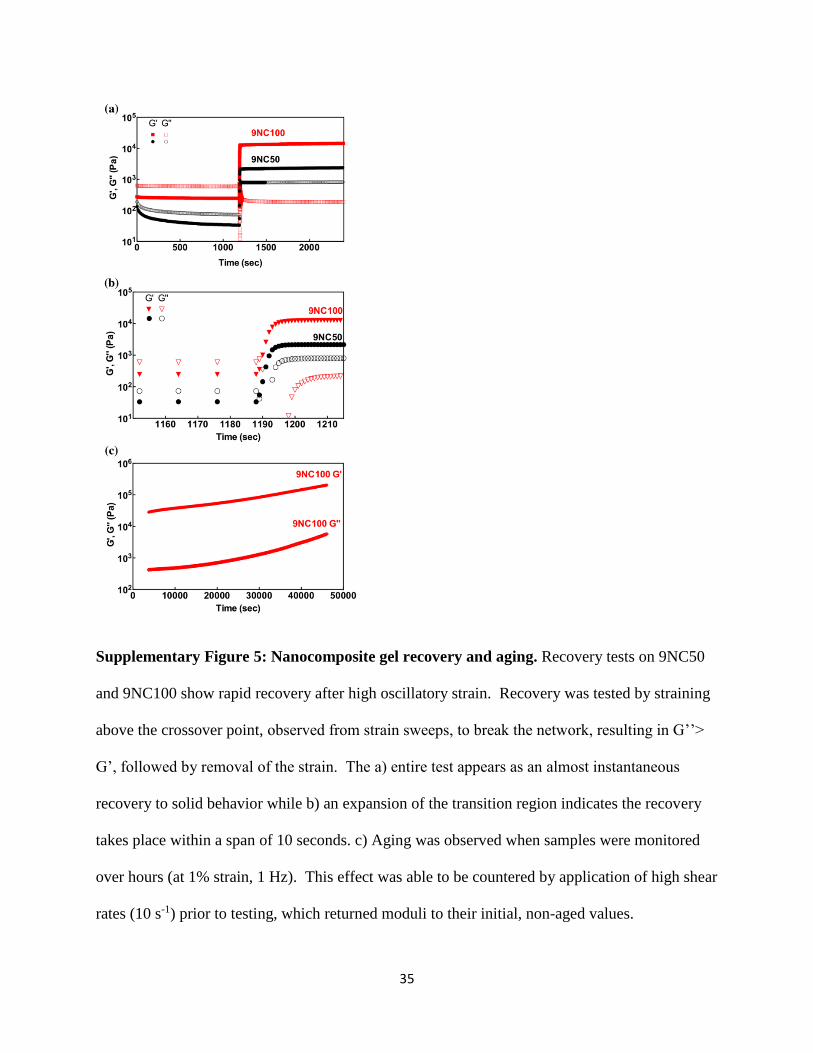
Supplementary Figure 5: Nanocomposite gel recovery and aging. Recovery tests on 9NC50
and 9NC100 show rapid recovery after high oscillatory strain. Recovery was tested by straining
above the crossover point, observed from strain sweeps, to break the network, resulting in G’’>
G’, followed by removal of the strain. The a) entire test appears as an almost instantaneous
recovery to solid behavior while b) an expansion of the transition region indicates the recovery
takes place within a span of 10 seconds. c) Aging was observed when samples were monitored
over hours (at 1% strain, 1 Hz). This effect was able to be countered by application of high shear
rates (10 s-1) prior to testing, which returned moduli to their initial, non-aged values.
36
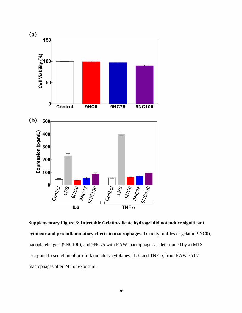
Supplementary Figure 6: Injectable Gelatin/silicate hydrogel did not induce significant
cytotoxic and pro-inflammatory effects in macrophages. Toxicity profiles of gelatin (9NC0),
nanoplatelet gels (9NC100), and 9NC75 with RAW macrophages as determined by a) MTS
assay and b) secretion of pro-inflammatory cytokines, IL-6 and TNF-α, from RAW 264.7
macrophages after 24h of exposure.

37
Supplementary Figure 7: Time series of clot formation. Time series of thrombus weight per
area for nanocomposite and hydrated QuikClotTM powder show similar trends in the formation of
a clot. The nanocomposite, an injectable system, begins to form a measureable clot at 2 minutes
while QuikClotTM, a solid hemostat, begins clotting at 5 minutes. Subsequent time points are not
statistically different from one another, indicating that the ability of 9NC75 to form a clot is
comparable to that of the commercial QuikClotTM powder. The background mass per area of 15
mg/cm2 is due to residual liquid remaining after the washing step is performed to halt clotting.

38
Supplementary Figure 8: Effect of nanocomposite on clot strength. Rotational strain sweep
of clot and clot-nanocomposite systems show a decreased peak stress for clots in contact with
9NC0 in comparison to the peak stress of a clot alone. However, the peak stress for a 9NC75-
clot system has a comparable peak stress to that of the clot alone.

39
Supplementary Figure 9: Nanocomposite surface initiates clotting through interactions
with blood components. (a) Fluorescent imaging reveals that the interfaces between the
nanocomposite and blood have localized aggregation of RBCs. (b) Silicate nanoplatelets in
contact with blood proteins and RBCs were observed using fluorescently labeled silicate
nanoplatelets. The presence of blood components (platelet poor plasma (PPP) or platelet rich
plasma (PRP)) in the nanoplatelets disrupt their ordering, observed by the non-uniform
fluorescence signal. The stability of 9NC100, previously shown by its large absolute value of
zeta potential, is decreased and aggregation occurs in systems of blood components and
nanocomposites, suggesting a decrease in the zeta potential and increase in attractive forces.

40
Supplementary Figure 10: In vivo procedure showing the liver bleeding model. To induce
lethal liver bleeding, the following standardized procedure was conducted prior to re-suturing: 1)
skin incision, 2) wound edge retraction to expose the liver, 3) addition of a hemostat after
laceration, 4) two minutes after intervention, 5) ten minutes after intervention and 6) saline
washing of the abdominal cavity. Images are from a trial with QuikClotTM intervention.