Shear-induced permeation and fusion of lipid...
9
Journal of Colloid and Interface Science 287 (2005) 298–306 www.elsevier.com/locate/jcis Shear-induced permeation and fusion of lipid vesicles Anne-Laure Bernard a , Marie-Alice Guedeau-Boudeville a,∗ , Valérie Marchi-Artzner b,1 , Thadeus Gulik-Krzywicki c , Jean-Marc di Meglio d , Ludovic Jullien e a Laboratoire de Physique de la Matière Condensée (CNRS UMR 7125) and Matière et Systèmes Complexes (CNRS FR 2438), Collège de France, 11 Place Marcelin Berthelot, 75231 Paris Cedex 05, France b Laboratoire de Chimie des Interactions Moléculaires (CNRS UPR 285), Collège de France, 11 Place Marcelin Berthelot, 75231 Paris Cedex 05, France c Centre de Génétique Moléculaire (CNRS UPR 2167), Bât. 26, avenue de la Terrasse, 91198 Gif sur Yvette Cedex, France d Laboratoire de Biorhéologie et d’Hydrodynamique Physico-chimique (CNRS UMR 7057) and Matière et Systèmes Complexes (CNRS FR 2438), Université Denis Diderot, 2 place Jussieu Case courrier 7056, 75251 Paris Cedex 05, France e Département de Chimie (CNRS UMR 8640), École Normale Supérieure, 24 rue Lhomond, 75231 Paris Cedex 05, France Received 2 August 2004; accepted 1 December 2004 Available online 25 April 2005 Abstract This paper introduces a novel approach to controlling membrane permeability in free unilamellar vesicles using shearing in the presence of a detergent with a large head-group to tune pore formation. Such shear-induced permeation could offer a simple means of postencapsulating bioactive molecules to prepare vesicle vectors for drug delivery. Using UV absorption, fluorescence emission, dynamic light scattering, and electron microscopy, we investigated the membrane permeability and the morphology of unilamellar lipid vesicles (diameter in the range 50–400 nm) subjected to a shear stress in the presence of a small amount of nonionic surfactant (Brij 76). Shear-induced leakage and fusion events were observed. We analyzed the significance of the vesicle size, the shear rate, and the surfactant-to-lipid ratio for the observed phenomena. The present approach is evaluated for postloading of preformed vesicles. 2004 Elsevier Inc. All rights reserved. Keywords: Vesicle; Shearing; Permeation; Surfactant; Fusion; Encapsulation 1. Introduction Chemical agents (addition of solvent, surfactant, or salt modifying the membrane properties, chemical degradation Abbreviations: ANTS, aminonaphthalene-3,6,8-trisulfonic acid; Brij 76, polyoxyethylene (10) stearyl ether; Brij 700, polyoxyethylene (100) stearyl ether; DLS, dynamic light scattering; DPX, N ,N -p-xyly- lene-bis(pyridinium bromide); EDTA, ethylenediamine tetraacetate; EPC, egg phosphatidylcholine; GUV, giant unilamellar vesicle; LUV, large unilamellar vesicle; SUV, small unilamellar vesicle; TES, N -tris[hy- droxymethyl]methyl-2-aminoethanesulfonic acid; Tris buffer, 2-amino- 2-(hydroxymethyl) 1,3-propanediol. * Corresponding author. E-mail address: [email protected] (M.-A. Guedeau-Boudeville). 1 Present address: SESO (CNRS UMR 6510), Université de Rennes I, 35042 Rennes Cedex, France. of the lipids) and physical actions (change of temperature, exposure to UV, osmotic shock) can be used to stress vesicles [1–10]. Among physical sources of stress, shearing exhibits especially attractive features such as tunability and selectiv- ity of action for encapsulating fragile objects such as drug molecules. Shearing found versatile applications for vesi- cle preparation: large unilamellar vesicles (LUV, diameter 100 to 400 nm) are commonly produced by extrusion and the so-called onion vesicles can be obtained from lamellar phases of monocatenar surfactants by shearing in a Couette viscometer [11]. Shearing can also be a strategy for delivering hydrophilic molecules trapped inside vesicles [12–16]. In a first attempt, bilayer permeability in giant unilamellar vesicles (GUV, di- ameter 1 to 100 μm) was induced by spreading them onto charged rough surfaces of sporopollenins [13]. We sub- sequently analyzed the spreading-induced permeability of 0021-9797/$ – see front matter 2004 Elsevier Inc. All rights reserved. doi:10.1016/j.jcis.2004.12.019
-
Upload
dinhkhuong -
Category
Documents
-
view
215 -
download
0
Transcript of Shear-induced permeation and fusion of lipid...

resence ofcapsulatingering, andthe range
and fusione observed
Journal of Colloid and Interface Science 287 (2005) 298–306www.elsevier.com/locate/jcis
Shear-induced permeation and fusion of lipid vesicles
Anne-Laure Bernarda, Marie-Alice Guedeau-Boudevillea,∗, Valérie Marchi-Artznerb,1,Thadeus Gulik-Krzywickic, Jean-Marc di Megliod, Ludovic Julliene
a Laboratoire de Physique de la Matière Condensée (CNRS UMR 7125) and Matière et Systèmes Complexes (CNRS FR 2438), Collège de France,11 Place Marcelin Berthelot, 75231 Paris Cedex 05, France
b Laboratoire de Chimie des Interactions Moléculaires (CNRS UPR 285), Collège de France, 11 Place Marcelin Berthelot, 75231 Paris Cedex 05, Francec Centre de Génétique Moléculaire (CNRS UPR 2167), Bât. 26, avenue de la Terrasse, 91198 Gif sur Yvette Cedex, France
d Laboratoire de Biorhéologie et d’Hydrodynamique Physico-chimique (CNRS UMR 7057) and Matière et Systèmes Complexes (CNRS FR 2438),Université Denis Diderot, 2 place Jussieu Case courrier 7056, 75251 Paris Cedex 05, France
e Département de Chimie (CNRS UMR 8640), École Normale Supérieure, 24 rue Lhomond, 75231 Paris Cedex 05, France
Received 2 August 2004; accepted 1 December 2004
Available online 25 April 2005
Abstract
This paper introduces a novel approach to controlling membrane permeability in free unilamellar vesicles using shearing in the pa detergent with a large head-group to tune pore formation. Such shear-induced permeation could offer a simple means of postenbioactive molecules to prepare vesicle vectors for drug delivery. Using UV absorption, fluorescence emission, dynamic light scattelectron microscopy, we investigated the membrane permeability and the morphology of unilamellar lipid vesicles (diameter in50–400 nm) subjected to a shear stress in the presence of a small amount of nonionic surfactant (Brij 76). Shear-induced leakageevents were observed. We analyzed the significance of the vesicle size, the shear rate, and the surfactant-to-lipid ratio for thphenomena. The present approach is evaluated for postloading of preformed vesicles. 2004 Elsevier Inc. All rights reserved.
Keywords: Vesicle; Shearing; Permeation; Surfactant; Fusion; Encapsulation
salttion
id;ne
PC,ge
no-
ure,icles
ibitsctiv-rugesi-terandllar
uette
ilic
i-
1. Introduction
Chemical agents (addition of solvent, surfactant, ormodifying the membrane properties, chemical degrada
Abbreviations: ANTS, aminonaphthalene-3,6,8-trisulfonic acBrij 76, polyoxyethylene (10) stearyl ether; Brij 700, polyoxyethyle(100) stearyl ether; DLS, dynamic light scattering; DPX,N ,N -p-xyly-lene-bis(pyridinium bromide); EDTA, ethylenediamine tetraacetate; Eegg phosphatidylcholine; GUV, giant unilamellar vesicle; LUV, larunilamellar vesicle; SUV, small unilamellar vesicle; TES,N -tris[hy-droxymethyl]methyl-2-aminoethanesulfonic acid; Tris buffer, 2-ami2-(hydroxymethyl) 1,3-propanediol.
* Corresponding author.E-mail address: [email protected]
(M.-A. Guedeau-Boudeville).1 Present address: SESO (CNRS UMR 6510), Université de Rennes I,
35042 Rennes Cedex, France.
0021-9797/$ – see front matter 2004 Elsevier Inc. All rights reserved.doi:10.1016/j.jcis.2004.12.019
of the lipids) and physical actions (change of temperatexposure to UV, osmotic shock) can be used to stress ves[1–10]. Among physical sources of stress, shearing exhespecially attractive features such as tunability and seleity of action for encapsulating fragile objects such as dmolecules. Shearing found versatile applications for vcle preparation: large unilamellar vesicles (LUV, diame100 to 400 nm) are commonly produced by extrusionthe so-called onion vesicles can be obtained from lamephases of monocatenar surfactants by shearing in a Coviscometer[11].
Shearing can also be a strategy for delivering hydrophmolecules trapped inside vesicles[12–16]. In a first attempt,bilayer permeability in giant unilamellar vesicles (GUV, d
ameter 1 to 100 µm) was induced by spreading them ontocharged rough surfaces of sporopollenins[13]. We sub-sequently analyzed the spreading-induced permeability of
e h
A.-L. Bernard et al. / Journal of Colloid and Interface Science 287 (2005) 298–306 299
Fig. 1. Schematic diagram of the strategy to induce leakage of vesicles by shearing. Upon shearing, the evenly distributed detergent bearing a largead group
partially segregates within the bilayer. This phenomenon promotes the formation of pores favoring exchanges of aqueous solution between the internal andis sto
tedr-ally
ch tom-oreos-ral,ar-r in
-entsringouldles
werine
entsres-ical
by
ressres-he
n as
-and
s
rate
d toIn aonsitud toat aent
ater-e thetarycat-opyple
encepeti-the
of armits-ed
es antal
es.i-ce.ob-
em-0–
external vesicle pools. Lipid distribution relaxes once the shear stress
GUV electrostatically interacting with smooth or decoraplanar surfaces[15,16]. In particular, we showed that pemeation was governed by pore formation that specificoccurred in heterogeneous domains of the lipid bilayer[17].The latter observation suggested to us a novel approacontrolling membrane permeability in free vesicles of diaeter smaller than that of GUV by using shearing to tune pformation. Practically, small vesicles are used in many cmetic or pharmaceutical products (topical, parenteral, oinhalation delivery), and control over delivery through sheing effects during application (spreading on skin, sheablood vessels, etc.) could be useful.
In relation to theoretical works[18], we suggest applying shear stress to deform vesicles made of two compona lipid matrix (EPC for instance) and a detergent beaa large head group. The predicted ellipsoidal shape shsterically favor concentration of the detergent at the poof the deformed vesicles. This phenomenon could lothe local membrane tension and correspondingly determthe pore formation and the leakage of the internal cont(Fig. 1). Indeed, it has already been shown that the pence of detergent favors the formation of pores of critradiusr = γ /σ , γ being the line tension of the pore andσ
the membrane tension[19].In order to determine if the vesicle shape is affected
shear, one introduces the capillary numberCa = Σstress/
PLaplace. The latter compares the strength of the shear stΣstressthat tends to stretch the vesicle to the Laplace psurePLaplace that tends to restore its spherical shape. Tvesicle shape is significantly affected by shear as sooCa > 1. Estimating the membrane tension asΣ = Kc/R
2,Ca is written
(1)Ca = Σstress
PLaplace= ηγ̇R3
Kc,
where Kc, R, η, and γ̇ respectively designate the rigidity constant, the vesicle radius, the solution viscosity,the shear rate. TakingR = 200 nm, η = 10−3 Pa s, andKc = 10kBT [20], one hasCa > 1 at room temperature a
−1
soon asγ̇ > 5000 s . Such numbers are compatible withusing large unilamellar vesicles (LUV) and spreading onskin (shear rate∼1000 s−1) [21], shearing in blood capil-pped.
,
lary vessels (shear rates∼100–1000 s−1) [22,23], or shear-ing through the nozzle of a hair spray aerosol (shear∼10,000 s−1) [24].
The present paper reports on our first results relatethe strategy exposed above. It is organized as follows.first part, we report on a new permeability test relyingUV–vis absorption spectroscopy that was designed for inobservation in the transparent Couette viscometer useinduce shearing. The latter test is used to evidence thshearing stress on the mixed lipid bilayer of EPC:detergvesicles in the range 50–400 nm induces leakage of wsoluble species through the membrane. Then we analyzshear effects on the vesicle morphology by complementechniques such as electron microscopy, dynamic light stering, and fluorescence emission. The electron microscanalysis reveals a mechanism more complex than simleakage leading to vesicle fusion. So we use a fluoresctest after the shear process in order to evaluate the comtion between leakage and fusion during the shearing ofvesicles. It was previously reported that the shearinglamellar phase creates onions and simultaneously peencapsulation of materials[25]. Here we consider the postencapsulation of external molecules inside already formunilamellar vesicles by shearing. The discussion proposmechanism that is analyzed in relation to the experimeresults and to the expected shearing effects.
2. Materials and methods
2.1. Materials
ANTS and DPX were obtained from Molecular ProbPlasmocorinth, EDTA, EPC, Brij 76, and all other chemcal compounds were obtained from Sigma-Aldrich, FranPrepacked PD 10 columns (Sephadex G-25M) weretained from Amersham Biosciences, Sweden. Dialysis mbrane (Spectra/Por 4, molecular weight cutoff 12,00
14,000) was obtained from Merck-Eurolab, France. Extru-sion membrane (Whatman) was obtained from VWR Inter-national, France.
and
ialfi-oticulaandap-
m-cellwerputaer-
armth
ex-s toaly-to
tionhaw
nate
medadexso-
nth
A,
S,);r
ts:ofsm.
ee
hatatureas aof a
ature
rian640ectrais-cita-dri-
ar-ple
edµmidlyrac-
Theithalor-
h anim-osit.aphs-
Mex-ere
50sercu-f the
300 A.-L. Bernard et al. / Journal of Colloid
2.2. Preparation of vesicle suspensions
Lipid concentration was calculated from the initamount of lipid used for vesicle preparation and thenal aqueous volume. To avoid bursting because of osmshocks during exchanges of aqueous solutions in vesicmedia, osmolarities were adjusted with buffer or glucosewere measured with a cryoscopic osmometer (Roeblingparatus).
SUV were prepared by ultrasonication with a 2.5-mdiameter disruptor tip mounted on a Branson Sonifier (disruptor B-30 manufactured by the Branson Sonic PoCompany, USA), used in the continuous mode with outlevel 2 out of 10. A lipid film resulting from evaporatingchloroform solution of EPC under vacuum at room tempature was hydrated with the appropriate solution in a wbath (40◦C). After 15 min of ultrasonication in an ice bato avoid any detrimental action of the generated heat, theternal solution was exchanged by submitting the vesicleseveral dialyses against an isoosmolar solution. After disis, the final lipid concentration typically ranges from 4070 mM depending on the preparation.
LUV were prepared by extrusion[26]. An EPC film washydrated at room temperature using the appropriate soluThe lamellar suspension was submitted to five freeze–tcycles using liquid nitrogen and water at 20◦C before be-ing extruded 10 times through a track-etched polycarbomembrane (100-, 200-, 400-nm pores; Whatman) at 45◦C.The exchange of the vesicle external medium was perforby gel exclusion chromatography on a prepacked SephG-10 column (Pharmacia) by elution with an isoosmolarlution.
2.3. Solutions to evaluate shear-induced leakage
Internal vesicle compartments: 5 mM Plasmocori[27], 5 mM MgCl2, 50 mM Tris pH 8 (osmolarity: 113mOsmol). External vesicle compartments: 12 mM EDT50 mM Tris pH 8 (osmolarity: 113 mOsmol).
2.4. Solutions to evaluate shear-induced fusion [28,29]
Internal vesicle compartments: either (i) 25 mM ANT10 mM TES pH 7.4, 120 mM glucose (ANTS LUV 100(ii) 90 mM DPX, 10 mM TES pH 7.4 (DPX LUV 100); o(iii) 12.5 mM ANTS, 45 mM DPX, 10 mM TES pH 7.4(ANTS/DPX LUV 100). External vesicle compartmen10 mM TES pH 7.4, 200 mM glucose. The osmolaritiesthe three solutions were adjusted with glucose to 216 mO
2.5. Couette cell
The Couette cell (CAPLIM, Bordeaux, France; s
Fig. 1S in Supplementary Material) consists of one innerstator and three different-sized outer rotors, allowing gapsof 0.3, 0.5, and 1 mm for shear rates up to 20,000 s−1. It isInterface Science 287 (2005) 298–306
r
.
made of transparent polymethylmethacrylate (PMMA) tallows spectroscopic measurements in situ. The temperof the sheared solution in the Couette was calibratedfunction of the shear rate by recording the absorbancePlasmocorinth solution whose dependence on temperwas independently measured in a cuvette.
2.6. Photophysical experiments
Absorption spectra were recorded with a Cary 217 Vaspectrometer (Couette experiments) or a Beckman DUspectrometer (cuvette experiments). Fluorescence spwere recorded with a SPEX Fluoromax fluorimeter. Emsion spectra were recorded under a monochromatic extion wavelength ofλexc = 320 nm. Temperature was fixeat 20◦C with a circulating water bath during these expements.
2.7. Freeze-fracture transmission electron microscopy
Samples of LUV were studied before and after sheing. In order to achieve the best preservation of the samstructure upon cryofixation, glycerol (33% v/v) was addinto the external media. A layer of the sample 20–30thick was deposited on a thin copper holder and then rapquenched in liquid propane. The frozen samples were ftured under vacuum (10−7 Torr) with a liquid-nitrogen-cooled knife inside a freeze-etching unit (Balzers 301).replication was done using unidirectional shadowing wPt–C at an angle of 35◦. The mean thickness of the metdeposit was 1.0–1.5 nm. The replicas were washed withganic solvents and distilled water and then observed witelectron microscope (Philips EM 410). The contrast inages is related to the depth fluctuations of the metal depThe mean diameters were measured on electron microgrand are corrected by a factor of 4/π to account for the random cleavage of the vesicle by cryofracture.
2.8. Dynamic light scattering (DLS)
All the vesicle samples at lipid concentration 0.1–0.5 mwere filtered (Nucleopore, pore size 0.25 µm) beforeperiments. The dynamic light-scattering measurements wcarried out on a 4700/PCS100 Malvern apparatus at◦to take into account the largest particles using an Ar la(514.5 nm). Correlation functions were analyzed by themulant method to estimate the hydrodynamic diameter ovesicles.
3. Results
3.1. Set-up of a test for direct observation ofshearing-induced vesicle leakage in a Couette cell
We have first looked for a technique to study the bilayerpermeability possibly induced during vesicle shearing. The
d and
tionation.
-cenele-ettesionasedonshe
tornal, thelly
propd P)
tionfa-
leseitionn-
bler-
en-s.d inom-
ter-
va-
) in
ab-mM
ter-siontion
the
A.-L. Bernard et al. / Journal of Colloi
(a)
(b)
Scheme 1. (a) Molecular structure of Plasmocorinth B (P); (b) complexaprocess occurring between Plasmocorinth B and the magnesium (II) c
classical test based on calcein[30] was first envisaged. Unfortunately we observed that the vesicles and the fluoresdye adsorbed onto the walls of the Couette cell at the rvant concentrations; in addition, the geometry of the Coucell was not favorable to recording fluorescence emisspectra. Thus we designed a new permeability test bon UV–vis absorption and relying on larger concentrati(typically 1 mM in dye and 10 mM in lipid) to decrease tsignificance of nonspecific adsorption.
The principle of this new permeability test is similarthat of the calcein one: when the contents of the inteand external vesicular compartments come into contactreaction occurring between a cation and a ligand initiaseparated induces a major change of the spectroscopicerties of the ligand. We chose Plasmocorinth B (denoteas a ligand (Scheme 1).
This azo dye gives a 1:1 complex with Mg2+ and cor-respondingly exhibits a significant change of its absorpvisible-spectrum in a favorable range of wavelengths tocilitate correction from light scattering induced by vesic(Fig. 2a) [27]. The symmetry of the curves giving relativabsorbances at 520 and 620 nm upon successive addof Mg2+ and EDTA (Fig. 2b) suggests that P can be regeerated from P:Mg(II) due to formation of the more sta1:1 EDTA:Mg(II) complex. The low curvatures of the coresponding curves indicate that complexation of Mg2+ by Por displacement of P:Mg(II) after EDTA addition are esstially complete under the present experimental condition
These properties, observed in solution, were then usethe case of our vesicular systems. The internal vesicle cpartment is loaded with the P:Mg(II) complex and the exnal compartment contains EDTA; the latter Mg2+ ligand isadded (i) to favor displacement of Mg2+ from the P:Mg(II)complex and (ii) to avoid any interaction between the di
lent cation and the polar head groups of the vesicle lipids.Before contact between the internal and external pools, thevesicle suspension is red, whereas it becomes blue after perInterface Science 287 (2005) 298–306 301
t
-
s
(a)
(b)
Fig. 2. (a) Evolution of the extinction coefficientε vs the wavelengthλof solutions of P (solid line) and of the P:Mg(II) complex (dashed line50 mM Tris pH 8: the formation of the 1:1 complex with Mg2+ inducesa blue shift of the P absorption spectrum. (b) Evolution of the relativesorbance at 520 nm (squares) and 620 nm (circles) during titration of 1P with MgCl2 up to 1 mM in MgCl2, followed by titration with EDTA, inTris 50 mM pH 8.
meation through the lipid bilayer. The corresponding alation of the absorption spectrum of the vesicle suspencan be used to follow the exchange of the aqueous solubetween the vesicle pools.
3.2. Shear-induced leakage in EPC vesicles
Four different batches of EPC vesicles containing
-
P:Mg(II) complex (respectively EDTA) in their internal (re-spectively external) compartments were prepared using ei-ther ultrasonication or extrusion through carbonate filters
and
n:lativedis-as a
t dif-
nm.specV
hear
re.n-
trat--dgentfa-
tiondij 76
nsical
1%nrast,r at-
he00nt
venm-
edced0
-
302 A.-L. Bernard et al. / Journal of Colloid
(a)
(b)
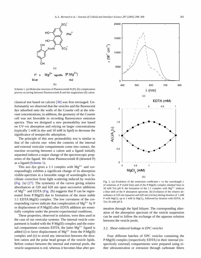
Fig. 3. (a) Evolution of the relative absorbance of LUV 100 (Clip =7.5 mM) suspension in the presence of 1% (mol/mol) Brij 76 at shearrate 10,000 s−1 as a function of shear time (0 min: solid line; 20 midashed line) at room temperature. In a purpose of comparison, the reabsorbance after cholate addition leading to vesicle disruption is alsoplayed (dotted line). (b) Evolution of the relative absorbance at 620 nmfunction of shear time at room temperature in LUV 100 suspension aferent concentrations in Brij 76: 0% (diamonds), 0.1% mol/mol (squares),1% mol/mol (circles) (shear rate= 10,000 s−1).
with holes of increasing diameter: 100, 200, and 400The corresponding suspensions of EPC vesicles are retively designated by SUV, LUV 100, LUV 200, and LU400 in the following.
LUV 100 were first submitted to shearing at 10,000 s−1
at room temperature[31] in the Couette viscometer. Asdisplayed inFig. 3b, only a weak leakage was observedafter 20 min. Then we added to the vesicle suspension a
Interface Science 287 (2005) 298–306
-
Fig. 4. Evolution of the relative absorbance at 620 nm as a function of srate for different suspensions of EPC vesicles (Clip = 7.5 mM) in the pres-ence of 1% (mol/mol) Brij 76 after 20 min of shearing at room temperatuLUV 100 (diamonds), LUV 200 (squares), LUV 400 (cross), SUV (triagle).
detergent bearing a large head group following the segy evoked in the Introduction[32]. The study was restricted to Brij 76 (C18OE10) which induced the desirephenomenon of permeation under shearing. This deterwas chosen because of its electric neutrality and itsvorable partition coefficient between the aqueous soluand the membrane[33]. This method could be extendeto other detergents presenting the same properties. Brwas added at 0.1 or 1% (mol/mol) with respect to EPC inLUV 100 (Clip = 7.5 mM). Such detergent concentratio(respectively 0.0075 and 0.075 mM) are above its critmicellar concentration (cmc= 0.003 mM at 25◦C) [34].Without shearing, no bilayer permeation occurred up to(mol/mol) in Brij 76: we did not observe any evolutioof the vesicle suspension at the day timescale. In contbilayer permeability dramatically increased under shea10,000 s−1 (Figs. 3a and 3b). The extent of permeation increases at short times and then saturates: at 1% (mol/mol)in Brij 76, the plateau is almost reached after 10 min. Tsame experiments carried out on LUV 200 and LUV 4led to similar results.Fig. 3b shows that increasing detergeconcentration increases the rate of permeation.
The significance of vesicle size was studied for a giconcentration of Brij 76 at different shear rates at room teperature.Fig. 4 displays the extent of permeation inducby shearing in LUV 100 and LUV 200 in the presenof 1% (mol/mol) Brij 76 after 20 min at 2000, 5000, an10,000 s−1. The permeation extents in SUV and LUV 40in the presence of 1% (mol/mol) Brij 76 after 20 min at10,000 s−1 are also shown inFig. 4. One notices that the ex
tent of permeation increases with the shear rate but reaches aplateau above 5000 s−1 for both LUV 100 and LUV 200. Inaddition, LUV are more sensitive to shearing than the SUV.
d and
UVom
rizeunderselteresh
- oriclesit issh
ith-nm
ce
iclef thef thetheeke.hat-thatg in-ter
min
rewer
ar-
g toon-onellar
asnce
s ofori-
ce
le.con-mM
c-
e
M
inmh thehe
:1
arvedionsthese
nch-ast,to
omin
oushear-
sideg al-ned,isso-
A.-L. Bernard et al. / Journal of Colloi
Fig. 5. Mean diameter (gray bars) and polydispersity (white bars) of L100 (A), LUV 200 (B), and LUV 400 (C) before and after shearing at rotemperature at 10,000 s−1 for 20 min in the presence of 1% (mol/mol)Brij 76.
3.3. Shear-induced fusion in EPC vesicles
Dynamic light scattering (DLS) was used to charactethe vesicle preparations. The diameter of the SUV was foin the 50-nm range.Fig. 5 compares the average diametand polydispersities[35] of the different LUV samples. Thaverage diameters are in line with the mesh size of the fiused (135, 225, and 325 nm for 100-, 200-, and 400-nm msize filters, respectively). As already noted elsewhere[36],the vesicle samples prepared by extrusion on the 100200-nm mesh size filters are less polydisperse than vesprepared on the 400-nm mesh size filters. In addition,already known that EPC LUV obtained with a 100-nm mesize filter are more unilamellar than EPC LUV made wa 200-nm mesh size filter, those prepared with the 400mesh size filters being rather multilamellar[26].
Fig. 5 shows that shearing of the LUV in the presenof 1% (mol/mol) Brij 76 at 10,000 s−1 for 20 min at roomtemperature strongly modifies the size distribution of vessuspensions: the average diameter of the LUV 100 and oLUV 200 is increased, whereas the average diameter oLUV 400 is decreased. Big objects were formed fromoriginally “small” LUV 100 and LUV 200, whereas somof the largest vesicles from the LUV 400 preparation broMoreover, the polydispersity increased with shearing, wever the initial diameter of the vesicles. In fact, it seemsthe EPC vesicles reached a similar state after shearindependent of their initial size distribution (average diamearound 260 nm and polydispersity around 0.3 after 20of shearing at 10,000 s−1).
With a view to determining if the created objects welipid aggregates or fused vesicles, the sheared samplesobserved by freeze-fracture electron microscopy.Figs. 6aand 6brespectively display LUV 100 before and after she
−1
ing for 20 min at 10,000 s in the presence of 1% mol/molBrij 76 at room temperature.Fig. 6b does not exhibit anyvesicle aggregate. Moreover, conservation of the surface ofInterface Science 287 (2005) 298–306 303
e
the lipid bilayer suggests that vesicle breakage leadinlipid aggregates is a minor phenomenon if present. In ctrast,Fig. 6b confirms that fusion of vesicles does occur upshearing: it evidences the presence of very large unilamvesicles with diameters up to 1 µm. In contrast, fusion wnot observed after shearing the LUV 100 nm in the abseof Brij 76.
3.4. During shearing
To evaluate the competition between the processeleakage and fusion during shearing, we turned to the flumetric ANTS/DPX fusion test[28,29]. Collisional quench-ing is responsible for extinction of ANTS fluorescenby DPX. At too low a concentration in a 1:1 (mol/mol)ANTS:DPX mixture, the fluorescence of ANTS is visibIn contrast, the ANTS fluorescence is quenched at largecentrations. We recorded the emission spectra from 2ANTS/DPX LUV 100, and from a 1:1 (mol/mol) mixture of2 mM ANTS LUV 100 and 2 mM DPX LUV 100 (see Setion 2) before and after 20 min of shearing at 10,000 s−1 inthe presence of 1% (mol/mol) Brij 76 at room temperatur(λexc= 320 nm).
The initially low fluorescence emission from 2 mANTS/DPX LUV 100 in the presence of 1% (mol/mol)Brij 76 increased by a factor of 10 after shearing for 20 mat 10,000 s−1. This observation confirmed the results frothe permeation test: a shearing-induced leakage througbilayer dilutes the internal 1:1 ANTS:DPX solution and tquenching of ANTS by DPX becomes less efficient.
The initially large fluorescence emission from a 1(mol/mol) mixture of 2 mM ANTS LUV 100 and 2 mMDPX LUV 100 decreased to 2/3 of its original value aftershearing for 20 min at 10,000 s−1 at room temperature. Ascontrol experiment no change in fluorescence was obsewithout shearing for several hours. Under the concentratused, 100% leakage upon shearing would hardly alterfluorescence intensity; dilution of ANTS can only increaits quantum yield of fluorescence and essentially no queing by DPX is expected after 100% leakage. In contr100% of fusion of the internal compartment would leadobservation of a fluorescence intensity similar to that frthe ANTS/DPX LUV 100. Thus the observed decreaseemission intensity clearly shows that fusion of the aquecompartments is a faster process than leakage during sing.
3.5. Vesicle postloading by shearing
We finally undertook to postencapsulate molecules invesicles under shear. In fact, this could be an interestinternative when fragile or expensive molecules are concersince vesicle preparation and encapsulation would be d
ciated. Indeed, the Brij 76 detergent could be removed in asubsequent step either by dialysis or using adsorption on la-tex beads[37].
304 A.-L. Bernard et al. / Journal of Colloid and Interface Science 287 (2005) 298–306
(a) (b)
(c)
Fig. 6. Freeze-fracture electron microscopy pictures of LUV 100 before (a) and after (b) shearing for 20 min at 10,000 s−1 in the presence of 1% mol/molhisto ite bars).pictu
,m,
r-.re-siono-tinghearµMthe
al-2%
p toide
f theion
gnif-sionrij 76size,
Brij 76 at room temperature (black bar= 1.5 µm); (c) corresponding sizeThese histograms were obtained from analysis of 220 vesicles in each
EPC LUV 100 (Clip = 6.6 mM) prepared without calceincontaining only 50 mM Tris at pH 8 in the internal mediuwere sheared in the presence of 1% (mol/mol) Brij 76 in asolution containing 0.9 µM calcein 50 mM Tris pH 8 (extenal medium) for 20 min at 10,000 s−1 at room temperatureThen the calcein remaining in the external medium wasmoved by two successive chromatographies on gel exclucolumns with 50 mM Tris pH 8 as elution solution. The flurescence emission of these filtered vesicles only originafrom the calcein entrapped inside the vesicles after swas identical to the fluorescence emission from a 0.02calcein solution. It corresponds to the fluorescence ofinternal volume of the vesicles filled with the 0.9 µM ccein external solution during the shear. This value (about
of the starting external medium concentration) satisfactorilycompares with the theoretical internal volume for EPC LUV100 (Clip = 6.6 mM): 2% of the total volume, assuming agram of the same samples before (black bars) and after shearing (whre.
polar head surface per lipid of 70 Å2 [1,38]. Thus 0.9 µMcalcein penetrates inside the vesicles during shearing uequilibration between the concentrations of calcein insand outside the vesicles. Therefore, the postloading ovesicles by shearing was only limited by the volume fractof their internal compartment.
4. Discussion
The present series of experiments evidences the siicance of a shear on the behavior of EPC vesicles. Fuand leakage events were observed in the presence of Bdepending on diverse factors such as average vesicle
molar fraction of the detergent, and shear rate.Fig. 7 dis-plays a tentative mechanism to account for our observa-tions.
d and
ear-hear-thethe
can
ioneter00.
romyer.
thethegestcurthe
linej 76
thetorscen-
law
-
ntdi-,wn-
be-Theecteduch
ings af-
tices-
iesceithi-ctive
y af-m at
ed.cedon.edvesi-ady
ventsles.sion
A.-L. Bernard et al. / Journal of Colloi
Fig. 7. Schematic diagram of the mechanism governing the shing-induced fusion and shearing-induced leakage of vesicles. Upon sing, the Brij 76 molecules partially segregate to accommodateless-curved positions within the bilayer. This phenomenon promotesformation of pores favoring leakage. In addition, interaction of vesicleslead to fusion. In contrast, the largest vesicles are prone to scission.
One first examines the effect of the Brij 76 concentraton the leakage/fusion phenomena. In the absence of dgent, we observed no significant leakage of EPC LUV 1In the presence of detergent (up to 1% mol/mol to EPC) butwithout shear, no permeation occurred, as anticipated fthe homogeneous distribution of the detergent in the bilaIn contrast, the analysis relying on the capillary numberCadefined in the Introduction supports the conclusion thatshape of the largest fraction of the LUV investigated inpresent study is significantly affected by shear at the larexamined shear rates; in view of the large spontaneousvature of Brij 76, shearing here induces segregation ofBrij 76 molecules in the bilayer, which decreases thetension. Leakage and fusion are facilitated; the more Brithe easier this is.
Fig. 4 shows that the rate of leakage increases withshear rate until a certain threshold is reached. Two facplay a role here. First shear increases the Brij 76 con
tration at the vesicle poles, as already discussed above. Inaddition, shear increases the collision frequency betweenvesicles. Indeed, Smoluchowski showed that the kinetics ofInterface Science 287 (2005) 298–306 305
-
-
coagulation of particles submitted to a shear stress is aof the second order with a characteristic timeτ
γ̇
coll if we con-sider soft shocks making them stick together[39]:
(2)τγ̇
coll =3
32γ̇ R3N0.
N0 is the concentration of vesicles andR their average radius. By comparison, the characteristic timeτ0
coll for colli-sion at rest, determined by the Brownian motion, is
(3)τ0coll =
1
8πDRN0,
whereD = kBT/6πηR designates the diffusion coefficieof vesicles (η is the external average viscosity). If the amensional ratioβ = τ
γ̇
coll/τ0coll = kBT/8ηγ̇R3 is less than 1
vesicle coalescence will rely on shear and not on Broian motion. In the case of vesicles, the conditionβ < 1 isfulfilled as soon asR > 40 nm (takingkT = 4 × 10⁻²¹ Jat room temperature,η = 10−3 Pa s, andγ̇ = 10,000 s−1).Thus shearing does increase the number of collisionstween most vesicles investigated in the present study.increase of the coalescence rate due to shearing is expto strongly depend on vesicle size. The phenomenon is mmore pronounced for large vesicles.
Eventually vesicle size plays a major role in determinthe significance of shearing; the smallest vesicles are lesfected by shearing than the largest (seeFig. 4). Indeed, SUVare the vesicles least responsive to shearing. One nothat LUV 100, LUV 200, and LUV 400 behave rather similarly. Explanation of the latter observation probably relon vesicle multilamellarity. Extrusion is prone to produa larger fraction of multilamellar vesicles when filters wlarge holes are used[26]. In the case of multilamellar vescles, the leakage/fusion mechanism should be less effethan expected for the largest vesicles.
5. Summary
This paper demonstrates that shearing considerablfects suspensions of EPC vesicles in the range 50–400 nrates beyond 5000 s−1 when suitable detergents are addShear-induced leakage through lipid bilayers was evidenwith a new permeability test based on UV–vis absorptiDiffusion light scattering and electron microscopy showthat shear can also promote the membrane fusion of thecles; whatever the initial size of the vesicles, a same stestate seems to be reached after fusion and scission erespectively involving the smallest and the largest vesicThe fluorescence ANTS/DPX test demonstrated that fu
occurs faster than leakage upon shearing. Finally, shearingis suggested as an attractive tool for post-loading preformedvesicles.
and
rn-ringn mi
up-
93.x-
0.68–
92)
97)
ni,
m.
ehn,
L.
tl.
Rev.
di
, L.
ysica
77
5)
54
n,
lag,
ate
oen-
s.
son,
em-
,hys.
re.58
thethe
dex
55
27
ase V.
.
all,
306 A.-L. Bernard et al. / Journal of Colloid
Acknowledgments
We thank Hubert Hervet for helpful discussions conceing the Cary spectrometer and the dynamic light scattemeasurements and Jean-Claude Dedieu for the electrocroscopy measurements.
Supplementary material
The online version on this article contains additional splementary material.
Please visit doi:10.1016/j.jcis.2004.12.019.
References
[1] D.D. Lasic, Liposomes: From Physics to Applications, Elsevier, 19[2] R.R.C. New, Liposomes: A Practical Approach, IRL Press, O
ford/New York/Tokyo, 1990.[3] Y. Nagawa, S.L. Regen, J. Am. Chem. Soc. 113 (1991) 7237–724[4] Y. Nagawa, S.L. Regen, J. Am. Chem. Soc. 114 (1992) 16
1672.[5] K. Naka, A. Sadownik, S.L. Regen, J. Am. Chem. Soc. 114 (19
4011–4013.[6] Y. Liu, S.L. Regen, J. Am. Chem. Soc. 115 (1993) 708–713.[7] M. Shibakami, M. Inagaki, S.L. Regen, J. Am. Chem. Soc. 119 (19
12,354–12,357.[8] K. Takiguchi, F. Nomura, T. Inaba, I. Takeda, A. Saitoh, H. Hota
Chem. Phys. Chem. 3 (2002) 571–574.[9] R. Blumenthal, M.J. Clague, S.R. Durell, R.M. Epand, Che
Rev. 103 (2003) 53–70.[10] J.C. Bradley, M.-A. Guedeau-Boudeville, G. Jeandeau, J.-M. L
Langmuir 13 (1997) 2457–2462.[11] O. Diat, D. Roux, F. Nallet, J. Phys. II Fr. 3 (1993) 1427–1452.[12] M. Dvolaitzky, P.-G. de Gennes, M.-A. Guedeau-Boudeville,
Jullien, C. R. Acad. Sci. Paris Sér. II 316 (1993) 1687–1690.[13] M.-A. Guedeau-Boudeville, L. Jullien, J.-M. di Meglio, Proc. Na
Acad. Sci. USA 92 (1995) 9590–9592.[14] N. Shahidzadeh, D. Bonn, O. Aguerre-Chariol, J. Meunier, Phys.
Lett. 81 (1998) 4268–4271.[15] A.-L. Bernard, M.-A. Guedeau-Boudeville, L. Jullien, J.-M.
Meglio, Langmuir 16 (2000) 6809–6820.[16] A.-L. Bernard, M.-A. Guedeau-Boudeville, O. Sandre, S. Palacin
Jullien, J.-M. di Meglio, Langmuir 16 (2000) 6801–6808.
Interface Science 287 (2005) 298–306
-
[17] See also F. Brochard-Wyart, P.-G. de Gennes, O. Sandre, PhA 278 (2000) 32–51.
[18] M. Kraus, W. Wintz, U. Seifert, R. Lipowsky, Phys. Rev. Lett.(1996) 3685–3688.
[19] C. Taupin, M. Dvolaitzky, C. Sauterey, Biochemistry 14 (1974771–4775.
[20] F. Brochard, J.F. Lennon, J. Phys. Fr. 36 (11) (1975) 1035–1047.[21] J.A. Bouwstra, P.L. Honeywell-Nguyen, Adv. Drug. Delivery Rev.
(2002) 541–555.[22] Y. Leong Yeow, S. Ranil Wickramasinghe, Y.-K. Leong, B. Ha
Biotechnol. Prog. 18 (2002) 1068–1075.[23] Y.C. Fung, Biomechanics Circulation, second ed., Springer-Ver
New York, 1997.[24] Estimation for a nebulizer or hairspray aerosol (flow r
∼1500 µl/min; nozzle diameter∼200 µm).[25] O. Freund, P. Mahy, J. Amedee, D. Roux, R. Laversanne, J. Micr
capsul. 17 (2) (2000) 157–168.[26] M.J. Hope, M.B. Bally, G. Webb, P.R. Cullis, Biochim. Biophy
Acta 812 (1985) 55–65.[27] G. Charlot, Les méthodes de la chimie analytique, fourth ed., Mas
Paris, 1961.[28] N. Düzgünes, T.M. Allen, J. Fedor, D. Papahadjopoulos, Bioch
istry 26 (1987) 8435–8442.[29] V. Marchi-Artzner, T. Gulik-Krzywicki, M.-A. Guedeau-Boudeville
C. Gosse, J.M. Sanderson, J.-C. Dedieu, J.-M. Lehn, Chem. PChem. 2 (2001) 367–376.
[30] D.A. Kendall, R. McDonald, Anal. Biochem. 134 (1983) 26–33.[31] See Section2. No significant difference in permeation rates we
observed between 10 and 40◦C for the chosen vesicle suspensions[32] R. Schubert, H. Wolburg, K. Schmidt, H. Roth, Chem. Phys. Lip.
(1991) 121–129.[33] As a comparison, we observed that Brij 700 (C18OE100) did not
induce any permeability during shearing.[34] From Sigma Product Information Sheet about Brij detergents.[35] The polydispersity coefficient characterizes the polydispersity of
diffusion coefficient but reasonably varies in the same direction aspolydispersity for the diameters. Above 0.3, the polydispersity inis meaningless.
[36] L.D. Mayer, M.J. Hope, P.R. Cullis, Biochim. Biophys. Acta 8(1986) 223–230.
[37] J.-L. Rigaud, D. Levy, G. Mosser, O. Lambert, Eur. Biophys. J.(1998) 305–319.
[38] The total internal volume fraction of such vesicle dispersion wevaluated to lie between 1 and 2% by NMR experiments. SeMarchi-Artzner, L. Jullien, L. Belloni, D. Raison, L. Lacombe, J.MLehn, J. Phys. Chem. 100 (1996) 13,844–13,856.
[39] V.G. Levich, Physicochemical Hydrodynamics, Prentice–HEnglewood Cliffs, NJ, 1962.