Shailee Jain*, Tarun Upadhyay*
22
Original Article Bio-Science Research Bulletin Vol. 33 No.2, July-December 2017: P.63-84 Print version ISSN 0970 0889 Online version ISSN 2320 3161 DOI 10.5958/2320-3161.2017.00010.4 QUORUM QUENCHING OF BACTERIA CAUSING DENTAL CARIES THROUGH HERBAL FORMULATION Shailee Jain*, Tarun Upadhyay* Author’s Affiliation: *Assistant Professor, Department of Life Sciences, Institute of Applied Medicines and Research, Ghaziabad (U.P). Corresponding Author: Tarun Upadhyay Assistant Professor, Department of Life Sciences, Institute of Applied Medicines and Research, Ghaziabad, NH-58, Delhi-Meerut Rd, Duhai, Ghaziabad, Uttar Pradesh-201206 E-mail: [email protected] Received on 02.11.2017, Accepted on 21.12.2017 Abstract Dental caries is the most prevalent and chronic diseases among dental problems like periodontal, tooth loss, gingivitis, toothache, mouth ulcers, gum bleeding and toothache. It is mainly caused by sugar content in diet and bacteria such as Streptococcus and Staphylococcus. Different methods are used for the prevention of dental caries such as chemical and natural methods. However, the present study was carried out to compare the antibacterial activity of aqueous preparation extract of dried and fresh leaves of Punica granatum, Eucalyptus, Mangifera indica and Acacia nilotica at 0.40% and 0.60% against the dental microflora of tobacco chewers plus smoking, tobacco chewers, smokers and normal persons (who are neither tobacco chewers nor smokers). Antibacterial activity of Punica granatum, Eucalyptus, Mangifera indica and Acacia nilotica have been determined by using agar well diffusion method against the sample. Out of the aqueous preparation of dried leaves extract of Punica granatum, Eucalyptus, Mangifera indica and Acacia nilotica, Punica granatum was found more effective at 0.40% concentration and Acacia nilotica was found least effective at 0.40%. But at 0.60% concentration, Eucalyptus was found more effective and Acacia nilotica was least effective. While, in case of aqueous extract prepared from fresh leaves of the selected plants, Eucalyptus was found more effective at 0.40% concentration, while at 0.60% concentration, Punica granatum was found more effective and Acacia nilotica was least effective. On the basis of quick response, Punica granatum was found more effective in comparison to other tested plant species (i.e. Eucalyptus, Mangifera indica, Acacia nilotica). Punica granatum showed zone of activity within 1days after implementation while other extracts showed zone of activity after 6-7 days. Keywords: Quorum Quenching, Dental caries, Herbal extract, Biofilm formation, Antibacterial activity.
Transcript of Shailee Jain*, Tarun Upadhyay*
Original Article Bio-Science Research Bulletin Vol. 33 No.2, July-December 2017: P.63-84
Print version ISSN 0970 0889 Online version ISSN 2320 3161
DOI 10.5958/2320-3161.2017.00010.4
QUORUM QUENCHING OF BACTERIA CAUSING DENTAL CARIES THROUGH HERBAL FORMULATION
Shailee Jain*, Tarun Upadhyay*
Author’s Affiliation:
*Assistant Professor, Department of Life Sciences, Institute of Applied Medicines and Research, Ghaziabad (U.P). Corresponding Author: Tarun Upadhyay Assistant Professor, Department of Life Sciences, Institute of Applied Medicines and Research, Ghaziabad, NH-58, Delhi-Meerut Rd, Duhai, Ghaziabad, Uttar Pradesh-201206 E-mail: [email protected] Received on 02.11.2017, Accepted on 21.12.2017
Abstract
Dental caries is the most prevalent and chronic diseases among dental problems like periodontal, tooth loss, gingivitis, toothache, mouth ulcers, gum bleeding and toothache. It is mainly caused by sugar content in diet and bacteria such as Streptococcus and Staphylococcus. Different methods are used for the prevention of dental caries such as chemical and natural methods. However, the present study was carried out to compare the antibacterial activity of aqueous preparation extract of dried and fresh leaves of Punica granatum, Eucalyptus, Mangifera indica and Acacia nilotica at 0.40% and 0.60% against the dental microflora of tobacco chewers plus smoking, tobacco chewers, smokers and normal persons (who are neither tobacco chewers nor smokers). Antibacterial activity of Punica granatum, Eucalyptus, Mangifera indica and Acacia nilotica have been determined by using agar well diffusion method against the sample. Out of the aqueous preparation of dried leaves extract of Punica granatum, Eucalyptus, Mangifera indica and Acacia nilotica, Punica granatum was found more effective at 0.40% concentration and Acacia nilotica was found least effective at 0.40%. But at 0.60% concentration, Eucalyptus was found more effective and Acacia nilotica was least effective. While, in case of aqueous extract prepared from fresh leaves of the selected plants, Eucalyptus was found more effective at 0.40% concentration, while at 0.60% concentration, Punica granatum was found more effective and Acacia nilotica was least effective. On the basis of quick response, Punica granatum was found more effective in comparison to other tested plant species (i.e. Eucalyptus, Mangifera indica, Acacia nilotica). Punica granatum showed zone of activity within 1days after implementation while other extracts showed zone of activity after 6-7 days. Keywords: Quorum Quenching, Dental caries, Herbal extract, Biofilm formation, Antibacterial activity.

Shailee Jain & Tarun Upadhyay / Quorum Quenching of Bacteria Causing Dental Caries Through Herbal Formulation
~ 64 ~
1. INTRODUCTION Dental caries is considered as a multifactorial, microbial infectious, chronic infectious, biofilm-dependent, site specific and transmissible disease [1, 2, 3] that results in localized dissolution and detoriation of the calcified tissues [4]. G.V. Black classified carious lession into six classes based on their location- Class I: caries lession on the occlusal areas or bucccal areas or lingual pits on the tooth surface; Class II: on the posterior occlusal and interior proximal surface of the teeth; Class III: on the anterior inter-proximal surface of tooth; Class IV: on the anterior inter-proximal surface of the tooth including the incisal corners; Class V: on the gingival third of the crown on facial or lingual surface of the tooth; Class VI: on tip of the cusp of posterior teeth. Stages of Dental Caries Dental caries occurs across a series of stages or a continuum of disease. Initial Stage Characterised by the first clinically non-cavitated visual changes in enamel on clean dry teeth. These lesions can be controlled and potentially reversed with changes to diet and personal dental hygiene practices, supplemented by fluorides [5]. Moderate Stage Characterized clinically either by a localized enamel breakdown (without clinical visual signs of dentinal involvement) or an underlying dark shadow from dentine. Extensive Stage Characterized by an extensive distinct cavity with visible dentin involving more than half of the tooth surface. There are several factors that can be responsible for dental caries such as biofilm formation, diet high rich in sugar and starch, bacteria in the mouth, stain (tobacco and smoking), poor oral hygiene, dry mouth, tooth size and shape, tooth position, thickness of enamel, poor calcium buffering capacity of saliva, high acid foods and drinks [6, 7, 8]. Out of these, biofilm play a major role in the development of dental caries [9]. Biofilm formation occurs into five distinct phases– (i) adsorption of host and bacterial molecules to the tooth surface, (ii) passive transport of oral bacteria to the pellicle-coated tooth surface, (iii) co-aggregation (co-adhesion) of later colonizers to already attached early colonizers, (iv) multiplication of attached microorganisms to produce confluent growth, (v) detachment [10]. Dental caries can be characterized by the appearance of a chalky white, brown, dark brown and shiny spot on teeth, bad breath, foul tastes, tooth sensitivity and toothache [7,8]. Dental caries can be influenced by several factors such as sex, age, socio-economic status, dietary patterns and oral hygiene habit, ethnic group [11, 12]. It can be transmitted either through caregivers, mothers by mouth-to-mouth transmission, by sharing a cup, utensil, toothbrush or even a shared toy that are contaminated with cariogenic bacteria [13]. Dental caries can be prevented by disturbing quorum sensing between bacteria within the biofilm and the phenomenon of the inhibition of quorum sensing called quorum quenching [14]. However, different methods are adopted to inhibit quorum sensing including behavior modification, chemical methods and natural method. Behavior modification includes dental health education, history of oral hygiene routine, dietary habit include sugar and starch consumption and regular dental check-up. While chemical method includes topical varnishes,

Bio-Science Research Bulletin / Volume 33 Number 2 / July-December 2017
~ 65 ~
use of fluoride tablets, chlorhexidine, casein phosphopeptide–amorphous calcium phosphate, magnesium, gelatin, root canal treatment, use of restoration material (include dental amalgam, composite resin, porcelain, gold and mercury filling), toothpaste containing fluorine, triclosan and remineralisation [7, 15) and natural method includes the use of herbs and herbal toothpaste powder (dant rattan herbal product, miswak toothpaste etc). However, dental treatment is highly expensive remedy because of the utilization of the chemical agents like triclosan, chlorexidine and amine based fluoride, antiseptics and antibacterial agents but their utilization has been limited due to their drawbacks such as toxicity, side effect, unaffordable, unsafe and uneconomic. While the major drawback of chemicals that they can alter oral micro biota such as vomiting, diarrhea, burning sensation and tooth staining [16]. Due to adverse effects of chemical based remedies, plant based products emerged out as a best alternative. Medicinal plants have been used as traditional treatments for numerous human diseases from thousands of years in many parts of the world. In rural areas of the developing countries, they continue to be used as the primary source of medicine. About 80% of the people in developing countries use traditional medicines for their health care. The natural products obtained from plants contain very rich biologically active constituents which have great potential against bacterial species and they act as main ingredient in various pharmaceutical products. A number of plants are used as chewing sticks in various parts of world which help in cleaning the buccal cavity. There are approximately 5, 00, 000 plant species occurring worldwide, of which only 1% has been phytochemically investigated. Thus, there is great potential for discovering novel bioactive compounds. There have been numerous reports of the use of traditional plants and natural products for the treatment of oral diseases. Many plant-derived medicines used in traditional medicinal systems have been recorded in pharmacopeias as agents used to treat infections and a number of these have been recently investigated for their efficacy against oral microbial pathogens [17]. Herbs are alkaline in nature with high antibacterial activity and help to maintain acid alkaline balance of saliva, decrease plaque, calculus formation, inhibit the growth of oral pathogens, influence the adhesion of bacteria to surfaces and reduce the symptoms of oral diseases. While, the plant extracts essential oils and purified photochemical have the potential to be developed into agents that can be used as preventative or for the treatment of oral diseases. Commercial mouthwashes containing essential oils are useful in the long-term control of plaque and mild- to-moderate gingivitis and are preferred those containing chlorhexidine for long-term daily use [18, 19]. Plants like Miswak has been used as chewing stick in many parts of world in various cultures with different names such as Miswak or Arak in Middle East, Datan in India and Pakistan. A number of plants like orange tree, lime tree have been used in Africa as chewing sticks whereas roots of plant like Senna were used in American continent. Neem plant has been widely used as chewing stick in the maintenance of oral hygiene throughout the Indian subcontinent. Some other plant parts are also used to maintain the oral hygiene such as Eucalyptus leaves are used to mask the bad mouth odour, onion and lime juices are used as gargles and to relieve in toothache. The use of medicinal plants belonging to the family of Fabaceae, Ebenaceae, Bombaceae and Anonnaceae has been reported for treatment of oral diseases [20].

Shailee Jain & Tarun Upadhyay / Quorum Quenching of Bacteria Causing Dental Caries Through Herbal Formulation
~ 66 ~
2. MATERIALS AND METHODS A total of sixteen persons were randomly selected as volunteers for the present study. Out of sixteen persons, four persons known to be tobacco chewer plus smoking, four tobacco chewers, four smokers and four were normal persons (who were neither smoker nor tobacco chewers). The plaque were removed with the help of sterile tooth picks from the oral cavity of each volunteers comes under these categories. Then the plaque samples from a given volunteer were placed in separate test tubes. In this way, four test tubes were used to collect the plaque samples from the volunteers of one category. Similarly, this process was repeated for the collection of plaque samples from the other three categories. All the four test tubes of each category were immediately brought to the laboratory for further processing. Isolation and selection of bacteria from dental plaque samples All the plaque samples from a given volunteer were transferred aseptically to a test tube containing 10 ml of sterile water and crushed using sterile glass rod to obtain a suspension of plaque particles. 1ml of this suspension was transferred to another test tube containing 9 ml of sterile water to obtain 1:100 dilution of this suspension. From this, further dilutions of 1:1000 and 1:10000 were obtained and the original stock suspension was discarded.1ml of suspension from the test tube containing 1:100 dilution was transferred aseptically to each of four Petri dishes pre-sterilized in an oven at 160°C for 4hrs. The Petri dishes were gently rotated to ensure uniform spread of the inoculums. 20 ml of sterilized and cooled nutrient agar medium were added to each Petri dish. Similar procedure was repeated for the other two dilutions i.e., 1:1000, 1:10000. The Petri dishes were incubated at 37°C for 24-48hrs. The remaining samples from other three categories volunteers were also proceed in similar manner. Then, the bacteria were selected on the basis of morphology and frequency. Selected culture of bacteria were transferred into cooled autoclaved nutrient agar broth and incubated at 37°C for 24 to 48hrs. Identification and characterization of bacteria Different types of bacterial cultures growing on the Petri plates were recognized as S1, S2 … S33 which contained 33 strains of bacteria. Each Petri plate was considered as a unit of study like a quadrate and the frequency percentage of each strain as well as the number of isolates of each strain were calculated. 24 hrs old cultures of all the 33 strains were used to study the Gram’s reaction and morphological characteristics. The strain S1 was found most frequently and highly dominant in the dental plaques of normal persons, S2 was found most frequently and highly dominant in the dental plaque of smokers, S3 was found most frequently and highly dominant in the dental plaque of tobacco chewers and S4 was found most frequently and dominant in the dental plaque of tobacco chewers plus smoking. Biochemical tests were performed to find out the difference exists between different strains with reference to biochemical properties. These include IMVIC test such as indole test, methyl red test, voges-proskauer test, citrate utilization and catalase test. Collection and preparation of leaves extract Sufficient amount of fresh leaves of Punica granatum, Eucalyptus, Mangifera indica, Acacia nilotica were collected from Tapovan, C.C.S. University, Meerut. Two methods were used to prepare leaves extract: Extract Prepared from Dried Leaves The collected leaves were washed with running tap water to remove the possible dust or surface contamination. The washed materials were dried at room temperature for 6 to 8 days under shade. After drying, the materials were powered using electric blender. Then, five gram powder of plant leaves were dissolved separately in 100ml of sterilized distilled water in Erlenmeyer flask of 250 ml and shake for 24hrs at a constant speed of 200rpm on a rotator shaker. The solution were filtered through muslin cloth and centrifuged at 8000rpm at 4°C for
Bio-Science Research Bulletin / Volume 33 Number 2 / July-December 2017
~ 67 ~
20 min. Supernatants were separately collected and filtered aseptically in the inoculation chamber with the help of bacterial filters, the filtrate were collected and make it up to 500ml with sterilized distilled water. Extract Prepared from Fresh Leaves Freshly collected leaves washed with double distilled water and cut into small disc. Five gram of leaves disc dissolved in 100ml double distilled water by boiling for 15 min and filtered through Whatman Number-1-filter paper. The filtrate was collected and makes it up to 500ml with sterilized distilled water. Antibacterial activity of aqueous leaves extract of plants Antibacterial activity of the aqueous solution of plant extracts were determined by using agar well diffusion bioassay. For this, 1ml of the bacterial suspension was poured in each of sixteen pre-sterilized petri plates for one plant extract i.e., Punica granatum. Luke warm NAM (Nutrient Agar Medium) was poured in each sixteen petri dish inoculated with 1ml bacterial suspension. After solidification of the medium, each petri dish was punched to make three wells of 0.8cm diameter with the help of sterilized cork-borer. In each petri dish, 50µl of the selected extract was added to two wells and 50µl of the autoclaved distilled water was added to the remaining third well. Well with distilled water was served as control. The plates were incubated overnight at 37°C for 24hrs. The antibacterial activity was interpreted from the size of the diameter of zone of inhibition measured in cm, it was evaluated as the clear zone appeared surrounding the well filled with extract. The remaining three plant extracts (prepared from Eucalyptus, Mangifera indica, Acacia nilotica) were also preceded in a similar way. Statistical analysis Data were expressed as mean±SEM of three-independent experiments. 3. RESULTS AND DISCUSSION The present study was conducted to assess the difference among the bacteria colonizing dental plaques of tobacco chewers with smoking, tobacco chewers, smokers and normal persons (non-smoker and non-chewer). Total isolates (TI), percentage isolates (PI), frequency (F) and frequency class (FC) of isolated oral microflora A total 33 strains of bacteria were isolated from all the samples. In all, 5009CFU were isolated from the dental plaque of chewers with smokers, 5820 CFU were isolated from dental plaques of tobacco chewers, 7148 CFU were isolated from the dental plaques of smokers while 7286 CFU were isolated from normal persons. The total isolates, percentage isolates, frequency and frequency class of all the 33 strains are represented in the table 1. Although a lot of work has been carried out in the past to observe the effect of smoking on oral bacteria and dental plaque formation but there is limited information on the relationship between smokeless tobacco user and periodontal diseases in reference to the role of microbes. Saini et al. (2009) reported a substantial reduction in the normal oral microbiota of the persons who are chronic tobacco eaters taking 25-30 gutkas per day (1431) as compared to (7910) in normal ones [21]. Rani (2010) also isolated 27 strains of bacteria from dental plaques. In all, 7910 CFU were isolated from dental plaque of normal persons and 1431 CFU from tobacco chewers [22]. While, Singh (2010) isolated 24 strains of bacteria from dental plaques. In all, 7901 CFU were isolated from dental plaque of normal persons and 1295 CFU were isolated from tobacco chewer [23]. Chandrabhan et al. (2012) isolated total of 39 bacteria from the dental plaques of persons belong to different age groups (1 - 20, 21 - 40, 41 - 60 year and above) [24]. Sapkota et al. (2010) observered more than 90% of the tobacco samples from the cigarettes contained Actinetobacter, Bacillus, Burkholderia, Closteridium, Klebsiella, Pseudomonas aerogenosa and Serratia [25].

Shailee Jain & Tarun Upadhyay / Quorum Quenching of Bacteria Causing Dental Caries Through Herbal Formulation
~ 68 ~
Table 3.1: Total isolate (TI), percentage isolate (PI), frequency (F) and frequency class (FC) of bacterial strains isolated from dental plaque of normal persons, smokers, chewers and chewers with smokers.
Bacterial strain
Normal persons Smokers
Tobacco Chewers
Chewers with Smokers
TI PI
F FC TI
PI
F FC TI
PI
F FC TI
PI
F FC
S1 1002 13.75 100 V 888 12.42 87.5 V 600 10.30 87.5 V 725 14.47 89.58 V
S2 985 13.52 89.58 V 782 10.94 93.75 V 800 13.74 83.33 V 666 13.29 87.5 V S3 885 12.14 95.83 V 625 8.74 79.17 IV 1012 17.38 97.91 V 555 11.08 83.33 V S4 995 13.66 93.75 V 850 11.89 85.42 V 725 12.45 93.75 V 900 17.96 93.75 V S5 150 2.05 66.66 IV 72 1.00 58.33 III 55 0.94 52.08 III - - - - S6 402 5.52 81.25 V 226 31.61 68.75 IV - - - - 156 3.11 66.66 IV S7 55 0.75 60.42 IV 225 3.15 75 IV 5 0.08 4.16 I 125 2.49 60.14 IV S8 185 2.54 79.12 IV 15 0.20 12.5 I 225 3.86 39.58 II 56 1.11 27.08 II S9 - - - - - - - - - - - - 355 7.08 77.08 IV S10 45 0.62 45.83 III 2 0.02 2.08 I 25 0.42 25 II - - - - S11 105 1.44 75 IV 185 2.59 37.5 II 55 0.94 35.14 II 66 1.31 54.16 III S12 333 4.57 81.25 V 194 2.71 70.83 IV 9 0.15 8.33 I 2 0.03 2.08 I S13 46 0.63 58.33 III 39 0.54 27.08 II 125 2.14 72.91 IV 225 4.49 58.33 III S14 8 0.10 8.33 I - - - - 75 1.28 54.16 III 55 1.09 45.83 III S15 444 6.09 66.66 IV 345 4.82 87.5 V 225 3.86 79.16 IV 5 0.09 4.16 I S16 89 1.22 75 IV 355 4.96 81.25 V - - - - - - - - S17 112 1.54 64.59 IV - - - - 255 4.38 72.91 IV 7 0.13 6.25 I S18 15 0.20 14.59 I 455 6.36 83.33 V 45 0.77 39.58 II - - - - S19 25 0.34 25 II 85 1.18 56.25 III 3 0.05 2.08 I - - - - S20 77 1.05 43.75 III - - - - 125 2.14 56.25 III 55 1.09 39.58 II

Bio-Science Research Bulletin / Volume 33 Number 2 / July-December 2017
~ 69 ~
S21 88 1.20 54.17 III 295 4.12 66.66 IV - - - - 2 0.03 4.16 I S22 9 0.12 8.33 I 189 2.64 52.08 III 67 1.15 66.66 IV 95 1.89 81.25 V S23 186 2.55 33.33 II 403 5.63 60.41 IV 160 2.74 75 IV - - - - S24 66 0.90 66.66 IV 17 0.23 8.33 I 33 0.56 25 II 298 5.94 72.91 IV S25 205 2.81 72.92 IV 195 2.72 70.83 IV 95 1.62 68.75 IV 23 0.45 31.25 II S26 175 2.40 52.09 III 110 1.53 47.91 III 325 5.58 91.66 V 75 1.49 54.16 III S27 18 0.25 12.5 I 11 0.15 12.5 I 255 4.38 77.08 IV 105 2.09 64.58 IV S28 5 0.07 4.17 I 75 1.04 39.58 II 25 0.42 22.91 II 215 4.29 70.83 IV S29 23 0.32 27.09 II - - - - 95 1.63 81.25 V - - - - S30 69 0.95 37.5 II - - - - 15 0.25 14.58 I 55 1.09 37.5 II S31 91 1.25 2.09 III 105 1.46 54.17 III 36 0.61 29.16 II 1 0.01 2.08 I S32 188 2.58 66.66 IV 255 3.56 89.58 V 95 1.63 58.33 III - - - - S33 195 2.68 79.16 IV 150 2.09 50 III 255 4.38 79.16 IV 187 3.73 56.25 III Total 7286 7148 5820 500
9

Shailee Jain & Tarun Upadhyay / Quorum Quenching of Bacteria Causing Dental Caries Through Herbal Formulation
~ 70 ~
As we can observed from the table 1, a total of 1002 isolates (100%) of the bacterial strain. S1 was isolated which is highly dominant in the plaques of healthy persons with a total of 888 isolates (87.5%). Bacterial strain S2 was isolated from the dental plaque which is highly dominant in the plaques of smokers with total of 1012 isolates (97.91%). Bacterial strain S3 was isolated which is highly dominant in the plaques of tobacco chewers, while the dental plaques of chewers with smokers were highly dominated by the bacterial strain S4 with total isolates of 900 with (93.75%) frequency. All the isolated and tested strains were gram positive. Tentative characterization of tested bacterial strains
Fig 1: Biochemical test of selected bacterial strains- (a) Indole test, (b) Methyl red test, (c) Voges-proskauer test, (d) Catalase test Different biochemical test (Table 2 and Fig. 1) were performed on the four selected strains of bacteria i.e., S1, S2, S3 and S4 to characterize them by performing gram staining reaction, indole test, methyl red, Voges-proskauer test, citrate utilization test and catalase test.

Bio-Science Research Bulletin / Volume 33 Number 2 / July-December 2017
~ 71 ~
Table 2: Biochemical test of four selected strain of bacteria
All four strains gave positive test except citrate utilization test by giving pink colour for indole test and methyl red, orange colour for voges-proskauer test and bubble formation for catalase test. All the isolated strains of bacteria were characterized as gram-positive by performing gram staining. Singh (2010) observed all the isolated strains of bacteria from dental plaques were gram positive and stated that Staphylococcus and Streptococcus were commonly occurring strains of bacteria in dental plaque. Out of which Staphylococcus was dominated in the dental plaque of normal persons and Streptococcus was dominated in the dental plaque of tobacco chewers [23]. Rani (2010) also observed that isolated strains of bacteria from dental plaques were gram positive and observed Staphylococcus and Streptococcus were two dominated bacterial strains in dental plaque. Out of which Staphylococcus was dominated in the dental plaque of normal persons and Streptococcus was dominated in the dental plaque of tobacco chewers [22]. Antibacterial activity of different plant extracts on tested bacterial strains However, the present study was carried out to compare the antibacterial activity of aqueous preparation of dried and fresh leaves extract of Punica granatum, Eucalyptus globulus, Mangifera indica and Acacia nilotica against the dental microflora of tobacco chewers plus smoking, tobacco chewers, smokers and normal persons (who are neither tobacco chewers nor smokers) by agar well diffusion method. Aqueous preparation of dried and fresh leaves extract of Punica granatum, Eucalyptus globulus, Mangifera indica and Acacia nilotica were applied at concentration of 0.40% and 0.60% against the dental microflora and their effectiveness were determined by measured the zone of inhibition at concentration of 0.40% and 0.60% as given in table 3, 4, 5 and 6 and fig. 2, 3, 4, 5.
BIOCHEMICAL TESTS REACTIONS Indole test + Methyl red test + Voges–Proskauer test +
Citrate utilization test - Catalase test + Gram staining +

Shailee Jain & Tarun Upadhyay / Quorum Quenching of Bacteria Causing Dental Caries Through Herbal Formulation
~ 72 ~
Fig 2: Zone of inhibition caused by dried leaves extract of Punica granatum (a), by Eucalyptus globulus (b), by Mangifera indica (c), by Acacia nilotica (d) Table 3: Zone of inhibition by aqueous preparation of dried leaves extract of Punica granatum, Eucalyptus globulus, Mangifera indica and Acacia nilotica
Test bacterial Strains
Punica granatum Eucalyptus globules
Mangifera indica Acacia nilotica
0.40% 0.60% 0.40% 0.60% 0.40% 0.60% 0.40% 0.60% S1 0.4 0.6 0.55 0.25 0.2 0.4 0.7 0.7 S2 0.3 0.4 0.75 0.6 0.55 0.65 0.65 0.75 S3 0.45 0.7 0.55 0.55 0.55 0.45 0.4 0.5 S4 0.25 0.2 0.45 0.2 0.4 0.5 0.6 0.75
Bio-Science Research Bulletin / Volume 33 Number 2 / July-December 2017
~ 73 ~
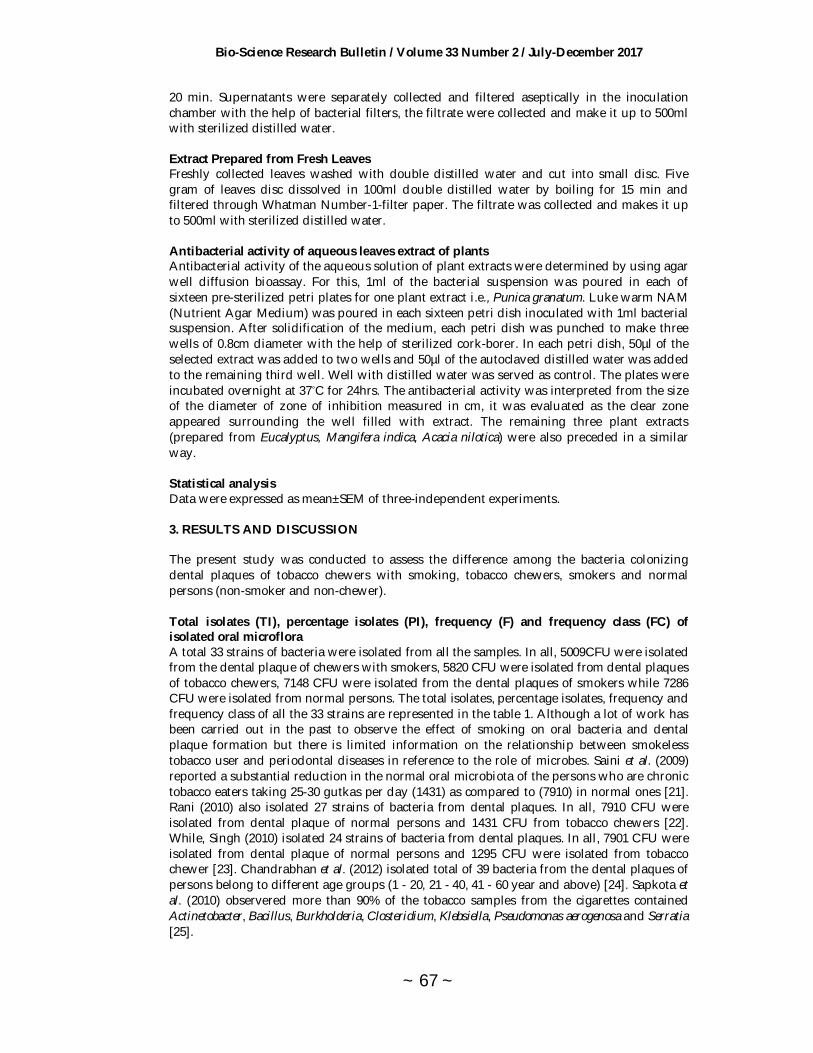
Fig 3: Zone of inhibition caused by fresh leaves extract of Punica granatum (a), by Eucalyptus globulus (b), by Mangifera indica (c), by Acacia nilotica (d) Table 4: Zone of inhibition by fresh leaves extract of Punica granatum, Eucalyptus globulus, Mangifera indica and Acacia nilotica
Test bacterial Strains
Punica granatum Eucalyptus globulus
Mangifera indica Acacia nilotica
0.40% 0.60% 0.40% 0.60% 0.40% 0.60% 0.40% 0.60% S1 0.2 0.3 0.30 0.7 0.75 0.45 0.45 0.35 S2 0.3 0.4 0.35 0.6 0.5 0.6 0.45 0.55 S3 0.5 0.4 0.35 0.20 0.6 0.4 0.6 0.7 S4 0.2 0.4 0.1 0.15 0.7 0.5 0.75 0.5
Table 5: Percentage of inhibition by aqueous preparation of dried leaves extract of Punica granatum, Eucalyptus globulus, Mangifera indica and Acacia nilotica
Test bacterial Strains
Punica granatum
Eucalyptus globulus
Mangifera indica Acacia nilotica
0.40% 0.60% 0.40% 0.60% 0.40% 0.60% 0.40% 0.60% S1 50 25 31.25 68.75 75 50 12.5 12.5 S2 62.5 50 62.5 25 31.25 18.75 18.75 6.25 S3 43.75 12.5 31.25 31.25 31.25 43.75 50 37.5 S4 68.75 75 43.75 75 50 37.5 25 6.25

Shailee Jain & Tarun Upadhyay / Quorum Quenching of Bacteria Causing Dental Caries Through Herbal Formulation
~ 74 ~
Fig 4: Percentage of inhibition caused by dried leaves extract of Punica granatu (a), by Eucalyptus globulus (b), by Mangifera indica (c), by Acacia nilotica (d) Table 6: Percentage of inhibition by fresh leaves extract of Punica granatum, Eucalyptus globulus, Mangifera indica and Acacia nilotica
Test bacterial Strains
Punica granatum Eucalyptus globulus
Mangifera indica Acacia nilotica
0.40% 0.60% 0.40% 0.60% 0.40% 0.60% 0.40% 0.60%
S1 75 62.5 62.5 12.5 6.25 43.75 43.75 56.25 S2 62.5 50 56.25 25 37.5 25 43.75 31.25 S3 37.5 50 56.25 75 25 50 25 12.5 S4 75 50 87.5 81.25 12.5 37.5 6.25 37.5

Bio-Science Research Bulletin / Volume 33 Number 2 / July-December 2017
~ 75 ~
Fig 5: Percentage of inhibition caused by fresh leaves extract of Punica granatum (a), by Eucalyptus globulus (b), by Mangifera indica (c), by Acacia nilotica (d) Out of the aqueous preparation of dried leaves extract of Punica granatum, Eucalyptus globulus, Mangifera indica and Acacia nilotica, Punica granatum was found more effective at 0.40% (50%, 62.5%, 43.75%, 68.75%) extract concentration followed by Mangifera indica (75%, 31.25%, 31.25%, 50%), Eucalyptus globulus (31.25%, 62.5%, 31.25%, 43.75%) and Acacia nilotica (12.5%, 18.75%, 50%, 25%) was found least effective at 0.40%. But at 0.60% extract concentration, Eucalyptus globulus (68.75%, 25%, 31.25%, 75%) was found more effective followed by Punica granatum (25%, 50%, 12.5%, 75%), Mangifera indica (50%, 18.75%, 43.75%, 37.5%) and Acacia nilotica (12.5%, 6.25%, 37.5%, 6.25%). In case of aqueous extract prepared from fresh leaves of the selected plants, Eucalyptus globulus (62.5%, 56.25%, 56.25%, 87.5%) was found more effective followed by Punica granatum (75%, 62.5%, 37.5%, 75%), Acacia nilotica (43.75%, 43.75%, 25%, 6.25%) and Mangifera indica (6.25%, 37.5%, 25%, 12.5%) at 0.40% extract concentration, while at 0.60% extract concentration, Punica granatum (62.5%, 50%, 50%, 50%) was found more effective followed by Eucalyptus globulus (12.5%, 25%, 75%, 81.25%), Mangifera indica (43.75%, 25%, 50%, 37.5%) and Acacia nilotica (56.25%, 31.25%, 12.5%, 37.5%). Thus, dried leaves extract of Acacia nilotica was found least effective at 0.40% and 0.60%. But in case of aqueous leaves extract, Mangifera indica was found least effective at 0.40% and Acacia nilotica at 0.60%. On the basis of quick response, Punica granatum was found more effective plant in comparison to other tested plant species (i.e. Eucalyptus globulus, Mangifera indica, Acacia nilotica). Punica granatum showed zone of activity within 1-2 days after implementation while other extracts showed zone of activity after 4-5 days. Hegde Chaitra et al. (2012) observed that the chloroform and ethyl acetate extracts exhibited maximum inhibition against S. aureus (10 ±0.05 mm and 16 ± 0.20 mm, respectively) and methanolic extract was found to exhibit better inhibition against the gram-positive bacteria than the gram-negative ones, with the highest inhibitory zone against B. cereus (30 ± 0.32

Shailee Jain & Tarun Upadhyay / Quorum Quenching of Bacteria Causing Dental Caries Through Herbal Formulation
~ 76 ~
mm)[26]. Braga et al. (2005) studied that pomegranate extract at a higher concentration (1%, v/v) was found to completely inhibit the growth of Staphylococcus aureus FRI 722 and subsequent enterotoxin production [27]. Machado et al. (2003) observed that the ethylacetate extract revealed a zone of 16 ± 0.20 mm in the present study, whereas, the highest activities were previously found in the ethyl acetate fraction when tested against different S. aureus strains [28]. Prashanth (2001) observed that extracts prepared from flowers showed better results than those of leaves and also observed that the acetone extracts had more antibacterial effect than the methanol extracts [29]. Mathabe et al. (2005) reported that methanol, ethanol, acetone and water extracts obtained from pomegranate were effective against the tested microorganisms (S. aureus, E. coli, S. typhi, Vibrio cholerae, Shigella dysenteriae, S. sonnei, S. flexneri, S. boydii) showing inhibition zones between 12-31 mm[30]. Janani and Estherlydia (2013) observed that methanolic rind extract showed highest antimicrobial activity against Staphylococcus epidermis at the concentration of 100 mg/ml with zone of inhibition of 14mm. While, aqueous rind extract showed highest antimicrobial activity against Staphylococcus aureus at the concentration of 100mg/ml with the diameter of zone of inhibition of 22mm [31]. Menezes et al. (2006) investigated an antibacterial effect of hydroalcoholic extract of Punica granatum fruit (HAEP) against dental plaque microorganisms and was found to be effective against Staphylococcus, Streptococcus, Klebsiella and Proteus species, as well as E. coli and the ellagitannin and punicalagin are thought to be fraction responsible for pomegranate’s antibacterial activity[32]. Singh et al. (2002) reported that extracts of Punica granatum peel in different concentrations were effective against S. epidermidis, S. aureus, S. mutans, S. sanguinis and S. salivarius. He also demonstrated that antibacterial activity is related to the presence of hydrolysable tannins and polyphenolics in the pomegranate extract specifically punicalagin and gallagic acid [33]. Reddy et al. (2007) and Naz et al. (2007) demonstrated that gallic acid (a tannic acid) has the highest anti-bacterial effect against tested sensitive strains even at low concentrations [34, 35]. Kote et al. (2011) showed that pomegranate juice is effective against dental plaque microorganisms. It decreased the CFU by 32% and showed 23% reduction in the growth of Streptococci and 46% reduction in the growth of Lactobacillus after rinsing with 30 ml of pomegranate juice [36]. While, Trivedi and Kazmi (1979) using extracts of fruit barks and observed antibacterial activity of pomegranate extract against Bacillus anthracis and Vibrio cholera[37]. Menezes et al. (2006) observed that the hydroalcoholic extract from pomegranate fruit decreased the colony formation unit (CFU) per milliliters of dental plaque by 84%[32]. Sastravaha et al. (2005) observed the effect of pomegranate fruit extract on periodontal therapy and also observed that a gel containing extracts of Cenetella asiatica and Punica granatum was an effective adjunct to conventional periodontal therapy [38]. Abdollahzadeh et al. (2011) showed that methanolic extract of pomegranate might be used in control of common oral pathogens responsible for caries stomatitis and periodontal diseases [39]. George and Sumathy (2012) observed that pomegranate peel on Lactobacillus sp. showed highest inhibition when compared to the antibiotics used except Chloramphenicol [40]. Menezes et al. (2006) and Pereira et al. (2006) also studied the antimicrobial activity of P. granatum [32, 41]. Machado-Thelma et al. (2002) and Haghighati et al. (2003) observed the antimicrobial activity of methanolic and acetone extracts on both gram-positive and gram-negative non-oral bacteria [42, 43]. Mc-Carrell and coworkers (2008) found aqueous macerated extract of pomegranate rind inhibits growth of Staphylococcus aureus and Pseudomonas aeurginosa and the extract of Punica granatum rind had an effect on C. albicans in all concentrations, while the pith and seed did not have any effect. The antibacterial activity of Punica granatum may be related to polyphenol structures because polyphenols may affect the bacterial cell wall, inhibit enzymes by oxidized agents, interact with proteins and disturb co-aggregation of microorganism. Thus, pomegranate fruit as a whole has superior bioactivity against oral pathogens as compared to
Bio-Science Research Bulletin / Volume 33 Number 2 / July-December 2017
~ 77 ~
its purified polyphenols, which illustrates the chemical synergy of the whole fruit's multiple compounds compared to single, purified, active ingredients [44]. Ishnava et al. (2013) observed that the distilled water extract of E. globules leaf extracts is active against L. acidophilus. Ethyl acetate extracts are active against all the selected bacterial strain but show maximum activity against L. acidophilus, S. mutans and L. casei with the zone of inhibition is 13mm, 13mm and 11mm respectively. Hexane extracts of E. globules are also active against all selected microorganisms but the maximum zone of inhibition was obtained against L. acidophilus, S. mutans and L. casei is 12mm, 8mm and 10mm respectively. While the maximum activity of methanolic extract of plant leaves against L. acidophilus, L. casei and S. mutans is 12mm, 12mm and 13mm respectively [45]. Ghalem and Mohamed (2008) reported the E. globules activity against Staphylococcus aureus Gram (+) and Escherichia coli Gram (–) bacteria [46]. Badrunnisa et al. (2011) observed that the ethanol leaf extracts of Eucalyptus tereticornis showed highest activity against Pseudomonas aeruginosa (zone inhibition 19.50 ± 0.71mm, n = 3), followed by Bacillus thurangenisis (13.00 ± 2.12mm, n = 3) and Bacillus cereus (12.75 ± 0.35mm, n = 3)[47]. Nezhad et al. (2009) reported that the antibacterial activity of Eucalyptus extracts was due to the components such as 1, 8-cineole, citronellal, citronellol, citronellyl acetate, p-cymene, eucamalol, limonene, linalool, β- pinene, γ-terpinene, α- terpinol, alloocimene and aromadendrene [48]. Sirivan et al. (2008) reported that the volatile oils showed good antimicrobial activity against Propionibacterium acnes (MIC, MBC = 9.38 mg/ml for eucalyptus oil) [49]. Kachhiya (2008) observed that the methanolic extract of Eucalyptus globules stem is effective against anticariogenic bacteria [50]. Rakotonirainy and Lavedrine (2005) reported the previous study of the growth inhibition ability of Eucalyptus sp. against of both Gram-positive and Gram-negative bacteria and also studied the antimicrobial activity methanol extracts of Eucalyptus globules against four Gram-negative and nine Gram-positive bacterial strains including Pseudomonas aeruginosa and Staphylococcus sp. with zone inhibition ranging from 10-20 mm [51]. Badrunnisa et al. (2011) concluded that the methanol and ethanol extracts of Eucalyptus tereticornis produced outstanding antibacterial activity against Pseudomonas aeruginosa, Bacillus cereus, Bacillus thurangenisis isolated from used steel industry coolants. Although, the MIC against Pseudomonas aeruginosa (2 mg/ml) was higher than that for Bacillus cereus (0.5 mg/ml) and Bacillus thurangenisis (0.2mg/ml) while the ethanol leaf extract showed highest zone of inhibition of 19.50 ± 0.71mm in Pseudomonas aeruginosa at a concentration of 50 mg/ml, while the same extract concentration produced a zone inhibition of only 12.75 mm ± 0.35 (52.9 % less) and 13.00 ± 2.12 (50% less) with Bacillus cereus and Bacillus thurangenisis respectively[47]. Ayepola and Adeniyi (2008) observed that the methanol extracts of Eucalyptus camaldulensis showed greater activity against Klebsiella sp. (15-16 mm) than Pseudomonas aeruginosa and Staphylococcus aureus (13-14 mm). Ayepola and Adeniyi (2008) observed that the methanol extracts of E. camaldulensis showed greater activity against Salmonella typhi, Staphylococcus aureus and Bacillus subtilis (15 -16mm) than Klebsiella sp., Yersinia enterocolitica and Pseudomonas aeruginosa (14mm). While, the dichloromethane fraction exhibited higher activity against Klebsiella sp., Salmonella typhi, Yersinia enterocolitica and Bacillus subtilis (15–16mm) than Staphylococcus aureus and Pseudomonas aeruginosa (13-14mm) and the methanol residue had a lower activity against all the test organisms except Klebsiella sp. and Salmonella typhi [52]. Akin-Osanaiye et al. (2007) observed the antibacterial activity of E. camaldulensis extracts against Staphylococcus aureus [53]. Babayi et al. (2004) observed that the methanol extract of E. camaldulensis has been found to be effective against staphylococcus aureus and Bacillus subtilis [54].

Shailee Jain & Tarun Upadhyay / Quorum Quenching of Bacteria Causing Dental Caries Through Herbal Formulation
~ 78 ~
Ghalem and Mohamed (2008) observed that the growth of Staphylococcus aureus and Escherichia coli were not inhibited at low concentrations (i.e. 1 and 2 μl) of eucalyptus oil from E. camaldulensis and E. globules while the increasing amount of essential oil gave a distinct zone of inhibition [46]. The similar result was found by Cimanga et al. (2002) and Nair et al. (2008) [55, 56]. Some researchers reported that the antimicrobial activity of an essential oil is linked to its chemical composition. Sartorelli et al. (2006), Mohammadreza (2008), Maksimovi et al. (2008) observed that the antimicrobial activity of plants are due to the presence of functional groups of some compounds such as alcohol, phenols, terpenes and ketones [57,58,59]. Bachir and Benali (2012) observed that the essential oil showed antibacterial activity with varying magnitudes, depending on the size of inoculums and the concentration of essential oil. Diameter of inhibition zone of essential oil of E. globulus leaves varied from 8 to 26 mm. The largest zone of inhibition was obtained for E. coli (10-3 dilution) with 100% concentration of essential oil of E. globulus and the lowest for S. aureus (10−1 dilution) with 25% concentration of essential oil leaves [60]. These similar results was also observed Ait- Ouazzou (2011), Bachheti et al. (2011); Elaissi et al. (2011)[61,62,63]. While, Nagata et al. (2008) reported that the Macrocarpals A, B, C are the major components of Eucalyptus globules leaf demonstrate relatively strong anticariogenic bacterial activity against Streptococcus mutans and Streptococcus sobrinus [64]. Hannan et al. (2013) observed that size of zone of inhibition against S. typhi was directly proportional to the increased concentration of acetone mango leaf extracts (AMLE). S. typhimurium (ATCC14028) and S. aureus (ATCC 25923) showed the largest zone of inhibition (20 ± 1.5mm) at the lowest concentration of extract (50mg/ml) followed by MDR and antibiotic sensitive S. typhi (18 ± 1.5mm). They also observed that S. typhi isolates showed zone of inhibition >12mm at all concentrations of AMLE (i.e. at 50mg/ml, S. typhi showed 15.5 ± 3.5, MDR S. typhi showed 15.25 ± 3.75, Antibiotic sensitive S. typhi showed 15.5 ± 3.5, ATCC S. aureus showed 20.0 ± 1.5, ATCC S. typhimurium showed 20.0 ± 1.5. While at 100mg/ml, they showed 17.25 ± 3.75, 17±3.5, 17.5±3.5, 22.0±1.5 and 20.0 ± 1.5. At 150mg/ml, they showed 18.0 ± 4.0, 17.5 ± 3.5, 18.5 ± 3.5, 24.0 ± 1.5, 22.0 ± 1.5. At 200mg/ml, they showed 18.5 ± 3.5, 18.25 ± 3.25, 18.5 ± 3.5, 26.0 ± 1.5, 22.0 ± 1.5 and at 250mg/ml, they showed 19.75 ± 3.75, 19.75 ± 3.75, 19.75 ± 3.75, 26.0 ± 1.5, 24 ± 1.5). These results against MDR S. typhi compared with Nigerian that showed zones of 4.0, 7.0, 9.0, 12.0, 14.0 mm at concentrations of 50, 100, 150, 200 and 250 mg/ml respectively and demonstrated that acetone extract of mango leaf exerted highest activity against S. typhi compared to aqueous and methanolic extracts of mango leaves (Doughari and Manzara, 2008)[65,66]. Zakaria et al. (2006) observed in the study conducted in USA that ethanolic and methanolic extract of mango leaf showed relatively high zones of inhibition against S. typhi. Both ethanolic and methanolic extract of mango leaf showed inhibition zones of >13.0 mm and <16.0 mm at 50 and 100 mg/ml [67]. Doughari and Manzara (2008) observed that the differences in the activities of various extracts may be due to varying degrees of solubility of the active constituents in the solvent used. And also documented that different solvents have different solubility capacities for different phytoconstituents [66]. Thus, the study conducted by Hannan et al. (2013) showed that high inhibition zones against S. aureus (ATCC 25923) followed by S. typhimurium (ATCC 14028) [65] but the previous study showed the high inhibition zones against S. aureus (ATCC 25923) followed by S. typhimurium (ATCC 14028) (Zakaria et al., 2006; Doughari and Manzara, 2008) [67, 66].

Bio-Science Research Bulletin / Volume 33 Number 2 / July-December 2017
~ 79 ~
Zakaria et al. (2006), observed that this may be due to presence of lipopolysaccharides (LPS) in the Gram- negative bacteria [67]. Bharti (2003) in her study observed the antimicrobial activity of different extract of Mangifera indica against nine bacteria (Salmonella typhi, Klebsiella pneumoniae, Enterobacter aerogens, Mycobacterium tuberculosis, Streptococcus pyrogens, Pseudomonas aeuroginosa, Proteus vulgaris, Escherichia coli and Staphylococcus aureus). He observed that the methanol extract showed maximum zone of inhibition against Entrobacter aerogens (1.3 cm). The acetone extract showed a maximum zone of inhibition against Salmonella typhi (3.0cm). The hexane extract showed maximum zone of inhibition against Mycobacterium tuberculosis (0.5 cm). The ethyl acetate extract showed maximum zone of inhibition against Enterobacter aerogens (1.9 cm). The hexane-ethyl acetate extract showed maximum zone of inhibition against Streptococcus pyrogens (2.6 cm) and also against Salmonella typhi (2.5 cm) [68]. Sahrawat et al. (2013) observed that the methanol extract of Mangifera indica leaves showed maximum growth inhibition i.e. 18.5% against Salmonella typhi and minimum growth inhibition i.e. 0% against Pseudomonas fluorescens. The Ethanol extracts of Mangifera indica leaves showed maximum growth inhibition 20% against Pseudomonas fluorescens and minimum growth inhibition -28.75% against Klebsiella pneumoniae. The Benzene extract of Mangifera indica leaves showed maximum growth inhibitions 85.1% against Pseudomonas fluorescence and also showed minimum growth inhibition 18.51% against Shigella flexneri [69]. Parvathi et al. (2012) showed the presence of alkaloids, phenols, flavonoids, carbohydrates and absence of saponins in ethanolic flower extract of Mangifera indica. Luka and Mohammed (2012) showed the presence of alkaloids, carbohydrates, phytosterols, flavonoids and protein in ethanolic leaves extract [70]. Abdalla et al. (2007) observed that seed kernel of mango extract showed inhibitory effect against coliform and E. coli [71]. GM et al. (2007) observed that mango extract at 50% concentration showed maximum zone of inhibition on Streptococcus mitis [72]. Rahman et al. (2012) observed that the leaves extract of A. nilotica was an effective inhibitors of bacterial and fungal growth and also observed that the extract showed varying degrees of activity against Gram-positive and Gram-negative bacteria as well as fungi. It showed inhibitory zones ranging from 4 to 29mm against E. coli, S. aureus, P. aeruginosa, K. pneumonia and B. subtilis [73]. Mahesh et al. (2008) observed that the methanolic extracts of Acacia nilotica showed highest antibacterial activity against B. subtilis and Staphylococcus aureus with inhibition zone 15 ± 0.66mm while its leaf extract showed highest activity against Bacillus subtilis with inhibition zone 20 ± 1.20mm [74]. Rahman et al. (2012) observed the antimicrobial activity of methanolic extract of A. nilotia showed highest activity against E. coli followed by K. pneumonia, B. subtilis and P. aeruginosa [73]. Banso (2009) found that the ethanolic stem bark of A. nilotica L. produced antimicrobial activity against Streptococcus viridians, B. subtilis, S. aureus, E. coli and Shigella sonnei. He observed MIC value against B. subtilis and E. coli was 35 and 45mg/ml [75]. While Rahman et al. (2012) observed the antimicrobial activity of ethanolic extract of A. nilotica against E. coli and B. subtilis was 1mg/ml [73]. Sharma et al. (2014) observed that at 5% concentration of babool extract showed no antimicrobial activity, but its babool extract showed antimicrobial activity while maximum antimicrobial activity was found with 50% concentration with mean zone of inhibition of 1.4 mm [76].

Shailee Jain & Tarun Upadhyay / Quorum Quenching of Bacteria Causing Dental Caries Through Herbal Formulation
~ 80 ~
Saini et al. (2008) found that the antimicrobial activity of babool may be due to hydrophilic compounds such as polyphenols, gums (poly-saccharides) and tannins while secondary compounds includes anti-cancer (triterpenoid and saponins), diuretic (glucosides), natriuretic (glucosides), important nutraceutical (poly-saccaride and gum) anti-digestive disorder (saponins, tannins and flavanoids), anti-oxidant (polyphenols), anti-plasmodial (treptamine, tannins, organic acids and saponins [77]. 4. CONCLUSION Conclusively, the study revealed that Bacteria are considered as causative agents of dental caries. It might occur either in healthy, smokers, chewers as well as chewers with smokers. Dental caries can be caused by both Gram positive and Gram negative bacteria, but Gram positive bacteria play a major role in causing dental caries. General oral microflora was high in healthy persons followed by smokers, chewers and chewers with smokers. But, as per our observation, caries causing microorganism were more present in smokers followed by chewers, chewers with smokers while the least numbers found in healthy persons. The dried leaves extract of Punica granatum was found more effective and Acacia nilotica was effective at 0.40% concentration. But in case of fresh leaves extract, Eucalyptus globulus was found more effective and Mangifera indica was least effective at 0.40% concentration. The dried leaves extract of Eucalyptus globulus was found more effective and Acacia nilotica was least effective at 0.60% concentration. But in case of fresh leaves extract, Punica granatum was found more effective and Acacia nilotica was least effective at 0.60% concentration. 5. ACKNOWLEDGEMENTS We would also like to extend our thanks to Dr. Pradip Panwar, Department of Biotechnology, CCS University, Meerut for necessary guidance during the work. Mr. Sanjay Bansal (Secretory), Ms. Anshu Bansal (Jt. Secretory), IAMR Group of Institution, Ghaziabad for his motivation and support and I would like to also express my gratitude to Rajya Vardhan Tripathi for necessary guidance during the manuscript preparation. 6. CONFLICT OF INTEREST The authors declare no conflict of interest. REFERENCES 1. Hurlbutt M. Dental caries: A pH mediated diseases. Winter. 2010; 25(1): 9-14. 2. Chandrabhan D, Hemlata R, Renu B, Pradeep V. Isolation of dental caries bacteria from
dental plaque and effect of toothpaste on acidogenic bacteria. Open J. Med. Microbiol. 2012; 2(3): 1-5.
3. Ayub F, Thomas B, Paulaian B, Emil J. Herbs and dental caries: A review. Uni. J. Pharm. 2013; 2(5): 18-21.
4. Shah N, Mathur VP, Logani A. Reference Manual on Oral Health for Allopathic and Ayush Practioners. In: Centre for dental education and research. All India Institute of Medical Science, New Delhi. 2012; 1-36.
5. Longbottom CL, Huysmans MC, Pitts NB, Fontana M. Glossary of key terms. Monographs Oral Sci. 2009; 21: 209-216.
6. Li YH, Lau PCY, Lee JH, Ellen RP, Cvitkovitch DG. Natural genetic transformation of Streptococcus mutans growing in biofilms. J. Bacteriol. 2001; 183: 897-908.
7. Cury JA, Tenuta LM. How to maintain a cariostatic fluoride concentration in the oral environment. Adv. Dent. Res. 2008; 20(1): 13-16.
8. Hurlbutt M. Cambra: Best Practices in Dental Caries Management. www. rdhmag.com. 2011.
9. Marsh D. Dental plaque as a microbial biofilm. Caries Res. 2004; 38: 204-211.

Bio-Science Research Bulletin / Volume 33 Number 2 / July-December 2017
~ 81 ~
10. Silva ACB, Souza DCC, Portela G S, Araujo DAM, Sampaio FC. Microbial Dynamics and Caries: The Role of Antimicrobials, Contemporary Approach to Dental Caries, (Ming-Yu, Li. Ed.).ISBN: 978-953-51-0305-0309. 2012.
11. Sudha P, Bhasin S. Anegundi RT. Prevalence of dental caries among 5 to 13 years old children in Mangalore city. J. Indian Soc. Ped. Preven. Dent. 2005; 23:74-79.
12. Moses J, Rangeeth BN, Gurunathan D. Prevalence of dental caries, socio-economic status and treatment needs among 5 to 15 year old school going children of chidambaram. J. Clin. Diag. Res. 2011; 5: 146-151.
13. Chan KM, King M, Kilpatrick NM. Can infants catch caries? A review of the current evidence on the infectious nature of dental caries in infants. N Z. Dent J. 2005; 101(1): 4-11.
14. Sahu B, Guduguntla S, Mangalekar SB, Shetty S, Thakur P, Mishra S . Quorum sensing and quorum quorum facebook of microbial world. Int. J. Sci. Res. 2014; 3: 423-426
15. Rugg-Gunn AJ, Edgar WM, Geddes DA, Jenkins GN. The effect of different meal patterns upon plaque pH in human subjects. Br. Dent J. 1975; 139(9): 351-356.
16. Bairwa R, Gupta P, Gupta VK, Srivastava B. Traditional medicinal plants: use in oral hygiene. Int. J. Pharm. Chem. Sci. 2012;1(4): 15-29
17. Cowan MM. Plant products as antimicrobial agents. Clin. Microbiol. Rev. 1999; 12(4): 564-582.
18. Rodrigues F, Lehmann M, do Amaral VS, Reguly ML, de Andrade HHR. Genotoxicity of three mouthwash products, cepacol, periogard, and plax, in the Drosophila wing-spot test. Environ. Mol. Mutagenesis. 2007; 48(8): 644-649.
19. Yadav R, Yadav SK. Dental diseases and its cure: A review. Asian J. Pharm. Clin. Res. 2013; 6: 16-20.
20. Hadissa T, Jean- Pierre D. Use of medicinal plants for treatment of oral diseases in Burkina Faso. J. Ethnopharmacol. 2005; 104: 68-78.
21. Saini S, Saini SR, Katiyar R, Bhalero DS, Munde A. The use of tobacco and betel leaf and its effect on the normal flora of oral cavity. Pravara Med. Rev. 2009; 4: 17-19.
22. Rani A. Studies on Certain Aspects of Dental Plaque Microbial Biofilm. M.Sc. Dissertation. Department of Botany. CCSU, Meerut. India. 2010.
23. Singh J. Quorum Quenching for the Management of Dental Plaque Microbes. M.Sc. Dissertation. CCSU, Meerut. India. 2010.
24. Chandrabhan D, Hemlata R, Renu B, Pradeep V. Isolation of dental caries bacteria from dental plaque and effect of toothpaste on acidogenic bacteria. Open J. Med. Microbiol. 2012; 2(3): 1-5.
25. Sapkota AR, Berger S, Vogel TM. Human pathogens abundant in the bacterial metagenome of cigarettes. Environ. Health Perspectives. 2010.118(3): 351-356.
26. Hegde RC, Madhuri M, Swaroop TN, Das A, Sourav B, Rohit KC. Evaluation of antimicrobial properties, phytochemical contents and antioxidant capacities of leaf extracts of Punica granatum L. ISCA J. Biol. Sci. 2012; 1(2): 32-37.
27. Braga LC, Leite AA, Xavier KG, Takahashi JA, Bemquerer MP, Chartone- Souza E, Nascimento AM. Synergic interaction between pomegranate extract and antibiotics against Staphylococcus aureus. Can. J. Microbiol. 2005; 51: 541-547.
28. Machado TB, Pinto AV, Pinto MCFR, Leal ICR, Silva M, Amaral ACF, Kuster RM, Nettodos SKR. In vitro activity of Brazilian medicinal plants, naturally occurring naphthoquinones and their analogues, against methicillin-resistant Staphylococcus aureus. Int. J. Antimicrob. Agents. 2003; 21: 279-284.
29. Prashanth D, Asha MK. Antibacterial activity of Punica granatum. Fitoterapia. 2001; 72: 171-173.
30. Mathabe MC, Nikolova RV, Lall N, Nyazema NZ. Antibacterial activities of medicinal plants used for the treatment of diarrhoea in Limpopo Province, South Africa. J. Ethnopharmacol. 2005; 105: 286-293.
31. Janani J, Estherlydia D. Antimicrobial activities of Punica granatum extracts against oral microorganism. Int. J. Pharm. Tech. Res. 2013; 5(3): 973-977.
32. Menezes SM, Cordeiro LN, Viana GS. Punica granatum (pomegranate) extract is active against dental plaque. J. Herb Pharmacother. 2006; 6: 79-92.

Shailee Jain & Tarun Upadhyay / Quorum Quenching of Bacteria Causing Dental Caries Through Herbal Formulation
~ 82 ~
33. Singh RP, Chidambara MKN, Jayaprakasha GK. Studies on the antioxidant activity of pomegranate (Punica granatum) peel and seed extracts using in vitro models. J. Agric. Food Chem. 2002; 50(1): 81-86.
34. Reddy MK, Gupta SK, Jacob MR, Khan SI, Ferreira D. Antioxidant, antimalarial and antimicrobial activities of tannin-rich fractions, ellagitannins and phenolic acids from Punica granatum L. Planta. Med. 2007; 73(5): 461-467.
35. Naz S, Siddiqi R, Ahmad S, Rasool SA, Sayeed SA. Antibacterial activity directed isolation of compounds from Punica granatum. J. Food Sci. 2007; 72(9): M341-M345.
36. Kote S, Nagesh L. Effect of pomegrenate juice on dental plaque microorganism (Streptococci and Lactobacilli). Anc. Sci. Life. 2011; 31(2). 49-51.
37. Trivedi VB, Kazmi SM. Kachnar and anar as antibacterial drugs. Ind. Drugs. 1979; 16: 295-298.
38. Sastravaha G, Gassmann G, Sangtherapitikul P, Grimm WD Adjunctive periodontal treatment with Centella asiatica and Punica granatum extracts in supportive periodontal therapy. J. Int. Acsd. Periodoxtal. 2005; 7: 70-79.
39. Abdollahzadeh SH, Mashouf RY, Mogahaddam MH, Roozbahani N, Vahedi M. Antibacterial and antifungal activities of Punica granatum extract against oral pathogens. J. Dent. Tehran Uni. Med. Sci. 2011; 8(1): 1-6.
40. George S, Sumathy VJH. Antibacterial and antifungal activity of botanical extracts against microorganisms isolated from mouthflora. Biosci. Int. 2012; 1(3): 74-77.
41. Pereira JV, Pereira MSV, Sampaio FC, Sampaio MCC, Alves PM, Ara jo CRF, Higino JS. In vitro antibacterial and ant adherence effect of Punica granatum Linn extract upon dental biofilm microorganisms. Braz. J. Pharmacogn. 2006; 16: 88-93.
42. Machado-Thelma de B, Leal Ivana CR, Amaral ACF, Santos KRN, Silva MG, Kuster RM Antimicrobial ellagitannin of Punica granatum fruits. J, Braz. Chem. Soc. 2002; 13(5): 606-610
43. Haghighati F, Jaafari S, Beyt–Elahi JM. Comparison of antimicrobial effects of ten herbal extracts with chlorhexidine on three different oral pathogens; an in vitro study. HAKIM. 2003; 6: 71-76.
44. McCarrell EM, Gould SWJ, Fielder MD, Kelly AF, Sankary WE, Naughton DP. Antimicrobial activities of pomegranate rind extracts: enhancement by addition of metal salts and vitamin C. BMC Comp. Altern. Med. 2008; 8: 64.
45. Ishnava KB, Chauhan JB, Barad MB. Anticariogenic and phytochemical evaluation of Eucalyptus globules Labill. Saudi J. Biol. Sci. 2013; 20: 69-74.
46. Ghalem BR, Mohamed B. Antibacterial activity of leaf essential oils of Eucalyptus globules and Eucalyptus camaldulensis. Afr. J. Pharm. Pharmacol. 2008; 2(10). 211-215.
47. Badrunnisa S, Pai VR, Shantaram M. Antibacterial activity of Eucaluptus tereticornis for in use coolants of steel industry. Int. J. Res. Pharm. Biomed. Sci. 2011; 2(4): 1789-1794.
48. Nezhad FM, Zeigham H, Mota A, Sattari M, Yadegar A. Antibacterial activity of Eucalyptus extracts on methicillin resistance Staphylococcus aureus. Res. J. Biol. Sci. 2009; 4(8): 905-908.
49. Sirivan A, Rith W, Sujimon T, Panida V, Paisarn K, Prapan, Sae-Jong, Nijsiri R. The development of anti-acne products from Eucalyptus globules and Psidium guajava oil. J. Health Res. 2008; 22(3): 109-113.
50. Kachhiya JB. Screening and Evaluation of Ethano-Botanical Plants for Their Efficacy Against Cariogenic Bacteria. Sardar Patel University, Vidyanagar.2008.
51. Rakotonirainy MS, Lavedrine B. Screening for antifungal activity of essential oils and related compounds to control the bio contamination in libraries and archives storage areas. Inter. Biodeter. Biodegrad. 2005; 55(2): 141-147.
52. Ayepola OO, Adeniyi BA. The antibacterial activity of leaf extracts of Eucalyptus camaldulensis (myrtacea). J. appl. Sci. Res. 2008; 4(11): 1410-1413.
53. Akin-Osanaiye BC, Agbaji AS, Dakare MA. Antimicrobial activity of oils and extracts of Cymbopogon citrates, Eucalyptus citriodora and Eucalyptus camaldulensis. J. Med. Sci. 2007; 7(4): 694-697.

Bio-Science Research Bulletin / Volume 33 Number 2 / July-December 2017
~ 83 ~
54. Babayi H, Kolo I, Okogun JI, Ijah UJJ. The antimicrobial activities of methanolic extracts of Eucalyptus camaldulensis and Terminalia catappa against some pathogenic microorganisms. Biokemistri. 2004; 16: 106-111.
55. Cimanga K, Kambu K, Tona L, Apers S, De–Bruyne T, Hermans N, Totte J, Pieters L, Vlietinck AJ. Correlation between chemical composition and antibacterial activity of essential oils of some aromatic medicinal plants growing in the Democratic Republic of Congo. J. Ethnopharmacol. 2002; 2(79): 213-220.
56. Nair R, Vaghasiya Y, Chanda S. Antibacterial activity of Eucalpytus citriodora Hk. oil on few clinically important bacteria. Afr. J. Biotechnol. 2008; 7: 25-26.
57. Sartorelli P, Marquioreto AD, Amaral-Baroli A, Lima MEL, Moreno PRH. Chemical composition and antimicrobial activity of the essential oils from two species of Eucalyptus. Phytother. Res. 2006; 21(3): 231-233.
58. Mohammadreza VR. Chemical composition and antimicrobial activity of Pimpinella affinis Ledeb. Essential oil growing in Iran. Int. J. Green Pharm.2008.138-140.
59. Maksimovi Z, Milenkovi M, Vu I, Evi D, Risti M. Chemical composition and antimicrobial activity of Thymus pannonicus All. (Lamiaceae) essential oil Central Eur. J. Biol. 2008; 3(2): 149-154.
60. Bachir RG, Benali M. Antibacterial activity of the essential oils from the leaves of Eucalyptus globules against Escherichia coli and Staphylococcus aureus. Asian J. Trop. Biomed. 2012; 2(9). 739-742.
61. Ait-Ouazzou A, Loran S, Bakkali M, Laglaoui A, Rota C, Herrera A. Chemical composition and antimicrobial activity of essential oils of Thymus algeriensis, Eucalyptus globulus and Rosmarinus officinalis from Morocco. J. Sci. Food Agric. 2011; 91(14): 2643-2651.
62. Bachheti RK, Joshi A, Singh A. Oil content variation and Antimicrobial activity of Eucalyptus leaves oils of three different Species of Dehradun, Uttarakhand, India. Int. J. Chem. Tech. Res. 2011; 3(2): 625-628.
63. Elaissi A, Hadj SK, Mabrouk S, Mohamed LK, Chemli R, Skhiri FH. Antibacterial activity and chemical composition of 20 Eucalyptus species' essential oils. Food Chem. 2011; 129(4): 1427-1434.
64. Nagata H, Inagaki Y, Tanaka M, Ojima M, Kataoka K, Kuboniwa M. Effect of Eucalyptus extract chewing gum on periodontal health: a double-masked randomized trial. J. Periodontol. 2008; 79(8): 1378-1385.
65. Hannan A, Asghar S, Naeem T, Ullah MI, Ahmed I, Aneela S, Hussain S. Antibacterial effect of mango (Mangifera indica Linn.) leaf extract against antibiotic sensitive and multi-drug resistant Salmonella typhi. Pak. J. Pharm. Sci. 2013; 26(4): 715-719.
66. Doughari JH, Manzara S. In vitro antibacterial activity of crude leaf extract of Mangifera indica Linn. Afr. J. Microbiol. Res. 2008; 2: 67-72.
67. Zakaria ZA, Mat JAM, Sulaiman MR, Mohamed ISSP, Riffin S. The in vitro antibacterial activity of methanol and ethanol extracts of Carica papaya flowers and Magnifera indica leaves. J. Pharmacol. Toxicol. 2006; 1: 278-283.
68. Bharti RP. Studies on antimicrobial activity and phytochemical profile of Mangifera indica leaf extract. ISOR J. Environ. Sci. Toxicol. Food Tech. 2013; 7(3): 74-78.
69. Sahrawat A, Pal S, Shahi SK. Antibacterial activity of Mangifera indica (mango) leaves against drug resistant bacterial strains. Int. J. Adv. Res. 2013; 1(6). 82-86.
70. Parvathi A, Sundari UT, Rekha S. Phytochemical analysis of some therapeutic medicinal flowers. Int. J. Pharm. 2012; 2(3): 583-585.
71. Abdalla AEM, Darwish SM, Ayad EHE, El-Hamahmy RM. Egyptian mango by product 2: Antioxidant and antimicrobial activities of extracts and oil from mango seed kernel. Food Chem. 2007; 103: 1141-1152.
72. GM P, GN C, KS M, MD S. The effect of mango and neem extract on four organisms causing dental caries: Streptococcus mutans, Streptococcus salivavius, Streptococcus mitis and Streptococcus sanguis: an in vitro study. Ind. J. Dent. Res. 2007; 18(4): 148-151.
73. Rahman AU, Shakoor A, Zaib G, Mumtaz A, Ihtesham Y, Napar AA. Comparative antimicrobial activity of Acacia nilotica L. leaves extracts against pathogenic bacteria and fungi. J. Med. Plant Res. 2012; 8(29): 976.

Shailee Jain & Tarun Upadhyay / Quorum Quenching of Bacteria Causing Dental Caries Through Herbal Formulation
~ 84 ~
74. Mahesh B, Satish S. Antimicrobial activity of some important medicinal plant against plant and human pathogens. World J. Agri. Sci. 2008; 4: 839-843.
75. Banso A. Phytochemical and antibacterial investigation of bark extracts of Acacia nilotica. J. Med. Plants Res. 2009; 3: 82-85
76. Sharma A, Sankhla B, Parkha SM, Hongal S, Thanveer K, Ajit KCG . Effect of traditionally used neem and baboolchewing stick (datun) on Streptococcus mutans: An in–vitro study. J. Clin. Diag. Res. 2014; 8(7): ZC15-ZC17.
77. Saini ML, Saini R, Roy S, KumarA. Comparative pharmacognostical and antimicrobial studies of Acacia species (Mimosaceae). J. Med. Plant Res. 2008; 2(12): 378-386.