IVis Technologies iVis Center Franchising Affiliation Program.

Session 11 -
Scroll down to
Disclaimer: Papers were printed as recchanged where necessary to confirm wit
This manuscript is reproduced in the Convention Org
Proceedings of the 13th and 5th Conference on
11th - 15th February 2
Close window to return to IVIS
International SymposiumLameness in Ruminants 004, Maribor, Slovenija
Miscellaneous
view documents
eived from the authors. Only format was h proceedings guidelines.
IVIS website with the permission of the anizing Comittee

Proceedings of the 13th International Symposium and 5th Conference on Lameness in Ruminants
Introduction
The carpus in cattle is frequently affected by a number ofdifferent diseases. Al-though studies of various regions ofinterest as well as pathological cases have been pub-lished (Kofler 1995, 2000), a complete ultrasonographicsurvey of the carpal re-gion in cattle is lacking. The aimof this study was therefore to create a standardised ultra-sonographic examination procedure of the carpal regionbased on the anatomi-cal structures.Material and MethodsAll examinations were carried out with a real-time ultra-sound unit Sonoline Prima (Siemens), equipped with a5/7,5-MHz multi-frequency linear transducer. For a betterimaging of surface-near structures, Aquaflex®-Gel-Pads(Parker Laboratories, Inc., NJ) were used.Initially, 8 isolated cadaver specimens were prepared toshow the topographic anat-omy, and directly scanned torecognize the typical ultrasonographic appearance. Laterin 13 cadaver limbs, frozen sections of the carpus weremade, prepared and photographed.
Plane 1: At the levelt of the distal physis of the radius. Plane 2: At the level of the palpable joint space of the
antebrachiocarpal joint.Plane 3: At the level of the palpable intercarpal joint
space, distal to the Os carpi ac-cessorium.Plane 4: At the level of the palpable carpometacarpal
joint space, proximal to the Os metacarpale quintum.
Fig. 1: Bony structures of the carpus (Sisson and Grossman 1938)and examination planes. Planes 1-4 are marked as E1-E4. Legend: R = Radius; U = Ulna;
I = Spatium interosseum ante-brachii; T and 4 = Tuberositasossis metacarpalis III; C.r. = Os carpi radiale; C.i. = Os carpi in-termedium; C.u. Os carpi ulnare; C.a. = Os carpi accessorium;C.2+3 = Os carpale secundum et tertium; C.4 = Os carpalequartum; Mc.3+4 = Os metacarpale tertium et quartum; Mc.5 =Os metacarpale quintum; 5 = Canalis metacarpi proximalis.
In accordance with Mettenleiter (1995) and Budde(1997), the carpal region was di-vided into four horizon-tal planes. They could be easily identified by the palpa-tion of prominent bony structures in both young and adultcattle (Fig. 1). Each plane was examined from eight dif-ferent sides of the carpal joint, from dorsal, dorsolateral,lateral, lateropalmar, palmar, mediopalmar, medial, anddorsomedial. The resulting 32 planes were scanned hor-izontally and vertically, moving the trans-ducer distallystep by step. By comparing each ultrasound image withthe corre-sponding 32 anatomical sections, the relevantstructures could be verified clearly. With correspondingillustrations, all muscles and their tendons could beimaged properly. In three cadaver specimens, the carpaljoint pouches were filled with saline solution to determinethe best transducer positions for the diagnosis of patho-logical joint conditions.The examination procedure was used in 11 healthy cattleof both sexes aged 9 months to 9 years. Furthermore, 24carpi from cattle with diseases of the carpal re-gion wereexamined and compared with the ultrasound images ofhealthy cattle.
Results
A concise picture of each of the 32 carpal planes couldbe produced using this ex-amination procedure. Thetopography of each ultrasonographic picture could be ex-plained in detail by means of illustrations and by compar-ing it to the corresponding frozen anatomical section (Fig.2). By doing so, a good comparison of the structures inthe ultrasonographic images and the anatomical partscould be made. The carpal flexor and extensor tendons, partially theirtendon sheaths, the medial and lateral collateral liga-ment, the ligamentum accessoriometacarpeum and thesur-faces of the carpal bones could be imaged withoutmajor problems. The only excep-tion was the musculusabductor pollicis longus. This muscle had to be examinedin an oblique direction. With this ultrasound system, itwas not always possible to dis-tinguish it clearly from thesurrounding tissue.
GUIDE TO ULTRASOUND EXAMINATIONOF THE BOVINE CARPUS
C. Saule*, K. Nuss*** Bovine Surgery Station, Clinic of Veterinary Surgery (Prof. U.Matis), Veterinary Faculty, University of Munich, Germany. ** Department of Food Animals (Prof. U. Braun), VeterinaryFaculty, University of Zuerich, SwitzerlandChristine Saule DVM, Klinik für Wiederkäuer, Sonnenstrasse 16, D- 85764 Oberschleissheim, Germany
11. Miscellaneous
Close window to return to IVIS

Proceedings of the 13th International Symposium and 5th Conference on Lameness in Ruminants
11. Miscellaneous
Fig. 2: Example images of one examination plane: Plane 3 -mediopalmar - horizontal.Upper left: transducer position. Upper right: ultrasound image.Lower left and right: anatomical sec-tion and drawn illustration ofthe same region as in the ultrasound image.R = radius; Ci = Os carpi intermedium; Ca = Os carpi accesso-rium; Flexdigsup = Musculus flexor digitorum superficialis (o: sur-face near, t: deep part); Flexdigpro = Musculus flexor digitorumprofun-dus; Flexcarprad = Musculus flexor carpi radialis;Flexcarpuln = Musculus flexor carpi ulnaris; MSB = medial col-lateral ligament; Haut = skin;
Ultrasonographic imaging of the larger vessels and themedian nerve, running over the palmar and medialaspects of the carpus, required some practice and wasnot possible in every case. The antebrachiocarpal jointspace and the intercarpal and carpometacarpal jointspaces could be clearly defined as interruptions of theecho-genic bone surfaces, whereas the joint pouchescould only be detected around the dorsal and the lateralaspects. The joint capsules could not be identified. Inyoung animals the cartilaginous growth plates of the dis-tal physes of radius and ulna could be seen as ane-chogenic zones interrupting the hyperechogenic bonesurface.The experimental filling of the joints showed that the jointpouches of the carpus could be illustrated most easilyclose to the puncture sites of joints described in the liter-ature. This worked out best when holding the transducervertically.
Discussion
For a reliable ultrasound examination, a thorough knowl-edge of anatomy is prerequi-site (Kofler 2000). To getaccurate information out of every region of interest, how-ever, typical images of each single plane together with thecorresponding anatomical sections and drawn illustra-tions are very helpful. Based on these reference images,an ultrasonographic examination of both healthy and dis-eased carpal joints can be performed easier.
References
Budde K. Sonographische Diagnostik im Bereich desKarpalgelenkes beim Pferd. Diss vet. med. Bern, 1997.Kofler J. Application of ultrasonic examination in thediagnosis of bovine locomotory system disorders. SchweizArch Tierheilk 1995;137:369-80.Kofler J. Ultrasonographic examination of the carpalregion in cattle - normal appearance. The VeterinaryJournal 2000;159:85-96. Mettenleiter E. Das Tarsalgelenk des Pferdes -Sonographischer Atlas. Stuttgart; New York: Schattauer1995.Sisson S, Grossman JD. The Anatomy of the DomesticAnimals. 3rd Ed. Philadelphia and London: W. B.Saunders Company 1938.Tnibar M, Kaser-Hotz B, Auer JA. Ultrasonography of thedorsal and lateral aspects of the equine carpus: tech-nique and normal appearance. Vet Radiol & Ultrasound1993; 34(6):413-425.
Close window to return to IVIS
Proceedings of the 13th International Symposium and 5th Conference on Lameness in Ruminants
1)Corrosive effects of concrete on buffalo hoovesA request from the attending Veterinarian to travel 400kilometres to examine a very lame, valuable buffalo bullwas fulfilled. A number of buffalo from a disease breed-ing facility had been purchased a few weeks previously.The animals had been placed under roof in concrete penssimilar to the previous lodgings. The new facilities wereover 1 year old.On arrival, the buffalo bull was crawling around on hiselbows and knees. Due to his aggressive behaviour it wasdecided to bleed and examine hooves of some of his herdmates. It was very noticeable that the hooves of the dart-ed buffalo were freshly and severely worn. The bull washeavily sedated and examined. All 8 claws had beenworn down to the sensitive lamina with a few areas cov-ered by a paper thin sole that was readily and simplypealed away. Prognosis was ZERO and the owner wasadvised to euthanasia on welfare grounds. Due to thevalue and extreme reluctance of the owner, and againstveterinary advice, it was decided to try and treated subse-quently the less severely claws on all 4 feet were blocked.The blocks were attached solely to the hoof walls with cot-ton wool padding between the exposed sensitive laminaand the blocks. Painkillers and antibiotics were adminis-trated systemically and the bull was placed in a rapidlyerected outside camp with relatively soft soil.A better history revealed that due to the bull's aggressivenature he had, with the previous owners, been kept in anadjoining camp (without concrete flooring) that was wetand damp in certain areas. Consequently, his hooveswere softer than the other buffalo and were not adaptedto concrete. The new facilities, although over 1 year old,had never been used nor hosed down and were well pro-tected from the elements. The concrete was therefore rel-atively fresh and very abrasive and together with theaggressiveness of the buffalo bull with the continual pac-ing and mock charging and his relatively soft, non-adapt-ed hooves, disaster was eminent.The case is well illustrated.
2)Progressive complications of the claws of a Holstein BullOccasionally requests to attend complicated hoof prob-lems of valuable bulls at a large semen producing con-cern arise. A 6-year-old Holstein bull with a very disjoint-ed history was one such request. The initial complaint wasof a severe all round lameness and that the claws seemedto be peeling away from the coronets. The first visitrevealed a lame bull with severe chronic interdigital der-matitis but as the resident veterinarian was unavailable it
was not certain if this was the correct animal. Curativetrimming was attempted and along with the severity of thelesions and sincethe bull was unproven as regards semenquality and genetics the prognosis was guarded. Twoweeks later, a second visit to the same bull was met witha significant deterioration with a severe weight bearinglameness, especially of the front feet. All 8 claws showedsevere, deep, black "V", eroded ridges typical of interdig-ital dermatitis but with "golf-ball" like swellings in zone 6,axial to the ridges, involving the heels. These swellingswere hard and painful on palpation. Curative trimmingwith reducing ridge wall and cap of the "golf-ball"swellings was attempted plus the right fore lateral clawwas blocked to try and ease the extremely painful medialclaw. Daily phenylbutazon per os was recommended. Itwas then discovered that the bull had been placed in aformalin footbath daily for over 3 weeks.A third visit, 2 weeks later, found the bull much improvedwith an increased appetite and more ambulatory but withpronounced the left hind leg lameness. Both hind feetexhibited lesions as described previously but also withlarge necrotic scabs between the claws and just above thebulb region. Removal of the sensitive scabs left raw, well-circumscribed lesions that possibly could be confusedwith digital dermatitis but probably were the aftermath ofexcessive formalin exposure. Both black feet producedvery large tylomas in addition to the already existing typ-ical tylomas which were extremely painful and had to beremoved surgically. Topical oxytetracycline and a lightbandage were applied with good results. The later tylo-mas were possible due to severe irritation from the corro-sive effect of the formalin and complicated by secondaryinfection from the interdigital dermatitis.The case is still pending and is well illustrated.
Summary
This paper presents ten cases of true neoplasms located onbovine extremities. Neoplasms are rarely diagnosed in cattle.Clinical presentation, ultrasonographic findings, the results ofthe necropsies and pathohistological findings are presented.The diagnoses were a synovial sarcoma, a chondrosarcomaoriginating at the cartilage of the right scapula, metastases ofan adenocarcinoma of unknown primary, a fibrosarcoma, two
NEOPLASMS OF THE EXTREMITIES INCATTLE - CLINICAL FINDINGS, SURGICALTREATMENT AND OUTCOME IN 10 CASES
Martinek, Birgit1, Kofler, J.2, Huber, J.1, Schilcher, F.31Clinic for Internal Medicine in Ruminants and Swine. 2Clinic for Orthopaedics in Large Animals.3Institute for Pathology and Forensic Medicine, 1 - 3: Universityfor Veterinary Medicine Vienna, Veterinärplatz 1, A - 1210 Vienna, Tel: 0043 1 250775201, Fax: 0043 1 5290; E - mail: [email protected]
TWO CLINICAL CASES OF LAMENESS INRUMINANTS FROM SOUTH AFRICA
Prof. dr. Shakespeare AnthonyOnderstepoort Veterinary Faculty; Production Animal StudiesP.O.BOX 12702 Onderstepoort, 0110 Pretoria, South Africae-mail: [email protected]
11. Miscellaneous
Close window to return to IVIS

Proceedings of the 13th International Symposium and 5th Conference on Lameness in Ruminants
11. Miscellaneous
fibromas as well as a fibropapillomas, two squamous cell car-cinomas of the distal phalanx and a carcinoma of the convo-luted glands.
Introduction
Contrary to humans with the highest frequency of neo-plasms followed by dogs, horses and cats, tumours areless common in cattle and have not been well document-ed in literature. There exist only a few reports about neo-plasms originating from the extremities of cattle (Altonand Kofler, 1998). Neoplasms of the cartilage areextremely rare in cattle (Richardson and Acland, 1982).Several studies on neoplasms showed an incidence ofchondrosarcoma on bovine species of less than 0.1% ofall tumours recorded (Anderson et al., 1969; Shortridgeand Cordes, 1971). Benign neoplasms on the limbs canalso disturb normal movement mechanically and thuscause lameness. Malignant neoplasms destroy the sur-rounding tissues by infiltrating growth. Benign limbneo-plasms can also disturb normal movement mechanicallyand thus cause lameness. Cartilaginous neoplasms,whether malignant or benign, are relatively rare in alldomestic species. Chondrosarcoma is more commonthan its benign counterpart. Only a few cases of bovinechondrosarcomas have been described in detail, all ofwhich being recorded as originating in the sternum andthe ribs.
Material and Methods
The case records comprising clinical, radiographic andultrasonographic findings as well as the results of necrop-sies and pathohistological findings from 10 cattle, rang-ing from 6 months to 8 years old, with various neoplasmsoriginating on the limbs are presented. All patientsunderwent routine clinical examination. Blood sampleswere taken during hospitalisation. Depending on the sizeand the localisation of the tumour, ultrasonographic andradiographic examination was done. Ultrasonographywas carried out with the Sonoline Sienna (Siemens) unit,equipped with a 7.5 MHz linear and a 3.5 MHz convextransducer. Radiography was performed with the Super100 CD (Philips). Digital radiographic examination wasdone with the Fuji FCR AC - 3 unit (Fuji). Biopsies and/orfine-needle aspiration cytology were performed in somecases. Tissue samples obtained either by biopsy or duringnecropsy were fixed in 7 per cent neutral buffered forma-lin and sections were stained with haematoxylin andeosin (HE).
Results
A 6-months-old male calf had a congenital synovial sar-coma. After the first operation done by the referring vet-erinarian the tumour had immediately recurred. The calfpresented with a partly crude, partly soft-to-elastic tumourdouble the size of a human fist midway between the righthock and stifle joints on the dorsolateral aspect of the
tibia, the surface being ulcerated and partly necrotic. Onpatho-histological examination a malignant synovial sar-coma was diagnosed.An 8-year-old Simmental cow presented with anorexiaand a firm, painful swelling of the right shoulder region.Weight loss and respiratory distress had occurred withinthe last weeks. Clinical examination revealed a poor bodycondition. The normal bone structure of the right shoul-der was totally destroyed, ultrasonography showed manyirregularly shaped, hyperechoic small reflexes. A chon-drosarcoma, which had already spread to the lungs, wasdiagnosed.An 8-year-old Simmental cow was referred a subcuta-neous phlegmon involving a melon-sized, crude tumouron the caudolateral aspect of the right hind limb. Thepatho-histological examination revealed metastases ofan adenocarcinoma of unknown primary. These werefound in the muscles of the right hind limb, the lung andthe pleura.A 3.5-year-old Simmental cow had been referred with thehistory of a slowly growing neoplasm of two years dura-tion. On the lateral aspect of the elbow a tumour nearlyspheroid, possibly having its origin in the skin, was diag-nosed. Pathohistological examination was consistent withthe suspected diagnosis of a fibropapilloma. The cow wasdischarged two weeks after surgical treatment.A 1.5-year-old Simmental heifer presented with a tumourof the claws of the right hind limb. After the first opera-tion done by the referring veterinarian the tumour hadimmediately recurred. After pathohistological examina-tion a diagnosis of fibrosarcoma was made.Two cows in this study were diagnosed with a fibromaafter pathohistological examination of the tissue samples.A 6-year-old Brown Swiss cow presented with a painlessgrowth double the size of a human fist localised on theleft tuber coxae. In the case of a 6-year-old Simmentalcow the melon-sized tumour was situated on the left cal-caneal region.A 3 year-old Simmental showed a severe lameness of theright frontlimb. Radiography revealed extensive osteolysisof the medial distal phalanx. The first clinical diagnosis ofan apical pedal bone necrosis was followed by amputa-tion and pathological examination of the claw.Macroscopically, the horn capsule showed no lesions. Thepathoanatomical examination of the sagittally transectedclaw revealed a soft consistency of the pedal bone and ayellowish crumbling material, had replaced most of thepedal bone. Histologically a poorly keratinized squamouscell carcinoma was identified. The cow was dischargedfive weeks after surgery.A similar tumour was found in a four years old Simmentalcow. This cow presented a moderate lameness of the rightfront limb caused by white line disease and necrosis ofthe pedal bone. A six year -old Simmental cow presented a severe lame-ness of the right hindlimb and a severe swelling of thecoronary region. The diagnosis of a purulent necrotisingarthritis of the distal interphalangeal joint was followed byamputation. On pathohistology a carcinoma of the con-voluted glands was diagnosed. Four weeks after surgerythe cow was discharged from the clinic. 5 cattle showing tumours on the claws, the elbow and cal-
Close window to return to IVIS

Proceedings of the 13th International Symposium and 5th Conference on Lameness in Ruminants
caneal region were surgically treated with good success:digital amputation allowed removal all the affected tis-sues in this area and the fibropapilloma of the elbowregion and the fibroma of the calcaneal region were welldemarcated from the underlying tissues.
Discussion
According to relevant literature, tumours are less com-mon in cattle than in other species. In the authors opin-ion this might be due to the fact that most cattle areslaughtered before reaching a higher age, which is prob-ably a predisposition for the development of neoplasms.Neoplasms of the extremities present with a slowly devel-oping growth. In some cases, lameness is not caused bypain or destruction of the joint itself or adjoining struc-tures, but by mechanical obstruction alone. Infiltration ofsurrounding tissues, metastases in regional lymph nodesor other organs as well as anorexia, loss of milk yield,emaciation and perhaps fever are signs of malignancy.Benign tumours, on the other hand, are characterised bysolitary appearance, slow growth, good demarcationfrom the surroundings, absence of metastases, goodgeneral condition and appetite. Diagnoses were made after clinical, radiographic andultrasonographic and most importantly the patho-histo-logical examinations of tissue samples fixed in 7 per centneutral buffered formalin. It is most important to take thesamples from various parts of the suspected neoplasm inthe zone between sound and diseased tissue. Surgicalexcision of neoplastic bone and cortical or osteochondralallograft is used in small animals and humans (Brownand Cruess, 1982; Hanson and Markel, 1992) but is notpracticable in cattle and horses. In these species weightbearing on the affected limb after extensive resection ofneoplastic bone can lead to instability and pathologicalfractures. Although reconstruction and use of prothesisafter surgery of malignant bone tumours is performed inhumans (Wada et al., 1999) this technique is not feasiblein large animals. Chemotherapy should be only per-formed in pets and not in production animals. Surgical intervention is of limited use in malign neo-plasms, because the animals will not be referred until thedisease has already spread to vital organs. In benigncases, the outcome of the operation depends on the sizeand site of the tumour and the treatment costs.
References
Alton, K., Kofler, J. (1998): Plattenepithelkarzinom derKlauenmatrix eines Rindes. Tierärztliche Praxis 26, 73-77.Anderson, L.J., Sandison, A.T., Jarett, S.H. (1969): ABritish abattoir survey of tumors in cattle, sheep andpigs. Vet. Rec. 84, 547 - 551.Brown, K.L.B., Cruess, R.L. (1982): Bone and cartilagetransplantation in orthopedic surgery. J. Bone Joint Surg.64, 270 - 279.Hanson, P.D., Markel, M.D. (1992): Bone and cartilagetransplantation. Vet. Comp. Orth. Trauma 5, 163 - 169.
Richardson, D.W., Acland, H.M. (1983):Chondrosarcoma in a cow. Cornell Vet. 73, 137 - 143. Shortridge, E.H., Cordes, D.O. (1971): Neoplasms incattle: a survey of 372 neoplasms examinated at theRua Kura Veterinary Diagnostic Station. New Zeal. Vet. J.19, 5 - 11.Wada, T., Usui, M., Isu, K., Yamawakii, S., Ishii, S.(1999): Reconstruction and limb salvage after resectionfor malignant bone tumour of the proximal humerus. Asling procedure using a free vascularised fibular graft. J.Bone Jt. Surg. B 81 (5), 808 - 813.
Introduction
Ruminants kept in a zoological garden under conditionssimulating the wild are difficult orthopaedic patients.Examination and treatment is only possible under gener-al anaesthesia and the necessary changes of bandagesuntil the wound is healed poses a problem. Repeatedgeneral anaesthesia carries a high risk in ruminants,quite apart from the costs. In many cases it is not evenpossible to separate the patients from the herd duringconvalescence. Even if it seems possible to shut thepatient in a stable, the wild animal may suddenly gobeserk, breaking its neck in the attempt to break free.
Material and Methods
A 2-year-old Péré David hind of the wild parkGänserndorf presented with a severe lameness of theright front limb. Not being domesticated, it had to be nar-cotised using a blow pipe (Immobilon®). The clinicalexamination including x- rays were performed under gen-eral anaesthesia. The operation was performed after theapplication of a tourniquet under additional regionalintravenous anaesthesia using 10 ml of procaine-hydrochloride (Minocain 2%, Atarost, Germany). Atourniquet of rubber tubing was applied directly to themiddle of the metacarpus.
TOE ULCER AND NECROSIS OF THEDISTAL PHALANX IN A PÉRÉ DAVID'S DEER
1: Clinic for Internal Medicine in Ruminants and Swine,University for Veterinary Medicine Vienna, Veterinärplatz 1, A-1210 Vienna, Tel: 0043 1 250775201, Fax: 0043 1 5290.Email: [email protected]: Tierklinik Deutsch Wagram, Hauptstrasse 45,A-2232 Deutsch Wagram.3: Clinic for Orthopaedics in Ungulates, University for VeterinaryMedicine Vienna, Veterinärplatz 1, A-1210 Vienna.
11. Miscellaneous
Close window to return to IVIS

Proceedings of the 13th International Symposium and 5th Conference on Lameness in Ruminants
11. Miscellaneous
Results
The right lateral claw showed a hole at the tip of the toe.The claw itself was partly exungulated. There was noswelling of the soft tissues proximal to the coronary band.The loose parts of the claw horn were removed. The cori-um was necrotic with a superimposed infection. The dis-tal third of the pedal bone showed a possible pathologi-cal fracture with bone necrosis, yellow to greenish incolour. The x-ray findings were consistent with a diagno-sis of pathological fracture showing marked osteolysis ofthe bone structure. The diseased bone was removedusing a hammer and mallet. The resulting wound cavitywas lavaged with isotonic saline solution with diluted0.1% polyvidon-iodine-solution. Ampivet® (100 mg/ml Ampicillin-trihydrat) was used as alocal antibiotic treatment. This was supplemented with along acting penicillin given intramuscularly (Duplocillin®,Mycoform, Netherlands). A protective wound bandage upto the middle of the metacarpus was applied. The band-age was changed three times having stayed in place fora week. At no time did the wound show any signs of infec-tion and healed very fast. Four weeks after the operationthe hind was successfully reintegrated in the herd.
Follow-Up
Three months later a slight lameness recurred. The hindwas in heat at that time and therefore pestered by themale deer. An orthopaedic control examination of theclaw showed normal findings. After a separation for threedays, she was reintegrated into the herd showing no signsof a lameness. The hind is still alive and well.
Discussion
Reports on the orthopaedic treatment of wild ruminantsare rarely found in the relevant literature with a prepon-derance of case-reports (Butt et al., 2001; Cruz et al.,1999; Kaneps, 1996; Toews et al., 1998). Treatment andsurgery was performed as described in cattle (Kofler,1999). The long-term prognosis depends not so much onthe medical problem itself as on the post-operative man-agement. As soon as lameness persists, the herd instinc-tively tries to rid itself of the animal in question becauseof a possible liability in a conflict with a predator. Thelame deer is attacked by the males of the herd and arenot strong enough to fight for their share of the feed lead-ing to death by starvation. If the deer can be separated,one has to take care that it is returned as quickly as pos-sible to its natural habitat. In our case, the wound healedquickly enough for the animal to be turned out in time toget the winter coat. Another problem with residual lame-ness is the public opinion of the visitors. With the bestintentions, visitors might report the zoo to the authoritiesleading to expensive law suits.
References
Butt, T.D., Cruz, A.M., Bailey, J.V., Crawford, W.H.(2001): Outcome of limb amputations in wapiti: 13cases (1995-2001). Can. Vet. J. 42(12); 936-939.Cruz, A.M., Bailey, J.V., Fretz, P.B. (1999): Managementof limb fractures in wapiti (Cervus elaphus): 22 cases(1993-1997). J. Am. Vet. Med. Assoc. 15,214(12);1829-1832.Kaneps, A.J. (1996): Orthopedic conditions of smallruminants. Llama, sheep, goat, and deer. Ve.t Clin.North Am. Food Anim. Pract., 12(1); 211-31.Kofler, J. (1999): Clinical study of toe ulcer and necrosisof the apex of the distal phalanx in 53 cattle. Vet J., 157(2); 139-147.Toews, A.R., Bailey, J.V., Theore, C. (1998): Externalskeletal fixation for treatment of comminuted fractures inwapiti: 5 cases. Can. Vet. J., 39(6); 370-372.
Introduction
Reports on the treatment of wounds in bovine patientsand wound healing abnormalities in bovines are rarecompared with the corresponding literature in horses. The treatment of traumatic injuries with subsequent infec-tion of the digital flexor tendon sheath and the therapy oftraumatic exungulation and bone sequestration in limbswas described in cattle by some researches (1, 3, 5, 7,10). Other authors reported on the surgical therapy ofinfected abdominal incisions after abomasopexy, cesare-an section and colic surgery in cattle (3, 6, 9). In cattle, regarding the treatment of wounds beside thesurgical techniques and the possibilities that are avail-able, it is necessary to incorporate also economic aspectsfor the prognosis. So, if there are dressing materialsavailable that are not expensive and have been proven inhuman medicine, they should be applied also for woundtherapy in cattle (11-12). Polyurethane-soft foam dressing material is frequentlyused in human patients since many years for treatment ofinfected wounds, as postoperative wound dressing mate-rial, for the prophylaxis of decubital ulcers and formechanical wound debridement (11, 12). This
TREATMENT OF INFECTED WOUNDSAND ABSCESSES IN BOVINE LIMBS WITH
LIGASANO® -POLYURETHANE-SOFT FOAMDRESSING MATERIAL
Kofler Johann1, Martinek, B.21Clinic for Orthopaedics in Large Animals, 2Clinic of Internal Medicine in Ruminants & Swine, University ofVeterinary Medicine Vienna, Veterinärplatz 1, A-1210 Vienna,Austria, Tel. +43 1 25077 5520, Fax. +43 1 25077 5590,[email protected]
Close window to return to IVIS

Proceedings of the 13th International Symposium and 5th Conference on Lameness in Ruminants
polyurethane-soft foam dressing material has been usedalso with good results for therapy of various large sizedwounds in equine and bovine patients since about 5 yearsat our clinic. The objective of this report is present this specialpolyurethane-soft foam dressing material for the treat-ment of wounds in bovine limbs, to show its most impor-tant indications and to illustrate the healing progress andthe outcome of selected clinical cases which were treatedwith Ligasano® polyurethane-soft foam dressing materi-al.
Material and Methods
For this study 23 bovine patients were selected whichwere treated between 2001-2003 at the clinic for infect-ed cuts and laceration wounds (n=14) on the limbs withinvolvement of digital joints, for large abscesses in thetarsal and thigh region (n=7), intertrigo on the disto-medial crural region (n=1) and for deep puncturewounds leading to purulent tenosynovitis of the digitalflexor tendon sheath (n=4) by carrying out a total resec-tion of both digital flexor tendons, and open surgicalwound management. After routine wound cleaning, wound debridement or sur-gical intervention with subsequentwound lavage,Ligasano®-polyurethane-soft foam dressing material(Ligamed medical products, Cadolzburg-Wachendorf,Germany) was applied as a direct wound dressinginstead of conventional cotton gauze swabs, or asdrainage material in all cases. Ligasano®is available sheets 59 x 49 x 1 cm, and can be cut to sizeand shape for any wound, and can also be sterilised.
Results
Depending on the wound depth Ligasano® can beapplied as a sheet onto the wound, or as a roll pushedinto the wound, to cover both the surface and margin, sothat they are in full contact with this material under mod-erate pressure. Its porous and alveolar surface structurecauses mechanical wound cleaning and a mechanicalstimulation of the wound surface resulting in an increasedexudate and decreased fibrinous adhesions and adhe-sions of bacteria. The large suction effect of this bulkyporous wound dressing material ensured good drainageof exudate, avoiding the accumulation of exudate andresultant maceration of the surface. Ligasano® stimulated the formation of healthy granula-tion tissue over a level wound surface. In all cases of digital joint and purulent digital flexor ten-don sheath infections, starting with surgical treatment, nopurulent exudate was observed during the postoperativehealing period. Also when this material was used fordrainage of large abscessses incised on the lateral aspectof the tarsus, distal crural and caudal thigh region, onlya slightly purulent exudate was noted and rapid second-ary closure by granulation tissue could be seen. The adhesion of this material to the wound surface, evenwhen mainkained in place for several dayswas not as
great as with gauze. The detachment of a gauze dressingusually caused extensive bleeding. In contrast theLigasano® dressing could always be easily detachedfrom the wound when soaked with saline solution.
Discussion
In contrast to many other wound dressings Ligasano®can be cut in one piece from sheets59 x 49 x 1 cm appro-priate to the particular the wound. In all the above men-tioned indications for Ligasano® treatment either no or aslight purulent exudate was observed. Especially in treat-ment of purulent tenosynovitis with complete resection ofthe superficial and deep digital flexor tendons of one digitthe healing period was much shorter (about 14 days) thenreported previously (5, 8). The therapeutic success with Ligasano® was so convinc-ing in these cattle, that currently this material is usedmore or less exclusively as primary dressing material inbovine orthopaedic patients in our clinic and convention-al cotton gauze swabs (1, 2, 8) have been discarded.
References
1 Anderson, D.E., St-Jean, G., Morin, D.E., Ducharme,N.G., Nelson, D.R., Desrochers, A. (1996): Traumaticflexor tendon injuries in 27 cattle. Veterinary Surgery 25,320-6.2 Bailey, J. (1997): Wounds. In Lameness in cattle. edsGreenough P.R., Weaver A.D., 3rd Edn, Philadelphia,W.B. Saunders, pp. 181-8.3 Bienek, A., Grunert, E. (1997): SonographischeVerlaufskontrolle der Wundheilung nach Sectio caesareabeim Rind. Deutsche Tierärztliche Wochenschrift 104,423-7.4 Hirsbrunner, G., Steiner, A., Martig, J. (1995):Knochensequestration des Röhrbeines beim Rind.Tierärztliche Praxis 23, 251-8.5 Kofler, J. (1994): Sonography as a new diagnostic toolfor septic tenosynovitis of the digital flexor tendonsheath in cattle - therapy and long term follow-up.Deutsche Tierärztliche Wochenschrift 101, 215-22. 6 Seger, T., Grunert, E., Ahlers, D. (1994): ZurEntstehung einer gestörten Heilung derBauchwandwunde nach Schnittentbindung beim Rind.Deutsche Tierärztliche Wochenschrift 101, 309-11.7 Stanek C. (1981): Beitrag zur primären, totalenExungulation beim Rind. Berliner MünchenerTierärztliche Wochenschrift 94, 126-8.8 Stanek, C. (1997): Tendons and tendon sheaths. In:Lameness in cattle. eds Greenough, P.R., Weaver, A.D.,3rd ed, Philadelphia: W.B. Saunders, pp. 188-90. 9 Tulleners, E.P., Donawick, W.J. (1983): Secondary clo-sure of infected abdominal incisions in cattle and hors-es. J. Am. Vet. Med. Assoc. 182 (12):1377-9.10 Valentino, L.W., St-Jean, G., Anderson, D.E.,Desrochers, A. et al. (2000): Osseous sequestration incattle: 110 cases (1987-1997). J. Am. Vet. Med. Assoc.217, 376-83. 11 Weber, G., Galli, K. (1980): Therapeutische
11. Miscellaneous
Close window to return to IVIS

Proceedings of the 13th International Symposium and 5th Conference on Lameness in Ruminants
11. Miscellaneous
Anwendung von Ligasano®-Schaumstoff. DeutschesÄrzteblatt - Ärztliche Mitteilungen 77 (25), 1621-5,Sonderdruck.12 Zemlin, C. (2001): Studie über die Anwendung vonLigasano als Primärwundverband bei diabetischenFussläsionen. Lazarus 16, 24-5.
Introduction
Actinobacillosis is a well-recognised condition in rumi-nants (Swarbrick, 1967) caused by infection with theGram-negative bacterium Actinobacillus lignieresii.Classically, the best known conditions of actinobacillosisin cows are "wooden tongue" and local infections of thepharyngeal region in cattle (Gillespie and Timoney,1981). Other forms of actinobacillosis in cattle aredescribed in different publications. In this poster, a pres-entation of Actinobacillus lignieresii infection is describedon the hind legs, which gave problems in two 1-2 yearsold animals. Although this form of actinobacillosis hasbeen described earlier e.g. by Weaver and others, it hasnever been reported before in the Netherlands.
History
Two Holstein-Friesian yearlings (18 and 20 months old)were presented to the local practitioner with "potato sized"nodules on one hind leg, which started to bleed whentouched. The farmer had moved from another dairy farmto this location last winter, where cows had never beforebeen kept. The housing of the milking cows was newbuild cubicles, while the young stock were housed in areconstructed existing building. The lesions were first noticed in spring and had increasedduring the last 2 months.
Clinical inspection and pathology
Clinical inspection, revealed that the two yearlings werein good condition, but smaller than animals of the sameage in the group. None of the other animals showedlesions. Nodules were present on the right hind legs ofboth animals and located on the dorsal and lateral sur-faces of the tarsus, metatarsus, ferlock and on phalanx I
(see Pictures). The round-shaped nodules had a maxi-mum diameter of 12cm, and had a broad contact placewith the skin. The nodules were firm and painful, andbled easily. It seemed that the nodules were located onskin or subcutis without contact with bone. The regionallymph nodes were not considered enlarged.One non-pregnant animal was slaughtered and theaffected hind leg presented to the pathologist for furtherinvestigation. Macroscopica examination revealed three well defined,protruding round processes in the skin with a firm walland a fibrous aspect on cross-section. The subcutis sur-rounding these masses was oedematous. At histological examination the diagnosis of actinobacil-losis was made, based on the characteristics of themacroscopical and histological aspects of the process.The specific microscopic features included: micro-abscesses with granules, consisting of colonies of bacte-ria with a circle of surrounding eosinophilic-like bodies. It was not possible to isolate the bacterium. This rarelyhappens and could have its origin in the fact that theprocess is often presented to the pathologist in a very latephase, and/or the animals have been treated with antibi-otics.The other yearling was treated with streptomycin (25mg/kg b/w.) for twelve days, following post-mortal diag-nosis. The leg lesions resolved satisfactorily.
Conclusion
The diagnosis usually can be made based on clinical andpost-mortem examination. Frequently, it is not possible toisolate the bacterium.
Introduction
According to many authors, laminitis is regarded as themost important predisposing factor for the developmentof lameness (GREENOUGH, 1985; BOOSMANN, 1991;LISCHER, 1994; COLLICK, 1997). Nevertheless, thepathomechanisms of laminitis in cattle are still poorlyunderstood. Many theories, whose experimental proofare still pending, exist. The knowledge on the pathophys-iology of equine laminitis derives extensively from animalexperiments. Animal experiments, however, are problem-atic under ethical and economical considerations.
THE ISOLATED HAEMOPERFUSED COWLIMB AS A MODEL FOR STUDYING THEPATHOGENESIS OF BOVINE LAMINITIS
Rya-Yvonne Wüstenberg, Ch. K. W. Mülling, K.-D. BudrasAll authors: Department of Veterinary Anatomy, FreieUniversität Berlin, Koserstr. 20, D- 14195, Germany, E-mail:[email protected]
CLINICAL ASPECTS OF AN ABNORMALPRESENTATION OF ACTINOBACILLOSIS IN
A DAIRY HERD
M. Holzhauer* and M.P.H.M. RoumenAnimal Health Service, P.O. Box 9, 7400 AA Deventer, TheNetherlands* corresponding author: e-mail: [email protected] Tel.:0031570660300
Close window to return to IVIS

Proceedings of the 13th International Symposium and 5th Conference on Lameness in Ruminants
Isolated limb perfusion models represent a potentialalternative for animal experiments. A number of differentisolated organ models, including an isolated porcine limbmodel already exist. These models are used by the phar-maceutical industry for development and testing of drugs.Examples are the isolated perfused pig heart (LUEGER etal, 2002) and the isolated perfused bovine udder (KIETZ-MANN et al, 1993).The aim of our work was to develop and characterise anisolated haemoperfused cow limb model, based on apre-existing porcine limb model (WAGNER et al, 2003).This work is part of the fundamental research work doneunder the Lamecow project. The first developmentalphase will be followed by an experimental phase, i.e. aseries of studies on the effects of bioactive molecules onthe claw tissue.
Material and methods
The perfusion apparatus and regularisation mechanismwere taken from the porcine limb model. All parameterswere under similar physiological conditions, only perfu-sion flow and pressure had to be calculated based on val-ues given in literature for equine and porcine limbs, sincethere are no cattle specific values published. ROBINSONrecorded in 1974 measurements of equine limbs. Hefound pressure values of 93-154 mmHg and flow valuesof 24-116 ml/min. SCOTT et al recorded in 1978 pres-sure values of 78-130 mmHg and flow values of 25,5-80ml/min in equine limbs. For the pig comparable valueswere published (flow values of 100-250 ml/min and apressure value of 100 mmHg (NOGUEIRA et al, 1999;WAGNER et al, 2003). Isolated limbs and blood were obtained from routinelyslaughtered healthy cows older than 24 months fromlocal abattoirs. Immediately after slaughter the arteriametacarpea dorsalis III and arteria digitalis palmariscommunis III were cannulated and injected with an oxy-genated electrolyte solution supplemented with glucose,heparin and insulin until the outflow solution was clear.Then the limb was transported to the laboratory at 4°C,where it was connected to the perfusion apparatus. Theperfusion apparatus developed by Vitrotec EntwicklungsGmbH, Berlin, Germany, comprised two circuits, a per-fusate and a dialysate circuit. They are connected througha capillary dialyzer (Hemoflow F7 low flux, Fresenius) foroxygenation and exchange of metabolites.First perfusions were conducted with a pressure of 100mmHg and a resulting flow of 230-250 ml/min.However, in some limbs the resulting flow was abnormal(400 ml/min), and no perfusion would have been possi-ble. Therefore later limbs were perfused with a constantflow of 200 ml/min. The resulting pressure might notexceed 150 mmHg.The limbs were perfused with a mixture (=perfusate) ofbovine albumin (4%)/ erythrocytes (10%) in 630 mldialysate, with a resulting haematocrit of 8% and anhaemoglobin concentration of 2-3 g/dl. The dialysateconsisted of NaCl, KCl, MgCl, CH3COOH, NaHCO3and H2O. An oxygen/room air mixture produced an oxy-gen saturation of 98-100 %. The pH value was adjusted
over the CO2 - and HCO3 content. Glucose, as a nutri-ent, and insulin were added half an hour later. Theparameters were recorded from the arterial, venous anddialysate half-hourly. In addition lactate was examined tocheck the adequate oxygen supply, pyruvate was meas-ured to characterise the metabolism and the increase ofweight was determined during the perfusion to detectpossible development of oedema within the claw. Whenneither the pressure exceeded 150 mmHg nor the potas-sium content increased above 5 mmol/l, it was possibleto perfuse limbs 6-8 hours. After each perfusion sampleswere taken from all regions (segments) of the claws forlight and electron microscopic examination of tissueintegrity. Samples were fixed in formaldehyde orKarnovsky´s solution and routinely processed for lightand electron microscopy.
Results
The first perfusions were conducted with a pressure of100 mmHg and a flow of 230-250 ml/min over a totaltime of 4 hours. The light microscopic analysis did notshow cell degeneration, but vascular dilations and inter-stitial oedema were located in all regions of the claw. Thefollowing limbs were perfused with a lower and constantflow of 150- 200 ml/min. The resulting pressure variedfrom 30 to 117 mmHg. The light microscopic analysisindicated only few thromboses in a couple of smaller ves-sels. There was no indication of cell and/or tissue dam-age. When the 4 hour perfusions provided satisfactoryresults, the perfusion period was extended to 5 hours. Thelight microscopic analysis showed pressure-related dam-age limited to the wall region, where we found vasculardilations and cellular necrosis in the apical third of thedermal lamellae. In all other regions of these claws how-ever, there was no histological indication of cell damage.The potassium content in no perfusion exceeded 5mmol/l.
Discussion
For perfusion of the isolated pig limb it was an acceptablemethod to adjust the pressure to 100 mmHg. The result-ing flow amounted generally to 230-250 ml/min. Afterthe first perfusion it seems, that these parameters couldbe transferred. However the light microscopic analysisindicated vascular dilations and oedema showing that theexposure to the vessels was too intensive.The limbs evinced a sizable variance in the vasculardiameters, so that in some limbs a pressure of 100mmHg effected a very pathological flow (> 400 ml/min).To achieve reproducible results in spite of the differentvascular diameters, a constant flow interval had to bedefined and the resulting pressure recorded (unless >150 mmHg). With very large vessels the flow interval con-tained pressures of 30-50 mmHg. But these low pres-sures, according to current knowledge, were not physio-logical and microscopic analysis also suggested that theperfusion is not adequate. Therefore it is recommendedone should eliminate these limbs in the slaughterhouse
11. Miscellaneous
Close window to return to IVIS

Proceedings of the 13th International Symposium and 5th Conference on Lameness in Ruminants
11. Miscellaneous
already.The degenerative alterations in perfusions 9 and 10 sug-gest an outflow disturbance. During a perfusion there isthe possibility of vascular obliteration initiated by throm-boses. If a bigger vessel is occluded, the pressure willincrease. Occlusion of a small vessel remain undetecteduntil light microscopic analysis.Morphological evaluation of tissue integrity turned out toprovide the most valuable information whereas the pyru-vate/lactate ratio was less valuable. In all perfusions thelight microscopic analysis supplied the most authenticinformation about the tissue vitality and integrity in theperfused claws.During 8 to 10 further perfusions the developmentalphase will be finished. Subsequently, the model will beused to study the effects of reduced flow rates andreduced oxygen levels on the tissue.
This work was funded by the EU Lamecow project OLRT-2001-00969.
Figure 1: Isolated limb and technical equipment during perfusion
Figure 2: Micrograph from the wall region of a perfused limbshowing intact dermal and epidermal laminae. H&E stain, x 125.Insert: Physiological cell morphology in the strata basale andspinosum. X 250.
Figure 3: Time course of blood pressure, perfusionflow and organ resistance during a limb perfusion
References
Boosman R. et al. Bovine laminitis: clinical aspects,pathology and pathogenesis with reference to acuteequine laminitis. Vet. Quart. 1991; 13(3): 163-71Collick DW. Pododermatitis Circumscripta. In Lamenessin Cattle. (Ed. Greenough, PR, Weaver, D.) WB SaundersCompany, Philadelphia 1997: 101-104Greenough PR. The subclinical Laminitis Syndrome. Bov.Pract. 1985; 20: 144-149Kietzmann M et al. The isolated perfused bovine udderas an in vitro model of percutaneous drug absorption,skin viability and percutaneous absorption of dexam-ethasone, benzoyl peroxide and etofanamate. JPharmacol Toxicol Methods. 1993 Oct; 30 (2): 75-84Lischer C, Ossent P. Klauenrehe beim Rind: eineLiteraturübersicht. Tierärztl. Prax. 1994; 22: 424-32Lueger A et al. The influence of magnesium on the elec-trophysiological effects of erythromycin in the isolatedGuinea pig heart. Cardiovasc. Drugs Ther. 2002 Jul; 16(4): 327-33Nogueira AC et al. The isolated normothermic hemop-erfused porcine leg as a model for pharmacologicaland toxicological investigations. Altrex 1999; 16: 90-94Robinson NE et al. Vascular Responses in the EquineDigit. Am J Res 1975 Aug; 36 (8):1249-53 Scott EA et al. Electromagnetic Measurements ofMetacarpal and Digital Blood Flow in the Pony. Am JRes 1978 Nov; 39 (11): 1853-55Wagner SM et al. The isolated normothermic hemoper-fused porcine forelimb as a test system for transdermalabsorption studies. J Artif Organs. 2003 Sept; 6 (3):183-91
Close window to return to IVIS

Proceedings of the 13th International Symposium and 5th Conference on Lameness in Ruminants
Introduction
General: The causes of claw diseases can be attributed tovarious factors, however, most frequently they are associ-ated with housing systems, nutrition, genetic factors,breeding influences and physiological state. Increasinglyintensified milk production in the last three decades hasled to an exaggerated rise in the occurrence of non-infec-tious claw diseases. Relatively little is known about thecellular and molecular processes behind these disorders.To date few investigations have dealt with the complexinteractions of proteins and other signal-producing mole-cules with cellular structures. On the contrary extensivestudies have been done with human skin in this field. Anumber of them involve GJIC (Gap JunctionalIntercellular Communication), may to play a crucial rolein maintaining tissue homeostasis including differentia-tion and stratification processes. GJ and connexins (figure 1): In almost all tissues thetransmission of information between cells takes place viagap junctions. These channels directly connect the cyto-plasm of neighbouring cells. They are formed throughconcentrations of certain proteins, so-called connexins.The connexins belong to a multigene family composed ofat least 20 members in humans. Based on similarities insequences they were divided into 2 groups, and con-nexins. The current nomenclature refers to the molecularweight of the protein. Six connexins together build up so-called hemichannels, or connexons, which are then inte-grated into the gap junctional plaques of the cell mem-branes. Because most of the cells express more than oneconnexon type, there are homomeric (consisting of a sin-gle connexin type) and heteromeric connexons (consistingof various connexins). In order to build a functional chan-nel, it is necessary that the extracellular domains of con-nexons from neighbouring cells interact with each other.Depending on the combination of homomeric or het-eromeric connexons, homotypical, heterotypical or het-eromeric gap junctional channels can be formed, whichallow the passage of low weight molecular substances ( 1kD ) such as ions, metabolites and second messengers. Inhuman skin 10 different transcripts for connexins (Cx26,Cx30, Cx30.3, Cx31, Cx31.1, Cx32, Cx37, Cx40, Cx43and Cx45) have been discovered so far. Seven of them(Cx26, Cx30, Cx31, Cx32, Cx40, Cx43 and Cx45) havebeen approved on the protein level by immunohisto-chemical methods (Di et al, 2001). Best studied in human
skin are Cx26 and Cx43. Gap junctions of human ker-atinocytes mainly consist of Cx43 (Guo et al, 1992) whichcan be found in all living epidermal layers in both inter-follicular epidermis and epidermal appendices. Cx26 ismostly found in the appendices where it is co-localisedwith Cx43 (Salomon et al, 1994), rare in interfollicularepidermis but conspicuously expressed in the skin ofpalms and soles (Salomon et al, 1994, Lucke et al,1999). Cx31 and 37 occur in human skin suprabasal inspiny cell and granular cell layers (Richard, 2000).
Materials and Methods
Tissue: Tissue samples were obtained from 5 regions ofbovine hooves of slaughtered dairy cows provided bylocal abattoirs. Claws were sectioned on a band saw andsamples were cut out. Subsequently tissue samples wereeither fixed in neutral buffered formaldehyde for 24 hoursor embedded in Tissue-Tek , fresh snap frozen in liquidnitrogen and stored at -80 C. Dehydration for waxembedding was done in a Histokinette (Shandon) and thetissue was embedded in paraffin wax. 4-5 m sections(cryosections 8 m) were deposited on Capillary GapMicroscope Slides 155 m (ChemMate ,DakoCytomation, Hamburg). Cryosections were fixed inice cold acetone for 8 min. before immunostaining.Immunohistochemistry: All reagents were from theChemMate series (DakoCytomation, Hamburg). Slideswith sections were washed with Buffer 1 and positionedinto a immunostainer (TechMate -Horizon,DakoCytomation, Hamburg). The automatically runstaining protocol included the following steps: rinsingwith buffer, incubation with the primary antibodies (Rabbitanti-Mouse Cx26, Rabbit anti-Mouse Cx31, Rabbit anti-Mouse Cx37, ADI, USA; Mouse anti-Mouse Cx43,Chemicon, USA) for 35 min, rinsing with buffer, peroxi-dase block, incubation with the secondary antibody Goatanti-Rabbit and Mouse coupled with peroxidase labelleddextran (ChemMate EnVision /HRP, Rabbit/Mouse,Dakocytomation, Hamburg), rinsing with buffer, incuba-tion with the chromogen diaminobenzidine, washing andcounterstaining with haematoxylin. Cover slides weremounted with Canadabalm. Specimens were examinedwith a Olympus BX51 light microscope equipped with aColour viewII digital camera (SIS).Electron microscopy Samples were fixed in Karnovsky´ssolution, rinsed with Cacodylatbuffer, postfixed with osmicacid solution (OsO4, 1%), dehydrated and embedded inepoxy-resin. Semithin sections were prepared and stainedwith Toluidine blue. Ultrathin sections of selected areaswere stained with uranyl acetate and lead citrate.Samples were examined with a Zeiss EM 10 transmissionelectron microscope.
CELLULAR COMMUNICATIONCHANNELS IN BOVINE CLAW EPIDERMIS
AND THEIR FUNCTIONAL ROLE FOR HORNFORMATION
D. Frohberg-Wang, Ch. K. W. Mülling, K.-D. BudrasAll authors: Department of Veterinary Anatomy, FreieUniversität Berlin, Koserstr. 20, D-14195, Germany, Email: [email protected]
11. Miscellaneous
Close window to return to IVIS

Proceedings of the 13th International Symposium and 5th Conference on Lameness in Ruminants
11. Miscellaneous
Results
Connexin 43 expression in bovine claw epidermis (figures3 and 4) To assess whether keratinocytes from bovineclaw epidermis express connexin proteins we performedimmunohistochemical analysis using four commerciallyavailable anti connexin antibodies (Cx26, Cx31, Cx37and Cx43). We found a punctuate staining pattern forCx43 at the plasma membrane from keratinocytes imply-ing that the protein occurred at gap junctional plaquesbetween adjacent cells. In the same position gap junctionformation was confirmed by electron microscopy (figure2). Basal cells expressed high levels of Cx43 protein. Insuprabasal layers (stratum spinosum) Cx43 expressiondeclined to a weak staining in the stratum granulosumand in terminal differentiating cells. The stratum corneumfrom bovine claw epidermis did not contain immunohis-tochemically detectable levels of Cx43. The staining pat-tern described above was present in epidermal tissuefrom all the five regions of the bovine claw (Coronary,Wall, Sole and Bulbar region). We were not able to detectunequivocal levels of Cx26, Cx31 and Cx37 by immuno-histochemistry in bovine keratinocytes.
Discussion As part of investigations of cellular biological causes forlameness in dairy cattle, we examined the expression ofCx26, 31, 37 and 43 in bovine hoof tissue immunohisto-
chemically and showed that Cx43 was expressed strong-ly in the stratum basale of the claw epidermis. Cells of thestratum spinosum have been found to express lower lev-els and terminal differentiating cells showed only weakexpression of Cx43. Cx26, 31 and 37 could not bedetected.Because antibodies against bovine connexin proteins arenot yet available, we used a mouse anti-mouse Cx43. Asynthetic peptide corresponding to position 252-270 ofthe mouse Cx43 represents the immunogen and itssequence is identical in bovine. Cx 26, 31 and 37 anti-bodies were anti-mouse from rabbit and actual antibodycrossreactivity with these connexins from other specieswas not known. Therefore further investigations arerequired to verify our findings and to examine these con-nexins by other immunological methods, PCR and in situhybridisation techniques, but it is likely that connexinsexist in bovine epidermis in a distribution similar tohuman epidermis (resp. epidermal appendices). Tissueswill show specific patterns of connexin expressiondepending on their state of differentiation and function.There is evidence for changing patterns of connexinexpression during human fetal skin development (Arita etal, 2002). Intercellular communication was shown to beintimately involved in regulating epidermal wound repair(Goliger and Paul, 1995) and changes in normal expres-sion of connexins are associated with changes in the pro-liferation and differentiation program which keratinocytesundergo in psoriasis (Labarthe et al, 1998). To date anumber of connexin gen mutations have been describedto cause several epidermal dysfunctions (Richard, 2000),which supports the importance of gap junctions in epider-mal differentiation. Therefore connexins in bovine clawepidermis are supposed to play a similar role in main-taining tissue homeostasis, controlling growth and devel-opment and consequently they must be involved in phys-iological and pathological processes of claw horn forma-tion.
This work was founded by the EU Lamecow project OLRT-2001-00969
References
Arita K, Akiyama M, Tsuji Y, McMillan JR, Eady RA,Shimizu H. Changes in gap junction distribution andconnexin expression pattern during human fetal skindevelopment. J Histochem Cytochem 2002: 50: 1493-1500Di WL, Rugg EL, Leigh IM, Kelsell DP. Multiple epidermalconnexins are expressed in different keratinocyte subpopulations including connexin 31. JInvest Dermatol 2001: 117: 958-964Evans WH, Martin PEM. Gap junctions: structure andfunction. Mol Membr Biol 2002: 19: 121-136Goliger JA, Paul DL. Wounding alters epidermal con-nexin expression and gap junction-mediatedintercellular communication. Mol Biol Cell 1995: 6:1491-1501Labarthe MP, Bosco D, Saurat JH, Meda P, Salomon D.Upregulation of connexin 26 between
Figure 3: Connexin 43 proteinin bovine claw epidermis,expression pattern in the distalbulbar region; Stratum basale(Sb), Stratum spinosum (Ssp),Terminal differentiating cells(Tdc)
Figure 4: Connexin 43 proteinin the epidermis of the wallregion of the claw. Dermis (D),Dermal lamella from the Wallregion (DLW), Stratumcorneum (Sc)
Figure 1: Schematic drawingof a gap junctional plaqueand the arrangement of con-nexin proteins. Illustrationmodified according to Richard2000.
Figure 2: Electron micrographof a gap junction (GJ) and adesmosome (D) between 2neighbouring cells in the stra-tum spinosum (sole). Cellmembranes have converged to1 nanometer (arrowhead).Desmosomal adhesion disk(HP), keratin filaments (KF);Figure according to Muelling1993
Close window to return to IVIS
Proceedings of the 13th International Symposium and 5th Conference on Lameness in Ruminants
keratinocytes of psoriatic lesions. J Invest Dermatol1998: 111: 72-76Lucke T, Choudhry R, Thom R, Selmer IS, Burden AD,Hodgins MB. Upregulation of connexin 26 is a feature of keratinocyte differentiation in hyperprolifer-ative epidermis, vaginal epithelium and buccal epitheli-um. J Invest Dermatol 1999: 112: 354-361Matic M, Evans WH, Brink PR, Simon M. Epidermal stemcells do not communicate through gap junctions. J Invest Dermatol 2002: 118: 110-116Richard G. Connexins: a connection with the skin. ExpDermatol 2000: 9: 77-96Salomon D, Masgrau E, Vischer S, Ullrich S, Dupout E,Sappino P, Saurat JH, Meda P. Topography of mammalian connexins in human skin. J InvestDermatol 1994: 103: 240-247Traub O, Hertlein B, Kasper M, Eckert R, Krisciukaitis A,Hulser D, Willecke K. Characterization of the gap junction protein connexin 37 in murineendothelium, respiratory epithelium and after transfec-tion in human HeLa cells. Eur J Cell Biol 1998: 77:313-322Wiszniewski L, Limat A, Saurat JH, Meda P, Salomon D.Differential expression of connexins during stratification of human keratinocytes. J Invest Dermatol2000: 115: 278-285
Introduction
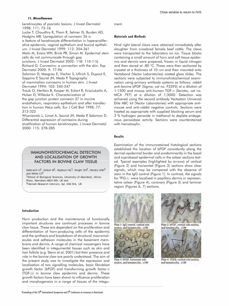
Horn production and the maintenance of functionallyimportant structures are continual processes in bovineclaw tissue. These are dependent on the proliferation anddifferentiation of horn-producing cells of the epidermisand the synthesis and breakdown of structural macromol-ecules and adhesion molecules in the basement mem-brane and dermis. A range of chemical messengers havebeen identified in integumental tissues such as skin andhair follicle (e.g. Stenn et al, 2001) but their presence androle in the bovine claw are poorly understood. The aim ofthe present study was to investigate the expression andlocalisation of two signalling molecules, basic fibroblastgrowth factor (bFGF) and transforming growth factor-b(TGF-b) in bovine claw epidermis and dermis. Thesegrowth factors have been shown to influence proliferationand morphogenesis in a range of tissues of the integu-
ment.
Materials and Methods
Hind right lateral claws were obtained immediately afterslaughter from crossbred female beef cattle. The clawswere transported to the laboratory on ice. Tissue blockscontaining a small amount of horn and soft tissue epider-mis and dermis were prepared, frozen in liquid nitrogenand then stored at -80 °C. These were then sectioned bycryostat at a thickness of 10 mm and then mounted ontoVectabond (Vector Laboratories) coated glass slides. Thesections were subjected to immunohistochemical exami-nation using primary antibody systems as follows: rabbitanti-bovine bFGF (Sigma, cat no. F3393) at a dilution of1:1500 and mouse anti-human TGF-b (Serotec, cat no.MCA 797) at a dilution of 1:3000. Detection wasachieved using the second antibody Vectastain UniversalElite ABC kit (Vector Laboratories) with appropriate anti-mouse and anti-rabbit negative controls. Sections weretreated as appropriate with supplied blocking serum and3 % hydrogen peroxide in methanol to deplete endoge-nous peroxidase activity. Sections were counterstainedwith hematoxylin.
Results
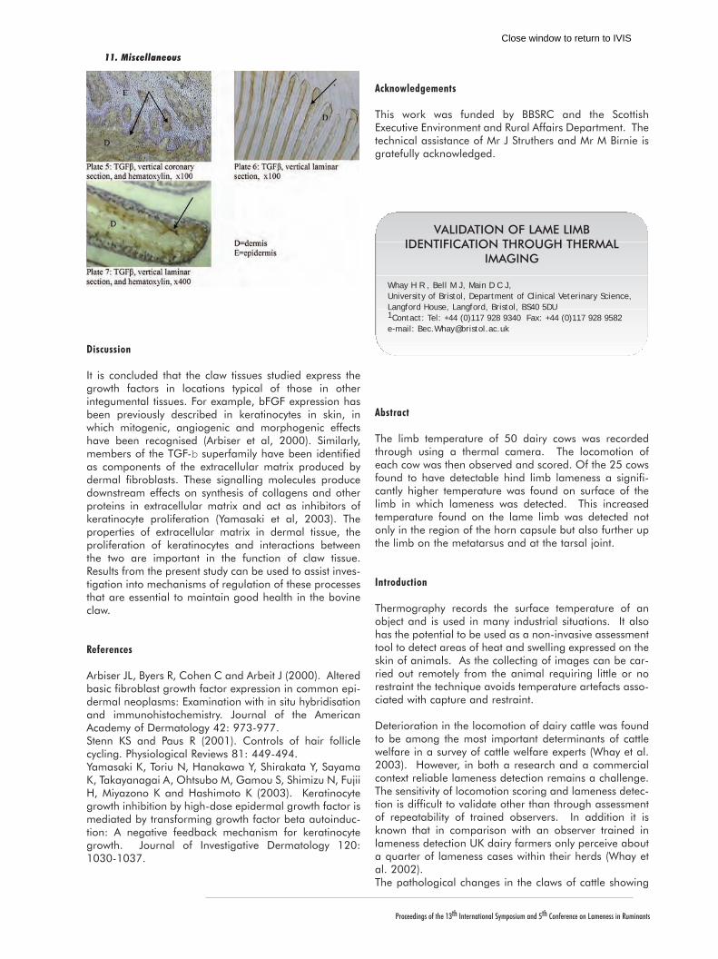
Examination of the immunostained histological sectionsestablished the location of bFGF consistently along thedermal-epidermal border and predominantly in the basaland suprabasal epidermal cells in the solear sections test-ed. Typical examples (highlighted by arrows) of vertical(Figure 2) and horizontal (Figure 3) sections show clearsignals, which may be compared with the absence ofstain in the IgG control (Figure 1). In contrast, the signalsfor TFG-b were localised in papillary dermis in represen-tative solear (Figure 4), coronary (Figure 5) and laminarregion (Figures 6, 7) sections.
IMMUNOHISTOCHEMICAL DETECTIONAND LOCALISATION OF GROWTHFACTORS IN BOVINE CLAW TISSUE
Galbraith H1, Dinkel M1, Hepburn NL1, Knight CH2, Hendry KAK2
and Wilde CJ21School of Biological Sciences, University of Aberdeen, HiltonPlace, Aberdeen AB24 4FA, UK and 2Hannah Research Institute, Ayr, KA6 5HL, UK
11. Miscellaneous
Close window to return to IVIS
Proceedings of the 13th International Symposium and 5th Conference on Lameness in Ruminants
11. Miscellaneous
Discussion
It is concluded that the claw tissues studied express thegrowth factors in locations typical of those in otherintegumental tissues. For example, bFGF expression hasbeen previously described in keratinocytes in skin, inwhich mitogenic, angiogenic and morphogenic effectshave been recognised (Arbiser et al, 2000). Similarly,members of the TGF-b superfamily have been identifiedas components of the extracellular matrix produced bydermal fibroblasts. These signalling molecules producedownstream effects on synthesis of collagens and otherproteins in extracellular matrix and act as inhibitors ofkeratinocyte proliferation (Yamasaki et al, 2003). Theproperties of extracellular matrix in dermal tissue, theproliferation of keratinocytes and interactions betweenthe two are important in the function of claw tissue.Results from the present study can be used to assist inves-tigation into mechanisms of regulation of these processesthat are essential to maintain good health in the bovineclaw.
References
Arbiser JL, Byers R, Cohen C and Arbeit J (2000). Alteredbasic fibroblast growth factor expression in common epi-dermal neoplasms: Examination with in situ hybridisationand immunohistochemistry. Journal of the AmericanAcademy of Dermatology 42: 973-977.Stenn KS and Paus R (2001). Controls of hair folliclecycling. Physiological Reviews 81: 449-494.Yamasaki K, Toriu N, Hanakawa Y, Shirakata Y, SayamaK, Takayanagai A, Ohtsubo M, Gamou S, Shimizu N, FujiiH, Miyazono K and Hashimoto K (2003). Keratinocytegrowth inhibition by high-dose epidermal growth factor ismediated by transforming growth factor beta autoinduc-tion: A negative feedback mechanism for keratinocytegrowth. Journal of Investigative Dermatology 120:1030-1037.
Acknowledgements
This work was funded by BBSRC and the ScottishExecutive Environment and Rural Affairs Department. Thetechnical assistance of Mr J Struthers and Mr M Birnie isgratefully acknowledged.
Abstract
The limb temperature of 50 dairy cows was recordedthrough using a thermal camera. The locomotion ofeach cow was then observed and scored. Of the 25 cowsfound to have detectable hind limb lameness a signifi-cantly higher temperature was found on surface of thelimb in which lameness was detected. This increasedtemperature found on the lame limb was detected notonly in the region of the horn capsule but also further upthe limb on the metatarsus and at the tarsal joint.
Introduction
Thermography records the surface temperature of anobject and is used in many industrial situations. It alsohas the potential to be used as a non-invasive assessmenttool to detect areas of heat and swelling expressed on theskin of animals. As the collecting of images can be car-ried out remotely from the animal requiring little or norestraint the technique avoids temperature artefacts asso-ciated with capture and restraint.
Deterioration in the locomotion of dairy cattle was foundto be among the most important determinants of cattlewelfare in a survey of cattle welfare experts (Whay et al.2003). However, in both a research and a commercialcontext reliable lameness detection remains a challenge.The sensitivity of locomotion scoring and lameness detec-tion is difficult to validate other than through assessmentof repeatability of trained observers. In addition it isknown that in comparison with an observer trained inlameness detection UK dairy farmers only perceive abouta quarter of lameness cases within their herds (Whay etal. 2002). The pathological changes in the claws of cattle showing
VALIDATION OF LAME LIMBIDENTIFICATION THROUGH THERMAL
IMAGING
Whay H R , Bell M J, Main D C J, University of Bristol, Department of Clinical Veterinary Science,Langford House, Langford, Bristol, BS40 5DU1Contact: Tel: +44 (0)117 928 9340 Fax: +44 (0)117 928 9582 e-mail: [email protected]
Close window to return to IVIS
Proceedings of the 13th International Symposium and 5th Conference on Lameness in Ruminants
signs of lameness may be detectable through thermo-graphic imaging. A pilot study was carried out to inves-tigate whether such changes in the surface temperature ofthe limbs of lame cows were detectable using thermalimaging.
Methods
Thermal images and surface skin temperature recordingswere taken from 50 lactating dairy cows during May2003 following Spring turnout on two farms in the SouthWest of England. The measurements were made using athermal imaging camera (ThermaCAMTM E2, FLIRSystems). Temperature recordings were taken of the later-al aspects of both hind-limbs at the metatarsal joint, midmetatarsus and the abaxial aspect of the lateral clawhorn capsule. The lowest, average and maximum surfacetemperatures were recorded from each region. The cowswere then walked on a flat concrete surface and theirlocomotion scored on a scale of 0 - 3 (0 - sound, 3 -severe lameness) and where detectable the lame limbwas identified.
The thermography data recorded from the limbs of thedairy cows was corrected against the average tempera-ture of the udder on the same side. This allowed a cor-rection for variations in skin temperature due to changesin ambient temperature. A general Linear Model wasthen used to examine the relationship between the pointson each limb where temperature was recorded andbetween the limb which was identified as being lamethrough locomotion scoring.
Results
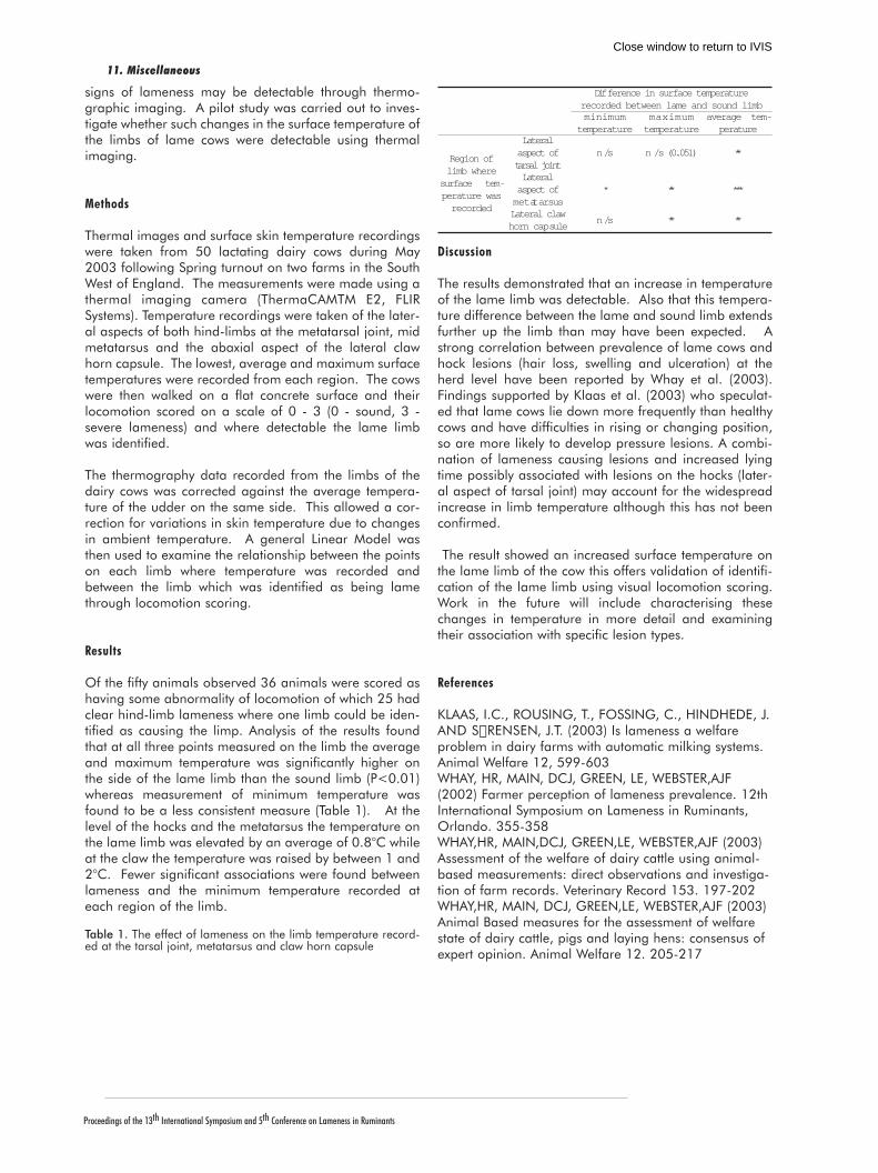
Of the fifty animals observed 36 animals were scored ashaving some abnormality of locomotion of which 25 hadclear hind-limb lameness where one limb could be iden-tified as causing the limp. Analysis of the results foundthat at all three points measured on the limb the averageand maximum temperature was significantly higher onthe side of the lame limb than the sound limb (P<0.01)whereas measurement of minimum temperature wasfound to be a less consistent measure (Table 1). At thelevel of the hocks and the metatarsus the temperature onthe lame limb was elevated by an average of 0.8°C whileat the claw the temperature was raised by between 1 and2°C. Fewer significant associations were found betweenlameness and the minimum temperature recorded ateach region of the limb.
Table 1. The effect of lameness on the limb temperature record-ed at the tarsal joint, metatarsus and claw horn capsule
Discussion
The results demonstrated that an increase in temperatureof the lame limb was detectable. Also that this tempera-ture difference between the lame and sound limb extendsfurther up the limb than may have been expected. Astrong correlation between prevalence of lame cows andhock lesions (hair loss, swelling and ulceration) at theherd level have been reported by Whay et al. (2003).Findings supported by Klaas et al. (2003) who speculat-ed that lame cows lie down more frequently than healthycows and have difficulties in rising or changing position,so are more likely to develop pressure lesions. A combi-nation of lameness causing lesions and increased lyingtime possibly associated with lesions on the hocks (later-al aspect of tarsal joint) may account for the widespreadincrease in limb temperature although this has not beenconfirmed.
The result showed an increased surface temperature onthe lame limb of the cow this offers validation of identifi-cation of the lame limb using visual locomotion scoring.Work in the future will include characterising thesechanges in temperature in more detail and examiningtheir association with specific lesion types.
References
KLAAS, I.C., ROUSING, T., FOSSING, C., HINDHEDE, J.AND SœRENSEN, J.T. (2003) Is lameness a welfareproblem in dairy farms with automatic milking systems.Animal Welfare 12, 599-603WHAY, HR, MAIN, DCJ, GREEN, LE, WEBSTER,AJF(2002) Farmer perception of lameness prevalence. 12thInternational Symposium on Lameness in Ruminants,Orlando. 355-358WHAY,HR, MAIN,DCJ, GREEN,LE, WEBSTER,AJF (2003)Assessment of the welfare of dairy cattle using animal-based measurements: direct observations and investiga-tion of farm records. Veterinary Record 153. 197-202WHAY,HR, MAIN, DCJ, GREEN,LE, WEBSTER,AJF (2003)Animal Based measures for the assessment of welfarestate of dairy cattle, pigs and laying hens: consensus ofexpert opinion. Animal Welfare 12. 205-217
Difference in surface temperaturerecorded between lame and sound limbminimum
temperaturemaximumtemperature
average tem-perature
Region oflimb where
surface tem-perature wasrecorded
Lateralaspect oftarsal joint
n /s n /s (0.051) **
Lateralaspect ofmetatarsus
* ** ***
Lateral clawhorn capsule
n /s ** **
11. Miscellaneous
Close window to return to IVIS

Proceedings of the 13th International Symposium and 5th Conference on Lameness in Ruminants
11. Miscellaneous
Introduction
Interdigital phlegmon is perhaps the disease with themost synonyms: infectious pododermatitis, interdigitalnecrobacillosis, foot rot , foul in the foot, foot abscess,panaritium are often mentioned. Traumatic lesions of theinterdigital skin, caused by rough floor, uneven ground,stones, straw or pieces of wood are the most commoncauses. Maceration of the skin by wet weather conditions,faeces and urine may predispose the claw to injuries (1-12). Pain, leading to mild or severe lameness and mod-erate to severe swelling of the interdigital space are majorsigns of the disease. Interdigital phlegmon has a world-wide occurence with massive economical losses in milkand meat production. Fusobacterium necrophorum andBacteroides melaninogenicus are mostly isolated fromthis infection. Clark et al. (1986) were able to reproducefoot rot by experimental cultures of F. necrophorumalone, as they injected the agent through the skin into thedermis. Interdigital phlegmon is usually sporadic, butmay be endemic in high intensity beef or dairy cattle pro-duction units (1, 3, 8, 10).
Material and Methods
In this retrospective study clinical and radiographic find-ings, treatment and outcome of 43 cases (1998-2003)with interdigital phlegmon are presented. All thesepatients had been referred by the local veterinarians tothe clinic, with an advanced stage of an interdigitalphlegmon. All patients underwent routine clinical andorthopaedic examination. The age of the animals rangedfrom 3 to 9 years (mean age 5 y). Thirty were Simmentalcows, 7 were Holstein Friesian, 3 were Brown-Swiss and2 were Crossbreds. Clinical signs included moderate or severe lameness withmarked swelling of the coronary region and the soft tis-sues of the interdigital space. In cases of advanced infec-tion a characteristic fetid smell caused by the interdigitalnecrotizing lesions was noted.Radiographic examination was performed in cases ofsevere lameness and obvious circular swelling over thewhole coronet from dorsal to abaxial to rule out infectionof the distal or/and proximal interphalangeal joint orinvolvement of the remaining claw.
The patients were prepared for surgical resection of theinfected and necrotic tissues and/or digital amputationwith an intravenous regional anaesthesia using 20 ml ofprocaine-hydrochloride (Minocain 2%, Atarost,Germany). A tourniquet of rubber tubing was applieddirectly in the middle of the metatarsus or metacarpus.
Results
Twenty-two cows showed a severe lameness (grade 3 and4 out of 4), thirteen cows presented a moderate lameness(grade 2 of 4) and in 8 cases only a slight lameness(grade 1 of 4) was noted. In 19 cows the left hindlimbwas affected, in 20 cases interdigital phlegmon waslocated at the right hindlimb. Two cows had infections ofboth hindlimbs. The right frontlimb was affected in 2cows. Radiography was performed in 15 cattle with asevere lameness. Radiographic signs of bone and/or jointinfection were widening of the joint space, loss of jointand bone architecture as well as reactive periostal newbone formation.Surgical resection of the affected soft tissues of the inter-digital space was performed in 19 out of 43 cattle.Starting from the causative penetrating wound the infect-ed and necrotic soft tissue structures of the interdigitalspace were resected completely. In one case the infectedand necrotic tissue reached the axial capsule of the distalinterphalangeal joint showing a serous, slightly turbideffusion: in this case the distal interphalangeal joint wasopened from this track and was lavaged with 2000 mlisotonic saline solution with diluted 0.1% polyvidon-iodine-solution. Although surgery and parenteral antibiosis was doneinfection spread to the distal interphalangeal joint in onecase and led to amputation of the affected claw.In 23 cows (out of 43) a purulent arthritis of the distalinterphalangeal joint and in 1 cow a purulent-necrotisingarthritis of the proximal interphalangeal joint had devel-oped from the interdigital phlegmon. 12 of the 24 cowswere euthanized or slaughtered due to the bad condition,the poor prognosis or economic reasons. Amputation of the infected claw was performed in 11cases, another one was treated by resection of the distalinterphalangeal joint. In addition, infections of the digitalflexor tendon sheaths occurred in five cases (out of 24).Three of them were euthanized after diagnosis: one cowhad already developed an infection of the fetlock joint,another one showed a severe swelling of the remaininghind claw because of a deep sole ulcer. In one caseamputation was refused by the owner. In 2 cows the deepand superficial flexor tendons and the infected flexor ten-don sheaths were resected.One out of these 24 cases showed a severe interdigitalphlegmon with a purulent arthritis of the distal interpha-langeal joint, multiple abscesses on the lateral digit anddistal limb and a purulent endocarditis and was eutha-nized.Systemic and local antibiosis was used for 3-5 days. Forsystemic antibiosis the cattle were treated with 1 mg cef-tiofur (Excenel , Pharmacia & Upjohn) per kg body weightor 10 mg oxytetracycline (Engemycin10% , Intervet,
TREATMENT AND OUTCOME OFINTERDIGITAL NECROBACILLOSIS (INTER-DIGITAL PHLEGMON, FOOT ROT) IN 43
COWS
Reinöhl-DeSouza, Cornelia, Martinek, B., Kofler, J.Clinic for Orthopaedics in Large Animals, University ofVeterinary Medicine Vienna, Veterinärplatz 1, A-1210 Vienna,Austria, Tel. +43 1 25077 5520, Fax. +43 1 25077 5590, [email protected]
Close window to return to IVIS

Proceedings of the 13th International Symposium and 5th Conference on Lameness in Ruminants
Vienna) per kg body weight. When amputation of theclaw was performed 20.000 IU benzyl-penicillin and 20mg streptomycin per kg body weight (Peni-strepto , VirbacLaboratoires) were given for 5 days.Depending on the surgical technique clinic hospitalisationranged from 2-28 days (mean 10.4).
Discussion
The first observed signs of foot rot are varying degrees oflameness, from barely noticeable to an extensive condi-tion in which cows get more or less recumbent (1, 12).Symptoms like widening of the interdigital space due to amoderate or severe swelling along with a reddening ofthe skin and the coronary band, heels and necrotic anddemarcated interdigital tissue could be detected in allcases. If the foot rot condition is severe and is not treatedin the early stages, complications may result such as sep-tic arthritis of the distal or/and proximal interphalangealjoint, septic inflammation of the digital flexor tendonsheath, other deep infections of the foot as local abscess-es and haematogenous spread of infectious material withsubsequent purulent endocarditis, abscesses in the lung,liver or kidneys (1, 12). In this study 24 cows showed apurulent arthritis of the distal and proximal interpha-langeal joint, in 5 cases an inflammation of the flexortendon sheaths and in one case a septic endocarditis.
References
1 Berg JN, Franklin, CL (2000): Interdigital phlegmon /interdigital necrobazillosis / acute foot rot of cattle: con-siderations in etiology, diagnosis and treatment. Proc.11th International Symposium on Disorders of theRuminant Digit, Parma, Italy, 3-7. September 2000, p.27-30.2 Bergsten Ch (1997): Infectious diseases of the digits.In: Greenough, PR, Weaver, AD (eds): Lameness in cat-tle. 3rd ed., Philadelphia, WB Saunders, pp. 89-94.3 Berry, St. (2001): Diseases of the digital soft tissues.Vet. Clin. North America: Food Animal Prac. 17, 35-37.4 Clark BL, Emery DL, Stewart DJ (1986): The aetiologyand prevention of interdigital necrobacillosis (footabscess) in cattle. In Stewart, DJ et others (ed.): Footrotin ruminants. Proc. Workshop, Melbourne, 1985, p.275-8.5 Clark BL, Stewart DJ, Emery DL (1985): The role ofFusobacterium necrophorum and Bacteroides melanino-genicus in the aetiology of interdigital necrobacillosis incattle. Australian Vet. J. 62, 47-9.6 Clark, B.L., Emery, D.L., Stewart, D.J., Dufty, J.H.,Anderson, D.A. (1986): Studies into immunisation ofcattle against interdigital necrobacillosis. Aust. Vet. J.63, 107 - 110.7 Henke CL, Howie NM, Smith FJ, Flaus L, Kausche FM(1994): Efficacy of Ceftiofur-Sodium (Excenel*) for thetreatment of acute foot-rot (interdigital necrobacillosis)in cattle. Proc. 18th World Buiatrics Congress, Bologna,Italy, August 29 - September 2, 1994, Vol. 2, 1355-8. 8 Iselin U, Lischer CJ, Zindel F (1998): Erfahrungen mit
der Prophylaxe des Panaritiums bei Alprindern inGraubünden. Abstracts Schweizer Tierärztetage 1998 inInterlaken, 4.-5. September 1998, S. 116-7.9 Kausche, FM, ROBB, EJ (2002): A comprehensivereview of Ceftiofur Na (Excenel sterile powder) andCeftiofur HCl (Excenel RTU suspension) in the treatmentof acute bovine foot rot. Proc. 22nd World BuiatricsCongress, Hannover, Germany, August 18-23, 2002,p.83. 10 Monrad J, Kassuku AA, Nansen P, Willeberg P(1983): An epidemiological study of foot rot in pasturedcattle. Acta Vet. Scand. 24, 403-17.11 Morck DW, Olson ME, Louie TJ, Koppe A, Quinn B(1998): Comparison of ceftiofur sodium and oxytetracy-cline for treatment of acute interdigital phlegmon (rootrot) in feedlot cattle. J. Am. Vet. Med. Assoc. 212, 254-7.12 Stokka, GL, Lechtenberg, K, Edwards, T, MacGregor,S, Voss, K, Griffin, D, Grotelueschen, DM, Smith, RA,Perino, LJ (2001): Lameness in feetlot cattle: Interdigitalnecrobacillosis. Vet. Clin. North America: Food AnimalPrac. 17, 189-190.
Abstract
In a digit of dairy cattle obtained from a slaughterhouse,three horizontal grooves were observed on the wall. In thevertical section of the sole, two horizontal thin layers wereseen, which had yellowish color.The first horizontal groove formed nearest under the digitalband was connected to the first horizontal thin layer in thevertical section of the sole. It showed that the horizontalgroove and the thin layer in the sole were formed at the sametime. From this observation, the growth of the sole could beestimated.The surface of the heel horn was detached at the groove onthe heel bulb.The relationship between horizontal grooves and the solechanges will be discussed.
CHANGES SEEN IN THE SOLE HORNCORRELATED WITH HORIZONTAL
GROOVES IN A DIGIT OF DAIRLY CATTLE
Kazunori,Yoshitani DVMUnakamimachi 464-5, Kaijougunn, Chiba-ken 289-2613, Japan
11. Miscellaneous
Close window to return to IVIS