seropositive juvenile rheumatoid arthritis. molecules ... · Specific HLA-DR4-associated...
6
Specific HLA-DR4-associated histocompatibility molecules characterize patients with seropositive juvenile rheumatoid arthritis. B S Nepom, … , P Antonelli, J A Hansen J Clin Invest. 1984; 74(1):287-291. https://doi.org/10.1172/JCI111413. The structural and functional heterogeneity of HLA-DR4-associated specificities was investigated in patients with seropositive juvenile rheumatoid arthritis, a DR4-associated disease. Using a combination of HLA-D analysis by mixed lymphocyte culture and electrophoretic analysis of immunoprecipitated Ia molecules by two-dimensional polyacrylamide gels, we observed a surprisingly homogeneous pattern of HLA-D antigen expression. All patients expressed common structural products of the DR and DS loci, and 7/12 homozygous DR4 patients expressed a rare and subtle HLA-D heterozygous phenotype. Research Article Find the latest version: http://jci.me/111413/pdf Pdf
Transcript of seropositive juvenile rheumatoid arthritis. molecules ... · Specific HLA-DR4-associated...
Specific HLA-DR4-associated histocompatibilitymolecules characterize patients withseropositive juvenile rheumatoid arthritis.
B S Nepom, … , P Antonelli, J A Hansen
J Clin Invest. 1984;74(1):287-291. https://doi.org/10.1172/JCI111413.
The structural and functional heterogeneity of HLA-DR4-associated specificities wasinvestigated in patients with seropositive juvenile rheumatoid arthritis, a DR4-associateddisease. Using a combination of HLA-D analysis by mixed lymphocyte culture andelectrophoretic analysis of immunoprecipitated Ia molecules by two-dimensionalpolyacrylamide gels, we observed a surprisingly homogeneous pattern of HLA-D antigenexpression. All patients expressed common structural products of the DR and DS loci, and7/12 homozygous DR4 patients expressed a rare and subtle HLA-D heterozygousphenotype.
Research Article
Find the latest version:
http://jci.me/111413/pdf
Rapid Publications
Specific HLA-DR4-associatedHistocompatibility MoleculesCharacterize Patients with SeropositiveJuvenile Rheumatoid Arthritis
Abstract. The structural and functional het-erogeneity of HLA-DR4-associated specificities was in-vestigated in patients with seropositive juvenile rheu-matoid arthritis, a DR4-associated disease. Using a com-bination of HLA-D analysis by mixed lymphocyte cultureand electrophoretic analysis of immunoprecipitated Iamolecules by two-dimensional polyacrylamide gels, weobserved a surprisingly homogeneous pattern of HLA-Dantigen expression. All patients expressed commonstruc-tural products of the DRand DS loci, and 7/12 homo-zygous DR4 patients expressed a rare and subtleHLA-D heterozygous phenotype.
Introduction
The presence of particular genetic markers has been linked todisease susceptibility in patients with some autoimmune or im-mune-mediated diseases ( 1-9). To test the hypothesis that specificclass II molecules associate with a particular HLA-linked disease,and thus identify putative disease-susceptibility Ir genes, wehave analyzed the HLA-DR4-associated specificities in patientswith seropositive juvenile rheumatoid arthritis (JRA).' HLA-
Address reprint requests to Dr. G. T. Nepom, Division of Immunology,Fred Hutchinson Cancer Research Center, 1124 Columbia, Seattle, WA98104.
Received for publication 31 January 1984 and in revised form 2April 1984.1. Abbreviations used in this paper: JRA, juvenile rheumatoid arthritis;RR, relative risk.
Barbara S. Nepom, Gerald T. Nepom, Eric Mickelson,Jane G. Schaller, Paolo Antonelli, and John A. HansenFred Hutchinson Cancer Research Center, Puget Sound BloodCenter, Pacific Northwest Research Foundation, Genetic SystemsCorporation, Division of Rheumatology, Children's OrthopedicHospital Medical Center, and the Departments of Pathology,Pediatrics and Medicine, University of Washington School ofMedicine, Seattle, Washington 98104
DR4 is a serologically defined specificity present in diverse pop-ulations, but in mixed lymphocyte response assays T cells rec-ognize at least six distinct HLA-DR4-associated specificities,which are defined as HLA-D antigens (10-12). These HLA-DR4-associated haplotypes also differ from each other in rec-ognizable electrophoretic variants of products of the DR locusand to a lesser extent of the DS locus (13, 14). Weinitiated thisstudy to look for a class II molecule(s) commonto JRA patientsthat would be a candidate for a DR4-associated disease-sus-ceptibility product contributing an Ir gene-regulated function.
Methods
Patient selection and typing. Patients attending the Arthritis Clinic atChildren's Orthopedic Hospital and Medical Center, Seattle, WAwitha diagnosis of JRA were used for this study. 27 out of 400 patients werediagnosed as having seropositive JRA by using previously describedcriteria (15). Most of these patients have a late childhood onset of sym-metrical, erosive, and usually severe and unremitting polyarthritis withhigh titer rheumatoid factor; they have frequent subcutaneous nodules;and they are female. The 22 Caucasian patients in this group, and theirfamilies, were selected for study.
HLA-DR typing was performed on nylon-wool-purified peripheralblood B cells or B lymphoblastoid cell lines as previously described, byusing a panel of antisera that recognize HLA-DRl through HLA-DRwIO(16). HLA-D typing was performed on peripheral blood lymphocytesby using a standard mixed lymphocyte culture as previously des-cribed (17).
Four homozygous typing cells were used to define Dw4(JAH, LGY,GW1, and JOL). Four homozygous typing cells were used to defineLD40 (2075, THO, BIN40, and LS40). Dw4 and LD40 are two of thesix recognized HLA-D clusters associated with HLA-DR4 (10, 12). Ho-mozygous cells that type as LD40 respond in mixed lymphocyte cultureagainst homozygous Dw4cells, even though they both express the DR4specificity (10). The double normalized value method of Ryder et al.(18) was used to adjust data from each HLA-D typing experiment.
287 Ia Molecules in Juvenile Rheumatoid Arthritis
J. Clin. Invest.© The American Society for Clinical Investigation, Inc.002 1-9738/84/07/0287/05 $ 1.00Volume 74, July 1984, 287-291

Double normalized values of <45 were considered to be "typing re-sponses", and assignments were made according to the Ninth Histo-compatibility Workshop method (17).
Antibodies and cells. Antibody P4.1 is a murine monoclonal antibodyspecific for a determinant on HLA-DR molecules (12). Monoclonalantibody SG465 recognizes a determinant present on DS molecules,which are molecules that are homologous by partial sequence analysisto murine I-A antigens (19). Cell lines were established from peripheralblood B lymphocytes of patients by Epstein-Barr virus transfor-mation (20).
Immunoprecipitation and gel analysis. Radiolabeling and neur-aminidase treatment of cell lysates and subsequent immunoprecipitationwere performed as previously described (12, 13). Gel analysis of theimmunoprecipitates used nonequilibrium electrophoresis in the first di-mension, followed by discontinuous 10% sodium dodecyl sulfate-poly-acrylamide slab gel electrophoresis in the second dimension. Gels weredried under vacuum and autoradiographed directly onto Kodak X-Omat film. HLA-DR a-chains did not display significant polymorphismor electrophoretic variation between haplotypes, so only i-chain resultsare shown.
Results
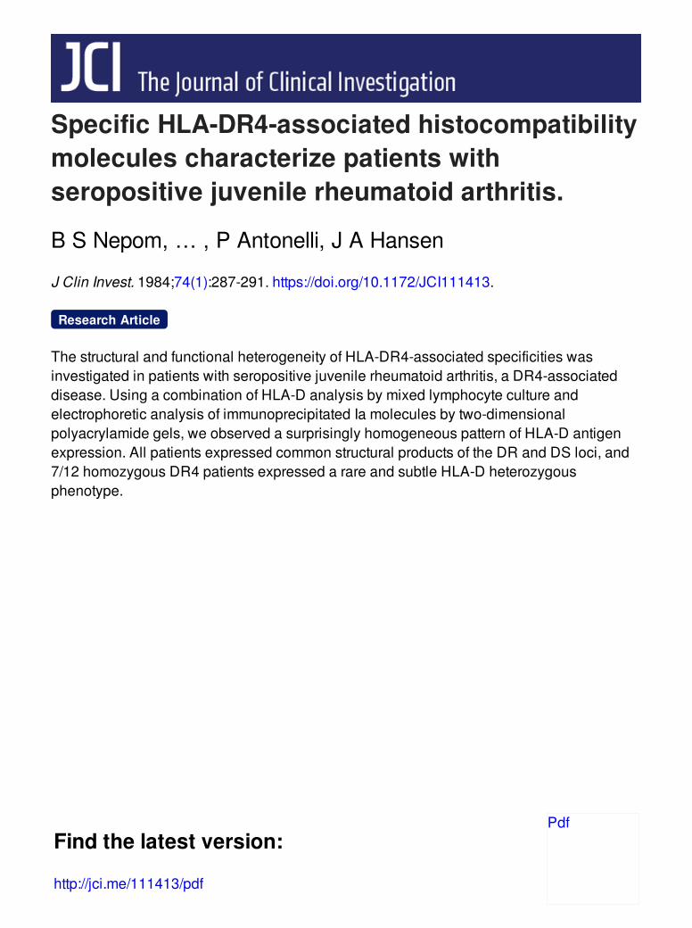
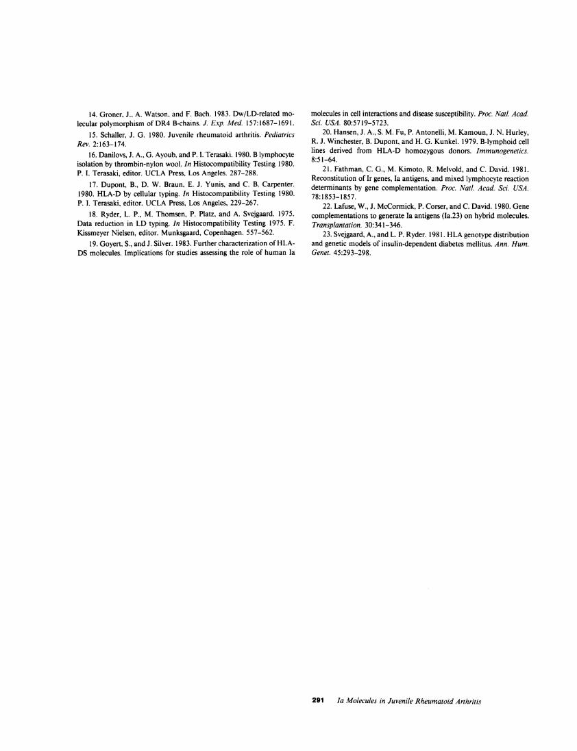
2-Dimensional gel analysis of DRand DSmolecules from ho-mozygous cell lines derivedfrom patients with seropositive JRA.B lymphoblastoid cell lines derived from eight of the patientswith seropositive JRA who were homozygous for HLA-DR4were radiolabeled and lysed for analysis by immunoprecipitationand gel electrophoresis, which is shown in Fig. 1. Ia ,8-chainsof the DRand DS products have characteristic electrophoreticpatterns that vary within the DR4 specificity, in a manner thatcorresponds with the HLA-D-defined haplotype (13). The JRADR fl-chain immunoprecipitates (left column), however, showa remarkably homogeneous electrophoretic pattern that has littlevariation between individuals. Three major spots are seen ineach case, with similar molecular weight and charge profiles.The patterns are indistinguishable between cell lines, except inthe case of cell line 728 where a faint fourth spot is also present,migrating more acidic than the others. This extra spot mayrepresent an additional DR4-associated product whose specificityhas not been defined by homozygous typing cells.
To analyze Ia ,-chains of the non-DR locus called DS, thecell lysates were precleared of DRmolecules with antibody P4.1before immunoprecipitation with anti-DS antibody SG465. Theobserved patterns (middle column) are distinct from the DRa-chain patterns but again show a remarkable degree of ho-mogeneity within the individuals tested; each cell line shows a
consistent gel pattern with two dominant spots, although subtledegrees of charge heterogeneity exist. Again the exception is cellline 728 where a faint basic spot is present.
After neuraminidase treatment and electrophoresis, most ofthe DRa-chain charge heterogeneity disappears (right column).In six cases, a single dark spot is seen, with a faint acidic shadow.In two of the cases, 728 and 813, the intensity of these same
two spots differs, which may reflect different susceptibilities todesialylation, or may indicate a second ,3-chain polypeptide. In
all cases the dominant spots co-migrate. Confirmation of thisconsistent electrophoretic migration was achieved both by over-lapping alignment of the gels, and by mixing experiments, whereimmunoprecipitates from two separate cell lines were mixedbefore the first-dimensional electrophoresis. Thus, subtle dif-ferences in charge or size would show up as separate spots whenrun simultaneously. In the lower right of Fig. 1 two representativemixing experiments are shown, which reveal that in fact themajor spots from different neuraminidase-treated cell lines ( 162+ 469, 125 + 372) are indistinguishable.
HLA-D and DR typing of patients with seropositive JRA.Complete HLAhaplotypes of the patients studied by biochemicalanalysis are shown in Table I. Surprisingly, although all eightpatients were homozygous for the serologically defined DR4specificity, seven were actually HLA-D heterozygotes. In theone exception, the patient appeared to be homozygous for theuncommon D-locus specificity LD"40", which is the same spec-ificity expressed by six of the seven heterozygotes. These dataare summarized along with HLA typing of the entire seropositiveJRA patient group in Table II.
DR4was present in 17 of 22 patients (77%) compared with32% of 219 controls for a relative risk (RR) of 7.2. 12 of the22 patients (54%) were homozygous for DR4. HLA-Dw4 waspresent in 16 patients (73%) and in 17% of controls, for a RRof 12.9. Eight of the DR4-positive patients exhibited the relativelyuncommon D-locus specificity LD"40". All eight patients withHLA LD"40" were homozygous for DR4 and seven of themwere heterozygous for Dw4 and LD"40". The expected fre-quency of LD"40", Dw4 heterozygotes was 0. 1%, thus the RRfor these two antigens together was 1 16. None of the controlpopulation typed as LD"40", Dw4 heterozygotes. None of 25seronegative JRA patients, including five DR4-positive females,expressed this heterozygous specificity.
Discussion
The expression of the HLA-DR4 specificity in patients withseropositive JRA is one of many such associations that correlatea single HLA specificity with a particular disease syndrome.These observations of association have generally been interpretedto represent linkage of a gene within the HLA region to a disease-susceptibility locus; in the case of HLA-D region associations,this is often attributed to an Ir gene-regulated type of event. Ifthis model is valid, it predicts that the specific products of Irgenes within the HLA-D region, namely the Ia antigens, maybe directly involved in disease susceptibility.
Wepresent our observations using structural and functionalanalysis of D region products that tested this hypothesis. Wefound a remarkably consistent expression of particular class IImolecules in our homozygous JRA patients by two-dimensionalgel analysis of DRand DS f,-polypeptide chains. In contrast tosimilar studies of nonselected HLA-DR4 homozygous cells,which revealed at least five distinct DR ,B-chain and three DS#-chain electrophoretic patterns, all patients cluster into very
288 Nepom, Nepom, Mickelson, Schaller, Antonelli, and Hansen

DR3 (Neurominidose)
4
i
i.
125 + 372
162 +469Figure ). Two-dimensional gel analysis of Ia molecules from DR4homozygous patients with JRA. Electrophoretic patterns of #-chainsradioiodinated with lactoperoxidase and '25I-NaI from immunopre-cipitates of DRand DS molecules are shown, obtained from each ofeight HLA-DR4 homozygous cell lines derived from patients withJRA. The DR a-gels (left and right columns) were obtained using an-
tibody P4.1; the DS A-gels (middle column) were obtained using anti-body SG465. Additive gels formed by mixing immunoprecipitates areshown (bottom right) for combinations of two different neuramini-dase-treated DR ,-chains. Equivalent portions of each autoradiogramare presented; the basic end of the gel (pH 8.5) is at the left and theacidic end (pH 5.0) at the right.
289 Ia Molecules in Juvenile Rheumatoid Arthritis
PatientNo.
925
DRf DS3
162
125 *4:0
253
813
728
372
469
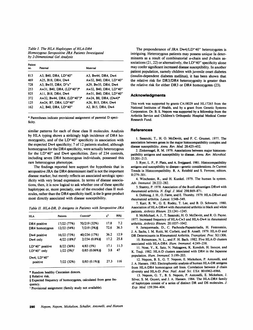
Table L The HLA Haplotypes of HLA-DR4Homozygous Seropositive JRA Patients Investigatedby 2-Dimensional Gel Analysis
Patientno. Paternal Maternal
813 A3, B40, DR4, LD"40" A3, Bw44, DR4, Dw4469 A25, B18, DR4, Dw4 Aw32, B40, DR4, LD"40"728 A3, Bw35, DR4, D"x" A29, Bw35, DR4, Dw4253 Aw3l, B40, DR4, (LD"40")* Aw32, B40, DR4, LD"40"925 A 1l, B18, DR4, Dw4 Aw3l, B40, DR4, LD"40"372 Aw32, Bw44, DR4, (LD"40")* Aw24, B8, DR4, (Dw4)*125 Aw24, B7, DR4, LD"40" A26, B15, DR4, Dw4162 A2, B40, DR4, LD"40" A2, B15, DR4, Dw4
* Parentheses indicate provisional assignment of parental D speci-ficity.
similar patterns for each of these class II molecules. Analysisby HLA typing shows a strikingly high incidence of DR4 ho-mozygosity, and of the LD"40" specificity in association withthe expected Dw4 specificity; 7 of 12 patients studied, althoughhomozygous for the DR4specificity, were actually heterozygousfor the LD"40" and Dw4 specificities. Zero of 234 controls,including seven DR4 homozygous individuals, possessed thisrare heterozygous phenotype.
The findings reported here support the hypothesis that inseropositive JRA the DR4determinant itself is not the importantdisease marker, but merely reflects an associated serologic spec-ificity with very broad expression. In terms of disease associa-tions, then, it is now logical to ask whether one of these specifichaplotypes or, more precisely, one of the encoded class II mol-ecules, rather than the DR4specificity itself, is the Ir gene productmost directly associated with disease susceptibility.
Table II. HLA-DR, D Antigens in Patients with Seropositive JRA
HLA Patients Controls x2 RRt
DR4 positive 17/22 (77%) 70/219 (32%) 17.8 7.2DR4 homozygous 12/22 (54%) 7/219 (3%)§ 72.6 36.3
Dw4 positive 16/22 (73%) 40/234 (17%) 36.2 12.9Dw4 only 4/22 (18%)" 2/234 (0.8%)§ 17.2 25.8
LD"40" positive 8/22 (36%) 4/83 (5%) 17.1 11.3LD"40" only 1/22 (5%)" 0/83 (0.06%)§ 3.8 47
Dw4, LD"40"positive 7/22 (32%) 0/83 (0. l%)§ 27.3 116
* Random healthy Caucasian donors.t Relative risk.§ Expected frequency of homozygotes, calculated from gene fre-quency.1' Provisional assignment (family study not available).
The preponderance of JRA Dw4/LD"40" heterozygotes isintriguing. Heterozygous patients may possess unique Ia deter-minants as a result of combinatorial a-chain and ,B-chain as-sociations (21, 22) or alternatively, the LD"40" specificity alonemay confer significant increased disease susceptibility. In anotherpatient population, namely children with juvenile onset diabetes(insulin-dependent diabetes mellitus), it has been shown thatthe relative risk for DR3/DR4 heterozygosity is greater thanthe relative risk for either DR3 or DR4 homozygotes (23).
Acknowledgments
This work was supported by grants CA18029 and HL17265 from theNational Institutes of Health, and by a grant from Genetic SystemsCorporation. Dr. B. S. Nepom was supported by a fellowship from theArthritis Service and Children's Orthopedic Hospital Medical CenterResearch Fund.
References
1. Sasazuki, T., H. 0. McDevitt, and F. C. Grumet. 1977. Theassociation between genes in the major histocompatibility complex anddisease susceptibility. Annu. Rev. Med. 28:425-452.
2. Zinkernagel, R. M. 1979. Associations between major histocom-patibility antigens and susceptibility to disease. Annu. Rev. Microbiol.33:201-213.
3. Ryer, L. P., P. Platz, and A. Svejgaard. 1981. Histocompatibilityantigens and susceptibility to disease-genetic considerations. In CurrentTrends in Histocompatibility. R. A. Reisfeld and S. Ferrone, editors.II:279-301.
4. Winchester, R., and H. Kunkel. 1979. The human Ia system.Adv. Immunol. 28;222-282.
5. Stastny, P. 1978. Association of the B-cell alloantigen DRw4withrheumatoid arthritis. N. Engl. J. Med. 298:869-871.
6. Dobloug, J. H., 0. F0rre, and E. Thorsby. 1979. HLA-DRw4andrheumatoid arthritis. Lancet. I:548-549.
7. Karr, R. W., G. E. Rodey, T. Lee, and B. D. Schwartz. 1980.Association of HLA-DRw4with rheumatoid arthritis in black and whitepatients. Arthritis Rheum. 23:1241-1245.
8. McMichael, A. J., T. Sasazuki, H. 0. McDevitt, and R. 0. Payne.1977. Increased frequency of HLA-Cw3 and HLA-Dw4 in rheumatoidarthritis. Arthritis Rheum. 20:1037-1042.
9. Jaraquemada, D., C. Pachoula-Papasteriadis, H. Festenstein,J. A. Sachs, I. M. Roitt, M. Corbett, and B. Ansell. 1979. HLA-D andDRDeterminants in Rheumatoid Arthritis. Transplant. Proc. XI: 1306.
10. Reinsmoen, N. L., and F. H. Bach. 1982. Five HLA-D clustersassociated with HLA-DR4. Hum. Immunol. 4:249-258.
11. Nose, Y., K. Sato, N. Nakagawa, K. Kondoh, H. Inouye, andK. Tsuji. 1982. HLA-D clusters associated with DR4 in the Japanesepopulation. Hum. Immunol. 5:199-203.
12. Nepom, B. S., G. T. Nepom, E. Mickelson, P. Antonelli, andJ. A. Hansen. 1983. Electrophoretic analysis of human HLA-DRantigensfrom HLA-DR4 homozygous cell lines. Correlation between ,B chaindiversity and HLA-D. Proc. Natl. Acad. Sci. USA. 80:6962-6966.
13. Nepom, G. T., B. S. Nepom, P. Antonelli, E. Mickelson, J.Silver, S. M. Goyert, and J. A. Hansen. 1984. The HLA-DR4 familyof haplotypes consist of a series of distinct DRand DS molecules. J.Exp. Med. 159:394-404.
290 Nepom, Nepom, Mickelson, Schaller, Antonelli, and Hansen
14. Groner, J., A. Watson, and F. Bach. 1983. Dw/LD-related mo-lecular polymorphism of DR4 B-chains. J. Exp. Med. 157:1687-1691.
15. Schaller, J. G. 1980. Juvenile rheumatoid arthritis. PediatricsRev. 2:163-174.
16. Danilovs, J. A., G. Ayoub, and P. I. Terasaki. 1980. B lymphocyteisolation by thrombin-nylon wool. In Histocompatibility Testing 1980.P. I. Terasaki, editor. UCLA Press, Los Angeles. 287-288.
17. Dupont, B., D. W. Braun, E. J. Yunis, and C. B. Carpenter.1980. HLA-D by cellular typing. In Histocompatibility Testing 1980.P. I. Terasaki, editor. UCLA Press, Los Angeles, 229-267.
18. Ryder, L. P., M. Thomsen, P. Platz, and A. Svejgaard. 1975.Data reduction in LD typing. In Histocompatibility Testing 1975. F.Kissmeyer Nielsen, editor. Munksgaard, Copenhagen. 557-562.
19. Goyert, S., and J. Silver. 1983. Further characterization of HLA-DS molecules. Implications for studies assessing the role of human Ia
molecules in cell interactions and disease susceptibility. Proc. Nail. Acad.Sci. USA. 80:5719-5723.
20. Hansen, J. A., S. M. Fu, P. Antonelli, M. Kamoun, J. N. Hurley,R. J. Winchester, B. Dupont, and H. G. Kunkel. 1979. B-lymphoid celllines derived from HLA-D homozygous donors. Immunogenetics.8:51-64.
21. Fathman, C. G., M. Kimoto, R. Melvold, and C. David. 1981.Reconstitution of Ir genes, Ia antigens, and mixed lymphocyte reactiondeterminants by gene complementation. Proc. Natl. Acad. Sci. USA.78:1853-1857.
22. Lafuse, W., J. McCormick, P. Corser, and C. David. 1980. Genecomplementations to generate Ia antigens (Ia.23) on hybrid molecules.Transplantation. 30:341-346.
23. Svejgaard, A., and L. P. Ryder. 1981. HLAgenotype distributionand genetic models of insulin-dependent diabetes mellitus. Ann. Hum.Genet. 45:293-298.
291 Ia Molecules in Juvenile Rheumatoid Arthritis