Serological Aspects of Myositis - cdn.intechweb.orgcdn.intechweb.org/pdfs/20186.pdf · Serological...
19
2 Serological Aspects of Myositis Johannes Schulte-Pelkum Dr. Fooke Laboratorien GmbH, Neuss Germany 1. Introduction Idiopathic polymyositis is a chronic inflammatory systemic disease with preponderant infestation of the musculature. Dermatomyositis also involves skin participation (Conrad et al., 2001c; Kalovidouris, 1992; Mimori, 1996). Idiopathic, secondary and paraneoplastic forms of the disease can also be observed. The first report about myositis was given in 1863 by Wagner (Wagner, 1863). Also Unverricht (Unverricht, 1887) reported in 1887 about a case about Myositis, and introduced the concept of dermatomyositis in 1891. In addition, distinct forms of interstitial myositis, marked by an appearing from foci of inflammation in the muscular interstitium and causative organism conditioned Myositis can be observed. The cause-conditioned myositis can be driven by Toxoplasma gondii or Trichinella spiralis infections (parasitcal myositis) and by Clostridium perfringens or staphylococcal infections (bacterial myositis). Although myositis is a rather rare disease with an annual incidence of 1:100 000- 280.000 inhabitants, this illness belonging to the collagenosis group is, on account of the therapeutic consequences (Glucocorticoides and Immunsuppressiva) and the clinically often observed muscle discomfort, of considerable differential-diagnostic meaning. Women are more frequently affected than men, the relation being about 2:1. The age distribution is marked by two maxima, for the juvenile form between the 5th and 15th year and the adult form beyond the 5 th decade (2004). The aetiology is unknown. A toxic infectious genesis is discussed among other causes and, in particular with juvenile dermatomyositis, Coxsackie viruses are considered as the cause, an allergic genesis on account of the appearance of a dermatomyositis after taking of antibiotics or sulphonamides and an immunologic genesis on account of the proof of autoantibodies. The illnesses relation with cancer could originate on the base of immunological cross reactions between tumour antigens and skin-muscle antigens or by a myotoxical substance secreted by the tumour tissue (Burmester et al., 2001). A causal connection between malignant and autoimmune disease could be seen in the disappearance or the decline of myositis symptoms after successful treatment of the tumour (in more than 10 % of the observed cases). Various studies show a raised appearance of cancer of the most different kind with patients with diagnosed Myositis (Manchul et al., 1985; Maoz et al., 1998; Zantos et al., 1994). 2. Serological aspects Myositis can be serologically diagnosed by autoreactivity against a distinct pattern of autoantibodies. Different subsets of autoantibodies targeting distinct groups of antigens www.intechopen.com
Transcript of Serological Aspects of Myositis - cdn.intechweb.orgcdn.intechweb.org/pdfs/20186.pdf · Serological...

2
Serological Aspects of Myositis
Johannes Schulte-Pelkum Dr. Fooke Laboratorien GmbH, Neuss
Germany
1. Introduction
Idiopathic polymyositis is a chronic inflammatory systemic disease with preponderant infestation of the musculature. Dermatomyositis also involves skin participation (Conrad et al., 2001c; Kalovidouris, 1992; Mimori, 1996). Idiopathic, secondary and paraneoplastic forms of the disease can also be observed. The first report about myositis was given in 1863 by Wagner (Wagner, 1863). Also Unverricht (Unverricht, 1887) reported in 1887 about a case about Myositis, and introduced the concept of dermatomyositis in 1891. In addition, distinct forms of interstitial myositis, marked by an appearing from foci of inflammation in the muscular interstitium and causative organism conditioned Myositis can be observed. The cause-conditioned myositis can be driven by Toxoplasma gondii or Trichinella spiralis infections (parasitcal myositis) and by Clostridium perfringens or staphylococcal infections (bacterial myositis). Although myositis is a rather rare disease with an annual incidence of 1:100 000- 280.000 inhabitants, this illness belonging to the collagenosis group is, on account of the therapeutic consequences (Glucocorticoides and Immunsuppressiva) and the clinically often observed muscle discomfort, of considerable differential-diagnostic meaning. Women are more frequently affected than men, the relation being about 2:1. The age distribution is marked by two maxima, for the juvenile form between the 5th and 15th year and the adult form beyond the 5th decade (2004). The aetiology is unknown. A toxic infectious genesis is discussed among other causes and, in particular with juvenile dermatomyositis, Coxsackie viruses are considered as the cause, an allergic genesis on account of the appearance of a dermatomyositis after taking of antibiotics or sulphonamides and an immunologic genesis on account of the proof of autoantibodies. The illnesses relation with cancer could originate on the base of immunological cross reactions between tumour antigens and skin-muscle antigens or by a myotoxical substance secreted by the tumour tissue (Burmester et al., 2001). A causal connection between malignant and autoimmune disease could be seen in the disappearance or the decline of myositis symptoms after successful treatment of the tumour (in more than 10 % of the observed cases). Various studies show a raised appearance of cancer of the most different kind with patients with diagnosed Myositis (Manchul et al., 1985; Maoz et al., 1998; Zantos et al., 1994).
2. Serological aspects
Myositis can be serologically diagnosed by autoreactivity against a distinct pattern of autoantibodies. Different subsets of autoantibodies targeting distinct groups of antigens
www.intechopen.com
Idiopathic Inflammatory Myopathies – Recent Developments
26
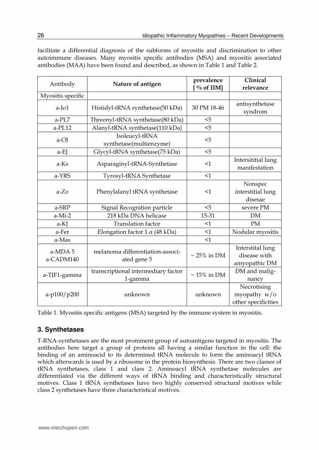
facilitate a differential diagnosis of the subforms of myositis and discrimination to other autoimmune diseases. Many myositis specific antibodies (MSA) and myositis associated antibodies (MAA) have been found and described, as shown in Table 1 and Table 2.
Antibody Nature of antigen prevalence
[ % of IIM]
Clinical
relevance
Myositis specific
a-Jo1 Histidyl-tRNA synthetase(50 kDa) 30 PM 18-46 antisynthetase
syndrom a-PL7 Threonyl-tRNA synthetase(80 kDa) <5 a-PL12 Alanyl-tRNA synthetase(110 kDa) <5
a-OJ Isoleucyl-tRNA
synthetase(multienzyme) <5
a-EJ Glycyl-tRNA synthetase(75 kDa) <5
a-Ks Asparaginyl-tRNA-Synthetase <1 Intersititial lung
manifestation a-YRS Tyrosyl-tRNA Synthetase <1
a-Zo Phenylalanyl tRNA synthetase <1 Nonspec
interstitial lung disesae
a-SRP Signal Recognition particle <5 severe PM a-Mi-2 218 kDa DNA helicase 15-31 DM a-KJ Translation factor <1 PM a-Fer Elongation factor 1 α (48 kDa) <1 Nodular myositis a-Mas <1
a-MDA 5 a-CADM140
melanoma differentiation-associ-ated gene 5
~ 25% in DM Interstital lung
disease with amyopathic DM
a-TIF1-gamma transcriptional intermediary factor
1-gamma ~ 15% in DM
DM and malig-nancy
a-p100/p200 unknown unknown Necrotising
myopathy w/o other specificities
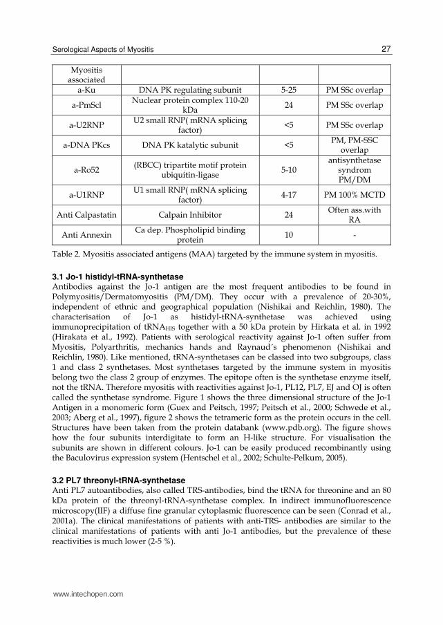
Table 1. Myositis specific antigens (MSA) targeted by the immune system in myositis.
3. Synthetases
T-RNA-synthetases are the most prominent group of autoantigens targeted in myositis. The antibodies here target a group of proteins all having a similar function in the cell: the binding of an aminoacid to its determined tRNA molecule to form the aminoacyl tRNA which afterwards is used by a ribosome in the protein biosynthesis. There are two classes of tRNA synthetases, class 1 and class 2. Aminoacyl tRNA synthetase molecules are differentiated via the different ways of tRNA binding and characteristically structural motives. Class 1 tRNA synthetases have two highly conserved structural motives while class 2 synthetases have three characteristical motives.
www.intechopen.com
Serological Aspects of Myositis
27
Myositis associated
a-Ku DNA PK regulating subunit 5-25 PM SSc overlap
a-PmScl Nuclear protein complex 110-20
kDa24 PM SSc overlap
a-U2RNP U2 small RNP( mRNA splicing
factor)<5 PM SSc overlap
a-DNA PKcs DNA PK katalytic subunit <5 PM, PM-SSC
overlap
a-Ro52 (RBCC) tripartite motif protein
ubiquitin-ligase 5-10
antisynthetase syndrom PM/DM
a-U1RNP U1 small RNP( mRNA splicing
factor)4-17 PM 100% MCTD
Anti Calpastatin Calpain Inhibitor 24 Often ass.with
RA
Anti Annexin Ca dep. Phospholipid binding
protein10 -
Table 2. Myositis associated antigens (MAA) targeted by the immune system in myositis.
3.1 Jo-1 histidyl-tRNA-synthetase Antibodies against the Jo-1 antigen are the most frequent antibodies to be found in Polymyositis/Dermatomyositis (PM/DM). They occur with a prevalence of 20-30%, independent of ethnic and geographical population (Nishikai and Reichlin, 1980). The characterisation of Jo-1 as histidyl-tRNA-synthetase was achieved using immunoprecipitation of tRNAHIS together with a 50 kDa protein by Hirkata et al. in 1992 (Hirakata et al., 1992). Patients with serological reactivity against Jo-1 often suffer from Myositis, Polyarthritis, mechanics hands and Raynaud´s phenomenon (Nishikai and Reichlin, 1980). Like mentioned, tRNA-synthetases can be classed into two subgroups, class 1 and class 2 synthetases. Most synthetases targeted by the immune system in myositis belong two the class 2 group of enzymes. The epitope often is the synthetase enzyme itself, not the tRNA. Therefore myositis with reactivities against Jo-1, PL12, PL7, EJ and OJ is often called the synthetase syndrome. Figure 1 shows the three dimensional structure of the Jo-1 Antigen in a monomeric form (Guex and Peitsch, 1997; Peitsch et al., 2000; Schwede et al., 2003; Aberg et al., 1997), figure 2 shows the tetrameric form as the protein occurs in the cell. Structures have been taken from the protein databank (www.pdb.org). The figure shows how the four subunits interdigitate to form an H-like structure. For visualisation the subunits are shown in different colours. Jo-1 can be easily produced recombinantly using the Baculovirus expression system (Hentschel et al., 2002; Schulte-Pelkum, 2005).
3.2 PL7 threonyl-tRNA-synthetase Anti PL7 autoantibodies, also called TRS-antibodies, bind the tRNA for threonine and an 80 kDa protein of the threonyl-tRNA-synthetase complex. In indirect immunofluorescence microscopy(IIF) a diffuse fine granular cytoplasmic fluorescence can be seen (Conrad et al., 2001a). The clinical manifestations of patients with anti-TRS- antibodies are similar to the clinical manifestations of patients with anti Jo-1 antibodies, but the prevalence of these reactivities is much lower (2-5 %).
www.intechopen.com

Idiopathic Inflammatory Myopathies – Recent Developments
28
Fig. 1. Histidyl tRNA Ligase monomer, according Aberg et al. (Aberg et al., 1997).
Fig. 2. Histidyl tRNA Ligase tetramer (Aberg et al., 1997).
3.3 PL12 alanyl-tRNA synthetase Anti-Pl 12-antibodies react with a 110 kDa protein, the alanyl-tRNA synthetase. Sera having this reactivity also contain antibodies directly binding the alanyl aminoacyl tRNA. Next to the relative low prevalence of less than 5 % in myositis, these antibodies are also found in interstitial lung diseases without myositis manifestation (Hirakata et al., 1995).
www.intechopen.com
Serological Aspects of Myositis
29
3.4 EJ glycyl-tRNA synthetase Anti-EJ-Antibodies bind a 75 kDa-protein of glycyl-tRNA synthetase together with four of the glycyl-tRNAs. This parameter is also of a low frequence. Of interest may be, that anti-EJ antibodies can, like anti Jo-1 antibodies precede the clinical symptoms of myositis (Targoff, 2000). Anti-EJ-antibodies have a prevalence of less than 5 % in myositis patients.
3.5 OJ- isoleucyl-tRNA synthetase Anti OJ antibodies have Isoleucyl-tRNA synthetases as a main target, but also bind to other synthetases. The main epitope of anti OJ antibodies seems to be directed against a multienzyme complex, containing aminoacyl-tRNA synthetase activity for up to nine different amino acid systems. Anti OJ-antibodies have a prevalence of less than 5 % in myositis
3.6 KS- asparaginyl tRNA-synthetase Anti KS antibodies bind Asparaginyl tRNA-synthetase. An anti KS-reactivity is not a clear marker for myositis, the majority of patients showing anti KS-reactivity suffers from interstitial lung disease and not from myositis (Hirakata et al., 1999).
3.7 ZO phenylalanyl tRNA synthetase First described by Betterige (Betteridge et al., 2007) in a patient showing clinical symptoms of an antisynthetase syndrome, but showing no positive serological reaction to the previously identified anti-synthetase autoantibodies. Up to now only one patient has been found with this reactivity.
4. Mi2 antigen: nucleosome remodeling deacetylase
Anti Mi2 Antibodies are well known markers for dermatomyositis with an apparent prevalence of 15-30 % in patients with DM (Conrad et al., 2001b), although the biological function of the Mi2 protein in the cell remained elusive for a long time (Targoff, 2000). Anti Mi2 Antibodies can be found in ca. 20 % of patients suffering from the adult form of dermatomyositis. The function of this protein seems to be to catalyze the unwinding of chromatin structures in chromosomal DNA. The protein possesses one DNA binding domain and one helicase domain (Targoff, 2000; Woodage et al., 1997). It is assumed that the Mi2 complex catalyses an ATP dependand mechanism of Nucleosome remodelling, which makes activated genes accessible on the chromosome. This complex, called Nucleosome Remodeling Deacetylase (NuRD) seems to play a central role in one previously unknown way of gene activation (Zhang et al., 1998). Due to the size of the protein (240 kDa), attempts were made to identify the epitope sequences and to produce a smaller protein for diagnostic purposes. Independently, Mi2 alpha and Mi2 beta were described in 1995 by Ge et al. (Ge et al., 1995) and Seelig et al. (Seelig et al., 1995) wich share a sequential homology of 68 % and have parts of high similarity (Seelig et al., 1996). Sera reacting with the natural form of the Mi2 autoantigen also react with the two different recombinant forms in a highly similar matter. First attempts to solve the three dimensional structure were made by Kwan et al. in 2003 seen in figure 3 (Kwan et al., 2003). The structure of the complex was revealed only lately by Lejon et al. (Lejon et al., 2011a), as shown in figure 4. Mi2 can be easily produced recombinantly using the Baculovirus expression system (Hentschel et al., 2002; Schulte-Pelkum, 2005).
www.intechopen.com

Idiopathic Inflammatory Myopathies – Recent Developments
30
Fig. 3. Chromodomain helicase-DNA-binding protein Mi2 subunit (Kwan et al., 2003).
Fig. 4. The NURD complex (Lejon et al., 2011b).
5. PM-Scl- and PM1 Alpha
Although found in high frequency in patients with myositis, autoantibodies against the PM/Scl Antigen are rather myositis associated antibodies (maa) than myositis specific antibodies (msa) (Targoff, 2000). The main antigen, targeted by nearly all anti-PM/Scl positive sera is the PM/Scl 100 protein, which is part of a major protein complex of 11 proteins. Many sera also react with a 75 kDa protein referred to as PM/Scl 75. This protein migrates with an apparent size of 75 kDa in SDS PAGE, but has a calculated size of about 40 kDa, a phenomenon which can be explained by the highly charged carboxyterminal half of the protein. The other proteins of this complex are not targeted by autoantibodies associated with polymyositis. The biological function of the protein complex containing PM/Scl 100 and PM/Scl 75 seems to be analogous to the exosome complex of yeast, in which RNA is
www.intechopen.com

Serological Aspects of Myositis
31
processed. Within this complex the degradation of RNA, the maturation of 5,8 S rRNA and the processing of small nuclear RNAs and AU-rich mRNAs (Raijmakers et al., 2003) is processed. Allmang et al. found out, that size and structure of the yeast exosome complex are the same as the PM/Scl complex in a human cell (Allmang et al., 1999). This complex can be found in the granular parts of the nucleoli and within the nucleoplasma. The PM/Scl 100 protein is the analogue to the yeast Rrp6p-protein, the PM/Scl 75 protein the analogue to the Rrp4p- protein (Van Eenennaam et al., 2002). As there was no three dimensional structure available, Parker and Song published a theoretical structure showing a ring-like structure of the proteins of the exosome complex (Parker and Song, 2004), shown in figure 5.
Fig. 5. The hypothetical exosome complex according to Parker et al. (Parker and Song, 2004).
Brouwer et al. (Brouwer et al., 2002) described reactivities towards the exosome complex for patients with idiopathic inflammatory myopathies, overlap syndromes and scleroderma. These reactivities were only found in a combination with reactivity against PM/Scl 100. Initial use of the PM/Scl 100 antigen showed good sensitivity and specificity using a full length 100 kDa antigen (Hentschel et al., 2002). While a debate was going on whether the PM/Scl 75 or the PM/Scl 100 antigen should be used as the preferred antigen for IVD testing (Raijmakers et al., 2004; Mahler and Raijmakers, 2007a; Brouwer et al., 2002), a peptide sequence between amino acids 231 and 245 was found which could be proven as the main site of autoantibody binding in the PM/Scl autoantigen (Bluthner et al., 2000a; Mahler et al., 2003; Mahler and Raijmakers, 2007b). The major epitope of PM/Scl referred to as PM1 alpha, consists of a local alpha helical structure. When an PM1 Alpha ELISA was recently compared to ELISA tests using the recombinant antigens with 75 and 100 kDa respectively, the best discrimination between preselected PM/Scl positive Sera (defined by Immunoblot, indirect immunofluorescence and Immunodiffusion) and control sera was observed with the PM1-Alpha ELISA with an area under the curve, AUC =1.0 compared to the PM/Scl-100 (AUC = 0.98) and PM/Scl-75 ELISA (AUC = 0.85) as revealed by receiver operating characteristics (ROC) analysis. This observation shows an interesting aspect: due to the higher local density of the immunodominant sequence, a peptide can even be the better antigen than the protein sequence it was derived from.
www.intechopen.com
Idiopathic Inflammatory Myopathies – Recent Developments
32
6. Ku –DNA-PK
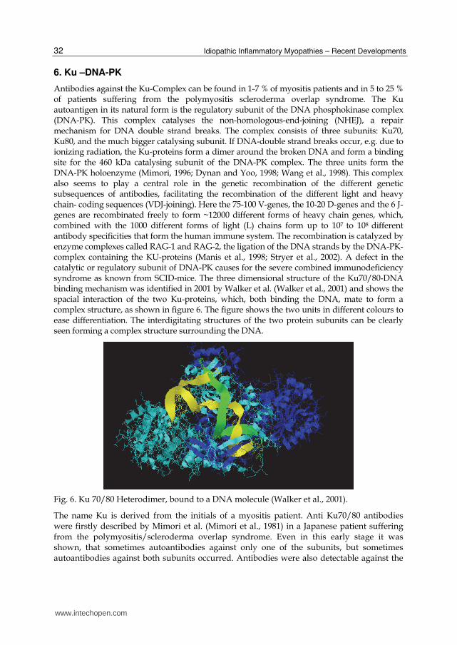
Antibodies against the Ku-Complex can be found in 1-7 % of myositis patients and in 5 to 25 % of patients suffering from the polymyositis scleroderma overlap syndrome. The Ku autoantigen in its natural form is the regulatory subunit of the DNA phosphokinase complex (DNA-PK). This complex catalyses the non-homologous-end-joining (NHEJ), a repair mechanism for DNA double strand breaks. The complex consists of three subunits: Ku70, Ku80, and the much bigger catalysing subunit. If DNA-double strand breaks occur, e.g. due to ionizing radiation, the Ku-proteins form a dimer around the broken DNA and form a binding site for the 460 kDa catalysing subunit of the DNA-PK complex. The three units form the DNA-PK holoenzyme (Mimori, 1996; Dynan and Yoo, 1998; Wang et al., 1998). This complex also seems to play a central role in the genetic recombination of the different genetic subsequences of antibodies, facilitating the recombination of the different light and heavy chain- coding sequences (VDJ-joining). Here the 75-100 V-genes, the 10-20 D-genes and the 6 J-genes are recombinated freely to form ~12000 different forms of heavy chain genes, which, combined with the 1000 different forms of light (L) chains form up to 107 to 108 different antibody specificities that form the human immune system. The recombination is catalyzed by enzyme complexes called RAG-1 and RAG-2, the ligation of the DNA strands by the DNA-PK-complex containing the KU-proteins (Manis et al., 1998; Stryer et al., 2002). A defect in the catalytic or regulatory subunit of DNA-PK causes for the severe combined immunodeficiency syndrome as known from SCID-mice. The three dimensional structure of the Ku70/80-DNA binding mechanism was identified in 2001 by Walker et al. (Walker et al., 2001) and shows the spacial interaction of the two Ku-proteins, which, both binding the DNA, mate to form a complex structure, as shown in figure 6. The figure shows the two units in different colours to ease differentiation. The interdigitating structures of the two protein subunits can be clearly seen forming a complex structure surrounding the DNA.
Fig. 6. Ku 70/80 Heterodimer, bound to a DNA molecule (Walker et al., 2001).
The name Ku is derived from the initials of a myositis patient. Anti Ku70/80 antibodies were firstly described by Mimori et al. (Mimori et al., 1981) in a Japanese patient suffering from the polymyositis/scleroderma overlap syndrome. Even in this early stage it was shown, that sometimes autoantibodies against only one of the subunits, but sometimes autoantibodies against both subunits occurred. Antibodies were also detectable against the
www.intechopen.com

Serological Aspects of Myositis
33
whole complex of both regulatory subunits and the catalyzing subunit of DNA-PK. This so called particle antigen was assembled from highly purified native components. Antibodies were found against subunits but also against three dimensional structures of the complete particle antigen (Jafri et al., 2001). The question which one of the two subunits of the Ku antigen offers the higher sensitivity and specificity for myositis diagnosis was raised and Wang et al. (Wang et al., 1997) published a study with showed that about 50 % of the patients showed reactivity against both subunits, 30 % reacted only with the 80 kDa subunit whereas only 3 % showed exclusive reactivity against the 70 kDa Subunit alone. Also of interest in this study was the observation that 18 % of the screened myositis patients had no reactivity against the one or the other subunit, but instead showed antibodies against the heterodimeric form of Ku70/80. For diagnostic purposes a native form of the heterodimeric Ku70/80 complex is the antigen of choice. Experiments showed a far better discrimination using the heterodimeric naitve antigen compared to separately purified subunits, which later were co-coated on an ELISA plate (Schulte-Pelkum, 2005)
7. SRP –Signal recognition particle
Anti Signal recognition particle (SRP, SRP 54) antibodies are rare autoantibodies in myositis with a prevalence of only 5 %, but the anti SRP reactivity coincides with a severe onset of polymyositis (Targoff et al., 1990). Normal corticosteroid treatment usually shows no positive effect on the disease progress on aSRP 54 pos patients. Newer data as described by Hengstman (Hengstman et al., 2006), after conducting an international study with 23 aSRP positive patient samples found anti SRP 54 reactivity as a marker for a necrotising myopathy rather than a classical myositis. The patients symptoms differed only in some cases from the symptoms of the anti-SRP negative myositis control group, but anti SRP pos patients suffered significantly more often from dysphagia and muscle atropy. Also the biopsy samples differed significantly, as there were no myositis specific histological features like inflammatory infiltrates, making anti SRP antibody positive patients a distinct group next to the more classical myositis. Firstly described as an autoantibody by Reeves et al. (Reeves et al., 1986), the SRP is the primary tool for the targeting of the nascent polypeptide chain. proteins which have to be folded and/or glycosylated within the endoplasmatic reticulum have to have a signal peptide sequence on the aminoterminus. The exact structure of this signal sequence seems to be of lesser importance, although all signal-sequences contain repeating motives of hydrophobic aminoacids alternating with serine and threonine residues, which can be found in many different signal sequences moderating the protein targeting. The complex which catalyzes the targeting of the de-novo sequences is the SRP. This complex contains the 7SL-RNA and six proteins of 9, 14, 19, 54, 68 and 72 kDa. The SRP 54 kDa is the protein which directly binds to the signal sequence of the nascent protein. The complex containing ribosome, nascent protein and SRP then binds to the SRP-receptor and to a translocon, a protein on the outside of the rough ER, which then forms a channel inside the ER-lumen. Inside the ER the signal sequence is cleaved by a signal peptidase, while the rest of the protein is now synthesized directly into the lumen of the ER. The three dimensional structure of SRP 54 as shown in figure 7, was determined by Gowda et al. (Gowda et al., 1998; Gowda et al., 1999). The epitopes of SRP 54 showed to be structural epitopes, which is easily understood looking at the structure of the protein, consisting nearly only from Helix-turn-Helix motives. Interestingly Beneviste et al. observed that the aSRP54 antibody titer in a cohort of 8 longitudinally followed patients correlated (Benveniste et al.,
www.intechopen.com

Idiopathic Inflammatory Myopathies – Recent Developments
34
2011) to a high degree with disease activity as measured e.g. by serum creatine kinase activity.
Fig. 7. Structure of SRP 54 (Gowda et al., 1998).
8. CADM140 / MDA 5
Newly shown by Nakashima (Nakashima et al., 2010) the antibody firstly described as a-CADM140 autoantibody recognises the melanoma differentiation-associated Gene 5 protein (MDA 5) which plays a role in innate immune responses. Patients with this reactivity suffer from clinically amyopathic Dermatomyositis (CADM) and have a high risk for life-threatening complications in DM, namely due to the rapidly progressing interstitial lung involvement. After the antibodies were first reported in Japan with a prevalence of around 25 % in DM cohorts (Hoshino et al., 2010), retrospective testing of dermatomyositis groups revealed also in Europe that up to 13 % of DM patients have antibodies against MDA 5, with a clear correlation for severe forms of rapidly progressing ILD and poor prognosis (Labirua and Lundberg, 2010; Fiorentino et al., 2011).
9. TIF1 gamma
First described as the anti P155 antibody the antigen bound by this antibody was revealed to be the TIF1 gamma protein of the tripartite motive family (TRIM 33) like other autoantigens, a zinc finger protein (Targoff et al., 2006a). It is (at the moment) thought to be a transcriptional corepressor. It was described first by Targoff et al. (Targoff et al., 2006b). The clinical manifestations of this reactivity involve high prevalences in dermatomyositis, as reported by Targoff et al., but with a high specificity the occurrence of this MSA correlates with a risk of malignancies as (Kaji et al., 2007). This observation was statistically underlined when Selva O´Canaghan et al. performed a meta analysis for anti TIF 1 gamma antibodies and predictive values for malignancies (Selva-O'Callaghan et al., 2010) and found that anti p155/TIF1 y antibodies have a 70 % sensitivity and a 90 % specificity to detect occult malignancies in DM. The TIF1 gamma protein as described by Venturini et al. shows a strong silencing activity towards gentic promoter sequences. In this promoter sequences, binding affinity of TIF1 gamma is dependant of a single motive (Venturini et al., 1999).
www.intechopen.com

Serological Aspects of Myositis
35
10. U1-snRNP
Antibodies against the U1-RNP complex are, like anti-Ku and anti-PM/Scl antibodies MAA. An association with PM can be found in 4-17 % of myositis cases, whereas these autoantibodies have a prevalence of 100 % in mixed connective tissue disease (MCTD). Absence of this antibodies rules out the diagnosis of MCTD (Conrad et al., 2001a). The U1 RNP consists of three small nuclear ribonuclear proteins (snRNP-“SNURPS”) A (34 kDa); C (22 kDa) and the 68 kDa protein. Most prominent is reactivity against the 68 kDa protein. Considering this reactivity the coincidence of an autoimmune disease and a cytomegalo virus infection was discussed (Newkirk et al., 2001), after high rates of coinciding SLE after CMV infections were reported (Newkirk et al., 2001). The U1-snRNP complex belongs to the snRNP protein and is involved in the splicing process of the pre-messenger RNA. In mammalian cells the U1-snRNP binds the 5´splicing site of an intron with a 15 nucleotide consensus sequence of its 165 base long snRNA sequence; the subunits of the U1-RNP then bind to this snRNA. Not all proteins bind directly to the snRNA, RNP-C for example binds with a zinc finger motive to the complex of RNA and the other RNP-proteins (Nelissen et al., 1991). The sequences were published by Sillekens et al (U1-RNP-A), Yamamoto et al. (U1-RNP-C) and by Theissen et al. (U1-RNP-68) (Sillekens et al., 1987; Yamamoto et al., 1988; Theissen et al., 1986). Figure 8 shows the structure of U1-RNP-A bound to RNA (Varani et al., 2000). A scheme of the complete U1 RNP is shown in Figure 9.
11. Ro52
Although Ro52 and Ro60 (SS-A), shown early to be separate proteins (Chan et al., 1991), were initially suggested to be closely related, no direct interaction of the proteins could be conclusively shown. Recent studies indicated that the proteins are even localized in different cell compartments and they perform rather different functions. The 52 kDa Ro antigen was eventually identified as tripartite motif protein (TRIM) 21 ubiquitin-ligase that is over-expressed in peripheral blood mononuclear cells in SjS and SLE patients (Rhodes et al., 2002; Wada and Kamitani, 2006). Ro52 is reported to interact with several different molecules, among them calreticulin and a 78 kDa glucose-regulated protein (GRP78), also known as immunoglobulin heavy chain-binding protein (BIP) and formerly proposed as an early marker for rheumatoid arthritis (Blass et al., 2001). Taking its function into consideration, Ro52 is thought to modify the role or stability of its substrates through ubiquitination, and this modification might result in the Ro52-mediated biological events (Wada and Kamitani, 2006; Gomez-Martin et al., 2008).
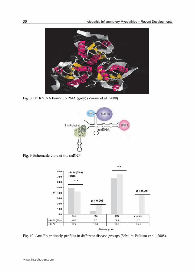
12. Association between anti-Ro52 and anti-Ro60 antibodies in different autoimmune diseases
Testing for SS-A/Ro60 and Ro52 with sera from SSA/Ro related autoimmune diseases showed differing prevalences of the two autoantibodies in the different disease entities (see Figure 10). The frequencies of anti-Ro52 antibodies and anti-Ro60 were comparable in all groups except the myositis and scleroderma cohort. The prevalences of anti-Ro52 reactivitiy without anti-Ro60 reactivity varied in the different groups from 14.5 % in SLE to 37.5 % in the myositis group. In the SjS group, 51.7 % of anti-Ro52 sera had also antibodies to Ro60.
www.intechopen.com
Idiopathic Inflammatory Myopathies – Recent Developments
36
Fig. 8. U1 RNP–A bound to RNA (grey) (Varani et al., 2000).
Fig. 9. Schematic view of the snRNP.
Fig. 10. Anti Ro antibody profiles in different disease groups (Schulte-Pelkum et al., 2008).
www.intechopen.com

Serological Aspects of Myositis
37
13. Coincidence of anti-Ro52 and anti-Jo-1 in patients with polymyositis
A high degree of correlation was found in a group of myositis sera tested for aab against Jo-1 and Ro52: a panel of 43 sera of myositis patients revealed reactivities against Ro52 and Jo-1 in 70 % (p=0.0002, Odds ratio=14.17, kappa=0.54) of Jo-1 positive sera when tested with ELISA (Dr. Fooke Laboratorien) and ALBIA. 22 (24) sera were found positive for anti-Jo-1 by ELISA and ALBIA (numbers in brackets), 16 (17) of these were also found positive for anti-Ro52 (72 % by ELISA, 70.8 % by ALBIA). These observations underline previous conclusions (Peene et al., 2002) that anti-Ro52 is indeed an independent aab in myositis. Rutjes et al. (Rutjes et al., 1997) found anti-Ro52 reactivity in 58 % of Jo-1 positive myositis sera, an observation confirmed in the subsequent years by Rozman et al. (2000), Brouwer et al. (2001) and Koenig et al. (2007) (Rozman et al., 2000; Brouwer et al., 2001; Koenig et al., 2007). In contrast, Langguth et al. (Langguth et al., 2007) indicated that isolated anti-Ro52 reactivity has limited clinical value in a non-obstetric population, a conclusion that could not be confirmed. Our study demonstrated the importance of detecting anti-Ro52 and anti-Ro60 aab separately when considering the diagnosis of patients and in particular myositis patients. This perspective was not included in the study performed by Langguth and colleagues. It can be concluded that anti-Ro52 clearly differs in reactivity from anti-Ro60 (SS-A): Anti-Ro52 is seen in relatively high frequency in myositis and SSc. Anti-Ro52 has a prevalence of up to 35 % in myositis and in this disease group co-occurs in up to 72 % of anti-Jo-1 positive sera. In our opinion it is strongly recommend that diagnostic assays and kits should test anti-Ro52 and anti-Ro60 (SS-A) separately (Schulte-Pelkum et al., 2008; Schulte-Pelkum et al., 2009).
14. Acknowledgements
I thank Dr. M. Fooke of Dr. Fooke Laboratorien GmbH for all the encouragement and the support. Also thanks M. Petschinka (Dr. Fooke Laboratorien GmbH) for technical assistance and support with the preparation of the data. Also I would like to thank Dr. C. Hentschel for quick help with needed data, and Dr. M. Mahler for his data. Thanks also to Dr. rer. nat. habil. W. Schoessler.
15. References
Aberg, A., Yaremchuk, A., Tukalo, M., RASMUSSEN, B., and Cusack, S. (1997). Crystal structure analysis of the activation of histidine by Thermus thermophilus histidyl-tRNA synthetase. Biochemistry 36, 3084-3094.
Allmang, C., Petfalski, E., Podtelejnikov, A., Mann, M., Tollervey, D., and Mitchell, P. (1999). The yeast exosome and human PM-Scl are related complexes of 3' --> 5' exonucleases. Genes Dev 13, 2148-2158.
Benveniste, O., Drouot, L., Jouen, F., Charuel, J.L., Bloch-Queyrat, C., Behin, A., Amoura, Z., Marie, I., Guiguet, M., Eymard, B., Gilbert, D., Tron, F., Herson, S., Musset, L., and Boyer, O. (2011). Anti-signal recognition particle auto-antibody levels correlate with creatine kinase activity in patients with necrotizing myopathy. Arthritis Rheum.
Betteridge, Z., Gunawardena, H., North, J., Slinn, J., and McHugh, N. (2007). Anti-synthetase syndrome: a new autoantibody to phenylalanyl transfer RNA synthetase (anti-Zo)
www.intechopen.com

Idiopathic Inflammatory Myopathies – Recent Developments
38
associated with polymyositis and interstitial pneumonia. Rheumatology. (Oxford) 46, 1005-1008.
Blass, S., Union, A., Raymackers, J., Schumann, F., Ungethum, U., Muller-Steinbach, S., de Keyser, F., Engel, J.M., and Burmester, G.R. (2001). The stress protein BiP is overexpressed and is a major B and T cell target in rheumatoid arthritis. Arthritis Rheum 44, 761-771.
Bluthner, M., Mahler, M., Muller, D.B., Dunzl, H., and Bautz, F.A. (2000a). Identification of an alpha-helical epitope region on the PM/Scl-100 autoantigen with structural homology to a region on the heterochromatin p25beta autoantigen using immobilized overlapping synthetic peptides. J. Mol. Med. 78, 47-54.
Bluthner, M., Mahler, M., Muller, D.B., Dunzl, H., and Bautz, F.A. (2000b). Identification of an alpha-helical epitope region on the PM/Scl-100 autoantigen with structural homology to a region on the heterochromatin p25beta autoantigen using immobilized overlapping synthetic peptides. J Mol Med 78, 47-54.
Brouwer, R., Hengstman, G.J., Vree, E.W., Ehrfeld, H., Bozic, B., Ghirardello, A., Grondal, G., Hietarinta, M., Isenberg, D., Kalden, J.R., Lundberg, I., Moutsopoulos, H., Roux-Lombard, P., Vencovsky, J., Wikman, A., Seelig, H.P., van Engelen, B.G., and van Venrooij, W.J. (2001). Autoantibody profiles in the sera of European patients with myositis. Ann Rheum Dis 60, 116-123.
Brouwer, R., Vree Egberts, W.T., Hengstman, G.J., Raijmakers, R., van Engelen, B.G., Seelig, H.P., Renz, M., Mierau, R., Genth, E., Pruijn, G.J., and van Venrooij, W.J. (2002). Autoantibodies directed to novel components of the PM/Scl complex, the human exosome. Arthritis Res 4, 134-138.
Burmester, G.R., Buttgereit, F., Keitel, W., and Sieper, J. (2001). Systemkrankheiten des Bindegewebes bei anderenorts klassifizierten Krankheiten. In Interdisziplinäre klinische Rheumatologie, H.Zeidler, J.Zacher, and F.Hiepe, eds. (Berlin: Springer Verlag), pp. 960-971.
Chan, E.K., Hamel, J.C., Buyon, J.P., and Tan, E.M. (1991). Molecular definition and sequence motifs of the 52-kD component of human SS-A/Ro autoantigen. J Clin Invest 87, 68-76.
Conrad, K., Schößler, W., and Hiepe, F. (2001a). Autoantikörper bei systemischen Autoimmunerkrankungen. (Lengerich; Berlin; Riga; Rom; Viernheim; Wien; Zagreb: Pabst Science Publishers).
Conrad, K., Schößler, W., and Hiepe, F. (2001b). Mi-2-Antikörper. In Autoantikörper bei systemischen Autoimmunerkrankungen, K.Conrad, W.Schößler, and F.Hiepe, eds. (Lengerich, Berlin, Riga, Rom, Viernheim, Wien, Zagreb: Pabst), pp. 89-90.
Conrad, K., Schößler, W., and Hiepe, F. (2001c). Polymyositis (PM) and Dermatomyositis (DM). In Autoantikörper bei systemischen Autoimmunerkrankungen, K.Conrad, W.Schößler, and F.Hiepe, eds. (Lengerich, Berlin, Riga, Rom, Viernheim, Wien, Zagreb: Pabst), pp. 203-205.
Dynan, W.S. and Yoo, S. (1998). Interaction of Ku protein and DNA-dependent protein kinase catalytic subunit with nucleic acids. Nucleic Acids Res 26, 1551-1559.
Fiorentino, D., Chung, L., Zwerner, J., Rosen, A., and Casciola-Rosen, L. (2011). The mucocutaneous and systemic phenotype of dermatomyositis patients with antibodies to MDA5 (CADM-140): A retrospective study. J. Am. Acad. Dermatol.
Ge, Q., Nilasena, D.S., O'Brien, C.A., Frank, M.B., and Targoff, I.N. (1995). Molecular analysis of a major antigenic region of the 240-kD protein of Mi-2 autoantigen. J Clin Invest 96, 1730-1737.
www.intechopen.com

Serological Aspects of Myositis
39
Gomez-Martin, D., Diaz-Zamudio, M., and Alcocer-Varela, J. (2008). Ubiquitination system and autoimmunity: the bridge towards the modulation of the immune response. Autoimmun. Rev. 7, 284-290.
Gowda, K., Black, S.D., Moeller, I., Sakakibara, Y., Liu, M.C., and Zwieb, C. (1998). protein SRP54 of human signal recognition particle: cloning, expression, and comparative analysis of functional sites. Gene 207, 197-207.
Gowda, K., Clemons, W.M., Jr., Zwieb, C., and Black, S.D. (1999). Expression, purification, and crystallography of the conserved methionine-rich domain of human signal recognition particle 54 kDa protein. protein Sci 8, 1144-1151.
Guex, N. and Peitsch, M.C. (1997). SWISS-MODEL and the Swiss-PdbViewer: an environment for comparative protein modeling. Electrophoresis 18, 2714-2723.
Hengstman, G.J., ter Laak, H.J., Vree Egberts, W.T., Lundberg, I.E., Moutsopoulos, H.M., Vencovsky, J., Doria, A., Mosca, M., van Venrooij, W.J., and van Engelen, B.G. (2006). Anti-signal recognition particle autoantibodies: marker of a necrotising myopathy. Ann. Rheum. Dis. 65, 1635-1638.
Hentschel, C., Schulte-Pelkum, J., Schößler, W., Hiepe, F., and Mierau, R. (2002). A new LINE Immuno assay for the detection of myositis specific autoantibodies. Report on the 6th Dresden Symposium on Autoantibodies 4, 649-650.
Hirakata, M., Mimori, T., Akizuki, M., Craft, J., Hardin, J.A., and Homma, M. (1992). Autoantibodies to small nuclear and cytoplasmic ribonucleoproteins in Japanese patients with inflammatory muscle disease. Arthritis Rheum 35, 449-456.
Hirakata, M., Nakamura, S., Okano, Y., Suwa, A., Inada, S., Akizuki, M., and Hardin, J.A. (1995). Anti-alanyl tRNA synthetase (PL12) antibodies are associated with interstitial lung disease in Japanese patients. Arthritis Rheum 38, 321.
Hirakata, M., Suwa, A., Nagai, S., Kron, M.A., Trieu, E.P., Mimori, T., Akizuki, M., and Targoff, I.N. (1999). Anti-KS: identification of autoantibodies to asparaginyl-transfer RNA synthetase associated with interstitial lung disease. J. Immunol. 162, 2315-2320.
Hoshino, K., Muro, Y., Sugiura, K., Tomita, Y., Nakashima, R., and Mimori, T. (2010). Anti-MDA5 and anti-TIF1-gamma antibodies have clinical significance for patients with dermatomyositis. Rheumatology. (Oxford) 49, 1726-1733.
Jafri, F., Hardin, J.A., and Dynan, W.S. (2001). A method to detect particle-specific antibodies against Ku and the DNA- dependent protein kinase catalytic subunit in autoimmune sera. J Immunol Methods 251, 53-61.
Kaji, K., Fujimoto, M., Hasegawa, M., Kondo, M., Saito, Y., Komura, K., Matsushita, T., Orito, H., Hamaguchi, Y., Yanaba, K., Itoh, M., Asano, Y., Seishima, M., Ogawa, F., Sato, S., and Takehara, K. (2007). Identification of a novel autoantibody reactive with 155 and 140 kDa nuclear proteins in patients with dermatomyositis: an association with malignancy. Rheumatology. (Oxford) 46, 25-28.
Kalovidouris, A.E. (1992). Immune aspects of myositis. Curr Opin Rheumatol 4, 809-814. Koenig, M., Fritzler, M.J., Targoff, I.N., Troyanov, Y., and Senecal, J.L. (2007). Heterogeneity
of autoantibodies in 100 patients with autoimmune myositis: insights into clinical features and outcomes. Arthritis Res. Ther. 9, R78.
Kwan, A.H., Gell, D.A., Verger, A., Crossley, M., Matthews, J.M., and Mackay, J.P. (2003). Engineering a protein scaffold from a PHD finger. Structure. (Camb. ) 11, 803-813.
Labirua, A. and Lundberg, I.E. (2010). Interstitial lung disease and idiopathic inflammatory myopathies: progress and pitfalls. Curr. Opin. Rheumatol. 22, 633-638.
Langguth, D.M., Morris, S., Clifford, L., Wilson, R.J., Neil, J., Hogan, P.G., and Wong, R.C. (2007). Specific testing for "isolated" anti-52 kDa SSA/Ro antibodies during
www.intechopen.com

Idiopathic Inflammatory Myopathies – Recent Developments
40
standard anti-extractable nuclear antigen testing is of limited clinical value. J. Clin. Pathol. 60, 670-673.
Lejon, S., Thong, S.Y., Murthy, A., AlQarni, S., Murzina, N.V., Blobel, G.A., Laue, E.D., and Mackay, J.P. (2011b). Insights into association of the NuRD complex with FOG-1 from the crystal structure of an RbAp48.FOG-1 complex. J. Biol. Chem. 286, 1196-1203.
Lejon, S., Thong, S.Y., Murthy, A., AlQarni, S., Murzina, N.V., Blobel, G.A., Laue, E.D., and Mackay, J.P. (2011a). Insights into association of the NuRD complex with FOG-1 from the crystal structure of an RbAp48.FOG-1 complex. J. Biol. Chem. 286, 1196-1203.
Mahler, M., Bluthner, M., and Pollard, K.M. (2003). Advances in B-cell epitope analysis of autoantigens in connective tissue diseases. Clin. Immunol. 107, 65-79.
Mahler, M. and Raijmakers, R. (2007a). Novel aspects of autoantibodies to the PM/Scl complex: clinical, genetic and diagnostic insights. Autoimmun. Rev. 6, 432-437.
Mahler, M. and Raijmakers, R. (2007b). Novel aspects of autoantibodies to the PM/Scl complex: clinical, genetic and diagnostic insights. Autoimmun. Rev. 6, 432-437.
Manchul, L.A., Jin, A., Pritchard, K.I., Tenenbaum, J., Boyd, N.F., Lee, P., Germanson, T., and Gordon, D.A. (1985). The frequency of malignant neoplasms in patients with polymyositis-dermatomyositis. A controlled study. Arch. Intern. Med. 145, 1835-1839.
Manis, J.P., Gu, Y., Lansford, R., Sonoda, E., Ferrini, R., Davidson, L., Rajewsky, K., and Alt, F.W. (1998). Ku70 is required for late B cell development and immunoglobulin heavy chain class switching. J. Exp. Med. 187, 2081-2089.
Maoz, C.R., Langevitz, P., Livneh, A., Blumstein, Z., Sadeh, M., Bank, I., Gur, H., and Ehrenfeld, M. (1998). High incidence of malignancies in patients with dermatomyositis and polymyositis: an 11-year analysis. Semin. Arthritis Rheum. 27, 319-324.
Mimori, T. (1996). Structures targeted by the immune system in myositis. Curr Opin Rheumatol 8, 521-527.
Mimori, T., Akizuki, M., Yamagata, H., Inada, S., Yoshida, S., and Homma, M. (1981). Characterization of a high molecular weight acidic nuclear protein recognized by autoantibodies in sera from patients with polymyositis- scleroderma overlap. J Clin Invest 68, 611-620.
Nakashima, R., Imura, Y., Kobayashi, S., Yukawa, N., Yoshifuji, H., Nojima, T., Kawabata, D., Ohmura, K., Usui, T., Fujii, T., Okawa, K., and Mimori, T. (2010). The RIG-I-like receptor IFIH1/MDA5 is a dermatomyositis-specific autoantigen identified by the anti-CADM-140 antibody. Rheumatology. (Oxford) 49, 433-440.
Nelissen, R.L., Heinrichs, V., Habets, W.J., Simons, F., Luhrmann, R., and van Venrooij, W.J. (1991). Zinc finger-like structure in U1-specific protein C is essential for specific binding to U1 snRNP. Nucleic Acids Res. 19, 449-454.
Newkirk, M.M., van Venrooij, W.J., and Marshall, G.S. (2001). Autoimmune response to U1 small nuclear ribonucleoprotein (U1 snRNP) associated with cytomegalovirus infection. Arthritis Res. 3, 253-258.
Nishikai, M. and Reichlin, M. (1980). Heterogeneity of precipitating antibodies in polymyositis and dermatomyositis. Characterization of the Jo-1 antibody system. Arthritis Rheum 23, 881-888.
Parker, R. and Song, H. (2004). The enzymes and control of eukaryotic mRNA turnover. Nat. Struct. Mol. Biol 11, 121-127.
www.intechopen.com

Serological Aspects of Myositis
41
Peene, I., Meheus, L., De, K.S., Humbel, R., Veys, E.M., and De, K.F. (2002). Anti-Ro52 reactivity is an independent and additional serum marker in connective tissue disease. Ann. Rheum. Dis. 61, 929-933.
Peitsch, M.C., Schwede, T., and Guex, N. (2000). Automated protein modelling--the proteome in 3D. Pharmacogenomics. 1, 257-266.
Raijmakers, R., Egberts, W.V., van Venrooij, W.J., and Pruijn, G.J. (2003). The association of the human PM/Scl-75 autoantigen with the exosome is dependent on a newly identified N terminus. J. Biol. Chem. 278, 30698-30704.
Raijmakers, R., Renz, M., Wiemann, C., Egberts, W.V., Seelig, H.P., van Venrooij, W.J., and Pruijn, G.J. (2004). PM-Scl-75 is the main autoantigen in patients with the polymyositis/scleroderma overlap syndrome. Arthritis Rheum. 50, 565-569.
Reeves, W.H., Nigam, S.K., and Blobel, G. (1986). Human autoantibodies reactive with the signal-recognition particle. Proc. Natl. Acad. Sci. U. S. A 83, 9507-9511.
Rhodes, D.A., Ihrke, G., Reinicke, A.T., Malcherek, G., Towey, M., Isenberg, D.A., and Trowsdale, J. (2002). The 52 000 MW Ro/SS-A autoantigen in Sjogren's syndrome/systemic lupus erythematosus (Ro52) is an interferon-gamma inducible tripartite motif protein associated with membrane proximal structures. Immunology 106, 246-256.
Rozman, B., Bozic, B., Kos-Golja, M., Plesivcnik-Novljan, M., and Kveder, T. (2000). Immunoserological aspects of idiopathic inflammatory muscle disease. Wien. Klin. Wochenschr. 112, 722-727.
Rutjes, S.A., Vree Egberts, W.T., Jongen, P., Van Den, H.F., Pruijn, G.J., and van Venrooij, W.J. (1997). Anti-Ro52 antibodies frequently co-occur with anti-Jo-1 antibodies in sera from patients with idiopathic inflammatory myopathy. Clin. Exp. Immunol. 109, 32-40.
Schulte-Pelkum, J. (2005). Entwicklung neuer Methoden zum Nachweis von Myositiden. (Berlin Erlangen.
Schulte-Pelkum, J., Fritzler, M.J., and Mahler, M. (2009). Latest update on the Ro/SS-A autoantibody system. Autoimmun. Rev. 8, 632-637.
Schulte-Pelkum, J., Simon, T., Szmyrka-Kaczmarek, M., Fritzler, M.J., and Mahler, M. (2008). Latest Update on the Ro/SS-A Autoantibody System. 6th International Congress on Autoimmunity, Porto (Lecture).
Schwede, T., Kopp, J., Guex, N., and Peitsch, M.C. (2003). SWISS-MODEL: An automated protein homology-modeling server. Nucleic Acids Res. 31, 3381-3385.
Seelig, H.P., Moosbrugger, I., Ehrfeld, H., Fink, T., Renz, M., and Genth, E. (1995). The major dermatomyositis-specific Mi-2 autoantigen is a presumed helicase involved in transcriptional activation. Arthritis Rheum 38, 1389-1399.
Seelig, H.P., Renz, M., Targoff, I.N., Ge, Q., and Frank, M.B. (1996). Two forms of the major antigenic protein of the dermatomyositis- specific Mi-2 autoantigen. Arthritis Rheum 39, 1769-1771.
Selva-O'Callaghan, A., Trallero-Araguas, E., Grau-Junyent, J.M., and Labrador-Horrillo, M. (2010). Malignancy and myositis: novel autoantibodies and new insights. Curr. Opin. Rheumatol. 22, 627-632.
Sillekens, P.T., Habets, W.J., Beijer, R.P., and van Venrooij, W.J. (1987). cDNA cloning of the human U1 snRNA-associated A protein: extensive homology between U1 and U2 snRNP-specific proteins. EMBO J. 6, 3841-3848.
Stryer, L., Berg, J.M., and Tymoczko, J.L. (2002). The Immune System. In Biochemistry, L.Stryer, J.M.Berg, and J.L.Tymoczko, eds. (New York: W.H. Freeman & Company New York), pp. 912-979.
www.intechopen.com

Idiopathic Inflammatory Myopathies – Recent Developments
42
Targoff, I., Trieu, E., None, M., Levy-Neto, M., Prasertsuntarasai, T., and Miller, F.W. (2006a). Autoantibodies to Transcriptional Intermediary Factor 1-gamma (TIF1-g) in Dermatomyositis. ACR Meeting 1241-Presentation.
Targoff, I.N. (2000). Update on myositis-specific and myositis-associated autoantibodies. Curr Opin Rheumatol 12, 475-481.
Targoff, I.N., Johnson, A.E., and Miller, F.W. (1990). Antibody to signal recognition particle in polymyositis. Arthritis Rheum 33, 1361-1370.
Targoff, I.N., Mamyrova, G., Trieu, E.P., Perurena, O., Koneru, B., O'Hanlon, T.P., Miller, F.W., and Rider, L.G. (2006b). A novel autoantibody to a 155-kd protein is associated with dermatomyositis. Arthritis Rheum. 54, 3682-3689.
Theissen, H., Etzerodt, M., Reuter, R., Schneider, C., Lottspeich, F., Argos, P., Luhrmann, R., and Philipson, L. (1986). Cloning of the human cDNA for the U1 RNA-associated 70K protein. EMBO J. 5, 3209-3217.
Unverricht, H. (1887). Über eine eigentümliche Form von akuter Muskelentzündung mit einem der Trichinose ähnlichen Krankheitsbilde. Münchener medizinische Wochenschrift 34, 488-492.
Van Eenennaam, H., Vogelzangs, J.H., Bisschops, L., Te Boome, L.C., Seelig, H.P., Renz, M., De Rooij, D.J., Brouwer, R., Pluk, H., Pruijn, G.J., van Venrooij, W.J., and Van Den Hoogen, F.H. (2002). Autoantibodies against small nucleolar ribonucleoprotein complexes and their clinical associations. Clin Exp Immunol 130, 532-540.
Varani, L., Gunderson, S.I., Mattaj, I.W., Kay, L.E., Neuhaus, D., and Varani, G. (2000). The NMR structure of the 38 kDa U1A protein - PIE RNA complex reveals the basis of cooperativity in regulation of polyadenylation by human U1A protein. Nat. Struct. Biol. 7, 329-335.
Venturini, L., You, J., Stadler, M., Galien, R., Lallemand, V., Koken, M.H., Mattei, M.G., Ganser, A., Chambon, P., Losson, R., and de The, H. (1999). TIF1gamma, a novel member of the transcriptional intermediary factor 1 family. Oncogene 18, 1209-1217.
Wada, K. and Kamitani, T. (2006). Autoantigen Ro52 is an E3 ubiquitin ligase. Biochem Biophys. Res. Commun. 339, 415-421.
Wagner, E.L. (1863). Fall einer seltenen Muskelkrankheit. Archiv der Heilkunde 4, 282-283. Walker, J.R., Corpina, R.A., and Goldberg, J. (2001). Structure of the Ku heterodimer bound
to DNA and its implications for double-strand break repair. Nature 412, 607-614. Wang, J., Dong, X., Myung, K., Hendrickson, E.A., and Reeves, W.H. (1998). Identification of
two domains of the p70 Ku protein mediating dimerization with p80 and DNA binding. J Biol Chem 273, 842-848.
Wang, J., Dong, X., Stojanov, L., Kimpel, D., Satoh, M., and Reeves, W.H. (1997). Human autoantibodies stabilize the quaternary structure of Ku antigen. Arthritis Rheum 40, 1344-1353.
Woodage, T., Basrai, M.A., Baxevanis, A.D., Hieter, P., and Collins, F.S. (1997). Characterization of the CHD family of proteins. Proc Natl. Acad Sci U. S. A 94, 11472-11477.
Yamamoto, K., Miura, H., Moroi, Y., Yoshinoya, S., Goto, M., Nishioka, K., and Miyamoto, T. (1988). Isolation and characterization of a complementary DNA expressing human U1 small nuclear ribonucleoprotein C polypeptide. J. Immunol. 140, 311-317.
Zantos, D., Zhang, Y., and Felson, D. (1994). The overall and temporal association of cancer with polymyositis and dermatomyositis. J. Rheumatol. 21, 1855-1859.
Zhang, Y., LeRoy, G., Seelig, H.P., Lane, W.S., and Reinberg, D. (1998). The dermatomyositis-specific autoantigen Mi2 is a component of a complex containing histone deacetylase and nucleosome remodeling activities. Cell 95, 279-289.
www.intechopen.com

Idiopathic Inflammatory Myopathies - Recent DevelopmentsEdited by Prof. Jan Tore Gran
ISBN 978-953-307-694-2Hard cover, 212 pagesPublisher InTechPublished online 15, September, 2011Published in print edition September, 2011
InTech EuropeUniversity Campus STeP Ri Slavka Krautzeka 83/A 51000 Rijeka, Croatia Phone: +385 (51) 770 447 Fax: +385 (51) 686 166www.intechopen.com
InTech ChinaUnit 405, Office Block, Hotel Equatorial Shanghai No.65, Yan An Road (West), Shanghai, 200040, China
Phone: +86-21-62489820 Fax: +86-21-62489821
The term "myositis" covers a variety of disorders often designated "idiopathic inflammatory myopathies".Although they are rather rare compared to other rheumatic diseases, they often cause severe disability andnot infrequently increased mortality. The additional involvement of important internal organs such as the heartand lungs, is not uncommon. Thus, there is a great need for a better understanding of the etiopathogenesis ofmyositis, which may lead to improved treatment and care for these patients. Major advances regardingresearch and medical treatment have been made during recent years. Of particular importance is thediscovery of the Myositis specific autoantibodies, linking immunological and pathological profiles to distinctclinical disease entities. A wide range of aspects of myopathies is covered in the book presented by highlyqualified authors, all internationally known for their expertice on inflammatory muscle diseases. The bookcovers diagnostic, pathological, immunological and therapeutic aspects of myositis.
How to referenceIn order to correctly reference this scholarly work, feel free to copy and paste the following:
Johannes Schulte-Pelkum (2011). Serological Aspects of Myositis, Idiopathic Inflammatory Myopathies -Recent Developments, Prof. Jan Tore Gran (Ed.), ISBN: 978-953-307-694-2, InTech, Available from:http://www.intechopen.com/books/idiopathic-inflammatory-myopathies-recent-developments/serological-aspects-of-myositis