SERIES ‘‘PULMONARY HYPERTENSION: BASIC CONCEPTS FOR Number 9 in this Series Cardiac magnetic...
13
SERIES ‘‘PULMONARY HYPERTENSION: BASIC CONCEPTS FOR PRACTICAL MANAGEMENT’’ Edited by M.M. Hoeper and A.T. Dinh-Xuan Number 9 in this Series Cardiac magnetic resonance imaging for the assessment of the heart and pulmonary circulation in pulmonary hypertension L.E.R. McLure and A.J. Peacock ABSTRACT: Pulmonary hypertension is a disease of the pulmonary arteries resulting in a progressive increase in pulmonary vascular resistance, ultimately leading to right ventricular failure and death. The functional capacity of the right ventricle is a major prognostic determinant. Our understanding of right ventricle performance in pulmonary hypertension has been hindered by the lack of techniques that give a reliable picture of right ventricle morphology and function. Cardiac magnetic resonance (CMR) imaging enables a unique combination of morphological and functional assessment of the right ventricle and pulmonary circulation. In this review article, we introduce the technique of CMR imaging, review its use in imaging of the heart and pulmonary circulation and discuss its current and future application to the management of patients with pulmonary hypertension. There have been recent major advances in our understanding of the mechanism of disease development, in the diagnostic process, and in the treatment of pulmonary hypertension. Therapeutic advances in the management have reinforced the requirement for noninvasive, accurate and reproducible methods of assessment to act as ‘‘end-points’’ to measure the effects of treatment. We anticipate CMR imaging will increasingly be utilised as the primary modality for combined anatomic and functional assessments that enable more complete and efficient evaluation of pulmonary hypertension patients. KEYWORDS: Cardiac magnetic resonance imaging, pulmonary circulation, pulmonary hyperten- sion, right heart I n the present article, it is our aim to introduce the technique of cardiovascular magnetic reso- nance imaging, to review its use in imaging of the heart and pulmonary circulation and to discuss its current and future application to the manage- ment of patients with pulmonary hypertension. This paper is part of a series of articles published in the European Respiratory Journal summarising and commenting on the latest developments in pul- monary vascular disease (see footnote). PULMONARY HYPERTENSION Pulmonary hypertension is a disease of the pul- monary arteries that is characterised by vascular proliferation and remodelling [1, 2]. It results in a progressive increase in pulmonary vascular resis- tance (PVR) and ultimately, right ventricular failure and death. The diagnostic classification of pulmon- ary hypertension is described in table 1. This current classification was established during the 2003 World Symposium on Pulmonary Hypertension [3]. AFFILIATIONS Scottish Pulmonary Vascular Unit, Golden Jubilee National Hospital, Glasgow, UK. CORRESPONDENCE A.J. Peacock Scottish Pulmonary Vascular Unit Golden Jubilee National Hospital Beardmore Street Clydebank G81 4HX West Dumbartonshire Scotland, UK E-mail: [email protected] Received: October 23 2007 Accepted after revision: December 23 2008 STATEMENT OF INTEREST A statement of interest for A.J. Peacock can be found at www.erj.ersjournals.com/misc/ statements.dtl European Respiratory Journal Print ISSN 0903-1936 Online ISSN 1399-3003 Previous articles in this series: No. 1: Dupuis J, Hoeper MM. Endothelin receptor antagonists in pulmonary arterial hypertension. Eur Respir J 2008; 31: 407–415. No. 2: Gomberg-Maitland M, Olschewski H. Prostcyclin therapies for the treatment of pulmonary arterial hypertension. Eur Respir J 2008; 31: 891–901. No. 3: Behr J, Ryu JH. Pulmonary hypertension in interstitial lung disease. Eur Respir J 2008; 31: 1357–1367. No. 4: Wilkins MR, Wharton J, Grimminger F, Ghofrani HA. Phosphodiesterase inhibitors for the treatment of pulmonary hypertension. Eur Respir J 2008; 32: 198–209. No. 5: Warwick G, Thomas PS, Yates DH. Biomarkers in pulmonary hypertension. Eur Respir J 2008; 32: 503–512. No. 6: Chaouat A, Naeije R, Weitzenblum E. Pulmonary hypertension in COPD. Eur Respir J 2008; 32: 1371–1385. No. 7: Montani D, Price LC, Dorfmuller P, et al. Pulmonary veno-occlusive disease. Eur Respir J 2009; 33: 189–200. No. 8: Faughnan ME, Granton JT, Young LH. The pulmonary vascular complications of hereditary haemorrhagic telangiectasia. Eur Respir J 2009; 33: 1186–1194. 1454 VOLUME 33 NUMBER 6 EUROPEAN RESPIRATORY JOURNAL Eur Respir J 2009; 33: 1454–1466 DOI: 10.1183/09031936.00139907 CopyrightßERS Journals Ltd 2009
Transcript of SERIES ‘‘PULMONARY HYPERTENSION: BASIC CONCEPTS FOR Number 9 in this Series Cardiac magnetic...
SERIES ‘‘PULMONARY HYPERTENSION: BASIC CONCEPTS FORPRACTICAL MANAGEMENT’’Edited by M.M. Hoeper and A.T. Dinh-XuanNumber 9 in this Series
Cardiac magnetic resonance imaging for
the assessment of the heart and pulmonary
circulation in pulmonary hypertensionL.E.R. McLure and A.J. Peacock
ABSTRACT: Pulmonary hypertension is a disease of the pulmonary arteries resulting in a
progressive increase in pulmonary vascular resistance, ultimately leading to right ventricular
failure and death. The functional capacity of the right ventricle is a major prognostic determinant.
Our understanding of right ventricle performance in pulmonary hypertension has been hindered
by the lack of techniques that give a reliable picture of right ventricle morphology and function.
Cardiac magnetic resonance (CMR) imaging enables a unique combination of morphological and
functional assessment of the right ventricle and pulmonary circulation. In this review article, we
introduce the technique of CMR imaging, review its use in imaging of the heart and pulmonary
circulation and discuss its current and future application to the management of patients with
pulmonary hypertension.
There have been recent major advances in our understanding of the mechanism of disease
development, in the diagnostic process, and in the treatment of pulmonary hypertension.
Therapeutic advances in the management have reinforced the requirement for noninvasive,
accurate and reproducible methods of assessment to act as ‘‘end-points’’ to measure the effects
of treatment. We anticipate CMR imaging will increasingly be utilised as the primary modality for
combined anatomic and functional assessments that enable more complete and efficient
evaluation of pulmonary hypertension patients.
KEYWORDS: Cardiac magnetic resonance imaging, pulmonary circulation, pulmonary hyperten-
sion, right heart
In the present article, it is our aim to introducethe technique of cardiovascular magnetic reso-nance imaging, to review its use in imaging of
the heart and pulmonary circulation and to discussits current and future application to the manage-ment of patients with pulmonary hypertension.This paper is part of a series of articles published inthe European Respiratory Journal summarising andcommenting on the latest developments in pul-monary vascular disease (see footnote).
PULMONARY HYPERTENSIONPulmonary hypertension is a disease of the pul-monary arteries that is characterised by vascularproliferation and remodelling [1, 2]. It results in aprogressive increase in pulmonary vascular resis-tance (PVR) and ultimately, right ventricular failureand death. The diagnostic classification of pulmon-ary hypertensionis described in table 1. This currentclassification was established during the 2003 WorldSymposium on Pulmonary Hypertension [3].
AFFILIATIONS
Scottish Pulmonary Vascular Unit,
Golden Jubilee National Hospital,
Glasgow, UK.
CORRESPONDENCE
A.J. Peacock
Scottish Pulmonary Vascular Unit
Golden Jubilee National Hospital
Beardmore Street
Clydebank
G81 4HX
West Dumbartonshire
Scotland, UK
E-mail: [email protected]
Received:
October 23 2007
Accepted after revision:
December 23 2008
STATEMENT OF INTEREST
A statement of interest for A.J.
Peacock can be found at
www.erj.ersjournals.com/misc/
statements.dtl
European Respiratory Journal
Print ISSN 0903-1936
Online ISSN 1399-3003
Previous articles in this series: No. 1: Dupuis J, Hoeper MM. Endothelin receptor antagonists in pulmonary arterial hypertension. Eur Respir J 2008; 31:
407–415. No. 2: Gomberg-Maitland M, Olschewski H. Prostcyclin therapies for the treatment of pulmonary arterial hypertension. Eur Respir J 2008; 31: 891–901.
No. 3: Behr J, Ryu JH. Pulmonary hypertension in interstitial lung disease. Eur Respir J 2008; 31: 1357–1367. No. 4: Wilkins MR, Wharton J, Grimminger F,
Ghofrani HA. Phosphodiesterase inhibitors for the treatment of pulmonary hypertension. Eur Respir J 2008; 32: 198–209. No. 5: Warwick G, Thomas PS, Yates DH.
Biomarkers in pulmonary hypertension. Eur Respir J 2008; 32: 503–512. No. 6: Chaouat A, Naeije R, Weitzenblum E. Pulmonary hypertension in COPD. Eur Respir
J 2008; 32: 1371–1385. No. 7: Montani D, Price LC, Dorfmuller P, et al. Pulmonary veno-occlusive disease. Eur Respir J 2009; 33: 189–200. No. 8: Faughnan
ME, Granton JT, Young LH. The pulmonary vascular complications of hereditary haemorrhagic telangiectasia. Eur Respir J 2009; 33: 1186–1194.
1454 VOLUME 33 NUMBER 6 EUROPEAN RESPIRATORY JOURNAL
Eur Respir J 2009; 33: 1454–1466
DOI: 10.1183/09031936.00139907
Copyright�ERS Journals Ltd 2009
The functional capacity of the right ventricle is a majorprognostic determinant in pulmonary hypertension. It isunknown why some patients with markedly elevated pul-monary artery pressure (Ppa) maintain well-preserved cardiacfunction for several years, while others with equal or lesssevere pulmonary hypertension suffer rapidly progressiveright heart failure. One factor that has hindered the under-standing of right ventricular performance in patients withpulmonary hypertension has been a lack of techniques thatgive a reliable picture of right ventricular morphological andfunctional change in the face of increasing outflow obstruction.
ASSESSMENT OF THE RIGHT VENTRICLE ANDPULMONARY CIRCULATION
Anatomy of the right ventricleThe right ventricle is characterised by a crescent-like shape anda thin wall. The right ventricle pumps the same stroke volumeas the left ventricle but with ,25% of the stroke work becauseof the low resistance of the pulmonary vasculature. Normally,the right ventricle has one-sixth of the muscle mass andperforms against one-tenth of the vascular resistance com-pared to the left ventricle. Of note, longitudinal shortening is agreater contributor to right ventricular stroke volume thanshort-axis (circumferential) shortening [4]. In contrast to thesymmetrical shape of the left ventricle, right ventricle
geometry is complex. The normal right ventricle has an inflowcomponent formed by the atrioventricular septum, tricuspidvalve and subvalvular apparatus, an apical trabecular compo-nent and an outflow tract that continues into the pulmonarytrunk. The right ventricular inflow and outflow regions areseparated by the crista ventricularis and the right ventricle is‘‘wrapped around’’ the left ventricle. This shape and orienta-tion makes the evaluation of right ventricular volumes, systolicfunction and myocardial mass difficult using two-dimensionalcross-sectional imaging modalities, such as echocardiography.
Assessment of the right ventricleSeveral imaging modalities are available for the assessment ofthe right ventricle.
1) Echocardiography is the most well established imagingtechnique for screening and diagnosis of pulmonary hyperten-sion [5]. As an imaging modality, it has the advantage of beingwidely available, inexpensive and safe. Echocardiographyprovides a quantitative estimate of systolic Ppa, using the peakvelocity of the regurgitant jet through the tricuspid valve. Itprovides an assessment of associated anatomical abnormal-ities, e.g. evidence of congenital heart disease and rightventricle enlargement. This investigation relies upon geometricassumptions that can be difficult to adopt for the rightventricle, which has a complex shape. Limiting factors include
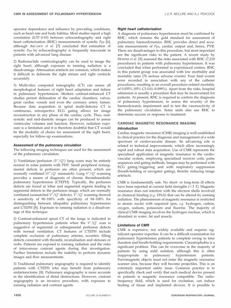
TABLE 1 Diagnostic classification of pulmonary hypertension (Venice 2003)
Pulmonary arterial hypertension
Idiopathic
Familial
Associated with:
Connective tissue disease
Congenital systemic to pulmonary shunts
Portal hypertension
HIV infection
Drugs and toxins
Other (thyroid disorders, glycogen storage disease, Gaucher disease, hereditary haemorrhagic telangiectasia, haemoglobinopathies, myeloproliferative
disorders or splenectomy)
Associated with significant venous or capillary involvement
Pulmonary veno-occlusive disease
Pulmonary capillary haemangiomatosis
Persistent pulmonary hypertension of the newborn
Pulmonary hypertension associated with left-sided heart disease
Left-sided atrial or ventricular heart disease
Left-sided valvular heart disease
Pulmonary hypertension associated with lung respiratory diseases or hypoxia
Chronic obstructive pulmonary disease
Interstitial lung disease
Sleep-disordered breathing
Alveolar hypoventilation disorders
Chronic exposure to high altitude
Developmental abnormalities
Pulmonary hypertension due to chronic thrombotic or embolic disease
Thromboembolic obstruction of proximal pulmonary arteries
Thromboembolic obstruction of distal pulmonary arteries
Nonthrombotic pulmonary embolism (tumour, parasites or foreign material)
Miscellaneous
Sarcoidosis, histiocytosis X, lymphangiomatosis, compression of pulmonary vessels (adenopathy, tumour or fibrosing mediastinitis)
L.E.R. MCLURE AND A.J. PEACOCK CMR IN ASSESSMENT OF PULMONARY HYPERTENSION
cEUROPEAN RESPIRATORY JOURNAL VOLUME 33 NUMBER 6 1455
operator dependence and influence by prevailing conditions,such as heart rate and body habitus. Most studies report a highcorrelation (0.57–0.93) between echocardiography and rightheart catheterisation (RHC) measurements of systolic Ppa [6],although ARCASOY et al. [7] concluded that estimation ofsystolic Ppa by echocardiography is frequently inaccurate inpatients with advanced lung disease.
2) Radionuclide ventriculography can be used to image theright heart, although exposure to ionising radiation is adisadvantage. Attenuation artefacts are common, which makesit difficult to delineate the right atrium and right ventricleaccurately.
3) Multi-slice computed tomography (CT) can assess allmorphological features of right heart adaptation and failurein pulmonary hypertension. Modern contrast-enhanced CTstudies permit delineation of the cardiac chambers, valves,great cardiac vessels and even the coronary artery lumen.Because data acquisition in spiral multi-detector CT iscontinuous, retrospective ECG gating allows for imagereconstruction in any phase of the cardiac cycle. Thus, end-systolic and end-diastolic images can be produced to assessventricular volumes and function. However, radiation expo-sure is a limitation and it is therefore doubtful that CT wouldbe the modality of choice for assessment of the right heart,especially for follow-up examinations.
Assessment of the pulmonary circulationThe following imaging techniques are used for the assessmentof the pulmonary circulation.
1) Ventilation/perfusion (V9/Q9) lung scans may be entirelynormal in some patients with PHT. Small peripheral nonseg-mental defects in perfusion are often present, which arenormally ventilated (V9/Q9 mismatch). Lung V9/Q9 scanningprovides a means of diagnosis of chronic thromboembolicpulmonary hypertension (CTEPH). Typically, the perfusiondefects are found in lobar and segmental regions leading tosegmental defects in the perfusion image, which are normallyventilated (unmatched V9/Q9 defects). V9/Q9 scanning showeda sensitivity of 90–100% with specificity of 94–100% fordistinguishing between idiopathic pulmonary hypertensionand CTEPH [8]. Exposure to ionising radiation is a disadvan-tage of this technique.
2) Contrast-enhanced spiral CT of the lungs is indicated inpulmonary hypertension patients when the V9/Q9 scan issuggestive of segmental or subsegmental perfusion defectswith normal ventilation. CT features of CTEPH includecomplete occlusion of pulmonary arteries, eccentric fillingdefects consistent with thrombi, recanalisation and stenoses orwebs. Patients are exposed to ionising radiation and the risksof intravenous contrast agents during this investigation.Further limitations include the inability to perform dynamicimages and flow measurements.
3) Traditional pulmonary angiography is required to identifypatients with CTEPH who may benefit from pulmonaryendarterectomy [8]. Pulmonary angiography is more accuratein the identification of distal obstructions. However, contrastangiography is an invasive procedure, with exposure toionising radiation and contrast agents.
Right heart catheterisationA diagnosis of pulmonary hypertension must be confirmed byRHC, which remains the gold standard for assessment ofpulmonary haemodynamics. RHC provides direct and accu-rate measurements of Ppa, cardiac output and, hence, PVR.There are disadvantages to this procedure, but most importantare the significant risks to the patient. A recent study byHOEPER et al. [9] assessed the risks associated with RHC (7,218procedures) in patients with pulmonary hypertension. It wasconcluded that when performed in experienced centres, RHCin this patient group was associated with low morbidity andmortality rates (76 serious adverse events). Four fatal eventswere recorded in association with any of the catheterprocedures, resulting in an overall procedure-related mortalityof 0.055% (95% CI 0.01–0.099%). Apart from the risks, hospitaladmission is usually a procedure that may be inconvenient forpatients. At present, RHC is required to confirm the diagnosisof pulmonary hypertension, to assess the severity of thehaemodynamic impairment and to test the vasoreactivity ofthe pulmonary circulation. Some units also use RHC todetermine success or response to treatment.
CARDIAC MAGNETIC RESONANCE IMAGING
IntroductionCardiac magnetic resonance (CMR) imaging is well establishedin clinical practice for the diagnosis and management of a widespectrum of cardiovascular disease. Its advancing role isrelated to technical improvements, which allow increasinglyrapid and robust data acquisition. Use of CMR represents thespecialised application of magnetic resonance to the cardio-vascular system, employing specialised receiver coils, pulsesequences and gating methods. Images may be performed withECG gating/triggering and with respiratory suppression(breath-holding or navigator gating), thereby reducing imageartefacts.
CMR is fundamentally safe. No short- or long-term ill effectshave been reported at current field strengths (,3 T). Magneticresonance does not interfere with the electron shells involvedin chemical binding (e.g. DNA) that can be altered by ionisingradiation. The phenomenon of magnetic resonance is restrictedto atomic nuclei with unpaired spin, e.g. hydrogen, carbon,oxygen, sodium, potassium and fluorine. The majority ofclinical CMR imaging involves the hydrogen nucleus, which isabundant in water, fat and muscle.
Limitations of CMRCMR is expensive, not widely available and requires sig-nificant operator expertise. It can be a difficult examination forpulmonary hypertension patients to complete owing to timeduration and breath-holding requirements. Claustrophobia is asignificant problem. This can be overcome in the majority ofpatients by using mild sedation, although this is ofteninappropriate in pulmonary hypertension patients.Ferromagnetic objects must not enter the magnetic resonancescanner area, because they will become projectiles. This is anextremely important safety issue. Common practice is tospecifically check and verify that each medical device presentin patients is magnetic resonance compatible. The radio-frequency field, which is used for excitation, can induceheating of tissue and implanted devices. It is possible to
CMR IN ASSESSMENT OF PULMONARY HYPERTENSION L.E.R. MCLURE AND A.J. PEACOCK
1456 VOLUME 33 NUMBER 6 EUROPEAN RESPIRATORY JOURNAL

stimulate sensitive tissues such as peripheral nerves owing tothe rapidly changing gradient magnetic fields used to generateimages. Myocardial stimulation has not been described withcurrent hardware.
Ventricular morphology and function by CMRMagnetic resonance imaging (MRI) produces tomographic stillimages that can accurately and reproducibly assess leftventricular and right ventricular chamber sizes, wall thicknessand mass. The multifaceted nature of MRI enables it to be usednot only for morphological assessment, but also for functionalassessment. Conventional gradient-recalled echo or steady-state free-precession pulse (SSFP) sequences can be used toconstruct a cine image, which is a movie of 15–20 frames inwhich the full cardiac cycle can be seen; each movie framerepresents 30–40 ms of the cycle. Recent technologicaladvances enable the implementation of SSFP sequences whichprovide a substantially higher signal-to-noise ratio than can beobtained by conventional gradient–echo techniques. Thecontrast between myocardium and cavity blood [10] makeplanimetry of the interface accurate and easily reproducible forassessment of left and right ventricular function. The SSFPtechnique is the preferred CMR pulse sequence for acquisitionof volumetric datasets of the left and right ventricles. Cinemode MRI allows regional and global systolic function to beevaluated because wall motion abnormalities can be identified.Ventricular volumes, ejection fraction and myocardial mass areusually obtained from a stack of contiguous ‘‘bright blood’’cine CMR 5–10 mm slices covering the left and right ventriclesacquired in short-axis or transverse orientation. Endocardialand epicardial contours are drawn during post-processing onend-diastolic and end-systolic frames, and left and rightventricular volumes are calculated as the sum of individualslice volumes (fig. 1). Ventricular mass is the product ofmyocardial volume and muscle-specific density (1.05 g?cm-3).A previous criticism of this technique has been the time requiredto analyse the cine data to generate accurate volume and massdata. New PC-based software solutions with intensity-basedthresholding for semiautomated myocardial blood borderdefinition has enabled analysis to become less time-consuming.
Impressive results for accuracy have been demonstrated byseveral investigators in various disease states [11–15]. Theinterstudy reproducibility of CMR-derived parameters ofventricular function and mass is good for both the left andright ventricles and is superior to two-dimensional and M-mode echocardiography [16–18]. The results from a studyperformed by GROTHUES et al. [19] demonstrate that theinterstudy reproducibility of the right ventricle is lower thanfor the left ventricle, although CMR is still a reliable methodand can be considered the gold standard for serial assessmentof right ventricular volumes, function and mass.
Flow analysisPhase contrast velocity mapping is an magnetic resonancesequence used to measure velocity and flow in blood vessels,or within the heart, in which each pixel in the image displaysthe signal phase, which is encoded. Volumetric flow (inmillilitres per second) is obtained in each time frame bymultiplying the spatial mean velocity (in centimetres persecond) of blood flow with the cross-sectional area of the vessel
(in square centimetres). Integrating the volumetric flow curveover systole gives the stroke volume. This imaging techniquehas been available for .20 yrs [20]. Velocity-encoded imaginghas been shown to be a reliable method to measure blood flowin different vessels of the body. Analogous to Dopplerechocardiography, this technique allows the calculation ofstroke volume, cardiac output, ejection fraction, valvularregurgitant fractions and quantification of cardiac shunts,while mitral and tricuspid transvalvular flow profiles allow theassessment of ventricular diastolic filling patterns (E and Awaves). Cardiac output and the pulmonary to systemic flowratio measured with the use of this technique have been shownto be accurate [21, 22]. Stroke volume calculated from flowmeasurements in the pulmonary artery corresponds well withvolumetric measurements of the right ventricle in healthysubjects. Phase contrast magnetic resonance flow is lessaccurate in patients with either cardiac arrhythmia duringacquisition or turbulent blood flow; the presence of these is ageneral limitation of this technique. Of note, even whenappropriate methods of acquisition have been used, there canbe inaccuracies of flow measurement on some CMR systemscaused by background phase errors due to eddy currents oruncorrected concomitant gradients.
Contrast-enhanced CMR imagingGadolinium is a contrast agent utilised in magnetic resonancescanning. It has seven unpaired electrons in its outer shell, andit hastens T1 relaxation, thereby increasing signal in the area ofinterest. Gadolinium alone is cytotoxic, but not if chelated withdiethylenetriamine pentaacetic acid. It has similar pharmaco-kinetic properties to iodinated X-ray contrast but with minimalnephrotoxicity and anaphylaxis risk. Attention has beendrawn, however, to recent reports identifying a possible link
RV
LV
FIGURE 1. Planimetry of right ventricle (RV). Epicardial and endocardial
borders of the right ventricular myocardium are manually traced at end-diastole on
this short-axis cardiac magnetic resonance image. This scan is taken from a patient
with idiopathic pulmonary hypertension. Right ventricular dilatation, hypertrophy
and increased trabeculation are evident. LV: left ventricle.
L.E.R. MCLURE AND A.J. PEACOCK CMR IN ASSESSMENT OF PULMONARY HYPERTENSION
cEUROPEAN RESPIRATORY JOURNAL VOLUME 33 NUMBER 6 1457
between exposure to gadolinium-containing agents used inpatients with end-stage renal disease and a rare, potentiallylife-threatening, condition referred to as nephrogenic systemicfibrosis. Regulatory authorities advise caution in the adminis-tration of gadolinium-containing agents in renally impairedpatients.
In addition to evaluating the first-pass transit of gadoliniumcontrast, images can be obtained 10–15 min later, in apseudoequilibrium phase. Gadolinium is avidly retained inabnormal myocardial regions, resulting in shortened T1 andincreased signal intensity. The bright areas on the resultingimages are described as areas of delayed contrast enhancement(DCE). DCE is not biologically specific and has been describedin a variety of illnesses. Myocardial infarction, fibrosis andinflammation have all been shown to result in DCE usinggadolinium as an i.v. contrast agent [23–27].
Magnetic resonance pulmonary circulationSeveral methods have been proposed for MRI imaging of thepulmonary vasculature, both with and without the use ofgadolinium. Three-dimensional gadolinium-enhanced mag-netic resonance angiography (MRA) is now the most commonlyapplied. Contrast-enhanced MRA utilises three-dimensionalultrafast imaging sequences (T1 weighted) after i.v. injection ofgadolinium and uses the first pass of this contrast agent [28].Limitations of MRA include a lower spatial resolution andlonger breath-hold when compared with CT.
Preliminary protocols are being developed to image lungperfusion into the diseased lung. These will allow forquantitative analysis of lung perfusion. This technique mayallow for perfusion/functional assessment before and afterdisease targeted therapy.
CMR ASSESSMENT OF PULMONARY HYPERTENSIONIt is becoming increasingly recognised that the right ventricleand the pulmonary vasculature should be approached, bothdiagnostically and therapeutically, as a unit in patients withpulmonary hypertension.
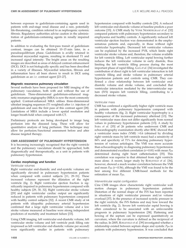
Cardiac morphology and functionVentricular volumesRight ventricular end-diastolic and end-systolic volumes aresignificantly elevated in pulmonary hypertension patientswhen compared with control subjects [11, 29–31]. Theseincreased volumes represent the dilatation of the rightventricle (fig. 2). Right ventricular ejection fraction is sig-nificantly impaired in pulmonary hypertension compared withhealthy subjects [29, 30, 32]. Right ventricular stroke volume[29] and right ventricular cardiac output are significantlyreduced in patients with pulmonary hypertension comparedwith healthy control subjects [32]. A recent CMR study of 64patients with idiopathic pulmonary arterial hypertensionconfirmed that a large right ventricular volume and a lowstroke volume measured at baseline were strong independentpredictors of mortality and treatment failure [33].
Using CMR imaging, left ventricular end-diastolic volume, leftventricular stroke volume and left ventricle peak filling rate(expressed as left ventricular end-diastolic volume per second)were significantly smaller in patients with pulmonary
hypertension compared with healthy controls [29]. A reducedleft ventricular end-diastolic volume at baseline predicts a pooroutcome [33]. A CMR study by VONK NOORDEGRAAF et al. [32]compared patients with pulmonary hypertension secondary toemphysema and healthy controls. A significantly reduced leftventricular ejection fraction was demonstrated in the emphy-sematous patients and, especially, in those without rightventricular hypertrophy. Decreased left ventricular volumescan be explained by the increased PVR, which limits rightventricular stroke volume and, therefore, the volume availablefor left ventricle filling. Left ventricular septal bowing furtherreduces the left ventricular volume in early diastole, thuslimiting the left ventricle filling process during the mostimportant phase of rapid filling. GAN et al. [34] investigated thecontribution of direct right to left ventricular interaction to leftventricle filling and stroke volume in pulmonary arterialhypertension patients and controls using CMR. They con-firmed a close relationship between left ventricular end-diastolic volume and stroke volume, and concluded thatventricular interaction mediated by the interventricular sep-tum (IVS) impairs left ventricle filling, contributing to adecreased stroke volume.
Ventricular massCMR has confirmed a significantly higher right ventricle massin patients with pulmonary hypertension compared withhealthy volunteers (fig. 2). Right ventricular hypertrophy is aconsequence of the increased pulmonary afterload [13]. Theleft ventricular mass does not differ significantly from normalvalues in pulmonary hypertension patients [13]. A study bySABA et al. [35] of 26 patients who underwent CMR andechocardiography examination shortly after RHC showed thata ventricular mass index (VMI) .0.6 (obtained by dividingright ventricle mass by left ventricle mass) had a sensitivity of84% and specificity of 71% for detecting pulmonary hyper-tension of various aetiologies. The VMI was more accuratethan echocardiography in diagnosing pulmonary hypertensionand demonstrated excellent correlation (r50.81) with mean Ppa
determined during right heart catheterisation [35]. Thiscorrelation was superior to that obtained from right ventriclemass alone. A recent, larger study by ROELEVELD et al. [36],however, showed a much weaker correlation between the VMIand mean Ppa (r50.56), although the VMI was found to be thebest among five different CMR-based methods for theestimation of mean Ppa.
Interventricular septal configuration
Cine CMR images show characteristic right ventricular wallmotion changes in pulmonary hypertension patients.Distortion of the normal shape of the IVS has been reportedin situations of right ventricular pressure and/or volumeoverload [37]. In the presence of increased systolic pressure inthe right ventricle, the IVS flattens and may bow toward theleft ventricle (fig. 2). Severe left ventricular septal bowing isoften considered to be associated with an unfavourableprognosis in pulmonary hypertension [38]. Flattening andbowing of the septum can be expressed quantitatively ascurvature, where the curvature is defined as the reciprocal ofthe radius. In 2005, ROELEVELD et al. [39] investigated whether arelationship existed between septum shape and systolic Ppa inpatients with pulmonary hypertension. It was concluded that
CMR IN ASSESSMENT OF PULMONARY HYPERTENSION L.E.R. MCLURE AND A.J. PEACOCK
1458 VOLUME 33 NUMBER 6 EUROPEAN RESPIRATORY JOURNAL

systolic Ppa was proportional to septal curvature (r50.77,p,0.001). Maximal distortion of the normal septal shape wasfound during the right ventricular relaxation phase. The causeof the leftward septum displacement appeared to be a pressureexcess in the right ventricle relative to the left ventricle. Dataobtained from 39 subjects showed a systolic Ppa .67 mmHgmight be expected if left ventricular septal bowing is seen.
Right ventricular diastolic function
Diastolic function has been shown to be abnormal in diseasesaffecting the left ventricle. Often, diastolic dysfunction is anearly sign of ventricular dysfunction and is currently beingtargeted therapeutically. GAN et al. [40] have shown that rightventricular diastolic dysfunction is present in pulmonaryhypertension patients and can be relatively easily measuredby CMR. Isovolumetric relaxation time (IVRT) may be amarker of right ventricular diastolic dysfunction and mightpredict burden of disease and clinical outcomes. IVRTcorrelates positively with both right ventricle mass and PVR,variables that are known to be of critical importance in theevaluation and prognosis of pulmonary hypertension [41].Perhaps, more importantly, IVRT improves in response tostandard therapies known to decrease right ventricular after-load, e.g. oral sildenafil [42]. These data suggest that MRI-measured right ventricular diastolic dysfunction and IVRT
might be a good surrogate end-point for clinical trials onpulmonary hypertension. This comes at a very good time,where hard end-points, directly relevant to the right ventricle,need to be identified and used in pulmonary hypertensionclinical trials; the validity of currently used primary end-points, such as the 6-min walk test (6MWT), are beingchallenged [43].
Right ventricular contractilityRecent advances in magnetic resonance scanner hardware andsoftware have enabled CMR guidance of endovascular cathetersunder real-time imaging (magnetic resonance fluoroscopy). ThisCMR approach is a promising tool for assessing rightventricular contractility in the clinical setting [44]. KUEHNE etal. [44] have demonstrated it is possible to combine CMR-guidedinvasive right ventricular pressure measurements with rightventricular volume values derived from cine CMR and to obtainright ventricular pressure–volume loops. This first study of sixpatients with early-stage idiopathic pulmonary arterial hyper-tension (mean¡SD Ppa 57¡21 mmHg) and six controls,demonstrated that the right ventricular and left ventricularstroke volumes and cardiac indices were significantly lower,despite higher right ventricular ejection fractions and rightventricular contractility in these patients. CMR-guided RHCwas successfully used by the same group to assess the changesin PVR after nitric oxide inhalation in patients with idiopathicpulmonary arterial hypertension [45]. Reduction or eliminationof X-ray radiation, added anatomic and functional informationavailable with magnetic resonance, and the relative ease andaccuracy of phase contrast magnetic resonance flow quantifica-tion may make this technique the method of choice for invasivemeasurement of PVR. This is a single-centre experience, andmajor limitations are cost and availability of magneticresonance-compatible equipment. This procedure is not suitablefor serial follow-up owing to its invasive nature.
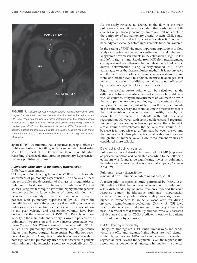
Contrast-enhanced perfusion CMRAn interesting pattern of hyperenhancement within the rightventricle is described with delayed-contrast CMR in patientswith pulmonary hypertension (fig. 3). This delayed-contrastenhancement pattern has a mid-wall distribution involving theright ventricle septal insertion points and the IVS [46]. A higherdegree of enhancement was correlated with poorer rightventricular function and haemodynamics. When contrastenhancement was present in the IVS, it was associated withseptal bowing on cine CMR. These data was confirmed byMCCANN et al. [27].
Stress CMRStress testing, by exercise or drug infusion, can be used todetermine cardiac reserve. Physical exercise within theconfines of the magnet is technically difficult and leads toimage degradation. HOLVERDA et al. [47], however, demon-strated that idiopathic pulmonary arterial hypertensionpatients were unable to significantly increase stroke volumefrom rest to exercise, using an magnetic resonance-compatibleergometer. Pharmacological CMR stress can be used inpatients with congenital heart disease to detect early rightventricular dysfunction. The physiological effects of exerciseare imitated by a continuous infusion of a short-acting agentsuch as dobutamine (a relatively selective b1-adrenoceptor
RVhypertrophy
D-shapedLV
IVSbowed
DilatedRV
RVLV
FIGURE 2. Cardiac magnetic resonance (CMR) short-axis image from a
patient with pulmonary hypertension. A short-axis cine image at mid-ventricular level
in early diastole. The CMR image was acquired from a patient with severe idiopathic
pulmonary arterial hypertension. The right ventricle (RV) is grossly dilated and
hypertrophied. The distorted interventricular septum (IVS) is bowed towards the left
ventricle (LV; D-shaped) owing to right ventricular pressure overload.
L.E.R. MCLURE AND A.J. PEACOCK CMR IN ASSESSMENT OF PULMONARY HYPERTENSION
cEUROPEAN RESPIRATORY JOURNAL VOLUME 33 NUMBER 6 1459
agonist) [48]. Dobutamine has a positive inotropic effect onright ventricular contractility, which can be determined usingMRI. To the best of our knowledge, there is no literatureregarding pharmacological stress in pulmonary hypertensionpatients published at present.
Pulmonary circulation in pulmonary hypertensionCMR flow measurementsVelocity-encoded imaging is another CMR approach for theassessment of pulmonary hypertension. The analysis of theseimages enables the description of changes or irregularities ofpulmonary blood flow in pulmonary hypertension. Previousstudies using this technique have found highly inhomogeneousvelocity profiles, a large volume of retrograde flow anddecreased distensibility of the main pulmonary artery inpatients with pulmonary hypertension [49, 50]. From thequantitative analysis of the pulmonary flow profile, noninvasiveindices (e.g. acceleration time, defined as time from onset of flowto the peak velocity, and acceleration volume) have beenderived for the assessment of PVR [51]. Peak blood flowvelocity in the main pulmonary artery is lower in patients withpulmonary hypertension and shows inverse correlation withmean Ppa and PVR. When examined in patients with CTEPH,values after pulmonary endarterectomy were significantlyhigher than before surgical intervention, but did not reachnormal range [52]. A significant reduction of peak velocity inboth right and left pulmonary arteries was observed in patientswith pulmonary hypertension secondary to cystic fibrosis [53].
As the study revealed no change in the flow of the mainpulmonary artery, it was concluded that early and subtlechanges of pulmonary haemodynamics are first noticeable inthe periphery of the pulmonary arterial system. CMR could,therefore, be the method of choice for detection of earlyhaemodynamic change before right ventricle function is altered.
In the setting of PHT, the most important applications of flowanalysis include measurement of cardiac output and pulmonaryto systemic flow measurements in the estimation of right-to-leftand left-to-right shunts. Results from MRI flow measurementscorrespond well with thermodilution data obtained but cardiacoutput determination using velocity-encoded MRI offersadvantages over the thermodilution method. It is noninvasiveand the measurements depend less on changes in stroke volumefrom one cardiac cycle to another, because it averages overmany cardiac cycles. In addition, the values are not influencedby tricuspid regurgitation to such a great extent.
Right ventricular stroke volume can be calculated as thedifference between end-diastolic and end-systolic right ven-tricular volumes, or by the measurement of volumetric flow inthe main pulmonary artery employing phase contrast velocitymapping. Stroke volume, calculated from flow measurementsin the pulmonary artery and from volumetric measurements ofthe right ventricle, corresponds well in healthy controls, andshow little divergence in patients with mild tricuspidregurgitation. However, with considerable tricuspid regurgita-tion (e.g. pulmonary hypertension patients), the volumetricstroke volume overestimates the actual stroke volume [29],because it is impossible to differentiate between the volumethat moves back through the tricuspid valve and forwardthough the pulmonary valve. Flow measurement values areconsidered more reliable.
Distensibility of pulmonary arteryPulmonary artery distensibility measured by CMR (expressedas per cent variation and calculated according to the followingequation) was found to be significantly lower in pulmonaryhypertension patients than it was in normal subjects (8% versus23%) [49].
Pulmonary artery distensibility5
((maximal area - minimal area)/minimal area)6100
A recent pilot, prospective study performed by JARDIM et al.[54] indicated that the noninvasive assessment of pulmonaryartery distensibility by magnetic resonance reflected the acuteresponse pattern in idiopathic pulmonary hypertensionpatients. Pulmonary artery distensibility was significantlyhigher in responders to an acute vasodilator test duringinvasive haemodynamic evaluation. GAN et al. [55] haverecently demonstrated that proximal pulmonary artery stiff-ness (in terms of area distensibility and noninvasively assessedrelative area change by CMR) predicted mortality in patientswith pulmonary hypertension.
CMR pulmonary angiographyThe typical findings of CTEPH (intraluminal webs and bands,vessel cut-offs, and organised thrombus) are well demon-strated by pulmonary MRA and can be seen in vessels tosegmental level. Beyond the segmental level, the higher spatialresolution of conventional angiography makes it superior.
DCE within IVS
DCE within RVIP
RV
LV
FIGURE 3. Delayed contrast-enhanced cardiac magnetic resonance (CMR)
images of a patient with pulmonary hypertension. A contrast-enhanced short-axis
CMR cine image was acquired at a basal ventricular level. The delayed-contrast
enhancement (DCE) pattern has a mid-wall distribution involving the right ventricular
insertion point (RVIP) and the interventricular septum (IVS). Trabeculations and
papillary muscles are deliberately included in the analysis, as this has been shown
to be a more accurate, although time-consuming, method. RV: right ventricle; LV:
left ventricle.
CMR IN ASSESSMENT OF PULMONARY HYPERTENSION L.E.R. MCLURE AND A.J. PEACOCK
1460 VOLUME 33 NUMBER 6 EUROPEAN RESPIRATORY JOURNAL

Surgical intervention is largely limited to proximal andsegmental vessels, and in a study by KREITNER et al. [52],contrast-enhanced MRA correctly predicted surgical success in33 out of 34 patients. The study demonstrated that three-dimensional contrast-enhanced MRA performed equally aswell as X-ray pulmonary angiography for the visualisation ofsegmental pulmonary vessels (533 out of 533 segments), wasslightly worse for subsegmental vessels (681 versus 733segments), but was superior for the depiction of the centralorigin of thromboembolic material. Pulmonary MRA may becombined in the same examination with a variety of cinetechniques to gauge cardiac function and flow. Contrast-enhanced MRA should identify patients with CTEPH thatdelineate typical findings and are potential candidates forsurgery.
CMR pulmonary perfusion imaging
OHNO et al. [56] have demonstrated that three-dimensionaldynamic contrast-enhanced MRI has the potential for assess-ment of disease severity in pulmonary hypertension patients.This technique showed significant differences in pulmonaryblood flow and mean transit time between healthy andpulmonary hypertension subjects.
PPA ESTIMATION BY CMRRepeated measurements of Ppa are sometimes used to assessdisease progression in pulmonary hypertension. Echocardio-graphy is safe and widely available but has limitations, aspreviously discussed [57]. MRI has been proposed to be anaccurate alternative for echocardiography in estimating Ppa.Investigators have attempted to use CMR as a noninvasivemeans of estimating mean Ppa but none have reported anyadvantages over echocardiography. Several estimators basedon different MRI techniques have been described in recentyears, including acceleration time (the time of onset offorward flow to the moment of maximum flow velocity inthe main pulmonary artery), acceleration time/ejection timeratio, pulse wave velocity, cross-sectional area of the mainpulmonary artery and ventricular mass index. Right ventri-cular end-diastolic wall thickness has been shown to correlatewell with mean Ppa in idiopathic pulmonary arterialhypertension and some cases of secondary pulmonaryhypertension [58, 59]. A linear relationship between rightventricle mass and mean Ppa has been described foridiopathic pulmonary arterial hypertension [13]. The ratio ofthe main pulmonary artery diameter over descending aorticdiameter has also been shown to correlate with mean Ppa inpulmonary hypertension. The VMI was found to be the bestamong five different CMR-based methods for the estimationof Ppa and similar to echocardiography (r50.55 using themodified Bernoulli equation and peak tricuspid regurgitationvelocity), but not accurate enough to replace RHC in clinicalpractice [36]. A computed method for the noninvasivemagnetic resonance assessment of pulmonary hypertensionhas been elaborated, in which a combination of physicalvariables, including main pulmonary artery blood flowvelocity at peak systole, maximal systolic main pulmonaryartery cross sectional area and biophysical parametersincluding patient height, weight and heart rate were used toestimate Ppa [60, 61].
FOLLOW-UP OF PATIENTS WITH PULMONARYHYPERTENSIONCMR is increasingly used in patients with pulmonaryhypertension for the evaluation of pathological and functionalchanges in the heart and pulmonary circulation. CMR providesa direct evaluation of right ventricular size, mass, morphologyand function [62]. Normal ranges have been established [13,50]. CMR findings in right ventricular failure include rightventricular dilatation, tricuspid regurgitation, right ventricularhypertrophy, interventricular septal flattening or paradoxicalmotion, and change in chamber morphology from a normalcrescent shape to a more concentric form. Noninvasiveassessments of blood flow (including stroke volume andcardiac output) and distensibility in the pulmonary arteriescan be made [63–65]. There is good correlation between RHCand magnetic resonance, suggesting that magnetic resonancedata could be used as a surrogate of right heart haemo-dynamics [64].
Pulmonary hypertension experts gathered in 2007 at the EndPoints Meeting held in Turnberry, UK. Physicians currentlyrely on the World Health Organization (WHO) functionalclass, 6MWT, biological markers (brain natriuretic peptide(BNP) levels), echocardiography and RHC to follow uppatients with pulmonary hypertension. These investigationshave acknowledged limitations. The question of which end-points are most relevant in the assessment of pulmonaryhypertension has been the topic of intense discussion. TheWHO functional class has been an important end-point inclinical trials of pulmonary hypertension, although the assign-ment of patients to categories is subject to the bias ofinvestigators, which limits its usefulness as an end-point. The6MWT is a submaximal exercise test which can be performedby patients who are incapable of tolerating maximal exercisetesting [66]. The 6MWT has been used widely as a primaryend-point in clinical trials, but flaws have been highlighted inits performance. The 6MWT must be performed correctly usingthe appropriate guidelines [66]. There are concerns that the 6-min walk distance (6MWD) is affected by a number of factorsother than pulmonary hypertension including age, sex, height,weight and musculoskeletal conditioning [67]. Furthermore, ithas been shown that the 6MWD can improve considerablywith rehabilitation measures alone [43]. Echocardiography isthe most well established and accessible imaging modality forfollow-up of patients with pulmonary hypertension. Dopplerechocardiography is suitable for serial assessments, although ithas some limitations, as discussed previously in the presentarticle. Serial measurement of plasma NT-proBNP (N-terminal-pro-BNP) has great attractions as an end-point. Itspresence in the blood is related to right ventricular dysfunc-tion, it is simple to measure and relatively inexpensive. Someremarkable relationships between plasma BNP/NT-proBNPand various elements of right ventricular dysfunction havebeen shown [46, 68, 69]. It would appear that BNP/NT-proBNP measurement is a dynamic measurement reflectingthe current state of the right ventricle. An increase in NT-proBNP over time reflects right ventricular dilatation con-comitant to hypertrophy and deterioration of systolic function[69]. We await the results of large-scale studies to determinethe role of BNP in the assessment and management of patientswith pulmonary hypertension. The normalisation of measures
L.E.R. MCLURE AND A.J. PEACOCK CMR IN ASSESSMENT OF PULMONARY HYPERTENSION
cEUROPEAN RESPIRATORY JOURNAL VOLUME 33 NUMBER 6 1461
of cardiovascular haemodynamics would be an ideal end-point. However, resting haemodynamics improve only mar-ginally in most patients, even when their clinical responseappears to be excellent [70], and do not reflect changes thatmay occur with exercise. Clinical improvement, therefore, isonly partly related to a modification of resting haemodynamicsin most patients. Furthermore, RHC is an invasive procedurethat is not ideal for serial evaluation.
It has been suggested that characteristics for an ideal marker inpulmonary hypertension might include [71]: 1) it should beheart or lung specific; 2) it should be abnormal in pulmonaryhypertension; 3) sample collection should be simple; 4) themarker should be easy to measure; 5) values should bereproducible; 6) values should follow the course of the disease(i.e. increasing if patients deteriorate and falling if patientsimprove); and 7) abnormal values should be indicative of apoor survival.
CMR imaging fulfils these stated characteristics. As discussed,modern CMR protocols provide us with abundant informationregarding the ventricular myocardium and pulmonary vascu-lature. Right ventricular volumes, muscle mass and functionalparameters, including stroke volume, ejection fraction andcardiac output, differ significantly in pulmonary hypertensioncompared to healthy subjects. CMR imaging is easilyperformed by trained MRI technicians/physicians and the
majority of patients tolerate this noninvasive investigationwell. Manual planimetry of the myocardium and flow analysis issimple to perform and reproducible although time-consumingat present. Sequential MRI is the optimal tool to monitortherapeutic effects on vascular remodelling and right heartperformance. CMR-derived right ventricle functional para-meters correlate well with established haemodynamic para-meters of prognostic significance. Although RHC remains thedefinitive assessment of pulmonary hypertension at present, thenoninvasive evaluation of cardiac morphology and function andof the pulmonary circulation is a new and promising applicationfor CMR imaging.
CMR AS AN END-POINTMRI is gaining a dominant role as the reference method forclinical trials assessing longitudinal changes in left ventricularfunction after therapeutic interventions [72–74]. The accuracyand reproducibility of CMR in assessing cardiac morphologicaland functional variables leads to low interstudy variability,which translates into a significant reduction in sample sizesrequired to test the efficacy of therapeutic interventions. It isexpected that the number of clinical trials using CMRparameters as study end-points will increase considerably inthe future. Ultimately, however, patient outcome is the relevantclinical issue. Future effort should be directed toward testingwhether changes in cardiac variables as measured by magnetic
a) b)
D-shaped LV
RVRV
Bowed IVS
LV
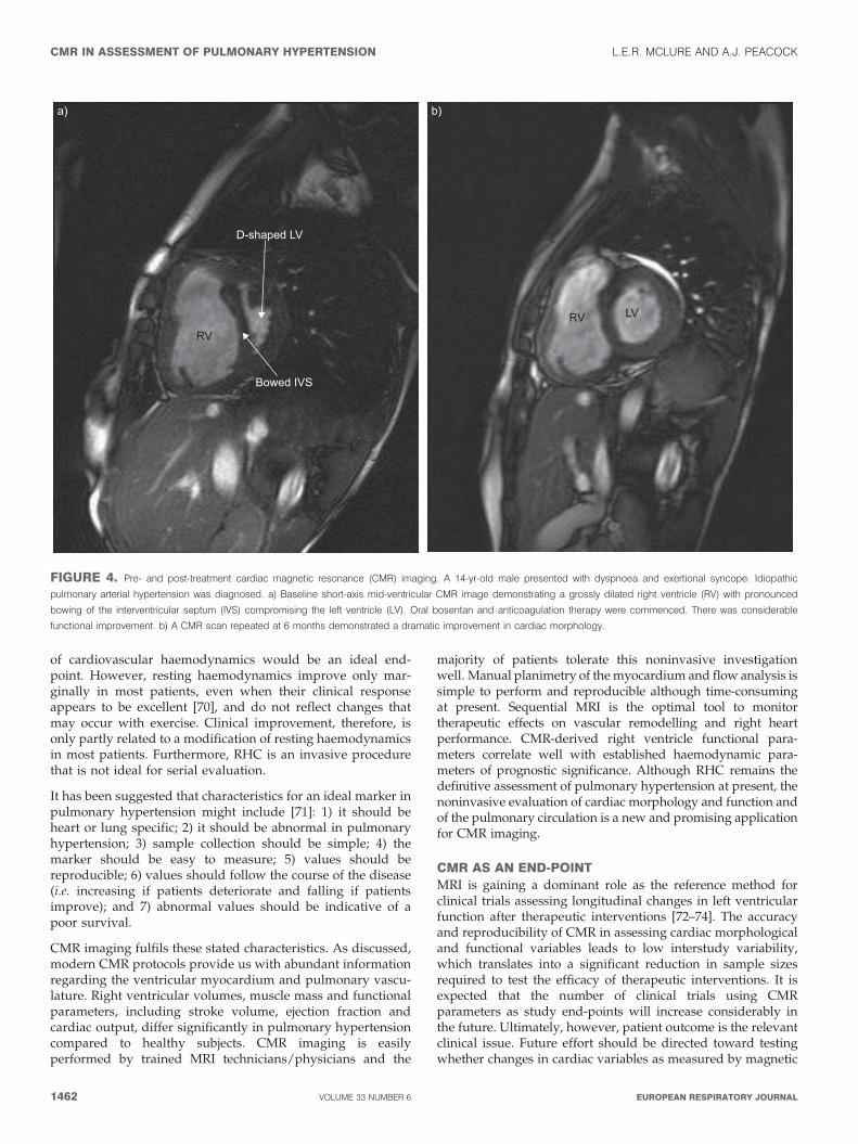
FIGURE 4. Pre- and post-treatment cardiac magnetic resonance (CMR) imaging. A 14-yr-old male presented with dyspnoea and exertional syncope. Idiopathic
pulmonary arterial hypertension was diagnosed. a) Baseline short-axis mid-ventricular CMR image demonstrating a grossly dilated right ventricle (RV) with pronounced
bowing of the interventricular septum (IVS) compromising the left ventricle (LV). Oral bosentan and anticoagulation therapy were commenced. There was considerable
functional improvement. b) A CMR scan repeated at 6 months demonstrated a dramatic improvement in cardiac morphology.
CMR IN ASSESSMENT OF PULMONARY HYPERTENSION L.E.R. MCLURE AND A.J. PEACOCK
1462 VOLUME 33 NUMBER 6 EUROPEAN RESPIRATORY JOURNAL

resonance indeed translate into differences in patient outcome.An example of CMR-measured improvement is seen in figure 4.
Deterioration of right ventricular function at follow-up exam-inations indicates an unfavourable prognosis because functionalimpairment of the right ventricle is the major factor in diseaseprogression and decline in life expectancy [75]. VAN WOLFEREN etal. [33] performed a longitudinal CMR study which confirmedright ventricular dilatation and a decrease in stroke volume andleft ventricular diastolic volume are strong predictors oftreatment failure and death at follow-up. Medical therapies orsurgical interventions may stop, or even reverse, this process,and the improvement of right ventricular function could bedetected by MRI.
At present, there are few studies in pulmonary hypertensionwhich have utilised CMR as an end-point so far. Changes inright ventricle mass, function and pulmonary artery bloodflow have been demonstrated by CMR following lungtransplantation in several studies [74, 76, 77]. In 2003,MICHELAKIS et al. [78] performed a small nonrandomised pilotstudy of five patients with pulmonary hypertension toinvestigate the effect of sildenafil 50 mg t.i.d. Right ventriclemass was utilised as an end-point. Sildenafil significantlyreduced right ventricle mass and increased the right ventri-cular stroke volume as measured by CMR. The pathologicalseptal shift towards the left ventricle was reversed by long-term sildenafil therapy. In a prospective study of pulmonaryhypertension patients with prostacyclin therapy, the signifi-cant increase in right ventricular stroke volume (pulmonaryarterial flow analysis) corresponded well with functionalimprovement (WHO functional class and 6MWT) [30]. Thefunctional and morphological effects of pulmonary endarterect-omy in patients with CTEPH was assessed by KREITNER et al. [52]using a combination of three-dimensional gadolinium contrast-enhanced MRA, cine CMR and velocity-encoded CMR. CMRdemonstrated surgical success in 33 out of the 34 patients, withimprovement of the initially depressed right ventricular ejectionfraction, which correlated with the decrease in mean Ppa anddisappearance of septal bowing in 68% of patients after surgery.In a comparison study between sildenafil and bosentan, rightventricle mass (measured by CMR) did not change after3 months of bosentan treatment, whereas sildenafil reducedright ventricle mass [79]. It was demonstrated by CMR that theaddition of sildenafil reversed right ventricular dilatation andhypertrophy in patients receiving treatment [80]. More recently,16 patients with pulmonary arterial hypertension were assessedby CMR at baseline and after 12 months treatment withbosentan [81]. After treatment, cardiac index, PVR and 6MWTdistance increased. There was a trend towards improvement inright ventricular stroke volume (p50.08), although there was nochange in right ventricular ejection fraction or right ventricularend-diastolic volume, as determined by CMR.
CONCLUSIONSCMR imaging enables a unique combination of morphologicaland functional assessment of the right ventricle and pulmon-ary circulation. CMR has emerged over recent years as the goldstandard for detailed study of the right ventricle and hasbecome an established modality for the physiological assess-ment of pulmonary hypertension patients in cross-sectionalstudies, longitudinal follow-up studies and clinical trials of
therapy. We anticipate that MRI will increasingly be utilised asthe primary modality for combined anatomic and functionalassessments that enable more complete and efficient evalua-tion of patients with pulmonary hypertension. CMR iscurrently being used as an end-point in the multinationalEuropean Union-funded Framework 6 EURO-MR project forpulmonary hypertension. We wait to see whether the promiseof CMR as a successful end-point is fulfilled.
REFERENCES1 Rubin LJ. Primary pulmonary hypertension. N Engl J Med
1997; 336: 111–117.2 Runo JR, Loyd JE. Primary pulmonary hypertension.
Lancet 2003; 361: 1533–1544.3 Simonneau G, Galie N, Rubin LJ, et al. Clinical classifica-
tion of pulmonary hypertension. J Am Coll Cardiol 2004;43, 12: Suppl. S, 5S–12S.
4 Kukulski T, Voigt JU, Wilkenshoff UM, et al. A comparisonof regional myocardial velocity information derived bypulsed and color Doppler techniques: an in vitro and in vivostudy. Echocardiography 2000; 17: 639–651.
5 Currie PJ, Seward JB, Chan KL, et al. Continuous waveDoppler determination of right ventricular pressure: asimultaneous Doppler-catheterization study in 127patients. J Am Coll Cardiol 1985; 6: 750–756.
6 Denton CP, Cailes JB, Phillips GD, et al. Comparison ofDoppler echocardiography and right heart catheterizationto assess pulmonary hypertension in systemic sclerosis. BrJ Rheumatol 1997; 36: 239–243.
7 Arcasoy SM, Christie JD, Ferrari VA, et al. Echocardio-graphic assessment of pulmonary hypertension in patientswith advanced lung disease. Am J Respir Crit Care Med 2003;167: 735–740.
8 Fedullo PF, Auger WR, Kerr KM, et al. Chronic throm-boembolic pulmonary hypertension. N Engl J Med 2001;345: 1465–1472.
9 Hoeper MM, Lee SH, Voswinckel R, et al. Complications ofright heart catheterization procedures in patients withpulmonary hypertension in experienced centers. J Am CollCardiol 2006; 48: 2546–2552.
10 Ley S, Kreitner KF, Fink C, et al. Assessment of pulmonaryhypertension by CT and MR imaging. Eur Radiol 2004; 14:359–368.
11 Boxt LM, Katz J, Kolb T, et al. Direct quantitation of rightand left ventricular volumes with nuclear magneticresonance imaging in patients with primary pulmonaryhypertension. J Am Coll Cardiol 1992; 19: 1508–1515.
12 Doherty NE 3rd, Fujita N, Caputo GR, et al, Measurementof right ventricular mass in normal and dilated cardio-myopathic ventricles using cine magnetic resonanceimaging. Am J Cardiol 1992; 69: 1223–1228.
13 Katz J, Whang J, Boxt LM, et al. Estimation of rightventricular mass in normal subjects and in patients withprimary pulmonary hypertension by nuclear magneticresonance imaging. J Am Coll Cardiol 1993; 21: 1475–1481.
14 Pattynama PM, Willems LN, Smit AH, et al. Earlydiagnosis of cor pulmonale with MR imaging of the rightventricle. Radiology 1992; 182: 375–379.
15 Suzuki J, Caputo GR, Masui T, et al. Assessment of rightventricular diastolic and systolic function in patients with
L.E.R. MCLURE AND A.J. PEACOCK CMR IN ASSESSMENT OF PULMONARY HYPERTENSION
cEUROPEAN RESPIRATORY JOURNAL VOLUME 33 NUMBER 6 1463
dilated cardiomyopathy using cine magnetic resonanceimaging. Am Heart J 1991; 122: 1035–1040.
16 Bottini PB, Carr AA, Prisant LM, et al. Magnetic resonanceimaging compared to echocardiography to assess leftventricular mass in the hypertensive patient. Am JHypertens 1995; 8: 221–228.
17 Grothues F, Smith GC, Moon JC, et al. Comparison ofinterstudy reproducibility of cardiovascular magneticresonance with two-dimensional echocardiography innormal subjects and in patients with heart failure or leftventricular hypertrophy. Am J Cardiol 2002; 90: 29–34.
18 Semelka RC, Tomei E, Wagner S, et al. Normal leftventricular dimensions and function: interstudy reprodu-cibility of measurements with cine MR imaging. Radiology1990; 174: 763–768.
19 Grothues F, Moon JC, Bellenger NG, et al. Interstudyreproducibility of right ventricular volumes, function, andmass with cardiovascular magnetic resonance. Am Heart J2004; 147: 218–223.
20 Nayler GL, Firmin DN, Longmore DB. Blood flow imagingby cine magnetic resonance. J Comput Assist Tomogr 1986;10: 715–722.
21 Beerbaum P, Korperich H, Barth P, et al. Noninvasivequantification of left-to-right shunt in pediatric patients:phase-contrast cine magnetic resonance imaging comparedwith invasive oximetry. Circulation 2001; 103: 2476–2482.
22 Robertson MB, Kohler U, Hoskins PR, et al. Quantitativeanalysis of PC MRI velocity maps: pulsatile flow incylindrical vessels. Magn Reson Imaging 2001; 19: 685–695.
23 Holman ER, van Jonbergen HP, van Dijkman PR, et al.Comparison of magnetic resonance imaging studies withenzymatic indexes of myocardial necrosis for quantificationof myocardial infarct size. Am J Cardiol 1993; 71: 1036–1040.
24 Laissy JP, Messin B, Varenne O, et al. MRI of acutemyocarditis: a comprehensive approach based on variousimaging sequences. Chest 2002; 122: 1638–1648.
25 Lima JA, Judd RM, Bazille A, et al. Regional heterogeneityof human myocardial infarcts demonstrated by contrast-enhanced MRI. Potential mechanisms. Circulation 1995; 92:1117–1125.
26 Teraoka K, Hirano M, Ookubo H, et al. Delayed contrastenhancement of MRI in hypertrophic cardiomyopathy.Magn Reson Imaging 2004; 22: 155–161.
27 McCann GP, Gan CT, Beek AM, et al. Extent of MRIdelayed enhancement of myocardial mass is related toright ventricular dysfunction in pulmonary artery hyper-tension. AJR Am J Roentgenol 2007; 188: 349–355.
28 Uematsu H, Ohno Y, Hatabu H. Recent advances inmagnetic resonance perfusion imaging of the lung. TopMagn Reson Imaging 2003; 14: 245–251.
29 Hoeper MM, Tongers J, Leppert A, et al. Evaluation of rightventricular performance with a right ventricular ejectionfraction thermodilution catheter and MRI in patients withpulmonary hypertension. Chest 2001; 120: 502–507.
30 Roeleveld RJ, Vonk-Noordegraaf A, Marcus JT, et al.Effects of epoprostenol on right ventricular hypertrophyand dilatation in pulmonary hypertension. Chest 2004; 125:572–579.
31 Vonk-Noordegraaf A, Marcus JT, Holverda S, et al. Earlychanges of cardiac structure and function in COPDpatients with mild hypoxemia. Chest 2005; 127: 1898–1903.
32 Vonk Noordegraaf A, Marcus JT, Roseboom B, et al. Theeffect of right ventricular hypertrophy on left ventricularejection fraction in pulmonary emphysema. Chest 1997;112: 640–645.
33 van Wolferen SA, Marcus JT, Boonstra A, et al. Prognosticvalue of right ventricular mass, volume, and function inidiopathic pulmonary arterial hypertension. Eur Heart J2007; 28: 1250–1257.
34 Gan CT, Lankhaar JW, Marcus JT, et al. Impaired leftventricular filling due to right-to-left ventricular interac-tion in patients with pulmonary arterial hypertension. Am JPhysiol Heart Circ Physiol 2006; 290: H1528–H1533.
35 Saba TS, Foster J, Cockburn M, et al. Ventricular massindex using magnetic resonance imaging accuratelyestimates pulmonary artery pressure. Eur Respir J 2002;20: 1519–1524.
36 Roeleveld RJ, Marcus JT, Boonstra A, et al. A comparison ofnoninvasive MRI-based methods of estimating pulmonaryartery pressure in pulmonary hypertension. J Magn ResonImaging 2005; 22: 67–72.
37 King ME, Braun H, Goldblatt A, et al. Interventricularseptal configuration as a predictor of right ventricularsystolic hypertension in children: a cross-sectional echo-cardiographic study. Circulation 1983; 68: 68–75.
38 D’Alonzo GE, Barst RJ, Ayres SM, et al. Survival in patientswith primary pulmonary hypertension. Results from anational prospective registry. Ann Intern Med 1991; 115:343–349.
39 Roeleveld RJ, Marcus JT, Faes TJ, et al. Interventricularseptal configuration at MR imaging and pulmonaryarterial pressure in pulmonary hypertension. Radiology2005; 234: 710–717.
40 Gan CT, Holverda S, Marcus JT, et al. Right ventriculardiastolic dysfunction and the acute effects of sildenafil inpulmonary hypertension patients. Chest 2007; 132: 11–17.
41 McLaughlin VV, Presberg KW, Doyle RL, et al. Prognosisof pulmonary arterial hypertension: ACCP evidence-basedclinical practice guidelines. Chest 2004; 126: 78S–92S.
42 Michelakis E, Tymchak W, Lien D, et al. Oral sildenafil isan effective and specific pulmonary vasodilator in patientswith pulmonary arterial hypertension: comparison withinhaled nitric oxide. Circulation 2002; 105: 2398–2403.
43 Mereles D, Ehlken N, Kreuscher S, et al. Exercise andrespiratory training improve exercise capacity and qualityof life in patients with severe chronic pulmonary hyper-tension. Circulation 2006; 114: 1482–1489.
44 Kuehne T, Yilmaz S, Steendijk P, et al. Magnetic resonanceimaging analysis of right ventricular pressure–volumeloops: in vivo validation and clinical application in patientswith pulmonary hypertension. Circulation 2004; 110:2010–2016.
45 Kuehne T, Yilmaz S, Schulze-Neick I, et al. Magneticresonance imaging guided catheterisation for assessmentof pulmonary vascular resistance: in vivo validation andclinical application in patients with pulmonary hyperten-sion. Heart 2005; 91: 1064–1069.
46 Blyth KG, Groenning BA, Martin TN, et al. Contrastenhanced-cardiovascular magnetic resonance imaging inpatients with pulmonary hypertension. Eur Heart J 2005;26: 1993–1999.
CMR IN ASSESSMENT OF PULMONARY HYPERTENSION L.E.R. MCLURE AND A.J. PEACOCK
1464 VOLUME 33 NUMBER 6 EUROPEAN RESPIRATORY JOURNAL
47 Holverda S, Gan CT, Marcus JT, et al. Impaired strokevolume response to exercise in pulmonary arterial hyper-tension. J Am Coll Cardiol 2006; 47: 1732–1733.
48 Tulevski, II, Lee PL, Groenink M, et al, Dobutamine-inducedincrease of right ventricular contractility without increasedstroke volume in adolescent patients with transposition ofthe great arteries: evaluation with magnetic resonanceimaging. Int J Card Imaging 2000; 16: 471–478.
49 Bogren HG, Klipstein RH, Mohiaddin RH, et al. Pulmonaryartery distensibility and blood flow patterns: a magneticresonance study of normal subjects and of patients withpulmonary arterial hypertension. Am Heart J 1989; 118:990–999.
50 Kondo C, Caputo GR, Masui T, et al. Pulmonaryhypertension: pulmonary flow quantification and flowprofile analysis with velocity-encoded cine MR imaging.Radiology 1992; 183: 751–758.
51 Mousseaux E, Tasu JP, Jolivet O, et al. Pulmonary arterialresistance: noninvasive measurement with indexes ofpulmonary flow estimated at velocity-encoded MR imaging– preliminary experience. Radiology 1999; 212: 896–902.
52 Kreitner KF, Ley S, Kauczor HU, et al. Chronic throm-boembolic pulmonary hypertension: pre- and postopera-tive assessment with breath-hold MR imaging techniques.Radiology 2004; 232: 535–543.
53 Ley S, Puderbach M, Fink C, et al. Assessment ofhemodynamic changes in the systemic and pulmonaryarterial circulation in patients with cystic fibrosis usingphase-contrast MRI. Eur Radiol 2005; 15: 1575–1580.
54 Jardim C, Rochitte CE, Humbert M, et al. Pulmonary arterydistensibility in pulmonary arterial hypertension: a MRIpilot study. Eur Respir J 2007; 29: 476–481.
55 Gan CT, Lankhaar JW, Westerhof N, et al. Noninvasivelyassessed pulmonary artery stiffness predicts mortality inpulmonary arterial hypertension. Chest 2007; 132: 1906–1912.
56 Ohno Y, Hatabu H, Murase K, et al. Primary pulmonaryhypertension: 3D dynamic perfusion MRI for quantitativeanalysis of regional pulmonary perfusion. AJR Am JRoentgenol 2007; 188: 48–56.
57 Yock PG, Popp RL. Noninvasive estimation of rightventricular systolic pressure by Doppler ultrasound inpatients with tricuspid regurgitation. Circulation 1984; 70:657–662.
58 Bouchard A, Higgins CB, Byrd BF 3rd, et al. Magneticresonance imaging in pulmonary arterial hypertension. AmJ Cardiol 1985; 56: 938–942.
59 Saito H, Dambara T, Aiba M, et al. Evaluation of corpulmonale on a modified short-axis section of the heart bymagnetic resonance imaging. Am Rev Respir Dis 1992; 146:1576–1581.
60 Laffon E, Vallet C, Bernard V, et al. A computed methodfor noninvasive MRI assessment of pulmonary arterialhypertension. J Appl Physiol 2004; 96: 463–468.
61 Lankhaar JW, Vonk Noordegraaf A, Marcus JT. A computedmethod for noninvasive MRI assessment of pulmonaryarterial hypertension. J Appl Physiol 2004; 97: 794.
62 Lorenz CH, Walker ES, Morgan VL, et al. Normal humanright and left ventricular mass, systolic function, andgender differences by cine magnetic resonance imaging. JCardiovasc Magn Reson 1999; 1: 7–21.
63 Marcus JT, Vonk Noordegraaf A, Roeleveld RJ, et al.Impaired left ventricular filling due to right ventricularpressure overload in primary pulmonary hypertension:noninvasive monitoring using MRI. Chest 2001; 119:1761–1765.
64 Paz R, Mohiaddin RH, Longmore DB. Magnetic resonanceassessment of the pulmonary arterial trunk anatomy, flow,pulsatility and distensibility. Eur Heart J 1993; 14: 1524–1530.
65 Tardivon AA, Mousseaux E, Brenot F, et al. Quantificationof hemodynamics in primary pulmonary hypertensionwith magnetic resonance imaging. Am J Respir Crit CareMed 1994; 150: 1075–1080.
66 ATS statement: guidelines for the six-minute walk test. AmJ Respir Crit Care Med 2002; 166: 111–117.
67 Rich S. The current treatment of pulmonary arterialhypertension: time to redefine success. Chest 2006; 130:1198–1202.
68 Nagaya N, Nishikimi T, Okano Y, et al. Plasma brainnatriuretic peptide levels increase in proportion to theextent of right ventricular dysfunction in pulmonaryhypertension. J Am Coll Cardiol 1998; 31: 202–208.
69 Gan CT, McCann GP, Marcus JT, et al. NT-proBNP reflectsright ventricular structure and function in pulmonaryhypertension. Eur Respir J 2006; 28: 1190–1194.
70 Castelain V, Chemla D, Humbert M, et al. Pulmonaryartery pressure-flow relations after prostacyclin in primarypulmonary hypertension. Am J Respir Crit Care Med 2002;165: 338–340.
71 Peacock A, Naeije R, Galie N, et al. End points inpulmonary arterial hypertension: the way forward. EurRespir J 2004; 23: 947–953.
72 Longmore DB, Klipstein RH, Underwood SR, et al.Dimensional accuracy of magnetic resonance in studiesof the heart. Lancet 1985; 1: 1360–1362.
73 Moon JC, Lorenz CH, Francis JM, et al. Breath-hold FLASHand FISP cardiovascular MR imaging: left ventricularvolume differences and reproducibility. Radiology 2002;223: 789–797.
74 Moulton MJ, Creswell LL, Ungacta FF, et al. Magneticresonance imaging provides evidence for remodeling ofthe right ventricle after single-lung transplantation forpulmonary hypertension. Circulation 1996; 94: II312–II319.
75 Sitbon O, Humbert M, Nunes H, et al. Long-termintravenous epoprostenol infusion in primary pulmonaryhypertension: prognostic factors and survival. J Am CollCardiol 2002; 40: 780–788.
76 Frist WH, Lorenz CH, Walker ES, et al. MRI complementsstandard assessment of right ventricular function afterlung transplantation. Ann Thorac Surg 1995; 60: 268–271.
77 Mohiaddin RH, Paz R, Theodoropoulos S, et al. Magneticresonance characterization of pulmonary arterial bloodflow after single lung transplantation. J Thorac CardiovascSurg 1991; 101: 1016–1023.
78 Michelakis ED, Tymchak W, Noga M, et al. Long-termtreatment with oral sildenafil is safe and improvesfunctional capacity and hemodynamics in patients withpulmonary arterial hypertension. Circulation 2003; 108:2066–2069.
79 Wilkins MR, Paul GA, Strange JW, et al. Sildenafil versusEndothelin Receptor Antagonist for Pulmonary
L.E.R. MCLURE AND A.J. PEACOCK CMR IN ASSESSMENT OF PULMONARY HYPERTENSION
cEUROPEAN RESPIRATORY JOURNAL VOLUME 33 NUMBER 6 1465

Hypertension (SERAPH) study. Am J Respir Crit Care Med2005; 171: 1292–1297.
80 van Wolferen SA, Boonstra A, Marcus JT, et al. Rightventricular reverse remodelling after sildenafil in pulmon-ary arterial hypertension. Heart 2006; 92: 1860–1861.
81 Chin KM, Kingman M, de Lemos JA, et al. Changes in rightventricular structure and function assessed using cardiacmagnetic resonance imaging in bosentan-treated patientswith pulmonary arterial hypertension. Am J Cardiol 2008;101: 1669–1672.
CMR IN ASSESSMENT OF PULMONARY HYPERTENSION L.E.R. MCLURE AND A.J. PEACOCK
1466 VOLUME 33 NUMBER 6 EUROPEAN RESPIRATORY JOURNAL