Serafim Paulo Melo de Oliveira PhD Thesis · Serafim Paulo Melo de Oliveira PhD Thesis ... I will...
182
Serafim Paulo Melo de Oliveira PhD Thesis Injectable system and scaffolds to promote endochondral mechanism for bone regeneration Tese submetida à Faculdade de Engenharia da Universidade do Porto para obtenção do grau de Doutor em Engenharia Biomédica Faculdade de Engenharia da Universidade do Porto 2008
Transcript of Serafim Paulo Melo de Oliveira PhD Thesis · Serafim Paulo Melo de Oliveira PhD Thesis ... I will...

Serafim Paulo Melo de Oliveira
PhD Thesis
Injectable system and scaffolds to promote endochondral
mechanism for bone regeneration
Tese submetida à Faculdade de Engenharia da Universidade do Porto para obtenção do grau
de Doutor em Engenharia Biomédica
Faculdade de Engenharia da Universidade do Porto
2008

This thesis was supervised by:
Professor Mário A. Barbosa
FEUP – Faculdade de Engenharia, Universidade do Porto;
INEB – Instituto de Engenharia Biomédica, Laboratório de Biomateriais.
Professor Cristina Teixeira
NYUCD – New York University College of Dentistry;
Department of Basic Sciences and Craniofacial Biology;
Department of Biomaterials and Biomimetics.
The research described in this thesis was conducted at:
INEB – Instituto de Engenharia Biomédica, Laboratório de Biomateriais;
FFUP – Faculdade de Farmácia, Universidade do Porto;
NYUCD – New York University College of Dentistry, Department of Basic Sciences and
Craniofacial Biology and Department of Biomaterials and Biomimetics.
The research described in this thesis was financially supported by:
PRODEP III – Programa para o Desenvolvimento Educativo para Portugal III;
Project Gaucher II – “An injectable enzyme delivery system based on apatite nanoparticles
and natural hydrogel microspheres for bone regeneration”, financed by FCT, ref:
POCTI/FCB/41523/2001;
FLAD – Fundação Luso-Americana para o Desenvolvimento: “Engineering growing bone
using three dimensional porous chitosan scaffolds – in vitro studies”;
Fundação Calouste Gulbenkian – “Engineering growing bone using three dimensional porous
chitosan scaffolds – in vivo studies”;
American Association of Orthodontics Foundation.


To my parents and Ana
and
To my brother, Isabel and Leonor


Acknowledgements
i
I would like to express my sincere gratitude to all the ones who have contributed to this long
term project, and made this thesis possible.
I will start acknowledging my supervisors Professor Mário Barbosa and Professor Cristina
Teixeira, for all the opportunities that they gave me, and for many hours of discussion that
they spend with me. THANK YOU.
My acknowledge goes also to the collaboration of the following persons who made this work
possible:
At FFUP: Professor Fernanda Bahia, Professor Paulo Costa, Professor Isabel Almeida and Dr.
Rosa Ferreira for helping in all studies concerning to polymeric solutions and for their
availability and friendship whenever needed.
At Hospital de São João: Professor Abel Trigo Cabral and Dr. Rui Pinto for having supplied
orthopaedic devices and explained the vertebroplasty procedure.
At CEMUP: to Daniela Silva for her availability in SEM analyses.
At ESTV: to Susana Ferreira for her very useful help when I was out of ESTV; to Angela
Neves and Octávio Cardoso, my partners in the office, for always encouraged me; to all my
other colleagues – Adelino Trindade, Admésio cabrita, Alexandre Aibéo, António Mário
Rodrigues, António Martins, António Teixeira Almeida, Carlos Pereira, Cristina Romão,
Daniel Gaspar, Francisco Lopes, Gabriel Ferreira, Henrique Silva, Hugo Ferreira, João Luís
Paiva, João Vinhas, José Fiuza, José Salgueiro, José Luís Silva, Luís Paiva, Odete Lopes,
Olga Contente and Paulo Vaz.
At NYUCD: to Louis Terracio for having always time to discuss and to suggest new
approaches to solve problems regardless his very busy days as Dean of the research; to
Deepak Saxena, John Ricci, Racquel LeGeros and Tim Bromage for helping me at any
moment when I needed. I want also to thank all the other faculties of the Department of
Biomaterials and Biomimetics and the Department of Basic Sciences and Craniofacial
Biology for their guidance and help in this research project and for providing me the
opportunity to use all the departmental facilities and resources (without any restriction):
Casey Kinnally, Dianne Rekow, Joan Phelan, John Legeros, Peter Sacks,

ii
Laurent DeJean, Nelson Silva, Page Caufield, Paulo Coelho, Van Thompson, Yihong Li.
Thanks also: to Rushali Ringshia and Yelena Nemelivsky for helping me in the laboratory; to
Dindo Mijares for his helping in all equipments in Biomaterials Department, and in FTIR
assays; to Gloria Turner for her availability to do so many histological sections; to Zhiming
He (James) for helping me in surgeries and to all other my friends who made my life easier at
NY: Alejandro, Carlos, Elizabeth, Honza, João, Zhou Chen (Joyce), Katya, Natalyia, Paul,
Seth, Shiela, Sonia, Upi, and Xuming.
Now, I would like to express my gratitude to all of my friends at INEB who always helped me
and tried to make me smile even during hard moments – thanks to all of you. I will start by
Cristina Barrias and Pedro Granja for always helped me, guided me, corrected me, and
encouraging me in every moment of this work; to Cristina Ribeira for also guided me at the
beginning of my project; to Isabel Amaral for helping in the preparation of chitosan solutions
and sponges and always suggesting new approaches; to Carlos Fonseca, Hugo Oliveira, Inês
Gonçalves and Sandra Teixeira – my great partners in the same room of the laboratory; to
Ana Paula Filipe for always has been there helping me – Thanks; to all my other colleagues
and friends at the INEB laboratory: Alis Mateus, Ana Cordeiro, Ana Queirós, Ana Paula
Pêgo, Ana Rosa Carvalho, Anabela Dias, Andreia Cabral, Carla Monteiro, Cristina Martins,
Dulce Carqueijó, Helder Machado, Eliana Vale, Fátima Pina, Prof. Fernando Jorge Monteiro,
Judite Barbosa, Manuela Brás, Lino Ferreira, Maria Ascenção Lopes, Maria Pia Ferraz, Marta
Evangelista, Meriem Lamghari, Patrícia Cardoso, Rui Azevedo, Sílvia Vidarra, Sofia
Rodrigues, Susana Carrilho, Susana Sousa, and Virgínia Alves.
I would like to thank Nuno Pontes for his very important collaboration in the design of some
images presented in this thesis and to Onélia Duarte for helping in the translating of the
abstract into French.
Finally, I would like to express my sincere gratitude to my wonderful parents and brother for
encouraging me and supporting me in any single need and moment. At last, to Ana for her
patience and support especially when I was not present.

Abstract
iii
Bone presents high mechanical properties and is the main support of the musculoskeletal
system. Additionally, bone structure is able to remodel and readapt under external mechanical
solicitations, and the remodeling mechanism is the result of the activity of osteocytes,
osteoblasts and osteoclasts. Despite its ability to remodel, bone loss occurs as result of aging,
disease, or injury. Therefore, medical intervention (conventional surgery or minimal invasion
surgery) is required to maintain skeletal functionality. Conventional surgery is associated with
higher risk of infection, while minimal invasion surgery has the advantage of lowering the
risk of infection and allowing faster recovery of patients, leading to gain in terms of patient
comfort as well as decreased hospital stay. Hence, many studies have been focused on the
development of injectable materials in order to improve minimal invasion surgeries. In this
project a novel injectable and osteoinductor material is described.
The injectable system was prepared from hydroxyapatite (HAp) microspheres and polymeric
vehicles. Hydroxyapatite microspheres with diameter around 500 µm were obtained with
enough strength to withstand extrusion procedures. To optimize the vehicle three polymers
were tested: carboxymethylcellulose, hydroxypropylmethylcellulose and alginate.
Rheological properties of the polymeric solutions were evaluated. The alginate solution
7.25% (w/w) was selected as vehicle for future studies since it presented the best rheological
properties. Finally, the stability of the alginate solution was analyzed for 3 months, showing
that either at 4 ºC or 25 ºC the rheological properties were maintained.
The gelation and the injectability of the mixtures (alginate solution/HAp microspheres) were
further analyzed for clinical applications. The vehicle gelation conditions were optimized in
order gelify in 10 to 15 minutes. To accomplish this objective, CaCO3 as source of Ca2+ and
GDL as acidifying agent were used. Additionally, the pH of the solution was maintained in
the range of physiological conditions. Using ratios of CaCO3/GDL=0.5 and
Ca2+/COO−=0.288 allowed gelation in about 11 minutes. Finally, several mixtures were
injected and allowed to gelify in order to evaluate their mechanical properties. Mixtures
prepared using 35% (w/w) of microspheres originated the best compromise between
injectability and mechanical properties. This system (vehicle/microspheres) may be adequate
for future clinical application, since it allowed gelification at 37 ºC, used biocompatible

iv
compounds, and its compression strength was closer to that of trabecular bone than most of
the materials that have been used in vertebroplasty.
To overcome the osteoinduction limitations of the injectable system studied, in the second
part of this work, a novel osteoinductor system with improved bone regeneration ability was
developed. It explored the endochondral mechanism using a 3D structure of chitosan
(sponges) as scaffold for chondrocyte culture.
Both in vitro and in vivo studies were completed to test the hypothesis that a mature cartilage
scaffold carries all the signals to make new bone. During in vitro studies the chitosan sponges
were seeded with chondrocytes harvested from caudal (CD) and cephalic (CP) regions of 14
days chick embryo sterna. Sponges seeded with CD cells worked as control whereas sponges
seeded with CP cells were the experimental group. Both groups were cultured for 20 days and
treated with retinoic acid (RA) over the last 10 days of culture to induce chondrocytes
maturation. Chondrocytes proliferated into the sponges and after 20 days pores were
completely filled with cells and matrix. However, only CP cells responded to the RA
treatment, undergoing hypertrophy characterized by high amounts of type X collagen and
active alkaline phosphatase enzyme. To investigate the ability of these scaffolds to induce
new bone formation, in vivo studies using nude mice were conducted. Both control and
experimental cartilage/chitosan scaffolds were implanted subcutaneously in the same animal,
and the formation of ectopic bone was evaluated over time. Animals were sacrificed monthly
for five months. In experimental scaffolds, mineralization was observed one month after
surgery and a bone-like layer was formed after two months. Moreover, the amount of mineral
and bone deposited increased during the period of study in those experimental scaffolds. After
five months, bone trabeculae and bone marrow cavities were formed inside the scaffolds, and
the bone deposited was similar to the bone of the mice vertebra. Interestingly, no bone
formation was observed in control implants. In conclusion, an engineered transient cartilage
template carries all the signals necessary to induce endochondral bone formation in vivo.

Resumo
O osso como o principal componente que sustenta estruturalmente o sistema músculo-
esquelético apresenta elevadas propriedades mecânicas. Além disso, a estrutura óssea tem a
capacidade de se regenerar e adaptar quando solicitada mecanicamente. Essa capacidade
regenerativa resulta da actividade de osteócitos, osteoblastos e osteoclastos. No entanto, a
perda de massa óssea ocorre ao longo do tempo devido ao envelhecimento, a doenças ou a
acidentes. Assim, para manter o sistema esquelético funcional, há necessidade de intervenção
médica, sendo as principais intervenções em estruturas ósseas realizadas através da cirurgia
convencional ou da cirurgia minimamente invasiva. Enquanto que a cirurgia convencional
está associada a um elevado risco de infecção, a cirurgia minimamente invasiva torna esse
risco mais baixo e possibilita uma mais rápida recuperação dos pacientes ganhando-se em
conforto para estes e em recursos hospitalares mobilizados. Por essas razões, diversos
trabalhos têm sido orientados no sentido de desenvolver materiais injectáveis apropriados a
cirurgias minimamente invasivas. No presente trabalho são descritos novos materiais
injectáveis e osteoindutores.
O sistema injectável foi preparado a partir de hidroxiapatite (HAp) e de polímeros naturais. A
HAp foi usada para a produção de microesferas, enquanto que soluções poliméricas foram
usadas para optimização de um veículo. As microesferas obtidas apresentam diâmetro médio
de aproximadamente 500 µm e a sua resistência à compressão é suficiente para suportar os
esforços aplicados durante a extrusão. Para a optimização do veículo, foram estudados três
polímeros: carboximetilcelulose, hidroxipropilmetilcelulose e alginato. As propriedades
reológicas das soluções poliméricas foram avaliadas e a solução de alginato 7,25% (w/w) foi
seleccionada como veículo para estudos futuros. No final foi analisada a estabilidade da
solução de alginato durante 3 meses, tendo os resultados mostrado que as propriedades
reológicas se mantiveram, quer a solução tenha sido armazenada a 4 ou a 25 ºC.
A injectabilidade e gelificação das misturas (solução de alginato/microesferas de HAp) bem
como a caracterização mecânica dos compósitos obtidos foram estudadas posteriormente. O
veículo foi optimizado de modo a gelificar num período de tempo entre 10 e 15 minutos. Para
tal, foi usado CaCO3 como fonte de iões Ca2+ e GDL como acidificante. O pH da mistura foi
mantido semelhante ao pH fisiológico usando a razão CaCO3/GDL=0,5. A razão
Ca2+/COO−=0,288 permitiu gelificar a solução em aproximadamente 11 minutos. No final
v

foram injectadas e gelificadas várias misturas para avaliação das propriedades mecânicas dos
compósitos obtidos. As misturas com 35% (em peso) de microesferas permitiram obter a
melhor relação injectabilidade/propriedades mecânicas. Este sistema (veículo/microesferas)
apresentou-se adequado para futuros testes, uma vez que gelifica à tempertura de 37 ºC, é
composto por materiais biocompatíveis e, após gelificação, apresenta resistência à
compressão mais próxima da do osso trabecular do que os materiais aplicados habitualmente
em vertebroplastia.
Para ultrapassar as limitações osteoinductoras do sistema injectável estudado, na segunda
parte deste trabalho foi desenvolvido e testado um novo sistema osteoinductor com
capacidade regenerativa melhorada. Esse novo sistema explora o mecanismo endocondral
usando estruturas 3D de quitosano (esponjas) como substrato para a cultura de condrócitos.
Para testar a hipótese de formação de osso endocondral a partir de cartilagem foram
realizados estudos in vitro e in vivo. Durante os estudos in vitro, as esponjas de quitosano
foram semeadas com condrócitos recolhidos das regiões caudal (CD) e cefálica (CP) do
externo de embriões de pinto com 14 dias de gestação. As esponjas semeadas com células CD
foram referenciadas como controlo, enquanto que as esponjas semeadas com células CP
funcionaram como o grupo experimental. Ambos os conjuntos foram mantidos em cultura
durante 20 dias. Durante os últimos 10 dias em cultura foi adicionado ácido retinoico (RA) ao
meio para induzir a maturação dos condrócitos. Foi observado que quer os condrócitos CP
quer os condrócitos CD, proliferaram para o interior das esponjas e que, após 20 dias em
cultura, os poros das esponjas estavam completamente preenchidos com células e matriz.
Apenas os condrócitos CP responderam ao tratamento com RA, produzindo elevadas
quantidades de colagénio tipo X e de enzima fosfatase alcalina muito activa.
Para avaliar a capacidade destas estruturas na indução de novo osso foram realizados estudos
in vivo usando ratos imunodeficientes. Ambos os grupos (controlo e experimental) foram
implantados no mesmo animal subcutaneamente, tendo o estudo decorrido durante 5 meses
com recolha de animais mensalmente. Os resultados mostraram que após um mês ocorreu
mineralização no grupo experimental e que, após dois meses, houve formação de uma camada
de osso na superfície das estruturas implantadas. Durante o estudo a quantidade de mineral e
de osso depositado aumentaram continuamente. Após cinco meses, foi observado osso na
secção transversal das amostras do grupo experimental, com características muito semelhantes
às do osso trabecular. Na superfície o osso formado é semelhante a osso cortical e apresenta
vi

Resumo
espessura próxima da dos ossos das vértebras dos ratos. Curiosamente, não foi observado osso
no controlo. Em conclusão, o modelo de cartilagem transiente desenvolvido possui todos os
sinais necessários para induzir a formação de osso endocondral in vivo.
vii


Résumé
L'os comme le principal composant qui soutient structurellement le système muscle-
squelettique exige d'élevés propriétés mécaniques. En outre, la structure osseuse a la capacité
de se régénérer et de s’adapter quand celle-ci sollicitée mécaniquement. Cette capacité
régénératrice résulte de l'activité d'ostéocytes, d'ostéoblastes et d'ostéoclastes. Néanmoins, la
perte de la masse osseuse peut se dérouler au long du temps dû au vieillissement, à des
maladies ou à des accidents. Ainsi, pour maintenir le système squelettique fonctionnel, il y a
la nécessité de l'intervention médicale. En étant les principales interventions dans des
structures osseuses réalisées à travers de la chirurgie classique ou de la minimal invasive
chirurgie. Tandis que la chirurgie classique est associée à un élevé risque d'infection, la
minimal invasive chirurgie rend ce risque plus bas et rend possible une récupération plus
rapide des patients en gagnant du confort pour ceux-ci et en ressource hospitaliers
disponibles. Pour ces raisons, de divers travaux ont été guidés dans le but de développer des
matériaux injectables qui permettent d'améliorer les minimales invasives chirurgies. Pendant
le présent travail sont décrits de nouveaux matériaux injectables et osteoinducteurs.
Le système injectable a été préparé à partir de hydroxyapatite (HAp) et de polymères naturels.
La HAp a été utilisé pour la production de microsphères tant dit que les solutions
polymériques ont été utilisées pour optimisation d'un véhicule. Les microsphères obtenues
présentent un diamètre moyen approximativement de 500 µm et sa résistance à la
compression est suffisante pour supporter les efforts appliqués pendant l'extrusion. Pour
l'optimisation du véhicule, ont été étudiés trois polymères: carboxyeméthylcellulose,
hydroxypropylméthylcellulose et alginate. Les propriétés rhéologiques des solutions
polymériques ont été évaluées et la solution d'alginate 7,25% (w/w) a été sélectionnée comme
véhicule pour futures études. À la fin s'est analysée la stabilité de la solution d'alginate
pendant 3 mois et les résultats ont montrés que les propriétés rhéologiques se sont maintenues
que la solution ait été stockée à 4 ou 25 ºC.
L’injectabilité et gélification des mélanges, solution d'alginate/microsphères de HAp, ainsi
que la caractérisation mécanique des composites obtenues se sont écoulées ultérieurement. Le
véhicule a été optimisé de manière à gélifier dans une période de temps entre 10 et 15
minutes. Pour cela s'est utilisé CaCO3 comme source d'ions Ca2+ et GDL comme acidifiante.
Le pH du mélange a été maintenu semblable au pH physiologique en utilisant la raison
ix
CaCO3/GDL=0,5. La raison Ca2+/COO−=0,288 a permis gélifier la solution dans
approximativement 11 minutes. À la fin ont été injectées et gélifiées plusieurs mélanges pour
évaluation des propriétés mécaniques des composites obtenues. Les mélanges avec 35% (en
poids) de microsphères ont permis d'obtenir la meilleure relation injectabilités/propriétés
mécaniques. Ce système (véhicule/microsphères) s'est présenté approprié pour de futurs essais
vu que permet la gélification à la température de 37 ºC, se compose de matériaux
biocompatibles et, après gélification, présente résistance à la compression la plus proche de
l'os trabéculaire que les matériaux appliqués habituellement en vertébroplastie.
Dans la deuxième partie de ce travail s'est développé et testé un nouveau système
osteoinducteur avec la capacité régénératrice améliorée. Ce nouveau système explore le
mécanisme endochondral de formation de l’os en utilisant des structures 3D de quitosan
(éponges) comme substrat pour la culture de chondrocytes.
Pour se tester l'hypothèse de formation d'os endochondral à partir de cartilage, des études ont
été réalisées in vitro et in vivo. Pendant les études in vitro, les éponges de quitosan ont été
semées avec des chondrocytes rassemblées des régions caudal (CD) et céphalique (CP) de
l'externe d'embryons de poussin avec 14 jours de gestation. Les éponges semées avec des
cellules CD ont été référencées comme contrôle tandis que les éponges semées avec des
cellules CP ont fonctionné comme le groupe expérimental. Les ensembles ont été maintenus
en culture pendant 20 jours. Pendant les 10 derniers jours en culture a été ajoutée acide
rétinoïque (RA) au moyen pour induire maturation des chondrocytes. S'est observé que, soit les
chondrocytes CP soit les chondrocytes CD ont proliféré vers l'intérieur des éponges et que,
après 20 jours en culture, les pores des éponges étaient complètement remplis avec cellules et
matrice. Seulement les chondrocytes CP ont répondus au traitement avec RA en produisant des
quantités élevées de collagène type X et d'enzyme phosphatase alcaline très active.
Pour évaluer la capacité de ces structures dans l'induction de nouvel os ont été réalisés des
études in vivo en utilisant des souris immunodéficients. Les deux groupes (contrôle et
expérimental) ont été implantés au même animal sous-cutanée et l'étude s'est déroulé pendant
5 mois avec collecte d'animaux mensuellement. Les résultats ont montré qu'après un mois
s'est produit minéralisation dans le groupe expérimental et que, après deux mois, il y a eu
formation d'une couche d'os à la surface des structures implantées. Durant l'étude la quantité
de minéral et d'os déposé ont augmenté continûment. Après cinq mois, a été observé de l’os
dans la section transversale des échantillons du groupe expérimental très semblable à l’os
x

Résumé
trabéculaire enveloppé par moelle osseuse. Dans la surface l'os formé est semblable à l’os
cortical et présente épaisseur proche à celle des os des vertèbres de souris.
xi


Contents
xiii
Acknowledgements Abstract Resumo Résumé
iiii v
ix
Aim and structure of the thesis
1
Chapter I Injectable system and scaffolds to promote endochondral mechanism for bone regeneration Introduction
Ceramic materials Polymeric materials
Cellulose derivatives Alginate Chitosan
Injectable systems
Types of injectable systems Pastes Gels Microspheres
Preparation of injectable systems Thermoplastic pastes In situ crosslinked systems
Endochondral ossification
Endochondral mechanism Growth plate Mineralization Vascular invasion Apoptosis Ossification
References
7
7
9
10111418
28
28293133
343535
39
394043444546
47
xiv
Chapter II Morphology and mechanical properties of injectable ceramic microspheres
67
Chapter III Optimization of polymeric solutions as vehicles for injectable hydroxyapatite microspheres
75
Chapter IV Injectability of a bone filler system based on hydroxyapatite microspheres and a vehicle with in situ gel-forming ability
91
Chapter V Engineering endochondral bone: in vitro studies
113
Chapter VI Engineering endochondral bone: in vivo studies
135
Chapter VII Conclusion remarks and future directions
153

Aim and structure of this thesis
1
AIM AND STRUCTURE
The search for materials to replace bone defects has been increasing over last decades.
Besides mechanical properties, most of these materials should be biocompatible and present
osteoindunction properties in order to improve bone formation. However, surgical techniques
should also be investigated in order to diminish patients’ pain. In this context, minimally
invasive surgery is becoming increasingly used, for which injectable materials have to be
developed.
Considering those aspects, the main aim of the work described in this thesis was the
development of injectable materials and the preparation of osteoinductor scaffolds able to
induce bone formation. Injectable materials were prepared using hydroxyapatite (HAp)
microspheres and an alginate solution 7.25% (w/w) as vehicle, whereas the osteoinductor
scaffolds were prepared using chondrocytes seeded in chitosan sponges.
This work is presented in VII chapters. A brief introduction is made in chapter I and the
complete experimental work is presented in the five following chapters (chapter II to VI).
Chapter VII presents the concluding remarks and future directions.
Chapter I
In this first chapter, an introduction to biomedical materials, namely ceramic and polymeric
materials, is presented. Ceramic materials (glass-ceramics and calcium phosphates) properties
and applications are discussed briefly, whereas polymeric materials (cellulose derivatives,
alginate and chitosan) structure and properties are presented in more detail. Besides the
pertinent discussion of the properties of these materials, a literature review about
biocompatibility, biodegradation, and biomedical applications (drug delivery, wound
dressing, orthopedics and cell culture) is presented. In addition, different types of injectable
systems (pastes, gels, and microspheres), their applications and preparation methods
(thermoplastic pastes and crosslinking techniques) are presented. Finally, an overview of
transient cartilage during endochondral mechanism (growth plate, mineralization, vascular
invasion, apoptosis, and ossification) is discussed.
2
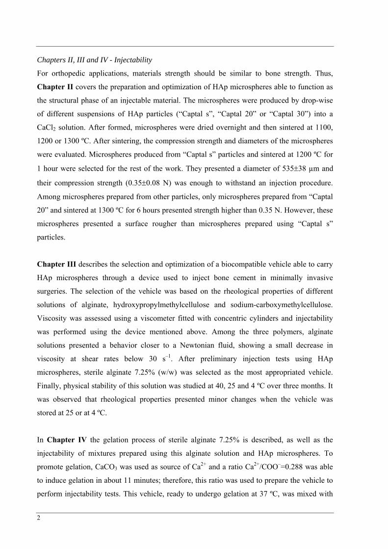
Chapters II, III and IV - Injectability
For orthopedic applications, materials strength should be similar to bone strength. Thus,
Chapter II covers the preparation and optimization of HAp microspheres able to function as
the structural phase of an injectable material. The microspheres were produced by drop-wise
of different suspensions of HAp particles (“Captal s”, “Captal 20” or “Captal 30”) into a
CaCl2 solution. After formed, microspheres were dried overnight and then sintered at 1100,
1200 or 1300 ºC. After sintering, the compression strength and diameters of the microspheres
were evaluated. Microspheres produced from “Captal s” particles and sintered at 1200 ºC for
1 hour were selected for the rest of the work. They presented a diameter of 535±38 µm and
their compression strength (0.35±0.08 N) was enough to withstand an injection procedure.
Among microspheres prepared from other particles, only microspheres prepared from “Captal
20” and sintered at 1300 ºC for 6 hours presented strength higher than 0.35 N. However, these
microspheres presented a surface rougher than microspheres prepared using “Captal s”
particles.
Chapter III describes the selection and optimization of a biocompatible vehicle able to carry
HAp microspheres through a device used to inject bone cement in minimally invasive
surgeries. The selection of the vehicle was based on the rheological properties of different
solutions of alginate, hydroxypropylmethylcellulose and sodium-carboxymethylcellulose.
Viscosity was assessed using a viscometer fitted with concentric cylinders and injectability
was performed using the device mentioned above. Among the three polymers, alginate
solutions presented a behavior closer to a Newtonian fluid, showing a small decrease in
viscosity at shear rates below 30 s–1. After preliminary injection tests using HAp
microspheres, sterile alginate 7.25% (w/w) was selected as the most appropriated vehicle.
Finally, physical stability of this solution was studied at 40, 25 and 4 ºC over three months. It
was observed that rheological properties presented minor changes when the vehicle was
stored at 25 or at 4 ºC.
In Chapter IV the gelation process of sterile alginate 7.25% is described, as well as the
injectability of mixtures prepared using this alginate solution and HAp microspheres. To
promote gelation, CaCO3 was used as source of Ca2+ and a ratio Ca2+/COO–=0.288 was able
to induce gelation in about 11 minutes; therefore, this ratio was used to prepare the vehicle to
perform injectability tests. This vehicle, ready to undergo gelation at 37 ºC, was mixed with

Aim and structure
3
different concentrations of HAp microspheres (20, 30, 35, and 40%) and, each mixture, was
extruded using the injectable device. After gelation at 37 ºC, mechanical properties of the
ceramic/polymeric composites were evaluated. Composites prepared using 35% of
microspheres presented the best compromise between injectability and compression strength.
Therefore, this composition was considered the most appropriated formulation to inject in
bone defects.
Chapters V and VI - Osteoinductible constructs
Chapter V describes the preparation and properties of chitosan sponges, and reports the in
vitro studies conducted to evaluate chondrocytes proliferation and maturation in those
scaffolds. Chitosan sponges were prepared by a freeze/drying process, resulting in pores of
about 100 µm of diameter. Chondrocytes were harvested from caudal (CD), permanent
cartilage, and cephalic (CP), transient cartilage, areas of the sterna of 14 days chick embryos.
After seeded in sponges, chondrocytes were cultured for 20 days, and treated with retinoic
acid to induce maturation and matrix synthesis. Results showed chondrocytes attachment,
proliferation and an abundant matrix synthesis, completely obliterating the pores of the
sponges. However, only CP chondrocytes underwent maturation and markedly changed the
mechanical properties of the CP chondrocytes/chitosan constructs. As a result, transient
cartilage scaffolds and permanent cartilage scaffolds were developed.
To investigate the ability of transient cartilage scaffolds to mimic the process of bone
formation occurring at growth plates, in vivo studies were performed. These studies are
reported in Chapter VI. After 20 days in culture, both transient and permanent cartilage
scaffolds were implanted subcutaneously into the back of nude mice. Animals were sacrificed
monthly and bone formation was evaluated over a period of five months. Mineralization was
assessed by Faxitron, micro computed tomography, scanning electron microscopy and Fourier
transform infrared spectroscopy analyses. Histological analysis provided further information
on tissue changes in the scaffolds. In transient cartilage scaffolds, bone formation was evident
as early as one month after surgery, and increased over the implantation period with bone and
bone marrow cavities developing throughout the implant. Interestingly, bone deposited was
similar to the bone of the mice vertebra and no bone formation was observed in permanent
cartilage scaffolds.

4
Chapter VII - Concluding remarks
In the last chapter of the thesis a short general discussion is presented, while detailed
discussions are provided in each of the preceding chapters. Possible directions for future
research are also proposed.

5
PAPERS RESULTING FROM THIS THESIS
1. Oliveira SM, Barrias CC, Ribeiro CC, Almeida IF, Bahia MF and Barbosa MA.
Morphology and mechanical properties of injectable ceramic microspheres. Key
Engineering Materials, In Press (Chapter II).
2. Oliveira SM, Almeida IF, Costa PC, Pena Ferreira MR, Barrias CC, Bahia MF and
Barbosa MA. Optimization of polymeric solutions as vehicles for injectable
hydroxyapatite microspheres. Submitted to European Journal of Pharmaceutical Sciences
in July 2008 (Chapter III).
3. Oliveira SM, Barrias CC, Almeida IF, Costa PC, Pena Ferreira MR, Bahia MF and
Barbosa MA. Injectability of a bone filler system based on hydroxyapatite microspheres
and a vehicle with in situ gel-forming ability. J Biomed Mat Res part B: Applied
Biomaterials. Published Online – 24 Apr 2008 (Chapter IV).
4. Oliveira SM, Amaral IF, Barbosa MA and Teixeira CC. Engineering endochondral bone:
in vitro studies. Tissue Engineering, Part A, In Press (Chapter V).
5. Oliveira SM, Mijares DQ, Turner G, Amaral IF, Barbosa MA and Teixeira CC.
Engineering endochondral bone: in vivo studies. Tissue Engineering, Part A, In Press
(Chapter VI).

6
CONTRIBUTION OF THE AUTHORS TO THE PAPERS RESULTING FROM THIS
THESIS
Oliveira SM planned and conducted all the experimental work and wrote the manuscripts.
Co-authors contributed with their expertise in the following areas:
1. Barrias CC and Ribeiro CC: assistance in the preparation of microspheres and in
planning the experimental work; Almeida IF: assistance in the characterization of
microspheres compression strength.
2. Almeida IF and Costa PC: performing of viscosity and stability tests; Pena Ferreira
MR: preparation of cellulose derivative and alginate solutions; Barrias CC:
assistance in the preparation of microspheres.
3. Barrias CC: assistance in planning the injectability of mixtures; Almeida IF:
assistance in injectability, gelation and mechanical tests; Costa PC: assistance in
viscosity tests; Pena Ferreira MR: preparation of alginate derivative solutions;
4. Amaral IF: assistance in the deacetylation of chitosan, and preparation and
characterization of chitosan sponges.
5. Mijares DQ: assistance in microCT and FTIR tests; Turner G: preparation of
histological sections; Amaral IF: assistance in the characterization of chitosan
sponges.

Chapter I
7
INJECTABLE SYSTEM AND SCAFFOLDS TO PROMOTE ENDOCHONDRAL MECHANISM FOR BONE REGENERATION
INTRODUCTION
Bone, a vigorous, well-vascularized tissue has an exceptional capability to heal and remodel,
and to rapidly activate mineral stores on metabolic demand.1 Its main role is to provide
structural support for the body and to serve as a mineral reservoir. It also supports muscular
contraction resulting in motion, withstands load bearing and protects internal organs.1,2
Therefore any major change in its structure due to injury or disease can significantly alter
one’s body equilibrium and quality of life.
Every year, more than a million bone-implants procedures are performed in the world, about
500,000 only in the United States, using a wide variety of bone implants materials. However,
the search for an ideal bone implant material is still going on. Although major advancements
occurred in the field of bone regenerative medicine in the past years, current therapies, such as
bone grafts still have several limitations. Despite the fact that materials science expertise has
resulted in numerous improvements in the field of bone grafting, no adequate bone substitute
has been developed. Thus, some of the severe injuries related to bone go inadequately treated.
Current orthopedic replacement materials do not perfectly adjust to the defect to be treated,
resulting in increased difficulties in adjacent tissue growth. In addition, these grafting
materials need to be produced in advance, thus increasing the risk of contamination. The fact
that the injury usually affects a large area and requires a considerable amount of time to
regenerate, also contributes to infection.
The first mention of bone transplantation goes far back to 1682 in the church literature where
a Russian soldier's cranial defect was successfully treated with a piece of dog skull.3 Since
then bone transplantation concept has changed and, nowadays, bone graft materials have been
divided in different groups on the basis of their origin: 1) Autografts or autogenous bone
grafts are considered the best grafting material in the craniofacial skeleton. They are obtained
from another site of the patient’s body. They can be cancellous (iliac crest) used to promote

Chapter I
8
osteogenesis, or cortical (tibia) used when stability is required. They have superior capacity to
promote osteogenesis and are not associated with immunologic problems. Disadvantages
include limited supply of the grafts, donor site morbidity and additional expense and trauma.4
2) Allografts – the grafts taken from another individual of same species. They are obtained 24
hours after the donor’s death and then freeze dried and processed. Demineralized human bone
matrix is also used as allogenic graft material. These grafts are easily obtainable but are
expensive, and associated with risk of disease transmission and immunogenic problems.5 3)
Xenografts – obtained from another species such as bovine (deproteinized bone mineral or
sintered deproteinized bone), pig (porcine amelogenin), or coral. They are available in good
supply but are associated with the risk of disease transmission such as bovine spongiform
encephalopathy – “mad cow disease”.5
Considering all the disadvantages above mentioned, a significant amount of research has been
done to develop synthetic bone graft materials or alloplastic materials. They are available in
powder, granules, blocks, cements and coatings. Those materials can be bioactive (capability
to chemically bond with surface of surrounding bone without fibrous involvement occurs), or
bioinert with no chemical bonding. Although some of alloplastic materials are
osteoconductive, they have been incorporated with growth factors and progenitor cells to
make them osteoinductive.6 Alloplastic materials include ceramics (alumina, zirconia,
calcium phosphate, calcium sulphate, calcium carbonate and bioglass), polymers (resorbable
and non-resorbable) of natural origin (collagen, chitosan, alginate, etc.) or synthetic
(polyethylene, polylactic acid, polyglycolic acid), metals (titanium and its alloy), and
composites.7,8
An ideal bone graft should be biocompatible,9-11 and have appropriate pore size, with
interconnected pores to allow cell ingrowth and an accurate cell distribution throughout the
porous structure. The porosity should facilitate the neovascularization, capillary ingrowth,
accurate diffusion of nutrients and gases, and the removal of metabolic waste resulting from
the activity of the cells that have grown into the scaffold.12 Pore size is an important issue to
address since small pores are unsuitable for tissue ingrowth while big pores would affect the
mechanical properties of the scaffold which might be important in areas of higher strength.
The scaffold should also be osteoinductive13 and present appropriate surface properties since
the chemistry and topography of surface affect both cellular adhesion and proliferation.14,15 In
addition, the scaffolds should withstand the hydrostatic pressures and maintain the spaces
required for cell ingrowth and matrix production, in vitro and ultimately in vivo.11 Because

Introduction
9
bone is always under continuous stress, the mechanical properties of the implanted construct
should ideally match those of living bone in order to enable a faster mobility of the injured
site.9-11 Furthermore, an ideal scaffold’s degradation rate must be tuned appropriately with the
growth rate of the new tissue, in such a way that by the time the injury site is totally
regenerated the scaffold is totally degraded.16
Ceramic materials
Over the past decades, ceramic materials have been given a lot of attention as candidates for
implant materials. Despite their low toughness, they possess certain highly desirable
characteristics as hardness and compression strength. For instance ceramics have been used in
dentistry for dental crowns owing to their inertness to the body fluids, high compressive
strength, and good aesthetical appearance.17,18 The two principal ceramic material groups used
in orthopedics are glass-ceramics and calcium phosphates, which are described in next
paragraphs.
In the early 1960s, polycrystalline ceramics (glass-ceramics) made by controlled
crystallization of glass were developed.19 The most used systems are SiO2-CaO-Na2O-P2O5
and Li2O-ZnO-SiO2 systems which are the base of Bioglass® and Ceravital®, respectively.
Glass-ceramics have several desirable properties compared to glasses and ceramics. The
thermal expansion coefficient is very low, typically 10-7/ºC to 10-5/ºC and in some cases, it
can be made even negative. Due to the controlled grain size and improved resistance to
surface damage, the tensile strength of these materials can be increased at least a factor of
two, from about 100 MPa to 200 MPa. Another important aspect is the mechanical strength of
bone-Bioglass® which is of the same order of magnitude of the bulk glass-ceramic strength
(83.3 MPa) and about three-fourths of the host bone strength.
The main drawback of glass-ceramics is their brittleness. Hence, they cannot be used for
making major load-bearing implants, such as joint implants. However, they can be used as
fillers for bone cement, dental restorative composites, and coating material.19-22
Calcium phosphates are a type of ceramic that has been widely used as artificial bone. It has
been synthesized and used for manufacturing of various forms of implants as well as for solid
or porous coatings. Applications include dental implants, periodontal treatment, alveolar ridge
augmentation, orthopedics, maxillofacial surgery, and otolaryngology, among others.
Different phases of calcium phosphate are used, depending upon whether a resorbable or
bioactive material is desired.18,20 The applications of calcium phosphates as implants are also

Chapter I
10
strongly influenced by their mechanical behavior. Tensile and compressive strength and
fatigue resistance depend on the total volume of porosity which can be in the form of
micropores (<1µm diameter, due to incomplete sintering) or macropores (>100µm diameter,
created to permit bone growth).23
The stable phase of calcium phosphate ceramics depends considerably upon temperature and
the presence of water, either during processing or after implantation. At body temperature,
only two calcium phosphates are stable when in contact with aqueous solution such as body
fluids. At pH<4.2, the stable phase is CaHPO4.2H2O (dicalcium phosphate, DCP), while at
pH≥4.2 the stable phase is Ca10(PO4)6(OH)2 (hydroxyapatite, HAp). At higher temperatures,
other phases such as Ca3(PO4)2 (tricalcium phosphate, β-TCP) and Ca4P2O9 (tetracalcium
phosphate) are present. The unhydrated high-temperature calcium phosphate phases interact
with water or body fluids at 37 ºC to form HAp which is the main mineral component of
bone. Therefore, synthetic porous HAp is widely used as bone substitute due to its
biocompatibility and its osteoconduction.20,24-26
Jarcho et al. described the bonding process to HAp implants. A cellular bone matrix from
differentiated osteoblasts appears at the surface, producing a narrow amorphous electron
dense band only 3 to 5 µm wide. Between this area and the cells, collagen bundles were seen.
Bone mineral crystals have been identified in that amorphous area. As the site matures, the
bonding zone shrunk to a depth of only 0.005 to 0.2 µm and the result was normal bone
attached through a thin epitaxial bonding layer to the bulk implant.27
Polymeric materials
Polymeric materials represent an important group of the biomaterials used today in medical
applications, since they exhibit properties (e.g. low density and high toughness) that cannot be
achieved by the other groups of biomaterials.28 Usually, polymeric materials for biomedical
applications are divided into two groups: biodegradable and non-biodegradable.29,30 In the
present text we will focus in biodegradable polymers due to their suitability for the
biomedical application.
The term biodegradable is associated with materials susceptible of decomposition by natural
biological processes, such as the action of bacteria, plants, and animals31 though other terms
like absorbable, erodible, and resorbable have also been used in the literature to indicate
biodegradation. The interest in biodegradable polymers for biomedical engineering use has
dramatically increased during the past decade. The reason is the two major advantages when

Introduction
11
compared with non-biodegradable materials. Firstly, they do not elicit permanent chronic
foreign-body reactions due to the fact that they are gradually absorbed and do not
permanently leave traces of residues in the implantation sites.29 Secondly, some of them have
recently been found to be able to regenerate tissues.32,33 Hence, surgical implants made from
biodegradable biomaterials could be used as a temporary scaffold for tissue regeneration. This
approach towards the reconstruction of injured, diseased, or aged tissues is one of the most
promising fields in this century.
Many biodegradable polymers have been studied however this thesis was focus only on some
of based natural polymers that have been widely used for biomedical applications, such as: cellulose
derivatives (hydroxypropylmethylcellulose and carboxymethylcellulose), alginate and
chitosan.
Cellulose derivatives
Cellulose is the world’s most abundant natural, renewable and biodegradable polymer, its
main sources are wood pulp and cotton and it can present high stiffness and high crystallinitty
serving well as a structural engineering material. Cellulose basic monomeric unit is D-glucose
which are linked through a glycosidic linkage in the β-configuration between carbon 1 and
carbon 4 of adjacent units to form long chain 1,4-glucans (Figure 1).
O
HO OH
O
O
HOOH
O
OH
4
4
n
CH2
OH
CH2
1 β
β
1
Figure 1. Cellulose structure.
Cellulose is not soluble in common solvents which make its use as pharmaceutical product
more difficult.34 To use cellulose as a pharmaceutical material it should be soluble and
flexible, therefore is common to prepare cellulose derivatives to undergo those limitations.
Cellulose derivatives preparation becomes an easy process since cellulose molecules contain
more than 30% of hydroxyl groups. In fact, each cellulose unit possesses one primary and two

Chapter I
12
secondary hydroxyl groups and those groups can undergo addition, substitution and oxidation
reactions. Although, hydroxyl groups are active, its availability to react can diverge from as
little as 10 to 15% in highly crystalline cellulose to as 98 to 100% in regenerated non-
crystalline cellulose.35
The preparation of cellulose derivatives depends on the average number of hydroxyls
substituted in D-glucose unit, which is known as the degree of substitution (DS). As result,
DS can vary from zero (cellulose itself) to a maximum of three (fully substituted cellulose).
The substitution of hydroxyl groups using ether groups results in cellulose ethers which are
the cellulose derivatives most used in medicine field and also the most widely used
polysaccharides in pharmaceutical industries36 like sodium carboxymethylcellulose and
hydroxypropylmethylcellulose.
Sodium carboxymethylcellulose (NaCMC)
Sodium carboxymethylcellulose is manufactured by an industrial process. Basically, NaCMC
(Figure 2) is prepared by treating cellulose with aqueous sodium hydroxide followed by
reaction with sodium chloroacetate. The main applications are the food industry as a thickener
or stabilizer compound and the pharmaceutical industry for personal care product. To achieve
n
O
HO OH
O
O
HO OH
O
4
4
CH2
OCH2COONa
CH2
OCH2COONa
1 β
1 β
Figure 2. Sodium carboxymethylcellulose structure.
a medical grade, a more refined material has to be prepared and the excess of salt removed
washing the materials with an alcohol-water solution. The DS is also an important parameter
to be controlled since at low DS (below 0.4) the polymer becomes insoluble. The DS control
is achieved controlling the time and the temperature of the reaction. NaCMC is non-toxic, and
generally non allergenic presenting high fluid absorbance and retention for long periods

Introduction
13
directly into its fibers which make it a good material for wound dressing applications
improving the wound healing process.37,38 When in contact with fluids, NaCMC forms a soft
gel or a viscous solution which has led its use for wound care, in gel formulations and in
hydrocolloid dressing.
The use of NaCMC for optical purposes has also been developed. It is common to use
NaCMC in the improvement of dry eyes and to prepare ophthalmic viscosurgical devices to
ensure the maintenance of the ocular space hence protecting of the corneal endothelium.39-41
Postoperative adhesion formation is the single greatest complication of a surgery. Fibrous
adhesions form at peritoneum, central nervous system, pericardium, pleura, and synovium.
The use of NaCMC as biomaterial was used in the form of films and gels reducing adhesion
in a variety of animal models.42,43 Most recently, diZerega et al.44 have observed a significant
reduction of adhesion formation in women undergoing pelvic surgery.
This cellulose derivative is also seen as material with antioxidant properties, as an anti-
inflammatory enzyme stabilizer and it is considered as a potential matrix system for drug
delivery or controlled release of bioactive agents.45-47 Its biocompatible properties also have
pushed NaCMC into bone growth research. Rodgers et al.48 have described NaCMC either
plain or combined with bone morphogenetic proteins as encouraging bone growth, suggesting
that NaCMC influence new bone formation because it is hydrophilic.
Hydroxypropylmethylcellulose (HPMC)
Hydroxypropylmethylcellulose as a cellulose derivative like NaCMC, is also prepared using
alkali cellulose, however, the reacting groups are methyl halide and propylene oxide resulting
the methyl and hydroxypropyl substitutes (Figure 3). HPMC has a wide range of industrial
applications particularly in food industry, pharmacy and medicine (health care, orthopaedics
and opthalmics).49,50 Specifically, HPMC is used whenever there is a need to thicken, gel,
emulsity, suspend, stabilize, water retention and good workability. In food industry, HPMC
uses are based more on the ability to gelify on heating while in pharmacy and biomedical
fields, the drug controlled-release and the biocompatibility are very important properties. In
opthalmics, the presence of extremely toxic free radicals damage the corneal endothelium
inducing corneal edema, however the protection with HPMC shows effective against it,
preventing those damages.50
In orthopaedics, HPMC (3% aqueous solution) has also been widely studied as a carrier of
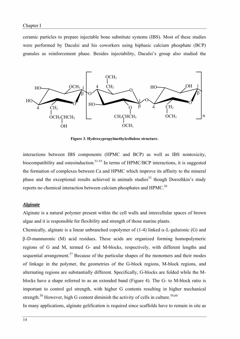
Chapter I
14
ceramic particles to prepare injectable bone substitute systems (IBS). Most of these studies
were performed by Daculsi and his coworkers using biphasic calcium phosphate (BCP)
granules as reinforcement phase. Besides injectability, Daculsi’s group also studied the
n
O
HO OH
O
O
HO O
O
4
4
CH2
OCH3
CH2
CH3CHCH2
OCH3
OCH3
HO OCH3
O 4 CH2
HO
OCH3CHCH2
OH
1 β
1 β
1 β
Figure 3. Hydroxypropylmethylcellulose structure.
interactions between IBS components (HPMC and BCP) as well as IBS nontoxicity,
biocompatibility and osteoinduction.51-55 In terms of HPMC/BCP interactions, it is suggested
the formation of complexes between Ca and HPMC which improve its affinity to the mineral
phase and the exceptional results achieved in animals studies53 though Dorozhkin’s study
reports no chemical interaction between calcium phosphates and HPMC.56
Alginate
Alginate is a natural polymer present within the cell walls and intercellular spaces of brown
algae and it is responsible for flexibility and strength of those marine plants.
Chemically, alginate is a linear unbranched copolymer of (1-4) linked α-L-guluronic (G) and
β-D-mannuronic (M) acid residues. These acids are organized forming homopolymeric
regions of G and M, termed G- and M-blocks, respectively, with different lengths and
sequential arrangement.57 Because of the particular shapes of the monomers and their modes
of linkage in the polymer, the geometries of the G-block regions, M-block regions, and
alternating regions are substantially different. Specifically, G-blocks are folded while the M-
blocks have a shape referred to as an extended band (Figure 4). The G- to M-block ratio is
important to control gel strength, with higher G contents resulting in higher mechanical
strength.58 However, high G content diminish the activity of cells in culture.59,60
In many applications, alginate gelification is required since scaffolds have to remain in site as

Introduction
15
well as they have to enable the encapsulation of drugs and cells. To obtain a hydrogel
structure, alginate crosslinking either covalently or ionically is common. The covalent
crosslinking has been reached using glutaraldehyde, isopropyl alcohol and genipin.61-65
OCOO−
HO OH
O
OCOO−
HOOH
Oβ
1
4 1
4
β
O COO−
HO
OH
O
OCOO−
OH
O
α 1
4 O
HO
1
4 GG
M M
Figure 4. Chemical structure of alginate. G-blocks are folded and M-blocks extended.
Although, covalent crosslink improves the mechanical strength, the use of chemical agents
may lead to toxic effects or to unwanted reactions with drugs.66 Therefore, ionic crosslinking
has been used successful in areas of pharmacy and medicine as alternative. The gelation of
alginate by ionic crosslinking can be achieved by reaction with divalent ions such as Ca2+,
Ba2+, and Sr2+. Monovalent cations and Mg2+ ions do not induce gelation while Ba2+ and Sr2+
ions produce stronger alginate hydrogels than Ca2+.67,68 This gelation takes place when those
divalent cations interact ionically with G-blocks, resulting in the formation of a three
dimensional network which is usually described as “egg-box” structure (Figure 5).69
Ca2+ Ca2+ Ca2+
G GO
COO−
HO
OH
O
COO−
OH
O
α 1
4 O
HO
α 1
4
O
G G
O
COO−
HO
OHO
−OOC
OH O
α 1
4
O
HO
α 1
4
O
Ca2+
Figure 5. Egg-box juntion of Ca2+ ion in polyguluronate blocks.

Chapter I
16
The preparation of such gels can be almost instantaneous forming microspheres or by
controlled gelation which is achieved controlling the divalent cations release into the solution.
The preparation of these controlled gelation hydrogels can be based in the use of CaCO3
suspensions, which are able to produce alginate gels using different gelation times since
CaCO3 dissociation can be controlled by the regulation of the pH solution.70
Alginate has been used for long time in the food industry as stabilizer and thickener.
Although, that industry is very important for alginate production, its applications in pharmacy
industries are also widespread. Non-toxicity and biodegradability of alginate make these
products very well accepted in this industry as well in biomedical industry.
In terms of biodegradability, alginate is one of the most promising biodegradable materials
and it can be resorbed in few weeks depending on the concentration of the solutions and on
the composition of alginate used. The use of sodium alginate containing gentamicin sulphate
for the treatment of bone infections was studied in terms of its biodegradability after has been
inserted into a femur defect in rats. The physical disruption of alginate in small fragments was
observed in one week and the complete removal of the implant from the body tissues happen
in two weeks.71 Comparing the behavior of three different concentrations of sodium alginate
solutions in bone defects made in the tibia of rats, it was observed that a solution using 0.5%
(w/w) of alginate had disappear after 4 weeks in situ whereas solutions using 1.0% or 1.5%
(w/w) remained in the implantation site.72 The preparation of alginate sponges using 1.0%
(w/w) alginate to repair a defect in the facial nerve of cats were absorbed gradually and no
alginate residue was detected remaining in the treated defect after 16 weeks post-
implantation.73 Mooney and coworkers evaluated the degradation of 3% (w/w) alginate
hydrogels by measuring the tensile strength and the molecular weight changes.74 In their study
they prepared solutions using high G content alginates (MVG) and high M content alginates
(LVM) and cultured them with rat bone marrow cells. The results showed that MVG
hydrogels retained more strength for longer than LVM hydrogels and, after 12 days in culture,
MVG still retained 27% of its initial strength. Additionally, MVG hydrogels worked as
substrates for cell growth for over 4 weeks in culture. Suzuki et al.75 had prepared an alginate
hydrogel by dissolving ethylenediamine and water-soluble carbodiimide in 1% sodium
alginate aqueous solution (AGA-100) to use in wound healing and they investigated in vivo
degradation by implanting those dressing materials intramuscularly in rabbits. After 3 months
AGA-100 disappeared without inflammation in the implanted site.

Introduction
17
External applications of alginate as wound dressing are common since it forms hydrophilic
gels providing a moist wound environment which promotes healing and epidermal
regeneration.75,76 More recently, it was developed and study a non-toxic and biodegradable
gel produced from gelatin, oxidized alginate and borax able to form a hydrogel in situ and to
mould into the shape of the wound defect which is an advantage over the preformed wound
dressing. On the other hand the use of gelatin can improve the haemostasis in bleeding
wounds and the borax improves the antiseptic and antiviral activity.77
The treatment of infections is commonly based in systemic drugs applications; however for
some infections local delivery systems have also been used. Studies using alginate alone or in
combination with other polymers or calcium phosphates as a carrier for products delivery has
been performed. Microspheres of alginate prepared by emulsification were able to obtain a
high bovine serum albumin (BSA) encapsulation efficiency as well as a slow release profile in
vitro though this release profile was slower for alginate microspheres coated with poly(L-
lysine) or prepared with high alginate molecular weight.78 Recently, the use of poly(L-lysine)-
coated alginate loaded with vancomycin was able to delivery this antibiotic locally in
concentrations above the minimum inhibitory concentration of staphylococcus aureus for 21
days.79
In orthopedics, the regeneration and the repair of cartilage defects after trauma, cancer or
metabolic disorders is still a major clinical challenge. Chondrocytes are known to
dedifferentiate when cultured in monolayer. However, dedifferentiated bovine articular
chondrocytes were able to redifferentiate after cultured in alginate beads subjected to a
pressure of 5% of oxygen.80 Even at atmospheric oxygen pressure, articular chondrocytes
cultured in alginate gels or in alginate beads retain a chondrocytic phenotype which was
showed by the synthesis of type II collagen and chondroitin-6-sulphate.81-83
The preparation of 3D scaffolds using alginate mixed with HAp or with chitosan also support
chondrocytes, enhancing its proliferation and maintaining their phenotype and spherical shape
with monoriented and sparse actin microfilaments network.84,85
The application of alginate and alginate derivatives scaffolds in the regeneration of bone
structures has been studied using osteoblasts cells encapsulated either in alginate
microspheres or just seeded in alginate solutions. Scaffolds prepared using alginate and
chitosan have allowed osteoblasts attachment, proliferation and deposition of a calcium
Chapter I
18
matrix in vitro.86 After in vivo implantation, calcium deposition occurred as earlier as the
fourth week and these hybrid scaffolds have showed a high degree of tissue compatibility. To
improve pre-osteoblasts attachment, proliferation and differentiation, alginate has been
modified with RGD-containing peptides revealing statistically significant increases in in vivo
bone formation compared with unmodified alginate.87 Studies with RGD-modified alginate
also improved myoblasts attachment, proliferation and differentiation.88,89
Alginate constructs also has been loaded with bone stromal cells and growth factors in order
to induce bone regeneration either in bone defects or in ectopic areas. A scaffold prepared
using 1% (w/w) alginate covalently crosslinked was loaded with morphogenetic protein-2-
derived peptide and implanted into the calf muscle of rats. Three weeks post-implantation,
vascular channels and an osteoblasts population followed by new bone formation were
observed in the pores of alginate hydrogel and after 8 weeks calcification and bone formation
increased showing that this oligopeptide possessed ectopic bone morphogenetic activity when
linked to alginate hydrogel.90 Also, an in vitro study was conducted with the aim of induce
chondrogenesis using 1.2% (w/w) alginate beads to encapsulate human mesenchymal stem
cells (HMSCs). The viability of cells was higher than 90% throughout the 4-week experiment
and cells started to express type II collagen after 1 week. Besides, cells also started to express
type X collagen after 2 weeks and its expression became stronger at the 4th week. Type X
collagen is a well known marker for hypertrophic cartilage which suggests the beginning of
endochondral ossification.91 Cai et al.92 used a 1.2% alginate solution to differentiate bone
marrow mesenchymal stem cells (BMSSCs) (in vitro) into two different lineages
(chondrogenic lineage and osteogenic lineage). After 14 days in culture, cells of chondrogenic
lineage expressed chondrocytes markers and cells of osteogenic lineage became osteoblast-
like in morphology. After in vivo implantation subcutaneously in the dorsum of nude mice,
they found that osteoblasts like-cells induced new bone formation after 8 weeks whereas
chondrocytes like-cells formed cartilage lacuna with a high proportion of type II collagen but
no sign of endochondral ossification.
Chitosan
Chitosan is the result of partial deacetylation of chitin which is, after cellulose, the most
abundant polysaccharide on earth. Chitin consists of β(1,4)-linked D-glucosamine (GlcN)
with a high degree of N-acetylation forming the N-acetyl D-glucosamine (GlcNAc) units
(Figure 6).

Introduction
19
n
OCH2OH
HO
O=C
CH3
NH
O
O CH2OH
HO
O=C
CH3
NH
Oβ 1
1 β
4
4
Figure 6. Chitin structure showing an N-acetyl D-glucosamine (GlcNAc).
In nature, chitin serves as a fibrous strengthening element that occurs as a structural
component of exoskeleton of insects and crustaceans as well in the cell wall of yeast and
fungi. The natural pathway of chitin metabolism includes enzyme-catalyzed hydrolysis by
chitinases. Lysozyme enzymes which are widely distributed in plants and in animals (present
in human body fluids) are also able to degrade chitin molecules.93 In fungi, chitin turnover
occurs by the action of chitin deacetylaze which deacetylate chitin to chitosan whereas, in
laboratory, chitin’ deacetylation is usually preformed in 50% of NaOH for 1 or 2 hours at 60
ºC under nitrogen atmosphere followed of washing in water at 70-80 ºC to neutralize. After
this first treatment a degree of deacetylation of about 80% can be obtained; further
deacetylation needs further treatments with alkaline solutions.
Chitin is a highly insoluble material with low chemical reactivity which is the major problem
for its processing and uses. Applications of chitin and chitin-based materials are widespread
and in many different areas such as environmental, food, pharmaceutical and medical
industries. In the pharmaceutical and medical applications, chitin film and fiber are commonly
used as wound dressing and for controlled drug release.94,95 Also the combinations of chitin
with other materials allowed the preparation of scaffolds. As example, hybrid scaffolds
composed of chitin and collagen showed good affinity and proliferation in culture with
fibroblasts.96 Another interesting application is a hydroxyapatite-carboxymethyl chitin
composite which was prepared and injected on the calvarial bone of rats with biocompatibility
as high as that achieved with HAp materials alone.97

Chapter I
20
As mentioned above, chitosan is the result of chitin deacetylation. However, chitosan name is
used when the deacetylated product becomes soluble in aqueous acidic solutions which
usually correspond to a deacetylation degree of 50%.98 Therefore, chitosan is a heteropolymer
containing both GlcN units and GlcNAc unis (Figure 7), and their relative proportion fixes the
degree of acetylation (DA) that controls many properties. The presence of the amine groups
explains its unique properties among biopolymers, specially its cationic behavior in acidic
solutions.
m
O CH2OH
HO
O=C
CH3
NH
O
1 β
4
OCH2OH
HO NH2
O
β
1
4 n
Figure 7. Chitosan structure showing an N-acetyl D-glucosamine (GlcNAc) and a D-glucosamine (GlcN)
unit.
The solubilization of chitosan occurs by protonation of the –NH2 group function of the C-2
position of the GlcN repeated unit, whereby the polysaccharide is converted to a
polyelectrolyte in acidic media.99,100 Solubililty is also greatly influenced by the addition of
salt to solution. The higher the ionic strength the lower is the solubility.101 This is due to the
fact that chitosan in solution exists in an extended conformation due to the repelling effect of
each positively charged deacetylated unit on the neighboring glucosamine unit.
The biocompatibility is an important requirement especially when chitosan is used as an
implant in contact with either hard or soft tissues. If biocompatible, chitosan will be less
susceptible to be rejected and more predisposed to create a good interface between host
tissues and implant. Focused in this issue, many in vitro studies have been performed showing
that chitosan and its derivatives are potentially favorable materials as substrates for the growth
of different type of cells.102-105 Other in vitro studies have evaluated how chitosan
Introduction
21
compatibility depends on the degree of acetylation. Chatelet et al.106 observed that higher
DA’s chitosan present lower cell adhesion but DA’s of chitosan films did not affect the
keratinocytes and fibroblasts cytocompatibility. Similar results were found by Amaral et
al.107,108 studying the influence of the DA in attachment, spreading and short-term
proliferation of human osteoblastic MG-63 cells either in three-dimension chitosan scaffolds
and on chitosan films. An in vivo study showed that a collagen matrix was built within pores
of chitosan scaffolds implanted into mice, and angiogenic activity was observed on the
external implant surface demonstrating the high degree of biocompatibility of chitosan in this
animal model.109
Chitosan conjugated with other chemical groups in its chain have also proved to be efficient
in improving the behavior of cells on its surface. The preparation of chitosan with
phosphorylcholine groups presented a good biocompatibility when in culture with human
umbilical vein endothelial cells110 and the conjugation of chitosan with RGDs improved the
capability for adhesion and proliferation of chondrocytes and fibroblasts.111,112
Besides the use of chitosan alone, its combination with other materials without chemical
reaction also enabled the preparation of scaffolds. Chitosan combined with alginate, as
mentioned elsewhere permitted to prepare scaffolds which have good biocompatibility in
cultures with different type of cells (osteoblasts, chondrocytes, and others).85,86,113 Using
similar approaches, calcium phosphate-chitosan composites and other chitosan derivatives
allowed mesenchymal stem cells and preosteoblasts viability, enhancing bone tissue
formation.114-116 On the other hand the combination of calcium phosphate cement (CPC) with
chitosan produced composites stronger than CPC and composites that allow osteoblasts cells
to adhere, spread and proliferate.117 To enhance chitosan’ mechanical properties is also
common to crosslink it with different chemical products like glutaraldhyde, diepoxide (1,4-
butanediol diglycidyl ether) and genipin which are usually more toxic compounds. However
their controlled use can maintain chitosan’ scaffolds biocompatibility.118-120
Biodegradability is another important issue in the use of a material for biomedical purposes.
Usually, natural polymers have the advantage of higher biodegradability. Chitosan as a
natural polymer shares that advantage, and its molecules can be broken down by lysozyme,
chitosanase and chitinase enzymes.121,122 However, chitosan’s physicochemical and biological
properties, such as biodegradability, are dependent on molecular weight (Mw) and DA. Zang
Chapter I
22
et al.122 compared chitosan susceptibility to degradation by rat cecal and colonic enzymes
with a commercial available almond emulsion β-glucosidase. The results show a higher
degradation of chitosan molecules with lower Mw and lower DA (DA=77.8).
In human serum, chitosan is mainly depolymerized by lysozyme which biodegrades the
polysaccharide by hydrolyzing the glycosidic bonds present in the chemical structure.123
Lysozyme contains a hexameric binding site, and hexasaccharide sequences containing 3 to 4
or more acetylated units, that contribute to the initial degradation rate of N-acetylated
chitosan.124,125 This mechanism explains the slow degradation of very low DA chitosan. Freir
et al.126 investigating materials for nerve regeneration using concentrations of lysozyme
similar to those found in the human body observed that samples with intermediated DA
values were almost completely degraded in one week. However, the degradation of samples
with very low or high DAs was minimal over the studied period.
In many studies, chitosan is mixed with other materials or is covalently crosslinked in order to
change its mechanical properties. In those chitosan based materials, while the degradability is
affected,120,127 it still occurs by an enzymatic mechanism and depolymerization.119,128
Chitosan crosslinking
Despite of easy gelation and good biocompatibility, chitosan has low mechanical integrity and
degrade rapidly especially in acid medium and in the presence of lysozyme.129 On the other
hand, for many applications, chitosan should be crosslinked due to its hydrophilic behavior. In
this context, various crosslinking reagents, most are synthetic, have been used to prepare
chitosan gels.130,131 However, these synthetic crosslinking reagents are usually cytotoxic
which may impair the biocompatibility of the crosslinked biomaterials.132 Therefore, it is
desirable to provide crosslinking reagents suitable for use in biomedical applications that are
not cytotoxic and may form stable and biocompatible products. Genipin is an alternative
crosslinker since it is about 5000-10000 times less cytotoxic than glutareldehyde and genipin-
crosslinked chitosan microspheres have shown a superior biocompatibility and slower
degradation rate than the glutaraldehyde-crosslinked chitosan microspheres.133 Also the use of
chitosan crosslinked with diepoxide-based bifunctional linkers increased its compression
modulus and stiffness while supporting chondrocytes typical round shape.119 The use of
radiation is another alternative to crosslink chitosan. The UV light is a common source of
energy to induce crosslinking and the use of this method enabled to successful transform
azide-chitosan-lactose aqueous solution into an insoluble and flexible hydrogel. In vivo, this

Introduction
23
hydrogel significantly induced wound contraction, accelerated wound closure and
epithelialization of treated wounds.134,135
Pharmaceutical applications of chitosan
Chitosan has been widely used in the pharmaceutical industry for its potential in the
development of controlled release drug delivery systems. The reason is its unique polymeric
cationic character and its gel and film forming properties. Such system should allow the
control of the rate of drug administration and prolong the duration of the therapeutic effect as
well as the targeting of the drug to specific sites.
The potential of chitosan derivatives as vehicle for the administration of proteins was
evaluated by several authors. Xu et al.136 synthesized a water soluble derivative of chitosan,
N-(2-hydroxyl) propyl-3-trimethylamonium chitosan chloride, and used it to incorporate
bovine serum albumin (BSA), considered as a model protein drug. A preparation of Poly
ethylene glycol (PEG) graft-chitosan crosslinked with genipin was able to carry BSA and
poly(vinyl alcohol) (PVA), while sodium bicarbonate mixed with chitosan also worked as
BSA delivery vehicle.137,138 Chitosan microspheres were used to release transforming growth
factor-β1 (TGF-β1) and its delivery profile was similar to BSA. When chitosan microspheres
were loaded with TGF-β1 and seeded into porous chitosan scaffolds, chondrocyte
proliferation and production of extracellular matrix were significantly increased.139 A paste of
chitosan glutamate (Protosan) and HAp was used as a delivery vehicle for bone marrow, bone
morphogenetic protein-2 (BMP-2) and osteoblasts, and implanted in cranial defects of rats
resulting in the formation of mineralized bone.140 Similar tests were performed using solutions
based in chitosan/polyol salt to delivery biologically active growth factors in vivo as well as to
encapsulate living chondrocytes.141
Besides applications in drug delivery, chitosan and its derivatives have been widely used in
the delivery of DNA, proteins and live cells. The use of DNA delivery system has the
potential to overcome extracellular barriers that limit gene therapy since controlled release
systems can improve gene delivery by maintaining high concentrations of DNA in the cellular
environment and increasing the duration of the transgene expression. Yun et al.142 prepared
DNA-chitosan complexes of microspheres physically combining PEG with poly(lactic
glycolic acid) (PLGA) able to sustain release for at least 9 weeks. In addition, chitosan-DNA
complexes were conjugated with alpha-methoxy-omega-succinimidyl-PEG by Zhang et al.143

Chapter I
24
improving the gene expression. The preparation of chitosan microspheres incorporating
adenovirus vectors for mucosal delivery was performed by Lameiro et al.144 This system
enabled an effective release when it was in contact with cells in monolayer while an
insignificant release was observed in aqueous media. Guo et al.145 combined tissue
engineering with gene therapy entrapping plasmid DNA in chitosan gelation complexes,
encoding TGF-β1 which has been proposed as promoter of cartilage regeneration. The
transfected chondrocytes expressed TGF-β1 protein stably in 3 weeks.
Chitosan for wound dressing
On last decades the treatment of wounds was revolutionized with the use of materials on
wound dressing which can replicate some of the skin’s properties, like 85% water content and
inherent permeability. Chitin and chitosan are known for accelerating the healing of
wounds.146,147 Chitosan stimulates the migration of cells and accelerates the regeneration of
normal skin as well as it was found to promote the attachment and proliferation of fibroblasts
on PVA/chitosan-blended hydrogel.148-150 The application of cotton fiber-type chitosan for 15
days on open skin wounds of dogs demonstrated that chitosan was efficient in wound healing
by increasing the formation of thick fibrin and the induction of macrophages and fibroblasts
migration into the wound area.147 Using similar materials in radiation-impaired wounds, also
in dogs, was detected advanced granulation and capillary formation combined with the
expressing of vascular endothelial growth factor confirming neovascularization.151
Additionally, chitosan membranes prepared by immersion-precipitation phase-inversion
method showed controlled evaporative water loss, excellent oxygen permeability and
promoted fluid drainage ability.152 In rats, wound covered with these chitosan membranes was
hemostatic and healed quickly. In another study the number of living bacteria responsible to
infect damaged skin was diminished using those membranes incorporating antibiotics for
controlled release.153 Using UV light, azide-chitosan-lactose hydrogels were crosslinked in
situ. Once applied on incisions made in mice’ skin, these hydrogels were enable to stop
bleeding and to adhere to the skin inducing significant wound contraction, closure and
healing.134 Recently, the preparation of chitosan combined with phosphorylcholine groups
through a heterogeneous reaction process showed a more hydrophilic characteristic, better
anti-protein-adsorption capability, better resistance to coagulation, an improved
hemocompatibility, as well as similar cell affinity and safety, compared with native
chitosan.110

Introduction
25
Chitosan for skeletal tissue engineering
A round cellular morphology is known to be a normal phenotypic characteristic of non-
dedifferentiated chondrocytes. Since chitosan has the ability to maintain round morphology of
chondrocytes and to preserve its cell specific extracellular matrix, chitosan and its derivatives
have been used for studies of cartilage reconstruction. Montembault et al.154 have used
chitosan hydrogel substrates for culture of human and rabbit chondrocytes. Those
chondrocytes were able to express mature type II collagen and aggrecan. Their phenotype was
maintained for as long as 45 days, forming cartilage like nodules, while chondrocytes cultured
without chitosan hydrogels dedifferentiated as indicated by the expression of immature type II
collagen and type I collagen. The use of chitosan-alginate scaffolds, for culture of
chondrocytes like cells (HTB-94), as well as chondroitin sulfate A combined with chitosan,
for culture of bovine articular chondrocytes, allowed cells to maintain their round
morphology, to produce cartilage specific type II collagen and proteoglycan, both markers of
chondrocytic phenotype.85,105 Comparing the behavior of rabbit chondrocytes on chitosan and
chitosan based hyaluronic acid, Yamane et al.155 suggested that hyaluronic acid improves
chondrocytes proliferation while maintaining their physiological phenotype and production of
a rich extracellular matrix. Additionally, scaffolds made of lactose-modified chitosan induced
cell aggregation when in contact with primary culture of pig chondrocytes, leading to the
formation of cartilage nodules rich in aggrecan and type II collagen.156 Another study, using
in situ gelling chitosan solutions (prepared by mixing chitosan disodium β-glycerol phosphate
and glycosamine) loaded with primary articular chondrocytes, also supported the
accumulation of cartilage matrix in vitro.157 On the other hand, rabbit subchondral bone
defects treated with a mixture of chitosan, glycerol phosphate and autologous blood, showed
an increased inflammatory reaction, vascularization, intramembranous bone formation and
subchondral bone remodeling, main factors supporting cartilage repair, when compared with
untreated defects.158
The use of cells different from chondrocytes, but with good characteristics of proliferation
and easy differentiation into chondrocytes were used also to produce cartilage using chitosan
derivatives as substrate. In that study, a thermosensitive (water chitosan-poly(N-
isopropylacrylamide) gel was seeded with HMSCs and injected in the submucosal layer of the
bladder of rabbits inducing HMSCs differentiation into chondrocytes and the formation of
cartilage in 14 weeks.116

Chapter I
26
The ability of chitosan and chitosan derivatives as substrate for osteoblasts and other cells
related with bone regeneration has been reported in several studies. Some of those studies will
be referenced in following paragraphs. The DA of chitosan plays an important role in cells
morphology and proliferation as it has been showed by Amaral et al.107,108. In those studies,
they demonstrated that chitosan of low DA (4%) improves cell attachment and spreading
displaying long cell filopodia and numerous cell-to-cell contacts when compared with
chitosan of higher DAs. Therefore low DAs chitosan as well as chitosan combined with other
materials are commonly used for osteoblasts cultures. The preparation of chitosan-alginate
scaffolds is one of those combinations. In vitro studies indicated that osteoblasts cells seeded
on porous chitosan-alginate scaffolds attached and proliferated well and promoted the
deposition of mineral. Additionally, in vivo studies, performed at same time, showed a rapid
vascularization, matrix deposition and calcification.86 It was also mentioned that all those
events happened within the entire scaffolds structure though no evidences were showed. The
preparation of chitosan-collagen composite sponges and CPC-chitosan scaffolds were studied
in vitro referencing those structures as biocompatible and as good substrates for osteoblasts
proliferation and differentiation.117,159 On the other hand RGDS-modified chitosan enhanced
the attachment of rat osteosarcoma cells making chitosan more biocompatible for the culture
of those osteoblasts-like cells.112
Chitosan scaffolds were also investigated for culture of mesenchymal stem cells (MSCs),
preosteoblasts and as a growth factor deliver with the purpose of bone regeneration. Chitosan
scaffolds cultured in vitro with MSCs and preosteoblasts allowed attachment and spreading of
those cells as well as their maturation into osteoblastic phenotype was successful.114 However,
comparing chitosan scaffolds with BCP embedded into chitosan scaffolds, a more uniform
extracellular matrix distribution were obtained when BCP was used. Besides, cells expressed
higher concentration of protein markers of osteoblastic phenotype (alkaline phosphatase and
osteocalcin). The use of a paste made of chitosan glutamate and HAp cultured with
osteoblasts or BMP-2 showed to be efficient in delivering these osteoinductive factors since
the presence of mineralized bone spicules were observed in rat calvaria defects filled with that
paste and osteoblasts or BMP-2.140 Osteogenesis was also observed using composite porous
matrix of chitosan-PLLA loaded with platelet-derived growth factor-BB (PDGF-BB) after it
has been implanted in rats calvaria defects and also after porous chondroitin-4-sulfate
chitosan sponges loaded with PDGF-BB has induced an increase of osteoblasts migration and

Introduction
27
proliferation in vitro.160,161 In another study performed in vivo, nude mice were injected
subcutaneously with alginate-chitosan gels loaded with MSCs and BMP-2 as well as loaded
with only one of those factors. Authors have observed that after 1 month scaffolds loaded
with only one factor either MSCs or BMP-2 did not formed bone. Moreover, they started to
be reabsorbed while scaffolds loaded with both factors were able to induce bone formation
forming a trabecular bone like structure.162 Another study using 3D alginate-chitosan
constructs cultured with human bone marrow cells and chondrocytes also showed optimal
osteocytes formation subcutaneously in immunocompromised mice. The interaction between
both cells type was also studied in the creation of specific tissues.163

Chapter I
28
INJECTABLE SYSTEMS
Invasive surgeries are associated to high risks of infection and high levels of pain, either
during or after the surgery, resulting in the need to develop alternative methods. One possible
alternative consists in injectable materials, which can fill the defects up using a needle. This
technique drastically decreases the affected area as well as the access to infections agents.
Additionally, it enables to fill any shape defect leading to an improved grow of adjacent tissue
as well as the materials can be used as delivery systems of growth factors, cells, and drugs.
Thus, a new concept of surgery appears with the injectable in situ polymerizable and
biodegradable materials, which may serve as scaffolds for guided bone regeneration and may
provide an alternative for bone defect treatment. Different authors are working on the
development of these new materials. Poly(propylene fumarate) (PPF) which is a unsatured
linear polyester that can be crosslinked through its fumarate double bonds using a vinyl
monomer is one of the materials tested. Based on this polymer, injectable composites
incorporating an osteoconductive matrix β-TCP and a porogen (sodium chloride) were
prepared.164,165 These injectable products, known as injectable bone substitutes (IBS), present
suitable rheological properties to ensure bonding of the mineral phase and good cell
permeability. Contrary to dense materials which do not have any intrinsic porosity, this
approach provides rapid and improved deep bone formation.
Other advantages of injectable formulations over conventional scaffolds are the possibility of
a minimally invasive implantation, ability to fill a desirable shape, and easy incorporation of
various therapeutic agents. Those advantages stimulate the search for new materials with
suitable characteristics to be injected. Some of those are: Alginate, chitosan, hyaluronan, and
polyethylene oxide/polypropylene oxide.165-168 In the following pages an overview of different
types of injectable systems (pastes, gels and microspheres) as well as their advantages,
disadvantages, applications, and preparations methods (thermoplastic pastes and crosslinking
techniques) are presented.
Types of injectable systems
The development of injectable materials were the result of different properties of the skeletal
tissues (cortical and trabecular bone, and cartilage), the different places in the body where the
implants are needed (femur, spinal column, pelvis, omoplate, cranial, etc.), and the different
shape of defects to be repaired (spherical, lamellar, and irregular). Those differences also
Injectable systems
29
require suitable mechanical properties and viscosity of the injectable materials. In terms of
viscosity, an injectable material should have low viscosity before injection, but whenever
injected, it should harden in situ (decrease viscosity) in order to provide mechanical properties
similar to the bone tissue. Additionally, these materials could work as osteoinductors.
For all of the reasons described above, injectable materials are sub-divided in three systems:
pastes, gels, and microspheres. Materials representing each of these categories will be
presented in the following sections.
Pastes
Pastes are an example of injectable systems and a promising candidate is the above mentioned
PPF. Poly(propylene fumarate) provides the orthopedic community with a new material that
holds great promise for use as an injectable biodegradable composite, for filling skeletal
defects.165
Peter et al.169 used an injectable composite paste of PPF, N-vynil pyrrolinidone (N-VP),
benzoyl peroxide (BP), sodium chloride (NaCl) and β-TCP to study the crosslinking
temperature, heat release upon crosslinking, gel point, and the composite compressive
strength and modulus. The maximum temperature reached did not vary widely among the
formulations tested, with the values ranging from 38 to 48 ºC, which is below poly(methyl
methacrylate) (PMMA) bone cement curing temperature, 94 ºC. Same authors also studied the
degradation of these composites in vivo. They observed that all implants were encased in
fibrous capsules within 4 days after implantation and granulation tissue was present,
accompanied by remnant neutrophils and necrotic tissue debris. Newly formed capillaries,
fibroblasts, and macrophages were evident at 1 week post-implantation and no multinuclear
giant cells were found at any implant site. After 3 weeks, all formulations demonstrated signs
of the late phase of healing, with the samples encased in a matured scar capsule of dense
collagen fibers.170
PPF filled with calcium gluconate/hydroxyapatite (CG/HAp), which cures to a hard
degradable cement by hydrolysis was investigated by Lewandrowski et al.171 to evaluate the
osteoconductive properties. The PPF bone cement which presented reasonable viscosity and
reasonable working (6 to 7 min) and hardening (10 to 12 minutes) times for injectability, was
injected into the proximal and distal portion of the intramedullary canal. The results showed
that the trabecular bone volume and remodeling index were lower in the injectable cement
that in the positive control but higher than in the negative control, supporting the concept of

Chapter I
30
osteoconductivity.
Self-hardening CPC are another group of pastes with interest in biomaterials research. The
first published CPCs consisted of TCP and dicalcium phosphate anhydrous (DCPA). Later,
additional CPCs highly biocompatible and able to set in situ were developed. Those properties
pushed CPCs into the dental and medical applications showing good results. Xu et al.172
investigated the effects of lubricants and particle sizes of the cements on the injectability of
the TCP/DCPA, α-TCP/CaCO3 and DCPA/CaCO3 cements. The study suggested that
excellent injectability can be obtained by incorporating CMC as gelling agent and by using a
cement powder that contains sufficiently small (<5µm) particles.
The purpose of Grimandi et al.54,173,174 was to develop an IBS for percutaneous orthopedic
surgery using a multiphasic material composed of 2% (w/w) aqueous solution of HPMC and
BCP (60% HAp and 40% β-TCP) where HPMC serves as the vehicle to carry BCP irregular
particles (80-200 µm of diameter). The optimum ratio for BCP and polymer solution tested
was 60/40 (w/w), and in vitro cytotoxicity assays showed that all cells survived, and there
were no significant differences between the values found for control and the test materials.
This system was considered nontoxic and suitable to use as IBS.54,173,174 Weiss et al.175,176
conducted in vivo studies also using the IBS system mentioned above and results showed high
permeability to cells and biological fluids, which allowed early osteoconduction and bone
substitution. Following those studies Gauthier et al.177 implanted the IBS into mandibular and
maxillary canine extraction sockets. After 19 days, gingival healing was achieved with no
suture dehiscence or any signs of infection or inflammation of tissues and three months after
implantation, extensive bone ingrowth was found.
To evaluate the importance of the particles size, Gauthier et al.,178,179 in other studies,
implanted two different IBS composites, each with different calcium phosphate particles
granulometries, 40-80 µm (IBS 40-80) and 200-500 µm (IBS 200-500), in the femur of
rabbits. Both IBS composites were prepared using BCP granules (60/40 HA/β-TCP weight
ratio) and HPMC 3% (w/w), and an extensive newly formed bone was noted 2 weeks after
implantation. However, earlier bone colonization occurred on IBS 40-80 than on IBS 200-
500. Others have also used HPMC to improve injectability of calcium phosphate
cements.172,180,181 However, the use of HPMC in IBS produced a non-hardening material
difficult to maintain within the defect during surgery so, recently, silane has been grafted to
the HPMC to induce its self-hardening.182,183 At basic pH, the silane radicals (R–Si–O–Na+)
grafted on cellulosic ether cycles, are ionized. Due to pH decrease, R–Si–O–Na+ are

Injectable systems
31
hydroxylated in R–Si–OH and covalent bridges linking hydroxyls radicals allow the
formation of a 3D network of Si-HPMC. This polymerization mechanism leads to the shift of
this cellulosic derivative fluid into a gel state without exothermic reaction. Besides Si-HPMC
self-hardening, this new material is also biocompatible and well-adapted matrix for 3D culture
and differentiation of osteoblast and chondrocytes.180,184 The combination of Si-HPMC with
BCP granules loaded with bone morphogenetic stem cells (BMSCs) showed osteoinductive
properties in ectopic areas and the culture of Si-HPMC with human nasal chondrocytes
revealed the formation of a cartilage like tissue with an extracellular matrix subcutaneously in
nude mice.185
Gels
A gel is defined as a three dimensional network swollen by a solvent where, usually, the
solvent is the major component of the gel system.31
Biocompatible polymeric formulations that exhibit sol-to-gel transition at physiological
conditions are good candidates for tissue engineering applications. To select an injectable gel
some factors should be considered: tissue specific cell-matrix interactions, gelation kinetics,
matrix resorption rates, toxicity of degradation products and their elimination routes. In terms
of gelation, it can occur by: thermal gelation (injectable polymer formulation can gel in vivo
in response to temperature change), pH change, ionic crosslinking, or solvent exchange.
Therefore, to design a tissue-supporting injectable scaffold, the correlation of the above
factors is required in order to obtain injectability and both new tissue formation and scaffold
reabsorption simultaneously. In the following paragraphs it will be presented some injectable
gels as well as their applications.
Poly ethylene-oxide (PEO)
Poly(ethylene-oxide-b-propylene oxide-b-ethylene oxide) (PEO-PPO-PEO), or Pluronics
(also known as Poloxamers), are the best known examples of thermally gelling polymers.
PEO-PPO-PEO copolymers are a family of more than 30 different nonionic surfactants and it
forms reversible gels at high temperatures and reverts to a liquid state upon lowering of
temperature. Poloxamer 407 solutions forms gels at 37 ºC for polymer concentrations above
20% (w/w). Solutions of this Poloxamer were used in burn treatment and other wound healing
applications more than 25 years ago. Cao et al.186 described a novel application of PEO-PPO-
PEO copolymers in the formation of a cartilage layer on the host bone. They demonstrated

Chapter I
32
cells suspended in the polymer formed higher volume of bone than cells suspended in saline.
Poly ethylene-glycol (PEG)
Recently, novel hydrogel triblock copolymers of PEG and poly(lactic glycolic acid) (PLGA),
or PEG-PLGA-PEG, were developed by Jeong et al.187-189 Aqueous solutions of these
copolymers also exhibit sol-to-gel transitions at body temperatures. Subcutaneous injection of
the copolymer formulation resulted in the formation of a transparent gel in situ maintaining its
structural integrity as well as the mechanical strength. Similar results were obtained using
poly(ethylene glycol-g-(DL-lactic acid-co-glycolic acid)) (PEG-g-PLGA) and poly((DL-lactic
acid-co-glycolic acid)-g-ethylene glycol) (PLGA-g-PEG).
Hyaluronic acid
A high-viscosity, shear-thinning polymer solution or slightly crosslinked gel may be injected
through a relatively small gage needle and upon removal of the injection force, a thick gel is
formed in situ. Hyaluronan gels form in situ upon injection of a viscous gel formulation.
Viscoelastic and shear-thinning properties of hyaluronan inspired researchers to use it as a
carrier for drugs, protein, and peptide delivery, as well as tissue engineering applications such
a bone fracture repair, wound healing and soft tissue augmentation procedures. Viscosity of
hyaluronic acid solutions decreases with increasing temperature, which makes it suitable for
infectable applications. Physicochemical properties of hyaluronan may also be improved by
chemical crosslinking with divinyl sulfone. Animals studies demonstrated that hyaluronan
polymers are nontoxic, nonantigenic, noninflammatory, and do not elicit a foreign body
reaction.190,191
Radomsky et al.192 evaluated sodium hyaluronate gel has a matrix to deliver basic fibroblast
growth factor (b-FGF) in bone defects. A simple injection of b-FGF and hyaluronan into a
freshly created fracture in the rabbit fibula resulted in increasing bone formation, and in an
earlier restoration of mechanical strength at the fracture site.
Chitosan
As discussed before chitosan is a charged, water soluble polymer that may form reversible
gels in response to a pH change in solution. As a specific example of pH-reversible polymeric
gels, chitosan solutions exhibit a liquid-gel transition around pH=7, when pH changes from
slightly acidic to neutral.
Since unmodified chitosan can be only dissolved in acidic solutions, its applications as

Injectable systems
33
injectable product are limited. Therefore the use of other compounds grafted on chitosan
molecules can adjust the properties of solutions in order to enable the preparation of
hydrogels at neutral pH and body temperature.116,141,193
To overcome those difficulties, Hoemann et al.157 studied the use of an in situ gelling
chitosan-disodium β-glycerol phosphate-glucosamine solution. This solution was used to
encapsulate chondrocytes supporting in vitro and in vivo accumulation of cartilage matrix.
When injected in osteochondral defects hydrogels persisted at least for 1 week. Also
chitosan/polyol salt solutions a liquid below room temperature, was used to encapsulate
chondrocytes. These solutions were able to gelify in situ at body temperature after injected in
vivo.141
The PEG covalently grafted to chitosan produced an injectable product able to transform into
a semisolid hydrogel at body temperature.137 This system was able to serve as carrier for BSA
delivery. Furthermore, crosslinking in situ of this copolymer with genipin produced a new
hydrogel also suitable to release BSA for 40 days with a quasi-linear profile. Another
hydrogel for BSA delivery was prepared mixing chitosan with poly(vinyl alcohol) and sodium
bicarbonate.138 This new hydrogel which is liquid at low temperatures (about 4 ºC) also
becomes a gel under physiological conditions and presents good mechanical properties.
Poly(N-isopropylacrylamide) (PNIPAAn), a well known thermosensitive polymer with a
thermoreversible phase transition at 32 ºC, has been used to create a PNIPAAn-chitosan
derivative with improved in situ gel formation.194-196 These polymers also presented high
potential for culturing of chondrocytes, meniscus cells, to differentiate human mesenchymal
stem cells into articular chondrocytes and also for drug delivery.116,197,198
The synthesis of chitosan graft to methacrylic acid produced a chitosan derivative readily
soluble in pure water below pH=9 but its gelation occurs by thermal treatment at body
temperature.199 Finally, the use of chitosan, citric acid and glucose solution to work as a liquid
phase to carry calcium phosphate particles enabled the production of a moldable and
biocompatible injectable bone substitute.200
Microspheres
The regular spherical shape of microspheres can be an advantage for injectability as well as in
terms of package and regular porosity interparticles. Therefore many studies have focus in
microspheres production for bone regeneration and other purposes. Different materials have
been used to produce microspheres. Schrier et al.201,202 prepared and implanted poly(lactic

Chapter I
34
glycolic acid) (PLGA) and CMC microspheres in New Zealand white rabbits observing that
bone was highly enhanced when microspheres were loaded with BMP-2. Similar results were
obtained in studies using PLGA sintered microspheres loaded with BMP-7. On the other hand
chondrocytes cultured on PLGA microspheres and injected in athymic mice improved the
production of extracellular matrix cartilage when compared with chondrocytes alone. In vivo
studies using bioactive glass microspheres loaded with BMP-2 implanted in rat tibia bone
defect highly promoted bone regeneration.203,204
As mentioned above HAp is widely used as bone substitute but it cannot be injected in its
massive form. However, HAp powder size is compatible with injection and it can be used to
mix with polymeric solutions or just to prepare HAp injectable granules. Pasquier et al.205
have coated HAp granules with type I collagen, forming microspheres (HAp-COL) in order to
obtain a bone substitute able to be injected and leading to better osteoconduction than HAp
powders and collagen separated. The HAp-COL system shows a good pathway for the
biological fluids and bone regeneration. Additionally, HAp granules released after collagen
degradation sustained the new bone formation, increasing bone strength. However, the delay
in collagen degradation, and its negative response in increasing new bone formation in time,
lowers the efficiency of the HAp-COL microspheres.
Ferraz et al.206 produced nanohydroxyapatite microspheres for antibiotics delivery observing
that either microspheres load or not with antibiotics have promoted osteoblasts proliferation
though in the presence of erythromycin, osteoblasts proliferation came enhanced. Barrias et
al.207,208 have studied HAp and calcium titanium phosphate (CTP) microspheres loaded with
human osteoblasts cells and bone marrow stromal cells. After 7 days in culture, both types of
cells proliferate and bone marrow stromal cells produced an abundant amount of fibrillar
extracellular matrix.
Preparation of injectable systems
An injectable material should have low viscosity and keeps its shape after injection into a
defect. Thus, new techniques must be developed in order to enable gelation in situ. To explain
this concept, different strategies to prepare in situ setting solid biodegradable injectable
implant systems will be presented. Those strategies can be divided into three categories based
on the mechanism of achieving solidification in vivo: thermoplastic pastes; in situ crosslinked
systems; and in situ precipitation.

Injectable systems
35
Thermoplastic pastes
Thermoplastic pastes are characterized as having a low melting point, ranging from 25 to 65
°C, but once injected into the body as a melt, they form a semi-solid upon cooling to body
temperature. They are easily injected, when heated slightly above their melting point, due to
their low molecular weight and low glass transition temperature (Tg). Furtheremore, they hold
their shape at room temperature and can be formed into different shapes by applying heat.209
Bioerodible thermoplastic pastes can be prepared from such monomers as D,L-lactide,
glycolide, ε-caprolactone, trimethylene carbonate, dioxanone, etc.210 Polymers and
copolymers of these monomers have been extensively used in a number of biomedical areas,
from carriers of pharmaceutical compounds to surgical sutures, ocular implants, soft tissue
repair and augmentation materials.211,212 They therefore have a demonstrated track record of
biocompatibility and thus are attractive starting points for new material development.
Walter et al.213 placed a Taxol-loaded poly [bis(p-carboxyphenoxy) propane-sebacic acid]
implant beside brain tumors or within tumor resection sites and demonstrated the
effectiveness of the method in rats after surgery. In an effort to develop a means of avoiding
surgery and circumventing the invasiveness of Walter's method, Zhang et al.214 developed a
thermoplastic triblock polymer system composed of poly(D,L-lactide )-block-poly(ethylene
glycol)-block-poly(D,L-lactide) and blends of low molecular weight poly(D,L-lactide) and
poly(ε-caprolactone) (PCL) for the local delivery of Taxol. Both polymeric systems were
capable of releasing Taxol for a long period of time (greater than 60 days), although at a very
low rate215,216.
In situ crosslinked systems
Crosslinked polymer networks can be formed in situ in a variety of ways, forming solid
polymer systems or gels. Means of accomplishing this end include free radical reactions
initiated by heat (thermosets) or absorption of photons, or ionic interactions between small
cations and polymer anions.
Termosets
Thermoset polymers can flow and be molded when initially constituted but, after heating, they
set into their final shape. This process is often called "curing" and involves the formation of
covalent crosslinks between polymer chains to form a macromolecular network. Reheating a
cured polymer only degrades the polymer. This curing is usually initiated chemically upon

Chapter I
36
addition of heat.
Dunn et al.210 used biodegradable copolymers of D,L-lactide or L-lactide with ε-caprolactone
to prepare a thermosetting system for prosthetic implants and slow release drug delivery
systems. This system is liquid outside the body and is capable of being injected via a syringe
and needle and, once inside the body, it cures. The multifunctional polymers in their
thermosetting system were first synthesized via copolymerization of either D,L-lactide or L-
lactide with ε-caprolactone using a multifunctional polyol initiator and a catalyst (e.g.,
peroxides) to form polyol terminated liquid pre-polymers. This pre-polymer was then
converted to an acrylic ester-terminated pre-polymer. Curing of the liquid acrylic-terminated
pre-polymer is initiated by the addition of either benzoyl peroxide or N,N-dimethyl-p-
toluidine, prior to injection into the body. After introduction of the initiator, the polymer
system is injected and polymer solidification occurs. The estimated time of reaction is
between 5 to 30 min.217 The advantage of using this system is its facile extrusion. However,
there are a main disadvantages associated with this system, which have limited its application.
Those disadvantages are related with the heat released upon curing due to the exothermic
nature of the crosslinking reaction, which can cause necrosis to the surrounding tissues.165
However, PMMA is still the most common system used in vertebroplasties though it is not
biodegradable and it has high hardness and stiffness.
Chemically crosslinked gels
Chemically crosslinked gels can be obtained by radical polymerization of low molecular
weight monomers in the presence of crosslinking agents. Poly(2-hydroxyethylmethacrylate)
(pHEMA) is a well known and frequently studied hydrogel system. This hydrogel was first
described by Wichterle and Lim and is obtained by polymerization of HEMA in the presence
of a suitable crosslinking agent (e.g. ethylene glycol dimethacrylate).218
A polymerizable dextran derivative was obtained by reaction of dextran (bacterial poly-
saccharide, which consists essentially of α-l,6 linked D-glucopyranose residues) with maleic
anhydride.219 These dextran derivatives can be converted into a hydrogel by UV-induced
polymerization. However, the gels obtained were not degradable under physiological
conditions and they did exhibit a strong pH-dependent swelling behavior due to the presence
of carboxylic acid groups in the network.220
Water-soluble polymers with hydroxyl groups, e.g. poly(vinyl alcohol), can be crosslinked

Injectable systems
37
using glutaraldehyde.221 Crosslinking of gelatin using polyaldehydes obtained by partial
oxidation of dextran has been reported. These gels were designed for application in wound
treatment and epidermal growth factor was incorporated to promote wound healing.222
Chan et al.223 prepared calcium alginate microspheres by an emulsification method and
crosslinked with various aldehydes using different methods.
Water-soluble polymers can be converted into hydrogels using bis (or higher) functional
crosslinking agents which react with functional groups of water-soluble polymers via addition
reactions. Polysaccharides can be crosslinked with l,6-hexamethylene-di-isocyanate,
divinylsulfone, or l,6-hexanedibromide and many other reagents.224
Recently, Hubbell and coworkers reported a degradable hydrogel in which crosslinks were
introduced by reaction of PEG-dithiol with PEG-acrylates. Gel formation occurred at room
temperature and physiological pH.225
He et al.226 investigated PPF crosslinking with poly(ethylene glycol)-dimethacrylate (PEG-
DMA) as a crosslinking reagent. Composites incorporating β-TCP, were also prepared. The
polymerizing composite pastes showed clinically favorable temperature profiles and gel
points.
Condensation reactions between hydroxyl groups or amines with carboxylic acids or
derivatives thereof are frequently applied for the synthesis of polymers to yield polyesters and
polyamides, respectively. These reactions can also be used for the preparation of hydrogels. A
very efficient reagent to crosslink water-soluble polymers with amide bonds is N,N-(3-
dimethylaminopropyl)-N-ethyl carbodiimide (EDC). Feijen and coworkers described the
preparation of gelatin hydrogels using this reagent. During the reaction N-
hydroxysuccinimide is added to suppress possible side-reactions and to have a better control
over the crosslink density of the gels.227 Alginate and PEG-diamines were crosslinked using
EDC. The mechanical properties could be controlled through amount of PEG-diamine in the
gel and the molecular weight of PEG.
Alginate is a well-known example of a polymer that can be crosslinked by ionic interactions.
Alginate is a polysaccharide with manuronic and glucuronic acid residues and can be
crosslinked by calcium ions. Crosslinking can be carried out at room temperature and
physiological pH. Therefore, alginate gels are frequently used as matrix for the encapsulation

Chapter I
38
of living cells and for the release of proteins.228
A synthetic polymer that, like alginate, can also be crosslinked by ionic interactions is poly-
[di(carboxylatophenoxy) phosphazene] (PCPP). Gel microbeads were prepared by spraying
an aqueous solution of PCPP in an aqueous solution of calcium chloride. The ionotropic
hydrogels degrade under physiological conditions and the degradation rates can be increased
by incorporation of hydrolysis-sensitive glycinato groups in the polymer.229
Dextran also forms an hydrogel in the presence of potassium ions. The mechanism was
elucidated by Watanabe et al.230 who showed that the ionic radius of the potassium ion
perfectly fits into the cage established by six oxygen atoms of glucose units of three polymer
chains, thereby forming a microstructure.
Photocrosslinked gels
Photopolymerizable, degradable biomaterials would provide many advantages over
chemically initiated thermoset systems. In this approach, pre-polymers are introduced to the
desired site via injection and photocured in situ with fiber optic cables. These characteristics
have encouraged the investigation of using this system for tissue engineering.226,231
Hubbell et al.232 described a photopolymerizable biodegradable hydrogel (PEG-oligoglycolyl-
acrylates) as a tissue contacting material and controlled release carrier using a ultraviolet or
visible light.
Muggli et al.233 studied anhydride monomers end-capped with methacrylate functionalities,
which were photopolymerized to produce highly crosslinked polymers with mechanical
properties were intermediate between those of cortical and trabecular bone.

Endochondral ossification
39
ENDOCHONDRAL OSSIFICATION
Skeletal development occurs through two different mechanisms: intramembranous and
endochondral ossification. Intramembranous bone forms by differentiation of mesenchymal
cells directly into osteoblasts, while endochondral ossification involves gradual and partial
replacement of a cartilage model by bone. The long bones, pelvis, vertebral column, base of
the skull, and mandible are formed through the endochondral mechanism. However, until
now, most of the attempts from the bone tissue engineering field have been to create bone by
intramembranous bone formation not considering the alternate pathway, the endochondral
mechanism. To mimic nature by creating a cartilage template in vitro which when implanted
in vivo will lead to endochondral bone formation might be a very good approach to overcome
the problems related to bone replacement. This alternative approach presents significant
advantages including the resistance of chondrocytes to low oxygen,234 and the fact that these
cells can induce vascular invasion and osteogenesis.4,235 We believe that the new bone formed
through this mechanism will be indistinguishable from natural bone (present the same
properties, respond to loading) and notably will have the potential to grow just as the patient’s
own bone.
This approach would simply require the production of the cartilage template, made of
chondrocytes able to undergo hypertrophy, in bioactive/biodegradable materials able to
support their proliferation and differentiation.
Endochondral mechanism
As mentioned above, most of bones form through a cartilage template followed by
endochondral ossification, whereas few develop by intramembranous ossification. In both
cases, bone formation begins when undifferentiated mesenchymal cells form condensations -
clusters of cells. In intramembranous ossification, these condensations differentiate directly
into bone-forming osteoblasts and producing a matrix particularly rich in type I collagen. On
the other hand, if cells in the condensation become chondrocytes, primary cell type in
cartilage, a process called chondrogenesis will take place. This process is regulated by Sox 9,
a DNA transcription factors that is required for expression of several chondrocytes-specific
proteins, including type II collagen, type IX collagen, type XI collagen, and proteoglycan
aggrecan.236,237 Type II collagen is the main collagenous protein found in cartilaginous tissue
and it represents 80–95% of total cartilage collagen.238-240 It can also be found in the
Chapter I
40
intervertebral discs and vitreous humor of the eye. In articular cartilage, type II collagen is
essential for the tensile stiffness and strength of cartilage and provides the basic architecture
of the tissue. Aggrecan, the largest aggregating proteoglycan, provides the osmotic properties
necessary for cartilage to resist compressive loads.241,242
Although some cartilaginous tissues, as articular or tracheal cartilage, persist for many years
(permanent cartilage), others (transient cartilage) will undergo further differentiation that lead
to organized alterations in chondrocytes size, extracellular matrix components, secreted
enzymes and growth factors, and receptors expression. The culmination of these events is
calcification of the cartilage matrix, vascular invasion, and chondrocytes apoptosis resulting
in endochondral ossification.
The alterations in transient cartilage start in the center of the cartilaginous template as early as
it is formed. There, chondrocytes stop proliferating and stop producing type II collagen and
aggrecan. A matrix rich in type X collagen starts to be produced and chondrocytes enlarge
becoming hypertrophic. The enlargement of chondrocytes, simply through its size, is the
principal engine of bone growth. Afterward, the hypertrophic cartilage is surrounded by a
layer of undifferentiated cells, which during development are believed to give origin to
osteoblasts; these secrete a characteristic matrix, forming a bone collar at the boundary
between hypertrophic chondrocytes and the undifferentiated cells. At the chondoosseous
junction hypertrophic chondrocytes undergo apoptosis, while the calcified cartilage matrix
serves as a scaffold for osteoblasts and osteoclasts to remodel and form new bone.243 Over
time, this center of ossification in the middle of the cartilage template grows in both directions
to the extremities, by recapitulating these events. Eventually, only two areas of cartilage will
remain at the end of bones, the articular surface and the growth plates. The growth plate is a
disc of cartilage responsible for postnatal bone growth, located between the primary and
secondary ossification centers (Figure 8).
Growth plate
In the growth plate, chondrocytes are present in a gradient of differentiation, organized in
three different zones: resting zone, proliferating zone and hypertrophic zone. In humans,
growth plate is completely replaced by bone following the puberty, resulting in fusion of the
epiphysis and diaphysis of long bones.

Endochondral ossification
41
Figure 8. Endochondral bone formation during development. (A), Some of the mesenchyme in the limb area
differentiates into chondrocytes to form a cartilage model of the future bone. (B), Cells in the center of the model
become hypertrophic (3) inducing vascular invasion (1). (C), Blood vessels bring osteoprogenitor cells that
remodel the cartilage, expand and form a marrow cavity (5), and a growth plate (8). (D), Secondary ingrowth of
blood vessels (9) in the epiphysis creates a secondary ossification center (7). The growth plate between the
primary (5) and secondary (7) ossification centers is formed by columns of chondrocytes that continue to
proliferate, hypertrophy, secrete extracellular matrix and mineralize. This matrix is partially resorbed by
osteoclasts as osteoblasts deposit new bone, resulting in bone growth. Adapted, Color Atlas of Histology, 3rd
Edition, Gatner & Hiatt.
Resting Zone
The resting zone is adjacent to proliferative zone and it is considered a reserve cartilage. The
chondrocytes are flat and often grouped in pairs. Chondrocytes in the resting zone are usually
embedded in an extracellular matrix, presenting a high ratio of extracellular matrix to cells
volume.243
Although, the function of resting zone is not clearly known several hypotheses have been
announced. First, the resting zone might contain stem-like cells capable of generating new
clones of proliferative zone chondrocytes and, after these cells divide, the daughter cell
closest to the proliferative zone may proliferate to populate a column of cells in proliferative
zone while the other cell may continue to serve as stem-like cell. Second, the resting zone

Chapter I
42
chondrocytes might produce a growth plate orientation factor, a morphogen that guides the
spatial orientation of proliferative zone chondrocytes into columns parallel to the long axis of
the bone. Third, the resting zone produces a factor that inhibits hypertrophic differentiation
which can regulate one of the paracrine factors (Parathyroid hormone related protein –
PTHrP, Indian hedgehog – Ihh, Bone morphogenetic proteins – BMPs, fibroblast growth
factors – FGF or retinoids) that are the switch to the hypertrophic phenotype.244-248
Proliferative zone
The proliferative zone plays a crucial role in endochondral bone formation since it is the
region of active cell replication and these chondrocytes secrete an extracellular matrix
composed mostly of type II collagen and proteoglycan.249-251 When a chondrocyte in
proliferative zone divides, the two daughter cells line up along the long axis of the bone. As a
result, groups of chondrocytes arrange in columns parallel to this axis directing growth in a
specific direction and are thus responsible for the elongate shape assumed by many
endochondral bones. The mechanism by which proliferative chondrocytes recognize and line
up along this axis is unknown. However, the mechanism that keeps cells proliferation seems
to be controlled by three signaling molecules synthesized by the growth plate chondrocytes
(PTHrP, Ihh and transforming growth factor-β1 (TGF-β1)) through a local feedback
loop.244,246,252-255
PTHrP is expressed in periarticular perichondrium during fetal life presumably to keep
chondrocytes proliferating and it has been shown to inhibit chondrocytes differentiation.
Additionally, PTHrP receptor is found in prehypertrophic cells and lower proliferating zone
cells indicating that PTHrP controls the rate of hypertrophic differentiation.256-261 Ihh was
observed to be expressed by prehypertrophic chondrocytes in the developing bone of mice
and it seems to send a signal back to the periarticular cells to increase production of
PTHrP.259,262 Finally, TGF-β1 acts on the perichondrial and periarticular cells to increase
PTHrP synthesis and also can act directly on chondrocytes to inhibit hypertrophy, type-X
collagen expression, and alkaline phosphatase activity.255,263-266
Hypertrophic zone
The hypertrophic zone is visually characterized by chondrocyte enlargement – chondrocytes
hypertrophy.243,267 These chondrocytes are generated by terminal differentiation of the
proliferative chondrocytes farthest from the epiphysis. The phenomenon starts when cells

Endochondral ossification
43
cease dividing and then start to enlarge and assume a more spherical shape, contributing
substantially to the growth process. The mechanism of the cell enlargement seems to be
associated with the synthesis of organelles though the cytoplasm and nuclear swelling also
occur. It was observed an increase of 126 percent of endoplasmic reticulum and mitochondria
and an increase of 779 percent of cytoplasm and nucleoplasm between the upper proliferative
zone and the lower hypertrophic zone.267 Terminal differentiation and chondrocytes
hypertrophy is also associated to the marked increase in alkaline phosphatase enzyme activity
and to the synthesis and secretion of type X collagen.268,269
Alkaline phosphatase (AP) is an inducible membrane glycoprotein known to be markedly
elevated in nonproliferating and fully differentiated cells.270 Furthermore, it is one of the most
frequently used markers of osteoblasts activity and it is responsible by the initiation of
mineral formation, lacking in mice suffering hypophosphatasia (a disease of defective AP
formation). Indeed, the absence of AP is associated with decreased mineralization of the
matrix and widening of the growth plate as well as defective mineralization of bone.271-273
The role of AP in the mineralization process seems to be connected with hydrolyzation of
organic phosphates releasing free inorganic phosphate at sites of mineralization and,
consequently, increasing the concentration of these ions which are necessary for the
calcification process. However, it is still not clear which organic phosphates are hydrolyzed
by AP.274,275
Type X collagen is a unique short chain collagen, which is only found in the hypertrophic
zone of the growth plate. Although type X collagen function is not completely known, its
interaction with the type II collagen and annexin V play a crucial role for Ca2+ loading and the
formation of the first crystal phase inside matrix vesicles (MV). The disruption of these
interactions has several consequences on mineralization of MV.276 Studies using normal mice
and transgenic mice to type X collagen indicate specific differences in the calcified cartilage
mineral in terms of both amount present and quality of mineral.277 Calcified cartilage mineral
from transgenic mice exhibited less crystallinity and higher acid phosphate content than the
corresponding mineral from normal mice. Surprisingly, transgenic mice lacking type X
collagen show only subtle alterations in hematopoiesis and growth plate architecture, but no
obvious skeletal changes.278
Mineralization
In the growth plate, MVs are extracellular microstructures that derived from chondrocytes

Chapter I
44
plasma membrane and they play an important role in the initiation of the process of
mineralization since they actively accumulate calcium and are rich in alkaline
phosphatase.279,280 Mineralization occurs through the action of MV-associated phosphatases
and calcium-binding phospholipids and proteins.281 The mineral formed inside the MVs
penetrates the vesicular membrane and in the presence of physiological concentrations of
extravesicular Ca2+, PO43- and pyrophosphate (PPi) serves as nuclei for the formation of
stellate clusters of needle-shaped biological apatite that proliferates to fill the interstices of the
longitudinal septa. The mineralized matrix is eventually degraded by osteoclasts and in time is
replaced with bone.282 Numerous studies have provided important evidence on how MVs
induce mineral formation. For instance, mineralization-competent MVs contain a nucleational
core composed of amorphous calcium phosphate and calcium phosphate phosphatidylserine
complex bound with the annexins while mineralization incompetent MVs lack these
components.283-285 Further, considerable data indicate that annexin V mediate Ca2+ influx into
the vesicles, enabling formation of the first solid phase mineral. Inorganic phosphate (Pi) is
also critical for formation of mineral within MVs and Pi is mediated, at least, by two Pi
transporters.286-288
AP was mentioned before as a mineralization factor and it seems to be related with mineral
crystal growth outward from the vesicles lumen to the extracellular matrix since in
hipophosphatasia, while mineralization occurs within the vesicle lumen, it does not spread
from there into extravesicular matrix. This outward growth of mineral involves the
degradation of the vesicles membrane phospholipids. Rupture of the membrane also releases
matrix processing enzymes present in MVs resulting in degradation of proteoglycan
aggregates, which facilitates apatite mineral formation.284,289,290
Vascular invasion
Various findings indicate that chondrocytes are able to synthesize both angiogenesis inhibitors
and stimulators, depending on their culture condition and state of differentiation.
Angiogenesis inhibition and stimulation seems to be related with hypoxia inducible factor-1a
(HIF-1a) which expression is stimulated in hypoxia conditions achieved during
chondrogenesis.291,292 That factor is known to be necessary for chondrocyte survival during
hypoxia, it may be required for expression of chondrocyte markers such as type II collagen,
and it induces expression of vascular endothelial growth factor (VEGF).293 VEGF is a protein
that targets vascular endothelial cells and stimulates their proliferation and migration and

Endochondral ossification
45
ultimately the formation of blood vessels.294 It is required for growth and survival during early
postnatal life and it is expressed by hypertrophic chondrocytes in the growth plate, but is
absent in resting and proliferating chondrocytes.295 In animals, inhibition of the receptor for
VEGF leads to loss of vascular invasion and to an increasing in the hypertrophic region and
diminution of trabecular bone formation due to a disorder in the architecture of the growth
plate.296,297 VEGF, produced by hypertrophic chondrocytes, seems to recruit endothelial cells
and thus induces blood vessels, which bring in nutrients, chondroclasts and osteoblasts.297
Additionally, exposure of endothelial cells to VEGF may trigger signaling cascades that lead
to production of cytokines, proteinases and other mediators that then influence chondrocytes,
chondroclast and osteoblasts, and prepare these structures for endochondral ossification.
Apoptosis
Apoptosis is a physiological form of cell death, during which the dying cells disappear
without any accompanying inflammatory response.298-300 Apoptosis occurs to control cell
number, as a strategy to remove infected, mutated, or damaged cells, and also as a
morphogenetic step during endochondral ossification. During apoptosis, a cell activates an
intrinsic suicide mechanism, which is extremely rapid (typically between few minutes and
few hours), and the apoptotic fragments are also rapidly cleared up. The final stage of
apoptosis, called execution, occurs through the activation and function of caspases, a highly
conserved family of cysteine proteases with specificity for aspartic acid residues in their
substrates.301 All cells contain caspases in their cytoplasm in an inactive form therefore they
are ready to undergo apoptosis. Another factor linked to apoptosis is the mitochondrial
function, which appears to be related with the delivery of cytochrome C into the cytoplasm
where it promotes the proteolytic activation of caspase 9 and thus initiate a caspase
cascade.302 In addition, a family of proteins (bcl-2 family) seems to be the major modulators
of caspases activity. Bcl-2 and other members of the family are able to prevent the release of
cytochrome C suggesting that this can be the mechanism by which bcl-2 prevents
apoptosis.303
Though controversial, several studies based largely on immunochemical detection of
osteoblast associated gene products, indicate that under some circumstances hypertrophic
chondrocytes may not apoptose but transform into bone forming cells. However, under
normal circumstances, it appears that the vast majority of hypertrophic chondrocytes in the
growth plate terminate their differentiation with apoptosis.304-306

Chapter I
46
In concordance with this proposal Bcl-2, an apoptosis inhibitor, was shown to be distributed
throughout mouse and rat growth plate with the highest levels in dividing and early
hypertrophic chondrocytes, and lower levels in late hypertrophic chondrocytes adjacent to the
metaphyseal vasculature.307
DNA fragmentation was also detected in some chondrocytes adjacent to the invading
vasculature in the region which contained chondrocytes with a condensed morphology.303
Phosphate ions which are involved in mineralization appear to play an important role in the
chondrocytes apoptosis. Phosphate ions effect was compared in embryonic sternal cephalic
and caudal chondrocytes and terminally differentiated tibial cells showing that differentiated
chondrocytes seem to be more sensitive to increased phosphate levels than less differentiated
cells.308 Moreover, this behavior appears to be linked to chondrocyte maturation and
mineralization of the extracellular matrix.
Although several studies have suggested that chondrocytes apoptosis may play a prominent
role in the nucleation of matrix calcification and chondrocyte apoptotic bodies have been
suggested to be a source of calcifying matrix vesicles, recently Pourmand et al.309 have
demonstrated that the stimulation of apoptosis prior to the onset of calcification resulted in a
decrease of total calcium uptake and the formation of abnormal mineral crystals meaning that
chondrocytes apoptosis is not essential for calcification.
Ossification
As a result of all the events described above, a cartilage template and later the growth plate is
invaded by blood vessels and osteoblasts, which will remove the calcified cartilage and
replace it by bone, completing the endochondral ossification pathway. In this process other
factors seen as essential for osteoblasts differentiation, such as runx2, come into action.310-312
Although runx2 is involved in osteoblasts differentiation, its inactivation results in severe
inhibition of chondrocyte maturation and in retardation of endochondral ossification.313-315 Ihh
which is presented as an inductor of chondrocytes proliferation is also involved in osteoblasts
differentiation.246 Ihh deficient mice present a disorganized growth plate and they also have
no osteoblasts cells in bone that is formed through endochondral ossification. Although poorly
understood, all these factors are engaged with only one purpose that culminates into
endochondral ossification.

References
47
REFERENCES
1. Sommerfeldt DW, Rubin CT. Biology of bone and how it orchestrates the form and function of the
skeleton. Eur Spine J 2001;10 Suppl 2:S86-95.
2. Rodan GA. Introduction to bone biology. Bone 1992;13 Suppl 1:S3-6.
3. Curtiss PH, Jr., Herndon CH. Immunologic factors in homogenous bone transplantation. I. Serological
studies. Ann N Y Acad Sci 1955;59(3):434-42.
4. Maes C, Carmeliet P, Moermans K, Stockmans I, Smets N, Collen D, Bouillon R, Carmeliet G. Impaired
angiogenesis and endochondral bone formation in mice lacking the vascular endothelial growth factor
isoforms VEGF164 and VEGF188. Mech Dev 2002;111(1-2):61-73.
5. Aichelmann-Reidy ME, Yukna RA. Bone replacement grafts. The bone substitutes. Dent Clin North Am
1998;42(3):491-503.
6. Reddi AH, Muthukumaran N, Ma S, Carrington JL, Luyten FP, Paralkar VM, Cunningham NS. Initiation
of bone development by osteogenin and promotion by growth factors. Connect Tissue Res 1989;20(1-
4):303-12.
7. Costantino PD, Hiltzik D, Govindaraj S, Moche J. Bone healing and bone substitutes. Facial Plast Surg
2002;18(1):13-26.
8. LeGeros RZ. Properties of osteoconductive biomaterials: calcium phosphates. Clin Orthop Relat Res
2002(395):81-98.
9. Hutmacher DW. Scaffolds in tissue engineering bone and cartilage. Biomaterials 2000;21(24):2529-43.
10. Agrawal CM, Ray RB. Biodegradable polymeric scaffolds for musculoskeletal tissue engineering. J
Biomed Mater Res 2001;55(2):141-50.
11. Leong KF, Cheah CM, Chua CK. Solid freeform fabrication of three-dimensional scaffolds for
engineering replacement tissues and organs. Biomaterials 2003;24(13):2363-78.
12. Freed LE, Martin I, Vunjak-Novakovic G. Frontiers in tissue engineering. In vitro modulation of
chondrogenesis. Clin Orthop Relat Res 1999(367 Suppl):S46-58.
13. Albrektsson T, Johansson C. Osteoinduction, osteoconduction and osseointegration. Eur Spine J 2001;10
Suppl 2:S96-101.
14. Lange R, Luthen F, Beck U, Rychly J, Baumann A, Nebe B. Cell-extracellular matrix interaction and
physico-chemical characteristics of titanium surfaces depend on the roughness of the material. Biomol
Eng 2002;19(2-6):255-61.
15. Cassinelli C, Morra M, Bruzzone G, Carpi A, Di Santi G, Giardino R, Fini M. Surface chemistry effects
of topographic modification of titanium dental implant surfaces: 2. In vitro experiments. Int J Oral
Maxillofac Implants 2003;18(1):46-52.
16. Langer R, Vacanti JP. Tissue engineering. Science 1993;260(5110):920-6.
17. Wolf WD, Vaidya KJ, Francis LF. Mechanical properties and failure analysis of alumina-glass dental
composites. Journal of the American Ceramic Society 1996;79(7):1769-1776.
18. Rey A, Hina S, Amrah-Bouali X. Surface reactions of calcium phosphate bioceramics, comparison with
bone mineral surface chemistry, . Bioceramics 1995;8:301-312.

Chapter I
48
19. Hench LL. La Fabbricazione dei bioceramici. Ceramurgia 1977;5:253-266.
20. Hench LL. Bioceramics - from Concept to Clinic. Journal of the American Ceramic Society
1991;74(7):1487-1510.
21. Lin FH, Hon MH. A Study on Bioglass Ceramics in the Na2O-Cao-Sio2-P2O5 System. Journal of
Materials Science 1988;23(12):4295-4299.
22. Ohtsuki C, Kushitani H, Kokubo T, Kotani S, Yamamuro T. Apatite formation on the surface of
Ceravital-type glass-ceramic in the body. J Biomed Mater Res 1991;25(11):1363-70.
23. Gauthier O, Goyenvalle E, Bouler JM, Guicheux J, Pilet P, Weiss P, Daculsi G. Macroporous biphasic
calcium phosphate ceramics versus injectable bone substitute: a comparative study 3 and 8 weeks after
implantation in rabbit bone. J Mater Sci Mater Med 2001;12(5):385-90.
24. Kotani S, Fujita Y, Kitsugi T, Nakamura T, Yamamuro T, Ohtsuki C, Kokubo T. Bone bonding
mechanism of beta-tricalcium phosphate. J Biomed Mater Res 1991;25(10):1303-15.
25. Fanovich MA, Lopez JMP. Influence of temperature and additives on the microstructure and sintering
behaviour of hydroxyapatites with different Ca/P ratios. Journal of Materials Science-Materials in
Medicine 1998;9(1):53-60.
26. Yamada S, Heymann D, Bouler JM, Daculsi G. Osteoclastic resorption of calcium phosphate ceramics
with different hydroxyapatite/beta-tricalcium phosphate ratios. Biomaterials 1997;18(15):1037-41.
27. Jarcho M. Calcium-Phosphate Ceramics as Hard Tissue Prosthetics. Clinical Orthopaedics and Related
Research 1981(157):259-278.
28. Dumitriu S. Polymeric biomaterials. New York USA: Marcel Dekker; 1994.
29. Chu CC. The biomedical Engineering Handbook. New York, USA: CRC/IEEE Press; 2000.
30. Lanza RP, Langer R, Vacanti J. Principles of tissue engineering; . San Diego, USA: Academic Press
2000.
31. Morris CG. Dictionary of science and technology. Academic Press, 1992.
32. Dumitriu S. Polysaccharides in medical applications. New York, USA: Marcel Dekker. 1996.
33. Benedetti L, Cortivo R, Berti T, Berti A, Pea F, Mazzo M, Moras M, Abatangelo G. Biocompatibility and
biodegradation of different hyaluronan derivatives (Hyaff) implanted in rats. Biomaterials
1993;14(15):1154-60.
34. Isogai A, Atalla RH. Dissolution of Cellulose in Aqueous NaOH Solutions. Cellulose 1998;5(4):309-319.
35. O'Sullivan A. Cellulose: the structure slowly unravels. Cellulose 1997;4(3):173-207.
36. Methacanon P, Chaikumpollert O, Thavorniti P, Suchiva K. Hemicellulosic polymer from Vetiver grass
and its physicochemical properties. Carbohydrate Polymers 2003;54(3):335-342.
37. Moseley R, Walker M, Waddington RJ, Chen WY. Comparison of the antioxidant properties of wound
dressing materials--carboxymethylcellulose, hyaluronan benzyl ester and hyaluronan, towards
polymorphonuclear leukocyte-derived reactive oxygen species. Biomaterials 2003;24(9):1549-57.
38. Waring MJ, Parsons D. Physico-chemical characterisation of carboxymethylated spun cellulose fibres.
Biomaterials 2001;22(9):903-912.
39. Lemp MA. Artificial Tear Solutions. International Ophthalmology Clinics Spring 1973;13(1):221-230.
40. Bruix A, Adan A, Casaroli-Marano RP. Efficacy of sodium carboxymethylcellulose in the treatment of
dry eye syndrome. Arch Soc Esp Oftalmol 2006;81(2):85-92.
References
49
41. Andrews GP, Gorman SP, Jones DS. Rheological characterisation of primary and binary interactive
bioadhesive gels composed of cellulose derivatives designed as ophthalmic viscosurgical devices.
Biomaterials 2005;26(5):571-80.
42. Rodgers KE, Schwartz HE, Roda N, Thornton M, Kobak W, diZerega GS. Effect of oxiplex films
(PEO/CMC) on adhesion formation and reformation in rabbit models and on peritoneal infection in a rat
model. Fertility and Sterility 2000;73(4):831-838.
43. Liu LS, Berg RA. Adhesion barriers of carboxymethylcellulose and polyethylene oxide composite gels.
Journal of Biomedical Materials Research 2002;63(3):326-332.
44. diZerega GS, Cortese S, Rodgers KE, Block KM, Falcone SJ, Juarez TG, Berg R. A modern biomaterial
for adhesion prevention. Journal of Biomedical Materials Research Part B: Applied Biomaterials
2007;81B(1):239-250.
45. Moseley R, Leaver M, Walker M, Waddington RJ, Parsons D, Chen WYJ, Embery G. Comparison of the
antioxidant properties of HYAFF(R)-11p75, AQUACEL(R) and hyaluronan towards reactive oxygen
species in vitro. Biomaterials 2002;23(10):2255-2264.
46. Chiumiento A, Dominguez A, Lamponi S, Villalonga R, Barbucci R. Anti-inflammatory properties of
superoxide dismutase modified with carboxymetil-cellulose polymer and hydrogel. J Mater Sci Mater
Med 2006;17(5):427-35.
47. Pal K, Banthia AK, Majumdar DK. Preparation of Novel pH-Sensitive Hydrogels of Carboxymethyl
Cellulose Acrylates: A Comparative Study. Materials & Manufacturing Processes 2006;21(8):877-882.
48. Rodgers JB, Vasconez HC, Wells MD, DeLuca PP, Faugere MC, Fink BF, Hamilton D. Two lyophilized
polymer matrix recombinant human bone morphogenetic protein-2 carriers in rabbit calvarial defects.
Journal of Craniofacial Surgery 1998;9(2):147-153.
49. Liu Z, Li J, Nie S, Liu H, Ding P, Pan W. Study of an alginate/HPMC-based in situ gelling ophthalmic
delivery system for gatifloxacin. International Journal of Pharmaceutics 2006;315(1-2):12-17.
50. Artola A, Alio JL, Bellot JL, Ruiz JM. Protective Properties of Viscoelastic Substances (Sodium
Hyaluronate and 2-Percent Hydroxymethylcellulose) against Experimental Free-Radical Damage to the
Corneal Endothelium. Cornea 1993;12(2):109-114.
51. Iooss P, Le Ray AM, Grimandi G, Daculsi G, Merle C. A new injectable bone substitute combining
poly(epsilon-caprolactone) microparticles with biphasic calcium phosphate granules. Biomaterials
2001;22(20):2785-94.
52. Gauthier O, Khairoun I, Bosco J, Obadia L, Bourges X, Rau C, Magne D, Bouler JM, Aguado E, Daculsi
G and others. Noninvasive bone replacement with a new injectable calcium phosphate biomaterial. J
Biomed Mater Res 2003;66A(1):47-54.
53. Bohic S, Weiss P, Roger P, Daculsi G. Light scattering experiments on aqueous solutions of selected
cellulose ethers: contribution to the study of polymer-mineral interactions in a new injectable biomaterial.
Journal of Materials Science-Materials in Medicine 2001;12(3):201-205.
54. Grimandi G, Weiss P, Millot F, Daculsi G. In vitro evaluation of a new injectable calcium phosphate
material. Journal of Biomedical Materials Research 1998;39(4):660-666.
55. Gauthier O, Muller R, von Stechow D, Lamy B, Weiss P, Bouler JM, Aguado E, Daculsi G. In vivo bone
regeneration with injectable calcium phosphate biomaterial: A three-dimensional micro-computed

Chapter I
50
tomographic, biomechanical and SEM study. Biomaterials 2005;26(27):5444-5453.
56. Sergey VD. Is there a chemical interaction between calcium phosphates and
hydroxypropylmethylcellulose (HPMC) in organic/inorganic composites? Journal of Biomedical
Materials Research 2001;54(2):247-255.
57. Grasdalen H, Larsen B, Smisrod O. 13C-n.m.r. studies of monomeric composition and sequence in
alginate. Carbohydrate Research 1981;89(2):179-191.
58. Simpson NE, Stabler CL, Simpson CP, Sambanis A, Constantinidis I. The role of the CaCl2-guluronic
acid interaction on alginate encapsulated [beta]TC3 cells. Biomaterials 2004;25(13):2603-2610.
59. Constantinidis I, Rask I, Long RC, Sambanis A. Effects of alginate composition on the metabolic,
secretory, and growth characteristics of entrapped [beta]TC3 mouse insulinoma cells. Biomaterials
1999;20(21):2019-2027.
60. Stabler C, Wilks K, Sambanis A, Constantinidis I. The effects of alginate composition on encapsulated
[beta]TC3 cells. Biomaterials 2001;22(11):1301-1310.
61. Kulkarni AR, Soppimath KS, Aminabhavi TM, Dave AM, Mehta MH. Glutaraldehyde crosslinked
sodium alginate beads containing liquid pesticide for soil application. Journal of Controlled Release
2000;63(1-2):97-105.
62. Kulkarni A, Soppimath K, Aralaguppi M, Aminabhavi T, Rudzinski W. Preparation of Cross-Linked
Sodium Alginate Microparticles Using Glutaraldehyde in Methanol. Drug Development & Industrial
Pharmacy 2000;26(10):1121.
63. Wan LSC, Heng PWS, Chan LW. Drug encapsulation in alginate microspheres by emulsification. Journal
of Microencapsulation 1992;9(3):309 - 316.
64. Chen S-C, Wu Y-C, Mi F-L, Lin Y-H, Yu L-C, Sung H-W. A novel pH-sensitive hydrogel composed of
N,O-carboxymethyl chitosan and alginate cross-linked by genipin for protein drug delivery. Journal of
Controlled Release 2004;96(2):285-300.
65. Ferreira Almeida P, Almeida AJ. Cross-linked alginate-gelatine beads: a new matrix for controlled release
of pindolol. Journal of Controlled Release 2004;97(3):431-439.
66. Esposito E, Cortesi R, Nastruzzi C. Gelatin microspheres: influence of preparation parameters and thermal
treatment on chemico-physical and biopharmaceutical properties. Biomaterials 1996;17(20):2009-2020.
67. Sutherland IW. Biomaterials: Novel Materials from Biological Sources. Stockton, New York; 1991. 309-
331 p.
68. Ouwerx C, Velings N, Mestdagh MM, Axelos MAV. Physico-chemical properties and rheology of
alginate gel beads formed with various divalent cations. Polymer Gels and Networks 1998;6(5):393-408.
69. Elcin YM. Encapsulation of urease enzyme in xanthan-alginate spheres. Biomaterials 1995;16(15):1157-
1161.
70. Kuo CK, Ma PX. Ionically crosslinked alginate hydrogels as scaffolds for tissue engineering: part 1.
Structure, gelation rate and mechanical properties. Biomaterials 2001;22(6):511-21.
71. Iannuccelli V, Coppi G, Bondi M, Pinelli M, Mingione A, Cameroni R. Biodegradable intraoperative
system for bone infection treatment II. In vivo evaluation. International Journal of Pharmaceutics
1996;143(2):187-194.
72. Ishikawa K, Ueyama Y, Mano T, Koyama T, Suzuki K, Matsumura T. Self-setting barrier membrane for

References
51
guided tissue regeneration method: Initial evaluation of alginate membrane made with sodium alginate
and calcium chloride aqueous solutions. Journal of Biomedical Materials Research 1999;47(2):111-115.
73. Wu S, Suzuki Y, Tanihara M, Ohnishi K, Endo K, Nishimura Y. Repair of facial nerve with alginate
sponge without suturing: an experimental study in cats. Scandinavian Journal of Plastic and
Reconstructive Surgery and Hand Surgery 2002;36(3):135-140.
74. Wang L, Shelton RM, Cooper PR, Lawson M, Triffitt JT, Barralet JE. Evaluation of sodium alginate for
bone marrow cell tissue engineering. Biomaterials 2003;24(20):3475-81.
75. Suzuki Y, Tanihara M, Nishimura Y, Suzuki K, Yamawaki Y, Kudo H, Kakimaru Y, Shimizu Y. In vivo
evaluation of a novel alginate dressing. J Biomed Mater Res 1999;48(4):522-7.
76. Suzuki Y, Nishimura Y, Tanihara M, Suzuki K, Nakamura T, Shimizu Y, Yamawaki Y, Kakimaru Y.
Evaluation of a novel alginate gel dressing: cytotoxicity to fibroblasts in vitro and foreign-body reaction
in pig skin in vivo. J Biomed Mater Res 1998;39(2):317-22.
77. Balakrishnan B, Mohanty M, Umashankar PR, Jayakrishnan A. Evaluation of an in situ forming hydrogel
wound dressing based on oxidized alginate and gelatin. Biomaterials 2005;26(32):6335-6342.
78. Lemoine D, Wauters F, Bouchend'homme S, Preat V. Preparation and characterization of alginate
microspheres containing a model antigen. International Journal of Pharmaceutics 1998;176(1):9-19.
79. Ueng SWN, Yuan L-J, Lee N, Lin S-S, Chan E-C, Weng J-H. In vivo study of biodegradable alginate
antibiotic beads in rabbits. Journal of Orthopaedic Research 2004;22(3):592-599.
80. Domm C, Schunke M, Christesen K, Kurz B. Redifferentiation of dedifferentiated bovine articular
chondrocytes in alginate culture under low oxygen tension. Osteoarthritis and Cartilage 2002;10(1):13-22.
81. Hauselmann HJ, Fernandes RJ, Mok SS, Schmid TM, Block JA, Aydelotte MB, Kuettner KE, Thonar EJ.
Phenotypic stability of bovine articular chondrocytes after long-term culture in alginate beads. J Cell Sci
1994;107 ( Pt 1):17-27.
82. Fragonas E, Valente M, Pozzi-Mucelli M, Toffanin R, Rizzo R, Silvestri F, Vittur F. Articular cartilage
repair in rabbits by using suspensions of allogenic chondrocytes in alginate. Biomaterials 2000;21(8):795-
801.
83. Lee DA, Reisler T, Bader DL. Expansion of chondrocytes for tissue engineering in alginate beads
enhances chondrocytic phenotype compared to conventional monolayer techniques. Acta Orthop Scand
2003;74(1):6-15.
84. Miralles G, Baudoin R, Dumas D, Baptiste D, Hubert P, Stoltz JF, Dellacherie E, Mainard D, Netter P,
Payan E. Sodium alginate sponges with or without sodium hyaluronate: In vitro engineering of cartilage.
Journal of Biomedical Materials Research 2001;57(2):268-278.
85. Li Z, Zhang M. Chitosan-alginate as scaffolding material for cartilage tissue engineering. Journal of
Biomedical Materials Research Part A 2005;75A(2):485-493.
86. Li Z, Ramay HR, Hauch KD, Xiao D, Zhang M. Chitosan-alginate hybrid scaffolds for bone tissue
engineering. Biomaterials 2005;26(18):3919-3928.
87. Alsberg E, Anderson KW, Albeiruti A, Franceschi RT, Mooney DJ. Cell-interactive alginate hydrogels
for bone tissue engineering. J Dent Res 2001;80(11):2025-9.
88. Rowley JA, Mooney DJ. Alginate type and RGD density control myoblast phenotype. J Biomed Mater
Res 2002;60(2):217-23.

Chapter I
52
89. Rowley JA, Madlambayan G, Mooney DJ. Alginate hydrogels as synthetic extracellular matrix materials.
Biomaterials 1999;20(1):45-53.
90. Suzuki Y, Tanihara M, Suzuki K, Saitou A, Sufan W, Nishimura Y. Alginate hydrogel linked with
synthetic oligopeptide derived from BMP-2 allows ectopic osteoinduction in vivo. Journal of Biomedical
Materials Research 2000;50(3):405-409.
91. Ma H, Hung S, Lin S, Chen Y, Lo W. Chondrogenesis of human mesenchymal stem cells encapsulated in
alginate beads. Journal of Biomedical Materials Research 2003;64A(2):273-281.
92. Cai X, Lin Y, Ou G, Luo E, Man Y, Yuan Q, Gong P. Ectopic osteogenesis and chondrogenesis of bone
marrow stromal stem cells in alginate system. Cell Biology International 2007;31(8):776-783.
93. Muzzarelli RA. Human enzymatic activities related to the therapeutic administration of chitin derivatives.
Cell Mol Life Sci 1997;53(2):131-40.
94. Yusof N, Wee A, Yong L, Lim L, Khor E. Flexible chitin films as potential wound-dressing materials:
Wound model studies. Journal of Biomedical Materials Research Part A 2003;66A(2):224-232.
95. Kato Y, Onishi H, Machida Y. Application of chitin and chitosan derivatives in the pharmaceutical field.
Curr Pharm Biotechnol 2003;4(5):303-9.
96. Lee SB, Kim YH, Chong MS, Lee YM. Preparation and characteristics of hybrid scaffolds composed of
beta-chitin and collagen. Biomaterials 2004;25(12):2309-17.
97. Uda H, Sugawara Y, Nakasu M. Experimental studies on hydroxyapatite powder-carboxymethyl chitin
composite: injectable material for bone augmentation. Journal of Plastic, Reconstructive & Aesthetic
Surgery 2006;59(2):188-196.
98. Rinaudo M. Chitin and chitosan: Properties and applications. Progress in Polymer Science
2006;31(7):603-632.
99. Rinaudo M, Pavlov G, Desbrieres J. Influence of acetic acid concentration on the solubilization of
chitosan. Polymer 1999;40(25):7029-7032.
100. Sorlier P, Denuziere A, Viton C, Domard A. Relation between the degree of acetylation and the
electrostatic properties of chitin and chitosan. Biomacromolecules 2001;2(3):765-772.
101. Filion D, Lavertu M, Buschmann MD. Ionization and Solubility of Chitosan Solutions Related to
Thermosensitive Chitosan/Glycerol-Phosphate Systems. Biomacromolecules 2007;8(10):3224-3234.
102. Lahiji A, Sohrabi A, Hungerford DS, Frondoza CG. Chitosan supports the expression of extracellular
matrix proteins in human osteoblasts and chondrocytes. J Biomed Mater Res 2000;51(4):586-95.
103. Mori T, Okumura M, Matsuura M, Ueno K, Tokura S, Okamoto Y, Minami S, Fujinaga T. Effects of
chitin and its derivatives on the proliferation and cytokine production of fibroblasts in vitro. Biomaterials
1997;18(13):947-51.
104. Mori T, Irie Y, Nishimura SI, Tokura S, Matsuura M, Okumura M, Kadosawa T, Fujinaga T. Endothelial
cell responses to chitin and its derivatives. J Biomed Mater Res 1998;43(4):469-72.
105. Sechriest VF, Miao YJ, Niyibizi C, Westerhausen-Larson A, Matthew HW, Evans CH, Fu FH, Suh JK.
GAG-augmented polysaccharide hydrogel: a novel biocompatible and biodegradable material to support
chondrogenesis. J Biomed Mater Res 2000;49(4):534-41.
106. Chatelet C, Damour O, Domard A. Influence of the degree of acetylation on some biological properties of
chitosan films. Biomaterials 2001;22(3):261-268.

References
53
107. Amaral IF, Sampaio P, Barbosa MA. Three-dimensional culture of human osteoblastic cells in chitosan
sponges: the effect of the degree of acetylation. J Biomed Mater Res A 2006;76(2):335-46.
108. Amaral IF, Cordeiro AL, Sampaio P, Barbosa MA. Attachment, spreading and short-term proliferation of
human osteoblastic cells cultured on chitosan films with different degrees of acetylation. J. Biomater. Sci.
Polymer Edn 2007;18(4):469-485.
109. VandeVord PJ, Matthew HW, DeSilva SP, Mayton L, Wu B, Wooley PH. Evaluation of the
biocompatibility of a chitosan scaffold in mice. J Biomed Mater Res 2002;59(3):585-90.
110. Meng S, Liu Z, Zhong W, Wang Q, Du Q. Phosphorylcholine modified chitosan: Appetent and safe
material for cells. Carbohydrate Polymers 2007;70(1):82-88.
111. Masuko T, Iwasaki N, Yamane S, Funakoshi T, Majima T, Minami A, Ohsuga N, Ohta T, Nishimura SI.
Chitosan-RGDSGGC conjugate as a scaffold material for musculoskeletal tissue engineering.
Biomaterials 2005;26(26):5339-5347.
112. Ho M, Wang D, Hsieh H, Liu HC, Hsien TY, Lai JY, Hou LT. Preparation and characterization of RGD-
immobilized chitosan scaffolds. Biomaterials 2005;26(16):3197-3206.
113. Yang Y, He Q, Duan L, Cui Y, Li J. Assembled alginate/chitosan nanotubes for biological application.
Biomaterials 2007;28(20):3083-3090.
114. Sendemir-Urkmez A, Jamison RD. The addition of biphasic calcium phosphate to porous chitosan
scaffolds enhances bone tissue development in vitro. Journal of Biomedical Materials Research Part A
2007;81A(3):624-633.
115. Zhang Y, Zhang MQ. Three-dimensional macroporous calcium phosphate bioceramics with nested
chitosan sponges for load-bearing bone implants. Journal of Biomedical Materials Research 2002;61(1):1-
8.
116. Cho JH, Kim SH, Park KD, Jung MC, Yang WI, Han SW, Noh JY, Lee JW. Chondrogenic differentiation
of human mesenchymal stem cells using a thermosensitive poly(N-isopropylacrylamide) and water-
soluble chitosan copolymer. Biomaterials 2004;25(26):5743-51.
117. Xu HH, Simon CG, Jr. Fast setting calcium phosphate-chitosan scaffold: mechanical properties and
biocompatibility. Biomaterials 2005;26(12):1337-48.
118. Jameela SR, Jayakrishnan A. Glutaraldehyde cross-linked chitosan microspheres as a long acting
biodegradable drug delivery vehicle: studies on the in vitro release of mitoxantrone and in vivo
degradation of microspheres in rat muscle. Biomaterials 1995;16(10):769-775.
119. Subramanian A, Lin HY. Crosslinked chitosan: its physical properties and the effects of matrix stiffness
on chondrocyte cell morphology and proliferation. J Biomed Mater Res A 2005;75(3):742-53.
120. Mi FL, Tan YC, Liang HF, Sung HW. In vivo biocompatibility and degradability of a novel injectable-
chitosan-based implant. Biomaterials 2002;23(1):181-91.
121. Hirano S, Tsuchida H, Nagao N. N-acetylation in chitosan and the rate of its enzymic hydrolysis.
Biomaterials 1989;10(8):574-576.
122. Zhang H, Neau SH. In vitro degradation of chitosan by a commercial enzyme preparation: effect of
molecular weight and degree of deacetylation. Biomaterials 2001;22(12):1653-8.
123. Varum KM, Myhr MM, Hjerde RJN, Smidsrod O. In vitro degradation rates of partially N-acetylated
chitosans in human serum. Carbohydrate Research 1997;299(1-2):99-101.

Chapter I
54
124. Pangburn SH, Trescony PV, Heller J. Lysozyme degradation of partially deacetylated chitin, its films and
hydrogels. Biomaterials 1982;3(2):105-8.
125. Nordtveit RJ, Varum KM, Smidsrod O. Degradation of partially N-acetylated chitosans with hen egg
white and human lysozyme. Carbohydrate Polymers 1996;29(2):163-167.
126. Freier T, Koh HS, Kazazian K, Shoichet MS. Controlling cell adhesion and degradation of chitosan films
by N-acetylation. Biomaterials 2005;26(29):5872-5878.
127. Wang X, Ma J, Wang Y, He B. Bone repair in radii and tibias of rabbits with phosphorylated chitosan
reinforced calcium phosphate cements. Biomaterials 2002;23(21):4167-4176.
128. Hong Y, Song H, Gong Y, Mao Z, Gao C, Shen J. Covalently crosslinked chitosan hydrogel: Properties of
in vitro degradation and chondrocyte encapsulation. Acta Biomaterialia 2007;3(1):23-31.
129. Madihally SV, Matthew HWT. Porous chitosan scaffolds for tissue engineering. Biomaterials
1999;20(12):1133-1142.
130. Kawamura Y, Mitsuhashi M, Tanibe H, Yoshida H. Adsorption of metal ions on polyaminated highly
porous chitosan chelating resin. Industrial and Engineering Chemistry Research 1993;32(2):386-391.
131. Remunan-Lopez C, Bodmeier R. Mechanical, water uptake and permeability properties of crosslinked
chitosan glutamate and alginate films. Journal of Controlled Release 1997;44(2-3):215-225.
132. Ballantyne B, Jordan SL. Toxicological, medical and industrial hygiene aspects of glutaraldehyde with
particular reference to its biocidal use in cold sterilization procedures. Journal of Applied Toxicology
2001;21(2):131-151.
133. Sung H, Huang R, Huang L, Tsai C, Chiu C. Feasibility study of a natural crosslinking reagent for
biological tissue fixation. Journal of Biomedical Materials Research 1998;42(4):560-567.
134. Ishihara M, Nakanishi K, Ono K, Sato M, Kikuchi M, Saito Y, Yura H, Matsui T, Hattori H, Uenoyama
M and others. Photocrosslinkable chitosan as a dressing for wound occlusion and accelerator in healing
process. Biomaterials 2002;23(3):833-840.
135. Renbutsu E, Hirose M, Omura Y, Nakatsubo F, Okamura Y, Okamoto Y, Saimoto H, Shigemasa Y,
Minami S. Preparation and Biocompatibility of Novel UV-Curable Chitosan Derivatives.
Biomacromolecules 2005;6(5):2385-2388.
136. Xu Y, Du Y, Huang R, Gao L. Preparation and modification of N-(2-hydroxyl) propyl-3-trimethyl
ammonium chitosan chloride nanoparticle as a protein carrier. Biomaterials 2003;24(27):5015-5022.
137. Bhattarai N, Ramay HR, Gunn J, Matsen FA, Zhang MQ. PEG-grafted chitosan as an injectable
thermosensitive hydrogel for sustained protein release. Journal of Controlled Release 2005;103(3):609-
624.
138. Tang YF, Du YM, Hu XW, Shi XW, Kennedy JF. Rheological characterisation of a novel thermosensitive
chitosan/poly(vinyl alcohol) blend hydrogel. Carbohydrate Polymers 2007;67(4):491-499.
139. Kim SE, Park JH, Cho YW, Chung H, Jeong SY, Lee EB, Kwon IC. Porous chitosan scaffold containing
microspheres loaded with transforming growth factor-beta1: implications for cartilage tissue engineering.
J Control Release 2003;91(3):365-74.
140. Mukherjee DP, Tunkle AS, Roberts RA, Clavenna A, Rogers S, Smith D. An animal evaluation of a paste
of chitosan glutamate and hydroxyapatite as a synthetic bone graft material. Journal of Biomedical
Materials Research Part B-Applied Biomaterials 2003;67B(1):603-609.
References
55
141. Chenite A, Chaput C, Wang D, Combes C, Buschmann MD, Hoemann CD, Leroux JC, Atkinson BL,
Binette F, Selmani A. Novel injectable neutral solutions of chitosan form biodegradable gels in situ.
Biomaterials 2000;21(21):2155-61.
142. Yun Y, Jiang H, Chan R, Chen W. Sustained release of PEG-g-chitosan complexed DNA from
poly(lactide-co-glycolide). Journal of Biomaterials Science, Polymer Edition 2005;16(11):1359-1378.
143. Zhang Y, Chen J, Zhang Y, Pan Y, Zhao J, Ren L, Liao M, Hu Z, Kong L, Wang J. A novel PEGylation
of chitosan nanoparticles for gene delivery. Biotechnol. Appl. Biochem. 2007;46(Pt 4):197-204.
144. Lameiro MH, Malpique R, Silva AC, Alves PM, Melo E. Encapsulation of adenoviral vectors into
chitosan-bile salt microparticles for mucosal vaccination. Journal of Biotechnology 2006;126(2):152-162.
145. Guo T, Zhao J, Chang J, Ding Z, Hong H, Chen J, Zhang J. Porous chitosan-gelatin scaffold containing
plasmid DNA encoding transforming growth factor-[beta]1 for chondrocytes proliferation. Biomaterials
2006;27(7):1095-1103.
146. Cho YW, Cho YN, Chung SH, Yoo G, Ko SW. Water-soluble chitin as a wound healing accelerator.
Biomaterials 1999;20(22):2139-2145.
147. Ueno H, Yamada H, Tanaka I, Kaba N, Matsuura M, Okumura M, Kadosawa T, Fujinaga T. Accelerating
effects of chitosan for healing at early phase of experimental open wound in dogs. Biomaterials
1999;20(15):1407-1414.
148. Kojima K, Okamoto Y, Miyatake K, Yukisato K, Minami S. Collagen typing of granulation tissue
induced by chitin and chitosan. Carbohydrate Polymers 1998;37(2):109-113.
149. Koyano T, Minoura N, Nagura M, Kobayashi K. Attachment and growth of cultured fibroblast cells on
PVA/chitosan-blended hydrogels. Journal of Biomedical Materials Research 1998;39(3):486-490.
150. Chuang WY, Young TH, Yao CH, Chiu WY. Properties of the poly(vinyl alcohol)/chitosan blend and its
effect on the culture of fibroblast in vitro. Biomaterials 1999;20(16):1479-1487.
151. Ueno H, Ohya T, Ito H, Kobayashi Y, Yamada K, Sato M. Chitosan application to X-ray irradiated wound
in dogs. Journal of Plastic, Reconstructive & Aesthetic Surgery 2007;60(3):304-310.
152. Mi FL, Wu YB, Shyu SS, Schoung JY, Huang YB, Tsai YH, Hao JY. Control of wound infections using a
bilayer chitosan wound dressing with sustainable antibiotic delivery. Journal of Biomedical Materials
Research 2002;59(3):438-449.
153. Mi FL, Shyu SS, Wu YB, Lee ST, Shyong JY, Huang RN. Fabrication and characterization of a sponge-
like asymmetric chitosan membrane as a wound dressing. Biomaterials 2001;22(2):165-173.
154. Montembault A, Tahiri K, Korwin-Zmijowska C, Chevalier X, Corvol MT, Domard A. A material decoy
of biological media based on chitosan physical hydrogels: application to cartilage tissue engineering.
Biochimie 2006;88(5):551-564.
155. Yamane S, Iwasaki N, Majima T, Funakoshi T, Masuko T, Harada K, Minami A, Monde K, Nishimura S.
Feasibility of chitosan-based hyaluronic acid hybrid biomaterial for a novel scaffold in cartilage tissue
engineering. Biomaterials 2005;26(6):611-9.
156. Donati I, Stredanska S, Silvestrini G, Vetere A, Marcon P, Marsich E, Mozetic P, Gamini A, Paoletti S,
Vittur F. The aggregation of pig articular chondrocyte and synthesis of extracellular matrix by a lactose-
modified chitosan. Biomaterials 2005;26(9):987-98.
157. Hoemann CD, Sun J, Legare A, McKee MD, Buschmann MD. Tissue engineering of cartilage using an

Chapter I
56
injectable and adhesive chitosan-based cell-delivery vehicle. Osteoarthritis Cartilage 2005;13(4):318-29.
158. Chevrier A, Hoemann CD, Sun J, Buschmann MD. Chitosan-glycerol phosphate/blood implants increase
cell recruitment, transient vascularization and subchondral bone remodeling in drilled cartilage defects.
Osteoarthritis and Cartilage 2007;15(3):316-327.
159. Arpornmaeklong P, Suwatwirote N, Pripatnanont P, Oungbho K. Growth and differentiation of mouse
osteoblasts on chitosan-collagen sponges. International Journal of Oral and Maxillofacial Surgery
2007;36(4):328-337.
160. Lee JY, Nam SH, Im SY, Park YJ, Lee YM, Seol YJ, Chung CP, Lee SJ. Enhanced bone formation by
controlled growth factor delivery from chitosan-based biomaterials. J Control Release 2002;78(1-3):187-
97.
161. Park YJ, Lee YM, Lee JY, Seol YJ, Chung CP, Lee SJ. Controlled release of platelet-derived growth
factor-BB from chondroitin sulfate-chitosan sponge for guided bone regeneration. Journal of Controlled
Release 2000;67(2-3):385-394.
162. Park DJ, Choi BH, Zhu SJ, Huh JY, Kim BY, Lee SH. Injectable bone using chitosan-alginate
gel/mesenchymal stem cells/BMP-2 composites. Journal of Cranio-Maxillofacial Surgery 2005;33(1):50-
54.
163. Pound JC, Green DW, Chaudhuri JB, Mann S, Roach HI, Oreffo RO. Strategies to promote
chondrogenesis and osteogenesis from human bone marrow cells and articular chondrocytes encapsulated
in polysaccharide templates. Tissue Eng 2006;12(10):2789-99.
164. Payne RG, Yaszemski MJ, Yasko AW, Mikos AG. Development of an injectable, in situ crosslinkable,
degradable polymeric carrier for osteogenic cell populations. Part 1. Encapsulation of marrow stromal
osteoblasts in surface crosslinked gelatin microparticles. Biomaterials 2002;23(22):4359-71.
165. Temenoff JS, Mikos AG. Injectable biodegradable materials for orthopedic tissue engineering.
Biomaterials 2000;21(23):2405-12.
166. Molinaro G, Leroux JC, Damas J, Adam A. Biocompatibility of thermosensitive chitosan-based
hydrogels: an in vivo experimental approach to injectable biomaterials. Biomaterials 2002;23(13):2717-
22.
167. Bodic F, Amouriq Y, Gayet-Delacroix M, Gauthier O, Bouler J-M, Daculsi G, Hamel L. Méthode nom
invasive d'evaluation d'un substitut osseux injectable / Non-invasive evaluation of an injectable bone
substitute. C. R. Biologies 2002;325:1-9.
168. Halberstadt C, Austin C, Rowley J, Culberson C, Loebsack A, Wyatt S, Coleman S, Blacksten L, Burg K,
Mooney D and others. A hydrogel material for plastic and reconstructive applications injected into the
subcutaneous space of a sheep. Tissue Eng 2002;8(2):309-19.
169. Peter SJ, Kim P, Yasko AW, Yaszemski MJ, Mikos AG. Crosslinking characteristics of an injectable
poly(propylene fumarate)/beta-tricalcium phosphate paste and mechanical properties of the crosslinked
composite for use as a biodegradable bone cement. Journal of Biomedical Materials Research
1999;44(3):314-321.
170. Peter SJ, Miller ST, Zhu G, Yasko AW, Mikos AG. In vivo degradation of a poly(propylene
fumarate)/beta-tricalcium phosphate injectable composite scaffold. J Biomed Mater Res 1998;41(1):1-7.
171. Lewandrowski KU, Gresser JD, Wise DL, White RL, Trantolo DJ. Osteoconductivity of an injectable and

References
57
bioresorbable poly(propylene glycol-co-fumaric acid) bone cement. Biomaterials 2000;21(3):293-8.
172. Xu HH, Weir MD, Burguera EF, Fraser AM. Injectable and macroporous calcium phosphate cement
scaffold. Biomaterials 2006;27(24):4279-87.
173. Daculsi G. Biphasic calcium phosphate concept applied to artificial bone, implant coating and injectable
bone substitute. Biomaterials 1998;19(16):1473-1478.
174. Schmitt M, Weiss P, Bourges X, Amador del Valle G, Daculsi G. Crystallization at the polymer/calcium-
phosphate interface in a sterilized injectable bone substitute IBS. Biomaterials 2002;23(13):2789-94.
175. Weiss P, Gauthier O, Bouler JM, Grimandi G, Daculsi G. Injectable bone substitute using a hydrophilic
polymer. Bone 1999;25(2 Suppl):67S-70S.
176. Daculsi G, Weiss P, Bouler JM, Gauthier O, Millot F, Aguado E. Biphasic calcium
phosphate/hydrosoluble polymer composites: a new concept for bone and dental substitution biomaterials.
Bone 1999;25(2 Suppl):59S-61S.
177. Gauthier O, Boix D, Grimandi G, Aguado E, Bouler JM, Weiss P, Daculsi G. A new injectable calcium
phosphate biomaterial for immediate bone filling of extraction sockets: a preliminary study in dogs. J
Periodontol 1999;70(4):375-83.
178. Gauthier O, Bouler JM, Weiss P, Bosco J, Daculsi G, Aguado E. Kinetic study of bone ingrowth and
ceramic resorption associated with the implantation of different injectable calcium-phosphate bone
substitutes. Journal of Biomedical Materials Research 1999;47(1):28-35.
179. Gauthier O, Bouler JM, Weiss P, Bosco J, Aguado E, Daculsi G. Short-term effects of mineral particle
sizes on cellular degradation activity after implantation of injectable calcium phosphate biomaterials and
the consequences for bone substitution. Bone 1999;25(2):71S-74S.
180. Trojani C, Weiss P, Michiels JF, Vinatier C, Guicheux J, Daculsi G, Gaudray P, Carle GF, Rochet N.
Three-dimensional culture and differentiation of human osteogenic cells in an injectable
hydroxypropylmethylcellulose hydrogel. Biomaterials 2005;26(27):5509-5517.
181. Dupraz A, Delecrin J, Moreau A, Pilet P, Passuti N. Long-term bone response to particulate injectable
ceramic. J Biomed Mater Res 1998;42(3):368-375.
182. Bourges X, Weiss P, Daculsi G, Legeay G. Synthesis and general properties of silated-hydroxypropyl
methylcellulose in prospect of biomedical use. Advances in Colloid and Interface Science
2002;99(3):215-228.
183. Trojani C, Boukhechba F, Scimeca JC, Vandenbos F, Michiels JF, Daculsi G, Boileau P, Weiss P, Carle
GF, Rochet N. Ectopic bone formation using an injectable biphasic calcium phosphate/Si-HPMC hydrogel
composite loaded with undifferentiated bone marrow stromal cells. Biomaterials 2006;27(17):3256-3264.
184. Vinatier C, Magne D, Weiss P, Trojani C, Rochet N, Carle GF, Vignes-Colombeix C, Chadjichristos C,
Galera P, Daculsi G and others. A silanized hydroxypropyl methylcellulose hydrogel for the three-
dimensional culture of chondrocytes. Biomaterials 2005;26(33):6643-6651.
185. Vinatier C, Magne D, Moreau A, Gauthier O, Malard O, Vignes-Colombeix C, Daculsi G, Weiss P,
Guicheux J. Engineering cartilage with human nasal chondrocytes and a silanized hydroxypropyl
methylcellulose hydrogel. Journal of Biomedical Materials Research Part A 2007;80A(1):66-74.
186. Cao YL, Ibarra C, Vacanti C. Preparation and use of thermosensitive polymers; 1999.
187. Jeong B, Bae YH, Kim SW. In situ gelation of PEG-PLGA-PEG triblock copolymer aqueous solutions

Chapter I
58
and degradation thereof. Journal of Biomedical Materials Research 2000;50(2):171-177.
188. Jeong B, Bae YH, Kim SW. Drug release from biodegradable injectable thermosensitive hydrogel of
PEG-PLGA-PEG triblock copolymers. J Control Release 2000;63(1-2):155-63.
189. Jeong B, Lee KM, Gutowska A, An YH. Thermogelling biodegradable copolymer aqueous solutions for
injectable protein delivery and tissue engineering. Biomacromolecules 2002;3(4):865-8.
190. Duranti F, Salti G, Bovani B, Calandra M, Rosati ML. Injectable hyaluronic acid gel for soft tissue
augmentation - A clinical and histological study. Dermatologic Surgery 1998;24(12):1317-1325.
191. Kobayashi K, Amiel M, Harwood FL, Healey RM, Sonoda M, Moriya H, Amiel D. The long-term effects
of hyaluronan during development of osteoarthritis following partial meniscectomy in a rabbit model.
Osteoarthritis and Cartilage 2000;8(5):359-365.
192. Radomsky ML, Aufdemorte TB, Swain LD, Fox WC, Spiro RC, Poser JW. Novel formulation of
fibroblast growth factor-2 in a hyaluronan gel accelerates fracture healing in nonhuman primates. Journal
of Orthopaedic Research 1999;17(4):607-614.
193. Felix L, Hernandez J, Arguelles-Monal WM, Goycoolea FM. Kinetics of gelation and thermal sensitivity
of N-isobutyryl chitosan hydrogels. Biomacromolecules 2005;6(5):2408-15.
194. Maeda T, Kanda T, Yonekura Y, Yamamoto K, Aoyagi T. Hydroxylated Poly(N-isopropylacrylamide) as
Functional Thermoresponsive Materials. Biomacromolecules 2006;7(2):545-549.
195. Hsiue GH, Chang RW, Wang CH, Lee SH. Development of in situ thermosensitive drug vehicles for
glaucoma therapy. Biomaterials 2003;24(13):2423-2430.
196. Chen J, Cheng T. Thermo-Responsive Chitosan-graft-poly(N-isopropylacrylamide) Injectable Hydrogel
for Cultivation of Chondrocytes and Meniscus Cells. Macromolecular Bioscience 2006;6(12):1026-1039.
197. Seetapan N, Mai-ngam K, Plucktaveesak N, Sirivat A. Linear viscoelasticity of thermoassociative
chitosan-g-poly(N-isopropylacrylamide) copolymer. Rheologica Acta 2006;45(6):1011-1018.
198. Hsiue G, Hsu S, Yang CC, Lee SH, Yang IK. Preparation of controlled release ophthalmic drops, for
glaucoma therapy using thermosensitive poly-N-isopropylacrylamide. Biomaterials 2002;23(2):457-462.
199. Hong Y, Mao Z, Wang H, Gao C, Shen J. Covalently crosslinked chitosan hydrogel formed at neutral pH
and body temperature. Journal of Biomedical Materials Research Part A 2006;79A(4):913-922.
200. Liu H, Li H, Cheng W, Yang Y, Zhu M, Zhou C. Novel injectable calcium phosphate/chitosan composites
for bone substitute materials. Acta Biomaterialia 2006;2(5):557-565.
201. Schrier JA, Fink BF, Rodgers JB, Vasconez HC, DeLuca PP. Effect of a freeze-dried CMC/PLGA
microsphere matrix of rhBMP-2 on bone healing. AAPS PharmSciTech 2001;2(3):E18.
202. Borden M, Attawia M, Khan Y, El-Amin SF, Laurencin CT. Tissue-engineered bone formation in vivo
using a novel sintered polymeric microsphere matrix. J Bone Joint Surg Br 2004;86(8):1200-8.
203. Kang SW, Yoon JR, Lee JS, Kim HJ, Lim HW, Lim HC, Park JH, Kim BS. The use of poly(lactic-co-
glycolic acid) microspheres as injectable cell carriers for cartilage regeneration in rabbit knees. J.
Biomater. Sci. Polymer Edn 2006;17(8):925–939.
204. Valimaki VV, Yrjans JJ, Vuorio EI, Aro HT. Molecular biological evaluation of bioactive glass
microspheres and adjunct bone morphogenetic protein 2 gene transfer in the enhancement of new bone
formation. Tissue Eng 2005;11(3-4):387-94.
205. Pasquier G, Flautre B, Blary MC, Anselme K, Hardouin P. Injectable percutaneous bone biomaterials: An

References
59
experimental study in a rabbit model. Journal of Materials Science-Materials in Medicine 1996;7(11):683-
690.
206. Ferraz MP, Mateus AY, Sousa JC, Monteiro FJ. Nanohydroxyapatite microspheres as delivery system for
antibiotics: Release kinetics, antimicrobial activity, and interaction with osteoblasts. Journal of
Biomedical Materials Research Part A 2007;81A(4):994-1004.
207. Barrias CC, Ribeiro CC, Lamghari M, Miranda CS, Barbosa MA. Proliferation, activity, and osteogenic
differentiation of bone marrow stromal cells cultured on calcium titanium phosphate microspheres.
Journal of Biomedical Materials Research 2005;72A(1):57-66.
208. Barrias CC, Ribeiro CC, Barbosa MA. Adhesion and proliferation of human osteoblastic cells seeded on
injectable hydroxyapatite microspheres. Bioceramics, Vol 16 2004;254-2:877-880.
209. Bezwada RS; Liquid copolymers of epsilon-caprolactone and lactide patent US 5 442 033. 1995.
210. Dunn RL, English JP, Cowsar DR, Vanderbelt DD; Biodegradable in-situ forming implants and methods
of producing the same. U.S. Pat. 5/340/849. 1994.
211. Einmahl S, Behar-Cohen F, Tabatabay C, Savoldelli M, D'Hermies F, Chauvaud D, Heller J, Gurny R. A
viscous bioerodible poly(ortho ester) as a new biomaterial for intraocular application. J Biomed Mater Res
2000;50(4):566-73.
212. Scopelianos AG, Bezwada RS, Arnold SC; Injectable liquid copolymers for soft tissue repair and
augmentation, U.S. Pat. 5/824/333, 20 October 1998.
213. Walter KA, Cahan MA, Gur A, Tyler B, Hilton J, Colvin OM, Burger PC, Domb A, Brem H. Interstitial
taxol delivered from a biodegradable polymer implant against experimental malignant glioma. Cancer Res
1994;54(8):2207-12.
214. Zhang X, Jackson JK, Wong W, Min W, Cruz T, Hunter WL, Burt HM. Development of biodegradable
polymeric paste formulations for taxol: An in vitro and in vivo study. International Journal of
Pharmaceutics 1996;137(2):199-208.
215. Dordunoo SK, Oktaba AMC, Hunter W, Min W, Cruz T, Burt HM. Release of taxol from poly(epsilon-
caprolactone) pastes: Effect of water-soluble additives. Journal of Controlled Release 1997;44(1):87-94.
216. Hatefi A, Amsden B. Biodegradable injectable in situ forming drug delivery systems. J Control Release
2002;80(1-3):9-28.
217. Moore LA, Norton RL, Whitman SL, Dunn RL. An injectable biodegradable drug delivery system based
on acrylic terminated poly n-caprolactone. 1995; CA USA.
218. Langer RS, Peppas NA. Present and future applications of biomaterials in controlled drug delivery
systems. Biomaterials 1981;2(4):201-14.
219. Brondsted H, Andersen C, Hovgaard L. Crosslinked dextran--a new capsule material for colon targeting
of drugs. J Control Release 1998;53(1-3):7-13.
220. Hennink WE, van Nostrum CF. Novel crosslinking methods to design hydrogels. Adv Drug Deliv Rev
2002;54(1):13-36.
221. Peppas NA, Benner RE. Proposed Method of Intracordal Injection and Gelation of Poly (Vinyl Alcohol)
Solution in Vocal Cords - Polymer Considerations. Biomaterials 1980;1(3):158-162.
222. Yamamoto M, Tabata Y, Hong L, Miyamoto S, Hashimoto N, Ikada Y. Bone regeneration by
transforming growth factor beta1 released from a biodegradable hydrogel. J Control Release 2000;64(1-
Chapter I
60
3):133-42.
223. Chan LW, Heng PW. Effects of aldehydes and methods of cross-linking on properties of calcium alginate
microspheres prepared by emulsification. Biomaterials 2002;23(5):1319-26.
224. Coviello T, Grassi M, Rambone G, Santucci E, Carafa M, Murtas E, Riccieri FM, Alhaique F. Novel
hydrogel system from scleroglucan: synthesis and characterization. J Control Release 1999;60(2-3):367-
78.
225. Elbert DL, Luthof MP, Pratt AB, Halstenberg S, Hubbel JA. Protein release from PEG hydrogels that are
similar to ideal Flory-Rehner Networks. Proc. Int. Symp. Controlled Rel. Bioact. Mat. 2001;28:987-988.
226. He S, Yaszemski MJ, Yasko AW, Engel PS, Mikos AG. Injectable biodegradable polymer composites
based on poly(propylene fumarate) crosslinked with poly(ethylene glycol)-dimethacrylate. Biomaterials
2000;21(23):2389-94.
227. Kuijpers AJ, van Wachem PB, van Luyn MJ, Engbers GH, Krijgsveld J, Zaat SA, Dankert J, Feijen J. In
vivo and in vitro release of lysozyme from cross-linked gelatin hydrogels: a model system for the delivery
of antibacterial proteins from prosthetic heart valves. J Control Release 2000;67(2-3):323-36.
228. Liu LS, Liu SQ, Ng SY, Froix M, Ohno T, Heller J. Controlled release of interleukin-2 for tumour
immunotherapy using alginate/chitosan porous microspheres. Journal of Controlled Release
1997;43(1):65-74.
229. Andrianov AK, Payne LG, Visscher KB, Allcock HR, Langer R. Hydrolytic Degradation of Ionically
Cross-Linked Polyphosphazene Microspheres. Journal of Applied Polymer Science 1994;53(12):1573-
1578.
230. Watanabe T, Ohtsuka A, Murase N, Barth P, Gersonde K. NMR studies on water and polymer diffusion in
dextran gels. Influence of potassium ions on microstructure formation and gelation mechanism. Magn
Reson Med 1996;35(5):697-705.
231. Lu SX, Anseth KS. Photopolymerization of multilaminated poly(HEMA) hydrogels for controlled release.
Journal of Controlled Release 1999;57(3):291-300.
232. Hubbell JA, Pathak CP, Sawhney AS, Desai NP, Hill JL; Photopolymerizable biodegradable hydrogels as
tissue contacting materials and controlled-release carriers. US patent 6,306,922. 2000.
233. Muggli DS, Burkoth AK, Anseth KS. Crosslinked polyanhydrides for use in orthopedic applications:
Degradation behavior and mechanics. Journal of Biomedical Materials Research 1999;46(2):271-278.
234. Rajpurohit R, Koch CJ, Tao Z, Teixeira CM, Shapiro IM. Adaptation of chondrocytes to low oxygen
tension: relationship between hypoxia and cellular metabolism. J Cell Physiol 1996;168(2):424-32.
235. Petersen W, Tsokos M, Pufe T. Expression of VEGF121 and VEGF165 in hypertrophic chondrocytes of
the human growth plate and epiphyseal cartilage. J Anat 2002;201(2):153-7.
236. Akiyama H, Chaboissier MC, Martin JF, Schedl A, Crombrugghe B. The transcription factor Sox9 has
essential roles in successive steps of the chondrocyte differentiation for expression of pathway and is
required Sox6 and Sox5
Genes & Dev. 2002;16:2813-2828.
237. Bi WM, Deng JM, Zhang ZP, Behringer RR, de Crombrugghe B. Sox9 is required for cartilage formation.
Nature Genetics 1999;22(1):85-89.
238. LeBaron RG, Athanasiou KA. Ex vivo synthesis of articular cartilage. Biomaterials 2000;21(24):2575-

References
61
2587.
239. Mow VC, Wang CCB. Some bioengineering considerations for tissue engineering of articular cartilage.
Clinical Orthopaedics and Related Research 1999(367):S204-S223.
240. Gemmiti CV, Guldberg RE. Fluid flow increases type II collagen deposition and tensile mechanical
properties in bioreactor-grown tissue-engineered cartilage. Tissue Engineering 2006;12(3):469-479.
241. Asanbaeva A, Masuda K, Thonar EJMA, Klisch SM, Sah RL. Mechanisms of cartilage growth -
Modulation of balance between proteoglycan and collagen in vitro using chondroitinase ABC. Arthritis
and Rheumatism 2007;56(1):188-198.
242. Byers S, vanRooden JC, Foster BK. Structural changes in the large proteoglycan, aggrecan, in different
zones of the ovine growth plate. Calcified Tissue International 1997;60(1):71-78.
243. Cancedda R, Cancedda FD, Castagnola P. Chondrocyte differentiation. Int Rev Citol 1995;159:265-358.
244. Minina E, Wenzel HM, Kreschel C, Karp S, Gaffield W, McMahon AP, Vortkamp A. BMP and
Ihh/PTHrP signaling interact to coordinate chondrocyte proliferation and differentiation. Development
2001;128(22):4523-4534.
245. Grimsrud CD, Romano PR, D'Souza M, Puzas JE, Reynolds PR, Rosier RN, O'Keefe RJ. BMP-6 is an
autocrine stimulator of chondrocyte differentiation. Journal of Bone and Mineral Research
1999;14(4):475-482.
246. St-Jacques B, Hammerschmidt M, McMahon AP. Indian hedgehog signaling regulates proliferation and
differentiation of chondrocytes and is essential for bone formation (vol 13, pg 2072, 1999). Genes &
Development 1999;13(19):2617-2617.
247. Minina E, Kreschel C, Naski MC, Ornitz DM, Vortkamp A. Interaction of FGF, lhh/Pthlh, and BMP
signaling integrates chondrocyte proliferation and hypertrophic differentiation. Developmental Cell
2002;3(3):439-449.
248. Shingleton WD, Jones D, Xu X, Cawston TE, Rowan AD. Retinoic acid and oncostatin M combine to
promote cartilage degradation via matrix metalloproteinase-13 expression in bovine but not human
chondrocytes. Rheumatology 2006;45(8):958-965.
249. Kember NF, Walker KVR. Control of Bone Growth in Rats. Nature 1971;229(5284):428-&.
250. Anderson HC, Sajdera SW. Fine Structure of Bovine Nasal Cartilage - Extraction as a Technique to Study
Proteoglycans and Collagen in Cartilage Matrix. Journal of Cell Biology 1971;49(3):650-&.
251. Miller EJ, Matukas VJ. Chick Cartilage Collagen . A New Type of Alpha1 Chain Not Present in Bone or
Skin of Species. Proceedings of the National Academy of Sciences of the United States of America
1969;64(4):1264-&.
252. Lee JE, Kim KE, Kwon IC, Ahn HJ, Lee SH, Cho HC, Kim HJ, Seong SC, Lee MC. Effects of the
controlled-released TGF-beta 1 from chitosan microspheres on chondrocytes cultured in a
collagen/chitosan/glycosaminoglycan scaffold. Biomaterials 2004;25(18):4163-4173.
253. Tschan T, Bohme K, Conscienceegli M, Zenke G, Winterhalter KH, Bruckner P. Autocrine or Paracrine
Transforming Growth-Factor-Beta Modulates the Phenotype of Chick-Embryo Sternal Chondrocytes in
Serum-Free Agarose Culture. Journal of Biological Chemistry 1993;268(7):5156-5161.
254. Kato Y, Iwamoto M. Fibroblast Growth-Factor Is an Inhibitor of Chondrocyte Terminal Differentiation.
Journal of Biological Chemistry 1990;265(10):5903-5909.

Chapter I
62
255. Bohme K, Winterhalter KH, Bruckner P. Terminal Differentiation of Chondrocytes in Culture Is a
Spontaneous Process and Is Arrested by Transforming Growth-Factor-Beta-2 and Basic Fibroblast
Growth-Factor in Synergy. Experimental Cell Research 1995;216(1):191-198.
256. Alvarez J, Sohn P, Zeng X, Doetschman T, Robbins DJ, Serra R. TGF beta 2 mediates the effects of
Hedgehog on hypertrophic differentiation and PTHrP expression. Development 2002;129(8):1913-1924.
257. Medill NJ, Praul CA, Ford BC, Leach RM. Parathyroid hormone-related peptide expression in the
epiphyseal growth plate of the juvenile chicken: Evidence for the origin of the parathyroid hormone-
related peptide found in the epiphyseal growth plate. Journal of Cellular Biochemistry 2001;80(4):504-
511.
258. Long FX, Linsenmayer TF. Regulation of growth region cartilage proliferation and differentiation by
perichondrium. Development 1998;125(6):1067-1073.
259. Vortkamp A, Lee K, Lanske B, Segre GV, Kronenberg HM, Tabin CJ. Regulation of rate of cartilage
differentiation by Indian hedgehog and PTH-related protein. Science 1996;273(5275):613-622.
260. Volk SW, Leboy PS. Regulating the regulators of chondrocyte hypertrophy. Journal of Bone and Mineral
Research 1999;14(4):483-486.
261. Lee K, Lanske B, Karaplis AC, Deeds JD, Kohno H, Nissenson RA, Kronenberg HM, Segre GV.
Parathyroid hormone-related peptide delays terminal differentiation of chondrocytes during endochondral
bone development. Endocrinology 1996;137(11):5109-5118.
262. Brouwers JEM, van Donkelaar CC, Sengers BG, Huiskes R. Can the growth factors PTHrP, Ihh and
VEGF, together regulate the development of a long bone? Journal of Biomechanics 2006;39(15):2774-
2782.
263. Serra R, Karaplis A, Sohn P. Parathyroid hormone-related peptide (PTHrP)-dependent and -independent
effects of transforming growth factor beta (TGF-beta) on endochondral bone formation. Journal of Cell
Biology 1999;145(4):783-794.
264. Alvarez J, Horton J, Sohn P, Serra R. The perichondrium plays an important role in mediating the effects
of TGF-beta 1 on endochondral bone formation. Developmental Dynamics 2001;221(3):311-321.
265. Ferguson CM, Schwarz EM, Reynolds PR, Puzas JE, Rosier RN, O'Keefe RJ. Smad2 and 3 mediate
transforming growth factor-beta 1-induced inhibition of chondrocyte maturation. Endocrinology
2000;141(12):4728-4735.
266. Pateder DB, Ferguson CM, Ionescu AM, Schwarz EM, Rosier RN, Puzas JE, O'Keefe RJ. PTHrP
expression in chick sternal chondrocytes is regulated by TGF-beta through Smad-mediated signaling. J
Cell Physiol 2001;188(3):343-51.
267. Buckwalter JA, Mower D, Ungar R, Schaeffer J, Ginsberg B. Morphometric Analysis of Chondrocyte
Hypertrophy. Journal of Bone and Joint Surgery-American Volume 1986;68A(2):243-255.
268. Schmid TM, Linsenmayer TF. Immunohistochemical Localization of Short Chain Cartilage Collagen
(Type-X) in Avian-Tissues. Journal of Cell Biology 1985;100(2):598-605.
269. Gerstenfeld LC, Landis WJ. Gene-Expression and Extracellular-Matrix Ultrastructure of a Mineralizing
Chondrocyte Cell-Culture System. Journal of Cell Biology 1991;112(3):501-513.
270. Ishikawa Y, Valhmu WB, Wuthier RE. Induction of Alkaline-Phosphatase in Primary Cultures of
Epiphyseal Growth Plate Chondrocytes by a Serum-Derived Factor. Journal of Cellular Physiology

References
63
1987;133(2):344-350.
271. Magnusson P, Larsson L, Magnusson M, Davie MWJ, Sharp CA. Isoforms of bone alkaline phosphatase:
Comparison between HPLC and two immunoassays in patients with severe renal insufficiency. Journal of
Bone and Mineral Research 1999;14:S422-S422.
272. Whyte MP. Hypophosphatasia and the role of alkaline phosphatase in skeletal mineralization. Endocr Rev
1994;15(4):439-61.
273. Narisawa S, Frohlander N, Millan JL. Inactivation of two mouse alkaline phosphatase genes and
establishment of a model of infantile hypophosphatasia. Developmental Dynamics 1997;208(3):432-446.
274. Hamade E, Azzar G, Radisson J, Buchet R, Roux B. Chick embryo anchored alkaline phosphatase and
mineralization process in vitro - Influence of Ca2+ and nature of substrates. European Journal of
Biochemistry 2003;270(9):2082-2090.
275. Robison R. The Possible Significance of Hexosephosphoric Esters in Ossification (Reprinted from
Biochem J, Vol 17, Pg 286, 1923). Clinical Orthopaedics and Related Research 1991(267):2-7.
276. Kirsch T, Harrison G, Golub EE, Nah HD. The roles of annexins and types II and X collagen in matrix
vesicle-mediated mineralization of growth plate cartilage. Journal of Biological Chemistry
2000;275(45):35577-35583.
277. Kansiz M, Heraud P, Wood B, Burden F, Beardall J, McNaughton D. Fourier Transform Infrared
microspectroscopy and chemometrics as a tool for the discrimination of cyanobacterial strains.
Phytochemistry 1999;52(3):407-417.
278. Gress CJ, Jacenko O. Growth plate compressions and altered hematopoiesis in collagen X null mice.
Journal of Cell Biology 2000;149(4):983-993.
279. Anderson HC. Vesicles Associated with Calcification in Matrix of Epiphyseal Cartilage. Journal of Cell
Biology 1969;41(1):59-&.
280. Anderson HC. Molecular-Biology of Matrix Vesicles. Clin Orthop Relat Res 1995;314:266-280.
281. Garimella R, Bi XH, Camacho N, Sipe JB, Anderson HC. Primary culture of rat growth plate
chondrocytes: an in vitro model of growth plate histotype, matrix vesicle biogenesis and mineralization.
Bone 2004;34(6):961-970.
282. Matsuzaw T, Anderson HC. Phosphatases of Epiphyseal Cartilage Studied by Electron Microscopic
Cyctochemical Methods. Journal of Histochemistry & Cytochemistry 1971;19(12):801-&.
283. Genge BR, Sauer GR, Wu LNY, Mclean FM, Wuthier RE. Correlation between Loss of Alkaline-
Phosphatase Activity and Accumulation of Calcium during Matrix Vesicle-Mediated Mineralization.
Journal of Biological Chemistry 1988;263(34):18513-18519.
284. Wu LNY, Yoshimori T, Genge BR, Sauer GR, Kirsch T, Ishikawa Y, Wuthier RE. Characterization of the
Nucleational Core Complex Responsible for Mineral Induction by Growth-Plate Cartilage Matrix
Vesicles. Journal of Biological Chemistry 1993;268(33):25084-25094.
285. Kirsch T, Nah HD, Shapiro IM, Pacifici M. Regulated production of mineralization-competent matrix
vesicles in hypertrophic chondrocytes. Journal of Cell Biology 1997;137(5):1149-1160.
286. Genge BR, Wu LNY, Wuthier RE. Identification of Phospholipid-Dependent Calcium-Binding Proteins
as Constituents of Matrix Vesicles. Journal of Biological Chemistry 1989;264(18):10917-10921.
287. Arispe N, Rojas E, Genge BR, Wu LNY, Wuthier RE. Similarity in calcium channel activity of annexin V

Chapter I
64
and matrix vesicles in planar lipid bilayers. Biophysical Journal 1996;71(4):1764-1775.
288. Kirsch T, Nah HD, Demuth DR, Harrison G, Golub EE, Adams SL, Pacifici M. Annexin V-mediated
calcium flux across membranes is dependent on the lipid composition: Implications for cartilage
mineralization. Biochemistry 1997;36(11):3359-3367.
289. Dean DD, Schwartz ZVI, Muniz OE, Gomez R, Swain LD, Howell DS, Boyan BD. Matrix Vesicles
Contain Metalloproteinases That Degrade Proteoglycans. Bone and Mineral 1992;17(2):172-176.
290. Boskey AL, Boyan BD, Schwartz Z. Matrix vesicles promote mineralization in a gelatin gel. Calcified
Tissue International 1997;60(3):309-315.
291. Schipani E, Ryan HE, Didrickson S, Kobayashi T, Knight M, Johnson RS. Hypoxia in cartilage: HIF-1
alpha is essential for chondrocyte growth arrest and survival. Genes & Development 2001;15(21):2865-
2876.
292. Pfander D, Cramer T, Schipani E, Johnson RS. HIF-1alpha controls extracellular matrix synthesis by
epiphyseal chondrocytes. J Cell Sci 2003;116(Pt 9):1819-26.
293. Ferrara N. Role of vascular endothelial growth factor in the regulation of angiogenesis. Kidney
International 1999;56(3):794-814.
294. Nakagawa M, Kaneda T, Arakawa T, Morita S, Sato T, Yomada T, Hanada K, Kumegawa M, Hakeda Y.
Vascular endothelial growth factor (VEGF) directly enhances osteoclastic bone resorption and survival of
mature osteoclasts. FEBS Lett 2000;473(2):161-4.
295. Gerber HP, Hillan KJ, Ryan AM, Kowalski J, Keller GA, Rangell L, Wright BD, Radtke F, Aguet M,
Ferrara N. VEGF is required for growth and survival in neonatal mice. Development 1999;126(6):1149-
1159.
296. Garcia-Ramirez M, Toran N, Andaluz P, Carrascosa A, Audi L. Vascular endothelial growth factor is
expressed in human fetal growth cartilage. Journal of Bone and Mineral Research 2000;15(3):534-540.
297. Gerber HP, Vu TH, Ryan AM, Kowalski J, Werb Z, Ferrara N. VEGF couples hypertrophic cartilage
remodeling, ossification and angiogenesis during endochondral bone formation. Nat Med 1999;5(6):623-
8.
298. Green DR. Apoptotic Pathways: The Roads to Ruin. Cell 1998;94(18):695-698.
299. Vaux DL, Korsmeyer SJ. Cell Death in Development. Cell 1999;96(22):245-254.
300. Fraser A, Evan G. A License to Kill. Cell 1996;85(14):781-784.
301. Salvesen GS, Dixit VM. Caspases: Intracellular Signaling by Proteolysis. Cell 1997;91(14):443-446.
302. Cohen GM. Caspases: the executioners of apoptosis. Biochemical Journal 1997;326:1-16.
303. Gibson G. Active role of chondrocyte apoptosis in endochondral ossification. Microscopy Research and
Technique 1998;43(2):191-204.
304. Gentili C, Bianco P, Neri M, Malpeli M, Campanile G, Castagnola P, Cancedda R, Cancedda FD. Cell-
Proliferation, Extracellular-Matrix Mineralization, and Ovotransferrin Transient Expression during in-
Vitro Differentiation of Chick Hypertrophic Chondrocytes into Osteoblast-Like Cells. Journal of Cell
Biology 1993;122(3):703-712.
305. Gerstenfeld LC, Shapiro FD. Expression of bone-specific genes by hypertrophic chondrocytes:
Implications of the complex functions of the hypertrophic chondrocyte during endochondral bone
development. Journal of Cellular Biochemistry 1996;62(1):1-9.

References
65
306. Yamada S, Heymann D, Bouler JM, Daculsi G. Osteoclastic resorption of biphasic calcium phosphate
ceramic in vitro. J Biomed Mater Res 1997;37(3):346-52.
307. Wang Y, Toury R, Hauchecorne M, Balmain N. Expression of Bcl-2 protein in the epiphyseal plate
cartilage and trabecular bone of growing rats. Histochemistry and Cell Biology 1997;108(1):45-55.
308. Mansfield K, Rajpurohit R, Shapiro IM. Extracellular phosphate ions cause apoptosis of terminally
differentiated epiphyseal chondrocytes. Journal of Cellular Physiology 1999;179(3):276-286.
309. Pourmand EP, Binderman I, Doty SB, Kudryashov V, Boskey AL. Chondrocyte Apoptosis Is Not
Essential for Cartilage Calcification: Evidence From an In Vitro Avian Model. J Cell Biochem
2007;100:43-57
310. Iwamoto M, Kitagaki J, Tamamura Y, Gentili C, Koyama E, Enomoto H, Komori T, Pacifici M,
Enomoto-Iwamoto M. Runx2 expression and action in chondrocytes are regulated by retinoid signaling
and parathyroid hormone-related peptide (PTHrP). Osteoarthritis and Cartilage 2003;11(1):6-15.
311. Komori T, Kishimoto T. Cbfa1 in bone development. Current Opinion in Genetics & Development
1998;8(4):494-499.
312. Ducy P, Karsenty G. Genetic control of cell differentiation in the skeleton. Current Opinion in Cell
Biology 1998;10(5):614-619.
313. Enomoto H, Enomoto-Iwamoto M, Iwamoto M, Nomura S, Himeno M, Kitamura Y, Kishimoto T,
Komori T. Cbfa1 is a positive regulatory factor in chondrocyte maturation. Journal of Biological
Chemistry 2000;275(12):8695-8702.
314. Ueta C, Iwamoto M, Kanatani N, Yoshida C, Liu Y, Enomoto-Iwamoto M, Ohmori T, Enomoto H,
Nakata K, Takada K and others. Skeletal malformations caused by overexpression of Cbfa1 or its
dominant negative form in chondrocytes. Journal of Cell Biology 2001;153(1):87-99.
315. Enomoto-Iwamoto M, Enomoto H, Komori T, Iwamoto M. Participation of Cbfa1 in regulation of
chondrocyte maturation. Osteoarthritis and Cartilage 2001;9:S76-S84.

Chapter II
67 Key Engineering Materials, In Press
Morphology and mechanical properties of injectable ceramic microspheres
S. M. Oliveira1,2,3, C. C. Barrias2, C. C. Ribeiro2,4, I. F. Almeida5, M. F. Bahia5, M.A.
Barbosa2,3 1ESTV – Escola Superior de Tecnologia de Viseu, Dep. de Eng. Mecânica e Gestão Industrial, Campus
Politécnico de Repeses, 3504-510 Viseu, Portugal; 2INEB – Instituto de Engenharia Biomédica, Divisão de Biomateriais, Rua do Campo Alegre 823, Porto,
Portugal; 3FEUP – Faculdade de Engenharia da Universidade do Porto, Departamento de Engenharia Metalúrgica e de
Materiais, Rua Roberto Frias, 4200-465 Porto, Portugal; 4ISEP – Instituto Politécnico do Porto, Departamento de Física, Rua Dr. António Bernardino de Almeida 431,
4200-465 Porto, Portugal; 5FFUP – Faculdade de Farmácia da Universidade do Porto, Departamento de Tecnologia Farmacêutica, Rua
Aníbal Cunha 164, 4050-047 Porto, Portugal.
ABSTRACT
The aim of this study was to analyze the effect of starting powder granulometry and sintering
conditions on the morphological structure and mechanical properties of injectable
hydroxyapaptite (HAp) microspheres. The mechanical properties of the microspheres were
evaluated to investigate if their integrity could be maintained during the injection process. To
obtain microspheres, HAp powders were dispersed in a sodium alginate solution and spherical
particles were prepared by droplet extrusion under a co-axial air stream, coupled with
ionotropic gelation in the presence of Ca2+. This was followed by a sintering process at
various temperatures and times. The morphology of microspheres was observed under SEM,
diameter measurements were performed in an optical microscope and the compression
strength was evaluated using a texture analyzer. Finally, microspheres prepared using lower
granulometry HAp powders and sintered at 1200 ºC for 1 hour presented the best properties
and were selected as the most suitable for the envisaged application.
Keywords: Injectability, hydroxyapatite, microspheres.
Chapter II
68
INTRODUCTION
Minimal invasive surgical procedures are becoming the first choice therapy for their lower
risk of infection and lower recovery time. In orthopaedics, the injectability of materials into
bone cavities is common practice (vertebroplasty) but, in our perspective, many
improvements can still be done. Therefore the search for new materials has to continue in
order to reach the suitable requirements. Our group has been working in the development of
injectable materials to be used as bone-fillers.1-3 The preparation and characterization of
ceramic microparticles of spherical-shape and uniform size have been previously described,
and in vitro studies revealed that they can be used as supports for culturing osteoblastic-like
and mesenchymal stem cells, suggesting their applicability as cell microcarriers for bone
regeneration applications.1,2,4 In this study, the granulometry of the starting ceramic powders
and sintering conditions were varied to enable the preparation of microspheres with different
porous sizes and surface textures, increasing the range of possible applications of the system.
For example, higher and interconnected porosity may allow cell migration into the
microspheres, as well as facilitate nutrient and metabolite exchange. Moreover, larger
particles are also expected to result in an increased surface roughness, which may improve
cell attachment and osteogenic differentiation.5 On the other hand, an increase in porosity may
negatively affect the mechanical properties of the microspheres, which is not desirable since
their integrity must be maintained during the injection process. Taking all this into
consideration, different HAp microspheres were prepared and characterized in terms of
morphological structure (dimension, sphericity and surface texture) and mechanical properties
(compression strength).
MATERIALS AND METHODS
As starting materials to prepare the microspheres, HAp and Na-alginate were used. HAp
powders – Captal s (Cs), Captal 20 (C20) and Captal 30 (C30) of different granulometric size
distributions and Na-alginate (Protanal LF 10/60) with a high α-L-guluronic acid content (65–
75%, as specified by the manufacturer) were kindly supplied by Plasma Biotal Ltd (Buxton,
UK) and Pronova BioPharma (Lysaker, Norway) respectively.
Characterization of HAp particles distribution was performed on laser diffraction particle size
analyzer (Coulter Electronics, Villepinte, France) and microspheres were prepared as
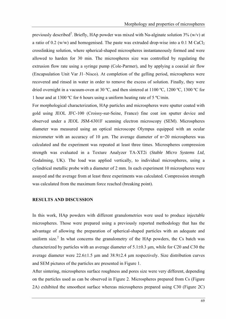
Morphology and properties of microspheres
69
previously described2. Briefly, HAp powder was mixed with Na-alginate solution 3% (w/v) at
a ratio of 0.2 (w/w) and homogenised. The paste was extruded drop-wise into a 0.1 M CaCl2
crosslinking solution, where spherical-shaped microspheres instantaneously formed and were
allowed to harden for 30 min. The microspheres size was controlled by regulating the
extrusion flow rate using a syringe pump (Cole-Parmer), and by applying a coaxial air flow
(Encapsulation Unit Var J1–Nisco). At completion of the gelling period, microspheres were
recovered and rinsed in water in order to remove the excess of solution. Finally, they were
dried overnight in a vacuum-oven at 30 ºC, and then sintered at 1100 ºC, 1200 ºC, 1300 ºC for
1 hour and at 1300 ºC for 6 hours using a uniform heating rate of 5 ºC/min.
For morphological characterization, HAp particles and microspheres were sputter coated with
gold using JEOL JFC-100 (Croissy-sur-Seine, France) fine coat ion sputter device and
observed under a JEOL JSM-6301F scanning electron microscopy (SEM). Microspheres
diameter was measured using an optical microscope Olympus equipped with an ocular
micrometer with an accuracy of 10 µm. The average diameter of n=20 microspheres was
calculated and the experiment was repeated at least three times. Microspheres compression
strength was evaluated in a Texture Analyzer TA-XT2i (Stable Micro Systems Ltd,
Godalming, UK). The load was applied vertically, to individual microspheres, using a
cylindrical metallic probe with a diameter of 2 mm. In each experiment 10 microspheres were
assayed and the average from at least three experiments was calculated. Compression strength
was calculated from the maximum force reached (breaking point).
RESULTS AND DISCUSSION
In this work, HAp powders with different granulometries were used to produce injectable
microspheres. Those were prepared using a previously reported methodology that has the
advantage of allowing the preparation of spherical-shaped particles with an adequate and
uniform size.2 In what concerns the granulometry of the HAp powders, the Cs batch was
characterized by particles with an average diameter of 5.1±0.3 µm, while for C20 and C30 the
average diameter were 22.6±1.5 µm and 38.9±2.4 µm respectively. Size distribution curves
and SEM pictures of the particles are presented in Figure 1.
After sintering, microspheres surface roughness and pores size were very different, depending
on the particles used as can be observed in Figure 2. Microspheres prepared from Cs (Figure
2A) exhibited the smoothest surface whereas microspheres prepared using C30 (Figure 2C)

Chapter II
70
particles presented a more irregular surface and bigger pores (around 20 µm) between
particles.
B
05
10152025
0 20 40 60 80 100Particles diameter (µm)
Wt %
CAPTAL S
A D
05
10152025
0 20 40 60 80 100Particles diameter (µm)
Wt %
CAPTAL 20
C F
05
10152025
0 20 40 60 80 100Particles diameter (µm)
Wt %
CAPTAL 30
E
Figure 1. Size distribution of HAp particles and their SEM images. Pictures A, C and E show the
granulometric size distribution of Cs, C20 and C30 particles respectively. Pictures B, D and F show the SEM
photomicrographs of Cs, C20 and C30 particles, respectively.
The shape was also slightly affected by the particle size, with larger particles resulting in less
spherical microspheres. However, a spherical shape is desirable in order to increase the
injectability yield since that is the shape used as model for fluid transportation studies, and it
is known as the easiest way to carry high concentration of solid particles using a fluid
vehicle.6
B
100 µm
A
100 µm
C
100 µm Figure 2. The SEM images of microspheres. Photomicrograph A corresponds to a microsphere prepared using
Cs particles and microspheres showed in photomicrographs B and C were prepared using C20 and C30 particles
respectively.
The microspheres size is also an important factor to take into consideration. The literature
shows that microspheres with diameters around 500 µm are considered injectable.7

Morphology and properties of microspheres
71
450
500
550
600
not s
int.
1100
-1
1200
-1
1300
-1
1300
-6
Temperature (ºC)-time(h)
Dia
met
er (µ
m)
CAPTAL S CAPTAL 20 CAPTAL 30
Figure 3. Microspheres’ diameter change with sintering temperatures. Microspheres prepared from C30
particles show a bigger diameter than microspheres prepared using Cs or C20 particles. An increase of sintering
temperature corresponds to a decrease of microspheres diameter.
Microspheres prepared with C30 particles have a diameter higher than those prepared using
Cs or C20. However, all obtained microspheres during this study fall in the range 500 to 600
µm which correspond to the values found in literature. Figure 3 shows the diameters of
microspheres versus sintering temperature. As expected, not sintered (not sint.) microspheres
are the largest ones. After sintering, microspheres diameter decreases due to the alginate
burning and to the compactation (decrease of porosity/volume) commonly associated with the
sintering process. Thus, microspheres diameter is not only a function of the type of particles
used, but also a function of the sintering temperature. A narrow size distribution of
microspheres sizes was obtained for all the formulations. This is an important aspect since a
uniform size is expected to create homogeneous spaces between microspheres when packed at
the bone cavity whereas a wide size distribution or a bimodal size distribution will create
smaller spaces.
During the injection process microspheres integrity must be maintained. However, if
microspheres compression strength is lower than the tension used to extrude the injectable
material, they will most probably break down. The rupture forces of the microspheres
prepared in this study are presented in Figure 4. Those forces can be as higher as 4.30±0.31 N
and as lower as 0.07±0.01 N, depending on the state of the microspheres (sintered or not) and
also on the type of HAp particles used. After sintering at 1100 ºC, all microspheres presented

Chapter II
72
low rupture strength. Moreover, the strength of microspheres prepared from C20 or C30
particles could not be measured since the force necessary to break them was out of the range
of the texture analyzer used. The rupture force increased along with the sintering temperature.
A force as high as 3.79±0.76 N was needed to break microspheres prepared from Cs after
sintered at 1300 ºC for 6 hours.
4.30
0.16
0.35
2.19
3.793.24
0.30
0.55
2.15
0.07
0.0
0.1
1.0
10.0
n sin
t.
1100
-1
1200
-1
1300
-1
1300
-6Temperature (ºC)-time (h)
Rup
ture
forc
e (N
)
CAPTAL S CAPTAL 20 CAPTAL 30
Figure 4. Rupture force of microspheres prepared using different sintering temperatures. Microspheres’
strength increases with sintering temperature. Rupture force of microspheres prepared using C20 and C30
particles could not be measured for sintering temperatures below 1300 ºC.
That increase of rupture force resulted from the establishment of bridges (known as “necks”)
among the HAp particles during the sintering process. According to the literature, during an
injectable procedure the force reached on an area similar to the equatorial area of our
microspheres is below 0.30 N.8,9 Therefore microspheres prepared using Cs particles and
sintered at temperatures equal or superior to 1200 ºC as well as microspheres prepared using
C20 particles and sintered at 1300ºC had enough strength to be injected. However, the
injection process will probably be facilitated when using the microspheres with the
smoothness surface.
CONCLUSIONS
The use of HAp powders with different granulometries enabled the preparation of

Morphology and properties of microspheres
73
microspheres with different morphological structures and mechanical properties.
Microspheres prepared using HAp powders with higher granulometry needed higher sintering
temperature to achieve enough compression strength, and presented a more irregular surface.
Microspheres prepared using HAp powders with the lower granulometry presented a
homogeneous size distribution (around 500 µm) and its strength was 0.35 N after sintering at
1200 ºC for 1 hour which make them the most suitable microspheres to use in our approach.
ACKNOWLEDGEMENTS
SM Oliveira is grateful to the Program for Education Development in Portugal III (PRODEP
III) for supporting his salary at Escola Superior de Tecnologia de Viseu, Portugal. This work
was supported by Foundation for Science and Technology (FCT) under contract
POCTI/FCB/41523/2001.
REFERENCES
1. Barrias CC, Ribeiro CC, Lamghari M, Miranda CS, Barbosa MA. Proliferation, activity, and osteogenic
differentiation of bone marrow stromal cells cultured on calcium titanium phosphate microspheres.
Journal of Biomedical Materials Research 2005;72A(1):57-66.
2. Barrias CC, Ribeiro CC, Barbosa MA. Adhesion and proliferation of human osteoblastic cells seeded on
injectable hydroxyapatite microspheres. Bioceramics, Vol 16 2004;254-2:877-880.
3. Ribeiro CC, Barrias CC, Barbosa MA. Calcium phosphate-alginate microspheres as enzyme delivery
matrices. Biomaterials 2004;25(18):4363-73.
4. Silva RV, Camilli JA, Bertran CA, Moreira NH. The use of hydroxyapatite and autogenous cancellous
bone grafts to repair bone defects in rats. International Journal of Oral and Maxillofacial Surgery
2005;34(2):178-184.
5. Boyan BD, Lossdörfer S, Wang L, Zhao G, Lohmann CH, Cochran DL, Schwartz Z. Osteoblasts
generate an osteogenic microenvironment when grown on surfaces with rough microtopographies. Eur
Cell Mater 2003;6:22-27.
6. Cardoso ON, Sotto Mayor T, Pinto AMFR, Campos JBLM. Axial dispersion of particles in a slugging
column--the role of the laminar wake of the bubbles. Chemical Engineering Science 2003;58(18):4159-
4172.
7. Gauthier O, Bouler JM, Weiss P, Bosco J, Aguado E, Daculsi G. Short-term effects of mineral particle
sizes on cellular degradation activity after implantation of injectable calcium phosphate biomaterials and
the consequences for bone substitution. Bone 1999;25(2):71S-74S.
8. Xu HH, Weir MD, Burguera EF, Fraser AM. Injectable and macroporous calcium phosphate cement
scaffold. Biomaterials 2006;27(24):4279-87.

Chapter II
74
9. Ginebra MP, Rilliard A, Fernandez E, Elvira C, San Roman J, Planell JA. Mechanical and rheological
improvement of a calcium phosphate cement by the addition of a polymeric drug. J Biomed Mater Res
2001;57(1):113-8.

Chapter III
75 Submitted for publication, 2008
Optimization of polymeric solutions as vehicles for injectable
hydroxyapatite microspheres
S.M. Oliveira1,2,3, I.F. Almeida4, P.C. Costa4, C.C. Barrias2, M.R. Pena Ferreira4, M.F.
Bahia4, M.A. Barbosa2,3
1ESTV – Escola Superior de Tecnologia de Viseu, Dep. de Eng. Mecânica e Gestão Industrial, Campus
Politécnico de Repeses, 3504-510 Viseu, Portugal; 2INEB – Instituto de Engenharia Biomédica, Divisão de biomateriais, Rua do Campo Alegre 823, 4150-180
Porto, Portugal; 3FEUP – Faculdade de Engenharia Universidade do Porto, Dep. de Eng. Metalúrgica e de Materiais, Rua
Roberto Frias, 4200-465 Porto, Portugal; 4FFUP – Faculdade de Farmácia da Universidade do Porto, Departamento de Tecnologia Farmacêutica, Rua
Aníbal Cunha 164, 4050-047 Porto, Portugal.
ABSTRACT
Injectable materials for bone defects fillers can be based in the combination of a vehicle and a
reinforcement phase. Therefore, the properties of the vehicle should be suitable to enable the
transport of that extra phase. Additionally, the use of biocompatible materials is a requirement
for tissue regeneration. Thus, we intended to optimize a biocompatible vehicle able to carry
hydroxyapatite microspheres into bone defects using an orthopaedic injectable device. To
achieve that goal, polymers usually regarded as biocompatible were selected, namely sodium
carboxymethylcellulose, hydroxypropylmethylcellulose and sodium alginate (ALG). The
rheological properties of the polymeric solutions at different concentrations were assessed by
viscosimetry before and after moist heat sterilization. In order to correlate rheological
properties with injectability, solutions were tested using an orthopaedic device applied for
minimal invasive surgeries. Among the three polymers, ALG solutions presented the most
suitable properties for our goal and a non-sterile ALG 6% solution was successfully used to
perform preliminary injection tests of hydroxyapatite microspheres. Sterile ALG 7.25%
solution was found to closely match non-sterile ALG 6% properties and it was selected as the
optimal vehicle. Finally, sterile ALG 7.25% physical stability was studied at different
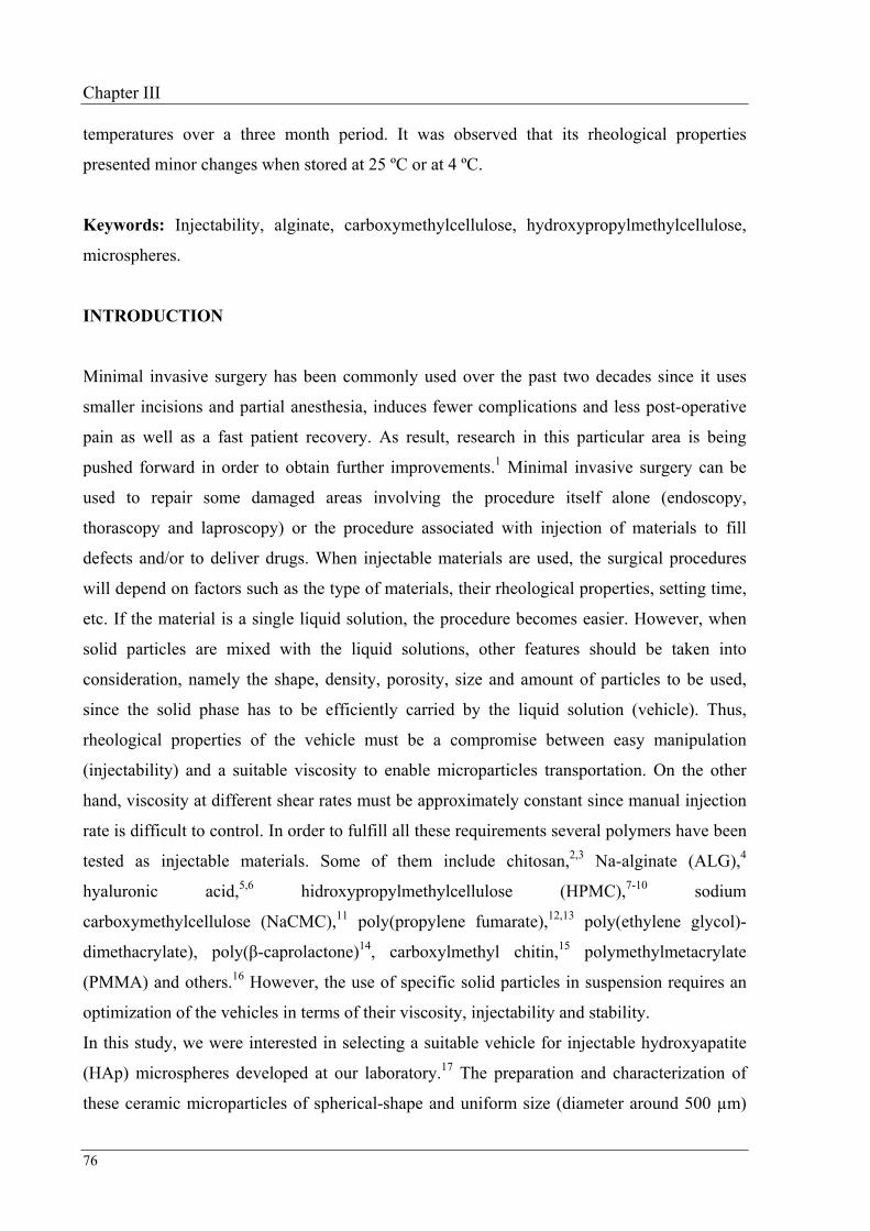
Chapter III
76
temperatures over a three month period. It was observed that its rheological properties
presented minor changes when stored at 25 ºC or at 4 ºC.
Keywords: Injectability, alginate, carboxymethylcellulose, hydroxypropylmethylcellulose,
microspheres.
INTRODUCTION
Minimal invasive surgery has been commonly used over the past two decades since it uses
smaller incisions and partial anesthesia, induces fewer complications and less post-operative
pain as well as a fast patient recovery. As result, research in this particular area is being
pushed forward in order to obtain further improvements.1 Minimal invasive surgery can be
used to repair some damaged areas involving the procedure itself alone (endoscopy,
thorascopy and laproscopy) or the procedure associated with injection of materials to fill
defects and/or to deliver drugs. When injectable materials are used, the surgical procedures
will depend on factors such as the type of materials, their rheological properties, setting time,
etc. If the material is a single liquid solution, the procedure becomes easier. However, when
solid particles are mixed with the liquid solutions, other features should be taken into
consideration, namely the shape, density, porosity, size and amount of particles to be used,
since the solid phase has to be efficiently carried by the liquid solution (vehicle). Thus,
rheological properties of the vehicle must be a compromise between easy manipulation
(injectability) and a suitable viscosity to enable microparticles transportation. On the other
hand, viscosity at different shear rates must be approximately constant since manual injection
rate is difficult to control. In order to fulfill all these requirements several polymers have been
tested as injectable materials. Some of them include chitosan,2,3 Na-alginate (ALG),4
hyaluronic acid,5,6 hidroxypropylmethylcellulose (HPMC),7-10 sodium
carboxymethylcellulose (NaCMC),11 poly(propylene fumarate),12,13 poly(ethylene glycol)-
dimethacrylate), poly(β-caprolactone)14, carboxylmethyl chitin,15 polymethylmetacrylate
(PMMA) and others.16 However, the use of specific solid particles in suspension requires an
optimization of the vehicles in terms of their viscosity, injectability and stability.
In this study, we were interested in selecting a suitable vehicle for injectable hydroxyapatite
(HAp) microspheres developed at our laboratory.17 The preparation and characterization of
these ceramic microparticles of spherical-shape and uniform size (diameter around 500 µm)

Optimization of a vehicle for injectable microspheres
77
have been previously described, and in vitro studies revealed that they can be used as supports
for culturing osteoblastic-like cells, suggesting their applicability as cell microcarriers for
bone regeneration applications18. However, in order to display adequate handling properties
and to enable minimally invasive implantation, the microspheres must be combined with a
vehicle. To accomplish our goal, we tested three polymers (ALG, HPMC and NaCMC), in
terms of viscosity and injectability. Finally, ALG was selected as the most promising solution
and its stability was evaluated over a three month period storage at different temperatures.
MATERIALS AND METHODS
Materials
Sodium alginate (Protanal 10/60LS) with a high α-L-guluronic acid content (65–75%, as
specified by the manufacturer) was kindly donated by Pronova Biopolymers, Norway and
used without further purification. Alginate solutions were freshly prepared as needed, using
deionised water. HPMC (Methocel E4M Premium) was a gift from Colorcon, USA and
NaCMC was purchased from Guinama, Spain. Hydroxyapatite was purchased from Plasma
Biotal, Ltd., UK. All products were of pharmaceutical grade.
Preparation of the polymer solutions
Sodium alginate was dissolved in deionised water for 24 hours in order to obtain a
homogeneous dispersion. The cellulose derivatives were dispersed in deionised water under
mechanical agitation.
Preparation of the microspheres
Hydroxyapatite microspheres were optimized in our laboratory in order to fill all the
requirements for injectability.19,20 Briefly, a suspension of HAp particles in an ALG 3% (w/v)
solution was extruded drop-wise into CaCl2 solution followed by sintering at 1200 ºC for 1
hour. After sintering, spherical-shaped microparticles with a uniform size were obtained as
depicted in Figure 1. The microspheres diameter and compression strength were found to be
around 500 µm and 0.35 N, respectively, as described in detail previous chapter.17

Chapter III
78
500 µm
A
Figure 1. HAp microspheres sintered at 1200 ºC for 1 hour. The picture was obtained by optical microscope,
showing the homogeneous size and sphericity of microspheres after sintering.
Evaluation of the rheological behaviour
Flow assay
The rheological behaviour was assessed by performing flow measurements at 20 ºC using a
viscometer Viscotester VT550 (ThermoElectron, UK), fitted with concentric cylinder
geometry (SV-DIN), between 1 and 1000 s-1 (unless high viscosity did not allowed it) and
backwards, with a 60 s delay period between measurements. The experimental results were
fitted with Power law model nk γτ &×= and consistency, k, and flow index, n, were
determined. In Power law equation τ is the shear stress and γ& is the shear rate.21
Injectability (extrusion) assay
The injectability of the mixtures was evaluated using an injection device (LP2 Stainless Steel
Delivery System - Biomet, Portugal) commonly used in vertebroplasty surgical procedures,
which consisted of a plastic syringe (20 mm internal diameter), a cannula (2.7 mm internal
diameter and 173 mm length) and a polymeric connection tube (Figure 2). The syringe was
filled up with the polymeric solution and the whole device was mounted on a Texture
Analyzer TA-XT2i (Stable Mycro Systems, UK) working in compression mode. During
extrusion tests, samples were assayed in triplicate applying the force vertically and using a

Optimization of a vehicle for injectable microspheres
79
plunger displacement rate of 1 mm/s. Results were expressed as the force needed to push the
solution out from the cannula. Some preliminary tests using non-sterile ALG 6% (NS-ALG
6%) mixed with 20% or 40% (w/w) of microspheres were also performed.
The effects of the ALG concentration and sterilization on the mechanical properties were
statistically evaluated by one-way ANOVA. Post hoc comparisons of the means of individual
groups were performed using Tukey’s Honestly Significant Difference test. A value of p=0.05
was taken to denote significance. Statistical analysis was performed with SPSS 15.0 for
Windows software (SPSS Inc., Chicago, IL, USA).
needle
syringe
force
vehicle
B
A
Figure 2. Injectability device. Schematic representation of the procedure and device used to evaluate the
injectability of the polymers. Injectability device scheme (A) and image of the injectability device (B).
Influence of sterilization on the rheological behaviour
Sterilization was carried out in an autoclave (15 minutes at 121 ºC) and the evaluation of the
mechanical properties was accessed as described previously (injectability assay) after a
storage period of 24 hours at 20 ºC.
Influence of sterilization on alginate molecular weight
Molecular weight was characterized at room temperature by High Performance Size

Chapter III
80
Exclusion Chromatography (HP-SEC) using a modular system, composed of an isocratic
pump (K-1001 Knaeur), a vacuum degasser (K-5002 Knaeur), a viscometer/right angle laser
light scattering (RALLS) dual detector (T60 Viscotek), and a refractive index detector (K5002
Knaeur) operating at the same wavelength as the RALLS detector (670 nm). Separations were
performed in a set of PL aquagel-OH mixed columns. The mobile phase consisted of 0.1 M
NaNO3 with 0.02% (w/v) NaN3 and the flow-rate was maintained at 1.0 ml/min. Samples
were dissolved in the mobile phase at 1 mg/ml, sterilised, filtered and injected through a
manual injection valve equipped with a 116 µl loop.
Evaluation of the physical stability
The physical stability of the sterile ALG 7.25% (S-ALG 7.25%) solution was studied after
storage for three months at 4 ºC, 25 ºC and 40 ºC. Rheological properties were assessed on
three batches performing flow measurements at 20 ºC using a viscometer (Viscotester VT550,
ThermoElectron, UK) fitted with concentric cylinder geometry, in the shear rate range from 1
to 500 s-1 and backwards, with 60 s delay period between measurements.
RESULTS AND DISCUSSION
Evaluation of the rheological behaviour
When polymeric solutions are used in injectable systems their rheological behaviour becomes
one of the most important properties to be studied. In this work, the rheograms obtained
showed that all polymeric solutions presented shear thinning behaviour characterized by a
decrease of viscosity for an increase of shear rate (Figure 3). The parameters of the Power law
model are presented in Table I. The flow index, n, varies from 0.140 to 0.834. The polymeric
solution with higher viscosity was NaCMC 4%.
An increase in consistency, k, following an increase in concentration was observed in all
cases. Increasing the concentration of NaCMC and HPMC solutions, the rheological
behaviour showed a trend to be more shear-thinning (lower n values) whereas ALG solutions
presented a behaviour closer to Newtonian fluid (n=1), showing a decrease in viscosity only
at medium to high shear rates. This behaviour did not change with concentration as flow
indexes remained approximately constant.
The polymeric solutions did not present thixotropy (represented by a hysteresis area between
up and down curves) with the exception of the NaCMC solutions at higher concentrations.

Optimization of a vehicle for injectable microspheres
81
0.1
1
10
100
1000
1 10 100 1000Shear rate (1/s)
Vis
cosi
ty (P
a.s)
NaCMC 1% NaCMC 2%
NaCMC 4% Alginato 2%
Alginato 4% Alginato 6%
Alginato 8% Alginate 10%
HPMC 2% HPMC 4%
Figure 3. Rheograms of polymeric solutions at different concentrations (T=20 ºC). NaCMC solutions
showed a decrease of viscosity for the whole range of shear rates tested while most of the Na-alginate solutions
presented a viscosity approximately constant at shear rates below 30 s-1.
Low modification of the mechanical properties after sterilization was found for cellulose
derivatives solutions (Figures 4 and 5). In contrast, the ALG solutions showed a marked
decrease of viscosity (Figure 6).
Table I. Parameters of the power law model.
Product Concentration (% w/w) K n R2
1 1.05 0.623 0.9987
2 51.63 0.279 0.9939 NaCMC
4 337.83 0.140 0.989
2 14.32 0.480 0.9928 HPMC
4 126.33 0.391 0.9850
2 0.34 0.834 0.999
4 4.69 0.669 0.9964
6 20.47 0.614 0.995
8 37.95 0.704 0.9972
ALG
10 74.62 0.695 0.9977
k - consistency, n - flow index, and R2- R-squared value.
Chapter III
82
Injectability
In order to evaluate the injectability, polymeric solutions were extruded through a specially
designed injection apparatus (Figure 2) and force versus distance values were recorded and
plotted (Figure 7). The curves show the evolution of applied force during polymeric solution
movement through the device.
0
1
10
100
1000
1 10 100 1000
Shear rate (1/s)
Vis
cosi
ty (P
a.s)
NaCMC 4% NaCMC 4% S
NaCMC 2% NaCMC 2% S
Figure 4. Effect of sterilization on NaCMC solutions viscosity. After sterilization, a decrease of 50% in
NaCMC solutions viscosity was detected for low shear rates, while at high shear rates the decrease was smaller.
Sterile solutions are represented by “S”.
0
1
10
100
1 10 100 1000
Shear rate (1/s)
Visc
osity
(Pa.
s)
HPMC 2% HPMC 2% S
HPMC 4% HPMC 4% S
Figure 5. Effect of sterilization on HPMC solutions viscosity. HPMC solutions viscosity was almost the same
before and after sterilization. Sterile solutions are represented by “S”.

Optimization of a vehicle for injectable microspheres
83
The evaluation of extrusion force showed that HPMC 2%, NaCMC 2% and ALG 2%
solutions were able to extrude at low forces, respectively 33.3±1.4 N, 35.4±3.8 N and
16.3±0.7 N (Mean±SD). Furthermore, ALG 4% and ALG 6% solutions were not extruded at
high forces (18.4±1.3 N and 34.3±0.8 N respectively) but HPMC 4% and NaCMC 4% needed
around 100 N to be extruded (123.5±7.4 N and 92.7±5.6 N respectively).
0
1
10
100
1000
1 10 100 1000
Shear rate (1/s)
Vis
cosi
ty (P
a.s)
Alginate 6% Alginate 6% S
Alginate 8% Alginate 8% S
Alginate 10% Alginate 10% S
Figure 6. Effect of sterilization on Na-alginate solutions viscosity. Alginate solutions viscosity dropped
sharply after sterilization. However, it is still approximately constant at low shear rates. Sterile solutions are
represented by “S”.
The injectability is highly dependent of viscosity as well as of the orthopedic device design as
can be observed from the extrusion curves (Figure 7). Each component of the device affects
the extrusion process. At the beginning, extrusion force increased in order to extrude the
solution out from the syringe into the connection tube and the maximum force did not go
above 20 N. As the connection tube was filled up (first plateau) the extrusion force becames
approximately constant. Once solution reached the cannula, extrusion force increased again
until the solution started to come out from it. Following this last slope, a second plateau was
observed (constant value of extrusion force). At high viscosities (HPMC 4% and NaCMC
4%), the influence of the cannula was marked by a high increase in the extrusion force.
Therefore, during a surgical procedure, the use of cannulas of smaller length and higher
diameter could eventually decrease the extrusion force needed to accomplish the injection
procedure.

Chapter III
84
0
20
40
60
80
100
120
140
0 5 10 15 20
Distance (mm)
Ext
rusi
on F
orce
(N)
NaCMC 2%
NaCMC 4%
ALG 2%
ALG 4%
ALG 6%
HPMC 2%
HPMC 4%
Figure 7. Curves of injectability of polymeric solutions after extrusion using the device shown in fig. 2. The
curves were drawn using the values recorded during extrusion process and correspond to different polymeric
solutions. The curves profile identifies the position of solutions inside of the device.
Both high and low viscosity solutions can be associated with poor injectability upon addition
of a solid phase. With high viscosity a high force has to be applied to extrude a mixture, while
for low viscosity solutions a phenomenon known as filtering will occur and solid particles
will not be pushed through the cannula.22 Furthermore, solutions with low viscosity may also
present the disadvantage of easily leaking and potentially invading blood vessels.23-26 On the
other hand, solution’s viscosity should be approximately constant at different shear rates
(close to a Newtonian fluid) since injection is usually performed manually (variable injection
rate). Among the studied vehicles, ALG solutions presented the closest behaviour to a
Newtonian fluid (n=1) and they were easily injected using the orthopaedic device above
described. Thus, NS-ALG 6% was used to perform some preliminary tests, using both 20%
and 40% (w/w) of HAp microspheres (diameter of 535±35 µm). In those assays both mixtures
were able to be extruded; however, the mixtures containing 40% (w/w) of microspheres were
only extruded at forces of 166±40 N while mixtures prepared with 20% (w/w) of
microspheres were extruded at 42±5 N. In the literature, values in the range 40 to 300 N (most
accepted 100N) have been found as forces suitable to perform material injections which
suggests that, in terms of rheological properties, NS-ALG 6% solution could be an
appropriate solution to work as an injectable vehicle.27-29

Optimization of a vehicle for injectable microspheres
85
Although viscosity of NS-ALG 6% proved, empirically, to be proper to keep microspheres in
suspension and to accomplish several injection assays, the sterilization method decreased its
viscosity and microspheres in suspension fall down. Therefore, new studies had to be
performed in order to find a suitable concentration that, after sterilization, presents rheological
properties similar to NS-ALG 6%.
In Figure 8 it is observed that the force to extrude sterile ALG 6% (S-ALG 6%) decrease to
about 20 N (18.7±0.6) as the result of sterilization. On the other hand, both sterile ALG 8%
and sterile ALG 10% needed higher force to be extruded, 47.5 ±2.6 N and 82.7±1.3 N
respectively.
0
10
20
30
40
50
60
70
80
90
6% NS 6% S 7.25% S 8% S 10% S
Na-Alginate Solution (w/w)
Ext
rusi
on F
orce
(N)
*
Figure 8. Extrusion force of Na-alginate solutions. Na-Alginate solutions were autoclaved at 121 ºC for
15min. “NS” represents a solution non-autoclaved and “S” represents autoclaved solutions. *Not significantly
different from 6%NS.
Besides injectability, high mechanical strength of the injected mixtures was also a goal in this
study. This objective can be accomplished by injecting a mixture combining the maximum
possible amount of ceramic microspheres. However, the extrusion of sterile ALG 8% and
sterile ALG 10% needed high extrusion forces, consequently a lower percentage of
microspheres would have to be used to enable extrusion at forces below 100 N. Therefore, we
focused on finding the alginate solution concentration with rheological properties similar to
those of NS-ALG 6%. The approach used was based in the correspondence between extrusion
forces for sterile and non-sterile ALG 6%, ALG 8% and ALG 10%. Using the regression
equations (Figure 9) we calculated that S-ALG 7.25% was the appropriate solution to replace

Chapter III
86
the NS-ALG 6%. To confirm our approach, injectability tests using S-ALG 7.25% were
performed and results showed that both S-ALG 7.25% and NS-ALG 6% presented similar
extrusion forces (Figure 8), which was also confirmed by the rheological tests. The statistical
tests showed that the ALG concentration influenced the extrusion force, and only the
extrusion forces found for S-ALG 7.25% and NS-ALG 6% were not significantly different
(p=0.662).
y = 0.1016x2.921
R2 = 0.9975
y = 0.8943x2.1246
R2 = 0.9948
0
20
40
60
80
100
120
140
0 2 4 6 8 10 12
Alginate concentration (%)
Ext
rusi
on fo
rce
(N)
Non-autoclaved
Autoclaved
Figure 9. Extrusion forces of Na-alginate after and before sterilization. Extrusion forces were measured on
both autoclaved and non-autoclaved ALG solutions. Sterile ALG concentration that matched NS-ALG 6%
extrusion force was computed from the regression equations of autoclaved and non-autoclaved solutions.
Influence of sterilization on the alginate molecular weight
Upon submission to moist heat sterilization (121ºC, 15 min), a decrease in the viscosity of
alginate solutions was observed, as discussed before. In order to understand this behaviour,
the molecular weight of those solutions was measured and a decrease of more than 15% was
observed. Before sterilization the molecular weight of alginate solutions was 85 kDa and after
sterilization it has dropped to 70 kDa. This behaviour was expected since moist heat
sterilization promotes polymer breakdown, resulting in the reduction of the average molecular
weight and consequently altering the rheological properties of the polymer solution.30
Evaluation of physical stability
A marked decrease on the viscosity of polymer solutions after storage at 40 ºC was found,

Optimization of a vehicle for injectable microspheres
87
while minor modifications were observed on the other test conditions (Figure 10). At this
temperature, a viscosity decrease was found even 1 day after preparation (data not shown), it
continued to decrease with time and after 3 months viscosity decreased more that four folds
(Figure 10). In contrast, at both 4 ºC and 25 ºC only a slight decrease of viscosity was
observed and no differences could be detected between 1 and 3 months.
0
10
20
30
40
50
60
70
80
90
100
4 ºC 25 ºC 40 ºC 4 ºC 25 ºC 40 ºC 4 ºC 25 ºC 40 ºC
t=0 1 month 2 month 3 month
Vis
cosi
ty c
hang
e (%
)
Figure 10. Influence of storage time on S-ALG 7.25% solutions viscosity. The viscosity was highly affected
after stored at 40 ºC. After 1 month the viscosity decrease was notorious and continued for the 2nd and 3rd
months. At both 4 ºC and 25 ºC only a slight decrease of viscosity was observed but no differences could be
detected between 1 and 3 months.
Since both NS-ALG 6% and S-ALG 7.25% presented similar rheological behavior and S-
ALG 7.25% was stable for a period of at least three months when stored either at 25 ºC or at 4
ºC, it was chosen as the vehicle to perform future injectability tests using different
concentrations of HAp microspheres.
CONCLUSIONS
In order to use a polymeric solution as a vehicle capable of carrying solid particles, suitable
rheological and chemical properties should be reached. In this investigation we were looking
for a vehicle suitable to carry HAp microspheres through an orthopaedic device during an
injection procedure. Among the polymeric solutions studied, sodium alginate presented the
Chapter III
88
closest behaviour to a Newtonian fluid, so it was selected as the polymer for future studies.
NS-ALG 6% proved to be able to carry 40% (w/w) of HAp microspheres through an
orthopaedic device. Moreover, S-ALG 7.25% presented rheological properties similar to NS-
ALG 6%. For injection into a bone defect, sterility is a mandatory feature, and so S-ALG
7.25% was selected as the vehicle. Its rheological properties presented minor changes over a
three months period when stored either at 25 ºC or at 4 ºC. This behaviour is considered
acceptable for a pharmaceutical product of extemporaneous preparation.
ACKNOWLEDGEMENTS
SM Oliveira is grateful to the Program for Education Development in Portugal III (PRODEP
III) for supporting his salary at Escola Superior de Tecnologia de Viseu, Portugal. This work
was supported by Foundation for Science and Technology (FCT) under contract
POCTI/FCB/41523/2001.
REFERENCES
1. Verlaan JJ, Oner FC, Dhert WJ. Anterior spinal column augmentation with injectable bone cements.
Biomaterials 2006;27(3):290-301.
2. Chenite A, Chaput C, Wang D, Combes C, Buschmann MD, Hoemann CD, Leroux JC, Atkinson BL,
Binette F, Selmani A. Novel injectable neutral solutions of chitosan form biodegradable gels in situ.
Biomaterials 2000;21(21):2155-61.
3. Hoemann CD, Sun J, Legare A, McKee MD, Buschmann MD. Tissue engineering of cartilage using an
injectable and adhesive chitosan-based cell-delivery vehicle. Osteoarthritis Cartilage 2005;13(4):318-29.
4. Balakrishnan B, Jayakrishnan A. Self-cross-linking biopolymers as injectable in situ forming
biodegradable scaffolds. Biomaterials 2005;26(18):3941-3951.
5. Shu XZ, Ghosh K, Liu Y, Palumbo FS, Luo Y, Clark RA, Prestwich GD. Attachment and spreading of
fibroblasts on an RGD peptide-modified injectable hyaluronan hydrogel. J Biomed Mater Res
2004;68A(2):365-75.
6. Shu XZ, Ahmad S, Liu Y, Prestwich GD. Synthesis and evaluation of injectable, in situ crosslinkable
synthetic extracellular matrices for tissue engineering. J Biomed Mater Res A 2006;79(4):902-12.
7. Weiss P, Gauthier O, Bouler JM, Grimandi G, Daculsi G. Injectable bone substitute using a hydrophilic
polymer. Bone 1999;25(2 Suppl):67S-70S.
8. Trojani C, Weiss P, Michiels JF, Vinatier C, Guicheux J, Daculsi G, Gaudray P, Carle GF, Rochet N.
Three-dimensional culture and differentiation of human osteogenic cells in an injectable
hydroxypropylmethylcellulose hydrogel. Biomaterials 2005;26(27):5509-5517.
9. Virto MR, Frutos P, Torrado S, Frutos G. Gentamicin release from modified acrylic bone cements with
lactose and hydroxypropylmethylcellulose. Biomaterials 2003;24(1):79-87.

Optimization of a vehicle for injectable microspheres
89
10. Bodic F, Amouriq Y, Gayet-Delacroix M, Gauthier O, Bouler J-M, Daculsi G, Hamel L. Méthode nom
invasive d'evaluation d'un substitut osseux injectable / Non-invasive evaluation of an injectable bone
substitute. C. R. Biologies 2002;325:1-9.
11. Andrews GP, Gorman SP, Jones DS. Rheological characterisation of primary and binary interactive
bioadhesive gels composed of cellulose derivatives designed as ophthalmic viscosurgical devices.
Biomaterials 2005;26(5):571-80.
12. He S, Yaszemski MJ, Yasko AW, Engel PS, Mikos AG. Injectable biodegradable polymer composites
based on poly(propylene fumarate) crosslinked with poly(ethylene glycol)-dimethacrylate. Biomaterials
2000;21(23):2389-94.
13. Temenoff JS, Mikos AG. Injectable biodegradable materials for orthopedic tissue engineering.
Biomaterials 2000;21(23):2405-12.
14. Iooss P, Le Ray AM, Grimandi G, Daculsi G, Merle C. A new injectable bone substitute combining
poly(epsilon-caprolactone) microparticles with biphasic calcium phosphate granules. Biomaterials
2001;22(20):2785-94.
15. Uda H, Sugawara Y, Nakasu M. Experimental studies on hydroxyapatite powder-carboxymethyl chitin
composite: injectable material for bone augmentation. J Plast Reconstr Aesthet Surg 2006;59(2):188-96.
16. Carrodeguas RG, Lasa BV, Del Barrio JS. Injectable acrylic bone cements for vertebroplasty with
improved properties. J Biomed Mater Res B Appl Biomater 2004;68(1):94-104.
17. Oliveira SM, Barrias CC, Ribeiro CC, Almeida IF, Bahia MF, Barbosa MA. Morphology and mechanical
properties of injectable ceramic microspheres. Accepted for publication in Key Engineering Materials,
2008.
18. Barrias CC, Ribeiro CC, Barbosa MA. Adhesion and proliferation of human osteoblastic cells seeded on
injectable hydroxyapatite microspheres. Bioceramics, Vol 16 2004;254-2:877-880.
19. Ribeiro CC, Barrias CC, Barbosa MA. Preparation and characterisation of calcium-phosphate porous
microspheres with a uniform size for biomedical applications. J Mater Sci Mater Med 2006;17(5):455-63.
20. Oliveira SM, Barrias CC, Almeida IF, Costa PC, Ferreira MP, Bahia MF, Barbosa MA. Injectability of a
bone filler system based on hydroxyapatite microspheres and a vehicle with in situ gel-forming ability. J
Biomed Mater Res B Appl Biomater 2008;In Press.
21. Barnes HA, Hutton JF, Walters K. An Introduction to Rheology. Amsterdam: Elsevier Science; 1998.
22. Bohner M, Baroud G. Injectability of calcium phosphate pastes. Biomaterials 2005;26(13):1553-63.
23. Hide IG, Gangi A. Percutaneous vertebroplasty: history, technique and current perspectives. Clin Radiol
2004;59(6):461-7.
24. Lewis G. Injectable bone cements for use in vertebroplasty and kyphoplasty: State-of-the-art review.
Journal of Biomedical Materials Research Part B-Applied Biomaterials 2006;76B(2):456-468.
25. Baumann A, Tauss J, Baumann G, Tomka M, Hessinger M, Tiesenhausen K. Cement embolization into
the vena cava and pulmonal arteries after vertebroplasty: Interdisciplinary management. European Journal
of Vascular and Endovascular Surgery 2006;31(5):558-561.
26. Mathis JM, Wong W. Percutaneous vertebroplasty: Technical considerations. Journal of Vascular and
Interventional Radiology 2003;14(8):953-960.
27. Xu HH, Weir MD, Burguera EF, Fraser AM. Injectable and macroporous calcium phosphate cement

Chapter III
90
scaffold. Biomaterials 2006;27(24):4279-87.
28. Gisep A, Curtis R, Hanni M, Suhm N. Augmentation of implant purchase with bone cements: an in vitro
study of injectability and dough distribution. J Biomed Mater Res B Appl Biomater 2006;77(1):114-9.
29. Krebs J, Ferguson SJ, Bohner M, Baroud G, Steffen T, Heini PF. Clinical measurements of cement
injection pressure during vertebroplasty. Spine 2005;30(5):E118-22.
30. Draget KI, Skjak-Braek G, Smidsrod O. Alginate based new materials. Int J Biol Macromol 1997;21(1-
2):47-55.

CHAPTER IV
91 Journal of Biomedical Materials Research Part B: Applied Biomaterials, published online 2008
Injectability of a bone filler system based on hydroxyapatite microspheres
and a vehicle with in situ gel-forming ability
S.M. Oliveira1,2,3, C.C. Barrias2, I.F. Almeida4, P.C. Costa4, M.R. Pena Ferreira4, M.F.
Bahia4, M.A. Barbosa2,3
1ESTV – Escola Superior de Tecnologia de Viseu, Dep. de Eng. Mecânica e Gestão Industrial, Campus
Politécnico de Repeses, 3504-510 Viseu, Portugal; 2INEB – Instituto de Engenharia Biomédica, Divisão de biomateriais, Rua do Campo Alegre 823, 4150-180
Porto, Portugal; 3FEUP – Faculdade de Engenharia Universidade do Porto, Dep. de Eng. Metalúrgica e de Materiais, Rua
Roberto Frias, 4200-465 Porto, Portugal; 4FFUP – Faculdade de Farmácia da Universidade do Porto, Departamento de Tecnologia Farmacêutica, Rua
Aníbal Cunha 164, 4050-047 Porto, Portugal.
ABSTRACT
The aim of this study was to test the injectability of a bone filler system based on the
combination of ceramic microspheres with a gel-like vehicle, for minimal invasive surgery.
Porous hydroxyapatite microspheres with a uniform size and an average diameter of 535±38
µm were prepared, and their compression strength and friability were tested. The sodium-
alginate solution with a concentration of 7.25% (w/w) was used as the vehicle. To promote its
in situ gelation, calcium carbonate and D-gluconic-δ-lactone were added to the solution.
Microspheres were mixed with the vehicle at different percentages, 20% to 40% (w/w).
Gelation times in the range of 8 to 20 min were obtained, depending on the formulation.
Mixtures of HAp microspheres with alginate solution at 7.25% originating a gel in 11 min
present an adequate handling time to perform an injection. Their injectability was evaluated
using an injection device commonly employed in vertebroplasty surgical procedures, coupled
to a texturometer in compression mode. Using an extrusion rate of 0.1 mm/s, the force
required to extrude any of the mixtures tested was lower than 100 N. For an extrusion rate of
1 mm/s mixtures with 40% (w/w) of microspheres were very difficult to inject. Mixtures with
35% (w/w) of microspheres presented the best compromise between injectability and

Chapter IV
92
compression strength of the gelled system. MicroCT analysis revealed a homogeneous
distribution of the microspheres inside the vehicle, as well as full interconnection of the intra-
microspheres spaces. The compression strength for the gelled systems ranged from 80 kPa
(gel itself) to 600 kPa (composite with 40% of microspheres).
Keywords: Microspheres, hydroxyapatite, ceramic, hydrogel, alginate.
INTRODUCTION
Loss of bone has been a problem, especially for women after menopause since their capacity
to rebuild new bone is diminished and bone absorption is high, resulting in osteoporosis.
Other identical processes, like the loss of bone in Gaucher disease (type I), which is related to
the absence of the enzyme glucocerebrosidase,1,2 can occur and hence fractures and failure of
some bones takes place. Usually, those losses result in the collapse of some vertebra causing
pain and, in many cases, an anatomically normal posture cannot be maintained anymore.3 In
order to relieve the pain and to strengthen osteoporotic bones surgery is frequently performed.
The most common procedure consists of injecting and filling up the inside of a vertebral body
with a polymer, polymethylmethacrylate (PMMA), using minimum invasion surgery such as
percutaneous vertebroplasty (PVP) or kyphoplasty. These are fast procedures and allow an
immediate pain relief and patient’s recovery in few days.4-6 However, some problems remain
unsolved: PMMA cannot be absorbed, does not allow or induce bone regeneration, has a high
setting temperature7 and its high hardness and strength after setting is associated with new
cracks in surrounding bone and fracture on adjacent vertebras.8-11 Taking into consideration
all the advantages associated with the PVP, we intend to use the same procedure to inject a
different material (composite) to overcome some of the difficulties related to the use of
PMMA. One of the strategies is to test the injectability of an alginate solution mixed with
ceramic microspheres of a uniform size that will gelify in situ within a few minutes at body
temperature and may be loaded with drugs and/or osteoblastic or osteoprogenitor cells to
customize for specific applications. In this way, drugs could be released in situ, allowing the
treatment of some diseases, and the cells could induce new bone formation.1,12
As a component of seaweed, alginate is a natural and abundant polymer which has been
described by several authors as a biocompatible and biodegradable material (ultra-pure
grade).13-19 When used as a solution, alginate can easily be crosslinked in situ using calcium

Injectability of a bone filler system based on HAp microspheres
93
or some other divalent positive ions.20-22 Some of the applications where it has been used are
as a vehicle for drug delivery and as a scaffold for tissue engineering, either as porous
structures or modified with RGD-containing peptide sequences.1,12,23 Among others polymers
that have been studied in this work, alginate was the one that had least variation in viscosity in
the range of shear rates applied in this study.
In the system described in this paper a ceramic phase has been used to reinforce the calcium
alginate phase. Hydroxyapatite (HAp) is one of the ceramics that can be used as
reinforcement because it has good strength and an excellent biocompatibility due to its
chemical similarity with the mineral phase of hard tissues.23,24
The injection of ceramic materials into a bone cavity is not a new concept; however, a
literature search revealed that most of the works used particles with an irregular shape, a
broad size distribution and too large or too small dimensions.25-29 Since spherical particles
with narrow size distribution can create more homogeneous interparticle spaces, as well as
being easier to inject, we have produced porous HAp microspheres with an average diameter
of 535±38 µm (mean±SD). The injectability tests were performed using a device applied in
PVP, in order to increase the similarity with surgical procedures.
MATERIALS AND METHODS
Materials
Commercial hydroxyapatite (HAp) (Captal S, Plasma Biotal) was used to produce
microspheres. Granulometric analysis of the powder was performed using a laser scanner
particle size analyzer (Coulter Electronics Incorporation), and a particle average diameter of
5.1±0.3 µm was found. Pharmaceutical-grade sodium alginate (Na-alginate, Protanal
10/60LS) with a high α-L-guluronic acid content (65–75%, as specified by the manufacturer)
was kindly donated by Pronova Biopolymers and used without further purification. Na-
alginate solutions were freshly prepared as needed, using deionised water. Calcium carbonate
(CaCO3) powder and D-glucono-δ-lactone (GDL) were obtained from Sigma.
Preparation and characterization of HAp microspheres
Preparation
HAp microspheres were prepared as reported previously.1 Briefly, the HAp powder was
dispersed at a ratio of 0.2 (w/w) with 3% (w/v) Na-alginate aqueous solution under gentle
Chapter IV
94
stirring until a homogeneous paste was obtained. The paste was extruded drop-wise into a 0.1
M CaCl2 crosslinking bath, where spherical-shaped particles were instantaneously formed and
allowed to harden for 30 min. The particle size was controlled by regulating the extrusion
flow rate using a syringe pump (Cole-Parmer), and by applying a coaxial air flow
(Encapsulation Unit Var J1–Nisco). At completion of the gelling period the microspheres
were recovered and rinsed in water in order to remove the excess of CaCl2. Finally, they were
dried overnight in a vacuum oven at 37 ºC, and then sintered at 1200 °C for 1 h, with a
uniform heating rate of 5 °C/min from room temperature.
Morphological characterization (SEM and dimensional analysis)
Morphological characterization of the microspheres was carried out using a stereomicroscope
(Olympus SZX9) and a scanning electron microscopy (SEM). For SEM, samples were sputter
coated with gold using a JEOL JFC-100 fine coat ion sputter device and observed using a
JEOL JSM-6301F SEM. The diameter was measured using an inverted plate microscope
(Olympus) equipped with an ocular micrometer with an accuracy of 10 µm. The average
diameter of n=20 microspheres was calculated and the experiment was repeated at least three
times.
Physicochemical characterization (XRD, FTIR)
For Fourier transform infrared spectroscopy (FTIR) microspheres were reduced to powder
and analysed as KBr pellets using a Perkin Elmer System 2000 spectrometer. For X-ray
diffraction (XRD), microspheres were reduced to powder and analysed with CuKα radiation
using a Rigaku PMG-VH diffractometer.
Mechanical properties (friability and compression strength)
Friability tests were performed according to a procedure described in the European
Pharmacopeia (5th edition) with minor modifications. Briefly, 2 g of HAp-microspheres were
loaded into a drum (SOTAX/F1) operating at 25 rpm. The fall height was 150 mm and the
same microspheres were used for 3 cycles of 4 min each. After each cycle, the desegregated
powder was blown out and the microspheres were collected and weighted again. Friability is
reported as percentage of total weigh lost.
Microsphere compression strength was evaluated in a Texture Analyzer (TA-XT2i, Stable
Micro Systems Ltd). The load was applied vertically, to individual microspheres, using a

Injectability of a bone filler system based on HAp microspheres
95
cylindrical metallic probe with a diameter of 2 mm. In each experiment 10 microspheres were
assayed and the average from at least three experiments was calculated. Compression strength
was calculated from the maximum force reached (breaking point).
Preparation and characterization of the alginate vehicle
Preparation
Preliminary studies using Na-alginate, carboxymethylcellulose and hydroxypropylmethyl
cellulose solutions at different concentrations, before and after sterilization, were carried out
(previous chapter). From those studies, a Na-alginate solution with a concentration of 7.25%
(w/w) was chosen as the most adequate vehicle. It was obtained by dissolving Na-alginate in
deionised water for 24 hours in order to obtain a homogeneous solution, and then sterilizing in
an autoclave according to the standard pharmacopoeia procedure (15 min at 121 ºC).
Characterization of the molecular weight and viscosity
The molecular weigh of Na-alginate, before and after sterilization, was calculated by high
performance-size exclusion chromatography (HP-SEC). The analysis was performed at room
temperature using a modular system, composed of an isocratic pump (K-1001 Knaeur), a
vacuum degasser (K-5002 Knaeur), a viscometer/right angle laser light scattering (RALLS)
dual detector (T60 Viscotek), and a refractive index detector (K-5002 Knaeur) operating at
the same wavelength as the RALLS detector (670 nm). Separations were performed in a set of
PL aquagel-OH mixed columns. The mobile phase consisted of 0.1 M NaNO3 with 0.02%
(w/v) NaN3 and the flow-rate was maintained at 1.0 ml/min. Samples were dissolved in the
mobile phase at 1 mg/ml, filtered and injected through a manual injection valve equipped with
a 116 µl loop.
The viscosity of Na-alginate solutions was assessed by performing flow measurements at 20
ºC using a viscometer (Viscotester VT550, ThermoElectron, UK), fitted with concentric
cylinder geometry, between 1 and 100 s-1 and backwards, with a 60 s delay period between
measurements. All measurements were performed in triplicate.
Gel formation
Internal gelation of the 7.25% (w/w) Na-alginate solution was promoted using a method
previously described by Kuo et al.30 with minor modifications. Briefly, a calcium salt with
limited solubility, in this case CaCO3, was mixed with the alginate solution and used as a

Chapter IV
96
source of calcium ions. The release of Ca2+ into the solution was promoted by the generation
of an acidic pH with D-glucono-δ-lactone (GDL), a slowly dissociating acid, which was also
incorporated in the solution. Once released, Ca2+ ions can participate in the interchain ionic
binding between carboxyl groups (COO−) of guluronic acids blocks in the polymer chain,
giving rise to a crosslinked gel.
To obtain a sterile gel, the CaCO3 powder was previously autoclaved (15 min at 121 ºC),
while GDL aqueous solutions were filtered through a 0.22 µm membrane filter and used
immediately after preparation. The CaCO3/GDL ratio was set at 0.5 to yield a neutral pH
value.30 All components were pre-equilibrated at room temperature (set at 20 ºC) before being
mixed. An aqueous CaCO3 suspension was added to the Na-alginate solution, which was
mixed and vortexed for 1 min and allowed to equilibrate at 20 ºC for 15 min. Several
Ca2+/COO− ratios were tested (0.180, 0.216, 0.252, 0.288, 0.324 and 0.360). To initiate gel
formation, a fresh GDL solution was subsequently added to each mixture and vortexed for 20
sec. Immediately after, the mixtures were poured into test tubes, incubated at 20 ºC in water
bath and allowed to gelify. The gelation time was calculated according to the procedure of
Oakenfull et al.31 It was defined as the time required for the mixtures to form a gel just strong
enough to remain held in position upon inversion of the test tubes. All samples were assayed
in triplicate.
Preparation and characterization of the microspheres-vehicle system
Preparation of the system
Microspheres were added to the vehicle (Na-alginate with CaCO3 and GDL) and the mixture
was carefully homogenised. Different percentages of microspheres – 20, 30, 35 and 40%
(w/w) – were tested. In this study, a Ca2+/COO− ratio of 0.288 was used, since preliminary
results showed that it promotes gelation in approximately 11 min. This was considered an
adequate handling period, giving the surgeon enough time to prepare, manipulate and inject
the system.
Osmolality
The osmolality (mOsm/kg) of extemporaneously prepared microspheres-vehicle mixtures was
measured in an osmometer (Semi-Micro Osmometer, Knauer).

Injectability of a bone filler system based on HAp microspheres
97
Injectability (extrusion) assay
The injectability of the mixtures was evaluated according to the scheme of Figure 1A. In the
procedure, an injection device commonly used in vertebroplasty surgical procedures was
used, which was coupled to a Texture Analyzer (Figure 1B). The device consisted of a plastic
syringe (20 mm internal diameter), a cannula (2.7 mm internal diameter, 173 mm length) and
a polymeric connection tube. The syringe was filled with extemporaneously prepared
microspheres-vehicle mixtures and the whole device was mounted on the Texture Analyzer
operating in the compression mode. For extrusion, the load was applied vertically using two
different plunger displacement rates (1 mm/s or 0.1 mm/s). Results are expressed as the force
needed to extrude the mixture out from the syringe. The percentage of injectability, defined as
the ratio between the mass of mixture expelled from the syringe and the total mass loaded,
was also calculated. All samples were assayed in triplicate.
needle
Syringe
Force
distance
Vehicle
wt % 20/30/35/40
HAp microspheres
Stirrer
Sterile alginate solution (7.25%)
CaCO3 + GDLA
Figure 1. Schematic representation of the procedure and device used to evaluate the injectability of the
microspheres-vehicle system. Global procedure (A), injectability device and texture analyzer TA-XT2i (B).
Gel formation
The microspheres-vehicle mixtures were extruded into cylindrical shape moulds, transferred
to an oven at 37 ºC under controlled humidity (to prevent dehydration) and incubated for 24 h.

Chapter IV
98
At the end, crosslinked systems with a cylindrical shape were obtained.
Characterization of the gelled systems (Compression strength and MicroCT)
The compression strength of the systems obtained after 24 hours of gelation was calculated
with a Texture Analyzer, with the load applied vertically using a cylindrical probe with a
diameter of 4 mm. In each cylindrical composite, three compression tests were performed and
the force profile curves were used to estimate the rupture force and the associated
compression strength.
The spatial distribution of the microspheres in the gelled systems was analysed by micro
computed tomography (MicroCT, Scanco Med), using a resolution of 10 µm scans and a
thresholding range of 260 to 1000 to yield three-dimension (3D) reconstruction. Only
crosslinked systems prepared with 35% (w/w) microspheres and extruded at 0.1 mm/s were
used in this test.
A
500 µm
CB
Figure 2. Photomicrographs of HA-microspheres. Image A was obtained with a stereomicroscope and
illustrates the spherical-shape of the microspheres and their uniform size. Images B and C were obtained by
SEM at different magnifications (150× and 2500× respectively).
RESULTS
Preparation and characterization of HAp-microspheres
HAp-microspheres were obtained from a HAp-alginate slurry by droplet extrusion into a
crosslinking bath containing Ca2+, where composite hydrogel beads instantaneously formed.
This was followed by a sintering stage (1 hour at 1200 ºC). During this process the polymer
was burned-off and the ceramic particles became associated yielding HAp-microspheres with
an average diameter of 535±38 µm (mean±SD, n=20), as assessed by optical microscopy.
Figure 2A illustrates the spherical-shape of the microspheres as well as their uniform size. At

Injectability of a bone filler system based on HAp microspheres
99
higher magnification it can be seen from Figure 2B and 2C that HAp-microspheres are porous
and present a rough surface.
-20
0
20
40
60
80
100
120
2θ (degrees)
Inte
nsity
(%) -
HA
pha
se
0
20
40
60
80
100
120
140
4 12 20 28 36 44 52 60 68 76
Int
ensi
ty (%
) - S
inte
red
mic
rosp
here
s
HA phase
Sintered microspheres
(101
)
(112
)
(323
)(310
)
(210
)(1
02) (2
02)
(300
)
(200
)
(211
)
(111
)(0
02)
(222
) (213
)(3
12)
(321
)
(004
)
(502
)
(513
)(420
)
Figure 3. XRD spectra of the sintered HA-microspheres and peaks of the HA phase.
Physical-chemical characterization was carried out by XRD and FTIR. In terms of phase
constitution, the HAp-microspheres do not present other crystalline phases than those
observed in the original HAp powder, as observed by XRD (Figure 3). Moreover, the FTIR
characteristics bands present in the microspheres match the ones of the HAp powders (Figure
4), confirming that the ceramic kept its integrity.
400
800
1200
1600
2000
2400
2800
3200
3600
4000
cm-1
0
10
20
30
40
50
60
70
80
% T
HA powder
Sintered microspheres
Figure 4. FTIR spectra of the sintered HA-microspheres and of the original HA powder.
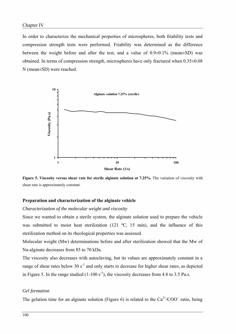
Chapter IV
100
In order to characterize the mechanical properties of microspheres, both friability tests and
compression strength tests were performed. Friability was determined as the difference
between the weight before and after the test, and a value of 0.9±0.1% (mean±SD) was
obtained. In terms of compression strength, microspheres have only fractured when 0.35±0.08
N (mean±SD) were reached.
Alginate solution 7.25% (sterile)
1
10
1 10 100
Shear Rate (1/s)
Vis
cosi
ty (P
a.s)
Figure 5. Viscosity versus shear rate for sterile alginate solution at 7.25%. The variation of viscosity with
shear rate is approximately constant.
Preparation and characterization of the alginate vehicle
Characterization of the molecular weight and viscosity
Since we wanted to obtain a sterile system, the alginate solution used to prepare the vehicle
was submitted to moist heat sterilization (121 ºC, 15 min), and the influence of this
sterilization method on its rheological properties was assessed.
Molecular weight (Mw) determinations before and after sterilization showed that the Mw of
Na-alginate decreases from 85 to 70 kDa.
The viscosity also decreases with autoclaving, but its values are approximately constant in a
range of shear rates below 30 s-1 and only starts to decrease for higher shear rates, as depicted
in Figure 5. In the range studied (1-100 s-1), the viscosity decreases from 4.8 to 3.5 Pa.s.
Gel formation
The gelation time for an alginate solution (Figure 6) is related to the Ca2+/COO− ratio, being
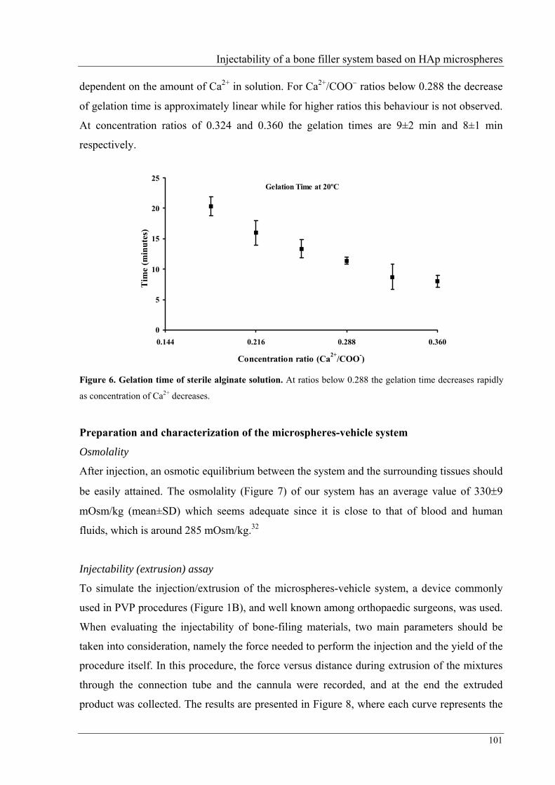
Injectability of a bone filler system based on HAp microspheres
101
dependent on the amount of Ca2+ in solution. For Ca2+/COO− ratios below 0.288 the decrease
of gelation time is approximately linear while for higher ratios this behaviour is not observed.
At concentration ratios of 0.324 and 0.360 the gelation times are 9±2 min and 8±1 min
respectively.
Gelation Time at 20ºC
0
5
10
15
20
25
0.144 0.216 0.288 0.360
Concentration ratio (Ca2+/COO-)
Tim
e (m
inut
es)
Figure 6. Gelation time of sterile alginate solution. At ratios below 0.288 the gelation time decreases rapidly
as concentration of Ca2+ decreases.
Preparation and characterization of the microspheres-vehicle system
Osmolality
After injection, an osmotic equilibrium between the system and the surrounding tissues should
be easily attained. The osmolality (Figure 7) of our system has an average value of 330±9
mOsm/kg (mean±SD) which seems adequate since it is close to that of blood and human
fluids, which is around 285 mOsm/kg.32
Injectability (extrusion) assay
To simulate the injection/extrusion of the microspheres-vehicle system, a device commonly
used in PVP procedures (Figure 1B), and well known among orthopaedic surgeons, was used.
When evaluating the injectability of bone-filing materials, two main parameters should be
taken into consideration, namely the force needed to perform the injection and the yield of the
procedure itself. In this procedure, the force versus distance during extrusion of the mixtures
through the connection tube and the cannula were recorded, and at the end the extruded
product was collected. The results are presented in Figure 8, where each curve represents the

Chapter IV
102
evolution of the force during extrusion for a particular formulation. At the end, a plateau is
observed, corresponding to the point at which the mixtures start being expelled from the
cannula. Since pressure does not subside immediately, the mixtures still continued to flow out
from the cannula, even after the force had been stopped.
250
270
290
310
330
350
370
390
15 20 25 30 35 40 45
Microspheres (wt %)
Osm
olal
ity (m
Osm
/kg)
Figure 7. Osmolality of mixtures. Osmolality was measured by comparing the freezing points of pure water and
the sample using 1 g of each mixture prepared just before measurement. The values among the samples are
approximately 330 mOsm/kg.
Figure 8 shows that only mixtures with 40% and 35% (w/w) of microspheres extruded at 1
mm/s needed the application of forces higher than 100 N (reference line), but even for those
mixtures the extrusion force does not go above 200 N. On the other hand, if such mixtures are
extruded at 0.1 mm/s the extrusion pressure falls below the reference line, to 233 and 153 kPa
respectively (Figure 9).
The yields of injection (% of injectability) found for each of the mixtures and rates of
extrusion respectively, are depicted in Table I. As can be observed, the percentage of
injectability presents a large range of values among the mixtures studied in this work. The
highest percentage of injectability (83.3%) was found for a mixture with 20% (w/w) of
microspheres, whereas the lowest (18.1%) was found for mixtures with 40% (w/w), both
extruded at 1 mm/s.

Injectability of a bone filler system based on HAp microspheres
103
0
20
40
60
80
100
120
140
160
180
200
0 5 10 15 20
Distance (mm)
Ext
rusi
on fo
rce
(N)
a
e'
e
c
d'b
d
a'
reference line
a - 0%-(1mm/s)a' - 0%-(0.1mm/s)b - 20%-(1mm/s)c - 30%-(1mm/s)d - 35%-(1mm/s)d' - 35%-(0.1mm/s)e - 40%-(1mm/s)e' - 40%-(0.1mm/s)
Figure 8. Extrusion force of different microspheres/alginate composites. During extrusion, the profile of
curves identifies the position where the mixture is in each moment. The first slope is the beginning of extrusion
and corresponds to the force need to extrude the mixture out from the syringe into the connection tube which will
be filled up at extrusion force approximately constant. Once mixture reached the cannula, extrusion force
increases again until mixture starts to come out from it. Following the slope is observed a decrease or constant
extrusion force.
104
163
289
365
641
47
153
233
0
100
200
300
400
500
600
700
0 20 30 35 40
Microspheres (wt%)
Ext
rusi
on P
ress
ure
(kPa
)
1 mm/s 0.1 mm/s
reference line (318 kPa /100 N)
Figure 9. Extrusion pressure of different microspheres/alginate composites. According to the syringe used,
a force of 100 N corresponds to a pressure of 318 N, our reference line. Mixtures extruded at 0.1 mm/s
experienced a pressure decrease of more than 50%.

Chapter IV
104
Mixtures with 35% (w/w) of microspheres presented an injectability of 76.4 % at an extrusion
rate of 0.1 mm/s, while mixtures with 40% (w/w) of microspheres extruded at 0.1 mm/s
present an injectability similar to that of mixtures with 30% (w/w) of microspheres extruded
at 1 mm/s.
Table I. Injectability obtained for different formulations and extrusion rates.
Microspheres
(%)
Extrusion
rate (mm/s)
Injectability
(%)
20 1 83.3
30 1 56.9
1 40.3 35
0.1 76.4
1 18.1 40
0.1 55.6
Characterization of the gelled systems (Compression strength and MicroCT)
MicroCT scans performed on composites with 35% (w/w) of microspheres extruded at 0.1
mm/s show a homogeneous distribution of the microspheres across the gel (Figure 10). Figure
10A is the result of a three dimensional (3D) reconstruction of the microCT scans and shows
Figure 10. MicroCT analysis of composites prepared with 35% of microspheres. Composites obtained after
mixtures (matrix – alginate and reinforcement phase – HAp microspheres) gelification; picture A0 represent a
composite prepared using 35% of microspheres. Scanning under microCT and three dimensional (3D)
reconstructions were performed using a threshold in the range of 255 to 1000. Picture A shows microspheres
distribution on composite. Picture B and C correspond to layers took at different depths inside the composite.

Injectability of a bone filler system based on HAp microspheres
105
microspheres distribution on the composite. Figures 10B and 10C correspond to layers taken
at different depths inside the composite using the same parameters.
The compression strength of the gelled systems was measured and the results are presented in
Figure 11. For an extrusion rate of 0.1 mm/s, the compression strength increased from ≅90
kPa to ≅600 kPa, as the percentage of microspheres increased from 0 to 40% (w/w). For an
extrusion rate of 1 mm/s, the compression strength is somewhat lower since lower extrusion
rates allow better accommodation of microspheres and at same time generate less residual
stress on alginate chains during gelification.
DISCUSSION
In this study the injectability of a bone filler system based on hydroxyapatite microspheres
and a vehicle with in situ gel-forming ability was evaluated.
Particles diameter and spherical shape are important parameters to take in consideration
during injection procedures. Therefore we have produced microspheres with diameters around
500 µm, which is the value considered acceptable according to Gauthier et al.33 Moreover,
mechanical properties of the microspheres are relevant for their functional behavior since
during the process of transportation, packing and handling, pharmaceutical and biomedical
products such as tablets, granules and other particulate materials generally erode to some
0
100
200
300
400
500
600
700
0 20 30 35 40
Microspheres (wt%)
Com
pres
sion
str
engt
h (k
Pa)
1 mm/s 0.1 mm/s
Figure 11. Compression strength of composites. Compression tests were performed on a TA-TX2i texture
analyzer and the probe used has 4 mm of diameter.

Chapter IV
106
extent, loosing some weight as well as breaking apart. One of the testing criteria of
mechanical strength of these kind of products is friability testing. Friability is determined as
the difference between the weight before and after the test and usually should not exceed 1%,
which is similar to the value obtained for our microspheres which was 0.9%.
During injection, the force applied on the syringe plunger will be transferred to the
microspheres, which have to resist the maximum pressure attained during extrusion (641 kPa
– Figure 9). For this reason, the compression strength of the microspheres was also evaluated.
If the compression strength of microspheres (force of 0.35±0.08 N) is converted into pressure,
by considering a cross-section area equivalent to the equatorial area of the microspheres, the
value will be 1556 kPa. Although this value may be underestimated, it is more than the double
of the maximum pressure mentioned above, suggesting that the microspheres are resistant
enough to withstand injection.
It is well known that autoclaving promotes polymer breakdown, changing its rheological
properties, and may also impair its gel-forming ability.20 Therefore, a decrease in alginate
molecular weight, and consequently a decrease in the viscosity of the solution, were expected.
Although, a decrease in Mw might present advantages, namely by facilitating in vivo
elimination, it results in the necessity of increasing the concentration of the original solution,
in order to reach the appropriated viscosity upon sterilization.
Another important parameter that has to be taken into consideration when adjusting the
formulation of the vehicle is its gelation time. If the gelation time is too long (30 min, long
time surgery) or too short (5 min, not enough time to prepare the mixture and inject it) the
PVP procedure may be impaired and the surgery can be at risk. Taking into consideration that
the time should be as short as possible, enabling a fast gelation and strengthening after
injection, but long enough to prepare the mixture, fill up the syringe, and inject, we selected a
Ca2+/COO− ratio of 0.288 in our mixtures, which gives the surgeon 10 to 15 minutes of
handling time (Figure 6).
The process to extrude either a material or a mixture always depends of its physical and
chemical properties, namely size, shape, surface roughness, chemical stability and chemical
reaction among the different compounds. In this particular case, we are trying to extrude a gel
incorporating solid particles, which brings additional difficulties. However, particles with a
narrow size distribution and a uniform spherical shape can help to solve problems.
Several maximum acceptable values for extrusion force have been reported as acceptable for a
product suitable for injection.26,34,35 According to Krebs et al.36, a force of 100 N is the value

Injectability of a bone filler system based on HAp microspheres
107
accepted to inject manually, while 260 N were measured when automated injection equipment
was used. In this study, we took 100 N as a reference value since our empirical data are
related more with manual injections.
The extrusion rate of 0.1 mm/s is closer to the ones used during surgeries, as experienced
empirically by an orthopaedic surgeon. On the other hand, extruding at 0.1 mm/s allowed a
decrease of 50% of extrusion pressure when compared with extrusion rate of 1 mm/s. This
behaviour was expected since lower extrusion rates corresponds to lower shear rates and
higher time for accommodation of the microspheres and relaxation of polymer molecules,
which is a characteristic of high fluency found in this group of materials.
The low values of injectability found for mixtures with 40% (w/w) of microspheres extruded
at 1 mm/s are the result of high volume of solid phase in mixture combined with high
extrusion rate and indicates that a filtering phenomenon is being experienced.37 This means
that the pressure required to filter the liquid in between the microspheres is lower than the
pressure required to extrude the mixture. When that mixture was extruded at 0.1 mm/s, its
injectability rose to more than 55% suggesting that filtering was eliminated. In the literature it
is common to find mixtures with injectability above 70%4,38 as acceptable to be injected,
which led us to eliminate mixtures with 30% and 35% (w/w) of microspheres extruded at 1
mm/s and also the mixture with 40% either extruded at 1 or 0.1 mm/s.
Looking at the extrusion force and injectability together, the mixture with 35% (w/w) of
microspheres extruded at 0.1 mm/s presented the best compromise, and was the one selected
for further studies.
As discussed before, these injectable materials can be used to fill empty spaces where
mechanical strength may or may not be the main property to achieve. In some cases,
injectable materials are used only to fill up a cavity, preventing invasion by fibrous tissues, or
to deliver drugs in order to treat some disease or to induce bone regeneration. In other
situations some mechanical strength is required either to fracture stabilization or to support
high loads. In order to achieve these goals, a homogeneous distribution of reinforcement
phase inside a composite is an important factor to be reached, which was clearly shown by
microCT scans. Moreover, large and fully interconnected spaces, between the microspheres,
are established, which can be an important feature, as it may improve regeneration by
facilitating vascularization and bone ingrowth.
Composites’ strength is directly related to phase reinforcement strength. Since microspheres
were obtained by sintering of HAp powder and its rupture force is 0.35 N, they represent a

Chapter IV
108
good reinforcement element inside the alginate matrix. The maximum value of compression
strength (600 kPa) obtained for these composites is lower than that of trabecular bone (3
MPa)39, but the composites’ strength is closer to trabecular bone strength than PMMA
strength (115 MPa)4 and other bone cements40 (>70 MPa). This property allows some gains in
toughness (not measured) since gels are good structures to absorb loads and avoids the
initiation of cracks or microcracks that are usually induced on bone tissue around the area
being treated and on adjacent vertebra.9-11 When considering composites with 35% (w/w) of
microspheres, the difference in compression strength to trabecular bone becomes eight times
lower but still closer than PMMA and other bone cements. However, it is important to
highlight that the mixture will be injected inside a space where some trabecular bone still
exist. The new structure that will be obtained (trabecular bone plus injected composite) will
present better compression strength than the composite itself since trabecular bone will work
as second reinforcement phase inside of alginate matrix.
CONCLUSIONS
Since PVP is considered a minimally invasive surgery, it involves minor pre-surgery
preparation of the patient and allows an immediate pain relief and a recovery in few days.
Those advantages led us to prepare HAp microspheres reinforced-alginate mixtures and to test
them as the injectable material. The HAp microspheres/alginate mixtures prepared were
extrudable through a PVP device, using forces below 100 N. After being injected, mixtures
gelified at 37 ºC and microspheres were homogeneously distributed across the composites
obtaining a mechanical strength of 407 kPa for composites with 35% (w/w) of microspheres.
Using this system we expect to diminish the formation of cracks that usually appear in
surrounding bone after conventional PVP.
ACKNOWLEDGEMENTS
SM Oliveira is grateful to the Program for Education Development in Portugal III (PRODEP
III) for supporting his salary at Escola Superior de Tecnologia de Viseu. This work was
supported by Foundation for Science and Technology (FCT) under contract
POCTI/FCB/41523/2001. The authors thank to Dr. Rui Pinto from Hospital de São João for
all its suggestions and for supplying the injection devices.
Injectability of a bone filler system based on HAp microspheres
109
REFERENCES
1. Ribeiro CC, Barrias CC, Barbosa MA. Calcium phosphate-alginate microspheres as enzyme delivery
matrices. Biomaterials 2004;25(18):4363-73.
2. Grabowski GA, Leslie N, Wenstrup R. Enzyme therapy for Gaucher disease: the first 5 years. Blood Rev
1998;12(2):115-33.
3. Melton LJ, 3rd, Kan SH, Frye MA, Wahner HW, O'Fallon WM, Riggs BL. Epidemiology of vertebral
fractures in women. Am J Epidemiol 1989;129(5):1000-11.
4. Carrodeguas RG, Lasa BV, Del Barrio JS. Injectable acrylic bone cements for vertebroplasty with
improved properties. J Biomed Mater Res B Appl Biomater 2004;68(1):94-104.
5. Hee HT. Percutaneous vertebroplasty: current concepts and local experience. Neurol India
2005;53(4):475-82.
6. Hide IG, Gangi A. Percutaneous vertebroplasty: history, technique and current perspectives. Clin Radiol
2004;59(6):461-7.
7. Verlaan JJ, Oner FC, Verbout AJ, Dhert WJ. Temperature elevation after vertebroplasty with polymethyl-
methacrylate in the goat spine. J Biomed Mater Res B Appl Biomater 2003;67(1):581-5.
8. Hardouin P, Grados F, Cotten A, Cortet B. Should percutaneous vertebroplasty be used to treat
osteoporotic fractures? An update. Joint Bone Spine 2001;68(3):216-221.
9. Berlemann U, Ferguson SJ, Nolte LP, Heini PF. Adjacent vertebral failure after vertebroplasty. A
biomechanical investigation. J Bone Joint Surg Br 2002;84(5):748-52.
10. Wilcox RK. The biomechanical effect of vertebroplasty on the adjacent vertebral body: a finite element
study. Proc Inst Mech Eng [H] 2006;220(4):565-72.
11. Uppin AA, Hirsch JA, Centenera LV, Pfiefer BA, Pazianos AG, Choi IS. Occurrence of new vertebral
body fracture after percutaneous vertebroplasty in patients with osteoporosis. Radiology 2003;226(1):119-
24.
12. Barrias CC, Ribeiro CC, Barbosa MA. Adhesion and proliferation of human osteoblastic cells seeded on
injectable hydroxyapatite microspheres. Bioceramics, Vol 16 2004;254-2:877-880.
13. Becker TA, Kipke DR, Brandon T. Calcium alginate gel: a biocompatible and mechanically stable
polymer for endovascular embolization. J Biomed Mater Res 2001;54(1):76-86.
14. Alsberg E, Anderson KW, Albeiruti A, Franceschi RT, Mooney DJ. Cell-interactive alginate hydrogels
for bone tissue engineering. J Dent Res 2001;80(11):2025-9.
15. Steinert A, Weber M, Dimmler A, Julius C, Schutze N, Noth U, Cramer H, Eulert J, Zimmermann U,
Hendrich C. Chondrogenic differentiation of mesenchymal progenitor cells encapsulated in ultrahigh-
viscosity alginate. J Orthop Res 2003;21(6):1090-7.
16. Wang L, Shelton RM, Cooper PR, Lawson M, Triffitt JT, Barralet JE. Evaluation of sodium alginate for
bone marrow cell tissue engineering. Biomaterials 2003;24(20):3475-81.
17. Shapiro L, Cohen S. Novel alginate sponges for cell culture and transplantation. Biomaterials
1997;18(8):583-90.
18. Eiselt P, Yeh J, Latvala RK, Shea LD, Mooney DJ. Porous carriers for biomedical applications based on

Chapter IV
110
alginate hydrogels. Biomaterials 2000;21(19):1921-7.
19. Wee S, Gombotz WR. Protein release from alginate matrices. Adv Drug Deliv Rev 1998;31(3):267-285.
20. Draget KI, Skjak-Braek G, Smidsrod O. Alginate based new materials. Int J Biol Macromol 1997;21(1-
2):47-55.
21. Bajpai SK, Sharma S. Investigation of swelling/degradation behaviour of alginate beads crosslinked with
Ca2+ and Ba2+ ions. Reactive & Functional Polymers 2004;59(2):129-140.
22. Ouwerx C, Velings N, Mestdagh MM, Axelos MAV. Physico-chemical properties and rheology of
alginate gel beads formed with various divalent cations. Polymer Gels and Networks 1998;6(5):393-408.
23. Paul W, Sharma CP. Development of porous spherical hydroxyapatite granules: application towards
protein delivery. Journal of Materials Science-Materials in Medicine 1999;10(7):383-388.
24. Jarcho M. Calcium-Phosphate Ceramics as Hard Tissue Prosthetics. Clinical Orthopaedics and Related
Research 1981(157):259-278.
25. Uda H, Sugawara Y, Nakasu M. Experimental studies on hydroxyapatite powder-carboxymethyl chitin
composite: injectable material for bone augmentation. J Plast Reconstr Aesthet Surg 2006;59(2):188-96.
26. Xu HH, Weir MD, Burguera EF, Fraser AM. Injectable and macroporous calcium phosphate cement
scaffold. Biomaterials 2006;27(24):4279-87.
27. Weiss P, Gauthier O, Bouler JM, Grimandi G, Daculsi G. Injectable bone substitute using a hydrophilic
polymer. Bone 1999;25(2 Suppl):67S-70S.
28. Iooss P, Le Ray AM, Grimandi G, Daculsi G, Merle C. A new injectable bone substitute combining
poly(epsilon-caprolactone) microparticles with biphasic calcium phosphate granules. Biomaterials
2001;22(20):2785-94.
29. Gauthier O, Khairoun I, Bosco J, Obadia L, Bourges X, Rau C, Magne D, Bouler JM, Aguado E, Daculsi
G and others. Noninvasive bone replacement with a new injectable calcium phosphate biomaterial. J
Biomed Mater Res 2003;66A(1):47-54.
30. Kuo CK, Ma PX. Ionically crosslinked alginate hydrogels as scaffolds for tissue engineering: part 1.
Structure, gelation rate and mechanical properties. Biomaterials 2001;22(6):511-21.
31. Oakenfull D, Scott A. Stabilization of gelatin by sugars and polyols. Food Hydrocolloyds 1986;1(2):163-
175.
32. Poon CY. Tonicity, osmoticity, osmolality and osmolarity. Philadelphia; 2006. 250-265 p.
33. Gauthier O, Bouler JM, Weiss P, Bosco J, Aguado E, Daculsi G. Short-term effects of mineral particle
sizes on cellular degradation activity after implantation of injectable calcium phosphate biomaterials and
the consequences for bone substitution. Bone 1999;25(2):71S-74S.
34. Gisep A, Curtis R, Hanni M, Suhm N. Augmentation of implant purchase with bone cements: an in vitro
study of injectability and dough distribution. J Biomed Mater Res B Appl Biomater 2006;77(1):114-9.
35. Ginebra MP, Rilliard A, Fernandez E, Elvira C, San Roman J, Planell JA. Mechanical and rheological
improvement of a calcium phosphate cement by the addition of a polymeric drug. J Biomed Mater Res
2001;57(1):113-8.
36. Krebs J, Ferguson SJ, Bohner M, Baroud G, Steffen T, Heini PF. Clinical measurements of cement
injection pressure during vertebroplasty. Spine 2005;30(5):E118-22.
37. Bohner M, Baroud G. Injectability of calcium phosphate pastes. Biomaterials 2005;26(13):1553-63.

Injectability of a bone filler system based on HAp microspheres
111
38. Hernandez L, Gurruchaga M, Goni I. Influence of powder particle size distribution on complex viscosity
and other properties of acrylic bone cement for vertebroplasty and kyphoplasty. J Biomed Mater Res B
Appl Biomater 2006;77(1):98-103.
39. Yoshimine F, Latta LL, Milne EL. Sliding characteristics of compression hip screws in the
intertrochanteric fracture: a clinical study. J Orthop Trauma 1993;7(4):348-53.
40. Jasper LE, Deramond H, Mathis JM, Belkoff SM. Material properties of various cements for use with
vertebroplasty. Journal of Materials Science-Materials in Medicine 2002;13(1):1-5.


Chapter V
113 Tissue Engineering, Part A, In Press
Engineering endochondral bone: in vitro studies
S.M. Oliveira1,2,3,4, I.F. Amaral2, M.A. Barbosa2,3, C.C. Teixeira4 1ESTV – Escola Superior de Tecnologia de Viseu, Dep. de Eng. Mecânica e Gestão Industrial, Campus
Politécnico de Repeses, 3504-510 Viseu, Portugal; 2INEB – Instituto de Engenharia Biomédica, Divisão de biomateriais, Rua do Campo Alegre 823, 4150-180
Porto, Portugal; 3FEUP – Faculdade de Engenharia Universidade do Porto, Dep. de Eng. Metalúrgica e de Materiais, Rua
Roberto Frias, 4200-465 Porto, Portugal; 4NYUCD – New York University College of Dentistry, Department of Basic Science and Craniofacial Biology,
345 E. 24th Street New York, NY 10010.
ABSTRACT
Chitosan scaffolds have been shown to possess biological and mechanical properties suitable
for tissue engineering and clinical applications. In the present work, chitosan sponges were
evaluated regarding their ability to support cartilage cell proliferation and maturation, which
are the first steps in endochondral bone formation. Chitosan sponges were seeded with
chondrocytes isolated from chicken embryo sterna. Chondrocyte/chitosan constructs were
cultured for 20 days, and treated with retinoic acid to induce chondrocyte maturation and
matrix synthesis. At different time points, samples were collected for microscopic,
histological, biochemical, and mechanical analyses. Results show chondrocyte attachment,
proliferation, and abundant matrix synthesis, completely obliterating the pores of the sponges.
Retinoic acid treatment caused chondrocyte hypertrophy, characterized by the presence of
type X collagen in the extracellular matrix and increased alkaline phosphatase activity. In
addition, hypertrophy markedly changed the mechanical properties of the
chondrocyte/chitosan constructs. In conclusion, we have developed chitosan sponges with
adequate pore structure and mechanical properties to serve as a support for hypertrophic
chondrocytes. In parallel studies, we have evaluated the ability of this mature cartilage
scaffold to induce endochondral ossification.

Chapter V
114
Keywords: Chitosan sponges, chondrocytes, maturation markers, growth cartilage,
endochondral ossification, bone regeneration.
INTRODUCTION
Bone is an important component of the musculoskeletal system and often suffers injuries
(caused by trauma, tumors, and others pathologies) that may result in considerable tissue loss.
To address these problems, different materials, inert or bioactive, have been used for bone
replacement or regeneration. Some of these materials are metals1-3 and ceramics4-6. More
recently, synthetic polymers7-10 and natural polymers11,12 have been used as templates for
bone growth and regeneration.
Chitosan, the product of the partial deacetylation of the naturally occurring polysaccharide,
chitin, has been shown to possess biological and mechanical properties suitable for clinical
applications.13-15 It is reported to be biocompatible and biodegradable in the presence of
lysozyme, and its degradation products are non-toxic and can be incorporated into the
extracellular matrix for rebuilding of normal tissues.16,17-20 These properties, together with the
ability to promote bone cell growth and differentiation have stimulated the use of chitosan as
a bone regeneration template.14,21 Moreover, its structural similarity to various
glycosaminoglycans and hyaluronic acid present in articular cartilage, makes chitosan one of
the most suitable materials for cartilage regeneration applications.17,22,23 Indeed, chondrocytes
have been successfully cultured on chitosan, and in vivo studies showed good results when
this material was used as an articular cartilage implant.22,24,25
Although chitosan has been used as a scaffold for articular cartilage and bone formation by
direct differentiation of mesenchymal cells into chondrocytes and osteoblast, respectively, to
the best of our knowledge it has not been used as a template for endochondral ossification.
The endochondral ossification pathway involves an intermediate cartilage stage and is
responsible for the formation of long bones, vertebra, and the cranial base during
development. During bone elongation, endochondral ossification mediates growth via the
activity of cells in the growth plates. The growth plates are discs of transient cartilage (not
permanent cartilage as in the articular surface), located at the end of long bones. Within the
growth plates, chondrocytes undergo maturation/hypertrophy coordinating the replacement of
the calcified cartilage matrix by new bone.
The goal of the present work was to investigate chitosan’s capability to support growth

Engineering endochondral bone: in vitro studies
115
cartilage cell proliferation and maturation, as well as its potential as a template for
endochondral ossification. There are numerous advantages in the use of the endochondral
pathway for bone tissue engineering. Chondrocytes are resistant to low oxygen levels26 and
can induce vascular invasion and osteogenesis27,28, therefore allowing the creation of larger
osteoinductive templates. Upon implantation, this template could behave like a growth plate,
remodeling into the required bone, mimicking the natural process of endochondral
ossification. In fact, a major problem in engineering articular cartilage is the tendency of the
cells to undergo further maturation.29-32 We sought to take advantage of this pathway for bone
regeneration.
MATERIALS AND METHODS
Preparation and characterization of chitosan powder and 3D scaffolds
Squid pen chitosan (Chitosan 123) was kindly supplied by France Chitine (Orange, France).
After a purification step, chitosan with a degree of acetylation (DA) of ≅4% was prepared by
heterogeneous deacetylation, according to Amaral et al.33 The physicochemical properties of
the resultant chitosan in terms of DA, weight average molecular weight (Mw), polydispersity
index (PDI), and intrinsic viscosity ([η]), are presented in Table I. The DA was determined by
Fourier transform infrared spectroscopy (FTIR) while the Mw, Mn, PDI, and [η] were
determined by high performance size-exclusion chromatography.33
Table I. Chitosan powder properties
DA
(%)
Mw (×105)
(Da)
PDI
[η]
(dL⋅g-1)
4.23 ± 0.46 2.1 ± 0.1 2.1 ± 0.2 8.20 ± 0.29
DA – degree of acetylation, Mw – weight average molecular weight,
PDI – polydispersity index = (Mw/Mn), [η]- intrinsic viscosity.
Three-dimensional (3D) porous scaffolds were prepared from 2% (w/v) chitosan acidic
solutions via thermally induced phase separation and subsequent sublimation of the ice
crystals. Briefly, chitosan solution was poured onto 24-well tissue culture polystyrene plates
(800 µL/well), frozen at −70ºC, and subsequently lyophilized during 48 hours. The resultant
planar sponges were cut into 4×4×1 mm3 pieces, immersed in absolute ethanol, hydrated in

Chapter V
116
serial diluted ethanol solutions, and finally equilibrated in Hank’s Balanced Salt Solution
(HBSS) (Gibco, Carlsbad, CA).
Scanning Electronic Microscopy (SEM)
Average pore diameter was measured on cross sections of SEM images. The maximum (l) and
minimum (h) pore length were measured using a Cell Observer System (Carl Zeiss, Germany)
and the average diameter determined using the following equation: hld .= . Results are the
average (±SD) of 20 measurements.
Cell morphology, attachment and proliferation were also assessed by SEM. Briefly, the
samples were collected, fixed with 2% glutaraldehyde for 5 min at 37 ºC, followed by 1 hour
incubation at room temperature (RT) and overnight incubation at 4 ºC. Samples were
dehydrated in serial ethanol solutions and critical-point dried. Finally, scaffolds were cut,
glued on steel stubs, coated with gold-palladium and analyzed on both top and cross sectional
areas by SEM.
Mechanical properties tests
Creep and load deformation tests were performed on sponges cultured with chondrocytes,
sponges without cells, as well as on tibia growth plates from 6 week old chickens. For the
creep test, samples were frozen in liquid nitrogen. The test was carried out on samples
confined to a chamber in a bath of media, and loaded using a porous indenter (EnduraTEC-
ELF 3200, Minnetonka, MM). Samples were rapidly loaded to 10 g, then the load held
constant, and the change in displacement monitored for 1200 seconds. Strain versus time
curves were analyzed using the biphasic theory of Mow et al.,34 and the aggregate modulus
and permeability computed. The load deformation test was performed on samples with 5 mm
diameter, loaded with a flat, non-porous plate (EnduraTEC-ELF 3200, Minnetonka, MM). A
load of 15 g was applied (47% strain produced). The stress versus strain plot was fitted with
an exponential curve and the slope at 30% strain computed as the elastic modulus.
Chondrocytes culture
Cephalic (CP) and caudal (CD) chondrocytes were isolated from the upper and lower region
of sternum of 14 days chicken embryos, according to the method described by Iwamoto et
al.35 Chondrocytes were allowed to proliferate in 100 mm dishes for 7 days at 37 ºC, 5% CO2
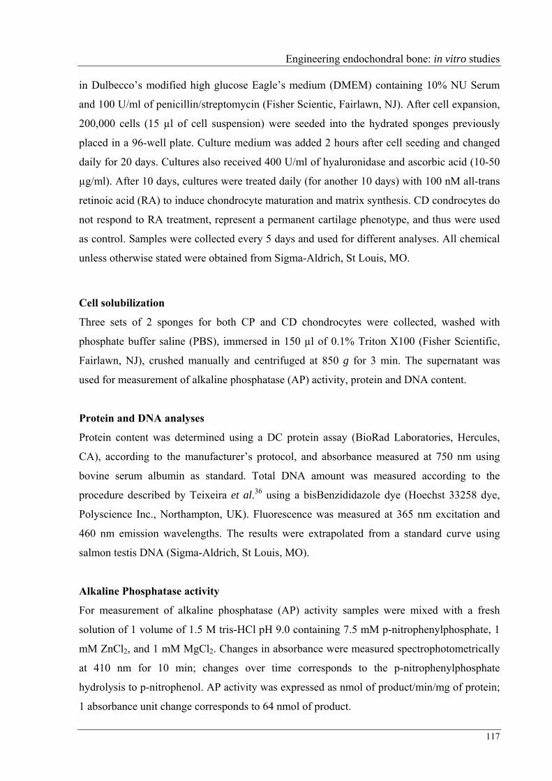
Engineering endochondral bone: in vitro studies
117
in Dulbecco’s modified high glucose Eagle’s medium (DMEM) containing 10% NU Serum
and 100 U/ml of penicillin/streptomycin (Fisher Scientic, Fairlawn, NJ). After cell expansion,
200,000 cells (15 µl of cell suspension) were seeded into the hydrated sponges previously
placed in a 96-well plate. Culture medium was added 2 hours after cell seeding and changed
daily for 20 days. Cultures also received 400 U/ml of hyaluronidase and ascorbic acid (10-50
µg/ml). After 10 days, cultures were treated daily (for another 10 days) with 100 nM all-trans
retinoic acid (RA) to induce chondrocyte maturation and matrix synthesis. CD condrocytes do
not respond to RA treatment, represent a permanent cartilage phenotype, and thus were used
as control. Samples were collected every 5 days and used for different analyses. All chemical
unless otherwise stated were obtained from Sigma-Aldrich, St Louis, MO.
Cell solubilization
Three sets of 2 sponges for both CP and CD chondrocytes were collected, washed with
phosphate buffer saline (PBS), immersed in 150 µl of 0.1% Triton X100 (Fisher Scientific,
Fairlawn, NJ), crushed manually and centrifuged at 850 g for 3 min. The supernatant was
used for measurement of alkaline phosphatase (AP) activity, protein and DNA content.
Protein and DNA analyses
Protein content was determined using a DC protein assay (BioRad Laboratories, Hercules,
CA), according to the manufacturer’s protocol, and absorbance measured at 750 nm using
bovine serum albumin as standard. Total DNA amount was measured according to the
procedure described by Teixeira et al.36 using a bisBenzididazole dye (Hoechst 33258 dye,
Polyscience Inc., Northampton, UK). Fluorescence was measured at 365 nm excitation and
460 nm emission wavelengths. The results were extrapolated from a standard curve using
salmon testis DNA (Sigma-Aldrich, St Louis, MO).
Alkaline Phosphatase activity
For measurement of alkaline phosphatase (AP) activity samples were mixed with a fresh
solution of 1 volume of 1.5 M tris-HCl pH 9.0 containing 7.5 mM p-nitrophenylphosphate, 1
mM ZnCl2, and 1 mM MgCl2. Changes in absorbance were measured spectrophotometrically
at 410 nm for 10 min; changes over time corresponds to the p-nitrophenylphosphate
hydrolysis to p-nitrophenol. AP activity was expressed as nmol of product/min/mg of protein;
1 absorbance unit change corresponds to 64 nmol of product.

Chapter V
118
RNA isolation and real time reverse transcriptase-polymerase chain reaction (RT-PCR)
RNA was isolated before the first day of RA treatment (day 10) and 10 days after RA
treatment (day 20). Total RNA was extracted with Trizol® reagent (Life Technologies,
Gaitherburg, MD) according to the manufacturer’s instructions with some modifications.
Briefly, samples were frozen in liquid nitrogen and crushed, Trizol® reagent was added
(10ml/g), the samples vortexed for 30 sec, and then kept at 4 ºC for 2 hours. Phase separation
was achieved by adding chloroform (0.2 volumes) to the mixture for 15 min and then
centrifuging it at 12,000 g for 30 min at 4 ºC. The upper aqueous phase containing the RNA
was collected, mixed with 0.25 volumes of high salt precipitation solution (0.8 M sodium
citrate and 1.2 M NaCl) and with 0.25 volumes of isopropanol and centrifuged at maximum
speed for 30 min at 4 ºC. RNA purification was completed using RNA micro kit (Qiagen Inc,
Chatsworth, CA) according to the RNeasy cleanup protocol. Real-time RT-PCR was
performed using QuantiTect SYBR Green RT-PCR kit (Qiagen Inc.), a DNA Engine
Optican2 system (Roche Molecular Systems, Pleasanton, CA), and primers specific for the
following chick genes: type X collagen (forward: AGTGCTGTCATTGATCTCATTGGA,
reverse: TCAGAGGAATAGAGACCATTGGATT), alkaline phosphatase (forward:
CCTGACATCGAGGTGATCCT, reverse: GAGACCCAGCAGGAAGTCCA), type I
collagen (forward: GCCGTGACCTCAGACTTAGC, reverse:
TTTTGTCCTTGGGGTTCTTG), runx2 (forward: CTTAGGAGAAGTGCCCGATG,
reverse: CCATCCACCGTCACCTTTAT) and VEGF (forward:
GGAAGCCCAACGAAGTTATC, reverse: AACCCGCACATCTCATCAG). Acidic
ribosomal protein (RP) mRNA was used as a reference for quantification (forward:
AACATGTTGAACATCTCCCC, reverse: ATCTGCAGACAGACGCTGGC). All primers
were purchased from Qiagen Inc (Valencia, CA). Relative transcript levels were presented as
‘‘fold change’’ in gene expression and calculated using the threshold cycle (Ct) and the
formula below, where ‘‘CD’’ refers to CD chondrocytes, ‘‘CP’’ refers to CP chondrocytes,
and ‘‘RP’’ refers to the acidic ribosomal protein: x=2∆∆Ct, in which ∆∆Ct=∆E–∆C, and
∆E=CtCP–CtRP, and ∆C=CtCD–CtRP. A ∆∆Ct<0 was considered a decrease while a
∆∆Ct>0 was considered an increase in gene expression.
Histology and immunohistochemistry
Scaffolds were collected and fixed in 10% phosphate buffer formalin, dehydrated in alcohol
series, embedded in paraffin, and 5 µm-thick sections cut and stained with Hematoxylin &

Engineering endochondral bone: in vitro studies
119
Eosin (H&E). For immunohistochemistry sections were deparafinized, rehydrated, and
immunostaining performed using antibodies specific for chick type X collagen with the
Vectastain ABC kit (Vector Laboratories, Burlingame, CA) according to the manufacturer’s
instructions. Sections were counter stained with 1% alcian blue and Light Green. The stained
sections were mounted under glass coverslips, and scanned on Scan Scope GL series optical
microscope (Aperio, Bristol, UK) at 20X magnification.
Statistical analysis
All experiments were repeated 3 to 4 times and the mean and standard error of the mean were
determined. Significant differences were assessed by ANOVA. A p-value refers to a
comparison of a measured parameter in the experimental group with that of the appropriate
control; significance was set at p<0.05.
RESULTS
Characterization of chitosan scaffolds
Images obtained by SEM (Figure 1) of cross-sections of sponges show a homogeneous pore
size and distribution.
A
1 mm
B
100µm Figure 1. Homogeneous distribution of pores in chitosan sponges. SEM photomicrographs of cross sections
of dehydrated chitosan sponges. Image A (lower magnification) shows high homogeneity in the size and
distribution of pores and image B (higher magnification) shows the pores interconnectivity.
At higher magnification (500X) the interconnectivity can be observed among pores with an
average pore size of 92±12 µm (Figure 1B). This pore dimension is considered to be within
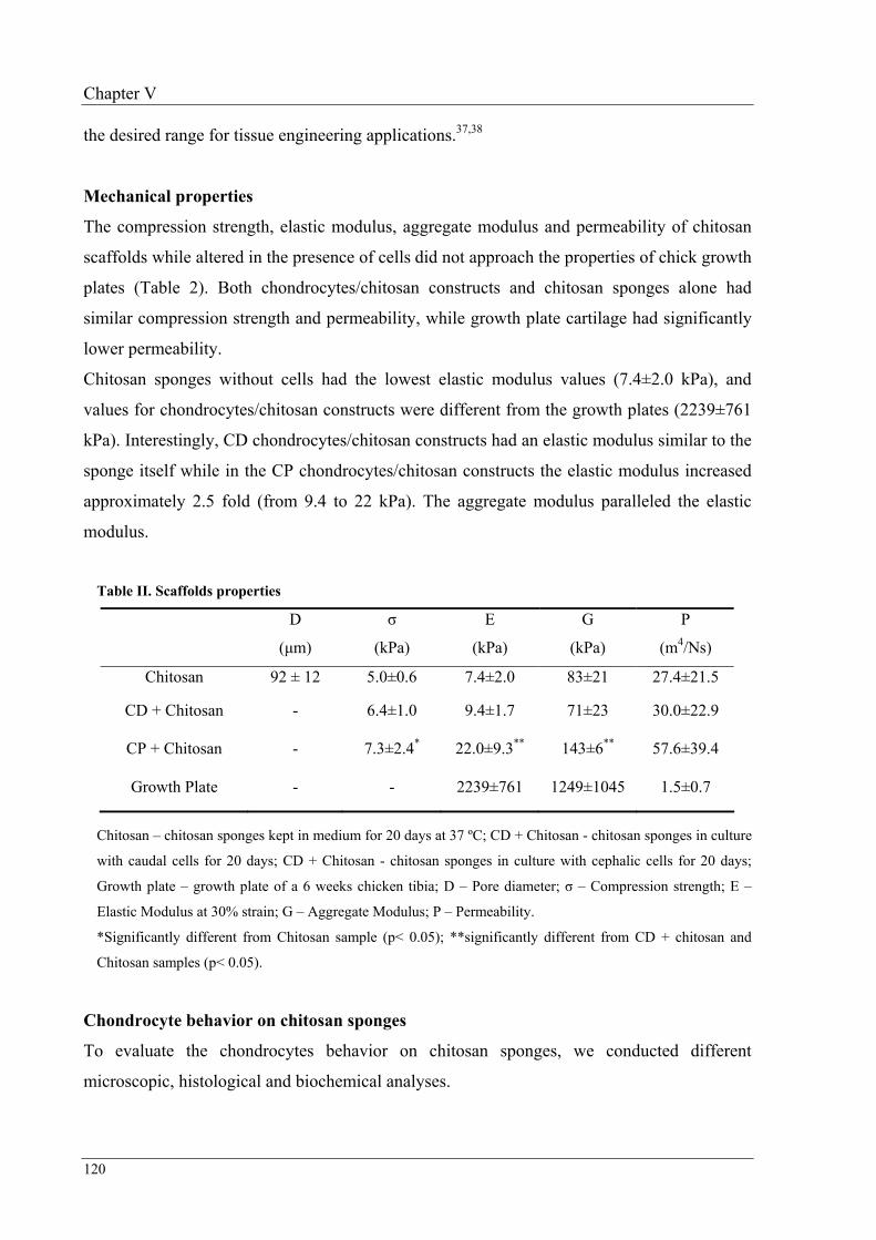
Chapter V
120
the desired range for tissue engineering applications.37,38
Mechanical properties
The compression strength, elastic modulus, aggregate modulus and permeability of chitosan
scaffolds while altered in the presence of cells did not approach the properties of chick growth
plates (Table 2). Both chondrocytes/chitosan constructs and chitosan sponges alone had
similar compression strength and permeability, while growth plate cartilage had significantly
lower permeability.
Chitosan sponges without cells had the lowest elastic modulus values (7.4±2.0 kPa), and
values for chondrocytes/chitosan constructs were different from the growth plates (2239±761
kPa). Interestingly, CD chondrocytes/chitosan constructs had an elastic modulus similar to the
sponge itself while in the CP chondrocytes/chitosan constructs the elastic modulus increased
approximately 2.5 fold (from 9.4 to 22 kPa). The aggregate modulus paralleled the elastic
modulus.
Table II. Scaffolds properties
D
(µm)
σ
(kPa)
E
(kPa)
G
(kPa)
P
(m4/Ns)
Chitosan 92 ± 12 5.0±0.6 7.4±2.0 83±21 27.4±21.5
CD + Chitosan - 6.4±1.0 9.4±1.7 71±23 30.0±22.9
CP + Chitosan - 7.3±2.4* 22.0±9.3** 143±6** 57.6±39.4
Growth Plate - - 2239±761 1249±1045 1.5±0.7
Chitosan – chitosan sponges kept in medium for 20 days at 37 ºC; CD + Chitosan - chitosan sponges in culture
with caudal cells for 20 days; CD + Chitosan - chitosan sponges in culture with cephalic cells for 20 days;
Growth plate – growth plate of a 6 weeks chicken tibia; D – Pore diameter; σ – Compression strength; E –
Elastic Modulus at 30% strain; G – Aggregate Modulus; P – Permeability.
*Significantly different from Chitosan sample (p< 0.05); **significantly different from CD + chitosan and
Chitosan samples (p< 0.05).
Chondrocyte behavior on chitosan sponges
To evaluate the chondrocytes behavior on chitosan sponges, we conducted different
microscopic, histological and biochemical analyses.

Engineering endochondral bone: in vitro studies
121
C
P ch
ondr
ocyt
es
A
200 µm
B
200 µm
C
200 µm
D
200 µm 100 µm
E
F
200 µm
G
200 µm
H
200 µm
I
200 µm
J
100 µm
CD
cho
ndro
cyte
s – RA
Day 5 Day 10 + RA
Day 15 Day 20 + RA
Day 20
Figure 2. Chondrocytes completely fill chitosan sponges during the culture period. Photomicrographs of
H&E staining of chitosan/chondrocytes constructs cultured for 20 days. A, B, C and D correspond to cross-
sections of chitosan sponges culture with CP chondrocytes for 5, 10, 15 and 20 days respectively. A complete
cross-section of each sponge is represented in the images. Photomicrographs F, G, H and I correspond to
sponges cultured with CD chondrocytes for same time points. E and J are higher magnification images of the
areas inside the square in D and I, respectively. Arrows in image E point to hypertrophic chondrocytes. At day 5,
sponges appear thinner because during processing they did not maintain the original thickness in absence of
cells/matrix.
Histological analyses clearly show attachment, proliferation and extracellular matrix synthesis
by chondrocytes seeded in the chitosan sponges (Figure 2). At day 5, cells (purple color)
migrating through connecting pores can be observed for both CP and CD chondrocytes. At
day 10, chondrocytes have migrated well into the depth of chitosan sponges though they do
not completely fill the pores. After 15 days in culture (5 days of treatment with RA),
deposition of matrix can be observed (pink material between cells). After 20 days in culture
(10 days of treatment with RA), both CP and CD chondrocytes deposited an abundant
extracellular matrix filling the pores of sponges. At higher magnification hypertrophic CP
chondrocytes (arrows in Figure 2E) can be observed while CD chondrocytes (Figure 2J)
appear smaller.

Chapter V
122
Scanning electron photomicrographs (Figure 3) confirmed the observations from histological
sections. Indeed at day 20, CP chondrocytes (Figures 3A and 3B) are totally embedded in a
rich extracellular matrix completely changing the appearance of the scaffold (compare Figure
3 with Figure 1). The CD chondrocytes (Figures 3C and 3D) also filled the pores of the
chitosan sponges with a compact matrix and no major differences can be observed between
CP and CD scaffolds in these photomicrographs.
CP
chon
droc
ytes
A
200µm
B
100µm
CD
cho
ndro
cyte
s
C
200µm 100µm
D
Figure 3. An abundant matrix fills the pores of chitosan sponges. SEM analyses of cross-sections of
chitosan/chondrocytes constructs. At the end of the culture period, CP (A and B) and CD (C and D)
chondrocytes migrated to the bottom of scaffolds and completely filled chitosan pores with a dense extra cellular
matrix.
DNA measurements confirmed the increase in chondrocyte number over the culture period for
both CP and CD cells. The average number of CD chondrocytes was higher than CP
chondrocytes at every time point, although not statistically significant (Figure 4). With
retinoic acid treatment, the proliferation rate of CP chondrocytes decreased. At the end of the
culture period, the number of CD chondrocytes was slightly higher since they maintained or
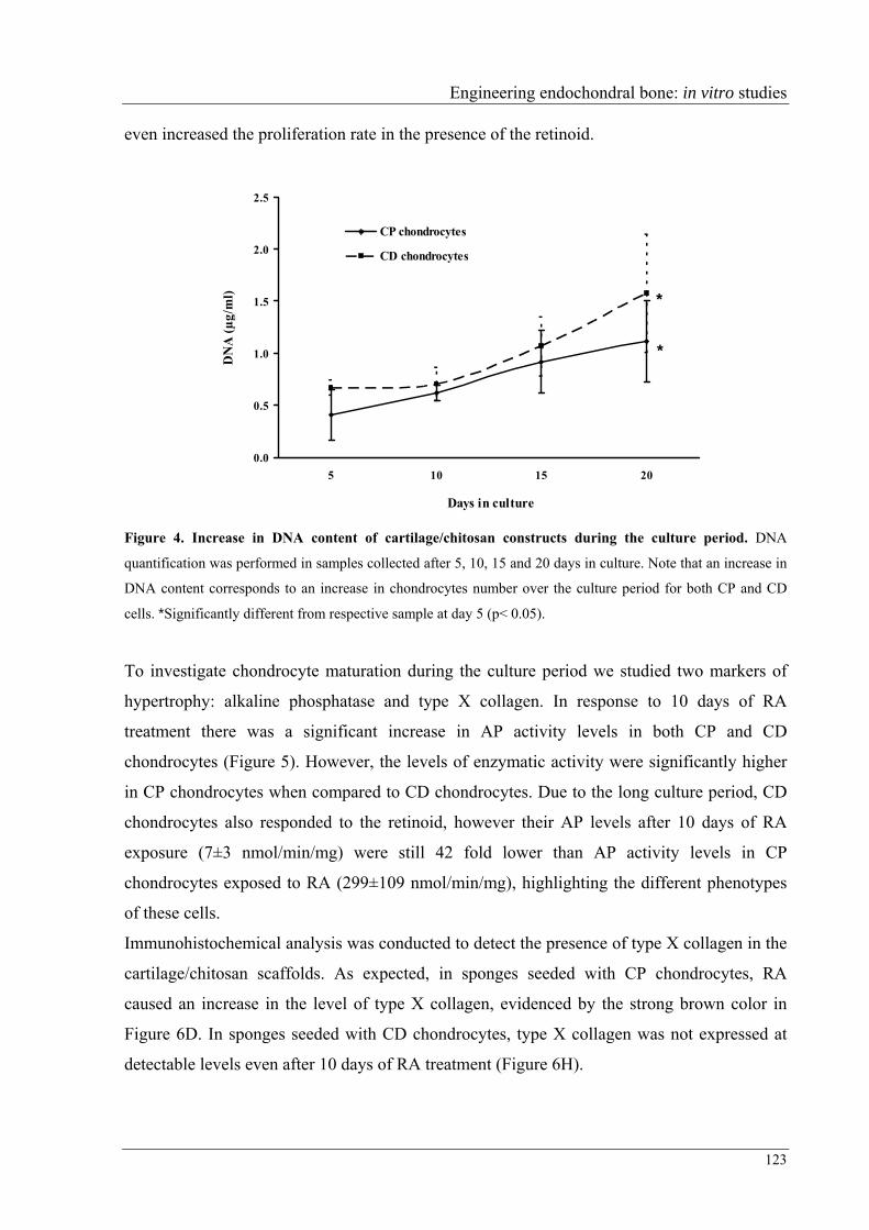
Engineering endochondral bone: in vitro studies
123
even increased the proliferation rate in the presence of the retinoid.
0.0
0.5
1.0
1.5
2.0
2.5
5 10 15 20
Days in culture
DN
A (µ
g/m
l)CP chondrocytes
CD chondrocytes
*
*
Figure 4. Increase in DNA content of cartilage/chitosan constructs during the culture period. DNA
quantification was performed in samples collected after 5, 10, 15 and 20 days in culture. Note that an increase in
DNA content corresponds to an increase in chondrocytes number over the culture period for both CP and CD
cells. *Significantly different from respective sample at day 5 (p< 0.05).
To investigate chondrocyte maturation during the culture period we studied two markers of
hypertrophy: alkaline phosphatase and type X collagen. In response to 10 days of RA
treatment there was a significant increase in AP activity levels in both CP and CD
chondrocytes (Figure 5). However, the levels of enzymatic activity were significantly higher
in CP chondrocytes when compared to CD chondrocytes. Due to the long culture period, CD
chondrocytes also responded to the retinoid, however their AP levels after 10 days of RA
exposure (7±3 nmol/min/mg) were still 42 fold lower than AP activity levels in CP
chondrocytes exposed to RA (299±109 nmol/min/mg), highlighting the different phenotypes
of these cells.
Immunohistochemical analysis was conducted to detect the presence of type X collagen in the
cartilage/chitosan scaffolds. As expected, in sponges seeded with CP chondrocytes, RA
caused an increase in the level of type X collagen, evidenced by the strong brown color in
Figure 6D. In sponges seeded with CD chondrocytes, type X collagen was not expressed at
detectable levels even after 10 days of RA treatment (Figure 6H).

Chapter V
124
1
10
100
1000
10 20
Days in culture
AP/
Pro
tein
(nm
ol/m
in/m
g)
CP chondrocytes
CD chondrocytes
*
***
Figure 5. Retinoic acid induces alkaline phosphatase activity in CP chondrocytes. Samples were collected
after 10 and 20 days in culture. Alkaline phosphatase (AP) activity was normalized to the total protein content of
each sample. The AP levels increased with time, in both CP and CD chondrocytes. *Significantly different from
CD chondrocytes at same time point (p< 0.05). **Significantly different from same sample at day 10 (p< 0.05).
Counter staining with alcian blue and Light Green allowed visualization of proteoglycan
deposition and chitosan, respectively. Although we did not quantify the staining, an intense
blue color can be observed in CP chondrocytes exposed to RA for 10 days, suggesting
increased proteoglycan synthesis in these samples (compare Figure 6C with Figures 6A and
6G).
To further characterize the phenotype of chondrocytes grown on the chitosan scaffold, we
conducted real time RT-PCR analyses. Results obtained are semi-quantitative and presented
as a “fold change” in mRNA levels in CP chondrocytes when compared to CD cells (Figure
7). A value higher than 1, corresponds to a higher gene expression by CP chondrocytes. As
expected, except for type II collagen, all genes studied were expressed at higher levels in CP
chondrocytes than in CD chondrocytes. In response to RA treatment, type X collagen
expression decreased (early hypertrophic marker) and AP gene expression increased (late
hypertrophic marker). Values obtained for runx2, type II collagen and VEGF are around 1; no
significant differences were observed between 10 and 20 days in culture.

Engineering endochondral bone: in vitro studies
125
CD chondrocytes Day 10 Day 20
CP chondrocytes Day 10 Day 20
Con
trol
Type
X C
ol D
50 µm
B
50 µm
C
50 µm
A
50 µm
F
50 µm
E
50 µm
H
50 µm
G
50 µm
Figure 6. Retinoic acid increases type X collagen in scaffolds seeded with CP chondrocytes.
Immunohistochemical analyses of type X collagen was performed on deparafinized sections of CP (A, B, C and
D) and CD (E, F, G and H) chondrocytes. Sections were counterstained with alcian blue to visualize
proteoglycans and Light Green to label the chitosan walls. Photomicrographs A, B, E and F correspond to 10
days in culture and pictures C, D, G and H corresponds to 20 days in culture. Upper panels show controls
(incubated with pre-immune serum) of respective bottom panel. Type X collagen is evidenced by the presence of
the brown color.
DISCUSSION
Chitosan has been used as biomaterial over the last decades and proved to be biocompatible
and biodegradable.39-41 Studies using low DA chitosan scaffolds showed that this material
allows osteoblast21,42 and articular chondrocyte21,43 attachment and proliferation. The goal of
the current study was to test the ability of this natural polymer to support proliferation and
maturation of transient cartilage cells, the early steps in endochondral bone formation. The
original chitosan powder from France Chitine had a DA of ≅30% and it was deacetylated to a
DA of 4.23±0.46%, using different temperature cycles. The deacetylation of chitosan using
alkali solutions is known to lead to a decrease in both average molecular weight (Mw) and
intrinsic viscosity (η) as a result of the scission of glycosidic linkages and end-group peeling.
Chitosan with a DA of 4% was previously reported to enable the attachment and proliferation
of both osteoblasts and rat bone marrow stromal cells.33 Therefore, chitosan with this DA was
selected to produce sponges. The 3D homogeneous structures prepared presented highly
interconnected pores of average size shown to allow robust chondrocyte proliferation and
metabolic activity.44 RA was used to induce chondrocyte maturation into hypertrophic cells.

Chapter V
126
RA is one of the more biologically active derivatives of retinol (vitamin A) and a well-known
regulator of cartilage and skeletal formation.45-47 We cultured both cells representative of a
transient cartilage phenotype, CP chondrocytes, and cells that maintain a more stable
phenotype, CD chondrocytes. Caudal cells do not readily undergo hypertrophy in response to
RA and therefore, were used as a “permanent” cartilage control.
0
1
10
100
Runx 2 AP Type II Type X VEGF
Genes expression
Fold
cha
nge
in m
RN
A le
vel
Day 10 Day 20
*
**
Figure 7. Gene expression profile of CP chondrocytes cultured in chitosan sponges. Real time RT-PCR
semi-quantitative results are presented as fold change in mRNA levels of CP chondrocytes when compared to
CD cells. Samples with CP and CD chondrocytes were both analyzed and a fold change higher than 1,
corresponds to a higher gene expression by CP chondrocytes. *Significantly different from CD chondrocytes at
same time point (p< 0.05).
In 10 days, both CP and CD chondrocytes migrated into pores deep in the chitosan sponges
completely obliterating some spaces, attesting to the excellent interconnectivity between the
pores. Cell proliferation continued for both CD and CP chondrocytes even during treatment
with RA, though CP cells showed a slight decrease after day 15. According to Gentili et al.48
chondrocytes proliferation stops only after onset of mineralization has been started. However,
in our studies we did not supplement the media with β-glycerophosphate or another phosphate
source. Thus our chondrocytes while expressing high AP levels, did not deposit mineral
(micro computerized tomography results not shown).
Cells proliferation and matrix deposition increased in both CD and CP chondrocytes seeded
scaffolds over the culture period. It was expected that such a robust synthesis of matrix would

Engineering endochondral bone: in vitro studies
127
drastically change the mechanical properties of the chitosan sponges. However, while elastic
modulus, a measure of stiffness, increased 3 fold for chitosan sponges seeded with CP
chondrocytes, it did not change significantly for chitosan sponges seeded CD chondrocytes.
Therefore, more than the quantity, it was the quality of the matrix deposited that mostly
affected stiffness of chondrocyte/chitosan constructs. Indeed, type X collagen as been
reported to increase the matrix stiffness by Naumann et al.49 However, the stiffness of CP
chondrocytes/chitosan constructs is hundred fold lower than the stiffness of chicken growth
plates. Since ceramic phases present higher compression strength than unmineralized tissues,
we believe that we would observe different mechanical properties had the cultured
chondrocytes been allowed to mineralize the extracellular matrix as it occurs in the native
growth plate.
Although both CP and CD chondrocytes responded to RA by increasing matrix deposition,
the nature of this matrix was found to be dependent on the type of cell. CP cells responded to
the retinoid by expressing the hypertrophic phenotype, characterized by the presence of type
X collagen in their matrix and increased AP activity. Indeed, all regulators or markers of
hypertrophy studied (runx 2, VEGF, type X collagen, AP) were expressed at significantly
higher levels in the CP cells when compared to the CD chondrocytes, supporting the different
phenotype of these cell populations. However, overtime we observed a decrease in type X
collagen expression and an increase in AP expression in CP cells. These results agree with in
vivo data showing that type X collagen expression occurs in early hypertrophic chondrocytes
while AP, a later maturation marker involved in matrix mineralization is expressed by
terminally differentiated chondrocytes in the calcified region of the growth plate.50-52
Several studies have demonstrated that chitosan supports the articular chondrocyte phenotype
and activity. The study of Lu et al.23 showed that chitosan injected into the knee articular
cavity of rats caused a significant increase in the density of chondrocytes in the knee articular
cartilage. Also, abundant cartilage extracellular matrix protein43 was obtained after injecting
nude mice subcutaneously with an in situ-gelling chitosan solution cultured with
chondrocytes. Furthermore, when pig chondrocytes were cultured on porous chitosan
scaffolds they were able to proliferate and to spread into the sponges; with larger
interconnected pores improving the cellularity and matrix content.44 Another study revealed
that primary chondrocytes cultured on chondroitin 4-sulfate/chitosan maintained the synthesis
of cartilage-specific collagens.17 Similar results were achieved using chitosan films seeded
with both human osteoblasts and chondrocytes.21 Osteoblasts were able to spread and to

Chapter V
128
express type I collagen whereas chondrocytes expressed type II collagen, suggesting that a
chitosan scaffold is able to maintain these cells original phenotype.
Amaral et al.53 investigated human osteoblast behavior in chitosan scaffolds similar to those
used in the present study. They found that osteoblasts attach well and spread on chitosan
sponges, displaying long cell filopodia and numerous cell-to-cell contacts. A major limitation
in tissue engineering has been the availability of nutrients and oxygen once the construct
reaches a critical mass. And in that aspect osteoblasts are particularly susceptible.54-57
Cartilage (growth plate and articular cartilage) however, is characterized by large avascular
regions and excellent adaptation to hypoxic conditions.58,59 In fact, evidence supports the role
of low oxygen levels in chondrocyte differentiation and endochondral bone formation.60
These properties provide support to the bone tissue engineering approach here investigated,
which explores the endochondral ossification pathway for the correction of extensive bone
defects.
All the studies mentioned earlier used chitosan as a scaffold for either articular cartilage or
bone. Here we reported the use of chitosan sponges as a template for transient cartilage and
the creation of an improved osteoinductive scaffold for bone regeneration. Indeed,
hypertrophic chondrocytes secrete factors capable of inducing osteoblast and osteoclast
activity, as well as vascularization.28,61-64 All these different cell types and their activity are
necessary and responsible for endochondral bone formation. Indeed, parallel studies
conducted in our laboratory show the ability of this transient cartilage/chitosan constructs to
induce endochondral bone formation in the subcutaneous region of nude mice.65
CONCLUSIONS
The use of osteoblasts or osteoprogenitor cells to promote bone regeneration is the method
most commonly exploited in current tissue engineering approaches. However, nature mostly
uses the endochondral ossification pathway during bone formation, growth and healing after
fracture. In addition to these considerations, there are numerous other advantages to explore
the endochondral pathway for bone tissue engineering. Chondrocytes are resistant to low
oxygen levels and can induce vascular invasion and osteogenesis. In this study, chitosan
sponges served as excellent support for chondrocyte proliferation and intense matrix
deposition. As a result of the activity of the transient cartilage cells, the mechanical properties
of the chitosan scaffold changed drastically and favorably. These scaffolds can now be used
Engineering endochondral bone: in vitro studies
129
as osteoinductive grafts in vivo, inducing and remodeling into bone, mimicking the natural
process of endochondral ossification.
ACKNOWLEDMENS
This work was supported by the Luso-American Foundation and PRODEP III from Portugal,
by the American Association of Orthodontics Foundation and NIDCR grant 5K08DE017426.
The authors thank Ms. Gloria Turner, Department of Pathology at New York University
College of Dentistry, for her important assistance in preparation of samples for histology and
immunohistochemistry. Mechanical tests were performed at the NIH sponsored
Musculoskeletal Repair and Regeneration Core Center at the Hospital for Special Surgery
(AR046121).
REFERENCES 1. Steinemann SG. Metal implants and surface reactions. Injury 1996;27 Suppl 3:SC16-22.
2. Disegi JA, Eschbach L. Stainless steel in bone surgery. Injury 2000;31 Suppl 4:2-6.
3. Reitman RD, Emerson R, Higgins L, Head W. Thirteen year results of total hip arthroplasty using a
tapered titanium femoral component inserted without cement in patients with type C bone. Journal of
Arthroplasty 2003;18(7):116-121.
4. Epinette JA, Manley MT, D'Antonio JA, Edidin AA, Capello WN. A 10-year minimum follow-up of
hydroxyapatite-coated threaded cups: clinical, radiographic and survivorship analyses with comparison to
the literature. J Arthroplasty 2003;18(2):140-8.
5. Xynos ID, Hukkanen MV, Batten JJ, Buttery LD, Hench LL, Polak JM. Bioglass 45S5 stimulates
osteoblast turnover and enhances bone formation In vitro: implications and applications for bone tissue
engineering. Calcif Tissue Int 2000;67(4):321-9.
6. Barrias CC, Ribeiro CC, Barbosa MA. Adhesion and proliferation of human osteoblastic cells seeded on
injectable hydroxyapatite microspheres. Bioceramics, Vol 16 2004;254-2:877-880.
7. Saito N, Takaoka K. New synthetic biodegradable polymers as BMP carriers for bone tissue engineering.
Biomaterials 2003;24(13):2287-2293.
8. Suzuki T, Kawamura H, Kasahara T, Nagasaka H. Resorbable poly-L-lactide plates and screws for the
treatment of mandibular condylar process fractures: a clinical and radiologic follow-up study. J Oral
Maxillofac Surg 2004;62(8):919-24.
9. Sittinger M, Reitzel D, Dauner M, Hierlemann H, Hammer C, Kastenbauer E, Planck H, Burmester GR,
Bujia J. Resorbable polyesters in cartilage engineering: affinity and biocompatibility of polymer fiber
structures to chondrocytes. J Biomed Mater Res 1996;33(2):57-63.
10. Vaccaro AR, Singh K, Haid R, Kitchel S, Wuisman P, Taylor W, Branch C, Garfin S. The use of

Chapter V
130
bioabsorbable implants in the spine. The Spine Journal 2003;3:227–237.
11. Jianqi H, Hong H, Lieping S, Genghua G. Comparison of calcium alginate film with collagen membrane
for guided bone regeneration in mandibular defects in rabbits. J Oral Maxillofac Surg 2002;60(12):1449-
54.
12. Chan C, Thompson I, Robinson P, Wilson J, Hench L. Evaluation of Bioglass/dextran composite as a
bone graft substitute. Int J Oral Maxillofac Surg 2002;31(1):73-7.
13. Mukherjee DP, Tunkle AS, Roberts RA, Clavenna A, Rogers S, Smith D. An animal evaluation of a paste
of chitosan glutamate and hydroxyapatite as a synthetic bone graft material. Journal of Biomedical
Materials Research Part B-Applied Biomaterials 2003;67B(1):603-609.
14. Sashiwa H, Aiba S. Chemically modified chitin and chitosan as biomaterials. Progress in Polymer Science
2004;29(9):887-908.
15. Hoekstra A, Struszczyk H, Kivekas O. Percutaneous microcrystalline chitosan application for sealing
arterial puncture sites. Biomaterials 1998;19(16):1467-71.
16. Lee KY, Ha WS, Park WH. Blood compatibility and biodegradability of partially N-acylated chitosan
derivatives. Biomaterials 1995;16(16):1211-1216.
17. Suh JK, Matthew HW. Application of chitosan-based polysaccharide biomaterials in cartilage tissue
engineering: a review. Biomaterials 2000;21(24):2589-98.
18. Hirano S, Tsuchida H, Nagao N. N-acetylation in chitosan and the rate of its enzymic hydrolysis.
Biomaterials 1989;10(8):574-576.
19. Prashanth KVH, Tharanathan RN. Chitin/chitosan: modifications and their unlimited application
potential--an overview. Trends in Food Science & Technology 2007;18(3):117-131.
20. Chellat F, Tabrizian M, Dumitriu S, Chornet E, Hilaire C, Yahia R. Study of biodegradation behavior of
chitosan-xanthan microspheres in simulated physiological media. Journal of Biomedical Materials
Research 2000;53(5):592-599.
21. Lahiji A, Sohrabi A, Hungerford DS, Frondoza CG. Chitosan supports the expression of extracellular
matrix proteins in human osteoblasts and chondrocytes. J Biomed Mater Res 2000;51(4):586-95.
22. Nettles DL, Elder SH, Gilbert JA. Potential use of chitosan as a cell scaffold material for cartilage tissue
engineering. Tissue Engineering 2002;8(6):1009-1016.
23. Lu JX, Prudhommeaux F, Meunier A, Sedel L, Guillemin G. Effects of chitosan on rat knee cartilages.
Biomaterials 1999;20(20):1937-44.
24. Abe M, Takahashi M, Tokura S, Tamura H, Nagano A. Cartilage-scaffold composites produced by
bioresorbable beta-chitin sponge with cultured rabbit chondrocytes. Tissue Engineering 2004;10(3-
4):585-594.
25. Sechriest VF, Miao YJ, Niyibizi C, Westerhausen-Larson A, Matthew HW, Evans CH, Fu FH, Suh JK.
GAG-augmented polysaccharide hydrogel: a novel biocompatible and biodegradable material to support
chondrogenesis. J Biomed Mater Res 2000;49(4):534-41.
26. Rajpurohit R, Koch CJ, Tao Z, Teixeira CM, Shapiro IM. Adaptation of chondrocytes to low oxygen
tension: relationship between hypoxia and cellular metabolism. J Cell Physiol 1996;168(2):424-32.
27. Maes C, Carmeliet P, Moermans K, Stockmans I, Smets N, Collen D, Bouillon R, Carmeliet G. Impaired
angiogenesis and endochondral bone formation in mice lacking the vascular endothelial growth factor

Engineering endochondral bone: in vitro studies
131
isoforms VEGF164 and VEGF188. Mech Dev 2002;111(1-2):61-73.
28. Petersen W, Tsokos M, Pufe T. Expression of VEGF121 and VEGF165 in hypertrophic chondrocytes of
the human growth plate and epiphyseal cartilage. J Anat 2002;201(2):153-7.
29. Pullig O, Weseloh G, Ronneberger D, Kakonen S, Swoboda B. Chondrocyte differentiation in human
osteoarthritis: expression of osteocalcin in normal and osteoarthritic cartilage and bone. Calcif Tissue Int
2000;67(3):230-40.
30. Hashimoto S, Ochs RL, Rosen F, Quach J, McCabe G, Solan J, Seegmiller JE, Terkeltaub R, Lotz M.
Chondrocyte-derived apoptotic bodies and calcification of articular cartilage. Proceedings of the National
Academy of Sciences of the United States of America 1998;95(6):3094-3099.
31. Pullig O, Weseloh G, Gauer S, Swoboda B. Osteopontin is expressed by adult human osteoarthritic
chondrocytes: protein and mRNA analysis of normal and osteoarthritic cartilage. Matrix Biol
2000;19(3):245-55.
32. Eerola I, Salminen H, Lammi P, Lammi M, von der Mark K, Vuorio E, Saamanen AM. Type X collagen,
a natural component of mouse articular cartilage: association with growth, aging, and osteoarthritis.
Arthritis Rheum 1998;41(7):1287-95.
33. Amaral IF, Sampaio P, Barbosa MA. Three-dimensional culture of human osteoblastic cells in chitosan
sponges: the effect of the degree of acetylation. J Biomed Mater Res A 2006;76(2):335-46.
34. Mow VC, Kuei SC, Lai WM, Armstrong CG. Biphasic creep and stress relaxation of articular cartilage in
compression? Theory and experiments. J Biomech Eng 1980;102(1):73-84.
35. Iwamoto M, Shapiro IM, Yagami K, Boskey AL, Leboy PS, Adams SL, Pacifici M. Retinoic acid induces
rapid mineralization and expression of mineralization-related genes in chondrocytes. Exp Cell Res
1993;207(2):413-20.
36. Teixeira CC, Hatori M, Leboy PS, Pacifici M, Shapiro IM. A rapid and ultrasensitive method for
measurement of DNA, calcium and protein content, and alkaline phosphatase activity of chondrocyte
cultures. Calcif Tissue Int 1995;56(3):252-6.
37. Nehrer S, Breinan HA, Ramappa A, Young G, Shortkroff S, Louie LK, Sledge CB, Yannas IV, Spector
M. Matrix collagen type and pore size influence behaviour of seeded canine chondrocytes. Biomaterials
1997;18(11):769-76.
38. Karande TS, Ong JL, Agrawal CM. Diffusion in musculoskeletal tissue engineering scaffolds: design
issues related to porosity, permeability, architecture, and nutrient mixing. Ann Biomed Eng
2004;32(12):1728-43.
39. VandeVord PJ, Matthew HW, DeSilva SP, Mayton L, Wu B, Wooley PH. Evaluation of the
biocompatibility of a chitosan scaffold in mice. J Biomed Mater Res 2002;59(3):585-90.
40. Hong Y, Song H, Gong Y, Mao Z, Gao C, Shen J. Covalently crosslinked chitosan hydrogel: Properties of
in vitro degradation and chondrocyte encapsulation. Acta Biomaterialia 2007;3(1):23-31.
41. Ho M, Wang D, Hsieh H, Liu HC, Hsien TY, Lai JY, Hou LT. Preparation and characterization of RGD-
immobilized chitosan scaffolds. Biomaterials 2005;26(16):3197-3206.
42. Amaral IF, Cordeiro AL, Sampaio P, Barbosa MA. Attachment, spreading and short-term proliferation of
human osteoblastic cells cultured on chitosan films with different degrees of acetylation. J. Biomater. Sci.
Polymer Edn 2007;18(4):469-485.

Chapter V
132
43. Hoemann CD, Sun J, Legare A, McKee MD, Buschmann MD. Tissue engineering of cartilage using an
injectable and adhesive chitosan-based cell-delivery vehicle. Osteoarthritis Cartilage 2005;13(4):318-29.
44. Griffon DJ, Sedighi MR, Schaeffer DV, Eurell JA, Johnson AL. Chitosan scaffolds: interconnective pore
size and cartilage engineering. Acta Biomater 2006;2(3):313-20.
45. Underhill TM, Weston AD. Retinoids and their receptors in skeletal development. Microscopy Research
and Technique 1998;43(2):137-155.
46. Cohen AJ, Lassova L, Golden EB, Niu Z, Adams SL. Retinoids directly activate the collagen X promoter
in prehypertrophic chondrocytes through a distal retinoic acid response element. J Cell Biochem
2006;99(1):269-78.
47. Pacifici M, Golden EB, Iwamoto M, Adams SL. Retinoic acid treatment induces type X collagen gene
expression in cultured chick chondrocytes. Exp Cell Res 1991;195(1):38-46.
48. Gentili C, Bianco P, Neri M, Malpeli M, Campanile G, Castagnola P, Cancedda R, Cancedda FD. Cell-
Proliferation, Extracellular-Matrix Mineralization, and Ovotransferrin Transient Expression during in-
Vitro Differentiation of Chick Hypertrophic Chondrocytes into Osteoblast-Like Cells. Journal of Cell
Biology 1993;122(3):703-712.
49. Naumann A, Dennis JE, Awadallah A, Carrino DA, Mansour JM, Kastenbauer E, Caplan AI.
Immunochemical and mechanical characterization of cartilage subtypes in rabbit. J Histochem Cytochem
2002;50(8):1049-58.
50. Wang W, Kirsch T. Retinoic acid stimulates annexin-mediated growth plate chondrocyte mineralization. J
Cell Biol 2002;157(6):1061-9.
51. Whyte MP. Hypophosphatasia and the role of alkaline phosphatase in skeletal mineralization. Endocr Rev
1994;15(4):439-61.
52. Lewinson D, Toister Z, Silbermann M. Quantitative and distributional changes in the activity of alkaline
phosphatase during the maturation of cartilage. J. Histochem. Cytochem. 1982;30(3):261-269.
53. Amaral IF, Sampaio P, Barbosa MA. Three-dimensional culture of human osteoblastic cells in chitosan
sponges: the effect of the degree of acetylation. J Biomed Mater Res A 2005;76(2):335.
54. Park JH, Park BH, Kim HK, Park TS, Baek HS. Hypoxia decreases Runx2/Cbfa1 expression in human
osteoblast-like cells. Mol Cell Endocrinol 2002;192(1-2):197-203.
55. Utting JC, Robins SP, Brandao-Burch A, Orriss IR, Behar J, Arnett TR. Hypoxia inhibits the growth,
differentiation and bone-forming capacity of rat osteoblasts. Exp Cell Res 2006;312(10):1693-702.
56. Warren SM, Steinbrech DS, Mehrara BJ, Saadeh PB, Greenwald JA, Spector JA, Bouletreau PJ, Longaker
MT. Hypoxia regulates osteoblast gene expression. J Surg Res 2001;99(1):147-55.
57. Salim A, Nacamuli RP, Morgan EF, Giaccia AJ, Longaker MT. Transient changes in oxygen tension
inhibit osteogenic differentiation and Runx2 expression in osteoblasts. J Biol Chem 2004;279(38):40007-
16.
58. Vu TH, Shipley JM, Bergers G, Berger JE, Helms JA, Hanahan D, Shapiro SD, Senior RM, Werb Z.
MMP-9/gelatinase B is a key regulator of growth plate angiogenesis and apoptosis of hypertrophic
chondrocytes. Cell 1998;93(3):411-22.
59. Skawina A, Litwin JA, Gorczyca J, Miodonski AJ. The vascular system of human fetal long bones: a
scanning electron microscope study of corrosion casts. J Anat 1994;185 ( Pt 2):369-76.

Engineering endochondral bone: in vitro studies
133
60. Pfander D, Cramer T, Schipani E, Johnson RS. HIF-1alpha controls extracellular matrix synthesis by
epiphyseal chondrocytes. J Cell Sci 2003;116(Pt 9):1819-26.
61. Deckers MM, Karperien M, van der Bent C, Yamashita T, Papapoulos SE, Lowik CW. Expression of
vascular endothelial growth factors and their receptors during osteoblast differentiation. Endocrinology
2000;141(5):1667-74.
62. Gerber HP, Vu TH, Ryan AM, Kowalski J, Werb Z, Ferrara N. VEGF couples hypertrophic cartilage
remodeling, ossification and angiogenesis during endochondral bone formation. Nat Med 1999;5(6):623-
8.
63. Nakagawa M, Kaneda T, Arakawa T, Morita S, Sato T, Yomada T, Hanada K, Kumegawa M, Hakeda Y.
Vascular endothelial growth factor (VEGF) directly enhances osteoclastic bone resorption and survival of
mature osteoclasts. FEBS Lett 2000;473(2):161-4.
64. Pfander D, Kortje D, Zimmermann R, Weseloh G, Kirsch T, Gesslein M, Cramer T, Swoboda B. Vascular
endothelial growth factor in articular cartilage of healthy and osteoarthritic human knee joints. Ann
Rheum Dis 2001;60(11):1070-1073.
65. Oliveira SM, Turner G, Mijares D, Amaral IF, Barbosa MA, Teixeira CC. Engineering endochondral
bone: In vivo studies. Tissue Engineering, Part A, In Press.


Chapter VI
135 Tissue Engineering, Part A, In Press
Engineering endochondral bone: in vivo studies
S.M. Oliveira1,2,3,4, D.Q. Mijares4, G. Turner4, I.F. Amaral2, M.A. Barbosa2,3, C.C.
Teixeira4,
1ESTV – Escola Superior de Tecnologia de Viseu, Dep. de Eng. Mecânica e Gestão Industrial, Campus
Politécnico de Repeses, 3504-510 Viseu, Portugal; 2INEB – Instituto de Engenharia Biomédica, Divisão de biomateriais, Rua do Campo Alegre 823, 4150-180
Porto, Portugal; 3FEUP – Faculdade de Engenharia Universidade do Porto, Dep. de Eng. Metalúrgica e de Materiais, Rua
Roberto Frias, 4200-465 Porto, Portugal; 4NYUCD – New York University College of Dentistry, Department of Basic Science and Craniofacial Biology
and Department of Biomaterials and Biomimetics, 345 E. 24th Street New York, NY 10010.
ABSTRACT
The use of biomaterials to replace lost bone has been a common practice for decades. More
recently, the demands for bone repair and regeneration have pushed research into the use of
cultured cells and growth factors in association with these materials. In this paper we describe
how a transient cartilage scaffold can induce endochondral ossification, a novel approach to
engineer new bone. Chondrocytes/chitosan scaffolds (both a transient cartilage scaffold -
experimental - and a permanent cartilage scaffold - control), were prepared and implanted
subcutaneously in nude mice. Bone formation was evaluated over a period of 5 months.
Mineralization was assessed by Faxitron, micro computed tomography, backscatter electrons,
and Fourier transform infrared spectroscopy analyses. Histological analysis provided further
information on tissue changes in and around the implanted scaffolds. The deposition of
ectopic bone was detected in experimental implants as early as 1 month after implantation
although it was only observed in few areas of the surface. After 3 months, bone trabeculae
and bone marrow cavities were formed inside the scaffolds. The bone deposited was similar to
the bone present in the mice vertebra. Interestingly, no bone formation was observed in
control implants. In conclusion, an engineered transient cartilage template carries all the
signals necessary to induce endochondral bone formation in vivo.

Chapter VI
136
Keywords: Chitosan sponges, chondrocytes, growth cartilage, endochondral ossification,
bone regeneration.
INTRODUCTION
With increasing time of life-expectation, the economic impact of musculoskeletal diseases,
may now exceed 2.5% of the Gross National Product in the United States.1 Among these
diseases, problems related to bone loss are one of the major causes of disability. To replace
lost bone, surgeons use autografts, considered the ideal grafting material. This approach
presents disadvantages such as donor site morbidity and limited supply, which in most of
cases is insufficient.2 Due to these limitations, it is imperative to find new alternatives to
autografts. Allografts and xenografs are reasonable substitutes but they also have serious
limitations like the risk of rejection or disease transmission. To overcome these shortcomings
several bone grafting materials have been engineered including ceramics, metals, synthetic
and natural polymers. In most cases, materials are implanted into surgically created bone
defects, and new bone grows on their surfaces and within their matrices. Under these
conditions, it is believed that the bone is formed by osteoblasts that migrated from the
adjacent, original bone and marrow cavities, a mechanism that is referred to as
osteoconduction. Although this osteointegration is important and essential, there are situations
(e.g. large defects) where the scaffolds, besides providing the template for tissue regeneration,
need to be osteoindutive, i.e. stimulate the migration of undifferentiated cells and induce their
differentiation into active osteoblasts in order to promote de novo bone formation. To improve
the osteoinductive properties of grafting materials they have been combined with growth
factors and different cells types with variable results, depending on the host regenerative
capability.3,4 Therefore, osteoinductive approaches are crucial when the bone regenerative
ability is diminished or lost. Here we propose a novel approach in bone tissue engineering and
the creation of an improved osteoinductive material for bone regeneration.
In the body, cartilaginous tissues can be divided into permanent and transient. While having
the same embryonic origin, permanent and transient cartilage follow distinct differentiation
pathways, fulfill different functions and ultimately as their names suggest have different fates.
Articular, tracheal, and other related cartilage structures are classified as permanent cartilage.
The chondrocytes in these structures maintain a relatively stable phenotype and persist for
many years5. In contrast, most of the embryonic cartilaginous skeleton, the epiphyseal growth

Engineering endochondral bone: in vivo studies
137
plates of long bones, the cartilaginous callus formed at fracture sites, and the tissue created
during distraction osteogenesis consist of transient cartilage.6,7 This transient cartilage is
gradually replaced by bone through a series of maturational changes during the process of
endochondral bone formation. Indeed, this is the mechanism responsible for formation of
most of our skeleton, and for postnatal growth.
Surprisingly, except for few reports, the bone tissue engineering field has focused on
generating bone directly from osteoprogenitor cells or osteoblasts. Montufar-Solis et al.8
prepared aggregates of mouse embryonic limb cells that after implantation in mice induced
endochondral bone formation. Indeed, the endochondral pathway for bone formation can be
activated using biomaterials carrying mesenchymal stem cells (MSCs),9 bone morphogenetic
proteins (BMPs)10, or plasmid DNA encoding for BMP’s.11,12 To date, only Alsberg et al.13
deliberately explored the endochondral pathway by implanting in mice, articular chondrocytes
mixed with calvarial osteoblasts using an alginate carrier. In vivo, the tissue organized in a
manner similar to a growth plate. This effect is not totally surprising if one considers that
hypertrophic chondrocytes secrete factors capable of inducing vascularization, osteoblast and
osteoclast activity.14-17 The interplay of these different cell types is required for endochondral
bone formation. Therefore, in the current work, we created a transient cartilage scaffold as a
template for in vivo endochondral ossification. We tested the hypothesis that this transient
cartilage, as an osteoinductive scaffold, carries all the signals necessary to induce new bone
formation once implanted in vivo, even subcutaneously.
MATERIALS AND METHODS
In vitro studies
Transient cartilage scaffolds
The chondrocyte/chitosan scaffolds used in this work were characterized extensively in
previous chapter.18 Briefly, chitosan sponges were prepared from a 2% (w/v) chitosan
solution by a freeze/drying process and then cut in small pieces (4x4x1 mm3). Cephalic (CP)
and caudal (CD) chondrocytes, isolated from the upper and lower regions of sternum of 14
days chicken embryos were allowed to proliferate in 100 mm dishes for 7 days in Dulbecco’s
modified high glucose Eagle’s medium (DMEM) containing 10% NU Serum and 100 U/ml of
penicillin/streptomycin (Fisher Scientic, Fairlawn, NJ), before being seeded into the chitosan
sponges and cultured for 20 days at 37 ºC and 5% CO2 in the same conditions. During the last

Chapter VI
138
10 days, cultures were treated with 100 nM all-trans retinoic acid (RA) in order to induce
chondrocyte maturation and matrix deposition. Prior to implantation, at day 20, alkaline
phosphatase (AP) activity and protein content were measured following the protocols
described in previous chapter, to confirm maturation.
In vivo studies
Surgical Procedures
Adult male athymic NCr-nu/nu mice (Charles River Laboratories, NY, US) were used as
surgical recipients. The animal experiments were conducted according to the guidelines of the
Institutional Animal Care and Use Committee (IACUC) of New York University. Briefly,
mice were received one week before scheduled surgery and feed a regular diet and sterile
water, ad libidum. After surgery and for one week, trimethropim and sulfamethoxazole (20
mg/l and 100 mg/l, respectively) were added to sterile drinking water. Surgeries were
performed under strict aseptic and warm conditions, with general anaesthesia produced by
intraperitoneal injection of ketamine (80 mg/kg) and xylazine (10 mg/kg). Lateral longitudinal
skin incisions were made in both sides of the mice dorsum and a subcutaneous pocket (1 to
1.5 cm) was created for implantation of the chondrocytes/chitosan scaffolds. On the right side,
we implanted the scaffolds seeded with cephalic chondrocytes (experimental implant)
whereas the left side received the scaffolds seeded with caudal chondrocytes (control).
Incisions were closed with surgical staples. Two or three sponges were implanted on each
side. Results shown represent three independent experiments, different surgeries, using three
independent groups of implants (i.e. sponges hydrated in different periods, primary cells
harvested from different chicken embryos, and mice received at different times). In the first
experiment 11 mice received implants, in the second and third experiments 14 and 22 mice
were implanted respectively. All animals, except 3 mice that did not recover from anesthesia,
survived the surgical procedures.
Euthanasia and samples collection
The mice were sacrificed at 1, 2, 3, 4 and 5 months postoperatively and implants and
surrounding tissues harvested. Briefly, animals were exposed to a gas mixture of 30% of CO2
and 70% of O2 for 1 min followed by CO2 100% during 4 minutes until no signal of breathing
was detected. Tissue/samples were collected and immediately immersed in 10% phosphate
buffer formalin for future analyses.

Engineering endochondral bone: in vivo studies
139
Faxitron and micro computed tomography (microCT)
All fixed tissues were radiographed in a high-resolution X-ray machine (Faxitron Series
43805N – Hewlett Packard, McMinnville, OR) set at 25 kVp, and 2.5 mA for 15 s, using a
KODAK dental film (Kodak, Rochester, NY, USA). Films were developed and scanned on a
Gendex GXP dental X-ray processor, (Gendex, Lake Zurich, IL) and on a Minolta Dimage
Scan Dual II, (Konica, Ney York, NY) respectively. The same samples were scanned by
microCT (µCT40 – Scanco Medical, Basserdorf, Switzerland). Wet fixed tissues were placed
in the microCT specimen holder, and sealed to prevent tissues from drying. Scans were
performed using high-resolution (10 µm nominal resolution) to assess the new mineralized
structure formed. Data were collected at 55 kVp and 145 µA, and reconstructed applying the
cone-beam algorithm supplied by Scanco. For visualization and 3D evaluation, images were
filtered using a constrained three-dimensional Gaussian filter to partially suppress noise in the
volumes (σ=1.2 and support=1), and binarized using a threshold in the range 255 to 1000. The
minimum threshold value was calculated comparing the 2D reconstructed images from our
scans to images obtained from µCT Evaluation Program V5.0. In addition, we performed CT-
based morphometrics analyses of bone volume/total volume (BV/TV).
Scanning electronic microscopy (SEM)
For SEM analysis, fixed samples were dehydrated in an ethanol series and critical-point dried.
Samples were then cut, glued on steel stubs, coated with carbon and analyzed on cross
sectional areas using SEM. Images of mineral phases were obtained from backscatter
electrons (BSE) and its chemical composition was determined by energy-dispersive X-ray
microanalysis (EDAX).
Fourier Transform Infrared spectroscopy (FTIR)
For FTIR, samples were reduced to powder and analyzed as KBr pellets using a Nicolet-
Magna 550 FTIR Series-II spectrometer (GMI, Ramsey, MN). Analyses were performed in
implanted materials as well as in mouse original bone (vertebras) for comparison. Before
preparation of pellets, implants were dried in an oven over night.
Histology evaluation
Implants were collected and fixed in 10% phosphate buffer formalin, demineralized for 15
days, dehydrated in alcohol series, embedded in paraffin, and 5 µm-thick sections cut using a

Chapter VI
140
microtome. Sections were mounted on glass slides, stained with Hematoxylin & Eosin
(H&E), and scanned on Scan Scope GL series optical microscope (Aperio, Bristol, UK).
Statistical analysis
All experiments were repeated 3 to 4 times and the mean and standard error of the mean were
determined. Significant differences were assessed by ANOVA. A p-value refers to a
comparison of a measured parameter in the experimental group with that of the appropriate
control; significance was set at p<0.05
RESULTS
Overview of experimental procedure
Figure 1 shows a schematic of the experimental approach in this study.
Images (SEM) of chitosan sponges, a mouse and an X-ray image of the implanted area three
months after implantation are displayed.
Figure 1. Schematic of experimental design. Cephalic (CP) and caudal (CD) chondrocytes were removed from
the sterna of chick embryos and seeded into chitosan sponges. After treatment with retinoic acid to induce
chondrocyte maturation, chondrocyte/chitosan scaffolds were implanted into subcutaneous pouches on each side
of the vertebral column of nude mice. Mice were euthanized (at least 5 mice each month for five months after
implantation) and different analyses performed to evaluate bone formation. The whole mouse radiographic
image shows mineralization only on CP chondrocyte/chitosan sponges, implanted on the right side of the mouse.
No significant increase in radiographic density was observed in CD chondrocyte/chitosan scaffolds implanted in
the mouse left flank at that time.

Engineering endochondral bone: in vivo studies
141
The implant on the right side (CP chondrocytes) reveals the ability of those cells to
mineralize, and the similarity between its density and the mouse bone density can be
observed. In contrast, the implant on the left side did not mineralize therefore, no radiopaque
area can be observed.
E
B A C D
CP
CD
CP
CD CPCD
CP
CDCP CD
vertebra
Figure 2: High resolution radiography shows an increase in mineralization during the implantation
period. Samples were collected every month for 5 months after implantation, and radiographed by Faxitron (25
kV/15s). Radiographic images A to E correspond to scaffolds implanted from 1 to 5 months, respectively (A=1
month, B=2 months, C=3 months, D=4 months, E=5 months). On the left side of each photomicrograph, inside
wire “boxes”, are scaffolds cultured with CD chondrocytes, and on the right side scaffolds cultured with CP
chondrocytes (same mouse). Wires were used to help localize the scaffolds, especially in the absence of
mineralization. Image E shows mouse vertebral bodies and ribs as well as a thin mineralized layer (arrow/CP
sample) similar to cortical bone.
Characterization of mineral distribution in implants
In vivo studies were conducted for 5 months and mice were collected for analyses every

Chapter VI
142
month. Mineralization of experimental implants (scaffolds seeded with CP chondrocytes) was
achieved in all scaffolds as confirmed by fine focus radiographs. As early as 1 month after
implantation, an increase in density was observed on the periphery of the experimental
implants (Figure 2A-CP) which is attributable to the presence of mineral. From the 2nd to the
5th month post-implantation (Figure 2B-CP to 2E-CP respectively) the amount of mineral
increased gradually. In Figure 2B and 2D implants can be observed in the vicinity of the
mouse ribs showing similar radiopacity to the mouse bones. In the 5th month after
implantation (Figure 2E-CP), radiographs of implants and the mouse skeleton allow
comparison between the vertebra and the scaffolds indicating considerable bone/mineral
deposition in response to CP chondrocyte signaling. In addition, a thin “cortical layer” is
observed (Figure 2E-white arrow) on the periphery of the implants. For all the time points
studied, no comparable radiopacity was observed on control implants (pictures marked with
CD); the density of these implants is similar to the soft tissue in the absence of mineralization.
1.0 mm
E 1.0 mm
A
1.0 mm
B
1.0 mm
D
1.0 mm
C
1.0 mm
F
G
y = 1.3952x - 1.2819R2 = 0.98
0
1
2
3
4
5
6
7
8
1 2 3 4 5
Time (months)
Vol
ume o
f min
eral
(cub
ic m
m/sp
onge
)
*
*
Figure 3. Accumulation of mineral in experimental scaffolds during the implantation period. Images
obtained from microCT scans (3D) show gradual accumulation of mineral in the experimental scaffolds (CP
chondrocytes) during the 5 months of implantation. Images A to E correspond to samples collected from month
1 to month 5 respectively and image F is the cross-sections of E (month 5). An increase of dense material is
observed over time. After 5 months of implantation mineralization is observed across the chondrocyte/chitosan
scaffold (F). At the threshold used to collect these data, no mineral was observed in the control implants (caudal
chondrocytes, not shown). Volume of mineral was calculated from microCT scans and graphed as mean ± SD
(panel G). At least 5 sponges were used to calculate the volume of mineral for each time point. * Significantly
different from previous months (p< 0.05).

Engineering endochondral bone: in vivo studies
143
Mineral distribution across the experimental implants was assessed by microCT scans as
shown in Figure 3. MicroCT results confirm that one month after implantation, mineralization
has occurred only in limited areas at the surface (Figure 3A) of the experimental implants.
Two months after implantation (Figure 3B) mineral can be found in almost all areas of the
implant with a homogeneous distribution. On the third (Figure 3C) and fourth (Figure 3D)
months after implantation, the mineral becomes denser and only few areas remain
unmineralized. In the fifth month post-implantation (Figure 3E), the mineral density is
significantly increased on the surface as well as inside of the implant, as observed in the cross
section image (Figure 3F). As in the faxitron images, a layer of mineral can be observed on all
the surface of the scaffold, suggesting the formation of a cortical bone like structure. Deeper
into the scaffold, mineral is observed both in cartilage as well as in areas of bone formation
(compare with histology images). Mineral quantification using microCT scans (Figure 3G)
confirms a time dependent increase (more than 10 fold) in mineral volume during the
implantation period studied. Control implants (CD chondrocytes) were also analyzed by
microCT, however, at the threshold used for data processing, only a few speckles of mineral
were observed (data not shown).
A B DC
Figure 4. Backscattering scanning electron microscopy and chemical composition after 5 months of
implantation. Photomicrographs were obtained from backscatter electrons (BSE) in a scanning electron
microscope and they show cross-sections of control implant (B) and the experimental implant (C). White areas
represent the mineralized regions and gray areas correspond to soft tissue. Qualitative analysis was determined
using SEM/Energy Dispersive X-ray Analysis (EDAX) (A and D). In the middle of experimental implant we
observed an area rich in “Ca” and “P”, while in control implant only “C” and “O” can be detected. Note the
presence of bone like layer similar to cortical bone on the surface of experimental implant (C), and the larger
thickness of the experimental implant suggesting growth.

Chapter VI
144
Characterization of the composition and structure of the mineral
The BSE results identified a thin layer of mineral on the surface of the experimental implants
(Figure 4C) and also show that the mineralization occurred across the implant. On control
implants (Figure 4B), minimal mineralization has occurred. The quantitative analyses
performed using EDAX allowed us to identify “Ca” and “P” (Figure 4D) as the main elements
present in the mineralized regions (white areas) of the experimental implants whereas in
control (Figure 4A) only “C” and “O” elements were detected.
0.00
0.10
0.20
0.30
0.40
0.50
400800120016002000
Wavenumbers (cm-1)
Abs
orba
nce
CPVertebra
Pho
spha
te
Car
bona
te
Am
ide
I
Am
ide
II
Phos
phat
e
Car
bona
te
Am
ide
III
A
0.00
0.10
0.20
0.30
0.40
0.50
400800120016002000
Wavenumbers (cm-1)
Abs
orba
nce
CP
CD
Pho
spha
te
Car
bona
te
Am
ide
I
Am
ide
II
Phos
phat
e
Car
bona
te
Am
ide
III
B
Figure 5. FTIR analyses of mouse vertebra, experimental and control scaffolds. Samples were dissected at
the end of 5 months implantation, dried and mixed with KBr to prepare pellets for FTIR analyses. (A) Spectra of
mouse vertebral bone and experimental (CP chondrocytes) scaffold both showing the presence of absorbance
bands for phosphate and carbonate groups. (B) In the control (CD chondrocytes) scaffold the absorbance bands
for phosphate and carbonate groups are much lower that in experimental scaffold.
In Figure 5 we compared the FTIR spectra of bone from vertebra of the mouse, with the
experimental and control implants. FTIR spectrum of material collected from vertebra
revealed the characteristic peaks corresponding to the mineral phase of the bone as well as
peaks corresponding to the organic phase. In the mineral phase, peaks represent carbonate
(CO3) and phosphate (PO4) groups. The experimental implants showed a very similar
spectrum. In the control implants, these peaks were not observed.
Histology
Histological sections stained with H&E (Figure 6) reveal the tissue changes that occurred
during the implantation period. Figures 6A to 6D show details of the experimental samples
during the first four months of implantation. A bone like layer (arrows) formed at the surface
of the scaffold as early as 1 month after implantation, while 3 and 4 months after implantation
we can observe the formation of bone marrow like structures (Figures 6C and 6D). The

Engineering endochondral bone: in vivo studies
145
Figure 6. Histological analysis of chondrocyte/chitosan scaffolds implanted in the subcutaneous region of
nude mice. Axial paraffin sections were cut through the body of the mouse (I) to include the experimental and
control scaffolds. Sections
were stained with
hematoxylin & eosin and
photographed. Details of
bone forming in experimental
scaffolds during the first 4
months of implantation are
shown in images A through
D. Images E, F, G and H
correspond to high
magnifications of A, B, C and
D respectively showing the
beginning of vascular
invasion (arrows head). The
control scaffolds do not
undergo visible changes
during the first 5 months of
implantation, and therefore
only the 5th month is included
in this figure. A complete
section is shown after 5
months of implantation
(image I). Control scaffold is
on the left and experimental
is seen on the right side of the
image. Details of control
scaffold (J), vertebral body
(K) and experimental scaffold
(L) can be observed. Higher
magnifications of control (M
and P), mouse vertebra (N
and Q) and experimental
scaffold (O and R) show
tissues details. Deep in the control sample (M), only cartilage tissue can be observed. Vascular invasion of
cartilage (O – arrow head) and bone deposition (O – arrow) can be observed in the experimental sample. Detail
of vertebra (N and Q) shows the presence of bone (arrow) and blood vessels (arrow head). Note bone formation
deep in the experimental scaffold as well as the presence of blood vessels (R).
5th month
Experimental Vertebra Control
I
O N M
R Q P
K L J
E F G H
1st month
A 2nd month
B3rd month
C4th month
D

Chapter VI
146
vascular invasion of the cartilage matrix is also observed as earlier as one month (arrow head
in Figure 6E). In the following months (Figures 6F to 6H) vascular invasion continues deep
into the sponges, and bone is deposited around these channels. In Figure 6I (a complete
section through the mouse body) we can observe the implants as well as the vertebral bone. In
the control implant (left side), only cartilage tissue can be seen. In the experimental implant
(right side), a cortical layer of bone has formed, strikingly very similar in thickness to the
cortical bone in the vertebral bodies (compare Figure 6K with 6L). Further, bone is forming
across the experimental implant, along with bone marrow cavities, while in the control
(Figures 6J, 6M and 6P) mostly chondrocytes and fibrous tissue can be seen invading the
scaffold. At high magnification and deep into the sponges (Figure 6O – arrow heads), we can
observe both vascular invasion of chondrocytes and bone deposition, consistent with changes
observed at the chondroosseous junction in growth plates.
DISCUSSION
Our in vitro studies demonstrated that a 3D chitosan sponge can support chondrocyte
proliferation and hypertrophy, characterized by expression of type X collagen and high
alkaline phosphatase activity.18 To test the hypothesis that such a transient cartilage template
carries all the signals necessary to induce new bone formation, we implanted these
cartilage/chitosan scaffolds into the subcutaneous region of nude mice and evaluated
mineralization and new bone formation over a period of five months. As control, we used
sponges seeded with CD sternal chondrocytes, shown to maintain a permanent cartilage
phenotype after treatment with RA.
A plethora of studies have successfully induced new bone formation after implantation of
different materials in areas adjacent to or in holes drilled into bone structures of animals.3,4,19-
21 Others have implanted materials subcutaneously or intramuscularly, and used stem cells,
bone differentiation factors or gene therapy to enhance new bone formation at these ectopic
areas.22,23 In addition, it was shown that combining human bone marrow stromal cells,
fibroblasts and primary articular chondrocytes could result in ectopic bone formation.12,24-26
On the other hand, most of the studies using chondrocytes focus on articular cartilage
reconstruction27-31 or implanted the cartilage in bone forming areas.8,32 However, in our
investigation we choose to implant the cartilage/chitosan scaffolds into a non bone forming
area (subcutaneous regions) to investigate the osteoinductive potential of hypertrophic

Engineering endochondral bone: in vivo studies
147
chondrocytes without the use of any bone differentiation factor or gene therapy. Indeed, as
soon as 1 month after implantation mineral deposition was observed by microCT and faxitron
analyses. These scaffolds were not mineralized in vitro, for a variety of reasons discussed in
previous chapter. However, in vivo, and as expected hypertrophic chondrocytes with high AP
activity in a matrix rich in type X collagen, rapidly triggered the mineralization phenomenon
throughout the scaffold.33-36 The main peaks in the EDAX spectrum showed the presence of
“Ca” and “P” in these scaffolds, major components of mineral both in bone and growth
cartilage.37,38 The FTIR spectrum of the mineral formed in the CP constructs is identical to
that obtained from the mouse vertebra, and the spectra described in the literature for bone.39,40
Interestingly, microCT, BSE, and histological analyses revealed the formation of a cortical
layer of bone on the surface of the experimental scaffolds20,32 as soon as one month after
implantation. The presence of osteocytes like cells in that layer was confirmed in the H&E
stained sections. Other studies have shown that the bone structure formed in response to
signals generated by the hypertrophic chondrocytes resemble the metaphyseal cortical bone41
formed during endochondral ossification. Indeed, in our experimental scaffolds we observed
phenotypical changes and tissue transformations that closely recapitulate the activity of the
cells in the growth plate and at the chondroosseous junction during endochondral bone
formation. Chondrocyte hypertrophy was induced in vitro,18 while mineralization of the
cartilage matrix occurred after implantation, in vivo. The experimental scaffolds also induced
abundant vascularization and extensive bone deposition, even in areas located deep into the
scaffold. Further, the bone deposited in our experimental scaffolds show striking similarities
(in the thickness of the cortical plate, the size of the marrow cavities and trabeculae forming
deep into the scaffold) to the endochondral bone of the mouse vertebra.
Interestingly, over the period of 5 months, the experimental cartilage/chitosan scaffold and the
bone forming in and around it, increased in size as can clearly be observed when comparing
control and experimental BSE images. This is not totally unexpected, since during
endochondral ossification there is interstitial growth resulting from proliferation, hypertrophy
and de novo bone deposition. All those processes are at play in our attempt to engineer
endochondral bone. While the scaffolds in the current implantation sites were not subject to
compressive forces (possibly stretching through the skin) evidence suggest that introducing
mechanical stimuli in future approaches will allow us to direct the bone growth process.42,43,44
Another interesting question warrants a detailed investigation. Since the instructions for bone
formation by our mammalian host originated from avian chondrocytes, we are curious about
Chapter VI
148
the nature of the bone tissue deposited in these scaffolds. Does the bone formed resemble
mammalian or avian bone? In addition, and as some researchers proposed,33 can chick
chondrocytes transdifferentiate into “bone like cells” depositing the first layers of bone?
Another question relates to the stability of the bone formed. Will endochondral ossification
proceed beyond 5-6 months of implantation and will we observe further increase in the size of
the original scaffold? Current and future studies in our laboratory will attempt to address these
issues.
CONCLUSIONS
We have presented evidence that a transient cartilage scaffold (CP chondrocytes) created in
vitro carries all the signals necessary to induce ectopic new bone formation in vivo, while a
permanent cartilage scaffold failed to do so (CD chondrocytes). We extended our study
beyond the 2 months of observation that characterize most bone tissue engineering
approaches,20,23,32,45 to study the stability of the bone formed and its potential to grow. We are
very excited to report that endochondral ossification (subcutaneously) proceeded in a manner
strikingly similar to the endochondral ossification occurring in the vertebra of the host, the
mice. The nature of the bone formed in response to the chick cartilage signals, the amount,
and the increase in bone tissue over the first year of implantation, are currently under
investigation in our laboratory. We believe that this represents an optimized approach to bone
grafting of large defects and in growing patients. The source of chondrocytes for such a
clinical application would be autologous cartilage from the host (nasal, ear, costochondral) or
chondrocytes obtained from different of stem cell sources, that could be induced to
hypertrophy in vitro before implantation. A very promising approach is the use of autologous
chondrocytes obtained from a bioreactor created in the space under the periosteum of the
host.46 Given that chitosan in different formulations and for multiple applications (health and
food industry) is currently FDA approved,47 we do not anticipate major obstacles to its use in
humans for the novel bone regeneration approach we propose in these studies.
ACKNOWLEDMENTS
This work was supported by the Luso-American Development Foundation (Portugal), the
Calouste Gulbenkian Foundation (Portugal), PRODEP (Portugal), the American Association

Engineering endochondral bone: in vivo studies
149
of Orthodontics Foundation and NIDCR grant 5K08DE017426. The authors would like to
thank Mr. Nuno Pontes for the illustration work.
REFERENCES
1. Yelin E, Callahan LF. The economic cost and social and psychological impact of musculoskeletal
conditions. National Arthritis Data Work Groups. Arthritis Rheum 1995;38(10):1351-62.
2. Service RF. Tissue engineers build new bone. Science 2000;289(5484):1498-500.
3. Dupraz A, Delecrin J, Moreau A, Pilet P, Passuti N. Long-term bone response to particulate injectable
ceramic. J Biomed Mater Res 1998;42(3):368-375.
4. Gauthier O, Khairoun I, Bosco J, Obadia L, Bourges X, Rau C, Magne D, Bouler JM, Aguado E, Daculsi
G and others. Noninvasive bone replacement with a new injectable calcium phosphate biomaterial. J
Biomed Mater Res 2003;66A(1):47-54.
5. Maroudas A, Bayliss MT, Uchitel-Kaushansky N, Schneiderman R, Gilav E. Aggrecan Turnover in
Human Articular Cartilage: Use of Aspartic Acid Racemization as a Marker of Molecular Age. Archives
of Biochemistry and Biophysics 1998;350(1):61-71.
6. Ferguson C, Alpern E, Miclau T, Helms JA. Does adult fracture repair recapitulate embryonic skeletal
formation? Mech Dev 1999;87(1-2):57-66.
7. Iwamoto M, Higuchi Y, Enomoto-Iwamoto M, Kurisu K, Koyama E, Yeh H, Rosenbloom J, Pacifici M.
The role of ERG (ets related gene) in cartilage development. Osteoarthritis Cartilage 2001;9 Suppl A:S41-
7.
8. Montufar-Solis D, Nguyen HC, Nguyen HD, Horn WN, Cody DD, Duke PJ. Using cartilage to repair
bone: An alternative approach in tissue engineering. Annals of Biomedical Engineering 2004;32(3):504-
509.
9. Pountos I, Jones E, Tzioupis C, McGonagle D, Giannoudis PV. Growing bone and cartilage. The role of
mesenchymal stem cells. J Bone Joint Surg Br 2006;88(4):421-6.
10. Valimaki VV, Yrjans JJ, Vuorio EI, Aro HT. Molecular biological evaluation of bioactive glass
microspheres and adjunct bone morphogenetic protein 2 gene transfer in the enhancement of new bone
formation. Tissue Eng 2005;11(3-4):387-94.
11. Huang YC, Simmons C, Kaigler D, Rice KG, Mooney DJ. Bone regeneration in a rat cranial defect with
delivery of PEI-condensed plasmid DNA encoding for bone morphogenetic protein-4 (BMP-4). Gene
Ther 2005;12(5):418-26.
12. Bikram M, Fouletier-Dilling C, Hipp JA, Gannon F, Davis AR, Olmsted-Davis EA, West JL.
Endochondral bone formation from hydrogel carriers loaded with BMP2-transduced cells. Annals of
Biomedical Engineering 2007;35(5):796-807.
13. Alsberg E, Anderson KW, Albeiruti A, Rowley JA, Mooney DJ. Engineering growing tissues. Proc Natl
Acad Sci U S A 2002;99(19):12025-30.
14. Deckers MM, Karperien M, van der Bent C, Yamashita T, Papapoulos SE, Lowik CW. Expression of
vascular endothelial growth factors and their receptors during osteoblast differentiation. Endocrinology

Chapter VI
150
2000;141(5):1667-74.
15. Petersen W, Tsokos M, Pufe T. Expression of VEGF121 and VEGF165 in hypertrophic chondrocytes of
the human growth plate and epiphyseal cartilage. J Anat 2002;201(2):153-7.
16. Nakagawa M, Kaneda T, Arakawa T, Morita S, Sato T, Yomada T, Hanada K, Kumegawa M, Hakeda Y.
Vascular endothelial growth factor (VEGF) directly enhances osteoclastic bone resorption and survival of
mature osteoclasts. FEBS Lett 2000;473(2):161-4.
17. Gerber HP, Vu TH, Ryan AM, Kowalski J, Werb Z, Ferrara N. VEGF couples hypertrophic cartilage
remodeling, ossification and angiogenesis during endochondral bone formation. Nat Med 1999;5(6):623-
8.
18. Oliveira SM, Amaral IF, Barbosa MA, Teixeira CC. Engineering endochondral bone: In vitro studies.
Tissue Engineering, Part A, In Press.
19. Hasegawa S, Ishii S, Tamura J, Furukawa T, Neo M, Matsusue Y, Shikinami Y, Okuno M, Nakamura T.
A 5-7 year in vivo study of high-strength hydroxyapatite/poly(L-lactide) composite rods for the internal
fixation of bone fractures. Biomaterials 2006;27(8):1327-1332.
20. Gauthier O, Muller R, von Stechow D, Lamy B, Weiss P, Bouler JM, Aguado E, Daculsi G. In vivo bone
regeneration with injectable calcium phosphate biomaterial: A three-dimensional micro-computed
tomographic, biomechanical and SEM study. Biomaterials 2005;26(27):5444-5453.
21. Chan C, Thompson I, Robinson P, Wilson J, Hench L. Evaluation of Bioglass/dextran composite as a
bone graft substitute. Int J Oral Maxillofac Surg 2002;31(1):73-7.
22. Trojani C, Boukhechba F, Scimeca JC, Vandenbos F, Michiels JF, Daculsi G, Boileau P, Weiss P, Carle
GF, Rochet N. Ectopic bone formation using an injectable biphasic calcium phosphate/Si-HPMC hydrogel
composite loaded with undifferentiated bone marrow stromal cells. Biomaterials 2006;27(17):3256-3264.
23. van Gaalen SM, Dhert WJA, van den Muysenberg A, Oner FC, van Blitterswijk C, Verbout AJ, de Bruijn
JD. Bone tissue engineering for spine fusion: An experimental study on ectopic and orthotopic implants in
rats. Tissue Engineering 2004;10(1-2):231-239.
24. Kim H, Suh H, Jo SA, Kim HW, Lee JM, Kim EH, Reinwald Y, Park SH, Min BH, Jo I. In vivo bone
formation by human marrow stromal cells in biodegradable scaffolds that release dexamethasone and
ascorbate-2-phosphate. Biochem Bioph Res Co 2005;332(4):1053-1060.
25. Krebsbach PH, Kuznetsov SA, Satomura K, Emmons RVB, Rowe DW, Robey PG. Bone formation in
vivo: Comparison of osteogenesis by transplanted mouse and human marrow stromal fibroblasts.
Transplantation 1997;63(8):1059-1069.
26. Musgrave DS, Bosch P, Lee JY, Pelinkovic D, Ghivizzani SC, Whalen J, Niyibizi C, Huard J. Ex vivo
gene therapy to produce bone using different cell types. Clin Orthop Relat Res 2000;378:290-305.
27. Kojima K, Bonassar LJ, Ignotz RA, Syed K, Cortiella J, Vacanti CA. Comparison of tracheal and nasal
chondrocytes for tissue engineering of the trachea. Annals of Thoracic Surgery 2003;76(6):1884-1888.
28. Van Susante JL, Buma P, Homminga GN, Van den Berg WB, Veth RP. Chondrocyte-seeded
hydroxyapatite for repair of large articular cartilage defects. A pilot study in the goat. Biomaterials
1998;19(24):2367-74.
29. Lu JX, Prudhommeaux F, Meunier A, Sedel L, Guillemin G. Effects of chitosan on rat knee cartilages.
Biomaterials 1999;20(20):1937-44.
Engineering endochondral bone: in vivo studies
151
30. Dorotka R, Windberger U, Macfelda K, Bindreiter U, Toma C, Nehrer S. Repair of articular cartilage
defects treated by microfracture and a three-dimensional collagen matrix. Biomaterials 2005;26(17):3617-
3629.
31. Marijnissen WJCM, van Osch GJVM, Aigner J, Verwoerd-Verhoef HL, Verhaar JAN. Tissue-engineered
cartilage using serially passaged articular chondrocytes. Chondrocytes in alginate, combined in vivo with
a synthetic (E210) or biologic biodegradable carrier (DBM). Biomaterials 2000;21(6):571-580.
32. Case ND, Duty AO, Ratcliffe A, Muller R, Guldberg RE. Bone formation on tissue-engineered cartilage
constructs in vivo: Effects of chondrocyte viability and mechanical loading. Tissue Engineering
2003;9(4):587-596.
33. Cancedda R, Cancedda FD, Castagnola P. Chondrocyte differentiation. Int Rev Citol 1995;159:265-358.
34. Hunziker EB. Mechanism of Longitudinal Bone-Growth and Its Regulation by Growth-Plate
Chondrocytes. Microscopy Research and Technique 1994;28(6):505-519.
35. Kirsch T, Nah HD, Shapiro IM, Pacifici M. Regulated production of mineralization-competent matrix
vesicles in hypertrophic chondrocytes. Journal of Cell Biology 1997;137(5):1149-1160.
36. Anderson HC. Molecular-Biology of Matrix Vesicles. Clin Orthop Relat Res 1995;314:266-280.
37. Yaylaoglu MB, Yildiz C, Korkusuz F, Hasirci V. A novel osteochondral implant. Biomaterials
1999;20(16):1513-1520.
38. Garimella R, Bi XH, Camacho N, Sipe JB, Anderson HC. Primary culture of rat growth plate
chondrocytes: an in vitro model of growth plate histotype, matrix vesicle biogenesis and mineralization.
Bone 2004;34(6):961-970.
39. Boskey A, Camacho NP. FT-IR imaging of native and tissue-engineered bone and cartilage. Biomaterials
2007;28(15):2465-2478.
40. Jikko A, Aoba T, Murakami H, Takano Y, Iwamoto M, Kato Y. Characterization of the Mineralization
Process in Cultures of Rabbit Growth Plate Chondrocytes. Developmental Biology 1993;156(2):372-380.
41. Cadet ER, Gafni RI, McCarthy EF, McCray DR, Bacher JD, Barnes KM, Baron J. Mechanisms
responsible for longitudinal growth of the cortex: Coalescence of trabecular bone into cortical bone. J
Bone Joint Surg Am 2003;85A(9):1739-1748.
42. Lee C, Grad S, Wimmer M, Alini M. The Influence of Mechanical Stimuli on Articular Cartilage Tissue
Engineering. Cartilage TE 2006;1:1-32.
43. Trepczik B, Lienau J, Schell H, Epari DR, Thompson MS, Hoffmann JE, Kadow-Romacker A, Mundlos
S, Duda GN. Endochondral ossification in vitro is influenced by mechanical bending. Bone
2007;40(3):597-603.
44. Tanck E, Hannink G, Ruimerman R, Buma P, Burger EH, Huiskes R. Cortical bone development under
the growth plate is regulated by mechanical load transfer. J Anatomy 2006;208(1):73-79.
45. Dong J, Uemura T, Kojima H, Kikuchi M, Tanaka J, Tateishi T. Application of low-pressure system to
sustain in vivo bone formation in osteoblast/porous hydroxyapatite composite. Mat Sci Eng C-Bio S
2001;17(1-2):37-43.
46. Stevens MM, Marini RP, Schaefer D, Aronson J, Langer R, Shastri VP. In vivo engineering of organs:
The bone bioreactor. Proceedings of the National Academy of Sciences 2005;102(32):11450-11455.

Chapter VI
152
47. Rinaudo M. Chitin and chitosan: Properties and applications. Progress in Polymer Science
2006;31(7):603-632.

Chapter VII
153
Concluding remarks and future directions
CONCLUDING REMARKS
The development of a bioactive and osteoinductor injectable system was the goal of this work.
Initially, the injectable system was studied in terms of its rheological properties. The system
used hydroxyapatite (HAp) microspheres as reinforcement phase and polymeric solutions as
the vehicle to carry the microspheres through an orthopedic injecting device. In addition, a
novel osteoinductor system using a cartilage template was developed. After implantation in
vivo extensive ectopic bone was formed.
Hydroxyapatite is a bioactive ceramic presenting high compression strength. Thus, HAp
particles with different size distribution were used to prepare microspheres. “Captal s”
(Cs) particles enabled the preparation of microspheres with pores smaller than those
formed in microspheres prepared using either “Captal 20” (C20) or “Captal 30” (C30)
particles. Additionally, the surface roughness of the first type of microspheres was the
smoothest. At sintering temperatures below 1300 ºC, microspheres prepared using either
C20 or C30 particles were not strong enough to enable compression strength
measurements, whereas Cs microspheres supported 0.35±0.08 N, after sintering at 1200
ºC for 1 hour. As expected, the diameter of all microspheres decreased as the sintering
temperature increased though C30 microspheres always presented larger diameter. After
sintering at 1200 ºC for 1 hour, the diameter of Cs microspheres was 535±38 µm. Thus,
Cs microspheres sintered at 1200 ºC for 1 hour were selected as the solid phase to
reinforce the polymeric solution (vehicle).
Polymeric solutions of Hydroxypropylmethylcellulose (HPMC), sodium
carboxymethylcellulose (NaCMC) and Alginate (ALG) were prepared at different
concentrations and tested in terms of their rheological properties and injectability. In terms of
rheology, viscosity was measured as a function of both shear rate and concentration of
Chapter VII
154
solutions, in order to predict injectability. The use of sterile materials is a demand in the
biomedical field, thus sterilization was done in an autoclave at 121 ºC for 15 minutes.
Stability tests were performed in order to evaluate changes in rheological properties after
solution preparation and storage.
Viscosity tests showed that either HPMC or NaCMC viscosities greatly changed with
both polymeric concentration and shear rate presenting shear thinning behavior.
Furthermore, NaCMC solutions presented thixotropy at high concentrations. Contrarily,
small variation of viscosity versus shear rate was observed on ALG solutions though its
viscosity increases slightly with concentration. Considering the above observations and
taking in account that ALG is widely applied as a biomedical material and its
crosslinking by ionic interactions allows a gelation at physiological pH and temperature,
alginate was selected to be used as the polymer to prepare the vehicle.
Among the alginate solutions tested, non-sterile ALG 6% (w/w) (NS-ALG 6%) and
sterile ALG 7.25% (w/w) (S-ALG 7.25%) presented similar properties in terms of
viscosity and injectability. Both solutions were extruded using forces below 40 N.
Additionally, in preliminary tests, microspheres were added to the NS-ALG 6%
solutions and extruded at forces below 100 N (an accepted force for injectability).1
Sterile materials are a requirement for biological purposes, thus S-ALG 7.25% was
chosen as the suitable vehicle and its physical stability was evaluated after storage for 3
months at 40 ºC, 25 ºC and 4 ºC. Results showed that rheological properties were
maintained after storage at 4 ºC and only minor changes occurred when stored at 25 ºC.
In contrast, rheological properties completely changed for solutions stored at 40 ºC.
Gelation, injectability, osmolality of mixtures (vehicle/microspheres), as well as mechanical
properties of composites obtained after injection and gelation, were then investigated.
Injectability tests were performed using an orthopaedic device applied in vertebroplasty
surgeries and the resulting composites were tested in compression to characterize their
strength.
Sterile ALG 7.25% gelation was based in CaCO3 and D-glucono-δ-lactone (GDL). The
concentrations were optimized to induce gelation in about 15 minutes, which seems to
be the most appropriated time to perform (simulate) an injection during vertebroplasty.
In these conditions, S-ALG 7.25% can work as a vehicle able to gelify in situ at 37 ºC.
A ratio Ca2+/COO−=0.288 was found as the most suitable to allow gelation of S-ALG

Concluding remarks
155
7.25% in a period of time closest to the one specified above and was used as the ratio to
prepare the injectable mixtures. Mixtures of the vehicle with different concentrations of
microspheres (20, 30, 35 and 40% (w/w)) were prepared and extruded through an
orthopedic device to evaluate injectability. Extruded mixtures were collected and
allowed to gelify at 37 ºC for 24 hours before compression tests were performed. In
those conditions only the mixture containing 40% of microspheres was difficult to
inject. Mixtures prepared using 20, 30 and 35% of microspheres were easily injected at
forces below 100 N. In terms of mechanical strength, mixtures prepared with higher
concentrations of microspheres lead to higher strength, which is favorable in a clinical
application. Among those mixtures, the ones using 35% of microspheres enabled the
strongest composites and their compression strength was closer to the trabecular bone
strength than most of the materials currently used in vertebroplasty.2-4
In the last part of this project a new approach to the development of an osteoinductive
material was investigated. Bone can form either by intramembranous or endochondral
mechanism. Interestingly, most bones are the result of endochondral ossification. This
mechanism is based on orchestrated events occurring in a cartilage template, the growth plate.
To overcome the osteoinduction limitations of the injectable system studied, we mimicked
nature by developing a cartilage template able to induce endochondral ossification.
Chondrocytes have the advantage to resist low oxygen concentrations and to deliver factors to
induce vascular invasion and osteogenesis.5-7 Therefore, we prepared cartilage templates
using 3D chitosan sponges and chondrocytes collected from the sterna of 14 days chick
embryos. In the sterna, cephalic (CP) chondrocytes differentiate and induce bone formation
whereas caudal (CD) chondrocytes maintain a stable cartilage phenotype and do not induce
bone formation.
Sponges were prepared by a freeze/dry process using a solution of 2% of chitosan (4%
acetylated) and were characterized in terms of compression strength, permeability and pores
size. Both elastic modulus and permeability were also measured on sponges cultured with
chondrocytes and on tibia growth plates from 6 weeks old chicken. Those sponges proved
suitable for in vitro cultures since they allowed proliferation and differentiation of
osteoblasts.8 After collection, chondrocytes were allowed to proliferate in culture dishes for
one week before they were seeded in chitosan sponges, and cultured for 20 days using regular
Chapter VII
156
medium. During the last 10 days the medium was supplemented daily with retinoic acid (RA)
to induce chondrocytes maturation.9-11 Scaffolds seeded with CD chondrocytes were used as
control while scaffolds loaded with CP chondrocytes were the experimental samples. The
proliferation of cells into the sponges was characterized by DNA measurements, scanning
electronic microscopy (SEM) and hematoxylin & eosin (H&E) staining. Maturation was
evaluated measuring alkaline phosphatase (AP) activity and gene expression by real time RT-
PCR. Addicionally, immunohistochemistry using specific antibody against type X collagen
was also used to evaluate chondrocytes maturation in cross-sections. Both AP and type X
collagen are markers of chondrocytes undergoing hypertrophy/maturation.12,13
Mechanical properties were evaluated before and after culturing the scaffolds with cells
and the results showed that those cultured with CP chondrocytes were the strongest.
Elastic modulus of experimental samples (22.0±9.3 kPa) was enhanced while sponges
without cells presented the lowest value (7.4±2.0 kPa), though similar to the elastic
modulus of the control scaffolds (9.4±1.7 kPa). However, the growth plate of 6 weeks
old chicken was hundred folds stiffer than experimental samples, most likely due to the
presence of mineral. In terms of permeability, values were similar among scaffolds
either seeded or not with cells but it decreased more that 30 folds on growth plate, in
opposition to elastic modulus.
As expected, DNA increased on both control and experimental samples during 20 days
in culture, showing chondrocytes proliferation even after RA treatment. Those results
were confirmed by SEM and H&E images since pores were completely filled by cells
and matrix.
Immunohistochemistry tests showed the deposition of type X collagen in experimental
samples. Besides type X collagen synthesis, CP chondrocytes also expressed the gene
for type X collagen 10 fold more than CD chondrocytes. Additionally, both AP activity
and AP gene expression were increased in the matured CP chondrocytes.
The in vitro studies opened good perspectives in terms of the potential of the mature cartilage
to induce bone formation in vivo. Therefore, it was planed to implant both control and
experimental samples subcutaneously in the back of nude mice after in vitro culture for 20
days. The implantation of both control and experimental samples in the same animal and
subcutaneously (area where bone is not formed naturally) were performed to clarify if the

Concluding remarks
157
implants are indeed the source of signals for ectopic bone formation. Animals were sacrificed
monthly for five months. It was expected to observe the events leading to endochondral
ossification (mineralization, vascularization and bone deposition) in experimental samples.
Faxitron, micro computed tomography (microCT) and SEM using backscattered electrons
were used to identify mineralization. Energy dispersive X-ray microanalysis was used to
analyze the mineral phase composition and Fourier transformed infrared spectroscopy
allowed the identification of the functional groups presented in explants. The structural
changes, namely vascularization and ossification, were detected by histology and H&E
staining.
In experimental samples, both Faxitron and microCT showed the presence of areas of
high opacity as earlier as one month after implantation that increased with the
implantation time. Furthermore, microCT quantification showed a linear increase in
mineral content over time. The mineral formed in experimental samples was rich in Ca
and P and its structures were composed by carbonate and phosphate functional groups,
suggesting the formation of apatite. Additionally, those functional groups matched the
same groups in the mice own bone.
Histology analyses demonstrated that blood vessels invaded scaffolds and a bone like
structure was deposited. In those areas, osteocyte-like cells and bone surrounded by
marrow cavities were clearly identified.
Formed bone was organized in cortical-like layer structure on the surface, while in the
interior it was organized in trabecular-like structure which was similar to the structure of
the mice vertebra. In control implants, neither mineralization nor bone formation were
identified. Besides, a structure similar to permanent cartilage remained, although some
areas were invaded by fibroblast-like cells.


Future directions
159
FUTURE DIRECTIONS
The research projects described in this thesis provides evidence for the potential of an
injectable system to fill and correct skeletal defects. The optimized rhelogical properties and
the gelation timing can serve as reference to prepare other injectable systems, either using the
same vehicle or a different one. Likewise, the growth cartilage scaffolds created to mimick
endochondral ossification, revealed high potential for bone regeneration even in ectopic areas.
While we are very excited about these different approaches to skeletal tissue regeneration,
questions remain unanswered and some topics await further investigation. Therefore, in the
following paragraphs we discuss some of these future directions.
Injectable System
Optimization of the opacity of the vehicle – During the injection of a material into a bone
defect some issues become important, such as position of the material inside the injecting
device, as well as the distribution of the material in the defect. Mechanisms need to be
devised to monitor the injected material. Materials should present opacity in order to be
distinguished from host tissues. In our injectable system, the HAp microspheres are opaque to
X-ray but the vehicle (alginate solution) is not. Thus, studies optimizing vehicle observation,
either adding X-ray opaque products to the vehicle or using a different technique
(computerized tomography), to follow the material’s flow, should be considered in future
studies.
Unwanted side effects – On the other hand, high amount of injected material, low viscosity of
material or high pressure in the defect may induce invasion of the surrounding tissue namely
the vascular system which should be avoid preventing thrombus formation. Therefore,
occurrence of these and other side effects need to be evaluated and techniques and properties
adjusted to avoid such problems.
Partial versus complete defect filling – The extent of the injection of a material into a specific
defect is an important question to be addressed. A completely filled defect enhances the
possibility of osteoconduction and osteointegration of material with host bone tissue. In
addition, a partially filled defect may compromise the strength. In this context is important to
investigate to what extent the defect needs to be filled and how much pressure should be used

Chapter VII
160
to accomplish optimal tissue response, integration and mechanical properties.
In vivo studies – The injectable system we described was able to gelify at 37 ºC. Also, both
materials used to prepare the system are considered biocompatible and have been tested in
vivo; however, when changes are implemented in materials or new surgeries techniques are
used, new in vivo testes are required. Furthermore, to evaluate the ability of these changes to
improve mechanical strength, either immediately after injection or at later stages, new in vivo
implantation studies will be necessary. We believe that the injection of these materials into
osteoporotic vertebras is a very good and simple model to test and to clarify these issues.
Cells viability after injection – The use of this injectable system as a drug delivery system or
as an osteoinductor system carrying cells or growth factors (bone morphogenic proteins) are
other possible applications. To address these new applications it is necessary to evaluate the
system in terms of its capacity to adsorve and to deliver drugs, and the viability of cells in the
mixture. In terms of drug and growth factors delivery, studies have been performed using
microspheres prepared from alginate and HAp particles15. However, the use of sintered HAp’
microspheres being carried by an alginate vehicle was not tested for this purpose yet. It is also
important to test if cells viability is maintained under injection conditions and in the alginate
(vehicle) environment. We believe that the use of chondrocytes will offer no major problems
since they are known to resist hypoxic6 conditions.
Endochondral ossification
In vivo long term study – The in vivo implantation of scaffolds to induce bone regeneration is
not a new concept though most of the studies are based on osteoblast cells. Even when
materials induce bone formation, the bone does not grow for long periods because it begins to
resorb. However, the use of chondrocytes (cartilage template) to induce bone may offer a
solution to some of these setbacks. We believe that bone will continue to be formed after the
five months (described in this thesis). Therefore, a long term implantation study is necessary
to address the stability of the bone formed and to potential for the bone to growth as it is
normally observed as the result of the activity of the growth plate cartilage.
Bone characterization – The evidence of endochondral mechanism was clear since ectopic
bone has been formed. However, the type of bone formed was not characterized. The

Future directions
161
implanted cells were harvested from chicken, whereas the host animals were mice. Therefore
the bone induction signals came from chicken cells while the induced bone cells had a mice
origin. The question is: what type of bone was formed, avian or mammalian? While bone
from different species is made up of the same proteins and mineral, some studies demonstrate
that bone from different species, present different osteocytes densities. Thus, future work
should be carried out to analyze the number of osteocytes in the ectopic bone formed and it
correlates with both chicken and mouse bone.
Test the cartilage template in a bone defect – The implanted materials induced bone
formation in an ectopic area so the connection between the new bone and the host bone
cannot be established though this is also an important issue to be addressed. Thus it is
suggested that similar experimental samples should be implanted in bone defects in order to
analyze how bone grow and which type of network is established between both new and host
bone.
Mechanical stress – Bone tissues are anisotropic materials because they are adapted to their
mechanical functions. Thus, bones from different regions, even in the same animal, present
different mechanical properties and dimensions. It is also well known that tissues, particularly
bone, responds to the mechanical stress adapting to it. Furthermore, cells in proliferative zone
of growth plate are aligned with load application. Therefore, it is important to study if
mechanical stress can induce any kind of orientation, either in chondrocytes undergoing
hypertrophy or in bone being formed in response to our cartilage template. During the present
work it was possible to observe that bone has formed in preferential directions, though it was
not possible to understand the primary reason.
REFERENCES
1. Bohner M, Baroud G. Injectability of calcium phosphate pastes. Biomaterials 2005;26(13):1553-63.
2. Krebs J, Ferguson SJ, Bohner M, Baroud G, Steffen T, Heini PF. Clinical measurements of cement
injection pressure during vertebroplasty. Spine 2005;30(5):E118-22.
3. Yoshimine F, Latta LL, Milne EL. Sliding characteristics of compression hip screws in the
intertrochanteric fracture: a clinical study. J Orthop Trauma 1993;7(4):348-53.
4. Carrodeguas RG, Lasa BV, Del Barrio JS. Injectable acrylic bone cements for vertebroplasty with
improved properties. J Biomed Mater Res B Appl Biomater 2004;68(1):94-104.
Chapter VII
162
5. Jasper LE, Deramond H, Mathis JM, Belkoff SM. Material properties of various cements for use with
vertebroplasty. Journal of Materials Science-Materials in Medicine 2002;13(1):1-5.
6. Rajpurohit R, Koch CJ, Tao Z, Teixeira CM, Shapiro IM. Adaptation of chondrocytes to low oxygen
tension: relationship between hypoxia and cellular metabolism. J Cell Physiol 1996;168(2):424-32.
7. Maes C, Carmeliet P, Moermans K, Stockmans I, Smets N, Collen D, Bouillon R, Carmeliet G. Impaired
angiogenesis and endochondral bone formation in mice lacking the vascular endothelial growth factor
isoforms VEGF164 and VEGF188. Mech Dev 2002;111(1-2):61-73.
8. Petersen W, Tsokos M, Pufe T. Expression of VEGF121 and VEGF165 in hypertrophic chondrocytes of
the human growth plate and epiphyseal cartilage. J Anat 2002;201(2):153-7.
9. Amaral IF, Sampaio P, Barbosa MA. Three-dimensional culture of human osteoblastic cells in chitosan
sponges: the effect of the degree of acetylation. J Biomed Mater Res A 2006;76(2):335-46.
10. Underhill TM, Weston AD. Retinoids and their receptors in skeletal development. Microscopy Research
and Technique 1998;43(2):137-155.
11. Pacifici M, Golden EB, Iwamoto M, Adams SL. Retinoic acid treatment induces type X collagen gene
expression in cultured chick chondrocytes. Exp Cell Res 1991;195(1):38-46.
12. Cohen AJ, Lassova L, Golden EB, Niu Z, Adams SL. Retinoids directly activate the collagen X promoter
in prehypertrophic chondrocytes through a distal retinoic acid response element. J Cell Biochem
2006;99(1):269-78.
13. Whyte MP. Hypophosphatasia and the role of alkaline phosphatase in skeletal mineralization. Endocr Rev
1994;15(4):439-61.
14. Wang W, Kirsch T. Retinoic acid stimulates annexin-mediated growth plate chondrocyte mineralization. J
Cell Biol 2002;157(6):1061-9.
15. Ribeiro CC, Barrias CC, Barbosa MA. Calcium phosphate-alginate microspheres as enzyme delivery
matrices. Biomaterials 2004;25(18):4363-73.