Auxin transport sites are visualized in planta using fluorescent auxin ...

Separating the Roles of Acropetal and Basipetal AuxinTransport on Gravitropism with Mutations in Two ArabidopsisMultidrug Resistance-Like ABC Transporter Genes W OA
Daniel R. Lewis,a Nathan D. Miller,b Bessie L. Splitt,a Guosheng Wu,a and Edgar P. Spaldinga,b,1
a Department of Botany, University of Wisconsin, Madison, Wisconsin 53706b Department of Biomedical Engineering, University of Wisconsin, Madison, Wisconsin 53706
Two Arabidopsis thaliana ABC transporter genes linked to auxin transport by various previous results were studied in a
reverse-genetic fashion. Mutations in Multidrug Resistance-Like1 (MDR1) reduced acropetal auxin transport in roots by 80%
without affecting basipetal transport. Conversely, mutations in MDR4 blocked 50% of basipetal transport without affecting
acropetal transport. Developmental and auxin distribution phenotypes associated with these altered auxin flows were studied
with a high-resolution morphometric system and confocal microscopy, respectively. Vertically grown mdr1 roots produced
positive and negative curvatures threefold greater than the wild type, possibly due to abnormal auxin distribution observed in
the elongation zone. However, upon 908 reorientation, mdr1 gravitropism was inseparable from the wild type. Thus, acropetal
auxin transport maintains straight growth but contributes surprisingly little to gravitropism. Conversely, vertically maintained
mdr4 roots grew as straight as the wild type, but their gravitropism was enhanced. Upon reorientation, curvature in this mutant
developed faster, was distributed more basally, and produced a greater total angle than the wild type. An amplified auxin
asymmetry may explain the mdr4 hypertropism. Double mutant analysis indicated that the two auxin transport streams are
more independent than interdependent. The hypothesis that flavanols regulate MDR-dependent auxin transport was
supported by the epistatic relationship of mdr4 to the tt4 phenylpropanoid pathway mutation.
INTRODUCTION
The hormone auxin is an important regulator of root growth and
development. The mechanisms responsible for distributing auxin
from sites of synthesis and their relationship to auxin-mediated
development have been major subjects of study since met-
abolically driven, polar auxin transport was first established
(Goldsmith, 1977; Muday and DeLong, 2001; Leyser, 2006).
Auxin entering the root from the shoot is transported through the
central tissues of the root toward the tip, where it is presumably
combined with apically produced auxin (Ljung et al., 2005),
redistributed toward the flanks, and then transported basipetally
through the lateral root cap and epidermis (Swarup and Bennett,
2003). The strong bias in the direction of transport within a tissue
results from asymmetry in the cellular localization of an efflux
apparatus that contains PIN-type efflux facilitators (Friml, 2003).
For example, localization of PIN1 at the apical ends of cells in the
stele of the root is thought to promote net movement of auxin
toward the root tip (Blilou et al., 2005). Laterally symmetric PIN3
in the columella cells of a vertically growing root facilitates a
uniform centrifugal flow of auxin toward the flanks. When the root
is rotated by 908, PIN3 distribution becomes asymmetric, accu-
mulating along the lower sidewall (Friml et al., 2002), which shifts
the lateral auxin stream to the lower flank of the root. The auxin is
presumed to enter the basipetal stream, which depends on the
basally localized PIN2 protein (Muller et al., 1998; Abas et al.,
2006) for its directionality. This results in a higher concentration
of auxin on the lower side of the root in the zone 50 to 800 mm
from the tip where cells are rapidly elongating. Because auxin
concentrations above the nanomolar range inhibit cell elongation
in this region of the root, which can be separated into distal and
central elongation zones (Evans et al., 1994; Wolverton et al.,
2002), expansion of cells on the lower side slows relative to the
upper side and downward curvature results. Mutations in PIN2
(Chen et al., 1998; Muller et al., 1998) or auxin transport inhibitors
such as naphthylphthalamic acid (NPA) impair basipetal auxin
transport and gravitropism (Muday, 2001), consistent with the
above explanation.
In addition to the PIN proteins, the multidrug resistance/
P-glycoprotein (MDR/PGP)-type ABC transporters also function
in the process of auxin transport and distribution (Noh et al.,
2001; Geisler and Murphy, 2006). These ATP binding, large,
glycosylated membrane proteins were first identified in plants by
Dudler and Hertig (1992) and shown by overexpression and
antisense manipulations to affect hypocotyl elongation by Sidler
et al. (1998). A connection to auxin and tropisms for two of the 22
members of the family was shown by studies of the mdr1 and
pgp1 mutants. (MDR1 [At3g28860] has also been referred to as
PGP19 [Martinoia et al., 2001] and MDR11 [Sanchez-Fernandez
1 To whom correspondence should be addressed. E-mail [email protected]; fax 608-262-7509.The author responsible for distribution of materials integral to thefindings presented in this article in accordance with the policy describedin the Instructions for Authors (www.plantcell.org) is: Edgar P. Spalding([email protected]).W Online version contains Web-only data.OA Open Access articles can be viewed online without a subscription.www.plantcell.org/cgi/doi/10.1105/tpc.107.051599
The Plant Cell, Vol. 19: 1838–1850, June 2007, www.plantcell.org ª 2007 American Society of Plant Biologists
et al., 2001].) Basipetal transport of auxin in the hypocotyl and
inflorescence stems of mdr1 mutants was impaired by ;80%
and nearly completely blocked in mdr1 pgp1 double mutants
(Noh et al., 2001). Surprisingly, tropisms were enhanced in mdr1
hypocotyls relative to the wild type and even more so in mdr1
pgp1 double mutants (Noh et al., 2003). The explanation offered
to reconcile a block in auxin transport with enhanced tropisms
was that reduced polar transport increased the potential for
lateral auxin gradients to form in response to tropic stimuli (Noh
et al., 2003).
Biochemical evidence of a role for these ABC transporters in
auxin transport has also been obtained. The MDR1 and PGP1
proteins when extracted from plant membranes bind to an NPA
affinity chromatography column, and yeast expressing MDR1
also bind NPA (Noh et al., 2001). Expression of PGP1 in cultured
HeLa cells conferred auxin efflux activity upon the heterologous
system, and auxin efflux from pgp1 protoplasts was reduced
relative to the wild type (Geisler et al., 2005). These and related
results (Petrasek et al., 2006) can be interpreted as evidence that
MDR/PGP proteins are auxin efflux transporters (Geisler and
Murphy, 2006), but they may also affect the mechanism respon-
sible for the subcellular localization of PIN proteins (Noh et al.,
2003), which has at least one NPA-sensitive step (Geldner et al.,
2003). Recent experiments indicated that a direct, synergistic
interaction between MDR1 and PIN1 affects the rate and sub-
strate specificity of auxin transport relative to the transport prop-
erties of either single molecule measured separately (Blakeslee
et al., 2007).
MDR4/PGP4 (At2g47000) is 46% identical to MDR1 at the
overall amino acid level and resides in a different subclade within
the MDR family. However, restricting comparisons to only the C
termini shows MDR4 to be the family member most similar to
MDR1. The C terminus is of particular interest because in the
case of MDR1 it has been shown to interact with the TWD1
immunophilin-like protein (Geisler et al., 2003). The wavy inflo-
rescence stems and hypocotyls and epinastic cotyledons of
twd1 seedlings create a phenotype very similar to that of mdr1
pgp1 mutants (Geisler et al., 2003), and TWD1 affects auxin efflux
activities attributable to MDR/PGP proteins in heterologous
systems (Bouchard et al., 2006), indicating that the TWD1 inter-
action at the C terminus is relevant to MDR1 function. Therefore,
the similarity between MDR1 and MDR4 C termini may be
evidence of functional similarity, even though one study shows
MDR4 mediates auxin accumulation, not efflux, in heterologous
systems (Terasaka et al., 2005). Particularly relevant to this work
is that the expression patterns of MDR4 and MDR1 in the root are
more complementary than overlapping. MDR4 is expressed
primarily in the outer cell layers of the root (Birnbaum et al.,
2003; Terasaka et al., 2005; www.arexdb.org), whereas MDR1 is
present primarily in cells of the central cylinder (Wu et al., 2007).
These two mutants were used here as genetic tools to dissect the
roles of the two antiparallel auxin streams in root growth and
development, which was quantified with unprecedented spatio-
temporal resolution by a novel morphometric platform based on
computer vision techniques (Miller et al., 2007). An accompany-
ing article (Wu et al., 2007) used the same mutants to investigate
the role of the auxin transport streams in lateral root growth and
development.
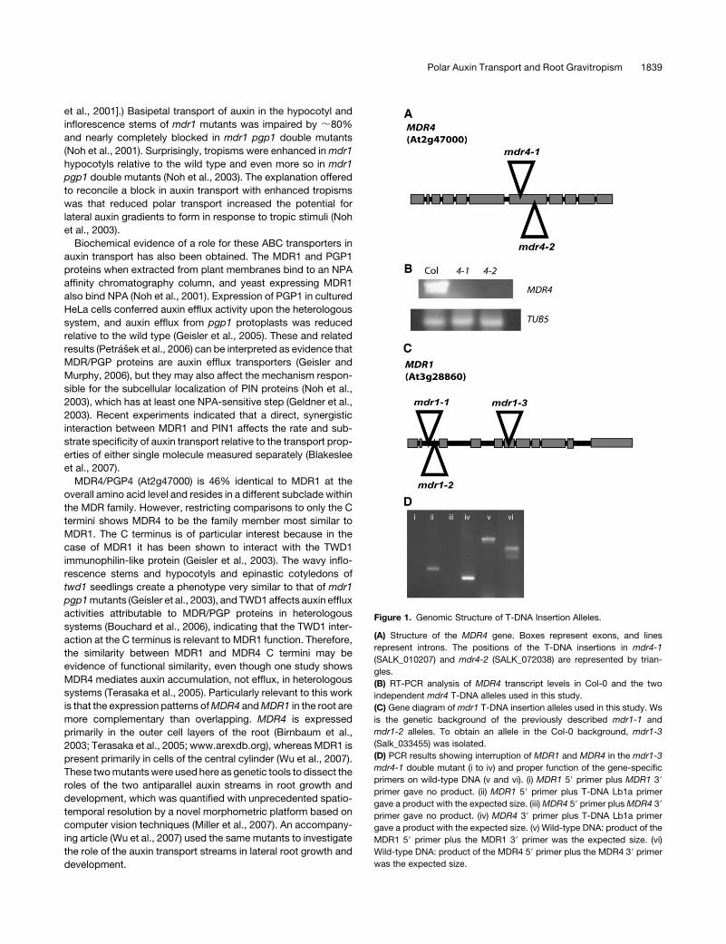
Figure 1. Genomic Structure of T-DNA Insertion Alleles.
(A) Structure of the MDR4 gene. Boxes represent exons, and lines
represent introns. The positions of the T-DNA insertions in mdr4-1
(SALK_010207) and mdr4-2 (SALK_072038) are represented by trian-
gles.
(B) RT-PCR analysis of MDR4 transcript levels in Col-0 and the two
independent mdr4 T-DNA alleles used in this study.
(C) Gene diagram of mdr1 T-DNA insertion alleles used in this study. Ws
is the genetic background of the previously described mdr1-1 and
mdr1-2 alleles. To obtain an allele in the Col-0 background, mdr1-3
(Salk_033455) was isolated.
(D) PCR results showing interruption of MDR1 and MDR4 in the mdr1-3
mdr4-1 double mutant (i to iv) and proper function of the gene-specific
primers on wild-type DNA (v and vi). (i) MDR1 59 primer plus MDR1 39
primer gave no product. (ii) MDR1 59 primer plus T-DNA Lb1a primer
gave a product with the expected size. (iii) MDR4 59 primer plus MDR4 39
primer gave no product. (iv) MDR4 39 primer plus T-DNA Lb1a primer
gave a product with the expected size. (v) Wild-type DNA: product of the
MDR1 59 primer plus the MDR1 39 primer was the expected size. (vi)
Wild-type DNA: product of the MDR4 59 primer plus the MDR4 39 primer
was the expected size.
Polar Auxin Transport and Root Gravitropism 1839
RESULTS
The experiments described here were performed with T-DNA
insertion mutants of two genes in the MDR family of ABC
transporters. The mdr1-1 and mdr1-2 null alleles (Wassilewskija
[Ws] ecotype) were isolated from the University of Wisconsin
collection and described in detail by Noh et al. (2001). The mdr4
alleles used here are in the Columbia-0 (Col-0) ecotype, and their
characterization is presented in Figures 1A and 1B. To generate
mdr1 mdr4 double mutants without mixing ecotypes, the mdr1-3
knockout allele in the Col-0 background was isolated. Descrip-
tion of the mdr1-3 allele and PCR results to document the double
mutant genotype are presented in Figures 1C and 1D.
Genetic Dissection of Basipetal and Acropetal Auxin
Transport Streams
The extent to which polar auxin transport depends on MDR1 in
both axial directions was examined by measuring the effects of
mdr1 knockout mutations on the movement of locally applied3H-indole-3-acetic acid (3H-IAA). The data in Figure 2A show that
acropetal transport of auxin placed near the primary root-shoot
junction of mdr1-1 and mdr1-2 seedlings was only ;20% of the
wild type. A ProDR5:b-glucuronidase (GUS)–based assay was
designed to independently test the conclusion that acropetal
auxin transport is highly MDR1 dependent in root. Endogenous
levels of ProDR5-driven GUS activity in apical portions of roots
quantified with a 4-methylumbelliferyl-b-D-glucuronide (MUG)
assay were 30% lower in mdr1 than the wild type, indicating that
impaired acropetal transport resulted in less auxin in general in
the mdr1 root. Application of IAA to the root-shoot junction raised
the GUS activity in the apical half of the root approximately
fivefold in the wild type but less than twofold in mdr1-1 (Figure
1B). The interpretation that much less IAA was transported
acropetally in mdr1-1 compared with the wild type is valid only
if sensitivity of the signaling mechanism linking auxin to the DR5
promoter is similar in the two genotypes. Figure 1C shows that
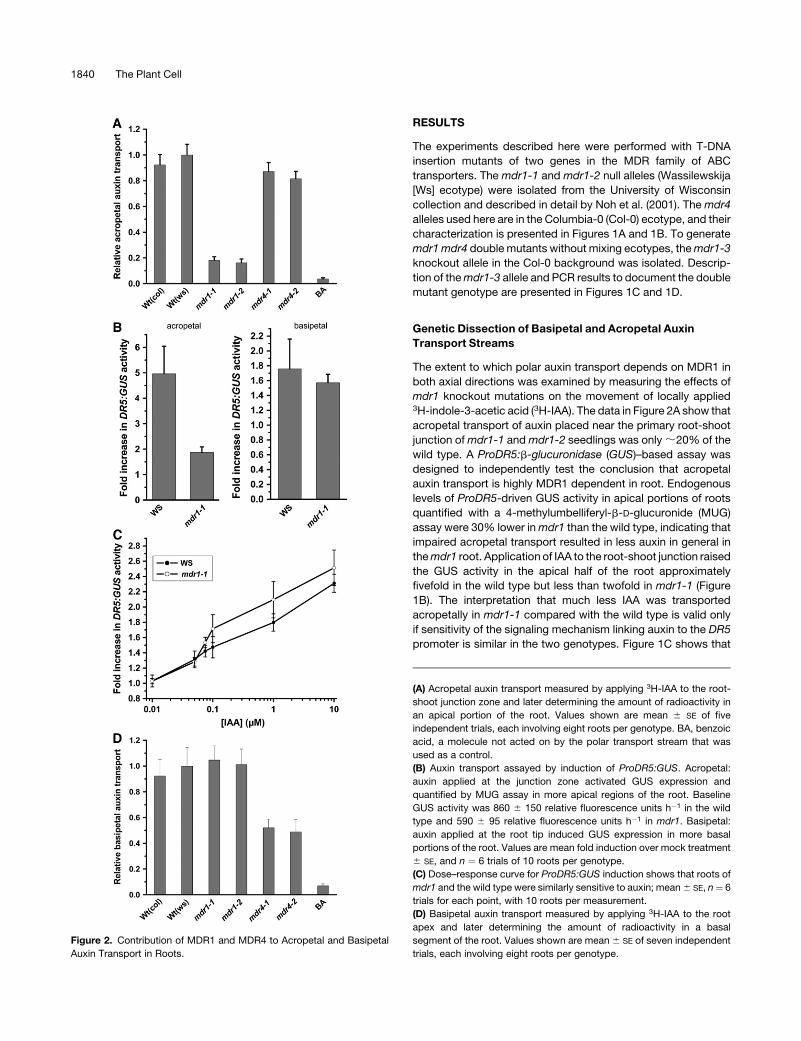
Figure 2. Contribution of MDR1 and MDR4 to Acropetal and Basipetal
Auxin Transport in Roots.
(A) Acropetal auxin transport measured by applying 3H-IAA to the root-
shoot junction zone and later determining the amount of radioactivity in
an apical portion of the root. Values shown are mean 6 SE of five
independent trials, each involving eight roots per genotype. BA, benzoic
acid, a molecule not acted on by the polar transport stream that was
used as a control.
(B) Auxin transport assayed by induction of ProDR5:GUS. Acropetal:
auxin applied at the junction zone activated GUS expression and
quantified by MUG assay in more apical regions of the root. Baseline
GUS activity was 860 6 150 relative fluorescence units h�1 in the wild
type and 590 6 95 relative fluorescence units h�1 in mdr1. Basipetal:
auxin applied at the root tip induced GUS expression in more basal
portions of the root. Values are mean fold induction over mock treatment
6 SE, and n ¼ 6 trials of 10 roots per genotype.
(C) Dose–response curve for ProDR5:GUS induction shows that roots of
mdr1 and the wild type were similarly sensitive to auxin; mean 6 SE, n¼ 6
trials for each point, with 10 roots per measurement.
(D) Basipetal auxin transport measured by applying 3H-IAA to the root
apex and later determining the amount of radioactivity in a basal
segment of the root. Values shown are mean 6 SE of seven independent
trials, each involving eight roots per genotype.
1840 The Plant Cell
ProDR5:GUS was induced by a wide range of exogenous auxin
concentrations similarly in the roots of mdr1-1 and the wild type.
Thus, the 3H-IAA and ProDR5:GUS results both demonstrate
that loss of MDR1 greatly impairs acropetal IAA transport. The3H-IAA assay is more sensitive than an assay of GUS activity, but
it relies on the IAA not being metabolized during the course of the
experiment. The ProDR5:GUS-based assay is less sensitive but
may have more physiological relevance because it measures the
output of an auxin signal transduction chain. A difference be-
tween two genotypes in the ProDR5:GUS assay means that the
difference in transport is sufficiently large to affect an auxin
response. The two independent methods of measuring polar
auxin transport both showed a major role for MDR1 in moving
physiologically relevant amounts of auxin toward the tip of roots.
The two assays were also used to test basipetal transport. Figure
1D shows that neither knockout allele of mdr1 differed from the
wild type with respect to basipetal auxin movement in the
primary root. The results of the radioactive assays were again
confirmed by experiments with ProDR5:GUS auxin reporter
plants (Figure 1B). Thus, MDR1 plays a critical role in acrop-
etal but not basipetal auxin transport. A complementary set
of experiments was performed with mdr4 knockout mutants.
Movement of 3H-IAA in the roots of two separate mdr4 knockout
alleles was measured. Both mdr4 alleles displayed normal
acropetal IAA transport (Figure 2A), but basipetal auxin transport
was reduced by ;50% (Figure 2D), consistent with previous
findings by Terasaka et al. (2005) who used a different allele and
different methods. Thus, ;80% of the acropetal auxin transport
stream depends on MDR1, and 50% of the basipetal stream
depends on MDR4.
Spurious Curvature and Altered Auxin Distribution in
Vertical Roots of mdr1
An observable mdr1 phenotype having a plausible connection
with its defect in acropetal auxin transport is a wavy root (Figure
3A). A newly developed computer vision tool was used to
quantify this phenotype. The technique is described in detail in
Miller et al. (2007). Briefly, electronic images of roots were
acquired at 7.5-min intervals by a CCD camera equipped with
a macro lens. Custom software developed by Miller et al. (2007)
extracted a smoothed set of root midline points from each image
in the time series and then fit polynomials to the family of midline
point sets. Mathematical analysis quantified curvature (K, in units
of mm�1) at ;12-mm intervals along the root axis. K is either
positive or negative (curvature is either concave or convex)
relative to a reference axis, which is the vertical in this case. To
quantify the waviness of the mdr1 roots, the absolute curvature
at each point along the midline was summed. A perfectly straight
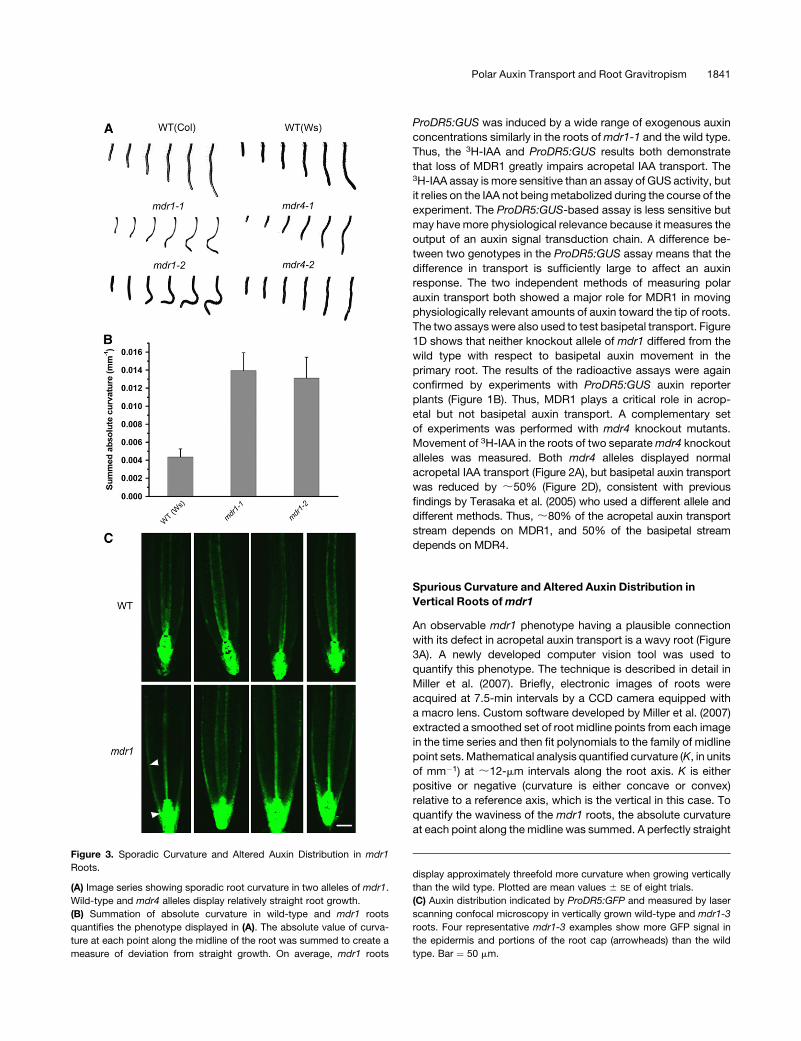
Figure 3. Sporadic Curvature and Altered Auxin Distribution in mdr1
Roots.
(A) Image series showing sporadic root curvature in two alleles of mdr1.
Wild-type and mdr4 alleles display relatively straight root growth.
(B) Summation of absolute curvature in wild-type and mdr1 roots
quantifies the phenotype displayed in (A). The absolute value of curva-
ture at each point along the midline of the root was summed to create a
measure of deviation from straight growth. On average, mdr1 roots
display approximately threefold more curvature when growing vertically
than the wild type. Plotted are mean values 6 SE of eight trials.
(C) Auxin distribution indicated by ProDR5:GFP and measured by laser
scanning confocal microscopy in vertically grown wild-type and mdr1-3
roots. Four representative mdr1-3 examples show more GFP signal in
the epidermis and portions of the root cap (arrowheads) than the wild
type. Bar ¼ 50 mm.
Polar Auxin Transport and Root Gravitropism 1841
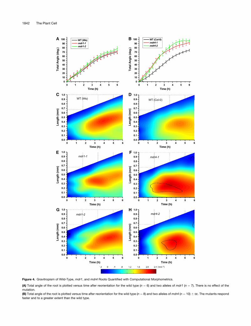
Figure 4. Gravitropism of Wild-Type, mdr1, and mdr4 Roots Quantified with Computational Morphometrics.
(A) Total angle of the root is plotted versus time after reorientation for the wild type (n ¼ 6) and two alleles of mdr1 (n ¼ 7). There is no effect of the
mutation.
(B) Total angle of the root is plotted versus time after reorientation for the wild type (n ¼ 8) and two alleles of mdr4 (n ¼ 10) 6 SE. The mutants respond
faster and to a greater extent than the wild type.
1842 The Plant Cell
root would give a sum of zero, and a wavy root would give a
positive value even if it had equal amounts of positive and
negative (convex and concave) curvature. Figure 3B shows that
both alleles of mdr1 displayed threefold more absolute curvature
than the wild type. The example time series of images in Figure
3A show that neither allele of mdr4 nor either wild-type ecotype
displayed the spurious curves of mdr1 roots. These data may be
taken as evidence that acropetal auxin transport, which is only
20% of wild-type levels in mdr1 roots (Figure 2), is required for
balancing rates of cell elongation across vertical roots, but
basipetal auxin transport, which is only 50% of wild-type levels
in mdr4 roots, plays no detectable role in this process.
Root waving in wild-type plants is exaggerated when the
plants are grown on back-tilted, stiff agar plates (Okada and
Shimura, 1990; Rutherford and Masson, 1996; Rutherford et al.,
1998; Thompson and Holbrook, 2004). The mdr1 phenotype
shown here was similar when seedlings were grown on 0.8%
agar or 1.5% agar plates (data not shown), indicating that it may
be mechanistically distinct from conventional root waving.
The spurious curvature may result from altered auxin distribu-
tion across mdr1 roots that somehow results from impaired
acropetal transport. To explore this possibility, the ProDR5:GFP
auxin reporter was crossed into mdr1-3 seedlings, and green
fluorescent protein (GFP) was visualized in mutant and wild-type
root apices by confocal microscopy. The results show that in
almost all cases, GFP signal extended back (basally) from the tip
substantially farther in mdr1 root epidermal and lateral root cap
cells compared with the wild type (Figure 3C). In mdr1, the signal
extended into the elongation zone. It is possible that higher auxin
levels in this part of the root, if not distributed symmetrically,
would cause a large imbalance in cell elongation rates, leading to
the observed curvatures. How loss of MDR1 results in higher
levels of auxin in the epidermis is considered in the Discussion.
Normal Gravitropism in mdr1 Despite Large Defects in
Acropetal Auxin Transport
The effect of disrupted acropetal auxin transport on gravitropism
was determined by subjecting mdr1 and wild-type roots under-
going gravitropism to the morphometric analysis developed by
Miller et al. (2007). Electronic images of roots were acquired at
2-min intervals following reorientation. Midline point sets com-
putationally extracted from these images were operated on by
the newly developed analytical algorithms to obtain the tip (total)
angle. This is the angle the root tip takes with respect to the
vertical, which conventionally would be measured by a protrac-
tor or its equivalent. Figure 4A shows, surprisingly, that total
angle accruement by mdr1-1 and mdr1-2 roots was similar in
extent and time course to the wild type. All three genoptypes
began to curve within 60 min of reorientation and reached an
angle of 708 to 808 within 6 h. The Miller et al. (2007) method also
determined the distribution of curvature (K) along the root axis
over time, with spatial and temporal resolution of ;5 mm and
2 min, respectively. These results are shown in two-dimensional
plots in which K is color-coded for the z-dimension. Moving from
left to right across the plots, in the direction of time, the increas-
ing length of the midline is shown. Superimposed are the color-
coded values of K, with blue indicating low curvature and red
indicating high curvature. The preponderance of blue along the
left edge of the plots indicates that the roots were mostly straight
at the onset of the experiment. Moving rightward, with time,
redder colors appear first ;0.25 mm from the tip within the first
hour. Curvature in the wild type continued to develop over time,
concentrating in a zone centered ;0.3 mm behind the tip. Both
alleles of mdr1 developed curvature very similarly to each other
and to the Ws wild type (Figures 4C, 4E, and 4G), consistent with
the total angle determinations in Figure 4A. Thus, a major
reduction in acropetal auxin transport does not measurably
affect gravitropic curvature development.
Enhanced Gravitropism in mdr4 Mutants Deficient in
Basipetal Auxin Transport
The mdr4 mutants were evaluated with the same method but
with notably different results. Total angle accrued faster in both
alleles of mdr4 compared with the wild type over the entire 6-h
period monitored, resulting in a complete 908 reorientation over a
period during which the wild type achieved only ;708 (Figure
4B). Also, the area of curvature concentration was localized more
basally than the wild type (Figures 4F to 4H). The black contour
lines in the mdr4 plots indicate the regions of the mutant
response that differ from the wild type to a statistically significant
extent (P ¼ 0.05) as determined by two-sample two-tailed
Student’s t tests executed at each point within the plot. Both
alleles of mdr4 displayed a region of significantly higher curva-
ture than the wild type 0.4 to 0.8 mm behind the tip within 2 h of
reorientation (area bounded by the contour). These data indicate
that reduced basipetal auxin transport through the elongation
zone of the root alters the location, persistence, and/or magni-
tude of the gravitationally induced lateral auxin gradient, an
interpretation that was experimentally tested as reported below.
Both alleles of mdr4 grew on average 20 to 25% faster than the
Figure 4. (continued).
(C) Spatiotemporal distribution of gravitropic curvature (K) in the Ws wild type. Length of the root axis is plotted in the y-dimension, and time is plotted
along the x-dimension. K is color-coded and plotted in the z-dimension. Straight areas of the root are shown in cool colors, and curvature is shown as
warm colors, as shown by the horizontal color scale bar. Shown is the average of six [0] individual roots.
(D) Spatiotemporal distribution of gravitropic curvature of Col wild-type roots, with an average of eight individuals.
(E) and (G) Spatiotemporal distribution of gravitropic curvature of mdr1 roots, with an average of seven individuals. The mdr1 mutations did not affect
the response.
(F) and (H) Spatiotemporal distribution of gravitropic curvature of mdr4 roots, with an average of 10 individuals. The area of main curvature is shifted
basally relative to the wild type. The black contour lines demark areas where the difference between the mutant and the wild type is significant to a level
of P ¼ 0.05.
Polar Auxin Transport and Root Gravitropism 1843
wild type during the measurement period, all with a standard
error of ;7.5%. To produce the fairest comparison of wild-type
and mdr4 curvature distributions, eight wild-type and 10 mdr4
individuals having similar growth rates (0.125 to 0.2 mm h�1)
were used to produce the results shown in Figures 4D, 4F, and
4H. Pooling all the trials regardless of growth rate increased the
difference between both mdr4 alleles and the wild type.
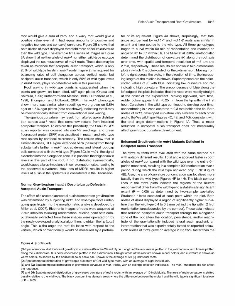
Visualizing the Auxin Gradient across Wild-Type and mdr4
Roots during Gravitropism
The ProDR5:GFP lines were used to visualize the change in
distribution of auxin signaling activity during gravitropism using a
horizontally mounted fluorescence microscope that permitted
the simultaneous monitoring of the curvature response and the
fluorescent auxin reporter. The results were somewhat variable,
with six out of 11 wild-type seedlings showing a clearly discern-
ible auxin gradient following reorientation. Three representatives
of these six are shown in Figure 5A as before-and-after pairs of
images. A distinct increase in ProDR5:GFP signal was observed
along the lower edge of the root. With mdr4 roots, 10 out of 12
trials showed an obvious auxin asymmetry, and the pattern, in
most cases, was more diffuse and against a somewhat higher
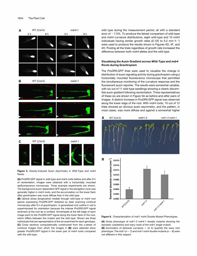
Figure 6. Characterization of mdr1 mdr4 Double Mutant Phenotypes.
(A) Gross phenotype of mdr1-3 mdr4-1 double mutants showing the
epinastic cotyledons and wavy roots of the mdr1 single mutant.
(B) Summation of absolute curvature 6 SE to quantify the wavy root
phenotype. The mdr1 (n¼ 7) and mdr1 mdr4 double mutants (n¼ 8) were
not different in this respect.
Figure 5. Gravity-Induced Auxin Asymmetry in Wild-Type and mdr4
Roots.
(A) ProDR5:GFP signal in wild-type and mdr4 roots before and after 8 h
of reorientation. Images were obtained with a horizontally mounted
epifluorescence microscope. Three example experiments are shown.
The background auxin-dependent GFP signal in the elongation zone was
generally higher in mdr4 roots, and the accumulation on the lower flank
after gravitropism was more diffuse than in the wild type.
(B) Optical slices (longitudinal medial) through wild-type or mdr4 root
apices expressing ProDR5:GFP obtained by laser scanning confocal
microscopy after 6 h of gravitropism. A generalized root outline in red is
superimposed for orientation because the intense ProDR5:GFP signal
landmark at the root tip is omitted. Arrowheads at the left edge of each
image point to the ProDR5:GFP signal along the lower flank of the root,
which differs between the mutant and the wild type. Shown are three
individuals that are representative of the six examined for each genotype.
(C) Cross sections computationally constructed from the z-series of
confocal images from which the images in (B) were selected show
greater ProDR5:GFP signal in the lower part of mdr4 roots compared
with the wild type.
1844 The Plant Cell
background of signal (Figure 5A). The general impression was
that mdr4 roots had more auxin signaling activity than the wild
type, and the gradient across the root resulting from reorientation
was not as tightly focused as the wild type. As an independent
test of this tentative conclusion, confocal microscopy was used
to examine GFP levels and distribution in serial longitudinal
optical sections through roots that had been gravistimulated for
6 h (Figure 5B). These results supported the conclusion that mdr4
roots produced a more robust gravity-induced auxin asymmetry
following gravistimulation than the wild type. Figure 5B displays
this result in two manners. First, a representative medial optical
slice is shown for three representative wild-type and three
representative mdr4 individuals. These optical slices show that
the ProDR5:GFP signal in mdr4 was brighter, more continuous,
and extended further basally than in the wild type. Second, a
z-stack of 16 optical slices was used to compute a cross-
sectional view of half of each root in the approximate region of the
distal elongation zone. The prominent crescent-shaped, contin-
uous green signal along the lower flank of mdr4 roots indicated
that a robust auxin asymmetry had developed across the root.
This signal was less extensive in the wild type, indicating that less
auxin was redistributed to the lower half of the root compared
with mdr4. Another view of the larger, broader auxin asymmetry
induced by gravity across the mdr4 root apex compared with the
wild type is presented in the form of rotating three-dimensional
reconstructions created from the series of z-sections (see Sup-
plemental Movies 1 and 2 online). Collectively, the data indicate
that slowing basipetal auxin transport (Figure 2D) by mutation of
MDR4 permits a larger, broader auxin asymmetry to develop in
response to root reorientation (Figure 5), which affects the time
course of gravitropism (Figure 4B) and the distribution of curva-
ture along the root axis (Figures 4F and 4H).
Morphometric Analysis of mdr1 mdr4 Double Mutants
Seedlings homozygous for the mdr1-3 and mdr4-1 mutations
were created by crossing and were identified by PCR genotyping
(documentation in Figure 1D). Surprisingly, combining those
mutations that individually impaired acropetal or basipetal auxin
transport in the root produced a plant without any visible phe-
notype more severe than the epinastic cotyledons and wavy root
characteristic of mdr1 mutants (Figure 6A). A detailed morpho-
metric analysis of root curvature in vertically maintained plants
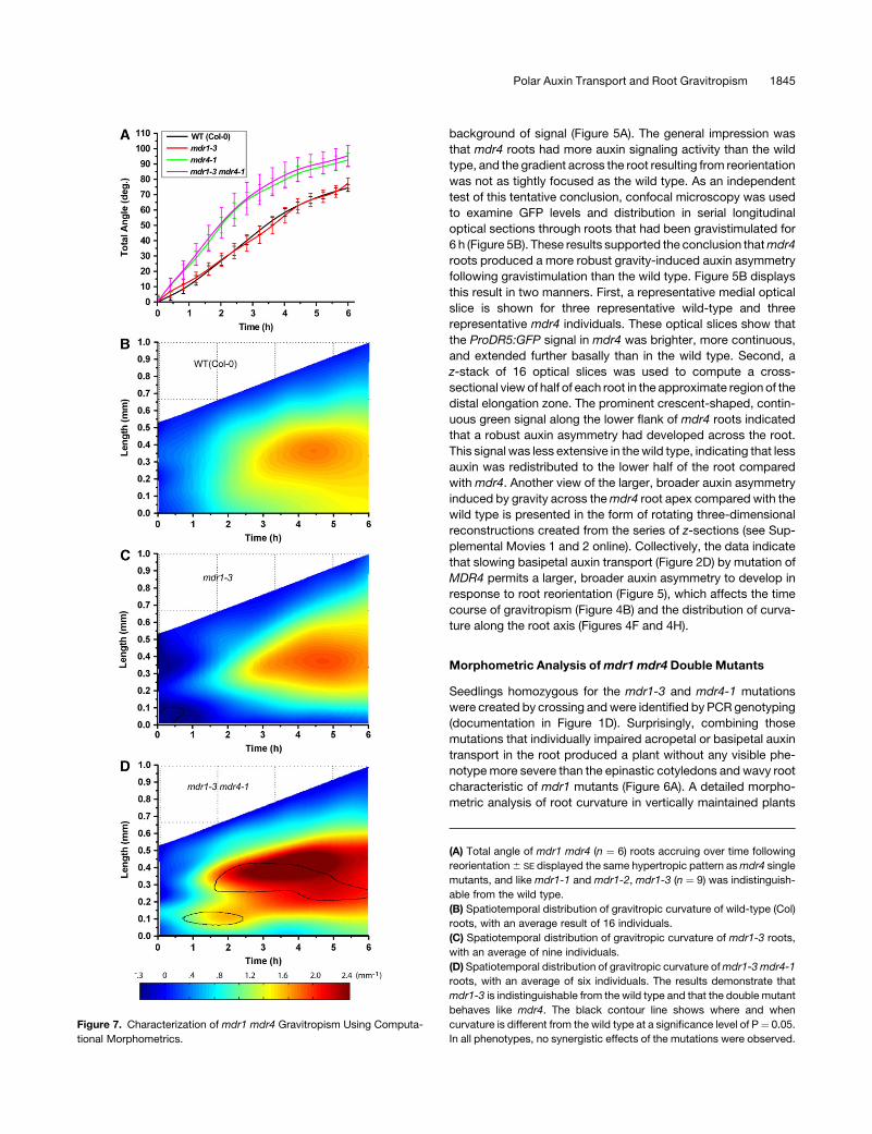
Figure 7. Characterization of mdr1 mdr4 Gravitropism Using Computa-
tional Morphometrics.
(A) Total angle of mdr1 mdr4 (n ¼ 6) roots accruing over time following
reorientation 6 SE displayed the same hypertropic pattern as mdr4 single
mutants, and like mdr1-1 and mdr1-2, mdr1-3 (n ¼ 9) was indistinguish-
able from the wild type.
(B) Spatiotemporal distribution of gravitropic curvature of wild-type (Col)
roots, with an average result of 16 individuals.
(C) Spatiotemporal distribution of gravitropic curvature of mdr1-3 roots,
with an average of nine individuals.
(D) Spatiotemporal distribution of gravitropic curvature of mdr1-3 mdr4-1
roots, with an average of six individuals. The results demonstrate that
mdr1-3 is indistinguishable from the wild type and that the double mutant
behaves like mdr4. The black contour line shows where and when
curvature is different from the wild type at a significance level of P¼ 0.05.
In all phenotypes, no synergistic effects of the mutations were observed.
Polar Auxin Transport and Root Gravitropism 1845
demonstrated that the spurious curvature of mdr1-3 roots, which
was similar to the mdr1-1 and mdr1-2 mutants in a different
ecotype, was not affected by the additional loss of MDR4 (Figure
6B). Therefore, the phenotype is tightly correlated with defective
acropetal transport but not affected by the addition of a major
decrease in basipetal transport. Morphometric analysis of mdr1-3
(in Col-0) provided an independent test of the results presented
in Figure 4 (Ws alleles). The results demonstrated that mdr1-3 did
not differ from its wild type in total angle accruement (Figure 7A)
or in the spatiotemporal distribution of gravitropic curvature
(Figures 7B and 7C). The mdr1 mdr4 double mutant displayed a
result much like mdr4 single mutants (Figure 7D). The effect of
impaired basipetal transport on gravitropism was neither ame-
liorated nor exacerbated by additionally impairing acropetal
transport. The fact that neither single mutation much affects
the phenotype produced by the other indicates that the aspects
of growth controlled by the acropetal auxin stream are indepen-
dent of those controlled by the basipetal stream. Growth and
development mediated by the two streams appear to be more
independent than interdependent.
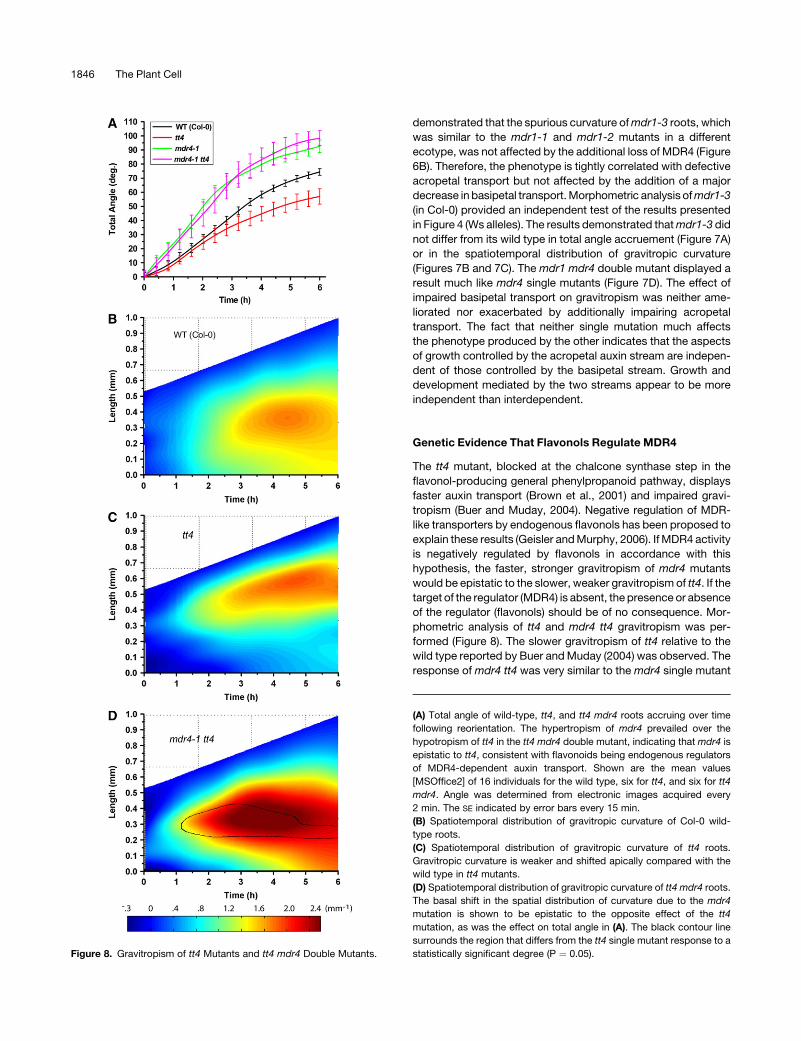
Genetic Evidence That Flavonols Regulate MDR4
The tt4 mutant, blocked at the chalcone synthase step in the
flavonol-producing general phenylpropanoid pathway, displays
faster auxin transport (Brown et al., 2001) and impaired gravi-
tropism (Buer and Muday, 2004). Negative regulation of MDR-
like transporters by endogenous flavonols has been proposed to
explain these results (Geisler and Murphy, 2006). If MDR4 activity
is negatively regulated by flavonols in accordance with this
hypothesis, the faster, stronger gravitropism of mdr4 mutants
would be epistatic to the slower, weaker gravitropism of tt4. If the
target of the regulator (MDR4) is absent, the presence or absence
of the regulator (flavonols) should be of no consequence. Mor-
phometric analysis of tt4 and mdr4 tt4 gravitropism was per-
formed (Figure 8). The slower gravitropism of tt4 relative to the
wild type reported by Buer and Muday (2004) was observed. The
response of mdr4 tt4 was very similar to the mdr4 single mutant
Figure 8. Gravitropism of tt4 Mutants and tt4 mdr4 Double Mutants.
(A) Total angle of wild-type, tt4, and tt4 mdr4 roots accruing over time
following reorientation. The hypertropism of mdr4 prevailed over the
hypotropism of tt4 in the tt4 mdr4 double mutant, indicating that mdr4 is
epistatic to tt4, consistent with flavonoids being endogenous regulators
of MDR4-dependent auxin transport. Shown are the mean values
[MSOffice2] of 16 individuals for the wild type, six for tt4, and six for tt4
mdr4. Angle was determined from electronic images acquired every
2 min. The SE indicated by error bars every 15 min.
(B) Spatiotemporal distribution of gravitropic curvature of Col-0 wild-
type roots.
(C) Spatiotemporal distribution of gravitropic curvature of tt4 roots.
Gravitropic curvature is weaker and shifted apically compared with the
wild type in tt4 mutants.
(D) Spatiotemporal distribution of gravitropic curvature of tt4 mdr4 roots.
The basal shift in the spatial distribution of curvature due to the mdr4
mutation is shown to be epistatic to the opposite effect of the tt4
mutation, as was the effect on total angle in (A). The black contour line
surrounds the region that differs from the tt4 single mutant response to a
statistically significant degree (P ¼ 0.05).
1846 The Plant Cell
and unlike the slow tt4 result, both in terms of total angle
accruement and spatiotemporal curvature distribution (Figure
5). Thus, mdr4 was epistatic to tt4, consistent with the hypothesis
that flavonols regulate MDR4 function in ways relevant to the
mechanism of gravitropism.
DISCUSSION
Much of root growth and development has some connection to
auxin transport. Sometimes the connection is first established by
an effect of a polar auxin transport inhibitor, such as NPA.
However, it isn’t known exactly how far the inhibitor has spread,
the extent of penetration, or the most causal site of action.
Instead of inhibitors, this work relies on mutations in a pair of
related genes to distinguish the effects of acropetal and basip-
etal auxin transport on root growth and gravitropism. One of the
surprising results is that acropetal transport can be impaired by
80% in the case of the mdr1 mutant without affecting the
gravitropic response, which was quantified with a new, high-
resolution technique. This result calls into question the prevailing
model of root gravitropism in which auxin from the acropetal
stream is asymmetrically redistributed to the basipetal stream so
that the lower portion of a reoriented root receives more.
According to this model, impaired gravitropism would be ex-
pected to result from a major disruption in acropetal transport,
but gravitropism proceeded normally in space and time in all
three alleles of mdr1 tested, in two different ecotypes. Possibly,
the remaining 20% of acropetal auxin transport is sufficient to
bring about normal gravitropism. That seems unlikely because
the remaining 20% was not sufficient for proper control of
straight growth (Figure 2). One possibility is that extra auxin
synthesis at the tip of mdr1 roots compensates for the reduced
delivery from the acropetal stream so that the basipetal stream is
adequately supplied. The auxin maximum at the apex of mdr1
roots (as visualized by ProDR5:GUS or ProDR5:GFP) was similar
to the wild type (Figure 3C; Wu et al., 2007), which indicates that
the acropetal stream either does not contribute to this feature or
that the hypothetical compensatory synthesis faithfully restores
the strength and pattern of the signal. The standard fountain
model of auxin flow in roots (Swarup and Bennett, 2003) has
been elaborated to include reflux loops that cause auxin to
recirculate from epidermal and cortical cells back into the stele,
where it rejoins the acropetal stream (Blilou et al., 2005). Perhaps
MDR1 participates in the mechanism that returns auxin to the
stele (Figure 9), which could explain why mdr1 roots appear to
have higher auxin signaling in the epidermal cells as far basally as
the elongation zone (Figure 3C). This hypothesis is supported by
work that describes MDR1 and PIN1 colocalization in the endo-
dermis and pericycle as a central component of the auxin reflux
loop (Blakeslee et al., 2007). Disruption of the reflux process
could be a proximal cause of the erratic changes in growth
direction. Indeed, the MDR1 protein is present on the inner, but
less so on the outer, periclinal cell membrane of cortical cells (Wu
et al., 2007), consistent with it being responsible for refluxing
auxin toward the central cylinder from the cortex.
The basipetal stream is thought to be the mechanism that
delivers auxin asymmetrically from the tip after reorientation, so
impairments in it might be expected to impair curvature devel-
opment, as is the case in pin2 mutants (Chen et al., 1998).
However, this work demonstrates that impaired basipetal trans-
port in mdr4 roots enhances rather than impairs gravitropism
(Figure 4). A previous study of mdr4 mutants (different alleles
than those used here) by Terasaka et al. (2005) concluded that
gravitropism was slower than the wild type, but their experiments
may have been compromised by some non-ideal environmental
influence or methodology because the wild type responded only
108 over 6 h and the mutant was even slower. The ;708 of re-
sponse accruing over 6 h for the wild-type presented in Figure 4
agrees well with several other quantitative studies from different
labs (Wolverton et al., 2002; Buer and Muday, 2004; Young et al.,
2006). The high resolution of the methods used here and the
statistical treatment of the responses of two well-characterized
mdr4 alleles make this finding of hypertropism in mdr4 mutants
very robust.
The results presented here leave unanswered questions about
how a decrease in basipetal auxin transport results in a more
vigorous gravitropic response, but some possible explanations
can be considered. According to Chen et al. (1998), the pin2
mutant is unable to transport an auxin asymmetry to the elon-
gation zone, leading to an agravitropic phenotype. Based on
quantitative expression data provided by Birnbaum et al. (2003),
PIN2 mRNA decreases with distance from the apex, while MDR4
mRNA increases. The contribution of these two proteins to ba-
sipetal auxin flux may follow the same complementary pattern.
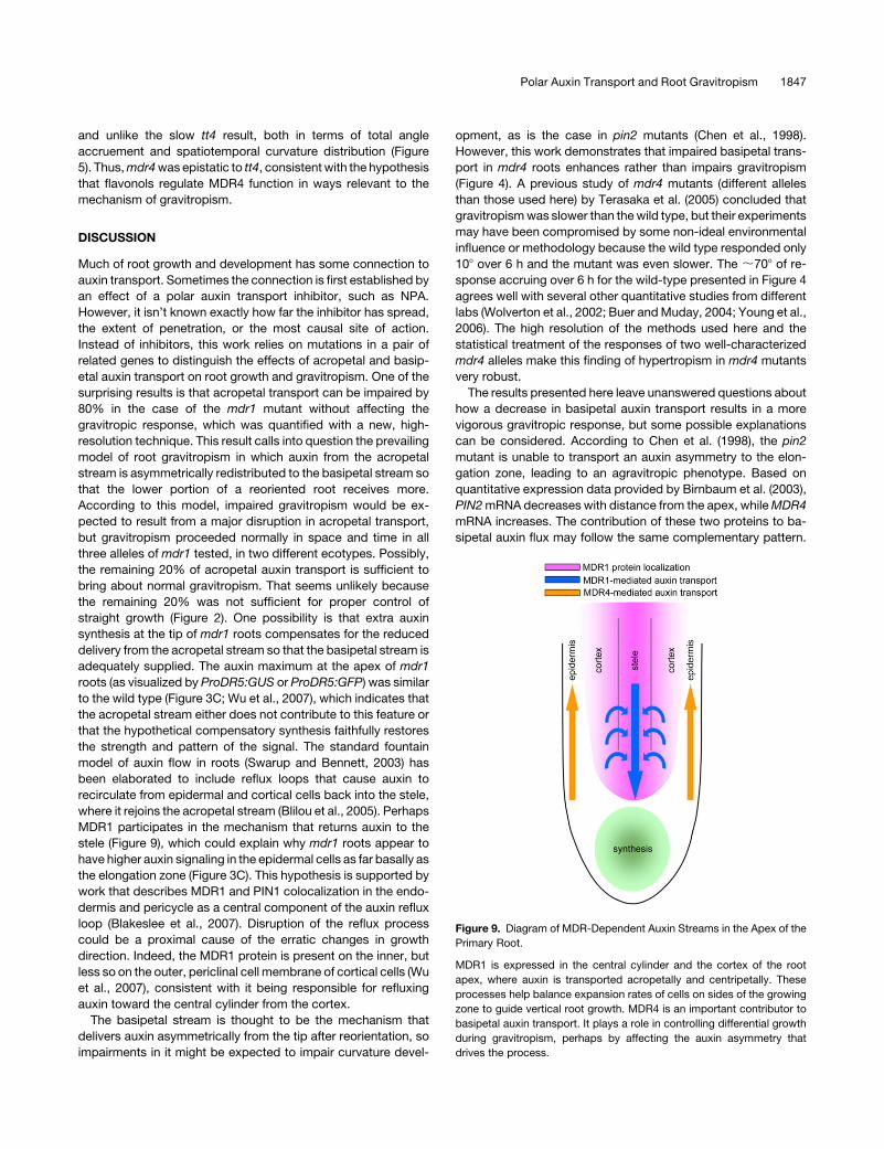
Figure 9. Diagram of MDR-Dependent Auxin Streams in the Apex of the
Primary Root.
MDR1 is expressed in the central cylinder and the cortex of the root
apex, where auxin is transported acropetally and centripetally. These
processes help balance expansion rates of cells on sides of the growing
zone to guide vertical root growth. MDR4 is an important contributor to
basipetal auxin transport. It plays a role in controlling differential growth
during gravitropism, perhaps by affecting the auxin asymmetry that
drives the process.
Polar Auxin Transport and Root Gravitropism 1847
Slower basipetal movement of auxin in the elongation zone of
mdr4 could lead to higher auxin levels behind the tip, altering the
gravity-induced auxin asymmetry in a way that leads to faster
curvature. The evidence for this is the higher background GFP
signal in most mdr4 roots viewed on the horizontal fluores-
cence microscope during gravitropism compared with the wild
type (Figure 3A). In addition, Terasaka et al. (2005) measured a
1.7-fold increase in free auxin content in the apical 1.5 mm of
mdr4 roots. The cross-sectional views computed from the con-
focal optical slices showed more broadly distributed ProDR5:
GFP signal in mdr4, consistent with the results of Terasaka et al.
(2005) and with the higher overall signal in Figure 3A. By contrast,
the elevated auxin signaling observed in pin2 mutants is re-
stricted to the lateral root cap (Swarup et al., 2005). Also, no
gravitationally induced auxin gradient reaches the elongation
zone of pin2 roots (Abas et al., 2006).
Another surprising result of this work is that combining a
mutation that reduces 80% of acropetal auxin transport with one
that blocks 50% of basipetal transport did not synergistically
impair root development. Instead, the modest phenotypes of
each mutant combined additively in the double mutant, as if they
were independent. A treatment with NPA that might have a
similar effect on acropetal and basipetal transport as these two
mutations would affect root growth and development to a much
more significant degree. The difference between major pharma-
cological and genetic blocks of auxin transport may be due to the
fact that a global NPA treatment would block auxin efflux from all
cells conducting NPA-sensitive auxin efflux, whereas the muta-
tions used here may specifically affect a certain subset of the
processes that NPA targets. It will be interesting to observe the
effects of knocking out additional MDR-like family members that
are known to be expressed in the root. Perhaps higher-order
mutants will more closely resemble the NPA-treated phenotype
characterized by highly distorted root development. Another
possible reason why the double mutant was not more severely
affected is that auxin accumulation due to altered auxin transport
may upregulate PIN gene expression in a way that mitigates the
developmental effects of the mdr mutations. Changes in PIN4
and PIN2 expression were observed in response to treatment
with NPA or mutation of PIN1, and these changes were interer-
preted as compensatory buffers against large developmental
defects arising from altered auxin distribution (Vieten et al., 2005).
The capabilities of the morphometric analysis platform em-
ployed here (Miller et al., 2007) made it possible to evaluate the
epistatic relationship between two mutants with subtle but
distinct gravitropism phenotypes. The tt4 mutant, which lacks
flavonols, has an elevated basipetal auxin transport rate and is
slower to respond than the wild type (Brown et al., 2001; Buer
and Muday, 2004). The mdr4 mutant shows reduced basipetal
auxin transport and responds faster. A tool capable of quantita-
tively evaluating these opposite responses made it possible to
test the hypothesis that MDR-like ABC transporters, and the
auxin transport processes they participate in, are regulated by
flavonoid compounds. The flavonoid compound quercetin is
known to affect auxin efflux and has been reported to bind
to mammalian (Ferte et al., 1999) MDR transporters. PGP1-
mediated auxin efflux in heterologous systems is also sensitive to
physiologically relevant amounts of quercetin (Geisler et al.,
2005). The fact that the tt4 mdr4 double mutant displayed the
phenotype of mdr4 instead of the slow tt4-type response indi-
cates that MDR4 is downstream of, or regulated by, products of
the chalcone synthase enzyme. These results support the idea
that flavanoids produced by the general phenylpropanoid path-
way are endogenous regulators of plant growth and develop-
ment that exert their effects at least in part by regulating the
activity of MDR-like ABC transporters, which control root growth
by participating in auxin transport.
METHODS
Plant Growth Conditions
Seeds of Arabidopsis thaliana were sown on Petri plates containing 0.8%
agarose, 0.53 Murashige and Skoog salts, and 0.5% sucrose (w/v). The
planted plates were stored for 2 to 4 d at 48C before being placed in a
growth chamber with a 16-h-light/8-h-dark cycle.
Radioactive Auxin Transport Assays
To measure acropetal auxin transport, 5-mL droplets containing 3 mM3H-IAA (ARC American Radio-Chemical) in 0.8% agarose (specific activ-
ity of 20 Ci/mmol) were applied to the junction zone of 5- or 6-d-old light-
grown seedlings. Total root length was ;20 mm. After 3 h, the root was
cut 4 mm below the junction zone. The remaining apical sections of eight
seedlings were placed in 5 mL of scintillation fluid overnight, and radio-
activity was counted in a Beckman LS6500 scintillation counter.
To measure basipetal auxin transport, droplets containing 4 mM 3H-IAA
in 0.8% agarose were placed in contact with the root apex. After 5 h, the
apical 5 mm of the root was excised and discarded, and the radioactivity
in the remainder of the root was determined. Benzoic acid control
experiments were performed by the same method. The same concen-
tration of benzoic acid was used in control experiments, but because its
specific activity was fourfold higher than for IAA, the counts per minute
were divided by four.
ProDR5:GUS-Based Auxin Transport Assay
To measure transport in the acropetal direction, solidified 10-mL droplets
of agarose containing 3 mM IAA were placed on the root-shoot junction of
seedlings grown for 5 d in continuous light. The droplets were removed
after 3 h. The 2 mm of root tissue closest to the junction zone was excised
and discarded. The 10 mm of tissue directly below this cut was harvested
from 10 similarly treated plants per trial and used in a MUG assay of GUS
activity by a method based on that of Cervera (2005). The tissue segments
were ground in 100 mL of extraction buffer consisting of 50 mM sodium
phosphate buffer, pH 7.0, 10 mM DTT, 1 mM Na2EDTA, 0.1% sodium
lauryl sarcosine, and 0.1% Triton X-100.
The samples were then centrifuged at 13,000 rpm for 5 min in a 48C
microcentrifuge. A 50-mL aliquot of the supernatant was added to 500 mL
of prewarmed extraction buffer containing 0.22 mg of MUG. The reaction
was incubated at 378C. A 100-mL aliquot of the reaction was added to
900 mL of MUG stop buffer (0.2M Na2CO3) at 3-h intervals. Fluorescence
was measured using a Tecan fluorimeter (Tecan Group). Independent
trials were performed, and the results averaged as indicated in the figure
legends.
To measure transport in the basipetal direction, IAA droplets were
placed in contact with the extreme tip of 10 roots growing on vertical agar
plates. After 3 h, all but the apical-most 2 mm of each root was harvested.
Tissue preparation and assay of GUS activity was performed as de-
scribed above. Independent trials were performed and the results aver-
aged as indicated in the figure legends.
1848 The Plant Cell
To determine the auxin sensitivity of mdr1 and wild-type roots, seed-
lings growing on plates as described above were lightly sprayed with an
IAA solution of the indicated concentration using an aerosol sprayer.
Three hours after IAA application, roots from 10 seedlings for each
genotype, per trial, were harvested and subjected to GUS activity analysis
as described above.
Morphometric Analysis of Vertical Root Growth and Gravitropism
To quantify the waviness of vertically grown roots, wild-type and mutant
plants grown in light for 5 d were transferred to fresh agarose plates and
aligned so that the root tips of both could be simultaneously imaged. After
30 min of recovery time, electronic images of the roots were captured at
7.5-min intevals at 80 pixels mm�1 resolution for 12.5 h. Using the
analytical methods described by Miller et al. (2007), absolute curvature
was calculated at each point along the midline and then summed to
obtain a single value for each root. The average of the indicated number of
separate trials per genotype is plotted along with standard error of the
mean.
To quantify gravitropic curvature development, one wild-type and one
mutant plant grown for 4 d as above were placed <2 mm apart on a new
0.8% agarose plate and maintained vertically for 30 min to recover from
handling before the plate was rotated 908 and imaged at 2-min intervals at
;160 pixels mm�1 resolution with electronic cameras as described by
Miller et al. (2007) for a period of 6 h. The spatiotemporal distribution of
curvature (K) was calculated for each individual root by the method of
Miller et al. (2007), and then the results for a given genotype were
averaged. A two way t test was performed at every point in the plot to
determine significantly different (P¼ 0.05) areas of curvature (Miller et al.,
2007). Regions of the spatiotemporal curvature plot that differ signifi-
cantly from the wild type are bounded by a black contour line.
Epifluorescence Microscopy
After crossing the ProDR5:GFP in Col-0 lines with mdr4-1, homozygous
plants were selected from the F2 generation by PCR screening. The
resulting plants were checked for GFP fluorescence, and those showing
signal were allowed to self-pollinate until a stable line homozygous for the
mutation and the auxin reporter was isolated. To image GFP fluorescence
in roots with a horizontal microscope while gravitropism was underway,
seedlings were mounted in a chamber constructed from 30 3 70 3 3-mm
plexiglass slides milled to create a central hole measuring 17 3 35 mm
surrounded by a 1 3 1-mm recess. A nonmilled slide was affixed to it to
create a backed chamber. Seeds were sown between a 30 3 15 3 2-mm
slice of 0.8% agarose media and a 22 3 40-mm cover slip. This assembly
was placed into the recessed microscope slide chamber and held in place
with vacuum grease. Each complete chamber was placed in a standard
Petri dish containing 2 mL of deionized water to prevent drying, and the
dish was sealed with Parafilm. After 48 h at 48C, the dishes were moved to
the growth chamber. After 3 d of growth, a chamber with seedlings was
attached to a rotatable stage of a horizontally mounted Nikon Optiphot
2 microscope equipped with a filter cube that excited the sample with
490-nm light and collected emission through a 525-nm filter after reflec-
tion from a 505-nm dichroic mirror. The chambers were maintained
vertical for 1 h, and then the stage was rotated 908. Images were captured
at 1-h intervals at standardized exposure and gain settings.
Laser Scanning Confocal Microscopy
Confocal microscopy was performed with a Zeiss LSM 510 laser scan-
ning confocal microscope equipped with a C-Apochromat 340 water
immersion lens and a plan-Neofluar 310 air lens. A root mounted in water
between a slide and cover slip was excited with the 488-nm line from a
30-mW argon gas laser. Channel mode detection was used to record the
emission. A dichroic mirror in the fluorescence emission path directed
wavelengths shorter than 545 nm to a 505-nm long-pass filter to isolate
the GFP signal. Propidium iodide staining to show outlines of cells was not
performed to prevent its fluorescence from contaminating the GFP signal
at the high detector gains used to capture the ProDR5:GFP signal in
Figures 3C, 5B, and 5C. Roots not containing ProDR5:GFP displayed no
signal, so the results shown in these figures can be interpreted as strictly
the product of the auxin-responsive promoter.
T-DNA Mutant Isolation and PCR Screening
Mutant lines were described by Noh et al. (2001) or in Figure 1. For mdr1-3
and the two alleles of mdr4, genomic DNA was chloroform extracted, and
PCR was performed with the T-DNA primer LB1a (59-TGGTTCACG-
TAGTGGGCCATCG-39) and the following gene-specific primers: mdr1-3
genotyping primers, MDR1 F (59-AAGTGTTGCTGTGATTCCCGGAATC-39)
and MDR1 R (59-ACTGCTCCCATGATTGAGTAAGGCCA-39); mdr4-1 and
mdr4-2 genotyping primers, MDR4 F (59-GCGCAATACCTCTTTGGTTCAT-
TAACTTCCCTGC-39) and MDR4 R (59-GCGCATTATCCAACACTCTTCCT-
GATTCCACAC-39).
Accession Numbers
The Arabidopsis Genome Initiative locus identifiers for genes described in
this article are as follows: MDR1 (also known as PGP19 and MDR11;
At3g28860), MDR4 (also known as PGP4; At2g47000), and TT4 (also
known as CHS; At5g13930).
Supplemental Data
The following materials are available in the online version of this article.
Supplemental Movie 1. Auxin Asymmetry in Gravistimulated Wild-
Type Root.
Supplemental Movie 2. Auxin Asymmetry in Gravistimulated mdr4
Root.
ACKNOWLEDGMENTS
This work was supported by National Science Foundation Grants
IOB-0517350 and DBI-0421266 to E.P.S. We would like to thank the
ABRC for supplying T-DNA mutant seeds and Patrick Masson (Univer-
sity of Wisconsin) for use of the horizontal fluorescence microscope.
Received March 13, 2007; revised May 7, 2007; accepted May 15, 2007;
published June 8, 2007.
REFERENCES
Abas, L., Benjamins, R., Malenica, N., Paciorek, T., Wisniewska, J.,
Moulinier-Anzola, J.C., Sieberer, T., Friml, J., and Luschnig, C.
(2006). Intracellular trafficking and proteolysis of the Arabidopsis
auxin-efflux facilitator PIN2 are involved in root gravitropism. Nat.
Cell Biol. 8: 249–256.
Birnbaum, K., Shasha, D.E., Wang, J.Y., Jung, J.W., Lambert, G.M.,
Galbraith, D.W., and Benfey, P.N. (2003). A gene expression map of
the Arabidopsis root. Science 302: 1956–1960.
Blakeslee, J.J., et al. (2007). Interactions among PIN-FORMED and
P-glycoprotein auxin transporters in Arabidopsis. Plant Cell 19:
131–147.
Blilou, I., Xu, J., Wildwater, M., Willemsen, V., Paponov, I., Friml, J.,
Heidstra, R., Aida, M., Palme, K., and Scheres, B. (2005). The PIN
Polar Auxin Transport and Root Gravitropism 1849
auxin efflux facilitator network controls growth and patterning in
Arabidopsis roots. Nature 433: 39–44.
Bouchard, R., Bailly, A., Blakeslee, J.J., Oehring, S.C., Vincenzetti, V.,
Lee, O.R., Paponov, I., Palme, K., Mancuso, S., Murphy, A.S., Schulz,
B., and Geisler, M. (2006). Immunophilin-like TWISTED DWARF1 mod-
ulates auxin efflux activities of Arabidopsis P-glycoproteins. J. Biol.
Chem. 281: 30603–30612.
Brown, D.E., Rashotte, A.M., Murphy, A.S., Normanly, J., Tague,
B.W., Peer, W.A., Taiz, L., and Muday, G.K. (2001). Flavonoids act
as negative regulators of auxin transport in vivo in Arabidopsis. Plant
Physiol. 126: 524–535.
Buer, C.S., and Muday, G.K. (2004). The transparent testa4 mutation
prevents flavonoid synthesis and alters auxin transport and the response
of Arabidopsis roots to gravity and light. Plant Cell 16: 1191–1205.
Cervera, M. (2005). Histochemical and fluorometric assays for uidA
(GUS) gene detection. Methods Mol. Biol. 286: 203–214.
Chen, R., Hilson, P., Sedbrook, J., Rosen, E., Caspar, T., and Masson,
P.H. (1998). The Arabidopsis thaliana AGRAVITROPICA 1 gene en-
codes a component of the polar-auxin-transport efflux carrier. Proc.
Natl. Acad. Sci. USA 95: 15112–15117.
Dudler, R., and Hertig, C. (1992). Structure of an mdr-like gene from
Arabidopsis thaliana. J. Biol. Chem. 267: 5882–5888.
Evans, M.L., Ishikawa, H., and Estelle, M.A. (1994). Responses of Arabi-
dopsis roots to auxin studied with high temporal resolution - Compar-
ison of wild-type and auxin-response mutants. Planta 194: 215–222.
Ferte, J., Kuhnel, J.M., Chapuis, G., Rolland, Y., Lewin, G., and
Schwaller, M.A. (1999). Flavonoid-related modulators of multidrug
resistance: Synthesis, pharmacological activity, and structure-activity
relationships. J. Med. Chem. 42: 478–489.
Friml, J. (2003). Auxin transport - Shaping the plant. Curr. Opin. Plant
Biol. 6: 7–12.
Friml, J., Wisniewska, J., Benkova, E., Mendgen, K., and Palme, K.
(2002). Lateral redistribution of auxin efflux regulator PIN3 mediates
tropism in Arabidopsis. Nature 415: 806–809.
Geisler, M., et al. (2005). Cellular efflux of auxin catalyzed by the
Arabidopsis MDR/PGP transporter AtPGP1. Plant J. 44: 179–194.
Geisler, M., et al. (2003). TWISTED DWARF1, a unique plasma
membrane-anchored immunophilin-like protein, interacts with Arabi-
dopsis multidrug resistance-like transporters AtPGP1 and AtPGP19.
Mol. Biol. Cell 14: 4238–4249.
Geisler, M., and Murphy, A.S. (2006). The ABC of auxin transport: The role
of p-glycoproteins in plant development. FEBS Lett. 580: 1094–1102.
Geldner, N., Anders, N., Wolters, H., Keicher, J., Kornberger, W.,
Muller, P., Delbarre, A., Ueda, T., Nakano, A., and Jurgens, G.
(2003). The Arabidopsis GNOM ARF-GEF mediates endosomal recy-
cling, auxin transport, and auxin-dependent plant growth. Cell 112:
219–230.
Goldsmith, M.H.M. (1977). The polar transport of auxin. Annu. Rev.
Plant Physiol. 28: 439–478.
Leyser, O. (2006). Dynamic integration of auxin transport and signalling.
Curr. Biol. 16: R424–R433.
Ljung, K., Hull, A.K., Celenza, J., Yamada, M., Estelle, M., Normanly,
J., and Sandberg, G. (2005). Sites and regulation of auxin biosyn-
thesis in Arabidopsis roots. Plant Cell 17: 1090–1104.
Martinoia, E., Klein, M., Geisler, M., Bovet, L., Forestier, C.,
Kolukisaoglu, U., Muller-Rober, B., and Schulz, B. (2001). Multi-
functionality of plant ABC transporters – More than just detoxifiers.
Planta 214: 345–355.
Miller, N.D., Parks, B.M., and Spalding, E.P. (2007). Computer-vision
analysis of seedling responses to light and gravity. Plant J., in press.
Muday, G.K. (2001). Auxins and tropisms. J. Plant Growth Regul. 20:
226–243.
Muday, G.K., and DeLong, A. (2001). Polar auxin transport: Controlling
where and how much. Trends Plant Sci. 6: 535–542.
Muller, A., Guan, C.H., Galweiler, L., Tanzler, P., Huijser, P.,
Marchant, A., Parry, G., Bennett, M., Wisman, E., and Palme, K.
(1998). AtPIN2 defines a locus of Arabidopsis for root gravitropism
control. EMBO J. 17: 6903–6911.
Noh, B., Bandyopadhyay, A., Peer, W.A., Spalding, E.P., and
Murphy, A.S. (2003). Enhanced gravi- and phototropism in plant
mdr mutants mislocalizing the auxin efflux protein PIN1. Nature 424:
999–1002.
Noh, B., Murphy, A.S., and Spalding, E.P. (2001). Multidrug resistance-
like genes of Arabidospis required for auxin transport and auxin-
mediated development. Plant Cell 13: 2441–2454.
Okada, K., and Shimura, Y. (1990). Reversible root tip rotation in
Arabidopsis thaliana seedlings is induced by obstacle-touching stim-
ulus. Science 250: 274–276.
Petrasek, J., et al. (2006). PIN proteins perform a rate-limiting function
in cellular auxin efflux. Science 312: 914–918.
Rutherford, R., Gallois, P., and Masson, P.H. (1998). Mutations in
Arabidopsis thaliana genes involved in the tryptophan biosynthesis
pathway affect root waving on tilted agar surfaces. Plant J. 16: 145–
154.
Rutherford, R., and Masson, P.H. (1996). Arabidopsis thaliana sku
mutant seedlings show exaggerated surface-dependent alteration in
root growth vector. Plant Physiol. 111: 987–998.
Sanchez-Fernandez, R., Davies, T.G.E., Coleman, J.O.D., and Rea,
P.A. (2001). The Arabidopsis thaliana ABC protein superfamily, a
complete inventory. J. Biol. Chem. 276: 30231–30244.
Sidler, M., Hassa, P., Hasan, S., Ringli, C., and Dudler, R. (1998).
Involvement of an ABC transporter in a developmental pathway
regulating hypocotyl cell elongation in the light. Plant Cell 10: 1623–
1636.
Swarup, R., and Bennett, M. (2003). Auxin transport: The fountain of
life in plants? Dev. Cell 5: 824–826.
Swarup, R., Kramer, E.M., Perry, P., Knox, K., Leyser, H.M.,
Haseloff, J., Beemster, G.T., Bhalerao, R., and Bennett, M.J.
(2005). Root gravitropism requires lateral root cap and epidermal
cells for transport and response to a mobile auxin signal. Nat. Cell
Biol. 7: 1057–1065.
Terasaka, K., Blakeslee, J.J., Titapiwatanakun, B., Peer, W.A.,
Bandyopadhyay, A., Makam, S.N., Lee, O.R., Richards, E.L.,
Murphy, A.S., Sato, F., and Yazaki, K. (2005). PGP4, an ATP binding
cassette P-glycoprotein, catalyzes auxin transport in Arabidopsis
thaliana roots. Plant Cell 17: 2922–2939.
Thompson, M.V., and Holbrook, N.M. (2004). Root-gel interactions
and the root waving behavior of Arabidopsis. Plant Physiol. 135:
1822–1837.
Vieten, A., Vanneste, S., Wisniewska, J., Benkova, E., Benjamins, R.,
Beeckman, T., Luschnig, C., and Friml, J. (2005). Functional redun-
dancy of PIN proteins is accompanied by auxin-dependent cross-
regulation of PIN expression. Development 132: 4521–4531.
Wolverton, C., Ishikawa, H., and Evans, M.L. (2002). The kinetics of
root gravitropism: Dual motors and sensors. J. Plant Growth Regul.
21: 102–112.
Wu, G., Lewis, D.R., and Spalding, E.P. (2007). Mutations in Arabi-
dopsis Multidrug Resistance-Like ABC transporters separate the roles
of acropetal and basipetal auxin transport in lateral root development.
Plant Cell 19: 1826–1837.
Young, L.S., Harrison, B.R., Narayana, M., Moffatt, B.A., Gilroy, S.,
and Masson, P.H. (2006). Adenosine kinase modulates root gravi-
tropism and cap morphogenesis in Arabidopsis. Plant Physiol. 142:
564–573.
1850 The Plant Cell
DOI 10.1105/tpc.107.051599; originally published online June 8, 2007; 2007;19;1838-1850Plant Cell
Daniel R. Lewis, Nathan D. Miller, Bessie L. Splitt, Guosheng Wu and Edgar P. Spalding ABC Transporter GenesArabidopsis Multidrug Resistance-Likein Two
Separating the Roles of Acropetal and Basipetal Auxin Transport on Gravitropism with Mutations
This information is current as of January 17, 2020
Supplemental Data /content/suppl/2007/05/30/tpc.107.051599.DC1.html
References /content/19/6/1838.full.html#ref-list-1
This article cites 41 articles, 21 of which can be accessed free at:
Permissions https://www.copyright.com/ccc/openurl.do?sid=pd_hw1532298X&issn=1532298X&WT.mc_id=pd_hw1532298X
eTOCs http://www.plantcell.org/cgi/alerts/ctmain
Sign up for eTOCs at:
CiteTrack Alerts http://www.plantcell.org/cgi/alerts/ctmain
Sign up for CiteTrack Alerts at:
Subscription Information http://www.aspb.org/publications/subscriptions.cfm
is available at:Plant Physiology and The Plant CellSubscription Information for
ADVANCING THE SCIENCE OF PLANT BIOLOGY © American Society of Plant Biologists


![An Auxin Transport Inhibitor Targets Villin-Mediated · An Auxin Transport Inhibitor Targets Villin-Mediated Actin Dynamics to Regulate Polar Auxin Transport1[OPEN] Minxia Zou,a Haiyun](https://static.fdocuments.net/doc/165x107/5f495bd623de363ead44b1aa/an-auxin-transport-inhibitor-targets-villin-an-auxin-transport-inhibitor-targets.jpg)


![PINOID Kinase Regulates Root Gravitropism through ... · PINOID Kinase Regulates Root Gravitropism through Modulation of PIN2-Dependent Basipetal Auxin Transport in Arabidopsis1[W][OA]](https://static.fdocuments.net/doc/165x107/5e1aefa914977d6de02f88d8/pinoid-kinase-regulates-root-gravitropism-through-pinoid-kinase-regulates-root.jpg)