
Sensory and Motor Mechanisms. Skeletal Muscle – attached to bones and is responsible for movement...
20
Chapter 49 Sensory and Motor Mechanisms
-
Upload
marlene-strickland -
Category
Documents
-
view
219 -
download
0
Transcript of Sensory and Motor Mechanisms. Skeletal Muscle – attached to bones and is responsible for movement...

Chapter 49
Sensory and Motor Mechanisms

Muscles
Skeletal Muscle – attached to bones and is responsible for movement
Cardiac Muscle – heart Do not need motor neurons
to produce action potentials Ion channels cause
rhythmic depolarizations Cannot reach tetanus
Smooth Muscle Contract only when
stimulated by neurons of ANS

Muscle Hierarchy
Muscle
Bundle of muscle fibers
Single muscle fiber
Myofibril
Myofilament Thin filament Thick filament

Sarcomere
The basic contractile unit of the muscle Thin filament – 2 strands of
actin (passive, attached to Z line) & 1 strand of regulatory protein coiled around one another
Thick filament – staggered arrays of myosin (active, does the work) molecules
Z Line – border of sarcomere – thin filaments run between Z line

Motor Unit
Consists of a single motor neuron and all the muscle fibers it controls When a motor neuron produces an action potential,
all the muscle fibers in its motor unit contract as a group
The strength of the contraction depends on how many muscle fibers the motor neuron controls.
Nervous system regulates strength of contraction in whole muscle by determining how many motor units are activated
The force developed by a muscle progressively increases as more and more of the motor units are activated – this is called Recruitment
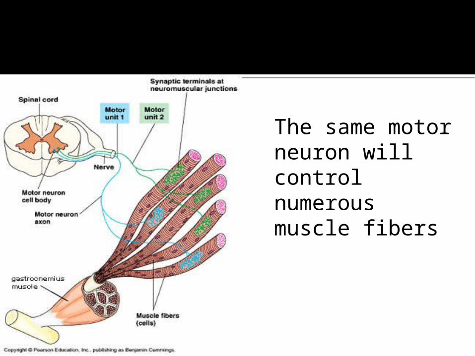
The same motor neuron will control numerous muscle fibers
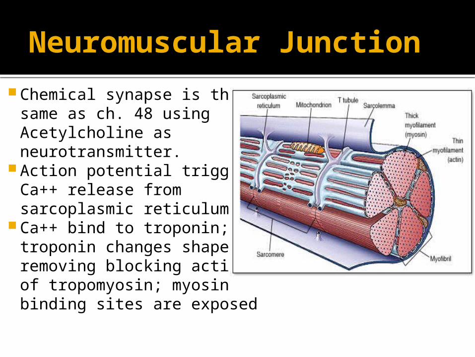
Neuromuscular Junction
Chemical synapse is the same as ch. 48 using Acetylcholine as neurotransmitter.
Action potential triggers Ca++ release from sarcoplasmic reticulum
Ca++ bind to troponin; troponin changes shape, removing blocking action of tropomyosin; myosin binding sites are exposed

Sliding Filament Theory
The neuron stimulate the sarcolemma
An Action Potential travels down the sarcolemma and T-tubules
Sarcoplasmic Reticulum releases calcium ions into the cytoplasm
Calcium uncovers myosin-binding sites on actin
Myosin heads attach to actin, bend and release, pulling the actin. ATP is used.

http://www.youtube.com/watch?v=0kFmbrRJq4w&safety_mode=true&persist_safety_mode=1

The Senses
Sense organs are the windows to the brain.
Sense receptors Interoceptors – respond to internal stimuli -
bodyposition, chemicals, blood pressure Exteroreceptors – respond to external stimuli -
light, pressure, chemicals, heat Integration – processing information
Some of this is often done at the receptor Sensory adaptation – decrease in response
due to continued stimulation

Types of sensory receptors
Mechanoreceptors – pulled or stretched Touch receptors in the skin Arteries detect blood pressure change Lungs respond to degree of lung inflation Proprioceptors – posture and balance Inner ear – sensitive to waves of fluid
which establishes equilibrium
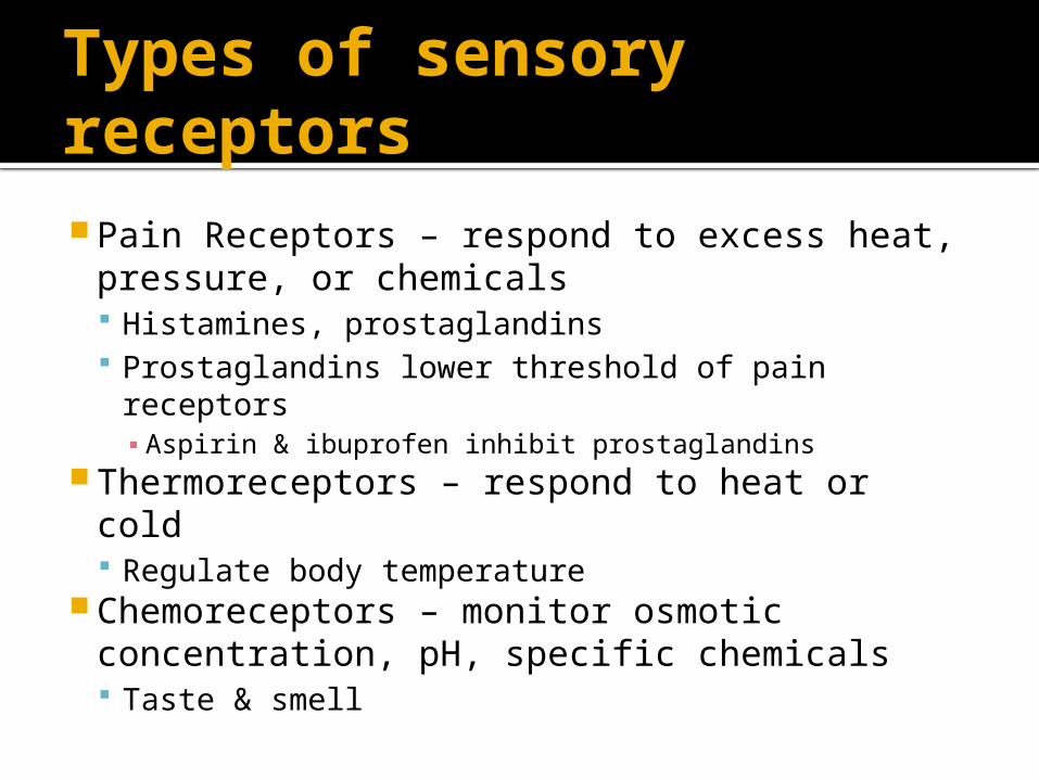
Types of sensory receptors Pain Receptors – respond to excess heat,
pressure, or chemicals Histamines, prostaglandins Prostaglandins lower threshold of pain
receptors▪ Aspirin & ibuprofen inhibit prostaglandins
Thermoreceptors – respond to heat or cold Regulate body temperature
Chemoreceptors – monitor osmotic concentration, pH, specific chemicals Taste & smell

Types of sensory receptors Electromagnetic receptors – light, electricity,
magnetism, photoreceptors (sight) Photoreceptors – respond to light energy
Types of eyes Simple eye cup – light intensity & direction Compound eye – 1000 or more ommatidia, each
with its own lens▪ Gives mosaic image able to detect very slight movement▪ Insects, some arthropods
Single lens eye – focuses image on retina▪ Spider, mollusk, polychaete, vertebrates

Compound Eye image


Eye parts & functionsPart Function
Iris Regulates size of pupil
Pupil Admits light
Retina Contains receptors for vision
Aqueous humour
Transmits light rays & supports eyeball
Vitreous humour
Transmits light rays & supports eyeball
Rods Allow black & white vision in dim light
Cones Allow color vision in bright light
Fovea Area of densely packed cone cells where vision is most accurate
Lens Focuses light rays
Sclera Protects & supports eyeball
Cornea Focusing begins here
Choroid Absorbs stray light
Conjunctiva Covers sclera & cornea; keeps eye moist
Optic nerve Transmits impulses to the brain
Eye lid Protects the eye
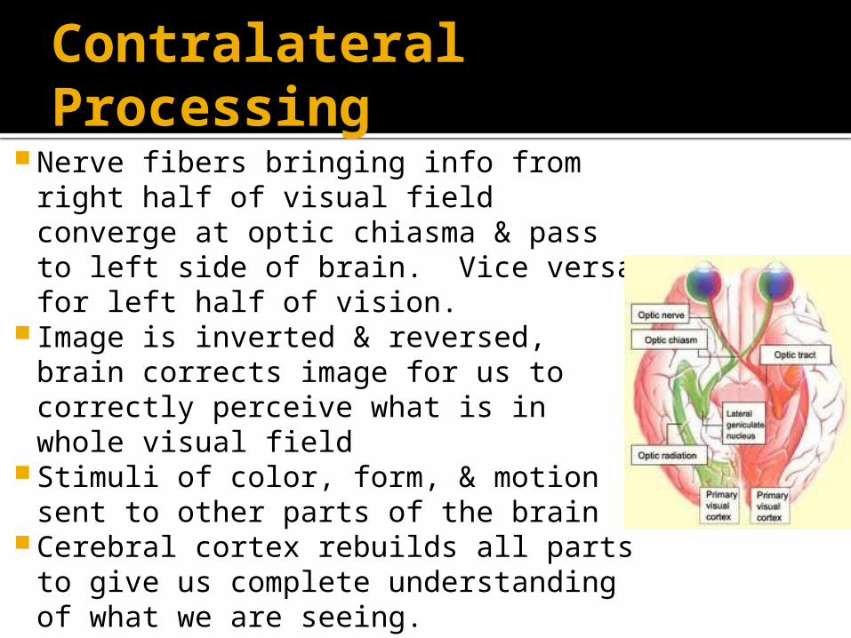
Contralateral Processing
Nerve fibers bringing info from right half of visual field converge at optic chiasma & pass to left side of brain. Vice versa for left half of vision.
Image is inverted & reversed, brain corrects image for us to correctly perceive what is in whole visual field
Stimuli of color, form, & motion sent to other parts of the brain
Cerebral cortex rebuilds all parts to give us complete understanding of what we are seeing.

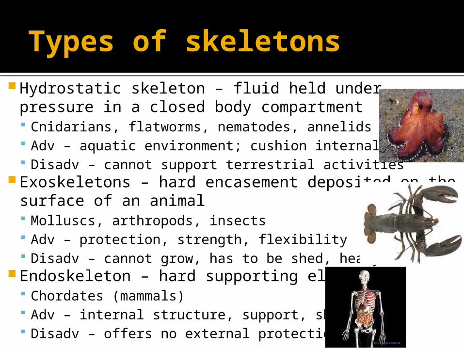
Types of skeletons Hydrostatic skeleton – fluid held under pressure in a
closed body compartment Cnidarians, flatworms, nematodes, annelids Adv – aquatic environment; cushion internal organs Disadv – cannot support terrestrial activities
Exoskeletons – hard encasement deposited on the surface of an animal Molluscs, arthropods, insects Adv – protection, strength, flexibility Disadv – cannot grow, has to be shed, heavy
Endoskeleton – hard supporting elements Chordates (mammals) Adv – internal structure, support, shape Disadv – offers no external protection

Joints
Ball & socket – rotation of arms & legs Humerus & shoulder Femur & hip
Hinge joint – single plane movement Elbow Knee
Pivot joint – rotation Forearm Head on neck


