Semmelweis University, Faculty of Medicine st Department ...
43
Surgery of hernias László NEHÉZ M.D. Semmelweis University, Faculty of Medicine, 1 st Department of Surgery
Transcript of Semmelweis University, Faculty of Medicine st Department ...

Surgery of hernias
László NEHÉZ M.D.
Semmelweis University, Faculty of Medicine, 1st Department of Surgery

Definition:
A hernia is where an internal part of the body, such
as an organ, pushes through a hereditary or
acquired weakness in the muscle or surrounding
tissue wall.
Semmelweis University, Faculty of Medicine, 1st Department of Surgery

The hernia repair is one of the most
common surgical procedures in the
world.!
US: 600000/y
HU: 30000/y
Semmelweis University, Faculty of Medicine, 1st Department of Surgery

Bassini 1887
• The modern era of the surgery
– Asepsis, anesthesia
Semmelweis University, Faculty of Medicine, 1st Department of Surgery

Use of implants and xenograft
• 1894. Phelps silver filigree
• 1949 PTFE-DuPont
• 1955 Gerendás Mihály:fibrin
layer from animals,
(Biethium)
• 1965 Horn B, Drobny S:
gynecological application
and liver surgery
Semmelweis University, Faculty of Medicine, 1st Department of Surgery

Types of hernias and distribution
Semmelweis University, Faculty of Medicine, 1st Department of Surgery

Anatomy
1. Hernia gate (ring)
2. Hernia sac (NB.: par glissement hernia)
3. Content
Semmelweis University, Faculty of Medicine, 1st Department of Surgery

Anatomy
No disease of the human body,
belonging to the province of the
surgeon, requires in its treatment a
better combination of accurate,
anatomical knowledge with surgical
skill than Hernia in all its varieties.
Sir Astley Cooper, 1804
Semmelweis University, Faculty of Medicine, 1st Department of Surgery

Inguinal hernia
Type:
• Indirect, lateral
– congenital
– acquired
• Direct
Semmelweis University, Faculty of Medicine, 1st Department of Surgery

Inguinal hernia
Pathogenesis:
Metabolic disorders
Impaired collagen synthesis
Weakness of connective tissue
Increased abdominal pressure pushes out the
abdominal content through the weak part
Semmelweis University, Faculty of Medicine, 1st Department of Surgery

Childhood risks
• Premature birth
• Cryptorchism
• Hypospadias
• Epispadias
• Failure in genital
development
• Peritoneal dialysis, ascites
• Abdominal wall defect
• Positive anamnesis in family
• Cystic fibrosis
• Connective tissue disorders
• Mucopolysaccharidosis
• Dislocated hip
• Ehlers-Danlos syndrome
• Marfan syndrome
• Fetal hydrops
• Ascites / liver disease
• Ventriculoperitoneal shunt
(hydrocephalus)
Semmelweis University, Faculty of Medicine, 1st Department of Surgery

Inguinal hernia
Treatment = Operation
(conservative treatment is
acceptable only at
contraindication of surgery)
Semmelweis University, Faculty of Medicine, 1st Department of Surgery

Before you start… OBJECTIVES
1. Identify pertinent external landmarks the inguinal region for use in
herniorrhaphy
2. Understand the layers of the abdominal wall and their relationship to the
inguinal canal
3. Define all of the components of the inguinal canal and spermatic cord
4. Describe the etiology of groin hernias by defining potential weak areas of the
inguinal and femoral canal
5. Describe the anatomic basis for hernia repair
Semmelweis University, Faculty of Medicine, 1st Department of Surgery

Inguinal hernia
Steps:
1. Dissection of hernia sac
2. Inspection of the content, replacement
3. Resection of the sac (Br J Surg. 2010 Mar;97(3):415-9)
4. Abdominal wall repair
Semmelweis University, Faculty of Medicine, 1st Department of Surgery

Inguinal hernia
TECHNIQUES:(difference in the abdominal wall reconstruction)
• Traditional (tension repair): (Bassini, Girard, Bassini-Kirschner, Shouldice)
• Tension Free Repair: with mesh(Lichtenstein,Trabucco, Stoppa, plug reconstruction, laparoscopic approaches)
Semmelweis University, Faculty of Medicine, 1st Department of Surgery

• Obturator hernia
• Lumbar hernia
• Sciatic hernia
• Perineal hernia
• Spiegel hernia
• Littre hernia
• Richter hernia (localisation independent)
Rare hernias
Semmelweis University, Faculty of Medicine, 1st Department of Surgery

Rare hernias
Internal hernias
(both gate and sac are located in the
abdominal cavity)
• Mesenterico-parietal hernia
• Paraduodenal hernia
• Hernia bursae omentalis
• Pericoecal hernia
Semmelweis University, Faculty of Medicine, 1st Department of Surgery

Sliding hernia (par glissement)
• in which the intestinal
wall forms part of the
hernial sac and the rest
of the sac is formed by
parietal peritoneum
Semmelweis University, Faculty of Medicine, 1st Department of Surgery

Diagnosis
• Case history
• Physical investigation:
– Inspection
– Sense of feeling,
possibility of replacement,
– Eavesdropping
• US, sometimes CT
Semmelweis University, Faculty of Medicine, 1st Department of Surgery

Differential diagnosis
• Hydrocele
• Varicocele
• Lipoma
• Lymphoma, lymphadenomegalia
• Tumor or inflammatory process
• Cyst
• Relaxation
Semmelweis University, Faculty of Medicine, 1st Department of Surgery

Possible complications
• Accrete hernia
• Inflammation
• Constipation, ileus
• Incarceration
(Richter hernia, retrograde incarceration,
risk of taxis(spontaneous repositioning))
Semmelweis University, Faculty of Medicine, 1st Department of Surgery

Semmelweis University, Faculty of Medicine, 1st Department of Surgery

Semmelweis University, Faculty of Medicine, 1st Department of Surgery

Inguinal herniaComplications:
• Nerve injury (n.ilioinguinalis,
n.genitofemoralis)
• Vascular injury (a.,v.femoralis, epigastrica
inferior, spermatica)
• Vas deferens injury
• Injury of bladder
• Intestinal injury
• Scrotal edema, hematoma
• Suppuration
• Testicle atrophy
Semmelweis University, Faculty of Medicine, 1st Department of Surgery

Inguinal hernia: Postoperative care
General wound care
Limited physical activity
– Normal work load• Up to 2 month after traditional surgery
• 2 weeks after tension free repair
• Few days after minimal invasive reconstruction
Semmelweis University, Faculty of Medicine, 1st Department of Surgery

Inguinal hernia
Reoccurrence (1-15%)
Frequency depends on:
– Surgery type
– Skills ant technique of the surgeon
–Overall condition of the patient
– Events in the postoperative period
Semmelweis University, Faculty of Medicine, 1st Department of Surgery

Femoral herniaAnatomy:
• under the Poupart
ligament
• lig.lacunare Gimbernati
• lig. iliopectinea Cooperi
• trigonum Hesselbachi
(N A V E - nerve-artery-
vein-empty space)
• Rosenmüller lymphnodes
Forms, properties:
• Always direct hernia
• Always acquired
• More common in female
• Incarceration is frequent
(Keep it in mind especially in case of old patients)
Semmelweis University, Faculty of Medicine, 1st Department of Surgery

Femoral hernia
Therapy = Surgery
1. Femoral approach (Fabricius)
2. Inguinal approach
(Lotheisen, Lotheisen-Reich)
Semmelweis University, Faculty of Medicine, 1st Department of Surgery

Umbilical herniaAnatomy, pathogenesis:
• Umbilical hernias - occur when fatty
tissue or a part of intestine pokes
through abdominal wall near the navel
(belly button)
• Insufficient closure of umbilical ring
• Weakening umbilical scar
Forms
• Congenital in children
(spontaneous closure before the
year 1.)
• Always acquired in adults
(increased physical load,
pregnancy, obesity, ascites)
• The risk of incarceration is high
Treatment
• The umbilical hernia of infants and
children can close up to the age of 2
years
• If not = surgery
• Umbilical hernia in adults = surgery
Surgical strategy:
• Children
– Navel preserving reconstruction
• Adult:
– Navel preserving reconstruction
– Mayo
– Mesh implantation
– Minimal invasive surgery

Hernia in linea alba
• EPIGASTRIC hernias develop in the mid upper abdomen, anywhere along a line drawn from the lower point of the breastbone straight down to the Umbilicus.
– can occur at any point along
– small in size and localized
– contents are easily pinched and these hernias therefore can cause a great deal of pain.
– well suited for repair using a Tension Free method
Semmelweis University, Faculty of Medicine, 1st Department of Surgery

Hernia in linea alba
• DIASTSIS RECTI is not a hernia
– Often confused and at times mis-diagnosed as an epigastrichernia
– Abdominal wall protrusions that occur due to a widened band of non-contractile fascia or tendon normally present between the rectus muscles
– This non-tender bulge extends from just below the breast bone, down to the navel.
– There is no pain associated with
– Surgery is not necessary except for cosmetic reason
Semmelweis University, Faculty of Medicine, 1st Department of Surgery

Incisional hernia
• Frequently combined with wound healing alterations (suppuration, fascia necrosis, hypoproteinemia, hypovitaminosis, hematoma)
• Therapy: reconstructionImportant: tension free methods improve the success rate, therefore mesh implantation necessary in case of big aperture and extended tissue defect.
Semmelweis University, Faculty of Medicine, 1st Department of Surgery

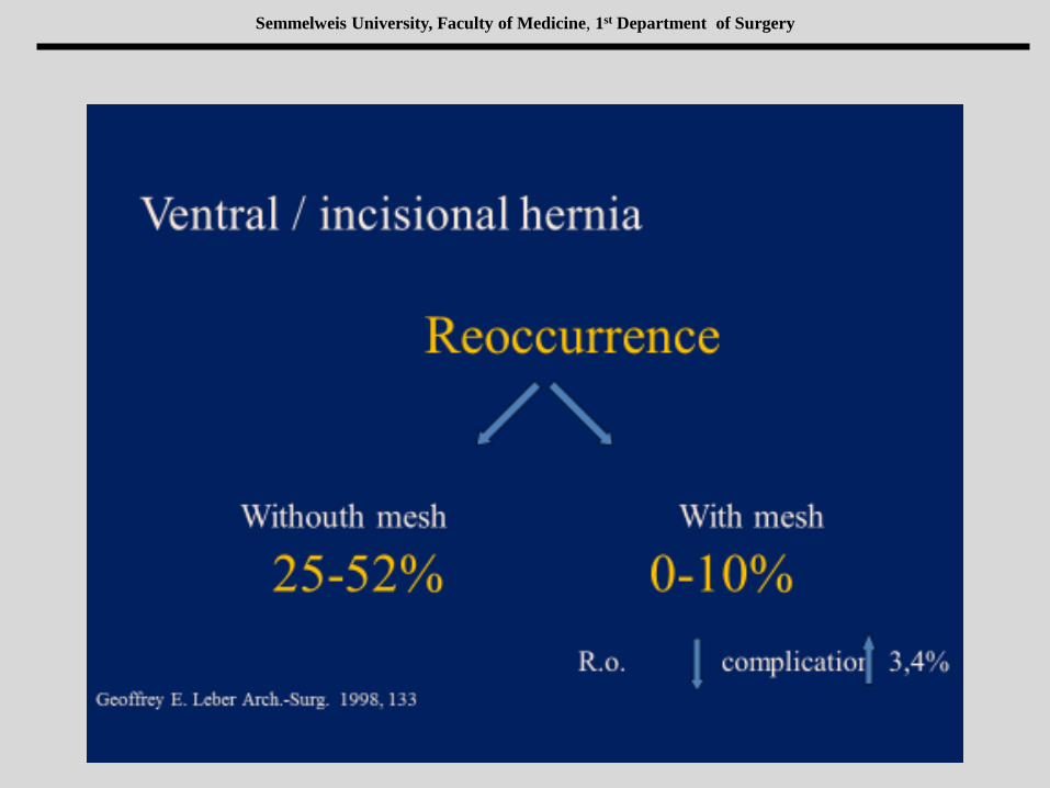
Ventral hernia
Frequency after laparotomy: 3-20%
Minimal invasive approach: 1-4%
Semmelweis University, Faculty of Medicine, 1st Department of Surgery

Traditional: open, sutures (reoccurrence: 41-52%)
Tension free: open, mesh (reoccurence: 12-24%,)
(Infection rate 20%-t)
Laparoscopic repair (1992, LVHR (Todd Heniford at
all)
Ventral hernia
Semmelweis University, Faculty of Medicine, 1st Department of Surgery

Incisional hernia
• High incidence of reoccurrence
• Mesh is useless in septic condition
• Giant hernia repair may lead to have
respiratory and cardiac failure
Semmelweis University, Faculty of Medicine, 1st Department of Surgery

Semmelweis University, Faculty of Medicine, 1st Department of Surgery
Semmelweis University, Faculty of Medicine, 1st Department of Surgery

Mesh implantation
• Less reoccurrence
• Less pain
• Early mobilization, and
physical load
• More complication
• Infection, seroma,
hematoma
• Shrinkage, migration
• Discomfort
(chronic pain, feel of mesh)
Semmelweis University, Faculty of Medicine, 1st Department of Surgery

Factors that influence the integration, and
complications:
• Amount of foreign materials
•The amount of implants correlate with
•Seroma
•Wound infection
•Chronic pain (granuloma)
•Other tissue reactions
•(20 x 30 cm mesh = 300 m suture!)
• Quality, rigidity
•Soft edges cause less complains.
Semmelweis University, Faculty of Medicine, 1st Department of Surgery

• Collagen development
•Impaired development around the mesh
•Discovered in 74% of recurrent hernia
Factors that influence the integration, and
complications:
Semmelweis University, Faculty of Medicine, 1st Department of Surgery

Monofilament polypropylene mesh
• Strong, most frequently used in surgery
• High pressure tolerance (caught),
• Porosity – tissue integration
• Blue stripe - orientation
PDS capsule
• Absorbable, tiny and flexible glue
O R C
• Gel formation to prevent adhesion
• Persist 14 days
• Vegetable derivate
Parts
Semmelweis University, Faculty of Medicine, 1st Department of Surgery

Application
• Complex abdominal wall repair
• Complex abdominal wall defects
• Parastomal and Hiatus hernia
• Any major tissue defect (e.g.
chest wall)
Semmelweis University, Faculty of Medicine, 1st Department of Surgery

Semmelweis University, Faculty of Medicine, 1st Department of Surgery