Self adjusting file - unizg.hr
56
Self adjusting file Hulenić, Kim Master's thesis / Diplomski rad 2015 Degree Grantor / Ustanova koja je dodijelila akademski / stručni stupanj: University of Zagreb, School of Dental Medicine / Sveučilište u Zagrebu, Stomatološki fakultet Permanent link / Trajna poveznica: https://urn.nsk.hr/urn:nbn:hr:127:890110 Rights / Prava: Attribution-NonCommercial-NoDerivs 3.0 Unported Download date / Datum preuzimanja: 2021-12-13 Repository / Repozitorij: University of Zagreb School of Dental Medicine Repository
Transcript of Self adjusting file - unizg.hr

Self adjusting file
Hulenić, Kim
Master's thesis / Diplomski rad
2015
Degree Grantor / Ustanova koja je dodijelila akademski / stručni stupanj: University of Zagreb, School of Dental Medicine / Sveučilište u Zagrebu, Stomatološki fakultet
Permanent link / Trajna poveznica: https://urn.nsk.hr/urn:nbn:hr:127:890110
Rights / Prava: Attribution-NonCommercial-NoDerivs 3.0 Unported
Download date / Datum preuzimanja: 2021-12-13
Repository / Repozitorij:
University of Zagreb School of Dental Medicine Repository

SCHOOL OF DENTAL MEDICINE
UNIVERSITY OF ZAGREB
Kim Hulenić
THE SELF-ADJUSTING FILE
GRADUATION THESIS
Zagreb, September 2015

Graduation thesis was completed at the
Department of Endodontics and Restorative Dentistry,
School of Dental Medicine,
University of Zagreb
Supervisor: Silvana Jukić, prof.dr.sc.
Proofreader in English: Tomaš Gaj, mag. philol. angl.
Proofreader in Croatian: Dina Valković, prof.
This thesis consists of:
- 50 pages
- 8 pictures
- 1 CD

I would like to thank prof.dr.sc.Silvana Jukić for her exceptional kindness, courtesy
and professional advice during the development of this thesis.
I would also like to thank my fellow colleagues for our unrepeatable moments
together that made my studying easier and unforgettable.
Special thanks goes to my family and friends for their support and understanding
during my education.
“Knowledge has to be improved, challenged,
and increased constantly, or it vanishes.”
Peter Druck

Contents:
1. INTRODUCTION ............................................................................................................ 1
2. THE AIM OF THIS WORK ............................................................................................ 2
3. HISTORY OF ENDODONTIC INSTRUMENTS .......................................................... 3
4. CLASSIFICATION OF ENDODONTIC INSTRUMENTS ........................................... 5
4.1. ENDODONTIC EXPLORER ................................................................................. 6
4.2. BARBED BROACH ............................................................................................... 6
4.3. K-TYPE FILE ......................................................................................................... 7
4.4. REAMER ................................................................................................................ 8
4.5. HEDSTROEM FILES ............................................................................................. 8
4.6. ENGINE DRIVEN INSTRUMENTS ................................................................... 10
4.7. PROFILE ............................................................................................................... 12
4.8. PROTAPER ........................................................................................................... 13
5. IRRIGATION ................................................................................................................ 15
5.1. SODIUM HYPOCHLORITE (NaOCl) ................................................................. 17
5.2. EDTA .................................................................................................................... 21
6. PULP ANATOMY ......................................................................................................... 22
7. THE SELF-ADJUSTING FILE ..................................................................................... 25
8. DISCUSSION ................................................................................................................ 31
9. CONCLUSION .............................................................................................................. 42
10. SUMMARY ................................................................................................................... 43
11. SAŽETAK...................................................................................................................... 44
12. LITERATURE ............................................................................................................... 45
13. CURRICULUM VITAE ................................................................................................ 50

LIST OF ABBREVIATIONS AND ACRONYMS
SICE Second International Congress of Endodontics
AAE American Association of Endodontics
CDMD Council on Dental Materials and Methods
ADA American Dental Association
ISO International Organization for Standardization
SAF self-adjusting file
RPM revolutions per minute
NiTinol Nickel Titanium Navol Ordinance Laboratory
RTN rotary nickel titanium system
EDTA ethylendiaminetetraacetic acid
NaOCl sodium hypochlorite
MTAD mixture of tetracycline, an acid and a detergent
CHX chlorhexidine
PGFA gutta-percha filled area
Micro-CT micro-computed tomography
VRF vertical root fracture
Kim Hulenić, graduation thesis
1
1. INTRODUCTION
Even though it may seemed that endodontics wasn't science with high jumps in
development due to the lack of visibility and unique shape and configuration of root
canals, the recent decade has produced great changes which resulted in new
instruments and concepts on how to adequately prepare root canal for filling. Over
the past few decades, instrument design has been considerably modified, as well as
alloy processing. These discoveries have altered the whole perspective of root
preparation even though the biological objectives of root canal treatment have not
changed over the years. The introduction of NiTi rotary system represented a major
leap in the development of endodontic treatment, with a wide variety of sophisticated
instruments presently available. Today, there is a new generation of rotary
instruments which have made one step forward with their design, concept, and
perhaps even changed the whole base of future endodontics and what it should be. It
is called self-adjusting file and its name gives a clue about where future endodontics
is headed (1).

Kim Hulenić, graduation thesis
2
2. THE AIM OF THIS WORK
The aim of this thesis is to provide an insight on how the development of
endodontic instruments in dental medicine went from simple hand pieces to machine
driven instruments, and to give detailed information about the new technique of
cleaning and shaping root canals.
The first part of the thesis walks you through the history of endodontic
instruments, their design, configuration and their purpose, and the second part
concerns the self-adjusting file, a new discovery in endodontic treatment. This thesis
sums it all up, from general specification to the studies based on clinical trial and
comparisons with well-known ProTaper and ProFile.
It also serves as a reminder on how important and necessary irrigation is, and
its activity in the root canal walls, smear layer and microorganisms that can diminish
our efforts in making it suitable environment for good filling.
As we all know, not every tooth is a textbook example, so this thesis draws
attention to all kinds of pulp anatomy, from calcifications inside pulp chamber to
varieties in directions, lengths, diameters, relationships in multi-rooted teeth and how
this can make our work a lot harder. With new instruments like SAF these once
called problems can now become manageable obstacles.

Kim Hulenić, graduation thesis
3
3. HISTORY OF ENDODONTIC INSTRUMENTS
The manufacture of the first instruments for endodontic use dates back to 1875.
These early instruments, made by hand from thin steel wires, performed more or less
the function of modern barbed broaches. In accordance with the lack of
sophistication of that time, more importance was given to obturation of the canal
space rather than to cleaning and advancements in bacteriology at the turn of the last
century. With the arrival of dental radiology, local anesthesia and advancements in
bacteriology at the turn of the last century, a new era opened in endodontic therapy.
In 1932, G. V. Skillen stated that it was necessary to curette the canal walls to
remove the pulp debris. His belief was that all residual tissue became degenerative
and would lead to failure of canal therapy. Skillen and his contemporaries were
occupied with establishing standards for the methods of root canal cleaning, which at
the time had not been standardized. Grove designed “standardized instruments and
gold cones”. His intent was to prepare the radicular canal space according to precise
norms of shape, size, and conicity. Jasper developed silver cones corresponding to
the sizes of the files that were in use at that time. In 1955 Ingle was the first to finally
express the need for the standardization of canal instruments, which he advocated
again in 1958 at the Second International Conference of Endodontics (SICE) in
Philadelphia. In 1961, Ingle established a basic, standardized shape for endodontic
instruments and a standardized endodontic technique using newly-designed
obturation instruments and materials. He substituted stainless steel for carbon steel
and introduced color-coded instruments that were smaller (06 and 08) and larger

Kim Hulenić, graduation thesis
4
(110-150) than those in use at the time. In 1965, the American Association of
Endodontists (AAE) adopted the terminology and nomenclature of the proposed
standardized system, and in June 1976 the Council on Dental Materials and Devices
(CDMD) of the American Dental Association (AAD) approved the specification #
28, which established the classification norms, required physical properties,
procedures for investigation, sampling tests, and preparation for the distribution of
root canal files and reamers. The system of standardization and agreements among
the various manufacturers to observe them is, therefore, a fairly recent development
(2). Edward Maynard has been credited with the development of the first endodontic
hand instrument. Notching a round wire (in the beginning watch springs, later piano
wires), he created small needles for extirpation of pulp tissue. In 1885, the Gates
Glidden drill were introduced and in 1915 the K-files. In 1889, William H. Rollins
developed the first endodontic hand piece for automated root canal preparation and
used specially designed needles with a 360 degree rotation and speed limited to 100
rpm in order to avoid instrument fracture. In 1892, Oltramare used fine needles with
a rectangular cross-section, mounted into a dental hand piece and passively
introduced into the root canal to the apical foramen, followed by the rotation started.
The Cursor filing contra-angle was developed in 1928 by W&H (Burmoos, Austria)
which combined rotational and vertical motion of the file. In 1958, W&H Company
started marketing The Racer-hand piece in Europe with a vertical file motion. Later,
in 1964, MicroMega (Besancon, France) started marketing the Giromatic in Europe
with a reciprocal 90 degree rotation. Endodontic hand-pieces such as the Endolift
(Kerr, Karlsruhe, Germany) with a combined vertical and 90 degree rotational

Kim Hulenić, graduation thesis
5
motion and similar devices were marketed during this period. A period of modified
endodontic hand pieces began with the introduction of the Canal Finder System (now
distributed by S.E.T., Grobenzell, Germany) by Levy. The Canal Finder was the first
endodontic hand-piece with a partially flexible motion. The amplitude of the vertical
file motion depended on the rotary speed and the resistance of the file inside the root
canal, and changing into a 90 degree rotational motion with increasing resistance. It
was an attempt to make the root canal anatomy or at least the root canal diameter one
main influencing factor on the behavior of the instrument inside the canal. The
Excalibur hand-piece (W&H), with laterally oscillating instruments, had a major
impact on canal preparation. NiTi rotary instruments introduced later, use a 360
rotation at low speed and offer new perspectives for root canal preparation that have
the potential to avoid some of the major drawbacks of traditional instruments and
devices (3).
4. CLASSIFICATION OF ENDODONTIC INSTRUMENTS
Endodontic instruments are traditionally divided into four categories:
1. exploring
2. extirpating
3. enlarging (cleaning and shaping), and
4. filling
ISO-FDI (Fédération Dentaire Internationale) grouped root canal instruments
according to their method of use:

Kim Hulenić, graduation thesis
6
Group I: Hand use only for example, K- and H-files, reamers, broaches etc.
Group II: Latch type Engine driven: same design as Group one but can be attached to
hand-piece.
Group III: Drills or reamers Latch type Engine driven for example Gates-Glidden,
Peeso reamers.
Group IV: Root canal points like gutta-percha, silver point, paper point.
Natural anatomy dictates the usual places for canals, but pulp stones,
dystrophic calcifications and restorations can alter the actual configuration
encountered (4).
4.1. ENDODONTIC EXPLORER
The endodontic explorer is used to locate orifices, and serves as a tool to
remove calcification.
4.2. BARBED BROACH
The barbed broach is an extirpating, not an enlarging, instrument. It is formed
from a tapered round shaft by lifting up portions of metal of the shaft almost at a
right angle to the shaft. These elevated barbs engage the pulp tissue and remove it
from the canal. In its use, the largest broach in size that will fit freely or loosely in
the canal is selected.
A broach must not engage the canal walls as it is advanced, or the pressure on
the barbs will flatten them against the shaft; as the instrument is withdrawn, the barbs

Kim Hulenić, graduation thesis
7
will then embed themselves in the walls, making it difficult or impossible to remove
the broach. Because the barbs are "nicked" out of the shaft this is an extremely
fragile instrument, and will break easily if misused. It must fit loosely.
Files are the mos common instruments used for cleaning and shaping the root
canal system. Traditionally they are manufactured from stainless steel in the form of
a filament with a round cross-section. First, they are ground in such a way as to have
a quadrangular cross-section and then twisted clock wise to achieve the definitive
form. The number of spirals per mm (pitch) for stainless steel files can very slightly
depend on the manufacturers but is always more (generally double) than that of the
reamers; their blades are furthermore positioned perpendicular to the long axis of the
instrument, giving files a particularly efficient cutting action during filing (5).
4.3. K-TYPE FILE
The K-type file was first introduced at the turn of the century (1901) and
receiving its name from the holder of its original patent, the Kerr Manufacturing
Company. The K-type file is manufactured by twisting or grinding a square or
triangular tapered shaft so that the cutting edges are almost perpendicular to the long
access of the instrument. The K-file works on the "pull" stroke,i.e., by scraping the
canal walls as it is withdrawn from the canal. It is advanced to the full working
length when rotated 1/4 to 1/2 turn clockwise, and withdrawn while being pressed
against one of the walls. The process is repeated against each of the walls in turn until
the canal is sufficiently enlarged to proceed to the next size instrument. It is preferable

Kim Hulenić, graduation thesis
8
to utilize an instrument to the maximum extent possible before proceeding to the next
instrument in the series. The file must be cleaned repeatedly during use (4).
4.4. REAMER
Reamers are generally obtained by twisting a steel wire with a triangular or
quadrangular cross-section. Compared to K-Files, the reamers have less spirals per
mm (about half) and a more acute blade cutting angle against the dentin. The main
consequence of the different blade angle of the reamers with regard to the K-Files is
their poor efficiency at cutting with a push and pull movement (filing); to be used
correctly the reamers should be rotated in the canal so that the blades have a 90
degrees angle contact with the dentin. The correct action of these instruments is
passive insertion to a depth permitted by the canal diameter, and a quarter clock-wise
rotation with simultaneous extraction of a few millimeters. The cutting action takes
place during the withdrawal phase. This movement is repeated a number of times
without ever forcing the instruments during their insertion but engaging the dentin
during rotation and removal from the canal. A rotation exceeding half a turn is not
recommended as it could cause engagement and fracture inside the canal (5). The
reamer must be in contact with the walls of the canal in order to be effective, but it
must not bind or it may break. They tend to remain self-centered in root canal
resulting in less chances of canal transportation (4).
4.5. HEDSTROEM FILES
The Hedstroem Files, or H Files, are obtained by microgrinding a conical steel
or NiTi wire with a round cross-section. The cutting angle of the blades (helical

Kim Hulenić, graduation thesis
9
angle) against the dentin is, for the Hedstroem, close to 90 degrees making this
instrument particularly aggressive when using the push and pull (filing) action. The
design of the blades is however also responsible for the structural weakness of the
Hedstroem files when used in a rotational manner. This is due to the fact that the
deep grinding of the surface has reduced the central mass of metal which determines
the torsional strength of the instrument. The efficient cutting action of the H-Files
seems to be superior to that of K-Files and this explains the popularity of this
instrument, especially for circumferential filing of canals with oval or elliptical cross-
section. The Hedstroem files are distributed by most manufacturers of endodontic
instruments, with diameters and lengths regulated by the ISO standard (5).
The uniqueness of the design and the method of fabrication of the barbed
broaches and rasps separates them from other intracanal hand instruments. They also
differ from H-type and K-type instruments because of taper and length of the
operating length of the shaft (10 mm). Barbed broaches are used primarily for the
removal of intact pulp tissue. The instrument is introduced slowly into the root canal
until gentle contact with the canal walls is made. It is rotated 360 degrees either
clockwise or counterclockwise, to entangle the pulpal tissue in the protruding barbs.
It is then withdrawn directly from the root canal. If the maneuver is successful, the
entire pulp comes out. If the vital pulp is so inflamed that the gel-sol state of the
ground substance has been altered by edema or the collagen fibrous network has
been destroyed, it probably cannot be removed intact by a barbed broach. The
instrument will only lacerate the already hemorrhagic tissues. Unless a necrotic pulp
maintains a high degree of cellular or fibrous integrity, it will not lend itself to
Kim Hulenić, graduation thesis
10
removal by the barbed broach. Because of these biological realities and the design of
the broach, this instrument has minimal clinical practice use. Rasps, being similar in
design to barbed broaches, but having shallower and more rounded barbs, produce
rougher walled canal preparations than other instruments for canal enlargement and
shaping. For this reason they have been superceded by H-type files (4).
4.6. ENGINE DRIVEN INSTRUMENTS
Traditional engine driven instruments are Gates-Glidden burs. The Gates-
Glidden drills are steel instruments for the contra-angled hand-piece characterized by
a long shank and an elliptical extremity which is flame-shaped with a “guiding” non-
cutting tip. The Gates-Glidden drills are available in six sizes marked with circular
notches on the part that attaches to the contra angled hand-piece; the Gates no. 1 has
one notch, the no. 2 has two notches, and so on. The calibration of the Gates Burs is
measured at the widest part of their elliptical portion; no. # 1 has a maximum
diameter of 0.50 mm, which increases by 0.20 mm for each successive size, until no.
6 which has a maximum diameter of 1.50 mm. Burs which have flame shaped cutting
point mounted on long thin shaft attached to a latch type shank (5). If its cutting tip
jams against the canal wall, the fracture should occur at the junction of the shank and
the shaft but not at the tip of the instrument. They can be used both in crown down as
well as step back fashion (4).
Peeso reamers are rotary instruments used mainly for post space preparations.
Disadvantages for using Peeso reamers are:

Kim Hulenić, graduation thesis
11
They do not follow the canal curvature and may cause perforation by cutting
laterally.
They are stiff instruments.
They have to be used very carefully to avoid iatrogenic errors.
When using stainless steel files, the occurrence of procedural errors cannot be
avoided, especially in case of curved canal. Deviation from the original shape, ledge
formation, zipping, stripping and perforation are common problems which are seen
in such cases. But the superelasticity of NiTi alloy allows these instruments to flex
more then stainless steel instruments before exceeding their elastic limit, thereby
allowing canal preparation with minimal procedural errors. NiTi was developed by
Buchler 40 years ago. NiTi is also known as NiTinol ( NiTi Navol Ordinance
laboratory in USA ). In endodontics, the commonly used Niti alloys are called 55
NiTi nol ( 55% weight of Ni and 45% Ti ) and 60 NiTi nol ( 60% weight of Ni and
40% of Ti ). The first use of NiTi in endodontics was reported in 1988 by Walia et al.
when a 15 No. NiTi was made from orthodontic wire and it showed superior
flexibility and resistance to torsional fractures (4).
Properties of Niti Alloys:
Shape Memory
Superelasticity
Low modules of elasticity
Kim Hulenić, graduation thesis
12
Good resilience
Corrosion resistance
Softer then stainless steel
Superelasticity and shape memory of NiTi alloys is present because of phase
transformation in their crystal structures when cooled from a stronger, high
temperature form (Austenite) to a weaker, low temperature form (Martensite). This
phase transformation is chiefly responsible for the above mentioned qualities.
Over the last few years great amount of rotary nickel titanium system (RTN)
have been made available. Although no system is perfect, if used in a proper way, it
can result in desired canal shape. Various rotary nickel titanium systems available in
the market are ProFile, ProTaper, Greater Taper Files, Quantec, Light Speed System,
K3 system, HERo 642, RaCe and Real World Endo Sequence file system (4).
4.7. PROFILE
ProFile instruments made by Tulsa Dental were one of the first NiTi
instruments available commercially. This system was introduced by Dr. Johnsson in
1944. The instrument set consists of orifice shapers (19mm long files with 5–8%
conicity), the conical ProFile 06 with a 6% conicity in sizes 15–40, as well as the
ProFile 04 with a conicity of 4% in sizes 15–90. Orifice shapers are used for the
instrumentation of the coronal third of the root canal. The ProFile 06 instrument with
a length 21 or 25mm is used for instrumentation of the middle third. Instrumentation
of the apical segment is accomplished with the ProFile 04 files (21, 25, and 31 mm).
Kim Hulenić, graduation thesis
13
The instruments have a U-shaped cross-sectional profile and “radial lands”. These
keep the instruments centered within the canal and enable easy smoothing of the
canal walls. They prevent any binding or catching/sticking in dentin. The smooth,
non-cutting tip serves to guide the file within the canal without scratching or
gouging. The ProFile instruments must always be coated with a lubricant and
between each change of instruments the canal must be copiously rinsed with 5%
NaOCl solution. The ProFile instruments are used with a slight in-and-out
movement, with a hub motion of no more than 2 mm and with only a slight
application of force. ProFile instruments can only be used in combination with a
motor that has a torsion limiter (e.g., ATR Technica)with low rpm (250–350). This
will prevent over-twisting of the instrument and resultant fracture. No ProFile
instrument should not be used more than 10 times (4).
4.8. PROTAPER
ProTaper instruments have a very unconventional shape. They combine several
(ascending) conicities in a single file. There are three shaping files for coronal
expansion and three finishing files for shaping the apical region. The diameter of the
instruments at the tip of the working portion is between 0.17/0.19 and 0.20 mm for
the shaping files, with 0.20/0.25/0.30 mm for the finishing files. In contrast to other
types of instruments, the ProTaper files exhibit conicities between 2 and 19% in a
single file. In the shaping files, the gradient toward the instrument tip is descending,
and with the finishing files ascending. Up to finishing file 3, all instruments exhibit a
triangular cross-sectional profile with convex cutting edges. The cross-section of file
F3 is characterized by a modified triangular form with concave surfaces, but does not

Kim Hulenić, graduation thesis
14
otherwise differ significantly from the other files. All instruments exhibit a rounded,
non-cutting tip; the cutting edges of the working area extend almost to the tip.
Because of the changing conicities, the tangent angle varies considerably, between
20 and 30 degrees. However, it increases in all instruments from the instrument tip to
the coronal end of the working portion. In contrast to the other two nitinol instrument
types, ProTaper instruments have the important advantage that even during initial
instrumentation of the coronal root canal segments, widening up to the size of the
corresponding Gates-Glidden drills 4–5 (sizes 110– 130) can be achieved; this
significantly simplifies further apical instrumentation, as well as the subsequent
filling of the canal via gutta percha condensation. The files are inserted into the canal
using gentle in-and-out movements with the amplitude of about 1 mm; the files must
be cleaned frequently, because large amounts of dentin are removed during initial
instrumentation. The removed dentin can accumulate between the cutting edges and
may lead to wedging of the file within the canal. Cleaning with sterile gauze and
frequent rinsing of the canal with NaOCl are necessary. Cleansing of the root canal
surface by instruments with a U-shaped cross-section and “radial lands” (ProFile,
GT-Rotary) provides better results when compared to the FlexMaster and ProTaper
systems with the conventional triangular cross-section. The U-shaped cross-section
evacuates dentin chips more effectively and, in addition, the improved centering of
the instruments provides efficient cleansing, without creating a pouch-like expansion
at the critical apical region (6).

Kim Hulenić, graduation thesis
15
5. IRRIGATION
According to an old and famous endodontic axiom, what is removed from the
root canal is more important than what is placed inside. Without minimizing the
importance of the obturation phase, it is nonetheless true that the phase of preparing
or emptying the root canal is undoubtedly the most important, the most complex, and
the most delicate procedure. It is difficult to imagine how one can completely
obturate a canal that has not been adequately cleaned and disinfected. On the other
hand, minor deficiencies in the filling of a root canal that has been totally debrided
and disinfected can be biologically tolerated, and they can also become contributing
causes of periapical inflammation in a root canal that remains infected (5).
The complexity of the root canal system, presence of numerous dentinal
tubules in the roots, invasion of the tubules by microorganism, formation of smear
layer during instrumentation, and presence of dentin as a tissue are the major
obstacles in achieving the primary objectives of complete cleaning and shaping of
root canal system.
Two terms - cleaning and shaping - encompass and characterize clearly the
most basic requirements for canal instrumentation, which were established by the
“old master” of endodontics, Professor Schilder, more than 30 years ago: cleansing
and giving the canal a proper form to receive the filling. Cleaning signifies the
removal of all materials from the root canal, including infiltrated tissues, antigenic
material, all organic components, bacteria and their products, but also caries, tissue
debris as well as denticles and other hard tissue accumulations, contaminated canal

Kim Hulenić, graduation thesis
16
filling material and other inflammation-inducing agents. Cleaning also means the
instrumentation and mechanical removal of canal constituents, chemical dissolution
of tissue debris, and rinsing them out of the canal.
Canal rinsing should:
• float dentin chips out of the canal, therefore preventing blockage
• dissolve vital and also necrotic tissue debris in those areas not accessible for
manual instrumentation
• provide a lubrication effect for the instruments
• have an antibacterial effect
• have some bleaching effect.
To effectively clean and disinfect the root canal system, an irrigant should be
able to disinfect and penetrate dentin and its tubules, offer long-term antibacterial
effect ( substantivity), remove the smear layer, and be nonantigenic, nontoxic and
noncarcinogenic. In addition, it should have no adverse effect on dentin or the
sealing ability of filling materials. Furthermore, it should be relatively inexpensive,
convenient for application and not cause tooth discoloration. Other desirable
properties for an ideal irrigant include the ability to dissolve pulp tissue and inactive
endotoxins. The irrigants that are currently used during cleaning and shaping can be
divided into antibacterial and decalcifying agents or their combinations. They include
sodium hypochlorite (NaOCl), chlorhexidine (CHX), ethylenediaminetetraacetic acid
(EDTA), and a mixture of tetracycline, an acid and a detergent (MTAD) (7).

Kim Hulenić, graduation thesis
17
5.1. SODIUM HYPOCHLORITE (NaOCl)
Potassium hypochlorite was first produced chemically in France by Claude
Louis Berthollet as an aqueous chlorine solution. This solution was produced
industrially by Percy in Javel near Paris, hence the name ‘Eau de Javel’.
Hypochlorite solutions were first used as bleaching agents. Subsequently, sodium
hypochlorite was recommended by Labarraque to prevent childbed fever and other
infectious diseases. After the controlled laboratory studies by Koch and Pasteur,
hypochlorite gained wide acceptance as a disinfectant by the end of the 19thcentury.
In World War I, chemist Henry Drysdale Dakin and the surgeon Alexis Carrel
extended the use of a buffered 0.5% NaOCl solution to the irrigation of infected
wounds, based on Dakin’s meticulous studies on the efficacy of different solutions
on infected necrotic tissue. Beside their wide spectrum, non-specific killing effects
on all microbes, hypochlorite preparations are sporicidal, viricidal and show far
greater tissue dissolving effects on necrotic than on vital tissues. These features
prompted the use of aqueous NaOCl in endodontics as the main irrigant as early as
1919, as recommended by Coolidge. (8)
NaOCl (household bleach) is the most commonly used root canal irrigant.
NaOCl solution varies from colorless to green/yellow and has a mild odor of
chlorine; the pH value is between 10.7 and 12.2. It is an antiseptic and inexpensive
lubricant that has been used in dilutions ranging 0.5% to 5.25%. When compared to a
0.5% solution, the antibacterial activity of a 2.5% solution was 3.5× higher, and that
of a 5.25% solution 5.5× higher. In a test of cleansing effectiveness, instrumented
root canals exhibited a cleaner surface after rinsing with a 2% NaOCl solution. Even

Kim Hulenić, graduation thesis
18
in the first 15 minutes after rinsing with a 2% NaOCl solution, 15% of the pulpal soft
tissues was dissolved; after 60 minutes, 45% and after two hours all pulpal tissue was
dissolved. The free chlorine in NaOCl dissolves vital and necrotic tissue by breaking
down proteins into amino acids. Decreasing the concentrations of the solution
reduces its toxicity, antibacterial effect and ability to dissolve tissues. Increasing its
volume or warming it increases its effectiveness as a root canal irrigant. The
advantages of NaOCl include its ability to dissolve organic substances present in the
root canal system and its affordability. The major disadvantages of this irrigant are
its cytotoxicity when injected into periradicular tissues, foul smell and taste, and the
ability to bleach clothes and cause corrosion of metal objects. In addition, it does not
kill all bacteria, nor does it remove all of the smear layer. It also alters the properties
of dentin. The results of a recent in vitro study show that the most effective irrigation
regimen is 5.25% at 40 minutes, whereas irrigation with 1.3% and 2.5% NaOCl for
this same time interval is ineffective in removing Enterococcus faecalis from infected
dentin cylinders. Enterococcus faecalis is a persistent organism that, despite making
up a small proportion of the flora in untreated canals, plays a major role in the
etiology of persistent periradicular lesions after root canal treatment. It is commonly
found in a high percentage of root canal failures and it is able to survive in the root
canal as a single organism or as a major component of the flora (9). It is
recommended to use other irrigants to increase the antibacterial effects during
cleaning and shaping of root canals. NaOCl is generally not utilized in its most active
form in a clinical setting. For proper antimicrobial activity, it must be prepared
freshly just before its use in the majority of cases. However, it is purchased in large

Kim Hulenić, graduation thesis
19
containers and stored at room temperature while being exposed to oxygen for
extended period of time. Exposure of the solution to oxygen, room temperature and
light can inactivate it significantly. The extrusion of NaOCl into periapical tissues
can cause severe injury to the patient. When NaOCl is extruded beyond the root
canal into the periradicular tissues, the effect is one of a chemical burn leading to a
localized or extensive tissue necrosis. Given the widespread use of hypochlorite, this
complication is fortunately very rare indeed. A severe acute inflammatory reaction of
the tissues develops. This leads to rapid tissue swelling both intraorally within the
surrounding mucosa and extra orally within the skin, and subcutaneous tissues. The
swelling may be oedematous, haemorrhagic or both, and may extend beyond the
region that might be expected with an acute infection of the affected tooth. A sudden
onset of pain is a hallmark of tissue damage, and may occur immediately or be
delayed for several minutes or hours. Involvement of the maxillary sinus will lead to
acute sinusitis. Associated bleeding into the interstitial tissues results in bruising and
ecchymosis of the surrounding mucosa and possibly the facial skin and may include
the formation of a haematoma. A necrotic ulceration of the mucosa adjacent to the
tooth may occur as a direct result of the chemical burn. This reaction of the tissues
may occur within minutes or may be delayed and appear some hours or days later. If
these symptoms develop, urgent telephone referral should be made to the nearest
maxillofacial unit. Patients will be assessed by the on call maxillofacial team. A
treatment is determined by the extent and rapidity of the soft tissue swelling but may
necessitate urgent hospitalization and administration of intravenous steroids and
antibiotics. Although the evidence for the use of antibiotics in these patients is

Kim Hulenić, graduation thesis
20
anecdotal, secondary bacterial infection is a distinct possibility in areas of necrotic
tissue and therefore they are often prescribed as part of the overall patient
management. Surgical drainage or debridement may also be required depending on
the extent and character of the tissue swelling and necrosis. (10) To minimize NaOCl
accidents, the irrigating needle should be placed short of the working length, fit
loosely in the canal and the solution must be injected using a gentle flow rate.
Constantly moving the needle up and down during irrigation prevents wedging of the
needle in the canal and provides better irrigation. The use of irrigation tips with side-
venting reduces the possibility of forcing solutions into the periapical tissues (7).
The smear layer is created upon the canal wall surfaces as a result of
instrumentation, and leads to closure of the dentinal tubuli orifices. Even canal
instrumentation using sonic and ultrasonic devices cannot prevent the formation of a
smear layer. One differentiates between the dentin debris forced into the dentinal
tubuli and the smear layer that accumulates on the root canal walls. The smear layer
creates a diffusion barrier that reduces the permeability of dentin by 25–30%. If the
smear layer is removed, root canal dressings containing medicaments can better
diffuse into the dentin of the canal walls, and the antibacterial effect increases.
Chelators have the capacity to limit the formation of smear layer on the canal
wall surfaces during root canal instrumentation. The effectiveness is more related to
the duration of application than to the choice of any particular preparation, and
decreases significantly from coronal toward apical. Only five minutes after
application there exists a 30 μm thick demineralization zone, which achieves 40 μm
Kim Hulenić, graduation thesis
21
after 30 minutes, and up to 50 μm after 48 hours. The interface between this layer
and the subjacent dentin is a clear line of demarcation (7).
5.2. EDTA
EDTA is ethylenediaminetetraacetic acid used as a chelator. It has a pH value
of 7.3, but a relatively low antibacterial effect. At a 10–15% concentration, however,
it is very effective in dissolving tissue debris and the smear layer. Therefore, like
citric-acid rinsing, EDTA solution is recommended before the placement of calcium
hydroxide. An EDTA solution does not penetrate diffusely into dentin, its effect is
rather selflimiting. Following complex formation with calcium, certain equilibrium is
established so that no further dissociation occurs. Even after five days, the maximum
penetration is only 0.28 mm. In combination with instrumentation, chelators can
significantly enhance dentin removal and therefore simplify the root canal
instrumentation process. However, the demineralization effect is limited. It appears
to be dependent upon the width of the root canal because, especially in narrow
canals, insufficient demineralizing substance can be applied. During instrumentation,
on the internal canal wall surfaces there is a 5 μm thick smear layer. The depth of the
densely packed dentinal tubuli extends up to an additional 40 μm. Constituents of the
smear layer include ground dentin and pulpal tissue residues, as well as bacteria in
some cases. EDTA dissolves the inorganic component of the smear layer. In
combination with NaOCl, the cleansing effect is significantly increased. The use of
ultrasonics does not further improve cleaning efficiency. Following the dissolution of
the smear layer with EDTA, dentin permeability increases because of enlargement of
the orifices of the dentinal tubuli. This results in enhancement of the efficacy of

Kim Hulenić, graduation thesis
22
medicament dressings in the root canal. Especially before placing calcium hydroxide
dressings, the canal should be rinsed for three minutes with EDTA. Following the
EDTA treatment, calcium phosphate crystals form in the depth of dentinal tubuli;
these effectively close the tubuli and reduce permeability. EDTA also possesses a
slight antibacterial potential. The antibacterial effect is enhanced by a combination of
EDTA and 5% NaOCl. The antibacterial effect of gel-type chelators, such as RC
Prep, Glyde, or File-Care, is primarily due to the addition of 10% urea or carbamide
peroxide, and depends on the length of time the preparation is actually in contact
with canal wall dentin. The instrumentation of the root canal should always be
performed using a gel-type chelator. Because of the improved cleaning efficiency
and the additional antibacterial effects, a 10% peroxide additive is recommended.
The paste-type chelators also serve simultaneously as a lubricant for the files and
reduce the risk of instrument fracture. A final rinsing with 17% EDTA removes the
smear layer and dramatically increases the antibacterial effect of interim dressings.
Rinsing with a chelator is also highly recommended before removal of broken
instruments or silver points from the root canal (11).
6. PULP ANATOMY
The result of successful endodontics revolves around knowledge, respect, and
appreciation for root canal anatomy and careful, thoughtful, and meticulously
performed cleaning and shaping procedures. A clinician is required to have an insight
of the morphology of tooth related to its shape, form and structure before
commencing treatment. This can be achieved by routine periapical radiographs to
Kim Hulenić, graduation thesis
23
assess the number, length, curvature and aberrations of the canal system of the tooth
(12). Variations in morphology have become very common. Successful endodontic
treatment is one where canal variations are detected, cleaned, shaped and obturated
(13). Several anatomical and histological studies have demonstrated the complexity
of the anatomy of the root canal system, including wide variations in the number,
length, curvature and diameter of root canals; the complexity of the apical anatomy
with accessory canals and ramifications; communications between the canal space
and the lateral periodontium and the furcation area; the anatomy of the peripheral
root dentin (3,14). As a cause of treatment failures, the lack of a working knowledge
of pulp anatomy ranks second only to errors in diagnosis and treatment planning.
Knowledge of the pulp must be three-dimensional. The pulp cavity must be mentally
visualized both longitudinally and in cross-section. In addition to general
morphologic features, irregularities and "hidden" regions of pulp are present within
each canal (Figure 1) (15). To clean and shape the pulp system maximally, intracanal
instruments must reach as many of these regions as possible to plain the wall in order
to loosen tissue and tissue remnants. The internal anatomy of teeth, i.e. pulp cavity
reflects the tooth form, yet various environmental factor s,whether physiological or
pathological, affect its shape and size because of pulpal and dentinal reaction to them
(4).

Kim Hulenić, graduation thesis
24
Figure 2. Illustration of complexity of the root canal system. Taken from: (16)

Kim Hulenić, graduation thesis
25
7. THE SELF-ADJUSTING FILE
The 3D cleaning, shaping and obturation of root canals have always been a
desired goal in endodontic treatment. However, most root canals are not round in
cross-section, which makes 3D preparation with rotary files a difficult and
challenging procedure (17).
The Self-Adjusting File (SAF) is a hollow file, without any internal core,
designed as an elastically compressible, thin-walled pointed cylinder, composed of a
unique nickel-titanium mesh. The SAF's patented lattice-like design makes the
instrument extremely flexible in all three dimensions, to fit the cross-section of the
canal at any vertical location. The file does not impose its shape to the canal, instead,
it will custom-fit itself to any root canal anatomy and shape it in a minimal invasive
way by removing more contaminated dentin while conserving healthy tooth
structure. The gentle scrubbing effect with its abrasive surface allows it to use
minimal instrumentation and preserve the original form of the root canal. It acts as
sandpaper to scrape dentin and promote root canal enlargement both
circumferentially and longitudinally (Figure 2 and 3). The long axis of the canal is
kept in its original place. The SAF is available in three standard lengths (21 mm, 25
mm and 31 mm) and two diameters (1.5 mm and 2.0 mm). The 1.5 mm SAF is
designed for canals with an initial apical size of ISO 20-35. The 2.0 mm SAF is
designed for use in wider canals with an initial apical size of ISO 35-60 commonly
found in retreatments or younger patients. The 2.0 mm SAF may also be used in
wider canals (≥#70), but would then require the dentist to pay attention to the

Kim Hulenić, graduation thesis
26
possible rotation of the file inside the canal. Unlike rotary files, the SAF’s lattice-like
design allows it to partially tear without separating completely (Figure 4).
Figure 2. Self-adjusting instrument. Taken from: (18)

Kim Hulenić, graduation thesis
27
Figure 3. The ability of SAF to adapt to the root canal walls. Taken from: (19)
Excessive pressure and rotation speed increase the torsional stress on the file.
In the rare case of complete file separation (chance of 0.6% or less according to a
recent study), use a Hedström file or a barbed broach to bypass and remove the
separated segment.
Figure 4. Lattice breakage. Taken from: (19)
The RDT3 hand-piece head operates the SAF with a 0.4 mm amplitude vertical
vibration and is designed to be operated at 5,000 rpm. The patented RDT3 / RDT3-
NX hand-piece heads are the only existing hand-piece heads that provide the
Kim Hulenić, graduation thesis
28
combined motion of vertical vibration and rotation required for proper operation of
the SAF. The RDT3 hand-piece head is designed to sustain prolonged exposure to
sodium hypochlorite and is available in two models that fit a wide variety of low-
speed endodontic motors hand-pieces. The file’s main mode of operation is vertical
vibration. These vibrations serve to circumferentially remove dentin from the canal
walls while also providing a gentle scrubbing motion and agitation of the irrigant.
The added rotation motion is very slow and almost torque-less. It only serves to
repeatedly change the file’s circular position in the root canal during treatment. The
rotation motion is not meant to remove any dentin, and it only occurs when the file is
not engaged with the canal walls. Continuous in-and-out hand pecking motions are
recommended during operation of the SAF and are essential for the circular
repositioning of the file to take place. Each out-bound stroke motion should reach far
enough coronally to disengage the file and allow it to rotate. When inserted into the
canal with the in-bound motion, the file should stop rotating but keep vibrating. The
head contains a clutch mechanism that inactivates the rotation when the file is
engaged in the canal.
The SAF may be operated with any of the two systems:
1. SAF System: includes the VATEA peristaltic irrigation device and the
RDT3/RDT3-NX hand-piece head (Figure 5). The SAF system enables the user to
work with various endodontic motors (which are not supplied with the system). The
VATEA system is a self-contained fluid delivery unit intended to deliver irrigant
solution during endodontic procedures. The irrigant is delivered via a disposable
silicone tube. The flow of irrigant is toggled using a foot pedal and the operator can

Kim Hulenić, graduation thesis
29
adjust the flow rate by using the -/+ push buttons located on the control panel. A
large LCD screen and audio notifications are used to indicate flow rate, working time
and battery status.
Figure 5. RDT3/RDT3-NX hand-piece head and KaVo KaVo GENTLEpower hand-
piece. Taken from: (15)
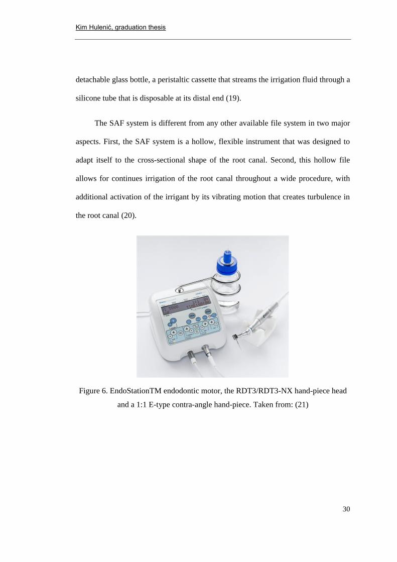
2. SAF proSystem: includes the EndoStationTM endodontic motor, the
RDT3/RDT3-NX hand-piece head and a 1:1 E-type contra-angle hand-piece (Figure
6). The SAF System includes its own endodontic motor and does not require any
additional endodontic motor. The EndoStationTM is a multi-functional endodontic
motor that enables the user to operate various motored endodontic systems,
including, alongside the Self-Adjusting File (SAF), all major brands of rotary and
reciprocating files. The EndoStationTM includes an integrated peristaltic pump and
irrigation system for work with the SAF System. The irrigation system and the
endodontic motor of the EndoStationTM are simultaneously operated by a foot
pedal. The irrigation system is at the back side of the device and includes a
Kim Hulenić, graduation thesis
30
detachable glass bottle, a peristaltic cassette that streams the irrigation fluid through a
silicone tube that is disposable at its distal end (19).
The SAF system is different from any other available file system in two major
aspects. First, the SAF system is a hollow, flexible instrument that was designed to
adapt itself to the cross-sectional shape of the root canal. Second, this hollow file
allows for continues irrigation of the root canal throughout a wide procedure, with
additional activation of the irrigant by its vibrating motion that creates turbulence in
the root canal (20).
Figure 6. EndoStationTM endodontic motor, the RDT3/RDT3-NX hand-piece head
and a 1:1 E-type contra-angle hand-piece. Taken from: (21)

Kim Hulenić, graduation thesis
31
8. DISCUSSION
Many researches have compared NiTi rotary system with SAF and all results
have been in favor of SAF (19). Here are some conclusions that were made in
various different studies with several topics and their main focuses.
Some studies reported that canal preparation can cause dentinal defects like
fractures, craze lines, incomplete crack and dentinal detachment. Over-
instrumentation might weaken the root and create apical root cracks. With NiTi
rotary system there is a possibility to go over the apical foramen and/or to make
craze lines, and microcracks resulted from the active cutting edges and continuing
rotation. The SAF system does not have the structure of a flute helix, cutting blades
or the internal core. This system does not have screw-in force and no pressure from a
metal shaft. Because of its ability to adapt to the canal's original anatomy and shape,
the result is a noninvasive root canal preparation. The unique SAF mesh structure
and cross sectional configuration minimizes the reaction forces against the root
canal. When the file moves in-and-out of the canal, the contacts between the
instrument and the root canal walls create many momentary stress concentrations in
dentin. Higher stresses in root during instrumentation can be generated by stiffer
instruments with bigger cross-sectional area, which may increase the risk of vertical
root fractures. SAF, with its minimal cross-sectional area, may pose minimal risk of
VRFS and maximum potential to preserve root canal integrity (22). Higher root
stress concentrations may cause more canal deviations and result in thinner dentin
areas. Thinner dentin weakens the root structure and increases the risk of apical

Kim Hulenić, graduation thesis
32
cracking. On the other hand, the SAF generated a minimal reaction force and resulted
in minimal dentin removal. This could contribute to the reduction of the fracture risk
and dentinal defects (Figure 7) (23).
Figure 7. Microcracks in dentinal walls (B,C,D and E) as a consequence of root canal
instrumentation with Hero Shaper (B), Revo-S (C), Twisted file (D) and ProTaper €.
Hand (A) and SAF (F) instrumentation did not produce microcracks. Taken from: (23)

Kim Hulenić, graduation thesis
33
The shaping of the root canal is one of the most important steps in endodontics.
For many years it has been common practice to enlarge a root canal to at least three
ISO sizes larger than the first file, so as to bind at the apical part of the canal. It was
assumed that such preparation will remove the inner layers of dentin while allowing
the irrigant to reach the canal space. This goal is easier to achieve today with NiTi
rotary system but the downside is its disability to make a 3D preparation where the
third dimension was continuously ignored. The goal of cleaning and shaping may be
achieved with rotary systems as far as relatively straight and narrow root canal with
round cross-section is concerned. Nevertheless, in flat-oval shaped root canals and in
curved ones, this goal is not easily attainable. The buccal and lingual areas of this
kind on root canals and the area facing the isthmus in tear-shaped ones cannot be
adequately prepared by current rotary files. Substantial untouched areas may be left
and current technology may mislead the operator to believe that the canal is
adequately prepared when there is a significant amount of infected tissue and debris
left. Such canals may never be well obturated or sealed due to remaining of tissue
and debris that provide a potential bacterial growth or future recontamination.
Thinner instruments will maintain the apical part of the curved canal in place, and as
for the stiffer ones, they have a tendency to remove more dentin on the outer side of
the curvature, leading to canal transportation. Transportation of the canal can have
two major deficiencies. First, the apical part of the canal on the inner side of the
curvature remains untouched and full of debris and, second, it may lead to ledging or
even subsequent perforation. To this day, most if not all file systems have the
inherent problem to one extent or another. Another closely related problem is

Kim Hulenić, graduation thesis
34
straightening of the root canal at the midroot section of curved root canals. Most files
system will straighten a part of the curvature to one extent or another, by removing
more dentin on the inner side of the curvature. This may reduce the thickness of the
remaining dentin on the inner side of the curvature to such measure that it increases
the risk of vertical fracture or even results in a strip perforation. The SAF file is
different from any current NiTi rotary system. Most of them will find the widest part
of the canal and gradually machine it, using several files of increasing diameter, to a
wider canal with round cross section, if the canal happens to be relatively narrow; the
whole original canal may be included in the preparation. However, if the canal is flat,
tear shaped or simply large, this preparation may result in untreated recesses. The
SAF file is used as a single file that starts as narrow and compressed shape and
gradually expands in the canal while removing a uniform layer of dentin from its
walls. Because the file adapts itself to the cross section of a given canal, a canal with
a round cross-section is enlarged as a round, whereas an oval canal is enlarged as an
oval canal of larger diameter. After preparation with SAF file, thickness of remaining
dentin wall is uniform and it is predisposing factor for vertical fractures. When rotary
files accidentally pass the apical foramen of an apically curved canal, due to
misleading length measurements or failure to maintain the marker in place, they may
soon ”zip” the apical foramen and form an oval opening. The SAF on the other hand
may be operated in such conditions even for few minutes with no zipping whatsoever
(20).
According to the present data and earlier pilot trials, the SAF is most effective
during the first two minutes of use, and hence under the present experimental

Kim Hulenić, graduation thesis
35
conditions, the bulk dentin removal was accomplished during the first two minutes of
preparation. However, there was about 40 % unprepared canal area with the SAF 2.0
mm at that time. Regression analysis indicated that the optimum of prepared canal
surface is more than 90 % on average, reached after five minutes of activation.
A recent micro-computed tomography (micro CT) study has shown that the
percentage of root canal area affected by the SAF method is larger than that affected
by popular rotary instruments. Consequently, less unprepared areas that might
potentially harbor bacterial biofilms remnants are observed. Other study reported that
SAF operation with continued irrigation resulted in root canal walls that were free of
debris in all specimens and almost completely free of smear layer. The SAF system
has a potential to be particularly advantageous in promoting disinfection of oval
shaped canals (25, 26).

Kim Hulenić, graduation thesis
36
Figure 8. Micro-CT of root canals prepared with SAF (A) and rotary system (B). A
good root canal filling adaptation with 98.1% of the canal wall in contact with the
root canal filling (C) and a poor root canal filling adaptation with only 68.9% of the
root canal wall in contact with the root canal filling material (D). Taken from: (27)
A recent study compared the prepared surfaces areas and revealed that the
mean in unaffected areas ranged from about 60-80% from the total canal length. All
the apical portion of the canal, the mean of untouched areas ranged from 65-75 %
(28). Another recent study comparing the cleaning effects of three instrumentation

Kim Hulenić, graduation thesis
37
techniques in oval-shaped canals reported that none of the techniques resulted in
completely prepared and cleaned canals (29).
C-shaped canals presented a challenge to both file systems which resulted in a
percentage of canal area unaffected by the procedures that was higher than
previously reported in normal canals. The SAF was more effective than the ProTaper
file system in shaping the walls of C-shaped root canals. In the SAF treated group, a
mean 41% ± 14% of the canal walls was unaffected by the procedure, with a range of
21% to 70%. In the ProTaper group, a mean of 66% ± 6% of the canal walls were
unaffected by the procedure with a range of 54 % to 75 % (30).
During root canal preparation procedures, dentin chips, pulp tissue,
microorganisms, and/or irrigants can be extruded into the periradicular tissue. The
extrusion of these elements may cause undesired consequences, such as the induction
of inflammation and postoperative pain, and delay of periapical healing. Currently,
all preparation techniques and instruments are associated with extrusion of debris;
however, there are notable differences among the techniques. Based on a statistical
data, the hand instrumentation group extruded significantly more debris than both of
the other tested groups. The SAF system extruded less than the ProTaper file.
According to the current results, apical debris extrusion occurred regardless of the
instrumentation technique used (28).
The present results may be explained by differences in the instrument design
and movement kinematics between SAF and ProTaper system. The metal mesh in the
SAF system is closely adapted to the canal walls and removes dentin with a back-

Kim Hulenić, graduation thesis
38
and-forth grinding motion, providing a scrubbing action on the canal walls.
Moreover, the SAF allows continues the irrigation of the root canal throughout the
procedures with additional activation of the irrigant by its vibrating motion, which
creates turbulence in the root canals. The irrigation fluid enters the file through a
free-rotating hub continuously replaced throughout the procedure, providing s fully
active supply of irrigant. No positive pressure can be developed in root canal space
because the solution easily escapes through openings in the file lattice. Under the
condition of this study, it can be concluded that all systems caused apical debris
extrusion. The SAF instrumentation was associated with significantly less debris
extrusion compared with the use of hand and rotary files (25, 27).
The SAF system substantially reduced the amount of the remaining pulp tissue
by 57% as compared with the conventional full sequence of ProTaper NiTi files. In
other words, the SAF system improved the debridement standard produced by the
conventional NiTi rotary preparation approach. In the ProTaper system group,
substantial amounts of pulp tissue remained in the canals, 21% of the root canal
cross-section contained pulp tissue remnants. This presents the inability of most
rotary systems to access buccal and/or lingual recesses of oval canals. Furthermore, it
represents the limited ability of sodium hypochlorite irrigant applied with a syringe
needle to compensate for the inadequacy of the file itself. The current study results
indicate that, in addition to its previously reported better efficiency for
circumferentially removing dentin from all canal walls, as shown by microCT
studies, the SAF system has also improved debridement and cleaning efficiency in

Kim Hulenić, graduation thesis
39
the oval-shaped canals. This may, in turn, also aid in explaining the recently reported
improved disinfection that the SAF system has in oval canals (25, 27, 31).
The SAF is extremely durable and may go through rather severe use before a
mechanical failure occurs. It does not have a core like the others. Any strain applied
to it is distributed along many of its delicate parts and the total endurance is a
function of the accumulated endurance of each individual part (32).
The irrigation of the root canal with sodium hypochlorite during root canal
treatment is widely recommended. It has been well documented that when exposed to
its target of bacteria and tissue debris, sodium hypochlorite loses its activity rather
quickly. Taking into account the extremely small volume of the root canal, the
amount of sodium hypochlorite contained in the canal loses its activity within a very
short time. Therefore, replacement of the irrigant as frequently as possible is
mandatory for maintaining its optimal potency and effect. The SAF operates with a
continuous flow of the irrigant, thus allowing continuously fresh irrigant to be
present in the canal at all times. The vibration of the file’s metal lattice within the
irrigant facilitates its cleaning and debridement effects. It is evident that syringe and
needle irrigation was ineffective in replacing the liquid in the apical part of a narrow
curved canal. The SAF, which was used with continuous irrigation, combined with
the vibrating action of the metal lattice, was affective in replacing the liquid in the
apical part of the narrow curved canal. No pressure builds up in the canal during the
SAF operation because the metal mesh allows free escape of the irrigant at all times.
No irrigant passes the apical foramen during SAF operation (32, 33).

Kim Hulenić, graduation thesis
40
The SAF instrumentation system was highly effective in reducing bacterial
population from infected root canals. Quantitative and qualitative results were
significantly superior to those attained by a hand instrumentation protocol. Some
bacterial species were identified in post-treatment samples and the excellent
antibacterial effectiveness of SAF, the fact that almost half of the teeth still had
detectable bacteria by molecular method, suggest that a supplementary step after
chemomechanical procedures with SAF is also required to improve disinfection (34).
The SAF has proven to be more effective in areas of the root canals that are
inaccessible to most other instruments. The SAF is expected to remove the remaining
gutta percha after conventional retreatment because of its scraping motion with
simultaneous irrigation and its ability to touch a higher percentage or root canal walls
than rotary instruments. Removing gutta-percha is an essential and difficult step
during root canal retreatment. Hand files, rotary files, and a solvent were not able to
remove all gutta-percha from root canals in previous studies. The SAF makes more
root canal-wall contact compared with other rotary files, resulting in a significantly
lower percentage of untouched canal areas on oval canals. Moreover, a SAF
operation with continuous irrigation, using alternating irrigants, resulted in root canal
walls that were free of debris. Therefore, it was hypothesized that the SAF might
scrape gutta-percha remnants of the root canal wall, resulting in cleaner canals after
retreatment under the experimental conditions. The SAF increased the amount of
gutta-percha removed from curved root canals when compared with ProTaper
retreatment instruments alone. However, the removal of gutta-percha from the apical
area of curved root canals still remains difficult (32).

Kim Hulenić, graduation thesis
41
A significantly higher PGFA (gutta-percha filled area) was found in oval
shaped canals prepared using SAF system with continuous irrigation compared with
similar canals prepared using a conventional NiTi rotary system with syringe and
needle irrigation. Recent reports have indicated that the SAF cleaning and shaping
irrigation system may improve both shaping and cleaning of oval shaped root canals
compared with traditional NiTi rotary system. A higher percentage of the canal wall
was affected by instrumentation with this method than with the rotary system,
whereas less tissue debris is left after instrumentation and almost no packing of hard
tissue particles occurs. Both instrumentation systems failed to achieve the desirable
goal of 100% PGFA in all cases (32).

Kim Hulenić, graduation thesis
42
9. CONCLUSION
As mentioned previously, cleaning and shaping of the root canal is the most
important step in every endodontic treatment. Currently, endodontic procedures are
performed with hand and rotary instruments that do not adapt to the canal walls. The
irrigation steps are intermittent and deliver limited amounts of irrigant to the apical
third of the root. Unfortunately, the literature is loaded with examples of instrument
breakage, poor results with chemomechanical preparation, canal transportation, and
over-thinning of the canal walls. Since hand and rotary files are round in cross
section, they leave more than half of the canal walls untouched and require multiple
sequences of filling and irrigation.
The new SAF instrument actively adapts to the shape of each canal, moves in a
new oscillating up-and-down motion, and allows simultaneous irrigation. After the
proper glide path is established, only one SAF instrument is required to finish the
entire 3-D instrumentation of the canal. This revolutionary technology has been
proven in numerous peer-reviewed, published articles to contact more canal surfaces,
improve canal disinfection, and provide better results. SAF technology provides a
virtually unbreakable instrument and is changing the way we perform endodontics.

Kim Hulenić, graduation thesis
43
10. SUMMARY
Since the beginning of modern day endodontics, there have been numerous
concepts, strategies, and techniques for preparing canals. Throughout decades, a
staggering array of files has emerged for negotiating and shaping canals. In spite of
the design of the file, the number of instruments required, and the surprising
multitude of techniques advocated, endodontic treatment has been typically
approached with optimism for probable success. The common misconception today
is that dentists tend to relate to all root canals as if they had a uniform round cross
section. This is true for the upper incisors, but wrong for every other tooth in maxilla
and mandible. The Self-adjusting file (SAF) brings a new concept to the table in
terms of root canal treatment. First of all, it is a system that combines irrigation and
instrumentation at the same time. The special design of the instrument allows its
compression inside the canals and three dimensional adjustment, no matter how
complicated the configuration of root canal is, without fear of breakage. In addition,
this instrument has the ability of circumferential removal of dentin to give better
adhesion of the future filling and the canal wall. Surface of the SAF has a roughness
of 2.8 µm ± 10 % which gives a file scraping feature and a possibility to smoothen
walls of the root canal. It generates a vibration of 5000 rpm. With the SAF, goals like
minimal reduction and uniformity of the remaining wall thickness become
achievable. The prevention of canal transportation, continuous irrigation with sodium
hypochlorite and high durability are just a few specifications that make this system a
step towards a whole new era in modern endodontics.

Kim Hulenić, graduation thesis
44
11. SAŽETAK
Od početka modernog doba endodoncije, javljali su se brojni koncepti, strategije i
tehnike za obradu kanala, te je do danas razvijen veliki broj različitih instrumenata.
Zajednička zabluda većine stomatologa u današnje vrijeme jest tendencija da se
odnose prema svim korijenskim kanalima kao da imaju jednak okrugli poprečni
presjek. To se možda odnosi na pojedine dijelove korijenskih kanala nekih zubi (npr.
gornjih sjekutića), no većina korijenskih kanala je izuzetno nepravilna.
Samoadaptirajući instrument je primjer potpuno novog koncepta u obradi korijenskih
kanala. Prije svega, to je sustav koji ujedinjuje irigaciju i instrumentaciju
istovremeno. Poseban dizajn instrumenta za obradu kanala omogućuje njegovo
komprimiranje unutar kanala i prilagodbu trodimenzionalnoj konfiguraciji
korijenskog kanala, ma koliko ona bila komplicirana, bez straha od pucanja. Pored
toga, taj instrument ima sposobnost cirkumferencijalnog uklanjanja dentina što
omogućuje bolju adheziju budućeg punjenja uz stjenke kanala. Površina
samoadaptirajućeg instrumenta ima hrapavost 2,8 µm ± 10%, što mu daje strugajuća
svojstva i zaglađuje stjenke kanala. Ostvaruje vibraciju od 5000 rpm. Sa
samoadaptirajućim instrumentom se postiže cilj minimalne redukcije i jednolikost
preostale debljine stjenke kanala. Prevencija preko instrumentacije, izvan granice
apikalnog foramena, kontinuirana irigacija korijenskog kanala natrijevim
hipokloritom i visoka izdržljivost, samo su neke karakteristike koje čine ovaj
instrument začetnikom nove moderne ere u endodonciji.

Kim Hulenić, graduation thesis
45
12. LITERATURE
1. Peters OA, Paqué F. Current developments in rotary root canal instrument
technology and clinical use: A review. Quintessence Int, 2010;41(6):479–88.
2. Castellucci A. Endodontics: Volume 1. 1st ed. California: Il Tridente; c2004.
Chapter 1, A brief history of endodontics; p. 2-6.
3. Hülsmann M, Peters OA, Dummer PMH. Mechanical preparation of root
canals: shaping goals, techniques and means. Endod Topics. 2005;10(1):30-
76.
4. Garg N, Garg A. Textbook of Endodontics. 1st ed. New Delhi: Jaypee
Brothers Medical Publisher Ltd; 2007. 504 p.
5. Castellucci A. Endodontics: Volume 2. 1st ed. California: Il Tridente; 2005.
750 p.
6. Messing JJ, Stock CJR. A Colour Atlas of Endodontics. 1st ed. London:
Wolfe Medical Publications Ltd; c1988. Chapter 5, Basic instruments and
materials; p. 54-84.
7. Kandaswamy D, Venkateshbabu N. Root canal irrigants. J Conserv Dent.
2010;13(4):256–64.
8. Spencer HR, Ike V, Brennan PA. Review: the use of sodium hypochlorite in
endodontics — potential complications and their management. Brit Dent J.
2007;202(9):555-9.

Kim Hulenić, graduation thesis
46
9. Stuart CH, Schwartz SA, Beeson TJ, Owatz CB. Enterococcus faecalis: its
role in root canal treatment failure and current concepts in retreatment. J
Endod. 2006;32(2):93-8.
10. Spencer HR, Ike V, Brennan A. Review: the use of sodium hypochlorite in
endodontics — potential complications and their management. Brit Dent J.
2007;202(9):555-9.
11. Hülsmann M, Heckendorff M, Lennon A. Chelating agents in root canal
treatment: mode of action and indications for their use. Int Endod J.
2003;36(12):810-30.
12. Carrotte P. Endodontics: Part 4 Morphology of the root canal system. Br Dent
J. 2004;197(7):379-83.
13. Vertucci FJ. Root canal morphology and its relationship to endodontic
procedures. Endod Topics. 2005;10(1):3-29.
14. Maniglia-Ferreira C, de Almeida-Gomes F, Carvalho de Sousa B, Cabral dos
Santos Acioli Lins C, Alves dos Santosc R. A Case of Unusual Anatomy in
Second Mandibular Molar with Four Canals. Eur J Dent. 2008;2(1):217-9.
15. Torabinejad M, Walton RE. Endodontics: Principles and practice. 4th ed.
St.Louis: Elsevier Health Sciences; c2009. Chapter 13, Internal Anatomy; p.
216-30.
16. Versiani MA, Pécora JD, Sousa Neto MD. Middle mesial canal: Maxillary
first molar [Internet]. Sao Paolo: Marco Versiani; 2013 Mar [cited 2015 Sep
1] Available from:
http://rootcanalanatomy.blogspot.hr/2013_03_01_archive.html.

Kim Hulenić, graduation thesis
47
17. Metzger, Z. From files to SAF: 3D endodontic treatment is possible at last.
Alpha Omegan. 2011;104(1-2):36-44.
18. Metzger Z, Teperovich E, Zary R, Cohen R, Hof R. The Self-adjusting file
(SAF): Respecting the root, canal anatomy - A new concept of endodontic
files and its implementation. J Endod. 2010;36(4 Pt 2):679-90.
19. ReDentNova.com [Internet]. Israel: ReDent Nova; c2015 [cited 2015 Sep
13]. Available from: http://www.redentnova.com/.
20. Metzger Z, Teperovich E, Cohen R, Zary R, Paqué F, Hülsmann M. The Self-
adjusting file (SAF): Removal of debris and smear layer - A scanning
electron microscope study. J Endod. 2010;36(4 Pt 3):697-702.
21. Metzger Z. The Self-adjusting file (SAF) system: An evidence-based update.
J Conserv Dent. 2014;17(5):401-19.
22. Kim HC, Sung SY, Ha JH, Solomonov M, Lee JM, Lee CJ et al. Stress
generation during Self-Adjusting file movement: minimally invasive
instrumentation. J Endod. 2013;39(12):1572-5.
23. Yoldas O, Yilmaz S, Atakan G, Kuden C, Kasan Z. Dentinal microcrack
formation during root canal preparations by different NiTi rotary instruments
and the Self-adjusting file. J Endod. 2012;38(2):232-5.
24. Hof R, Perevalov V, Eltanani M, Zary R, Metzger Z. The Self-adjusting file
(SAF): Mechanical analysis, J Endod. 2010;36(4 Pt 2):691-6.
25. Peters OA, Paqué F. Root canal preparation of maxillary molars with the
Self-adjusting file: A micro-computed tomography study. J Endod.
2011;37(1):53-7.
Kim Hulenić, graduation thesis
48
26. Peters OA, Boessler C, Paqué F. Root canal preparation with a novel nickel-
titanium instrument evaluated with micro-computed tomography: Canal
surface preparation over time. J Endod. 2010;36(6):1068-72.
27. Metzger Z, Zary R, Cohen R, Teperovich E, Paqué F. The quality of root
canal preparation and root canal obturation in canals treated with rotary
versus Self-adjusting files: A three-dimensional micro-computed
tomographic study. J Endod. 2010;36(9):1569-73.
28. De-Deus GA, Nogueira Leal Silva EJ, Moreira EJ, de Almeida Neves A,
Belladonna FG, Tameirão M. Assessment of apically extruded debris
produced by the Self-adjusting file system. J Endod. 2014;40(4):526-9.
29. Taha NA, Ozawa T, Messer HH. Comparison of three techniques for
preparing oval-shaped root canals. J Endod. 2010;36(3):532-5.
30. Solomonov M, Paqué F, Fan B, Eilat Y, Berman LH. The challenge of C-
shaped canal systems: A comparative study of the Self-adjusting file and
ProTaper. J Endod. 2012;38(2):209-14.
31. De-Deus G, Souza EM, Barino B, Maia J, Zamolyi RQ, Reis C et al. The
Self-adjusting file optimizes debridement quality in oval-shaped root canals. J
Endod. 2011;37(5):701-5.
32. De-Deus G, Barino B, Marins J, Magalhães K, Thuanne E, Kfir A. Self-
adjusting file cleaning - shaping - irrigation system optimizes the filling of
oval-shaped canals with thermoplasticized gutta-percha. J Endod.
2012;38(6):846-9.

Kim Hulenić, graduation thesis
49
33. Yoldas O, Yilmaz S, Atakan G, Kuden C, Kasan Z. Dentinal microcrack
formation during root canal preparations by different NiTi rotary instruments
and the Self-adjusting file. J Endod. 2012;38(2):232-5.
34. Neves MA, Rôças IN, Siqueira JF Jr. Clinical antibacterial effectiveness of
the Self-adjusting file system. Int Endod J. 2014;47(4):356-65.

Kim Hulenić, graduation thesis
50
13. CURRICULUM VITAE
Kim Hulenić was born on 11th of October 1990 in Zagreb, where she finished
primary school. In 2006 she enrolled in general high school in Zagreb, where she
graduated in 2009. The same year she entered in University of Dental Medicine in
Zagreb. During her studies she worked as an assistant in a private dental practice,
published an article in the student newspaper, did one semester Erasmus exchange in
Portugal and participated in several dental congresses in Croatia and abroad. She
speaks English, German and Portuguese.