SELECTIVE ANTEGRADE CEREBRAL PERFUSION IS THECEREBRAL ... · SELECTIVE ANTEGRADE CEREBRAL PERFUSION...
42
SELECTIVE ANTEGRADE CEREBRAL PERFUSION IS THE CEREBRAL PERFUSION IS THE TECHNIQUE OF CHOICE MARKO TURINA University of Zurich Zurich, Switzerland
Transcript of SELECTIVE ANTEGRADE CEREBRAL PERFUSION IS THECEREBRAL ... · SELECTIVE ANTEGRADE CEREBRAL PERFUSION...

SELECTIVE ANTEGRADE CEREBRAL PERFUSION IS THECEREBRAL PERFUSION IS THE
TECHNIQUE OF CHOICE
MARKO TURINAUniversity of ZurichZurich, Switzerland,

What is so special about the poperation on the aortic arch?
Di i ll ll d fi d• Disease process is usually well defined: degenerative, dissecting or spurious aneurysm; or a ruptured aortic plaque.a ruptured aortic plaque.
• Standard methods of cardiopulmonary bypass (with avoidance of aortic cannulation) and myocardial protection can be utilized.
• Classic aneurysm repair with graft replacement is usually possibleusually possible.
• It is the necessity for interrupting the normal brain circulation which makes this operationscirculation which makes this operations challenging.

How to protect the brain during p gprocedures on the aortic arch?
Hi t i ll l diff t th d tili dHistorically, several different methods were utilized for brain protection when operating on the aortic arch:arch:
• Antegrade perfusion of the cerebral vessels.• Deep hypothermic circulatory arrest.p yp y• Retrograde perfusion of superior vena cava (or
even total body retrograde perfusion)• Perfusion via subclavian artery, or combined
subclavian and left carotid perfusion.N d l th l t t ti l d• Nowadays, only the last two are routinely used.


Venogram of the SVC during cough: tight vein valves in internal jugular and subclavian vein
From Fisher et al., Circulation 1982

From Fisher et al., Circulation 1982

From Fisher et al., Circulation 1982


Retrograde cerebral perfusion can be severely impaired by competent jugular vein valvesimpaired by competent jugular vein valves

Anatomic and physiologic arguments p y g gfor antegrade cerebral perfusion
• Antegrade perfusion through carotid and vertebral arteries reaches the brain in a normal flow pattern and is distributed alongnormal flow pattern and is distributed along established arterial channels.
• Vein valves exist in jugular veins, which inVein valves exist in jugular veins, which in retrograde perfusion can prevent the nutritive flow to reach the brain tissue.
• Retrograde perfusion must be administered in deep hypothermia; otherwise it is not reliable.

Percent changes in middle cerebral artery blood flow velocities measured by transcranial Doppler technique in humans
Tanoue Y. et al.; Ann Thorac Surg 1999;67:672-675
Copyright ©1999 The Society of Thoracic Surgeons
Tanoue Y. et al.; Ann Thorac Surg 1999;67:672 675

Brain flow during hypothermia with antegrade (ante) and retrograde cerebral perfusion (RCP) in primates. During RCP, extremely low flow values were found in all areas examined Those flow values differedvalues were found in all areas examined. Those flow values differed significantly from the pre-arrest values at 18°C (p < 0.05).
Copyright ©1995 The Society of Thoracic Surgeons
Boeckxstaens C. J. et al.; Ann Thorac Surg 1995;60:319-327


Percentage changes in rCBV in right and left hemispheres during U-ACP at 15°C and at 60 minutes of reperfusion with CPB (R-CPB) at 37°C. No
i ifi t diff b d b t i ht d l ft h i hsignificant difference was observed between right and left hemispheres.
Ye, J. et al. Circulation 1999;100:II-309-II-315315

During retrograde perfusion through SVC only 1/10’000 of flow reaches brain capillaries
Copyright ©2001 The American Association for Thoracic Surgery
Ehrlich M. P. et al.; J Thorac Cardiovasc Surg 2001;122:331-338

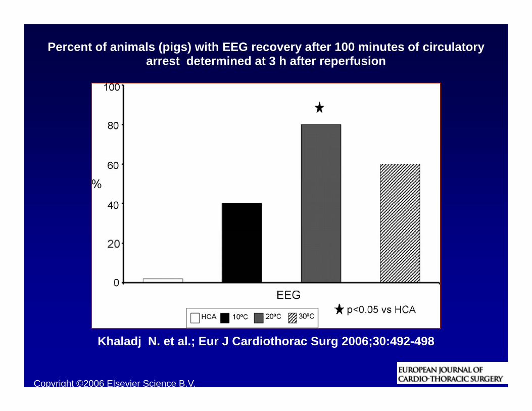
Percent of animals (pigs) with EEG recovery after 100 minutes of circulatory arrest determined at 3 h after reperfusion
Copyright ©2006 Elsevier Science B.V.
Khaladj N. et al.; Eur J Cardiothorac Surg 2006;30:492-498


Figure 1. Changes in PCO2 (A) and PO2 (B) in retrograde cerebral perfusion (RCP) outflow blood specimens measured at two different time points in individual patients who had two
or more serial measurements available for analysis (n = 15)
Copyright restrictions apply.
Cheung, A. T. et al. Anesth Analg 1999;88:8-15

Influence of antegrade cerebral perfusion on averaged SF-36 ({+/-}1 SD) score for patients from group 1, group 2, and group 3
Immer F F et al Circulation 2004;110:II 250 II 255
Copyright ©2004 American Heart Association
Immer, F. F. et al. Circulation 2004;110:II-250-II-255

In this prospective randomized trial, neuropsychiatry tests show less postoperative decline in the selective cerebral perfusionpostoperative decline in the selective cerebral perfusion
Okita Y. et al.; Ann Thorac Surg 2001;72:72-79
Copyright ©2001 The Society of Thoracic Surgeons

Perfusion Via Axillary Artery Results In Less Cerebral Embolization (Deflection?)
Cerebral microsphere distribution, expressed as mean {+/-} SD for aortic and axillary cannulation (n = 5), indicating fewer spheres in the cerebral circulation
during axillary cannulation (canine study)
Hedayati N. et al.; J Thorac Cardiovasc Surg 2004;128:386-390

Various methods of administering the gantegrade brain perfusion
• Cannulation of the right axillary artery, via ft l t th dgraft or cannula; most common method.
• Antegrade cannulation of the innominate d i ht tid tand right carotid artery.
• Combined axillary and femoral artery f iperfusion.

(A) A pursestring suture (5-0 polypropylene) is placed anteriorly in the exposed subclavian artery
Copyright ©2006 The Society of Thoracic Surgeons
Olsson C. et al.; Ann Thorac Surg 2006;81:868-874

Simplified technique for selective cerebral perfusion
Copyright ©2002 European Association for Cardio-Thoracic Surgery. Published by Elsevier B.V. All rights reserved.
Mazzola A. et al.; Eur J Cardiothorac Surg 2002;21:930-931

The distal anastomosis phase in group B2 patients
Copyright ©2006 The Society of Thoracic Surgeons
Della Corte A. et al.; Ann Thorac Surg 2006;81:1358-1364

F B b t l AFrom Barbeau et al, Ann Thor Surg 1999

Subclavian cannulation techniqueSubclavian cannulation technique

From Barbeau et al, Ann Thor Surg 1999

ACUTE TYPE A DISSECTION: C l tiCannulation
University Hospital Zurich, 1997 - 2002 (122 patients)
2425
30
% 0 0179
15
20
talit
y in
% p=0.0179
8
5
10
15
Ear
ly m
or
0
5
Femoral Subclavian
E
Femoral Subclavian

Yearly number of axillary artery cannulations performed either directly or with a side graft
Sabik J. F. et al.; Ann Thorac Surg 2004;77:1315-1320
Copyright ©2004 The Society of Thoracic Surgeons
g

Gulbins et al, Ann Thor Surg 2007;83:1219

Incidence of neurological damage after arch surgery increases with patient’s ageincreases with patient s age
Hagl C. et al.; J Thorac Cardiovasc Surg 2001;121:1107-1121
Copyright ©2001 The American Association for Thoracic Surgery

Brain circulation during right axillary artery perfusion
Copyright ©2006 The Society of Thoracic Surgeons
Merkkola P. et al.; Ann Thorac Surg 2006;82:74-79

Willis circle: is it safe to perfuse only p yone carotid and subclavian artery?

The minimally invasive computed tomography angiogram with good illustration of the brain vascular anatomy
Copyright ©2006 The Society of Thoracic Surgeons
Merkkola P. et al.; Ann Thorac Surg 2006;82:74-79

Willis circle: in reality not so simple as in the textbooks


A, The triple-branched stent graft is a branched 1-piece graft consisting of a self-expandable nitinol stent and polyester vascular graft fabric
Copyright ©2010 American Heart Association
Chen, L.-W. et al. Circulation 2010;122:1373-1378

Postoperative computed tomographic scans show that all stent grafts were fully opened and not kinked; there was no space or blood flow surrounding the triple-branched stent graft
Copyright ©2010 American Heart Association
Chen, L.-W. et al. Circulation 2010;122:1373-1378

Unresolved questions about antegrade cerebral perfusioncerebral perfusion
• Necessary flow rate: usually quoted as 10 ml/kg/min, but no hard data in support of this particular flow ratebut no hard data in support of this particular flow rate.
• Perfusion pressure: usually given as 50 – 70 mmHg, with some experimental evidence; but where to
? Th i ht di l i di t bl d l ftmeasure? The right radial is unpredictable, and left radial is obviously lower. Use cannula tip?
• What to do about the lower body: when innominate artery is clamped and left carotid and subclavian blocked, what is the ischemic tolerance of abdominal organs, and at what temperature?
• Optimal method of cannulation: axillary artery (graft or cannula?); with a single cannula or additional one in left carotid; or innominate and subclavian arteryin left carotid; or innominate and subclavian artery directly, after sternotomy.

Antegrade vs. retrograde cerebral perfusion SummarySummary
• Antegrade cerebral perfusion provides superior g p p pbrain protection in arch surgery.
• Isolated subclavian artery perfusion is sufficient h l h t (15 20 i t ) i t tiwhen only a short (15 – 20 minutes) interruption
of normal flow pattern is necessary.• Perfusion via subclavian and right carotid artery• Perfusion via subclavian and right carotid artery
is advised for longer periods of flow interruption.• Additional lower body perfusion might be y p g
advantageous in complex cases (redos, etc.).• With these techniques, perfusion at 280 C is
ibl tl h t i th CPB tipossible, greatly shortening the CPB time.