
Section C: Assessing Sentinel Facial Features - FASD Hub · Australian guide to the diagnosis of...
11
Australian guide to the diagnosis of FASD Section C: Assessing sentinel facial features and Appendices C & D Pages 31-34 & 74-78 Section C: Assessing Sentinel Facial Features Fetal exposure to alcohol during the first trimester affects development of facial features. The areas most affected are the orbital region (eyes) and mid-face. The effect of prenatal alcohol exposure on fetal brain growth is also thought to affect the size and shape of the face. A range of facial anomalies can occur as result of prenatal alcohol exposure. There are three features which commonly occur across age, gender and ethnic groups: ▪ Small palpebral fissures: short horizontal length of the eye opening, defined as the distance from the endocanthion to the exocanthion (points A and B on photo below) ▪ Smooth philtrum: diminished or absent ridges between the upper lip and nose ▪ Thin upper lip: with small volume These features are shown in the photo below. (Photo reproduced with permission from Susan Astley, University of Washington) Although these facial features may also occur independently as normal variations in the general population (unrelated to prenatal alcohol exposure), when seen in combination,
Transcript of Section C: Assessing Sentinel Facial Features - FASD Hub · Australian guide to the diagnosis of...
Australian guide to the diagnosis of FASD Section C: Assessing sentinel facial features and Appendices C & D Pages 31-34 & 74-78
Section C: Assessing Sentinel Facial Features
Fetal exposure to alcohol during the first trimester affects development of facial features. The areas
most affected are the orbital region (eyes) and mid-face. The effect of prenatal alcohol exposure on
fetal brain growth is also thought to affect the size and shape of the face. A range of facial anomalies
can occur as result of prenatal alcohol exposure.
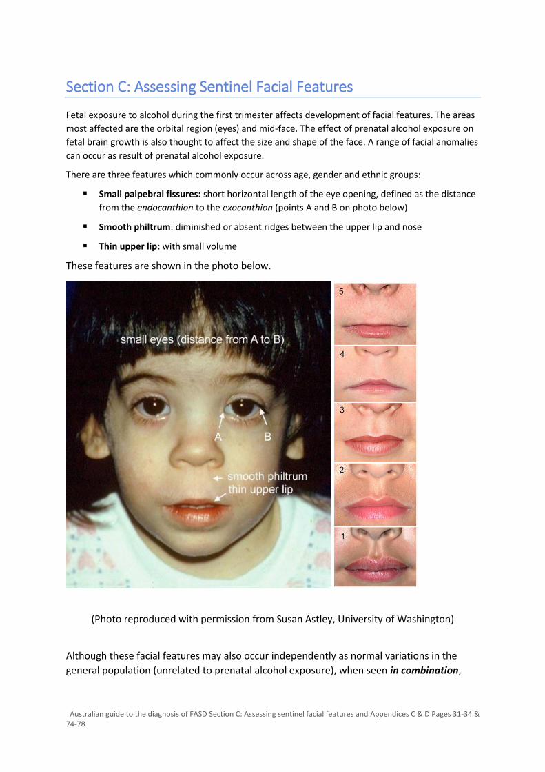
There are three features which commonly occur across age, gender and ethnic groups:
▪ Small palpebral fissures: short horizontal length of the eye opening, defined as the distance
from the endocanthion to the exocanthion (points A and B on photo below)
▪ Smooth philtrum: diminished or absent ridges between the upper lip and nose
▪ Thin upper lip: with small volume
These features are shown in the photo below.
(Photo reproduced with permission from Susan Astley, University of Washington)
Although these facial features may also occur independently as normal variations in the
general population (unrelated to prenatal alcohol exposure), when seen in combination,

Australian guide to the diagnosis of FASD Section C: Assessing sentinel facial features and Appendices C & D Pages 31-34 & 74-78
these facial features are pathognomonic of and highly specific to prenatal alcohol
exposure. They are termed the ‘sentinel’ facial features of FASD.
Facial anomalies are one of the three diagnostic criteria for FASD, together with prenatal
alcohol exposure and neurodevelopmental impairment. A diagnosis of FASD may be made
with or without facial features.
▪ A diagnosis of FASD with three sentinel facial features means that the individual has
all 3 of the characteristic (or ‘sentinel’) facial features that have been associated with
prenatal alcohol exposure.
▪ A diagnosis of FASD with less than 3 sentinel facial features means that the individual may have 0,1 or 2 of the characteristic facial features
The University of Washington FAS Prevention and Diagnostic Network has developed criteria for FASD sentinel facial features:
Assessment can be using direct measurement and clinical examination and/or computerised
analysis of a digital facial photograph (as described by Astley and Clarren (1, 2). Facial features
may alter with age. Diagnosis should be based on the point in time when the features were
most clearly expressed.
Further details regarding how to assess sentinel facial features are found in Appendix C.
Considerations regarding assessment of sentinel facial features
Palpebral fissure length (PFL) PFL growth charts have been developed for populations overseas. In the absence of
Australian reference data, we recommend using:
▪ Scandinavian (Stromland) charts if a child is under 6 years of age
▪ Canadian (Clarren) charts if a child, adolescent or adult is over 6 years
The Canadian charts are based on a multi-racial population considered to be a better
representation of Australian children, although this has not been qualified by research. As
the charts start at 6 years of age, Scandinavian charts need to be used in children under 6
years of age.
For infants and children under 2 years of age, the corrected age of an ex-premature child
should be used if they are under 2 years of age (similar to other growth parameters such as
head circumference, height and weight).
▪ Short palpebral fissure length (PFL) 2 or more standard deviations below the
population mean (or <3rd percentile). This equates to a z-score of -2 or more.
▪ Smooth philtrum – Rank 4 or 5 on the University of Washington Lip-Philtrum
Guide
▪ Thin upper lip – Rank 4 or 5 on the University of Washington Lip-Philtrum Guide

Australian guide to the diagnosis of FASD Section C: Assessing sentinel facial features and Appendices C & D Pages 31-34 & 74-78
For older adolescent and adults, since PFL matures by 16 years without further changes, PFL
norms and z scores for 16 year olds can be used for individuals over 16 years of age (from
the Clarren charts).
Upper Lip Thinness and Philtrum Smoothness Upper lip thinness and philtrum smoothness should be assessed using the University of
Washington (UW) Lip-Philtrum Guides, which comprise photographs according to a 5 rank
scale, which the range of lip thickness and philtrum depth seen in a population (i.e. the
normal distribution).
▪ Ranks 1, 2 and 3 are not associated with prenatal alcohol exposure, and are below
diagnostic threshold for FASD
▪ Ranks 4 and 5 are also caused by and characteristic of prenatal alcohol exposure and
FASD, but are also seen in a small proportion of the general population.
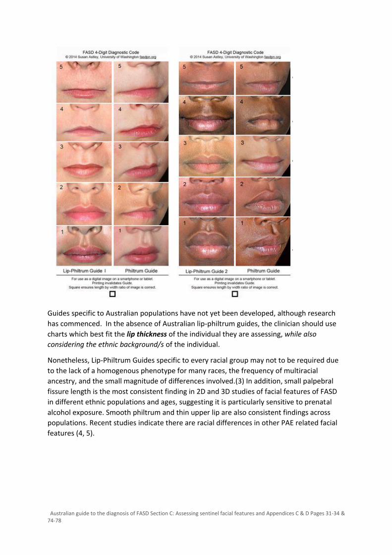
The University of Washington has developed guides for two ethnic populations: Caucasian
(Guide 1) and African American (Guide 2) – see Appendix C. They recommend:
▪ Lip-Philtrum Guide 1 should be used for Caucasians and all races (or combinations of
races) with lips like Caucasians.
▪ Lip-Philtrum Guide 2 should be used for African Americans and all races (or
combinations of races) with thicker lips like African Americans.
Australian guide to the diagnosis of FASD Section C: Assessing sentinel facial features and Appendices C & D Pages 31-34 & 74-78
Guides specific to Australian populations have not yet been developed, although research
has commenced. In the absence of Australian lip-philtrum guides, the clinician should use
charts which best fit the lip thickness of the individual they are assessing, while also
considering the ethnic background/s of the individual.
Nonetheless, Lip-Philtrum Guides specific to every racial group may not to be required due
to the lack of a homogenous phenotype for many races, the frequency of multiracial
ancestry, and the small magnitude of differences involved.(3) In addition, small palpebral
fissure length is the most consistent finding in 2D and 3D studies of facial features of FASD
in different ethnic populations and ages, suggesting it is particularly sensitive to prenatal
alcohol exposure. Smooth philtrum and thin upper lip are also consistent findings across
populations. Recent studies indicate there are racial differences in other PAE related facial
features (4, 5).

Australian guide to the diagnosis of FASD Section C: Assessing sentinel facial features and Appendices C & D Pages 31-34 & 74-78
Other dysmorphic features Other dysmorphic features have been observed in FASD but are not specific to FASD. These
should be documented during assessment and include:
▪ Facial features: Flat nasal bridge, midface hypoplasia (flat midface), epicanthic folds,
differences in craniofacial width, ear length and facial depth, widened intercanthal
distance, anteverted nares (short upturned nose), micrognathia (6, 7)
▪ Other minor congenital anomalies: clinodactyly (abnormal curving of the fifth finger
toward the fourth finger), "Hockey stick" configuration of the upper palmar crease,
other palmar crease abnormalities, “railroad track” ears, ptosis, strabismus,
decreased elbow pronation/supination, incomplete extension of one or more digits,
camptodactyly (permanent flexion of one or both finger interphalangeal joints, most
commonly fifth and fourth fingers), shortened fifth digits (7)
▪ Major birth defects of the cardiac, renal, ocular, auditory and skeletal systems such
as optic nerve hypoplasia and septal defects (8-10)
Individual dysmorphic features can occur in multiple syndromes and examination for
features that differentiate alternate or co-existing syndromes and other disorders during
the diagnostic assessment is essential. Differential diagnosis should include consideration of
conditions that have a clinical presentation that is similar to FASD.(9)
If a genetic disorder is suspected, or any uncertainty regarding differential diagnosis exists,
review by a clinical geneticist is indicated.
See Appendix D for Syndromes with constellations of features which overlap with FASD. (8)

Australian guide to the diagnosis of FASD Section C: Assessing sentinel facial features and Appendices C & D Pages 31-34 & 74-78
1. Astley SJ, Clarren SK. Measuring the facial phenotype of individuals with prenatal alcohol exposure: correlations with brain dysfunction. Alcohol Alcohol. 2001;36(2):147-59. 2. Astley S. FAS Facial Photographic Analysis Software. 1.0.0 ed. Seattle: FAS Diagnostic & Prevention Network, University of Washington; 2003. 3. Astley SJ. Diagnosing Fetal Alcohol Spectrum Disorders (FASD). Prenatal Alcohol Use and FASD: A Model Standard of Diagnosis, Assessment and Multimodal Treatment: Benthan Science Publishers Ltd; 2011. 4. May PA, Gossage JP, Smith M, Tabachnick BG, Robinson, L.K., Manning M, et al. Population differences in dysmorphic features among children with fetal alcohol spectrum disorders. J Developmental and Behavioral Pediatrics. 2010;31:304-16. 5. Fang S, McLaughlin J, Fang J, Huang J, Autti-Ramo I, Fagerlund A, et al. Automated diagnosis of fetal alcohol syndrome using 3D facial image analysis. Orthod Cranio Fac Res. 2012;11:162-71. 6. Foroud T, Wetherill L, Vinci-Booher S, Moore ES, Ward RE, Hoyme HE, et al. Relation over time between facial measurements and cognitive outcomes in fetal alcohol exposed children. Alcohol Clin Exp Res. 2012;36:1634-46. 7. Jones KL, Hoyme HE, Robinson LK, del Campo M, Manning MA, Prewitt LM, et al. Fetal alcohol spectrum disorders: Extending the range of structural defects. American Journal of Medical Genetics Part A. 2010;152A(11):2731-5. 8. Chudley A, Conry J, Cook J, Loock C, Rosales T, LeBlanc N. Fetal Alcohol Spectrum Disorder: Canadian Guidelines for Diagnosis. Can Med Assoc J. 2005;172:S1 - S21. 9. Leibson T, Neuman G, Chudley A, Koren G. The differential diagnosis of fetal alcohol spectrum disorder. J Popul Ther Clin Pharmacol. 2014;21((1)). 10. O'Leary CM, Nassar N, Kurinczuk JJ, de Klerk N, Geelhoed E, Elliott EJ, et al. Prenatal alcohol exposure and risk of birth defects. Pediatrics. 2010;126:e:834-50.

Australian guide to the diagnosis of FASD Section C: Assessing sentinel facial features and Appendices C & D Pages 31-34 & 74-78
Appendix C: Assessment of Sentinel Facial Features
1. Measuring Palpebral Fissure Length Follow these steps to accurately measure PFL manually:
▪ Use a small transparent ruler
▪ Align yourself directly in front of the patient's eye
▪ Remove glasses, if the patient wears them
▪ Place the ruler as close to the eye without touching the lashes
▪ Get the patient to open their eyes wide by looking up at the ceiling without tilting
their head upwards
▪ Repeat this for the other eye
Using the PFL Z-score calculator
The mean PFL measurement (average of the left and right PFL) is typed into the PFL
calculator (on the right of the screen). The patient’s birth date and the date of measurement
is also entered in order to calculate the patient’s current age.
The PFL Z scores are then automatically calculated (right column).
To download the PFL Z-score calculator follow this link:
https://depts.washington.edu/fasdpn/htmls/diagnostic-tools.htm#pfl
Using software to assess PFL
PFL can be measured on digital facial photographs using software developed by the
University of Washington. https://depts.washington.edu/fasdpn/htmls/face-software.htm
Australian guide to the diagnosis of FASD Section C: Assessing sentinel facial features and Appendices C & D Pages 31-34 & 74-78
Considerations
▪ Manual measurement of palpebral fissure length is prone to error and variation
between examiners.
▪ Measurement by photographic facial analysis is more accurate
▪ If clinicians may not have access to the software then direct manual measurement
should be used.
▪ When software is available, using both manual and photographic facial analysis is
recommended. If there is significant discrepancy between measurements, clinical
judgement is required regarding which is more accurate.
o For example, manual measurements may have been inaccurate due to a child
moving or not opening their eyes properly.
o Photographs might be affected by similar issues leading to poor quality photos
for analysis.
2. Measuring the Philtrum and Lip
The lip and philtrum can be assessed clinically by direct examination using Lip-Philtrum
guides developed by the University of Washington.
To obtain Lip and Philtrum Guides
▪ Digital version for smart phones or tablets can be downloaded
▪ Hard copies can be ordered.
▪ Following this link: https://depts.washington.edu/fasdpn/htmls/lip-philtrum-guides.htm
Using the Lip-Philtrum Guides during assessment
To use the guide properly, the clinician should:
▪ Be just below eye level in front of the patient, at the so-called frankfort level.
o The frankfort horizontal plane is a line (green line) that passes through the
patient's external auditory canal and the lowest border of the bony orbital rim
(eye socket).
o The physician’s eyes (or camera lens) should be directly in line with this plane
(see photo on page 77)
o This is important, e.g. if the physician stands above the plane looking down on
the patient, the patient’s upper lip could appear thinner than it truly is.
▪ Hold the guide next to their face (see photo on page 77).
▪ The patient must have a relaxed facial expression, because a smile can alter lip
thinness and philtrum smoothness.
▪ A short video tutorial on assessing the lip and philtrum using the guides is available
at: https://depts.washington.edu/fasdpn/htmls/lip-philtrum-guides.htm

Australian guide to the diagnosis of FASD Section C: Assessing sentinel facial features and Appendices C & D Pages 31-34 & 74-78
Using software to assess the lip and philtrum
The lip and philtrum can be assessed by analysis of digital facial photographs using software
developed by the University of Washington.
The software allows the clinician to visually re-assess the patient using the digital
photographs, and to calculate lip thickness (lip circularity)
https://depts.washington.edu/fasdpn/htmls/face-software.htm

Australian guide to the diagnosis of FASD Section C: Assessing sentinel facial features and Appendices C & D Pages 31-34 & 74-78
Lip Philtrum Guides
Sentinel Facial Features
Not associated with prenatal alcohol exposure, below diagnostic threshold for FASD
Frontal view and ¾ view Frontal view and ¾ view
Images: Courtesy of
Professor Susan Astley
Photo demonstrating how to use lip-philtrum guides including positioning at the frankfort level (green line).
Caucasian African American
Australian guide to the diagnosis of FASD Section C: Assessing sentinel facial features and Appendices C & D Pages 31-34 & 74-78
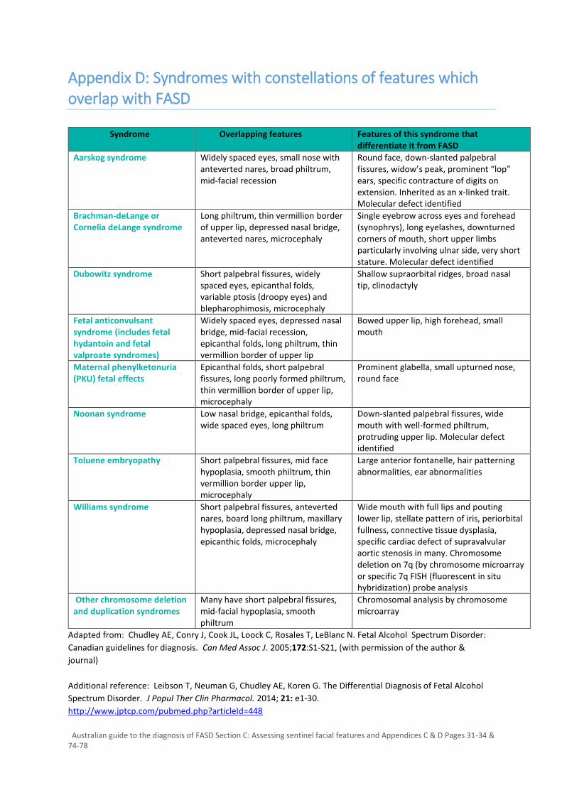
Appendix D: Syndromes with constellations of features which overlap with FASD
Syndrome Overlapping features Features of this syndrome that differentiate it from FASD
Aarskog syndrome Widely spaced eyes, small nose with anteverted nares, broad philtrum, mid-facial recession
Round face, down-slanted palpebral fissures, widow’s peak, prominent “lop” ears, specific contracture of digits on extension. Inherited as an x-linked trait. Molecular defect identified
Brachman-deLange or Cornelia deLange syndrome
Long philtrum, thin vermillion border of upper lip, depressed nasal bridge, anteverted nares, microcephaly
Single eyebrow across eyes and forehead (synophrys), long eyelashes, downturned corners of mouth, short upper limbs particularly involving ulnar side, very short stature. Molecular defect identified
Dubowitz syndrome Short palpebral fissures, widely spaced eyes, epicanthal folds, variable ptosis (droopy eyes) and blepharophimosis, microcephaly
Shallow supraorbital ridges, broad nasal tip, clinodactyly
Fetal anticonvulsant syndrome (includes fetal hydantoin and fetal valproate syndromes)
Widely spaced eyes, depressed nasal bridge, mid-facial recession, epicanthal folds, long philtrum, thin vermillion border of upper lip
Bowed upper lip, high forehead, small mouth
Maternal phenylketonuria (PKU) fetal effects
Epicanthal folds, short palpebral fissures, long poorly formed philtrum, thin vermillion border of upper lip, microcephaly
Prominent glabella, small upturned nose, round face
Noonan syndrome Low nasal bridge, epicanthal folds, wide spaced eyes, long philtrum
Down-slanted palpebral fissures, wide mouth with well-formed philtrum, protruding upper lip. Molecular defect identified
Toluene embryopathy Short palpebral fissures, mid face hypoplasia, smooth philtrum, thin vermillion border upper lip, microcephaly
Large anterior fontanelle, hair patterning abnormalities, ear abnormalities
Williams syndrome Short palpebral fissures, anteverted nares, board long philtrum, maxillary hypoplasia, depressed nasal bridge, epicanthic folds, microcephaly
Wide mouth with full lips and pouting lower lip, stellate pattern of iris, periorbital fullness, connective tissue dysplasia, specific cardiac defect of supravalvular aortic stenosis in many. Chromosome deletion on 7q (by chromosome microarray or specific 7q FISH (fluorescent in situ hybridization) probe analysis
Other chromosome deletion and duplication syndromes
Many have short palpebral fissures, mid-facial hypoplasia, smooth philtrum
Chromosomal analysis by chromosome microarray
Adapted from: Chudley AE, Conry J, Cook JL, Loock C, Rosales T, LeBlanc N. Fetal Alcohol Spectrum Disorder:
Canadian guidelines for diagnosis. Can Med Assoc J. 2005;172:S1-S21, (with permission of the author &
journal)
Additional reference: Leibson T, Neuman G, Chudley AE, Koren G. The Differential Diagnosis of Fetal Alcohol
Spectrum Disorder. J Popul Ther Clin Pharmacol. 2014; 21: e1-30.
http://www.jptcp.com/pubmed.php?articleId=448