Secondary Aneurysmal Bone Cyst in a Craniofacial Fibrous ... · 86 INTRODUCTION Aneurysmal bone...
6
86 INTRODUCTION Aneurysmal bone cyst (ABC), a term now regarded as a misnomer, is a non-neoplastic bony lesion that consists of cys- tic cavernous cavities without lining endothelium filled with blood and that occurs commonly in teenagers or young adults. ABCs occur mostly in long bones and the spinal column, and craniofacial involvement is a very rare occurrence. ABCs are either primary or secondary; secondary ABCs develop in pre- existing bony pathologies primarily including giant cell tu- mor, osteoblastoma, angioma and chondroblastoma and less commonly including fibrous dysplasia (FD), ossifying fibro- ma and osteosarcoma. FD is also a rare pathology that represents 2.5% of all bone tumors and 7% of benign bone tumors [1] and occurs mostly in the craniofacial skeleton of adolescent and young adult pa- Secondary Aneurysmal Bone Cyst in a Craniofacial Fibrous Dysplasia: Case Report Hyun-Seok Lee 1 , Young-Cho Koh 1 , Hong Gee Roh 2 , Hyung Kyu Park 3 , Soo Yeon Kim 1 * Departments of 1 Neurosurgery, 2 Radiology, 3 Pathology, Konkuk University Medical Center, Seoul, Korea Received August 1, 2018 Revised September 22, 2018 Accepted September 28, 2018 Correspondence Young-Cho Koh Department of Neurosurgery, Konkuk University Medical Center, 120-1 Neungdong-ro, Gwangjin-gu, Seoul 05030, Korea Tel: +82-2-2030-7357 Fax: +82-2-2030-7359 E-mail: [email protected] *Current address: Department of Neurosurgery, Seoul National University Bundang Hospital, Seongnam, Korea Aneurysmal bone cyst (ABC) is a rare non-neoplastic bone lesion that involves mostly the long bones and vertebrae and may occur very rarely in the craniofacial bones. ABCs may occur as secondary bony pathologies in association with various benign and malignant bone tumors and with fibrous dys- plasia (FD). FD is a common non-neoplastic bony pathology mostly affecting craniofacial bones. Secondary ABC occurring in craniofacial FD is extremely rare, with only approximately 20 cases re- ported in the literature to date. Here, we report on a case of secondary ABC in a 25-year-old woman who has had a craniofacial deformity for over 10 years and who presented to us with a rapidly grow- ing painful pulsatile mass in the right frontal region that began over 2 months prior to admission. On thorough examination of computed tomography and magnetic resonance imaging brain scans taken at two-month interval, an aggressive, rapidly enlarging ABC, arising from the right frontal FD, was di- agnosed. The patient underwent preoperative embolization followed by gross total resection of the ABC and cranioplasty. The 6-month follow up showed no recurrence of the ABC, nor was any progres- sion of the FD noticed. Key Words Aneurysmal bone cysts; Fibrous dysplasia of bone; Frontal bone; Craniotomy. tients. FDs are benign non-neoplastic lesions commonly pre- senting with craniofacial disfigurement or compressive cranial neuropathies such as vision loss. A few cases have been docu- mented with oculomotor palsy due to involvements of the neu- ral foramina at the base of the skull. Most FDs are self-limiting, with stabilization aſter puberty. Reports of secondary ABC occurring in craniofacial FDs are extremely rare in the literature, accounting for less than 20 cases. Patients usually present with a rapidly enlarging, soft, painful mass in the preexisting FDs, for which treatment is a curative resection of the ABC with a conservative resection of the FD or a conservative treatment that includes percutane- ous sclerosing therapy. The case review presented here was conducted with the approval of the Institutional Review Board (KUH10700028). CASE REPORT A 25-year-old female, with no significant medical or familial history, presented with rapidly growing right forehead mass beginning 2 months prior to admission. She had been followed for a diffuse forehead bulging since early in her second decade CASE REPORT Brain Tumor Res Treat 2018;6(2):86-91 / pISSN 2288-2405 / eISSN 2288-2413 https://doi.org/10.14791/btrt.2018.6.e15 is is an Open Access article distributed under the terms of the Creative Commons Attribution Non-Commercial License (https://creativecommons.org/licenses/by-nc/4.0) which permits unrestricted non-commercial use, distribution, and reproduction in any medium, provided the original work is properly cited. Copyright © 2018 e Korean Brain Tumor Society, e Korean Society for Neuro- Oncology, and e Korean Society for Pediatric Neuro-Oncology
Transcript of Secondary Aneurysmal Bone Cyst in a Craniofacial Fibrous ... · 86 INTRODUCTION Aneurysmal bone...

86
INTRODUCTION
Aneurysmal bone cyst (ABC), a term now regarded as a misnomer, is a non-neoplastic bony lesion that consists of cys-tic cavernous cavities without lining endothelium filled with blood and that occurs commonly in teenagers or young adults. ABCs occur mostly in long bones and the spinal column, and craniofacial involvement is a very rare occurrence. ABCs are either primary or secondary; secondary ABCs develop in pre-existing bony pathologies primarily including giant cell tu-mor, osteoblastoma, angioma and chondroblastoma and less commonly including fibrous dysplasia (FD), ossifying fibro-ma and osteosarcoma.
FD is also a rare pathology that represents 2.5% of all bone tumors and 7% of benign bone tumors [1] and occurs mostly in the craniofacial skeleton of adolescent and young adult pa-
Secondary Aneurysmal Bone Cyst in a Craniofacial Fibrous Dysplasia: Case ReportHyun-Seok Lee1, Young-Cho Koh1, Hong Gee Roh2, Hyung Kyu Park3, Soo Yeon Kim1*
Departments of 1Neurosurgery, 2Radiology, 3Pathology, Konkuk University Medical Center, Seoul, Korea
Received August 1, 2018Revised September 22, 2018Accepted September 28, 2018
CorrespondenceYoung-Cho KohDepartment of Neurosurgery, Konkuk University Medical Center, 120-1 Neungdong-ro, Gwangjin-gu, Seoul 05030, KoreaTel: +82-2-2030-7357Fax: +82-2-2030-7359E-mail: [email protected]
*Current address: Department of Neurosurgery, Seoul National University Bundang Hospital, Seongnam, Korea
Aneurysmal bone cyst (ABC) is a rare non-neoplastic bone lesion that involves mostly the long bones and vertebrae and may occur very rarely in the craniofacial bones. ABCs may occur as secondary bony pathologies in association with various benign and malignant bone tumors and with fibrous dys-plasia (FD). FD is a common non-neoplastic bony pathology mostly affecting craniofacial bones. Secondary ABC occurring in craniofacial FD is extremely rare, with only approximately 20 cases re-ported in the literature to date. Here, we report on a case of secondary ABC in a 25-year-old woman who has had a craniofacial deformity for over 10 years and who presented to us with a rapidly grow-ing painful pulsatile mass in the right frontal region that began over 2 months prior to admission. On thorough examination of computed tomography and magnetic resonance imaging brain scans taken at two-month interval, an aggressive, rapidly enlarging ABC, arising from the right frontal FD, was di-agnosed. The patient underwent preoperative embolization followed by gross total resection of the ABC and cranioplasty. The 6-month follow up showed no recurrence of the ABC, nor was any progres-sion of the FD noticed.
Key Words Aneurysmal bone cysts; Fibrous dysplasia of bone; Frontal bone; Craniotomy.
tients. FDs are benign non-neoplastic lesions commonly pre-senting with craniofacial disfigurement or compressive cranial neuropathies such as vision loss. A few cases have been docu-mented with oculomotor palsy due to involvements of the neu-ral foramina at the base of the skull. Most FDs are self-limiting, with stabilization after puberty.
Reports of secondary ABC occurring in craniofacial FDs are extremely rare in the literature, accounting for less than 20 cases. Patients usually present with a rapidly enlarging, soft, painful mass in the preexisting FDs, for which treatment is a curative resection of the ABC with a conservative resection of the FD or a conservative treatment that includes percutane-ous sclerosing therapy. The case review presented here was conducted with the approval of the Institutional Review Board (KUH10700028).
CASE REPORT
A 25-year-old female, with no significant medical or familial history, presented with rapidly growing right forehead mass beginning 2 months prior to admission. She had been followed for a diffuse forehead bulging since early in her second decade
CASE REPORT Brain Tumor Res Treat 2018;6(2):86-91 / pISSN 2288-2405 / eISSN 2288-2413https://doi.org/10.14791/btrt.2018.6.e15
This is an Open Access article distributed under the terms of the Creative Commons Attribution Non-Commercial License (https://creativecommons.org/licenses/by-nc/4.0) which permits unrestricted non-commercial use, distribution, and reproduction in any medium, provided the original work is properly cited.Copyright © 2018 The Korean Brain Tumor Society, The Korean Society for Neuro-Oncology, and The Korean Society for Pediatric Neuro-Oncology
HS Lee et al.
87
of life at a neurosurgical center in Kazakhstan. She underwent a magnetic resonance imaging (MRI) scan to assess the soft bulging in her right frontal region 2 months prior to admis-sion. The mass grew rapidly and she began to feel pain when the mass reached the size of a tennis ball. The patient was trans-
ferred to our hospital for definite diagnosis and treatment. The physical examination was normal except for a bulging, soft mass measuring 5.5 cm on the right forehead. She was free of neurological symptoms, and routine laboratory examinations were within normal limits. A computed tomography (CT) scan
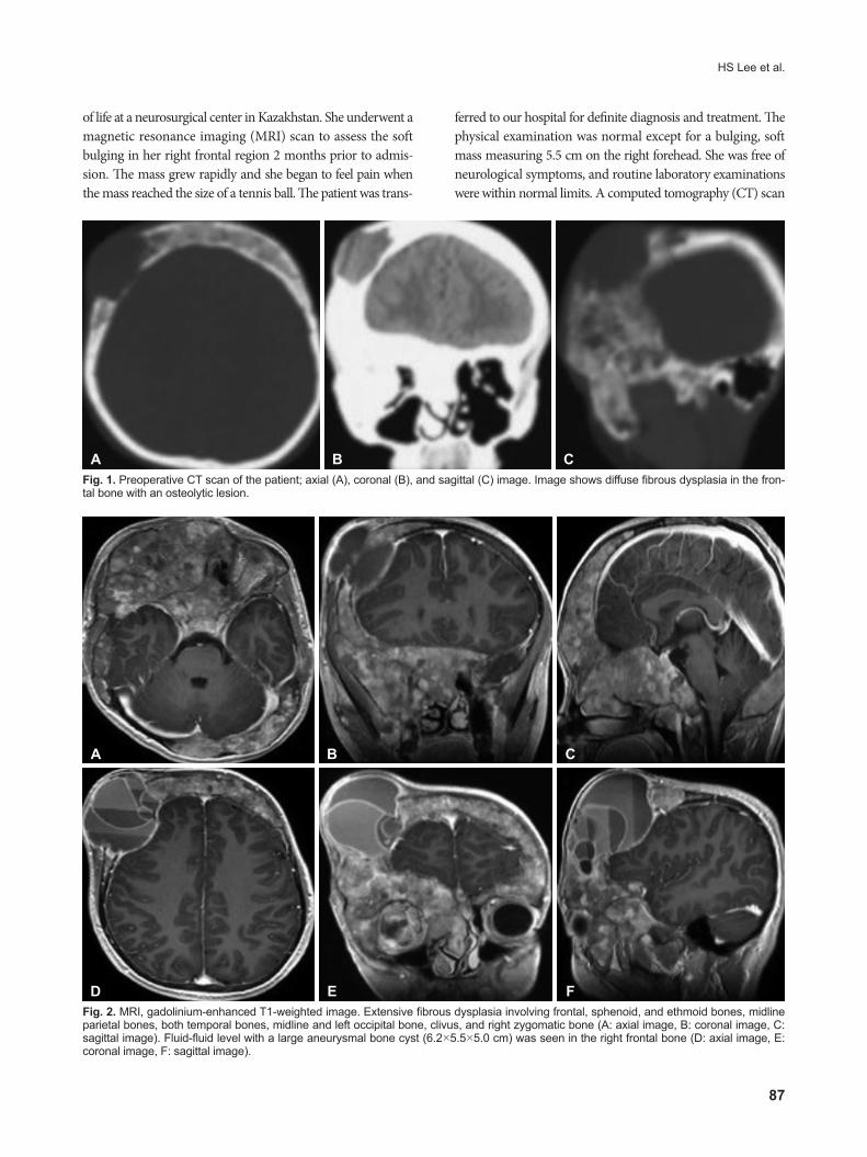
Fig. 2. MRI, gadolinium-enhanced T1-weighted image. Extensive fibrous dysplasia involving frontal, sphenoid, and ethmoid bones, midline parietal bones, both temporal bones, midline and left occipital bone, clivus, and right zygomatic bone (A: axial image, B: coronal image, C: sagittal image). Fluid-fluid level with a large aneurysmal bone cyst (6.2×5.5×5.0 cm) was seen in the right frontal bone (D: axial image, E: coronal image, F: sagittal image).
Fig. 1. Preoperative CT scan of the patient; axial (A), coronal (B), and sagittal (C) image. Image shows diffuse fibrous dysplasia in the fron-tal bone with an osteolytic lesion.
A B C
A
D
B
E
C
F

88 Brain Tumor Res Treat 2018;6(2):86-91
Secondary Aneurysmal Bone Cyst
There was no evidence of malignancy in the surgical speci-men. The FD was partially removed around the ABC, and af-ter surgery (Fig. 5), the patient’s cranio-facial deformity was dramatically improved (Fig. 6). No recurrence of the ABC nor progression of the FD was found at 6 months postoperatively.
DISCUSSION
FD, Langhans cell histiocytosis (Histiocytosis X), and ABC are the three most common juvenile craniofacial bony lesions. Among these, FD is the most common proliferating craniofa-cial bony lesion. In FD, abnormal growth of fibroblasts replac-es medullary bone with fibrocellular tissue [2,3]. The biologic behavior of FD is known to be self-limiting, with no progression after puberty [4-6]. According to reported cases, in FD patient, presented with painful swelling over a short period of time, which may clinically represent the very unusual sarcomatous transformation of FD [7].
ABC is neither an aneurysm nor a true cyst but is made of channels containing flowing blood. The pathogenetic mecha-nism of ABC remains unclear. Other report suggested that it may arise from abnormal hemodynamic conditions causing an increase in venous pressure, which then leads to hemorrhage
revealed characteristic findings of FD with a heterogeneous ground-glass appearance of both the frontal, sphenoid, and ethmoid bones, as well as a discrete cystic mass in the right frontal bone (Fig. 1). MRI also showed findings of FD in the bilateral craniofacial bone and indicated multi-septated fluid-fluid levels suggestive of ABC (Fig. 2). We performed a preoper-ative ophthalmological evaluation, in which visual acuity was 1.0 in both eyes, and there were no visual field, fundus, or eye movement abnormalities. For evaluation of the vascularity of the cystic mass and embolization of feeder arteries prior to surgery, a trans-femoral cerebral angiography was done. The angiography demonstrated moderate vascularity along the rim of the cystic mass and embolization via the right middle meningeal artery and right superficial temporal artery was performed. After embolization, the vascularity of the mass was markedly reduced (Fig. 3).
The patient underwent right frontal craniectomy with gross total resection of the brownish soft-tissue mass with multiple septated, brownish fluids and sediment-filled, cystic cavities followed by simple cranioplasty. The outer table thinned out as fine as paper, and the inner table was nearly absent. The histopathological findings of the skull lesion indicated FD with degenerative changes, forming typical ABC-like areas (Fig. 4).
Fig. 3. Trans-femoral cerebral angiography image of the patient. The pre-embolization image showed prominent vascular stain in the right frontal area, at the margin of the aneurysmal bone cyst (A: anterior-posterior view, B: right lateral view). An endovascular embolization was performed in a branch of the right middle meningeal artery and the right superficial temporal artery. After embolization, vascular staining was markedly decreased, but residual staining was seen in the posterior and inferior margins (C: anterior-posterior view, D: right lateral view).
A B
C D

HS Lee et al.
89
[8]. ABC may occur as either a primary (70%) or a secondary (30%) bone cyst [9]. Diseases known for predisposing the pa-tient to lesions of ABC include osteoclastoma, osteosarcoma, osteoblastoma, and hemangioma [10]. ABC arising from FD
is extremely rare, especially in the craniofacial bone. This lesion was first reported by Branch et al. [10] in 1986, and fewer than twenty cases have been reported since then [3,9-21]. We sum-marized the case reports of ABC with FD in the craniofacial
Fig. 4. Histological examination of the resected lesion. A: The aneurysmal bone cyst wall (hematoxylin and eosin staining, original magnifi-cation ×100). B: A multinucleated osteoclastic giant cell (hematoxylin and eosin staining, original magnification ×200). C: Woven bone with proliferating fibrous tissue (hematoxylin and eosin staining, original magnification ×200).
Fig. 5. Postoperative axial CT scan of the setting bone shows the partial removal of the fibrous dysplasia around the aneurysmal bone cysts (A and B).
Fig. 6. Cosmetic improvement of the patient’s forehead (A: preoperative image, B: postoperative image).
A B C
A B
A B

90 Brain Tumor Res Treat 2018;6(2):86-91
Secondary Aneurysmal Bone Cyst
bone in a literature search from PubMed in Table 1. In our pa-tient and in seven of the reported cases, a growing craniofacial mass was a presenting symptom. The rapidly distended con-tour of a secondary ABC may create suspicion of a malignant change in the pre-existing, benign bone tumor. The incidence of malignant transformation in FD has been reported to be between 0.5% and 4% [7]. When a patient presents with a rap-id growth of the skull, the possibility of malignant change of the benign bone pathology and the development of secondary ABC should both be considered, with the latter given enhanced consideration in younger patients. Screening through imaging may provide useful clues as to the correct preoperative diagno-sis. The presence of fluid-fluid levels on a CT scan or MRI is a typical feature of ABCs, although it has also been reported in many other osseous lesions containing hemorrhage [22]. When fluid-fluid levels are demonstrated in FD, this outcome may be associated with simple cystic degeneration or sarcomatous transformation of FD [23].
In a literature review, the treatment of choice for an ABC with FD is total resection of the ABC and cranioplasty. Close follow up is mandatory due to the likelihood of recurrence of the lesion. Where possible, embolization of the feeding artery has been reported to reduce bleeding during surgery and en-able maximal resection, as seen in our case [15].
We are reporting on a rapidly developing ABC in a pre-exist-ing FD that presented with painful swelling and disfigurement in the right frontal region. Because subtotal resection is asso-ciated with a 50% recurrence rate, the patient underwent ag-
gressive total resection of the ABC with partial removal of the FD followed by cranioplasty [24-27]. At the six-month post-operative follow up with CT and MRI images, there was no re-currence.
In conclusion, if a patient complains of a rapidly growing, soft mass with pain and pre-existing FD, secondary ABC de-velopment should be considered. In our case, preoperative angiography with embolization of the feeders was helpful in reducing intra-operative bleeding during an aggressive total resection.
Conflicts of InterestThe authors have no financial conflicts of interest.
REFERENCES
1. Edgerton MT, Persing JA, Jane JA. The surgical treatment of fibrous dysplasia. With emphasis on recent contributions from cranio-maxil-lo-facial surgery. Ann Surg 1985;202:459-79.
2. Casselman JW, De Jonge I, Neyt L, De Clercq C, D’Hont G. MRI in craniofacial fibrous dysplasia. Neuroradiology 1993;35:234-7.
3. Mattei TA, Mattei JA, Ramina R, Aguiar PH. Fibrous dysplasia in com-bination with aneurysmal bone cyst presenting as a subarachnoid haemorrhage. Neurol Sci 2005;26:178-81.
4. Dumont AS, Boulos PT, Jane JA Jr, Ellegala DB, Newman SA, Jane JA Sr. Cranioorbital fibrous dysplasia: with emphasis on visual impair-ment and current surgical management. Neurosurg Focus 2001;10:E6.
5. Maher CO, Friedman JA, Meyer FB, Lynch JJ, Unni K, Raffel C. Surgical treatment of fibrous dysplasia of the skull in children. Pediatr Neurosurg 2002;37:87-92.
6. Chong VF, Khoo JB, Fan YF. Fibrous dysplasia involving the base of the skull. AJR Am J Roentgenol 2002;178:717-20.
7. Ruggieri P, Sim FH, Bond JR, Unni KK. Malignancies in fibrous dys-
Table 1. Published cases of secondary aneurysmal bone cyst with craniofacial fibrous dysplasia
Authors Years Sex Age Site Presenting symptom TreatmentBranch et al. [10] 1986 M 19 Parietal Growing mass Surgical resection
F 9 Parietal, frontotemporal Painful mass Surgical resectionRappaport [11] 1989 M 25 Occipital Painful mass Surgical resectionWojno and McCathy [12] 1994 F 14 Temporal Painless mass Surgical resection
M 40 Frontal Exophthalmos and diplopia Surgical resectionHaddad et al. [13] 1998 M 6 Temporal Growing mass Surgical resectionSaito et al. [14] 1998 M 11 Nasal cavity and skull base Nasal obstruction and headache Surgical resectionItshayek et al. [15] 2002 M 19 Occipital and clivus Painless mass Endovascular embolization and
surgical resectionPasquini et al. [16] 2002 M 5 Skull base Sinusitis Endoscopic trans-nasal resectionLin et al. [9] 2004 M 18 Frontal Severe headache Surgical resectionIseri et al. [17] 2005 F 35 Occipital and clivus Severe headache Medical treatment*Mattei et al. [3] 2005 M 19 Occipital Severe headache and nuchal rigidity Surgical resectionLee et al. [18] 2010 F 18 Fronto-parietal Growing mass Surgical resectionTerkawi et al. [19] 2011 F 7 Nasal cavity and skull base Vision loss and nasal obstruction Surgical resectionManjila et al. [20] 2013 M 10 Nasal cavity and skull base Cognitive impairment Surgical resection and
endoscopic resectionHnenny et al. [21] 2015 F 28 Nasal cavity and skull base Vision worsening Surgical resection*Patient refused surgery, medical treatment was done with bisphosphonate

HS Lee et al.
91
plasia. Cancer 1994;73:1411-24.8. Tuna H, Karatas A, Yilmaz ER, Yagmurlu B, Erekul S. Aneurysmal bone
cyst of the temporal bone: case report. Surg Neurol 2003;60:571-4.9. Lin WC, Wu HT, Wei CJ, Chang CY. Aneurysmal bone cyst arising
from fibrous dysplasia of the frontal bone (2004:2b). Eur Radiol 2004; 14:930-2.
10. Branch CL Jr, Challa VR, Kelly DL Jr. Aneurysmal bone cyst with fi-brous dysplasia of the parietal bone. Report of two cases. J Neurosurg 1986;64:331-5.
11. Rappaport ZH. Aneurysmal bone cyst associated with fibrous dyspla-sia of the skull. Neurochirurgia (Stuttg) 1989;32:192-4.
12. Wojno KJ, McCarthy EF. Fibro-osseous lesions of the face and skull with aneurysmal bone cyst formation. Skeletal Radiol 1994;23:15-8.
13. Haddad GF, Hambali F, Mufarrij A, Nassar A, Haddad FS. Concomi-tant fibrous dysplasia and aneurysmal bone cyst of the skull base. Case report and review of the literature. Pediatr Neurosurg 1998;28:147-53.
14. Saito I, Asano T, Sano K, et al. Neuroprotective effect of an antioxidant, ebselen, in patients with delayed neurological deficits after aneurysmal subarachnoid hemorrhage. Neurosurgery 1998;42:269-77; discussion 277-8.
15. Itshayek E, Spector S, Gomori M, Segal R. Fibrous dysplasia in combi-nation with aneurysmal bone cyst of the occipital bone and the clivus: case report and review of the literature. Neurosurgery 2002;51:815-7; discussion 817-8.
16. Pasquini E, Ceroni Compadretti G, Sciarretta V, Ippolito A. Transnasal endoscopic surgery for the treatment of fibrous dysplasia of maxillary sinus associated to aneurysmal bone cyst in a 5-year-old child. Int J Pediatr Otorhinolaryngol 2002;62:59-62.
17. Iseri PK, Efendi H, Demirci A, Komsuoglu S. Fibrous dysplasia of the cranial bones: a case report and review of the literature. Yale J Biol Med 2005;78:141-5.
18. Lee JW, Kim JH, Han SH, Kang HI. Fibrous dysplasia with aneurysmal
bone cyst presenting as painful solitary skull lesion. J Korean Neuro-surg Soc 2010;48:551-4.
19. Terkawi AS, Al-Qahtani KH, Baksh E, Soualmi L, Mohamed Ael-B, Sabbagh AJ. Fibrous dysplasia and aneurysmal bone cyst of the skull base presenting with blindness: a report of a rare locally aggressive ex-ample. Head Neck Oncol 2011;3:15.
20. Manjila S, Zender CA, Weaver J, Rodgers M, Cohen AR. Aneurysmal bone cyst within fibrous dysplasia of the anterior skull base: continued intracranial extension after endoscopic resections requiring craniofa-cial approach with free tissue transfer reconstruction. Childs Nerv Syst 2013;29:1183-92.
21. Hnenny L, Roundy N, Zherebitskiy V, Grafe M, Mansoor A, Dogan A. Giant aneurysmal bone cyst of the anterior cranial fossa and paranasal sinuses presenting in pregnancy: case report and literature review. J Neurol Surg Rep 2015;76:e216-21.
22. Davies AM, Evans N, Mangham DC, Grimer RJ. MR imaging of brown tumour with fluid-fluid levels: a report of three cases. Eur Radi-ol 2001;11:1445-9.
23. Nguyen BD, Lugo-Olivieri CH, McCarthy EF, Frassica FJ, Ma LD, Zer-houni EA. Fibrous dysplasia with secondary aneurysmal bone cyst. Skeletal Radiol 1996;25:88-91.
24. Cakirer S, Basak M, Celebi I, Kabukcuoglu F, Erdem Y. Aneurysmal bone cyst of the temporal bone. Curr Probl Diagn Radiol 2003;32:169-75.
25. Chidambaram B, Santosh V, Balasubramaniam V. Aneurysmal bone cyst of the temporal bone. Childs Nerv Syst 2001;17:411-4.
26. Lippman CR, Jallo GI, Feghali JG, Jimenez E, Epstein F. Aneurysmal bone cyst of the temporal bone. Pediatr Neurosurg 1999;31:219-23.
27. O’Brien DP, Rashad EM, Toland JA, Farrell MA, Phillips J. Aneurys-mal cyst of the frontal bone: case report and review of the literature.Br J Neurosurg 1994;8:105-8.