Second messenger system - Patna University
30
1 E-content M.Sc. Zoology (Semester IV) Elective Paper: Cell and Molecular biology Unit: 3.7 Second messenger system Dr Gajendra Kumar Azad Assistant Professor Post Graduate Department of Zoology Patna University, Patna Email: [email protected]
Transcript of Second messenger system - Patna University
1
E-content
M.Sc. Zoology (Semester IV) Elective Paper: Cell and Molecular biology
Unit: 3.7
Second messenger system
Dr Gajendra Kumar Azad Assistant Professor
Post Graduate Department of Zoology Patna University, Patna
Email: [email protected]

Second Messenger System Signals received by receptors at the cell surface or, in some cases, within the cell are often relayed throughout the cell via generation of small, rapidly diffusing molecules referred to as second messengers. These second messengers broadcast the initial signal (the “first message”) that occurs when a ligand binds to a specific cellular receptor. Ligand binding alters the protein conformation of the receptor such that it stimulates nearby effector proteins that catalyze the production or, in the case of ions, release or influx of the second messenger. The second messenger then diffuses rapidly to protein targets elsewhere within the cell, altering the activities as a response to the new information received by the receptor.
2

There are three classic second messenger pathways as shown in figure 1: 1. Activation of adenylyl cyclase by G-protein-coupled receptors (GPCRs) to
generate the cyclic nucleotide second messenger 3′-5′-cyclic adenosine monophosphate (cAMP)
2. Stimulation of phosphoinositide 3-kinase (PI3K) by growth factor receptors to generate the lipid second messenger phosphatidylinositol 3,4,5-trisphosphate (PIP3)
3. activation of phospholipase C (PLC) by GPCRs to generate the two second messengers, membrane-bound messenger diacylglycerol (DAG) and soluble messenger inositol 1,4,5-trisphosphate (IP3), which binds to receptors on subcellular organelles to release calcium into the cytosol.
3

Second messengers disseminate information received from cell-surface receptors.
Figure 1: Second messengers: On the right, binding of agonists to a GPCR (the receptor) can activate adenylyl cyclase (the effector) to produce cAMP (the second messenger) to activate protein kinase A (PKA; the target). On the left, binding of growth factors to a receptor tyrosine kinase (RTK; the receptor) can activate PI3K (the effector) to generate PIP3 (the second messenger), which activates Akt (the target). In the center, binding of ligands to a GPCR (receptor) activates phospholipase C (PLC; the effector), to generate two second messengers, DAG and IP3, which activate protein kinase C (PKC; the target) and release calcium from intracellular stores, respectively. Cold Spring Harb Perspect Biol 2016;8:a005926
4

Second messengers fall into four major classes: 1. Cyclic nucleotides, such as cAMP and other soluble molecules that signal
within the cytosol
2. Lipid messengers that signal within cell membranes
3. Ions that signal within and between cellular compartments
4. Gases and free radicals that can signal throughout the cell and even to neighboring cells.
Second messengers from each of these classes bind to specific protein targets, altering their activity to relay downstream signals. In many cases, these targets are enzymes whose catalytic activity is modified by direct binding of the second messengers. The activation of multiple target enzymes by a single second messenger molecule further amplifies the signal. Second messengers are not only produced in response to extracellular stimuli, but also in response to stimuli from within the cell 5

1. Cyclic Nucleotides
a) cAMP and a Major Target, PKA
Earl Sutherland first showed that epinephrine bound to cell-surface receptors stimulates membrane-associated adenylyl cyclases to synthesize the chemical messenger cAMP. G-protein subunits are the intermediates that shuttle between receptors and a family of eight adenylyl cyclase isoforms in the plasma membrane. Each G protein is a trimer consisting of Gα, Gβ, and Gγ subunits. The Gα subunits in the G proteins Gs and Gi are distinct and provide the specificity for activation and inhibition of adenylyl cyclase, respectively. Gs and Gi can thus couple binding of ligands to GPCRs with either activation or inhibition of adenylyl cyclase, depending on the receptor type. This increases or reduces production of cAMP, which diffuses from the membrane into the cell.
6
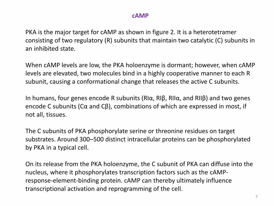
cAMP PKA is the major target for cAMP as shown in figure 2. It is a heterotetramer consisting of two regulatory (R) subunits that maintain two catalytic (C) subunits in an inhibited state. When cAMP levels are low, the PKA holoenzyme is dormant; however, when cAMP levels are elevated, two molecules bind in a highly cooperative manner to each R subunit, causing a conformational change that releases the active C subunits. In humans, four genes encode R subunits (RIα, RIβ, RIIα, and RIIβ) and two genes encode C subunits (Cα and Cβ), combinations of which are expressed in most, if not all, tissues. The C subunits of PKA phosphorylate serine or threonine residues on target substrates. Around 300–500 distinct intracellular proteins can be phosphorylated by PKA in a typical cell. On its release from the PKA holoenzyme, the C subunit of PKA can diffuse into the nucleus, where it phosphorylates transcription factors such as the cAMP-response-element-binding protein. cAMP can thereby ultimately influence transcriptional activation and reprogramming of the cell.
7

cAMP as a second messenger.
Cold Spring Harb Perspect Biol 2016;8:a005926
Figure 2: (A) cAMP levels increase rapidly following receptor-mediated activation of adenylyl cyclase (AC), which catalyzes the conversion of adenosine monophosphate (AMP) to cAMP. This small second messenger activates PKA at specific cellular locations as a result of anchoring of PKA to A-kinase-anchoring proteins (AKAPs). In addition, cAMP activates EPACs. (B) AC activity is controlled by the opposing actions of the Gs and Gi proteins. The cAMP produced by AC activates PKA by binding to its R subunit. This releases the C subunit. The signal can be terminated by the action of phosphodiesterase (PDE) enzymes.
8

b) cGMP 3′-5′-cyclic guanosine monophosphate (cGMP) is another cyclic nucleotide that serves as a second messenger. cGMP is often synthesized by receptor guanylyl cyclases, which have their catalytic domains close to the inner face of the plasma membrane and are stimulated by small peptide factors that bind to their extracellular domains. It can also be produced by soluble cytoplasmic guanylyl cyclases, which are stimulated by nitric oxide. cGMP targets cGMP-dependent protein kinase (PKG), a dimeric enzyme that has an R and C domain within the same polypeptide chain. When cGMP levels are low, PKG is dormant; however, when cGMP levels are elevated, two molecules bind to each R domain in the dimer, exposing the active catalytic domains. PKA and PKG have overlapping substrate specificities. However, a major difference is that PKG expression is restricted to the brain, lungs, and vascular tissue, where this enzyme controls important physiological processes.
9

Regulation of cAMP and cGMP signal Termination of cAMP and cGMP signals is mediated by phosphodiesterases (PDEs). These enzymes represent a vast gene family of 11 distinct subtypes and more than 100 isoforms that can break the phosphoester bond of either cyclic nucleotide to liberate AMP or GMP.
10
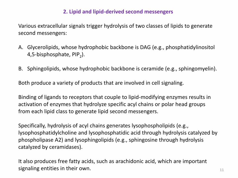
2. Lipid and lipid-derived second messengers Various extracellular signals trigger hydrolysis of two classes of lipids to generate second messengers: A. Glycerolipids, whose hydrophobic backbone is DAG (e.g., phosphatidylinositol
4,5-bisphosphate, PIP2).
B. Sphingolipids, whose hydrophobic backbone is ceramide (e.g., sphingomyelin).
Both produce a variety of products that are involved in cell signaling. Binding of ligands to receptors that couple to lipid-modifying enzymes results in activation of enzymes that hydrolyze specific acyl chains or polar head groups from each lipid class to generate lipid second messengers. Specifically, hydrolysis of acyl chains generates lysophospholipids (e.g., lysophosphatidylcholine and lysophosphatidic acid through hydrolysis catalyzed by phospholipase A2) and lysophingolipids (e.g., sphingosine through hydrolysis catalyzed by ceramidases). It also produces free fatty acids, such as arachidonic acid, which are important signaling entities in their own. 11
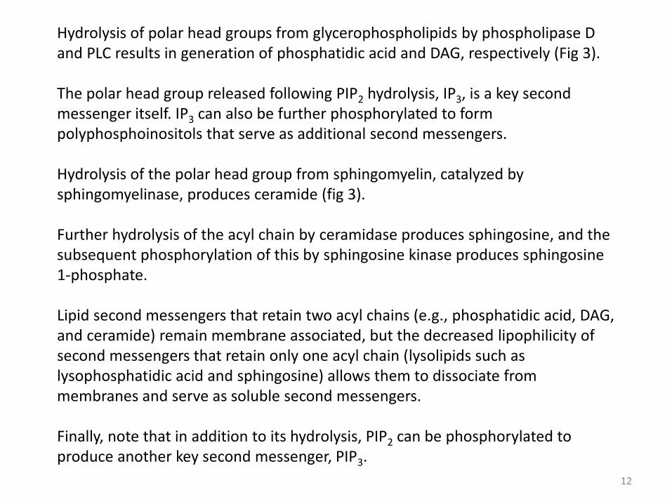
Hydrolysis of polar head groups from glycerophospholipids by phospholipase D and PLC results in generation of phosphatidic acid and DAG, respectively (Fig 3). The polar head group released following PIP2 hydrolysis, IP3, is a key second messenger itself. IP3 can also be further phosphorylated to form polyphosphoinositols that serve as additional second messengers. Hydrolysis of the polar head group from sphingomyelin, catalyzed by sphingomyelinase, produces ceramide (fig 3). Further hydrolysis of the acyl chain by ceramidase produces sphingosine, and the subsequent phosphorylation of this by sphingosine kinase produces sphingosine 1-phosphate. Lipid second messengers that retain two acyl chains (e.g., phosphatidic acid, DAG, and ceramide) remain membrane associated, but the decreased lipophilicity of second messengers that retain only one acyl chain (lysolipids such as lysophosphatidic acid and sphingosine) allows them to dissociate from membranes and serve as soluble second messengers. Finally, note that in addition to its hydrolysis, PIP2 can be phosphorylated to produce another key second messenger, PIP3. 12
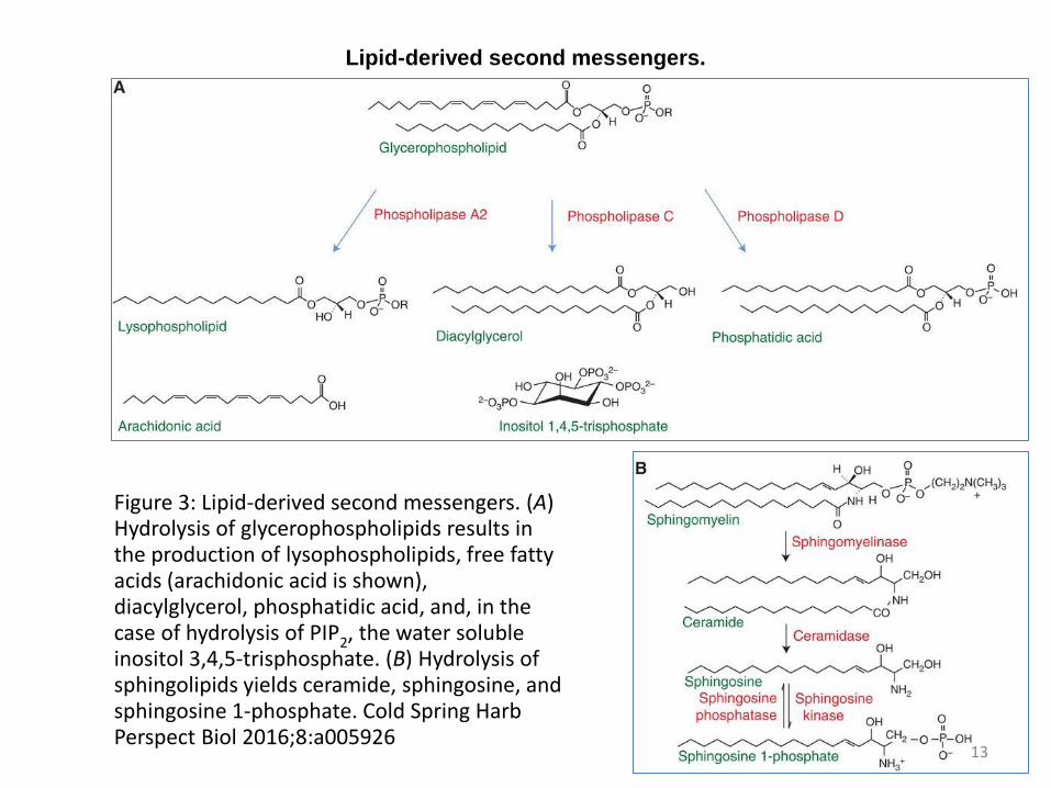
Lipid-derived second messengers.
Figure 3: Lipid-derived second messengers. (A) Hydrolysis of glycerophospholipids results in the production of lysophospholipids, free fatty acids (arachidonic acid is shown), diacylglycerol, phosphatidic acid, and, in the case of hydrolysis of PIP2, the water soluble inositol 3,4,5-trisphosphate. (B) Hydrolysis of sphingolipids yields ceramide, sphingosine, and sphingosine 1-phosphate. Cold Spring Harb Perspect Biol 2016;8:a005926
13

Lipid and lipid-derived second messengers
A-i) IP3 and DAG
Signals from both GPCRs and RTKs can result in activation of PLC, which cleaves phospholipids to generate DAG and IP3. If the lipid cleaved is PIP2 (rather than phosphatidylcholine [PC]), then water-soluble IP3 is also produced. IP3 binds to IP3 receptors on the endoplasmic reticulum, and other organelles, causing release of calcium into the cytosol. It can be further modified to yield additionally phosphorylated phosphoinositols, including diphosphoryl inositol phosphates. Inositol 1,3,4,5-tetrakisphosphate (IP4) activates chloride channels. The DAG sensor is a small globular domain, called the C1 domain, originally identified in PKC. Approximately 30 other proteins contain a C1 domain, including protein kinase D, Ras, gastrin-releasing polypeptides, DAG kinase, and n-chimaerin.
14

IP3 and DAG PKC isozymes require the coordinated presence of both calcium (C2 domain) and DAG (C1 domain) for activation and thus transduce signals that trigger PIP2 hydrolysis DAG- or calcium-dependent translocation of conventional and novel PKC isozymes to the membrane is a hallmark of their activation (see Fig. 4). In unstimulated cells, PKC localizes to the cytosol. PIP2 hydrolysis provides calcium, which binds to the C2 domain and thus recruits cytosolic PKC to the plasma membrane; there, PKC binds to the membrane-embedded second messenger DAG via its C1 domain. Binding of both the C2 and C1 domains to membranes activates PKC to phosphorylate substrates and relay signals. Reduction in cytoplasmic calcium levels and phosphorylation of DAG by DAG kinase effectively terminates PKC signaling.
15

PKC transduces signals that promote phospholipid hydrolysis.
Figure 4: PKC transduces signals that promote phospholipid hydrolysis. Signals that cause phospholipase-C-mediated hydrolysis of PIP2 activate conventional PKC isozymes by a two-step mechanism. First, calcium (whose levels increase following IP3 production) binds to the C2 domain of PKC. This increases the affinity of the module for the plasma membrane, causing PKC to translocate to the membrane. Here, it binds its allosteric activator, DAG. This binding produces a conformational change that expels the autoinhibitory pseudosubstrate segment from the active site, allowing substrate phosphorylation and downstream signaling. Cold Spring Harb Perspect Biol 2016;8:a005926 16
A-ii) PIP3 and Akt Signaling Binding of growth factors to RTKs results in the activation of PI3K isoforms (Fig. 5), which catalyze the phosphorylation of PIP2 at the 3′ position to generate the very minor, but highly efficacious, lipid second messenger PIP3 in the plasma membrane. PI3K also phosphorylates phosphatidylinositol and phosphatidylinositol 4-phosphate (PIP) at the 3′ position to generate corresponding 3′-phosphoinositides that also serve as second messengers. The best defined target for PIP3 is the kinase AKT. Like PKC, AKT is present in the cytosol in unstimulated cells. PIP3 produced following PI3K activation recruits AKT to the plasma membrane via its PH domain. This triggers the phosphorylation of AKT at two sites, leading to its activation and downstream signaling. Once phosphorylated, AKT is locked in an active and signaling-competent conformation and can be released from the plasma membrane to signal throughout the cell, including the nucleus. Signaling is terminated by dephosphorylation of PIP3 and dephosphorylation of Akt.
17

PIP3 signaling
Cold Spring Harb Perspect Biol 2016;8:a005926
Figure 5: Activation of PI3K following engagement of growth factor receptors such as insulin receptor generates the phospholipid PIP3, which recruits the kinases PDK1 and Akt to the membrane. Subsequent phosphorylation events lead to activation of Akt.
18

2. Sphingolipid Hydrolysis and its targets The major signaling lipids derived from sphingomyelin are ceramide, sphingosine, and sphingosine 1-phosphate (Fig. 3). Ceramide propagates information in stress-response pathways; it is produced following activation of receptors by stimuli such as cytokines or death ligands and also by nonreceptor signals in response to radiation, cytotoxic insult, or infection by pathogens. Sphingosine has apoptotic roles. Sphingosine 1-phosphate promotes prosurvival signaling. When secreted, it binds to a class of GPCRs that promote diverse cellular effects, including cell-survival, differentiation, inflammation, and angiogenesis. Intracellular sphingosine 1-phosphate acts as a second messenger, activating enzymes such as the TRAF2 E3 ligase and some histone deacetylases.
19

3. Ions as intracellular messengers
Ions such as sodium, potassium, calcium, magnesium, chloride, iron, and copper play essential roles in cell function. Ions such as calcium and magnesium can also play direct roles as dynamic intracellular messengers that regulate specific protein targets during signal transduction (Fig. 6). These ions are imported from the extracellular milieu or mobilized from intracellular stores and control the activity of protein targets by binding to specific motifs on the protein itself or upstream regulators (e.g., calmodulin). A key reason for using ions as messengers is speed of response. Cells use energy to maintain gradients of ions across their lipid membranes. By activating channels or transporters, cells can use the potential energy established by the electrochemical gradient of an ion to rapidly generate a cellular signal. Unlike other intracellular messengers, ionic signals can be generated with no enzymatic steps. The speed of the response depends on the rate at which the intracellular concentration of the ion changes and the proximity of the ions to their cellular targets.
20
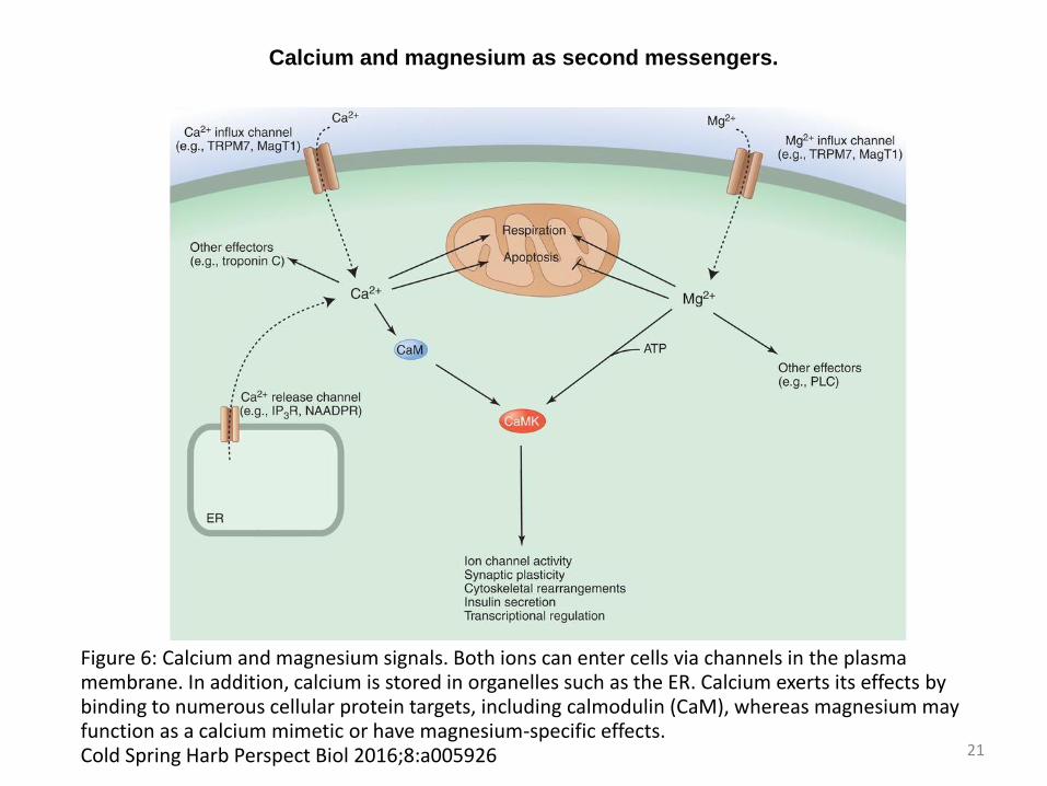
Calcium and magnesium as second messengers.
Figure 6: Calcium and magnesium signals. Both ions can enter cells via channels in the plasma membrane. In addition, calcium is stored in organelles such as the ER. Calcium exerts its effects by binding to numerous cellular protein targets, including calmodulin (CaM), whereas magnesium may function as a calcium mimetic or have magnesium-specific effects. Cold Spring Harb Perspect Biol 2016;8:a005926 21

3.1 Calcium as a secondary messenger Calcium is an extremely versatile intracellular messenger that controls a wide range of cellular functions by regulating the activity of a vast number of target proteins. The cellular effects of calcium are mediated either by direct binding to a target protein, or stimulation of calcium sensors that detect changes in calcium concentration and then activate different downstream responses. The multitude of sensors that mediate effects of calcium can be characterized by the nature of their calcium-binding site(s). The most common calcium-binding motifs are EF-hands and C2 domains. Synaptotagmin and troponin C are examples of proteins with C2 domains and EF-hands, respectively. Calcium sensors also act by recruiting a range of intermediary effectors, such as calcium-sensitive enzymes—for example, calcium/calmodulin-dependent protein kinases (CaMKs), calcineurin (also known as protein phosphatase 2B), myosin light chain kinase, and phosphorylase kinase. The cellular effect of Calcium on one of its target Calmodulin is shown in figure 7. 22
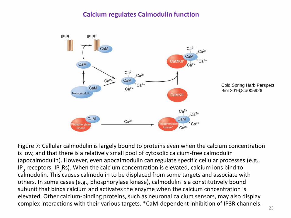
Figure 7: Cellular calmodulin is largely bound to proteins even when the calcium concentration is low, and that there is a relatively small pool of cytosolic calcium-free calmodulin (apocalmodulin). However, even apocalmodulin can regulate specific cellular processes (e.g., IP3 receptors, IP3Rs). When the calcium concentration is elevated, calcium ions bind to calmodulin. This causes calmodulin to be displaced from some targets and associate with others. In some cases (e.g., phosphorylase kinase), calmodulin is a constitutively bound subunit that binds calcium and activates the enzyme when the calcium concentration is elevated. Other calcium-binding proteins, such as neuronal calcium sensors, may also display complex interactions with their various targets. *CaM-dependent inhibition of IP3R channels.
Cold Spring Harb Perspect
Biol 2016;8:a005926
Calcium regulates Calmodulin function
23

Control of Calcium Levels
Intracellular calcium levels are controlled by an assortment of channels, pumps, transporters, buffers, and effector moieties. At rest, cytosolic calcium is maintained at ∼100 nM. In most cases, its levels are elevated by the opening of channels located either on various organellar stores or in the plasma membrane. Release of calcium from internal stores represents a major source of signal calcium for many cells. The principal calcium stores are the ER/SR, Golgi, and acidic organelles of the endolysosomal system. Calcium release channels are present on the membranes of these organelles and gate the flux of calcium from the ER/SR lumen into the cytosol. Ubiquitous calcium-release channels include the IP3Rs that respond to IP3 produced by hydrolysis of PIP2, ryanodine receptors , and two-pore channels that respond to nicotinic acid adenine dinucleotide phosphate. Channels that permit the influx of calcium across the plasma membrane are characterized by their activation mechanism. Receptor-operated calcium channels (NMDA receptors) and second messenger-operated calcium channels (Orai channels ) are opened by the binding of an external or internal ligand. 24

Calcium Buffers Cells express a number of calcium-binding proteins that buffer calcium changes within various cellular compartments and modulate calcium signals. The rapid binding of calcium by the cytosolic buffers parvalbumin and calbindin D-28k shapes both the spatial and temporal properties of intracellular calcium signals. ER/SR calcium-binding proteins (e.g., calsequestrin, calreticulin, GRP78, and GRP94) facilitate the accumulation of large amounts of calcium, which is necessary for rapid cell signaling. Mitochondria also play a key buffering role in that they express a calcium uniporter capable of taking up substantial amounts of calcium whenever the cytosolic calcium concentration increases during signaling.
25
Calcium Signal Termination Calcium pumps and exchangers are responsible for pumping calcium out of the cell or back into the ER/SR to terminate a calcium signal. Sucha as NCX proteins have low affinities for calcium, but have high capacities that enable them to rapidly remove large quantities of calcium. The plasma membrane calcium-ATPase and SR/ER calcium-ATPase enzymes have lower transport rates but higher affinities, which means that they operate at relatively low cytosolic calcium concentrations. Calcium/hydrogen exchangers are important for the loading of calcium into endo/lysosomal compartments.
26

3.2 Magnesium as secondary messenger Magnesium can also be considered an intracellular messenger because its concentration can change dynamically in response to cellular stimulation. Like calcium, there is an electrochemical gradient of magnesium across the plasma membrane that can serve as a reservoir for signal generation. The extracellular concentrations of magnesium and calcium are similar (1.1–1.5 mM), and magnesium can also act as a “calcimimetic” (e.g., by binding to the calcium-sensing receptor), a GPCR that has pleiotropic actions. Like calcium, magnesium has a plethora of transport pathways. Of these, TRPM6 and TRPM7 (members of the TRP family) are important. TRPM6 is restricted to kidney tubules and the intestinal epithelium, and plays an important role in magnesium (re)adsorption, whereas TRPM7 is ubiquitously expressed in mammals. Both TRPM6 and TRPM7 are “chanzymes”: ion channels that incorporate a kinase domain.
27

Magnesium as secondary messenger MagT1 is a key cellular magnesium channel. Like TRPM6 and TRPM7, it is located on the plasma membrane of mammalian cells. However, whereas TRPM6 and TRPM7 allow both calcium and magnesium fluxes, MagT1 is a specific pathway for magnesium. Some extracellular ligands elicit transient increases in free cytosolic Mg2+, which can increase Mg⋅ATP availability and stimulate Mg2+-sensitive enzymes. More than 500 cellular enzymes require Mg2+ for activity; therefore, Mg2+ affects multiple pathways when its intracellular level fluctuates. The Intracellular signal transduction by Mg2+ is summarized in figure 8.
28
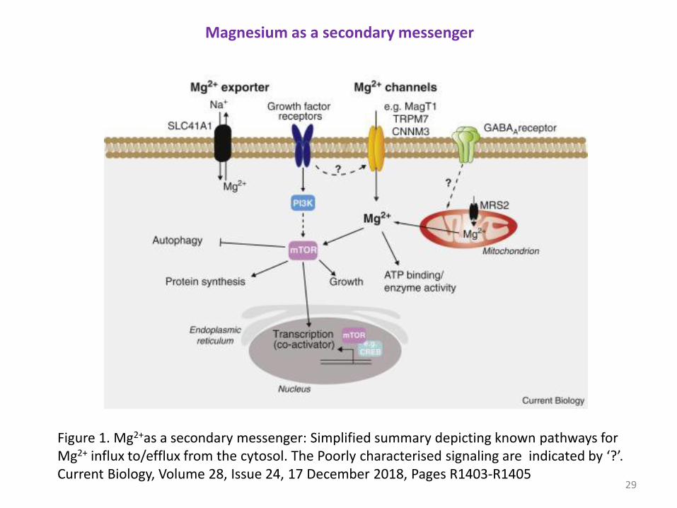
Figure 1. Mg2+as a secondary messenger: Simplified summary depicting known pathways for Mg2+ influx to/efflux from the cytosol. The Poorly characterised signaling are indicated by ‘?’. Current Biology, Volume 28, Issue 24, 17 December 2018, Pages R1403-R1405
Magnesium as a secondary messenger
29

References
The Cell, A molecular approach, Cooper Cold Spring Harb Perspect Biol 2016;8:a005926
Current Biology, Volume 28, Issue 24, 17 December 2018, Pages R1403-R1405
End
30