SCURRULA FERRUGINEA METHANOL EXTRACT INDUCES...
64
SCURRULA FERRUGINEA METHANOL EXTRACT INDUCES REACTIVE OXYGEN SPECIES-MEDIATED AND MITOCHONDRIAL-DEPENDENT APOPTOSIS IN BREAST CANCER CELLS MOHSEN MARVI BAIGI A thesis submitted in fulfilment of the requirements for the award of the degree of Doctor of Philosophy (Biomedical Engineering) Faculty of Biosciences and Medical Engineering Universiti Teknologi Malaysia MARCH 2016
Transcript of SCURRULA FERRUGINEA METHANOL EXTRACT INDUCES...

SCURRULA FERRUGINEA METHANOL EXTRACT INDUCES REACTIVE
OXYGEN SPECIES-MEDIATED AND MITOCHONDRIAL-DEPENDENT
APOPTOSIS IN BREAST CANCER CELLS
MOHSEN MARVI BAIGI
A thesis submitted in fulfilment of the
requirements for the award of the degree of
Doctor of Philosophy (Biomedical Engineering)
Faculty of Biosciences and Medical Engineering
Universiti Teknologi Malaysia
MARCH 2016

iii
I would like to dedicate this thesis to my beloved wife, my lovely unborn child and
my lovely father and mother
for their endless support and encouragement

iv
ACKNOWLEDGEMENT
Firstly, I would like to express my sincere gratitude to my main thesis
supervisor, Professor Dr. Ing Eko Supryianto for his encouragement, patience,
motivation, immense knowledge and friendship. Besides my supervisor, I am also
very thankful to my co-supervisor Associate Professor Dr. Fadzilah Adibah Abdul
Majid, who provided me an opportunity to join TCERG group, and who gave access
to the laboratory and research facilities. Without her precious support it would not be
possible to conduct this research. My sincere appreciation also extends to my co-
supervisors Dr. Shajarahtunnur Jamil and Dr. Saravana Kumar Jaganathan for their
insightful comments, guidance, advices and motivation. My sincere appreciation also
extends to all my colleagues and others who have provided assistance at various
occasions. Their views and tips are useful indeed. I would like to thank all members
of tissue culture engineering research group (TCERG) for their friendship and kind
assistance. Finally yet importantly, I would like to express utmost appreciation to my
lovely wife for her love, support and encouragements. In addition, especially I would
like to thank my beloved father, mother, sister and brothers for their sacrifice and
endless encouragement. My sincere thanks also goes to my father and mother in law
for their kind encouragements.

v
ABSTRACT
The purpose of this study is to investigate antioxidant and anticancer
activities of Scurrula ferruginea extracts. The antioxidant activities of the extracts
were evaluated using various assays. The extracts were further investigated to
examine their cytotoxic activity on human breast cancer cell lines; MDA-MB-231,
MDA-MB-468 and MCF-7 using MTT assay. Microscopic examinations of cells
were carried out to elucidate the modes of cell death. The effect of the extracts on
cancer cells colony formation and migration were determined. Changes in
mitochondrial membrane potential and level of reactive oxygen species (ROS) were
measured. Western blot and cell cycle analysis were performed to unravel the
mechanism of action of extracts against the breast cancer cells. Using GC-MS
analysis, chemical composition of extracts were characterized to reveal the presence
of anti-cancerous compounds. Our study on stem methanol extract has shown the
highest amount of phenolic, flavonoid contents, strong DPPH radical scavenging and
metal chelation activity in comparison to other extracts. The stem aqueous and
methanol extracts have shown higher cytotoxic effect towards MDA-MB-231 cells
compared to other cell lines with IC50 value of 50.35 and 19.27 µg/mL, after 72 h of
treatment, respectively. Morphological observations revealed properties of apoptosis
in the treated cells. The results displayed that the extracts have the ability to stop
migration of cancer cells and also inhibit the colony formation of cancer cells.
Moreover, the results have shown that the extracts induced apoptosis in breast cancer
cells by ROS generation and mitochondrial depolarization. Furthermore, this study
demonstrated that methanol extract inhibited the proliferation of breast cancer cells
via induction of cell cycle arrest at G0/G1 phase and apoptosis through a
mitochondria-dependent apoptosis pathway. The findings of present study revealed
the potential antioxidant and anticancer activities of S. ferruginea stem methanol
extract which may serve as a promising candidate in the search of a new anti-cancer
drug.

vi
ABSTRAK
Tujuan kajian penyelidikan ini adalah untuk mengkaji aktiviti antioksida dan
antikanser bagi ekstrak Scurrula ferruginea. Aktiviti antioksida bagi ekstrak
dianalisa menggunakan pelbagai kaedah asai. Ekstrak tersebut juga dikaji secara
lebih mendalam untuk mengenal pasti aktiviti sitotoksik terhadap garisan sel kanser
payudara; MDA-MB-231, MDA-MB-468 dan MCF-7 menggunakan asai MTT.
Analisis mikroskopik terhadap sel-sel telah dilaksanakan untuk menghuraikan mod
kematian sel. Kesan daripada ekstrak terhadap pembentukan dan migrasi koloni
kanser telah ditentukan. Perubahan kepada keupayaan membran mitokondria dan
tahap reaktif spesies oksigen (ROS) telah diukur. Western blot dan analisis kitaran
sel telah digunapakai untuk menguraikan mekanisme tindakan bagi ekstrak terhadap
sel-sel kanser payudara. Dengan menggunakan analisis GC-MS, komposisi kimia
bagi ekstrak telah dicirikan dan menunjukkan kehadiran sebatian anti-kanser.
Ekstrak methanol batang memberikan kuantiti fenolik dan flavonoid yang sangat
tinggi serta aktiviti penyingkiran radikal DPPH dan pengelatan logam yang kukuh
berbanding ekstrak yang lain. Ekstrak akueus dan methanol dari batang
menunjukkan kesan sitotoksik yang lebih tinggi terhadap sel MDA-MB-231
berbanding garisan sel yang lain dengan nilai IC50 masing-masing sebanyak 50.35
dan 19.27 µg/mL setelah 72 jam rawatan. Pemerhatian morfologi mendedahkan ciri-
ciri apoptosis dalam sel-sel yang dirawat. Hasil kajian menunjukkan bahawa ekstrak-
ekstrak tersebut mempunyai keupayaan untuk memberhentikan migrasi sel-sel
kanser di samping menghalang pembentukan koloni sel-sel kanser. Tambahan pula,
hasil kajian menunjukkan bahawa ekstrak-ekstrak mencetuskan apoptosis dalam sel-
sel kanser payu dara melalui penjanaan ROS dan penyahkutuban mitokondria. Selain
itu, kajian ini juga menunjukkan bahawa ekstrak methanol menghalang penyebaran
sel-sel kanser payu dara dengan merencatkan kitaran sel pada fasa G0/G1 dan
apoptosis melalui satu laluan apoptosis yang mempunyai pergantungan terhadap
mitokondria. Hasil kajian menunjukkan bahawa aktiviti antioksida dan antikanser
bagi ekstrak methanol dari batang S. ferruginea berpotensi menjadi calon kepada
pencarian ubat anti-kanser baru.
vii
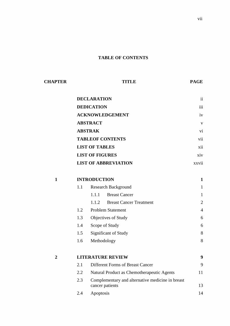
TABLE OF CONTENTS
CHAPTER TITLE PAGE
DECLARATION ii
DEDICATION iii
ACKNOWLEDGEMENT iv
ABSTRACT v
ABSTRAK vi
TABLEOF CONTENTS vii
LIST OF TABLES xii
LIST OF FIGURES xiv
LIST OF ABBREVIATION xxvii
1 INTRODUCTION 1
1.1 Research Background 1
1.1.1 Breast Cancer 1
1.1.2 Breast Cancer Treatment 2
1.2 Problem Statement 4
1.3 Objectives of Study 6
1.4 Scope of Study 6
1.5 Significant of Study 8
1.6 Methodology 8
2 LITERATURE REVIEW 9
2.1 Different Forms of Breast Cancer 9
2.2 Natural Product as Chemotherapeutic Agents 11
2.3 Complementary and alternative medicine in breast
cancer patients 13
2.4 Apoptosis 14
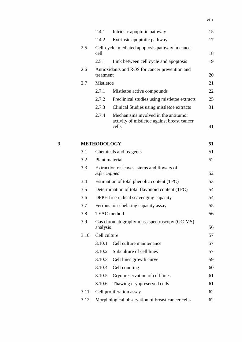
viii
2.4.1 Intrinsic apoptotic pathway 15
2.4.2 Extrinsic apoptotic pathway 17
2.5 Cell-cycle–mediated apoptosis pathway in cancer
cell 18
2.5.1 Link between cell cycle and apoptosis 19
2.6 Antioxidants and ROS for cancer prevention and
treatment 20
2.7 Mistletoe 21
2.7.1 Mistletoe active compounds 22
2.7.2 Preclinical studies using mistletoe extracts 25
2.7.3 Clinical Studies using mistletoe extracts 31
2.7.4 Mechanisms involved in the antitumor
activity of mistletoe against breast cancer
cells 41
3 METHODOLOGY 51
3.1 Chemicals and reagents 51
3.2 Plant material 52
3.3 Extraction of leaves, stems and flowers of
S.ferruginea 52
3.4 Estimation of total phenolic content (TPC) 53
3.5 Determination of total flavonoid content (TFC) 54
3.6 DPPH free radical scavenging capacity 54
3.7 Ferrous ion-chelating capacity assay 55
3.8 TEAC method 56
3.9 Gas chromatography-mass spectroscopy (GC-MS)
analysis 56
3.10 Cell culture 57
3.10.1 Cell culture maintenance 57
3.10.2 Subculture of cell lines 57
3.10.3 Cell lines growth curve 59
3.10.4 Cell counting 60
3.10.5 Cryopreservation of cell lines 61
3.10.6 Thawing cryopreserved cells 61
3.11 Cell proliferation assay 62
3.12 Morphological observation of breast cancer cells 62

ix
3.13 Ethidium bromide and acridine orange staining for
apoptosis detection 63
3.13.1 Apoptotic Index 63
3.14 Nuclear morphological studies by propidium
iodide and Hoechst 33342 staining 64
3.15 Clonogenic inhibition assay 65
3.16 In vitro scratch motility assay 65
3.17 Determination of intracellular reactive oxygen
species (ROS) generation 66
3.18 Mitochondrial membrane potential assay (JC-1
assay) 67
3.19 Extraction of proteins 68
3.20 Determination of protein concentration using BCA
assay 68
3.21 Sodium dodecyl sulfate polyacrylamide gel
electrophoresis (SDS-PAGE) 69
3.22 Western Blotting 71
3.23 Flow cytometry based analysis of cell cycle
distribution 72
3.24 Statistical analysis 73
4 RESULTS AND DISCUSSION 75
4.1 Extraction of plant materials 75
4.1.1 Extraction yields 76
4.2 Amount of phenolic compounds in S. ferruginea
extracts (TPC) 78
4.3 Determination of total flavonoid content (TFC) 79
4.4 DPPH scavenging activity 80
4.5 Fe 2+ chelating ability 85
4.6 Determination of TEAC value 87
4.7 Correlation between antioxidant activity assays,
phenolic and flavonoids contents 89
4.8 Chemical composition of S. ferruginea methanolic
extracts 90
4.8.1 Lupeol 98
4.8.2 Cinnamic acid 99
4.8.3 Linolenic acid 99
4.8.4 Humulane 100

x
4.8.5 Squalene 100
4.8.6 Hydrocarbons 100
4.8.7 Phytol 101
4.8.8 Vitamin E 101
4.9 Growth curves of breast cancer cell lines 102
4.10 In vitro cytotoxic activity of methanol and aqueous
extracts of S. ferruginea stems 105
4.10.1 Cytotoxic activity of selected extracts
against MDA-MB-231 cells 106
4.10.2 Cytotoxic activity of selected extracts
against MDA-MB-468 cells 108
4.10.3 Cytotoxic activity of selected extracts
against MCF-7 cells 110
4.10.4 Cytotoxic activity of selected extracts
against HSF-1184 cells 112
4.11 Morphological changes of breast cancer cells
following treatment with S. ferruginea extracts 115
4.11.1 Morphological changes of MDA-MB-231
cells treated with S. ferruginea extracts 116
4.11.2 Morphological changes of MDA-MB-468
cells treated with S. ferruginea extracts 118
4.11.3 Morphological changes of MCF-7 cells
treated with S. ferruginea extracts 120
4.12 Apoptosis detection by acridine orange/ethidium
bromide staining 123
4.12.1 Acridine orange/ethidium bromide
(AO/EB) staining of MDA-MB-231 cells 123
4.12.2 Acridine orange/ethidium bromide
(AO/EB) staining of MDA-MB-468 cells 127
4.12.3 Acridine orange/ethidium bromide
(AO/EB) staining of MCF-7 cells 131
4.13 Nuclear morphological studies using propidium
iodide and Hoechst staining 136
4.13.1 Propidium iodide and Hoechst staining of
MDA-MB-231 cells 137
4.13.2 Propidium iodide and Hoechst staining of
MDA-MB-468 cells 140
4.13.3 Propidium iodide and Hoechst staining of
MCF-7 cells 143
4.14 Effect of S. ferruginea extracts on colony
formation in breast cancer cell lines 146

xi
4.14.1 Effect of selected extracts on
clonogenicity of MDA-MB-231 cells 146
4.14.2 Effect of selected extracts on
clonogenicity of MDA-MB-468 cells 148
4.14.3 Effect of S. ferruginea extracts on
clonogenicity of MCF-7 cells 150
4.15 Cell migration inhibition efficiency of S.
ferruginea extracts 154
4.15.1 Effect of selected extracts on MDA-MB-
231 cell migration 154
4.15.2 Effect of selected extracts on MDA-MB-
468 cell migration 157
4.15.3 Effect of selected extracts on MCF-7 cell
migration 160
4.16 Measurement of reactive oxygen species (ROS)
generation 164
4.16.1 Qualitative measurement of ROS
formation in breast cancer cells 165
4.16.2 Quantitative measurement of ROS
formation in breast cancer cells 168
4.17 Measurement of mitochondrial membrane
potential (MMP) by JC-1 assay 170
4.17.1 Qualitative measurement of MMP 170
4.17.2 Quantitative measurement of MMP 173
4.18 Effect of S. ferruginea methanol extract on
apoptosis-associated proteins expression in MDA-
MB-231 cells 175
4.18.1 Effect of Methanol Extract on Pro-
apoptotic and Anti-apoptotic Proteins 175
4.18.2 Effect of Methanol Extract on Caspase 3,
Caspase 7 and PARP 178
4.19 Effects of S. ferruginea methanol extract on cell
cycle progression in breast cancer cells 182
5 CONCLUSION AND RECOMMENDATIONS 188
5.1 Conclusion 188
5.2 Recommendations 190
REFERENCES 191

xii
LIST OF TABLES
TABLE NO. TITLE PAGE
1.1 Summary of various methods of breast cancer treatment and
their common side effects. 3
2.1 Classification of invasive breast cancer according to Fisher
et al. 1975 and Linell et al. 1984. 10
2.2 Classification of breast cancer based on molecular markers. 11
2.3 Summary of in vitro and in vivo studies of mistletoe extracts
on breast cancer cells and animal models. 26
2.4 Summary of the clinical trials on efficacy of mistletoe
therapy in breast cancer patients. 38
2.5 A list of systematic and meta-analysis reviews including
controlled randomized, non-randomized and matched pair
clinical trials on different aspects of HRQoL in breast
cancers patients. 40
3.1 Preparation of resolving and stacking gels. 70
4.1 Percentage of S. ferruginea extraction yields obtained from
different parts using different solvents. 77
4.2 Total phenolic content of S. ferruginea leaves, stems and
flowers using different solvents expressed in mg Gallic
acid/g extract. 79
4.3 Total flavonoid content of S. ferruginea leaves, stems and
flowers using different solvents expressed in mg catechin/ g
extract. 80
4.4 Radical scavenging activity (IC50 value) of S. ferruginea
extracts against DPPH radical. 82
4.5 Metal chelation ability of various parts of S. ferruginea
extracts using different solvents. All analysis are mean of
three replicate determinations ± standard deviation (n = 3). a
Data expressed in percent of Fe (ii) chelation. b Data are
represented as IC50 and the values are presented with their
respective 95% confidence interval (95% CI). c Positive
reference standard. 87

xiii
4.6 ABTS+ radical scavenging capacity of S. ferruginea
extracts. Data were expressed as µM Trolox/g extract. 88
4.7 Pearson’s correlation coefficients of antioxidant activities,
total flavonoid and total phenolic content in S. ferruginea. 90
4.8 Phytochemical compounds identified in the methanolic
extracts of the S. ferruginea stem by GC-MS. 92
4.9 Phytochemical compounds identified in the methanolic
extracts of the S. ferruginea leaf by GC-MS. 94
4.10 Phytochemical compounds identified in the methanolic
extracts of the S. ferruginea flower by GC-MS. 96
4.11 Inhibitory effect of S. ferruginea extracts against MDA-
MB-231 breast cancer cell line at different incubation times. 108
4.12 Inhibitory effect of S. ferruginea extracts against MDA-
MB-468 breast cancer cell line at different incubation times. 110
4.13 Inhibitory effect of S. ferruginea extracts against MCF-7
cells at different incubation times. 112
4.14 Inhibitory effect of S. ferruginea extracts against different
breast cancer cells after 72h of incubation. 114
4.15 Comparison of the percentage of apoptotic cells at various
breast cancer cells treated with different concentrations of
methanol extract, aqueous extract and IC50 concentration of
positive control (tamoxifen). 136
4.16 Comparison of the percentage of colony forming potential
at various breast cancer cells treated with different
concentrations of methanol extract, aqueous extract and
positive control (tamoxifen). 153
4.17 Comparison of the percentage of migration of breast cancer
cells treated with different concentrations of methanol and
aqueous extracts after 24 h. 164

xiv
LIST OF FIGURES
FIGURE NO. TITLE PAGE
2.1 Schematic diagram of extrinsic and intrinsic apoptotic
pathways adapted from (Vries et al., 2006). 15
2.2 Origin and scientific names of mistletoes belonging to
Loranthaceae family. 23
2.3 Origin and scientific names of mistletoes belonging to
Viscaceae family. 23
2.4 Schematic diagram of the various cellular activities and
mechanisms involved in mistletoe extracts effects on QOL.
As depicted, the mechanisms involved in anti-tumor
properties of mistletoe are interrelated biological
phenomena including apoptosis, β-endorphin release,
immunomodulation (stimulation of pro-inflammatory
cytokines). ADCC: antibody-dependent cell-mediated
cytotoxicity. 42
2.5 Schematic illustration explaining the possible mechanisms
of apoptosis induction by mistletoe in breast cancer cells
(stimulatory effect represented by arrow with directed blue
lines and inhibitory effect indicated by red lines). 44
4.1 S. ferruginea (Jack) Danser on host tree (Bauhinia x
blakeana) collected from the campus of Universiti
Teknologi Malaysia (Latitude N 1° 33’ 54.9", Longitude E
103° 38’ 29.2"), Skudai, Johor, Malaysia. 75
4.2 Close-up view of S. ferruginea (jack) Danser stem, leaf and
flower. 76
4.3 Extract evaporation using rotary evaporator (BUCHI,
Switzerland, R210). 77
4.4 DPPH radical scavenging capacities of (a) stem, (b) leaf and

xv
(c) flower extracts of S. ferruginea and positive control (Ascorbic acid).
Values are mean of three replicate determinations ±
standard deviation (n = 3). Points marked with different
letters are significantly different at 𝑃 < 0.05 when compared
at the same concentration point as determined by two-way
ANOVA. The positive control (ascorbic acid) showed a
significantly higher scavenging capacity as compared to the
samples (at 𝑃 < 0.05 as determined by two-way ANOVA). 84
4.5 Metal chelating activities of (a) stem, (b) leaf and (c) flower
extracts of S. ferruginea and positive control (EDTA).
Values are mean of three replicate determinations ±
standard deviation (n = 3). Points marked with different
letters are significantly different at 𝑃 < 0.05 when compared
at the same concentration point as determined by two-way
ANOVA. The positive control (EDTA) showed a
significantly higher scavenging capacity as compared to the
samples (at 𝑃 < 0.05 as determined by two-way ANOVA). 86
4.6 GC-MS chromatogram of methanolic extract of S.
ferruginea stem. 92
4.7 GC-MS chromatogram of methanolic extract of S.
ferruginea leaf. 94
4.8 GC-MS chromatogram of methanolic extract of S.
ferruginea flower. 96
4.9 The complete growth curve of MDA-MB-231 cell line.
Population doubling time (PDT) is 24.63 h (1.02 day). Data
are represented as means ± SD. 104
4.10 The complete growth curve of MDA-MB-468 cell line.
Population doubling time (PDT) is 26.97 h (1.12 day). Data
are represented as means ± SD. 104
4.11 The complete growth curve of MCF-7 cell line. Population
doubling time (PDT) is 39.04 h (1.62 day). Data are
represented as means ± SD. 105
4.12 S. ferruginea extracts inhibits MDA-MB-231 cells
proliferation in a time- and dose-dependent manner. The
cells were treated with indicated concentrations of (a)
methanol and (b) aqueous extracts of S. ferruginea for
indicated time intervals. The results were expressed versus
percentage of the value observed with control. Cytotoxic
activity of extracts was compared to reference drug,
tamoxifen on MDA-MB-231 cell line (c). Cell viability was
measured by MTT assays. Data are represented as means ±
SD of three replicates in three independent experiments. All
data showed statistically significant difference from control
(one way ANOVA, P < 0.05). 107

xvi
4.13 S. ferruginea extracts inhibits MDA-MB-468 cells
proliferation in a time- and dose-dependent manner. The
cells were treated with indicated concentrations of (a)
methanol and (b) aqueous extracts of S. ferruginea for
indicated time intervals. The results were expressed versus
percentage of the value observed with control. Cytotoxic
activity of extracts was compared to reference drug,
tamoxifen on MDA-MB-468 cell line (c). Cell viability was
measured by MTT assays. Data are represented as means ±
SD of three replicates in three independent experiments. All
data showed statistically significant difference from control
(one way ANOVA, P < 0.05). 109
4.14 S. ferruginea extracts inhibits MCF-7 cells proliferation in a
time- and dose-dependent manner. The cells were treated
with indicated concentrations of (a) methanol and (b)
aqueous extracts of S. ferruginea for indicated time
intervals. The results were expressed versus percentage of
the value observed with control. Cytotoxic activity of
extracts was compared to reference drug, tamoxifen on
MCF-7 cell line (c). Cell viability was measured by MTT
assays. Data are represented as means ± SD of three
replicates in three independent experiments. All data
showed statistically significant difference from control (one
way ANOVA, P < 0.05). 111
4.15 In vitro anti-proliferative activity of the methanol and
aqueous extracts of S. ferruginea stems against HSF-1184
normal cell lines. The results were expressed versus
percentage of the value observed with control. The result
indicated that extracts were non-selective towards normal
cell line. Extracts showed negligible toxicity in normal cell
line. Data are represented as means ± SD of three replicates
in three independent experiments. * indicates statistically
significant different from control (one way ANOVA, P <
0.05). 113
4.16 Morphological changes of MDA-MB-231 cells treated with
methanol and aqueous extracts at their respective IC50
concentrations. As a positive control, the MDA-MB-231
cells were treated with 8.5 µg/mL tamoxifen. After 24, 48
and 72 hours of treatment, the cell morphological alterations
were observed with an inverted-phase contrast microscope
(20× magnification). 117
4.17 Close-up views of MDA-MB-231 cells treated with S.
ferruginea methanol and aqueous extracts viewed under an
inverted light microscope (20× magnification). The cells
showed hallmark properties of apoptosis such as cell
shrinkage (A, B, D) and cell rounding (C). 118
xvii
4.18 Morphological changes of MDA-MB-468 cells treated with
methanol and aqueous extracts at their respective IC50
concentrations. As a positive control, the MDA-MB-468
cells were treated with 8.5 µg/mL tamoxifen. After 24, 48
and 72 hours of treatment, the cell morphological alterations
were observed with an inverted-phase contrast microscope
(20× magnification). 119
4.19 Close-up views of MDA-MB-468 cells treated with S.
ferruginea methanol and aqueous extracts viewed under an
inverted light microscope (20× magnification). The cells
showed characteristics of apoptosis such as cell shrinkage
(A, B), cell rounding (C) and membrane blebbing (D). 120
4.20 Morphological changes of MCF-7 cells treated with
methanol and aqueous extracts at their respective IC50
concentrations. As a positive control, the MCF-7 cells were
treated with 8.5 µg/mL tamoxifen. After 24, 48 and 72
hours of treatment, the cell morphological alterations were
observed with an inverted-phase contrast microscope (20×
magnification). 121
4.21 Close-up views of MCF-7 cells treated with S. ferruginea
methanol and aqueous extracts viewed under an inverted
light microscope (20× magnification). The cells showed
characteristics of apoptosis such as membrane blebbing (A)
and cell shrinkage and rounding (B, C, D) 122
4.22 Morphological observation of AO/EB-stained MDA-MB-
231 cells incubated for 24 hours with methanol and aqueous
extracts. As a positive control, the cells were treated with
8.5 µg/mL tamoxifen. The morphological alterations in the
cells were visualized under fluorescence microscope (20×).
Viable cells stained uniformly in green color with normal
morphology. Treated cells showed early apoptotic cells with
membrane blebbing and bright green nuclei, late apoptotic
cells with fragmented and condensed orange-red nuclei and
necrotic cells with deep orange nucleus. 124
4.23 Close-up views of AO/EB double-stained MDA-MB-231
cells treated with S. ferruginea extracts viewed under
fluorescence microscope (20×). Membrane blebbing were
seen in treated cells (A, B). Early and late apoptotic cells
with nuclear fragmentation and margination were observed
in treated cells after 24 h incubation with extracts (C, D). 125

xviii
4.24 Quantification of the percentage of live, apoptotic, and
necrotic cells at different concentrations (31.25-1000
µg/mL) of (a) methanol and (b) aqueous extracts. The
percentage of apoptotic cells at different concentrations
were observed higher in methanol extract compare to
aqueous extract. Data are represented as means ± SD of
three replicates in three independent experiments, counting
a minimum of 200 total cells each. * indicates statistically
significant different from their respective control (one way
ANOVA, P < 0.05). 126
4.25 Morphological observation of AO/EB-stained MDA-MB-
468 cells after 24 hours incubation with methanol and
aqueous extracts. As a positive control, the cells were
treated with 8.5 µg/mL tamoxifen. The morphological
alterations in the cells were visualized under fluorescence
microscope (20×). Viable cells stained uniformly in green
color with normal morphology. Treated cells showed early
apoptotic cells with membrane blebbing and bright green
nuclei, late apoptotic cells with fragmented and condensed
orange-red nuclei and necrotic cells with deep orange
nucleus. 128
4.26 Close-up views of AO/EB double-stained MDA-MB-468
cells treated with S. ferruginea extracts viewed under
fluorescence microscope (20×). Plasma membrane blebbing
were seen in treated cells (A). Early and late apoptotic cells
with nuclear fragmentation and margination were observed
in treated cells after 24 h incubation with extracts (B, C, D). 129
4.27 Quantification of the percentage of live, apoptotic, and
necrotic cells at different concentrations (31.25-1000
µg/mL) of (a) methanol and (b) aqueous extracts. The
percentage of apoptotic cells at different concentrations
were observed higher in methanol extract compare to
aqueous extract. Data are represented as means ± SD of
three replicates in three independent experiments, counting
a minimum of 200 total cells each. * indicates statistically
significant different from their respective control (one way
ANOVA, P < 0.05). 130
4.28 Morphological observation of AO/EB-stained MCF-7 cells
after 24 hours treatment with methanol and aqueous
extracts. As a positive control, the cells were treated with
8.5 µg/mL tamoxifen. The morphological alterations in the
cells were visualized under fluorescence microscope (20×).
Viable cells stained uniformly in green color with normal
morphology. Treated cells showed early apoptotic cells with
membrane blebbing and bright green nuclei, late apoptotic
cells with fragmented and condensed orange-red nuclei and
necrotic cells with deep orange nucleus. 132

xix
4.29 Close-up views of AO/EB double-stained MCF-7 cells
treated with S. ferruginea extracts viewed under
fluorescence microscope (20×). Cell shrinkage and
membrane blebbing were seen in treated cells (A, B). Early
and late apoptotic cells with nuclear fragmentation were
observed in treated cells after 24 h incubation with extracts
(C, D). 133
4.30 Quantification of the percentage of live, apoptotic, and
necrotic cells at different concentrations (31.25-1000
µg/mL) of (a) methanol and (b) aqueous extracts. The
percentage of apoptotic cells at different concentrations
were observed higher in methanol extract compare to
aqueous extract. Data are represented as means ± SD of
three replicates in three independent experiments, counting
a minimum of 200 total cells each. * indicates statistically
significant different from their respective control (one way
ANOVA, P < 0.05). 134
4.31 Fluorescence imaging for detection of apoptosis in MDA-
MB-231 cells treated with methanol extract for 24 hours at
concentrations of 31.25 and 250 μg/mL. Left panel displays
Hoechst 33342 staining while right panel displays PI
staining of the same field. The morphological alterations in
the cells were visualized under fluorescence microscope
(20×). Both viable and dead cells nuclei were stained with
Hoechst 33342 while PI was unable to stain viable cells
nuclei. Treated cells at both concentrations showed
condensed and fragmented nuclei. The number of deranged
nuclei and apoptotic cells increased at concentration of
250 μg/mL. 138
4.32 Fluorescence imaging for detection of apoptosis in MDA-
MB-231 cells treated with aqueous extract for 24 hours at
concentrations of 31.25 and 250 μg/mL. Left panel displays
Hoechst 33342 staining while right panel displays PI
staining of the same field. The morphological alterations in
the cells were visualized under fluorescence microscope
(20×). Both viable and dead cells nuclei were stained with
Hoechst 33342 while PI was unable to stain viable cells
nuclei. Treated cells at both concentrations showed
condensed and fragmented nuclei. The number of deranged
nuclei and apoptotic cells increased at concentration of
250 μg/mL. 139
xx
4.33 Fluorescence imaging for detection of apoptosis in MDA-
MB-231 cells treated with tamoxifen as positive control for
24 hours at concentrations of 31.25 and 250 μg/mL. Left
panel displays Hoechst 33342 staining while right panel
displays PI staining of the same field. The morphological
alterations in the cells were visualized under fluorescence
microscope (20×). Both viable and dead cells nuclei were
stained with Hoechst 33342 while PI was unable to stain
viable cells nuclei. Treated cells were observed with
condensed and fragmented nuclei. 140
4.34 Fluorescence imaging for detection of apoptosis in MDA-
MB-468 cells. Cells were treated with methanol extract for
24 hours at concentrations of 31.25 and 250 μg/mL. Left
panel displays Hoechst 33342 staining while right panel
displays PI staining of the same field. The morphological
alterations in the cells were visualized under fluorescence
microscope (20×). Both viable and dead cells nuclei were
stained with Hoechst 33342 while PI was unable to stain
viable cells nuclei. Treated cells at both concentrations
showed condensed and fragmented nuclei. 141
4.35 Fluorescence imaging for detection of apoptosis in MDA-
MB-468 cells. Cells were treated with aqueous extract for
24 hours at concentrations of 31.25 and 250 μg/mL. Left
panel displays Hoechst 33342 staining while right panel
displays PI staining of the same field. The morphological
alterations in the cells were visualized under fluorescence
microscope (20×). Both viable and dead cells nuclei were
stained with Hoechst 33342 while PI was unable to stain
viable cells nuclei. 142
4.36 Fluorescence imaging for detection of apoptosis in MDA-
MB-468 cells. Cells were treated with tamoxifen (positive
control) for 24 hours at concentrations of 31.25 and
250 μg/mL. Left panel displays Hoechst 33342 staining
while right panel displays PI staining of the same field. The
morphological alterations in the cells were visualized under
fluorescence microscope (20×). 143
4.37 Fluorescence imaging for detection of apoptosis in MCF-7
cells. Cells were treated with methanol extract for 24 hours
at concentrations of 31.25 and 250 μg/mL. Left panel
displays Hoechst 33342 staining while right panel displays
PI staining of the same field. The morphological alterations
in the cells were visualized under fluorescence microscope
(20×). Both viable and dead cells nuclei were stained with
Hoechst 33342 while PI was unable to stain viable cells
nuclei. Treated cells at both concentrations showed
condensed and fragmented nuclei. 144
xxi
4.38 Fluorescence imaging for detection of apoptosis in MCF-7
cells. Cells were treated with aqueous extract for 24 hours at
concentrations of 31.25 and 250 μg/mL. Left panel displays
Hoechst 33342 staining while right panel displays PI
staining of the same field. The morphological alterations in
the cells were visualized under fluorescence microscope
(20×). Both viable and dead cells nuclei were stained with
Hoechst 33342 while PI was unable to stain viable cells
nuclei. Treated cells at both concentrations showed
condensed and fragmented nuclei. 145
4.39 Fluorescence imaging for detection of apoptosis in MCF-7
cells. Cells were treated with tamoxifen (positive control)
for 24 hours at concentrations of 31.25 and 250 μg/mL. Left
panel displays Hoechst 33342 staining while right panel
displays PI staining of the same field. The morphological
alterations in the cells were visualized under fluorescence
microscope (20×). 146
4.40 Effect of S. ferruginea extracts on colony-forming abilities
of MDA-MB-231 cells. Methanol (a) and aqueous (b)
extracts suppressed colony formation in a dose dependent
manner. The methanol extract inhibited the clonogenicity of
MDA-MB-231 cells more effectively than aqueous extract.
The images were taken using an inverted phase contrast
microscope (Zeiss Axiovert 100) at 4× magnification. 147
4.41 Quantitative measurement of colony formation of selected
extracts on MDA-MB-231 cells at different concentrations
(31.25-1000 µg/mL). The numbers of the colonies were
estimated under dissection (stereo) microscope (Wild
Heerburgg M3). The colony forming ability of the cells at
each dose of extracts is expressed in terms of percent of
control and represented as means ± SD of three replicates in
three independent experiments. * indicates statistically
significant different from their respective control (one way
ANOVA, P < 0.05). 148
4.42 Effect of S. ferruginea extracts on colony-forming abilities
of MDA-MB-468 cells. Methanol (a) and aqueous (b)
extracts suppressed colony formation in a dose dependent
manner. The methanol extract inhibited the clonogenicity of
MDA-MB-468 cells more effectively than aqueous extract.
The images were taken using an inverted phase contrast
microscope (Zeiss Axiovert 100) at 4× magnification. 149

xxii
4.43 Quantitative measurement of colony formation of selected
extracts on MDA-MB-468 cells at different concentrations
(31.25-1000 µg/mL). The numbers of the colonies were
measured under dissection (stereo) microscope (Wild
Heerburgg M3). The colony forming ability of the cells at
each dose of extracts is expressed in terms of percent of
control and represented as means ± SD of three replicates in
three independent experiments. * indicates statistically
significant different from their respective control (one way
ANOVA, P < 0.05). 150
4.44 Effect of S. ferruginea extracts on colony-forming abilities
of MCF-7 cells. A. Methanol (a) and aqueous (b) extracts
suppressed colony formation in a dose dependent manner.
The methanol extract inhibited the clonogenicity of MCF-7
cells more effectively than aqueous extract. The images
were taken using an inverted phase contrast microscope
(Zeiss Axiovert 100) at 4× magnification. 151
4.45 Quantitative measurement of colony formation of selected
extracts on MCF-7 cells at different concentrations (31.25-
1000 µg/mL). The numbers of the colonies were estimated
under dissection (stereo) microscope (Wild Heerburgg M3).
The colony forming ability of the cells at each dose of
extracts is expressed in terms of percent of control and
represented as means ± SD of three replicates in three
independent experiments. * indicates statistically significant
different from their respective control (one way ANOVA, P
< 0.05). 152
4.46 Effect of S. ferruginea methanol and aqueous extracts on
the cell migration of MDA-MB-231 cells. Scratch closure
activity of treated MDA-MB-231 cells upon creation of
scratch using a scratcher in control and treated well. The
images of scratched MDA-MB-231 cell monolayer treated
with extracts captured under an inverted phase-contrast
microscope at different time intervals (0, 6, 12 & 24 h). 155
4.47 Quantitative measurement of cell migration of methanol (a)
and aqueous (b) extracts on MDA-MB-231 cells at different
concentrations (31.25-1000 µg/mL). Scratch closure rates
were analyzed quantitatively as the difference between
scratch width at 0, 6 and 12 or 24 h and results are
expressed as percentage of cell migration. Results showed
that in presence of selected extracts the migration of the
MDA-MB-231 cells was dose- and time-dependently
inhibited. Data are represented as means ± SD of three
replicates in three independent experiments. * indicates
statistically significant different from their respective
control (one way ANOVA, P < 0.05). 156

xxiii
4.48 Effect of S. ferruginea methanol and aqueous extracts on
the cell migration of MDA-MB-468 cells. Scratch closure
activity of treated MDA-MB-468 cells upon creation of
scratch using a scratcher in control and treated well. The
images of scratched MDA-MB-468 cell monolayer treated
with extracts captured under an inverted phase-contrast
microscope at different time intervals (0, 6, 12 & 24 h). 158
4.49 Quantitative measurement of cell migration of methanol (a)
and aqueous (b) extracts on MDA-MB-468 cells at different
concentrations (31.25-1000 µg/mL). Scratch closure rates
were analyzed quantitatively as the difference between
scratched width at 0, 6 and 12 or 24 h and results are
expressed as percentage of cell migration. Results showed
that in presence of selected extracts the migration of the
MDA-MB-468 cells was dose- and time-dependently
inhibited. Data are represented as means ± SD of three
replicates in three independent experiments. * indicates
statistically significant different from their respective
control (one way ANOVA, P < 0.05). 159
4.50 Effect of S. ferruginea methanol and aqueous extracts on
the cell migration of MCF-7 cells. Scratch closure activity
of cells upon creation of scratch using a scratcher in control
and treated well. The images of scratched MCF-7 cell
monolayer treated with extracts captured under an inverted
phase-contrast microscope at different time intervals (0, 6,
12 & 24 h). 161
4.51 Quantitative measurement of cell migration of methanol (a)
and aqueous (b) extracts on MCF-7 cells at different
concentrations (31.25-1000 µg/mL). Scratch closure rates
were analyzed quantitatively as the difference between
scratch width at 0, 6 and 12 or 24 h and results are
expressed as percentage of cell migration. Results showed
that in presence of selected extracts the migration of the
MCF-7 cells was dose- and time-dependently inhibited.
Data are represented as means ± SD of three replicates in
three independent experiments. * indicates statistically
significant different from their respective control (one way
ANOVA, P < 0.05). 162
4.52 Qualitative evaluation of ROS generation in MDA-MB-231
cells using the fluorescent probe DCF-DA. MDA-MB-231
cells were treated with 31.25 μg/mL, 250 μg/mL and
positive control (50 μM H2O2) for 12 h. Fluorescence
microscopic images (10×) indicated that methanol extract
induced intracellular ROS formation in MDA-MB-231
cells. 166

xxiv
4.53 Qualitative evaluation of ROS generation in MDA-MB-468
cells using the fluorescent probe DCF-DA. MDA-MB-468
cells were treated with 31.25 μg/mL, 250 μg/mL and
positive control (50 μM H2O2) for 12 h. Fluorescence
microscopic images (10×) indicated that methanol extract
induced intracellular ROS formation in MDA-MB-468
cells. 167
4.54 Qualitative measurement of ROS generation in MCF-7 cells
using the fluorescent probe DCF-DA. MCF-7 cells were
treated with 31.25 μg/mL, 250 μg/mL and positive control
(50 μM H2O2) for 12 h. Fluorescence microscopic images
(10×) indicated that methanol extract induced intracellular
ROS formation in MCF-7 cells. 168
4.55 Effects of S. ferruginea methanol extract on ROS
generation in different breast cancer cell lines. Cells were
treated with different concentrations of methanol extract
and positive control (50 μM H2O2) for 12 h. Data are
represented as means ± SD of three replicates in three
independent experiments. * indicates statistically significant
different from corresponding controls (one way ANOVA, P
< 0.05). 169
4.56 Effect of methanol extract on mitochondrial membrane
potential (MMP) in MDA-MB-231 cells using JC-1
fluorescence dye. Methanol extract induced MMP
depolarization in MDA-MB-231 cells. The cells were
treated with 31.25 μg/mL, 250 μg/mL and positive control
(50 μM CCCP) for 12 h. Images were obtained with an
inverted fluorescent microscope (Zeiss Axiovert A) (40×).
The emitted green fluorescence indicates MMP
depolarization which is an early event in apoptosis. 171
4.57 Effect of methanol extract on mitochondrial membrane
potential (MMP) in MDA-MB-468 cells using JC-1
fluorescence dye. Methanol extract induced MMP
depolarization in MDA-MB-468 cells. The cells were
treated with 31.25 μg/mL, 250 μg/mL and positive control
(50 μM CCCP) for 12 h. Images were obtained with an
inverted fluorescent microscope (Zeiss Axiovert A) (40×).
The emitted green fluorescence indicates MMP
depolarization which is an early event in apoptosis. 172
4.58 Effect of methanol extract on mitochondrial membrane
potential (MMP) in MCF-7 cells using JC-1 fluorescence
dye. Methanol extract induced MMP depolarization in
MCF-7 cells. The cells were treated with 31.25 μg/mL, 250
μg/mL and positive control (50 μM CCCP) for 12 h. Images
were obtained with an inverted fluorescent microscope
(Zeiss Axiovert A) (40×). The emitted green fluorescence
indicates MMP depolarization which is an early event in
apoptosis. 173

xxv
4.59 Relative quantity of mitochondrial membrane potential
(ΔΨm) in different breast cancer cell lines. Cells were
treated with different concentrations of methanol extract
and positive control (50 μM CCCP) for 12 h. Methanol
extract disrupts mitochondrial transmembrane potential
(ΔΨm). Data are represented as means ± SD of three
replicates in three independent experiments. * indicates
statistically significant different from corresponding
controls (one way ANOVA, P < 0.05). 174
4.60 Western blot analysis of pro-apoptotic Bax protein in
MDA-MB-231 cells. MDA-MB-231 cells were treated with
IC50 concentration of S. ferruginea methanol extract and
control cells (0.1% DMSO) for indicated times. β-actin was
used as loading control. Densitometry analysis showed
time-dependent up-regulation of Bax protein. The
expression of Bax protein increased as early as 2 hour. The
densitometer-intensity data are represented as means ± SEM
of three replicates in three independent experiments. *
indicates statistically significant different from control (one
way ANOVA, P < 0.05). 177
4.61 Western blot analysis of anti-apoptotic Bcl-2 protein in
MDA-MB-231 cells. MDA-MB-231 cells were treated with
IC50 concentration of S. ferruginea methanol extract and
control cells (0.1% DMSO) for indicated times. β-actin was
used as loading control. Densitometry analysis showed
time-dependent down-regulation of Bcl-2 protein. The
densitometer-intensity data are represented as means ± SEM
of three replicates in three independent experiments. *
indicates statistically significant different from control (one
way ANOVA, P < 0.05). 178
4.62 Western blot analysis of caspase-3 protein in MDA-MB-
231 cells. MDA-MB-231 cells were treated with IC50
concentration of S. ferruginea methanol extract and control
cells (0.1% DMSO) for indicated times. β-actin was used as
loading control. Densitometry analysis demonstrated that
procaspase-3 (32-kDa) was cleaved to yield a catalytically
active 17-kDa fragment after treatment with methanol
extract. The densitometer-intensity data are represented as
means ± SEM of three replicates in three independent
experiments. * indicates statistically significant different
from control (one way ANOVA, P < 0.05). 180

xxvi
4.63 Western blot analysis of caspase-7 protein in MDA-MB-
231 cells. MDA-MB-231 cells were treated with IC50
concentration of S. ferruginea methanol extract and control
cells (0.1% DMSO0 for indicated times. β-actin was used as
loading control. Densitometry analysis demonstrated that
procaspase-7 (35-kDa) was cleaved to yield a catalytically
active 17-kDa fragment after treatment with methanol
extract. The densitometer-intensity data are represented as
means ± SEM of three replicates in three independent
experiments. * indicates statistically significant different
from control (one way ANOVA, P < 0.05). 181
4.64 Western blot analysis of PARP protein in MDA-MB-231
cells. MDA-MB-231 cells were treated with IC50
concentration of S. ferruginea methanol extract and control
cells (0.1% DMSO) for indicated times. β-actin was used as
loading control. The PARP protein (116-kDa) was cleaved
into its signature 85-kDa fragment, a marker of apoptosis,
after treatment with methanol extract. The densitometer-
intensity data are represented as means ± SEM of three
replicates in three independent experiments. * indicates
statistically significant different from control (one way
ANOVA, P < 0.05). 182
4.65 Effect of S. ferruginea methanol extract on the cell cycle
progression in MDA-MB-231 cell. MDA-MB-231 cells
were treated with IC50 concentration of methanol extract for
24 and 48 h, stained with PI and its content was analyzed by
flow cytometry. The data are represented as means ± SEM
of three replicates in three independent experiments. *
indicates statistically significant different from control (one
way ANOVA, P < 0.05). 184
4.66 Effect of S. ferruginea methanol extract on the cell cycle
progression in MCF-7 cell. MCF-7 cells were treated with
IC50 concentration of methanol extract for 24 and 48 h,
stained with PI and its content was analyzed by flow
cytometry. The data are represented as means ± SEM of
three replicates in three independent experiments. *
indicates statistically significant different from control (one
way ANOVA, P < 0.05). 185
4.67 Proposed schematic diagram of S. ferruginea methanol
extract-induced apoptosis in human breast cancer cells
MDA-MB-231. Treatment of MDA-MB-231 cells with S.
ferruginea methanol extract induced high level of ROS
generation and subsequently reduced ΔΨm levels which
leading to changes in the expression levels of Bax/Bcl-2.
This results in mitochondrial dysfunction and caspase-3 and
caspase-7 activation. These events all contribute to the
subsequent degradation of PARP in MDA-MB-231 cells via
G0/G1 cell cycle arrest, which mediates apoptosis. 187
xxvii
LIST OF ABBREVIATION
NCR - The National Cancer Registry
DCIS - Ductal Carcinoma In Situ
LCIS - Lobular Carcinoma In Situ
HER - Human Epidermal Growth Factor Receptor
ER - Estrogen Receptor
PR - progesterone receptor
NCI - National Cancer Institute
ROS - Reactive Oxygen Species
CAM - Complementary and Alternative Medicine
DPPH - Diphenyl-2-picryl hydrazine
ABTS - 2, 2'-azino bis-(3-ethyl benzo thiazoline-6-sulphonic acid)
EDTA - Ethylenediaminetetraacetic acid
DMEM - Dulbecco’s Modified Eagle Medium
FBS - Fetal Bovine Serum
PI - Propidium Iodide
PBS - phosphate buffer saline
DMSO - Dimethyl Sulfoxide
TPC - Total Phenolic Content
TFC - Total Flavonoid Content
GC-MS - Gas chromatography-mass spectroscopy
RPMI - Roswell Park Memorial Institute

xxviii
PDT - Population Doubling Time
MTT - Thiazolyl Blue Tetrazolium Bromide
AO/EB - Acridine orange/Ethidium bromide
MMP - Mitochondrial Membrane Potential
BCA - Bicinchoninic Acid
BSA - Bovine Serum Albumin
SDS-PAGE - Sodium dodecyl sulfate polyacrylamide gel electrophoresis
AP - Alkaline Phosphatase
MOMP - Mitochondrial Outer Membrane Permeabilization
DISK - Death-Inducing Signaling Complex
ML-I - Mistletoe Lectin I
HR-QOL - Health-Related Quality Of Life
VA - Viscum album
ADCC - Antibody-Dependent Cell-mediated Cytotoxicity
TNFα - Tumor Necrosis Factor alfa
CRF - Cancer Related Fatigue
TCM - Traditional Chinese Medicine
LS - Life Satisfaction
TEAC - Trolox Equivalent Antioxidant Capacity
RT - Retention Time

CHAPTER 1
1 INTRODUCTION
1.1 Research Background
1.1.1 Breast Cancer
Cancer of breast formed due to formation of malignant tumor in the cells of
breast. Initially the growth of breast cancer is local which is followed by extension
within lymph vessels into regional lymph nodes and invasion of small vein which
results in systematic metastatic spread (Spratt & Tobin, 1995). Breast cancer is the
most common type of non-skin malignancy among women worldwide. It has been
reported that the incidence and mortality of breast cancer have increased during the
last two decades (American Cancer Society Global Cancer Facts & Figures 2nd
Edition, 2011; Jemal et al., 2011; Ferlay et al., 2013). Based on 2006-2010 statistics,
the number of deaths in the United States was 22.6 per 100,000 women per year. It is
predicted that an estimated 231,840 new cases of breast cancer and 40,730 breast
cancer-related deaths will occur among women in 2015 worldwide (“American
Cancer Society. Cancer Facts & Figures,” 2015)
The incidence rate of breast cancer is highest in North America with the age
standardized rates of 99.4 per 100,000 population, followed by countries in the
Eastern Europe, South America, Southern Africa, and western Asia with moderate
incidence rates, while the lowest incidence rates are reported in most African
countries (Yip et al., 2006; Ferlay et al., 2010).

2
It is reported that approximately one million female are diagnosed with breast
malignancy with an estimated 410,000 deaths every year, worldwide (Coughlin &
Ekwueme, 2009). The incidence and mortality of breast cancer was reported lower in
low-resource countries compared to high-resource countries (Smith, 2006). In most
of the Asian countries, the incidence rate of breast cancer is increasing (Abdullah et
al., 2013). An increasing in the prevalence of breast cancer was reported in Malaysia
as well (Abdullah et al., 2013). The highest incidence rate for breast cancer in
Malaysia was observed at women between 50-60 years old (Dahlui et al., 2011). It is
estimated that one out of twenty Malaysian women have chance to get breast cancer
at some point of their lives (Dahlui et al., 2011).
Breast cancer is the most common cancer among Malaysian women (Lim et
al., 2008). The National Cancer Registry (NCR) 2003-2005 reported an age-
standardized rate (ASR) of 47.3 per 100 000. The incidence is highest in Chinese
(59.9 per 100 000) followed by Indians (54.2 per 100 000) and Malays (34.9 per 100
000) (Lim et al., 2008). The International Agency for Research in Cancer
(GLOBOCAN) 2012 estimated the ASR of breast cancer in Malaysia as 38.7 per
100,000 with 5410 new cases in 2012 (“http://globocan.iarc.fr,”).
1.1.2 Breast Cancer Treatment
Different treatment options are currently available including local therapy and
systemic therapy. Local therapy includes surgery, radiotherapy or a combination of
the two, applied to kill cancer cells from a limited (local) area such as lymph nodes,
breast and chest wall. Systemic therapy includes endocrine or hormone therapy and
chemotherapy which administered following primary surgery or radiotherapy to kill
or inhibit metastases and to improve survival. Table 1.1 represents various methods
of breast cancer treatment and their common side effects. Selection of treatment
strategies depend on tumor size, metastatic potential, axillary lymph node status and
molecular and patient profile (Liao et al., 2013). Systemic therapy with cytotoxic
chemotherapy and endocrine therapy were found to be effective in prolonging
disease-free and survival time (Peto et al., 2000).

3
Table 1.1: Summary of various methods of breast cancer treatment and their
common side effects.
Methods Mechanism of
action Side effects References
Surgery Conservative and
mastectomy
Lymphedema,
chronic nerve
damage, infection at
the incision site,
armpit discomfort
(Karen et al., 2002;
Ridner et al., 2011)
Radiotherapy Using high dose of
radiation
Skin reactions of the
area being radiated
(Sjövall et al.,
2010)
Biological targeted
therapy
Using monoclonal
antibody and
medicine
Herceptin
(Trastuzumab)
Weakness, diarrhea,
Pain, fever
(Nahta et al., 2006)
Tykerb (lapatinib)
Itchy and dry skin,
diarrhea
Endocrine or
hormone therapy
Using aromatase
inhibitors and
tamoxifen by
blocking the action
of estrogen
(Kalidas & Brown,
2005;
Connor & Attai,
2013)
Tamoxifen:
Vaginal discharge,
an increase in
thromboembolic
events and uterine
sarcoma
Aromatase
inhibitors:
Musculoskeletal
adverse effect ,hot
flashes, increased
LDL, loss of libido,
vaginal dryness
Chemotherapy
The most commonly
type of treatment
using anti-breast
cancer drugs
(Yao et al., 2007;
Chandwani et al.,
2012;
Gianni et al., 2008)
Carboplatin,
Cisplatin:
Cyclophosphamide:
Nephrotoxicity
Pulmonary toxicity

4
Despite of varied side effects, using chemotherapy either as a single
compound or combination therapy with multiple-agents is still the most commonly
used treatment option by breast cancer patients (Ozer et al., 2000). Chemotherapy
uses anti-breast cancer drugs and cytotoxic agents for treatment of metastatic breast
cancer (ER-negative tumors). Tumor cell response to chemotherapy and cytotoxic
agents through an active form of cell death is known as apoptosis or programmed
cell death. It is now well stablished that other modes of cell death such as necrosis
and autophagy also take place following chemotherapy in tumor cells (Brown &
Attardi, 2005).
1.2 Problem Statement
Although many treatment methods are currently established including
surgery, radiotherapy, biological therapy, hormone therapy and chemotherapy, these
therapies are less effective and recurrence is still occurring in breast cancer patients
due to side effects and toxicity of drugs in normal cell and aggressive behaviour of
the tumours (Table 1.3). In spite of many improvement in the use of hormonal and
adjuvant cytotoxic therapies in breast cancer patients, there is no considerable
reduction in mortality of breast cancer today (Eggenschwiler et al., 2007). Costly
treatment methods and serious side effects associated with available therapies may
cause greater tendencies among people to use herbal medicines for health care.
Complementary and alternative medicine (CAM) as one of the major aspect
of cancer therapy has been developed in last few years in order to alleviate drug side
effects and relief pain in breast cancer patients (Ostermann et al., 2009). A large
proportion of cancer patients (up to 80%) use complementary and alternative
medicine (CAM) (Vardy et al., 2013). Breast cancer patients are among the most
likely users of CAM (Bennett et al., 2009). Among CAM, herbal supplements (anti-
oxidants) is the most commonly used group of cancer treatment. Cancer treatment
using herbal medicine has a history of more than 2000 years (Craig, 1999).

5
Harmful effects of conventional treatment as well as toxicity of
chemotherapy create a significant problem in breast cancer therapy. The alternate
solution to decrease side effects of chemotherapeutic drugs is the use of medicinal
plants. Use of medicinal plants which have fewer side effects as compared to
synthetic drugs can provides an alternative to the use of conventional allopathic
medicine for treatment of breast cancer. In addition, any practical solution to manage
cancer progression is of paramount importance. Therefore, there is a need to evaluate
whether medicinal plant extracts are able to act as potent anticancer agent by
controlling the cancer progression or arresting the carcinogenic process.
Previous research findings have shown that various European mistletoe
extracts from different host trees are capable of inducing apoptosis and cell death in
numerous tumor cells and human cancer cell lines (Ramaekers et al., 2007;
Harmsma et al., 2006).
Although various studies investigated the effect of European mistletoe on
cancer, not many studies focused on other species of mistletoe from other continents.
Malaysia’s rainforest being part of the world’s tropical rainforest is also considered
as one of the most evolved and diverse rainforest in the world. Scurrula. ferruginea
is one of the mistletoe species in Malaysia which is used as a folk medicine for
treatment of several ailments (Barlow, 1991). It has been reported that a decoction of
S. ferruginea leaves along with Millettia sericea used for bathing malarial patients.
In addition, a poultice of the pounded leaves administered as a post-partum
protective medicine and also applied for snake bite and wound (Burkill et al., 1966)
(Perry, 1978). Moreover, this plant are traditionally employed in the treatment of
many diseases including gastrointestinal malfunction, high blood pressure and
hypertension (Ameer et al., 2009).
Ethno-medical knowledge plays an important role in selection of plants for
discovery of novel drugs. Therefore, S. ferruginea was selected for the present study
based on its reputation in folk medicine. There is no report on antioxidant capacity,
anticancer activity and mechanism of action of S. ferruginea. The current study

6
provide the scientific rational for antioxidant and anti-breast cancer activities of S.
ferruginea.
1.3 Objectives of Study
Based on the above-mentioned problem statements, the objectives of the
present study are as follow:
1. To evaluate potential of S. ferruginea crude extracts based on the
antioxidant activity and phytochemical analysis
2. To investigate the selective cytotoxic effects of selected extracts on
breast cancer cells and study apoptosis-inducing effects of extracts
3. To study the mechanism of growth arrest and unravel apoptotic
pathway involve in breast cancer cell death by selected extract
1.4 Scope of Study
Aerial parts of S. ferruginea (Jack) Danser including stems, leaves and
flowers were used in the present study. Different types of breast cancer cell lines
including MCF-7 (luminal A breast carcinoma), MDA-MB-231(Claudin-low breast
carcinoma) and MDA-MB-468 (basal-like breast carcinoma) which are differ in
molecular markers status and invasiveness have been selected for the present study.
To achieve the listed objectives, the study was confined to the following
scopes:
1. Determination of total phenolic and total flavonoid content by Folin-
Ciocalteu and aluminum chloride methods, respectively and
antioxidant activities of different extracts by assessing DPPH free

7
radical scavenging activity, ABTS and metal chelation capacity of S.
ferruginea extracts.
2. Analysis of chemical composition using GC-MS of S. ferruginea
extracts.
3. Evaluation of selective cytotoxic activities of selected extracts against
breast cancer cell lines and non-cancerous cell line using MTT assay
and characterization of the cell death using AO/EB and Hoechst/PI
staining methods.
4. Determination of cell migration inhibition efficiency and colony
forming ability of treated cancer cells using scratch assay and colony
forming assay respectively.
5. Measurement of mitochondrial membrane potential by JC-1 assay and
investigation on the potential mechanism of apoptosis as the result of
oxidative stress by measuring intracellular ROS level using DCF-DA
assay.
6. Determination of cell death mechanism pathway of selected extract
against breast cancer cell through the regulation of bcl-2, bax,
caspase-3, caspase-7 and PARP proteins using western blot analysis
and possible cell cycle arrest using flow cytometric analysis.

8
1.5 Significant of Study
i. Growth inhibitory effects on different carcinoma cell types may be
crucial for effective control of breast cancer; therefore, the present
study is of great importance to introduce a novel candidate in battling
breast cancer particularly ER-negative breast carcinoma.
ii. The present study is also paving the way for further research on S.
ferruginea in the field of pharmaceutical industry and anti-cancer drug
discovery for the development of anticancer agents.
iii. This study provides an experimental basis for systematic and clinical
research of medicines for treatment of breast cancer in the future.
1.6 Methodology
REFERENCES
Abdullah, N. A., Rozita, W., Mahiyuddin, W., Muhammad, N. A., Ali, Z. M.,
Ibrahim, L., Kamaluddin, M. A. (2013). Survival rate of breast cancer
patients in Malaysia : a population-based study. Asian Pacific Journal of
Cancer Prevention, 14, 4591–4594.
Abhyankar, G., Suprasanna, P., Pandey, B. N., Mishra, K. P., Rao, K. V., & Reddy,
V. D. (2010). Hairy root extract of Phyllanthus amarus induces apoptotic cell
death in human breast cancer cells. Innovative Food Science & Emerging
Technologies, 11(3), 526–532.
Ahmad, P., Jaleel, C., Salem, M., Nabi, G., & Sharma, S. (2010). Roles of enzymatic
and nonenzymatic antioxidants in plants during abiotic stress. Critical
reviews in biotechnology, 3, 161–75.
Ameer, O. Z., Salman, I. M., Yam, M. F., Abd Allah, H. H., Abdulla, M. H., Shah,
A. M., Asmawi, M. Z. (2009). Vasorelaxant Properties of Loranthus
ferruginea Roxb. Methanolic Extract. International Journal of
Pharmacology, 5(1), 44–50.
American Cancer Society Global Cancer Facts & Figures 2nd Edition. (2011).
Global Cancer Facts & Figures 2nd Edition. Atlanta: American Cancer
Society.
American Cancer Society. Cancer Facts & Figures. (2015). American Cancer
Society.
Amudha, M., & Rani, S. (2014). GC-MS Analysis of Bioactive components of
Cordia retusa (Boraginaceae). Hygeia: journal for drugs and medicines,
6(April), 12–19.
Andersson, I. (2005). Invasive Breast Cancer. In Radiologic-Pathologic Correlations
from Head to Toe (pp. 757–766).

192
Anna-Maria, L.-L., Velasco, M. G., & Reinhard, B. (2006). Mistletoe treatments for
minimising side effects of anticancer chemotherapy. GMS Health Technology
Assessment, 2, 1–8.
Arora, S., Bhardwaj, A., Srivastava, S. K., Singh, S., McClellan, S., Wang, B., &
Singh, A. P. (2011). Honokiol arrests cell cycle, induces apoptosis, and
potentiates the cytotoxic effect of gemcitabine in human pancreatic cancer
cells. PloS one, 6(6), e21573.
Ashraf, M. F., Aziz, M. A., Stanslas, J., Ismail, I., & Kadir, M. A. (2013).
Assessment of Antioxidant and Cytotoxicity Activities of Saponin and Crude
Extracts of Chlorophytum borivilianum. The Scientific World Journal, 2013.
Atasever-Arslan, B., Yilancioglu, K., Bekaroglu, M., Taskin, E., Altinoz, E., &
Cetiner, S. (2015). Cytotoxic effect of extract from Dunaliella salina against
SH-SY5Y neuroblastoma cells. General physiology and biophysics, 34(2),
201–207.
Auerbach, L., Dostal, V., Václavik-Fleck, I., Kubista, E., Rosenberger, A., Rieger,
S., Schierholz, J. . (2005). Signifikant höherer Anteil aktivierter NK-Zellen
durch additive misteltherapie bei chemotherapierten Mamma-Ca-
Patientinnen in einer prospektiv randomisierten doppelblinden Studie. In R.
Scheer, R. Bauer, H. Becker, V. Fintelmann, F. Kemper, & H. Schilcher
(Eds.), Fortschritte in der Misteltherapie (pp. 543–554). Essen: KVC Verlag.
Bailly, C. (2009). Ready for a comeback of natural products in oncology.
Biochemical pharmacology, 77(9), 1447–1457.
Bali, E., Açık, L., Elçi, P., Sarper, M., Avcu, F., & Vural, M. (2015). In vitro anti-
oxidant, cytotoxic and pro-apoptotic effects of Achillea teretifolia Willd
extracts on human prostate cancer cell lines. Pharmacognosy magazine, 11,
308–315.
Balsano, C., & Alisi, A. (2009). Antioxidant effects of natural bioactive compounds.
Current pharmaceutical design, 15(26), 3063–3073.
Bantel, H., Engels, I. H., Voelter, W., Klaus, S.-O., & Sebastian, W. (1999).
Mistletoe lectin activates caspase-8 / FLICE independently of death receptor
signaling and enhances anticancer drug-induced apoptosis. Cancer Research,
59, 2083–2090.
Barlow, B. A. (1991). Provisional key to the genera of Loranthaceae and Viscaceae
of the flora Malesiana region. Flora Malesiana Bull, 10, 335–338.

193
Bar-Sela, G. (2011). White-Berry Mistletoe (Viscum album L.) as complementary
treatment in cancer: Does it help? European Journal of Integrative Medicine,
3(2), e55–e62.
Becker, H. (1986). Botany of European Mistletoe (Viscum Album L.). Oncology,
43(Suppl 1), 2–7.
Becker, H. (2000). European mistletoe: Taxonomy, host trees, parts used,
physiology. (A Büssing & Mistletoe The Genus Viscum, Eds.) (pp. 31–41).
Harwood Academic Publishers.
Bennett, J. a, Cameron, L. D., Whitehead, L. C., & Porter, D. (2009). Differences
between older and younger cancer survivors in seeking cancer information
and using complementary/alternative medicine. Journal of general internal
medicine, 24(10), 1089–94.
Berger, M., & Schm, D. (1983). Studies on the Tumor-inhibiting Efficacy of Iscador
in Experimental Animal Tumors. J Cancer Res Clin Oncol, 262–265.
Beuth, J, Gabius, H., Steuer, M., Geisel, J., Ko, H., & Pulverer, G. (1993). Influence
of mistletoe lectin administration on defined acute phase reactants in cancer
patients. Medizinische Klinik, 88(5), 287–290.
Beuth, J, Ko, H., Gabius, H., Burrichter, H., Oette, K., & Pulverer, G. (1992).
Behavior of lymphocyte subsets and expression of activation markers in
response to immunotherapy with galactoside-specific lectin from mistletoe in
breast cancer patients. Clinical Investigator, 70, 658–661.
Beuth, J, Ko, H. L., Schneider, H., Tawadros, S., Kasper, H. U., Zimst, H., &
Schierholz, J. M. (2006). Intratumoral application of standardized mistletoe
extracts down regulates tumor weight via decreased cell proliferation,
increased apoptosis and necrosis in a murine model. Anticancer research,
26(6B), 4451–456.
Beuth, J, Ko, H., Tunggal, L., Geisel, J., & Pulverer, G. (1993). Comparative studies
on the immunoactive action of galactoside-specific mistletoe lectin. Pure
substance compared to the standardized extract. Arzneimittelforschung, 43(2),
166–169.
Beuth, J, Schneider, B., & Schierholz, J. M. (2008). Impact of complementary
treatment of breast cancer patients with standardized mistletoe extract during
aftercare: a controlled multicenter comparative epidemiological cohort study.
Anticancer research, 28(1B), 523–527.

194
Beuth, J, Stoffel, B., Ko, H., Buss, G., Tunggal, L., & Pulverer, G. (1995).
Immunoactive effects of various mistletoe lectin-1 dosages in mammary
carcinoma patients. Arzneimittelforschung, 45(4), 505–507.
Beuth, Josef. (2009). Evidence-Based Complementary Medicine in Breast Cancer
Therapy. Breast care (Basel, Switzerland), 4(1), 8–12.
Block, K. I., Koch, A. C., Mead, M. N., Tothy, P. K., Newman, R. a, & Gyllenhaal,
C. (2007). Impact of antioxidant supplementation on chemotherapeutic
efficacy: a systematic review of the evidence from randomized controlled
trials. Cancer treatment reviews, 33(5), 407–18.
Bock, P. R., Friedel, W. E., Karasmann, M., & Schneider, B. (2004). Efficacy and
Safety of Long-term Complementary Treatment with Standardised European
Mistletoe Extract (Viscum album L) in Addition to the Conventional
Adjuvant Oncological Therapy in Patients with Primary Non-metastatic
Breast Cancer. Arzneimittelforschung, 54(8), 456−466.
Bogoeva, V., Ivanov, I., Kulina, H., Russev, G., & Atanasova, L. (2013). A novel
cytokinin-binding property of mistletoe lectin I from viscum album.
Biotechnology & Biotechnological Equipment, 27(1), 3583–3585.
Bont, R. De, & Larebeke, N. Van. (2004). Endogenous DNA damage in humans : a
review of quantitative data. Mutagenesis, 19(3), 169–185.
Borrelli, E. (2001). Evaluation of the quality of life in breast cancer patients
undergoing lectin standardized mistletoe therapy. Minerva Medica, 92, 105–
107.
Brandenberger, M., Simões-Wüst, A. P., Rostock, M., Rist, L., & Saller, R. (2012).
An exploratory study on the quality of life and individual coping of cancer
patients during mistletoe therapy. Integrative cancer therapies, 11(2), 90–
100.
Brand-Williams, W., Cuvelier, M. E., & Berset, C. (1995). Use of a Free Radical
Method to Evaluate Antioxidant Activity. Lebensm Wiss Technology, 28, 25–
30.
Bras, M., Queenan, B., & Susin, S. A. (2005). Programmed Cell Death via
Mitochondria : Different Modes of Dying. Biochemistry, 70(2), 231–239.
Brien, K. M. O., Cole, S. R., Tse, C., Perou, C. M., Carey, L. A., Foulkes, W. D.,
Millikan, R. C. (2011). Intrinsic breast tumor subtypes, race, and long-term

195
survival in the Carolina Breast Cancer Study. Clinical cancer research,
16(24), 6100–6110.
Brown, J., & Attardi, L. (2005). The role of apoptosis in cancer development and
treatment response. Nature Reviews Cancer Cancer, 5, 231–237.
Bruno, S., Bino, G. Del, Gorczyca, W., Hotz, M. A., Lassota, P., & Traganos, F.
(1992). Features of apoptotic cells measured by flow cytometry. Cytometry,
808, 795–808.
Burger, A., Mengs, U., Kelter, G., Schüler, J., & Fiebig, H. (2003). No evidence of
stimulation of human tumor cell proliferation by a standardized aqueous
mistletoe extract in vitro. Anticancer Research, 23(5A), 3801–3806.
Burger, A., Mengs, U., Schüler, J., & Fiebig, H. (2001). Antiproliferative activity of
an aqueous mistletoe extract in human tumor cell lines and xenografts in
vitro. Arzneimittelforschung, 51(9), 748–757.
Burkill, I. H., Birtwistle, W., Foxworthy, F. W., Scrivenor, J. B., & Watson, J. G.
(1966). A dictionary of the economic products of the Malay peninsula. Vol. ΙI
(I-Z). Kualalumpure, Malaysia: on behalf of the gevernment of Malaysia and
Singapore by the ministry of Agriculture and co-operatives.
Büssing, A. (2000a). Biological and Pharmacological Properties of Viscum Album
L. In Mistletoe. The Genus Viscum. (A Büssing, Ed.) (pp.123–182).
Amsterdam: Harwood Academic Publishers.
Büssing, A. (2000b). Mistletoe. The Genus Viscum. Amsterdam: Hardwood
Academic Publishers.
Büssing, A. (2006). Immune Modulation Using Mistletoe (Viscum Album L.)
Extracts Iscador. Arzneimittelforschung, 56(6A), 508–515.
Büssing, A, Bücknerb, U., Enser-Weisb, U., Schnellec, M., Schumannc, A.,
Schietzeld, M., Hackmanne, J. (2008). Modulation of chemotherapy-
associated immunosuppression by intravenous application of Viscum album
L. Extract (Iscador): A randomised phase II study. European Journal of
Integrative Medicine, 1(1), 2–3.
Büssing, A, Stumpf, C., Tröger, W., & Schietzel, M. (2007). Course of mitogen-
stimulated T lymphocytes in cancer patients treated with Viscum album
extracts. Anticancer research, 27(4C), 2903–2910.
Bussing, A., Troger, W., Stumpf, C., & Schietzel, M. (2008). Local Reactions to
Treatments with Viscum album L . Extracts and their Association with T-

196
Lymphocyte Subsets and Quality of Life. Anticancer research, 28, 1893–
1898.
Büssing, Arndt, Bischof, M., Hatzmann, W., Bartsch, F., Soto-Vera, D., Fronk, E.-
M., Stein, G. M. (2005). Prevention of surgery-induced suppression of
granulocyte function by intravenous application of a fermented extract from
Viscum album L. in breast cancer patients. Anticancer research, 25(6C),
4753–4757.
Büssing, Arndt, Raak, C., & Ostermann, T. (2012). Quality of life and related
dimensions in cancer patients treated with mistletoe extract (iscador): a meta-
analysis. Evidence-based complementary and alternative medicine, 2012.
Bussinga, A., Suzartb, K., Bergmannc, J., Pfiillefl, U., Schietzek, M., & Schweizerb,
K. (1996). Induction of apoptosis in human lymphocytes treated with Viscum
album L . is mediated by the mistletoe lectins. Cancer Letters, 99, 59–72.
Butler, M. S. (2004). The Role of Natural Product Chemistry in Drug Discovery.
Journal of Natural Products, 67, 2141–2153.
Carey, L. A., Perou, C. M., Livasy, C. A., Dressler, L. G., Cowan, D., Conway, K.,
Earp, H. S. (2006). Race , Breast Cancer Subtypes , and Survival in the
Carolina Breast Cancer Study. Journal of American Medical Association,
295(21), 2492–2502.
Chandwani, K. D., Ryan, J. L., Peppone, L. J., Janelsins, M. M., Sprod, L. K.,
Devine, K., Mustian, K. M. (2012). Cancer-related stress and complementary
and alternative medicine: a review. Evidence-based complementary and
alternative medicine : eCAM, 2012, 979213.
Chien Lee, A. T., Azimahtol, H. L. P., & Tan, A. N. (2003). Styrylpyrone Derivative
(SPD) induces apoptosis in a caspase-7-dependent manner in the human
breast cancer cell line MCF-7. Cancer cell international, 3(16), 1–8.
Chinnaiyan, A., O’Rourke, K., Tewari, M., & Dixit, V. (1995). FADD, a novel death
domain-containing protein, interacts with the death domain of Fas and
initiates apoptosis. Cell, 81(4), 505–512.
Chipuk, J. E., & Green, D. R. (2011). How do BCL-2 proteins induce mitochondrial
outer membrane permeabilization? Trends in cell biology, 18(4), 157–164.
Christen, P., & Cuendet, M. (2012). Plants as a source of therapeutic and health
products. Chimia, 66(5), 320–323.
197
Connor, C., & Attai, D. (2013). Adjuvant endocrine therapy for the surgeon: Options,
side Effects, and their management. Annals of surgical oncology, 20(10),
3188–3193.
Coriell, L. (1979). Preservation, storage, and shipment. Methods in enzymology, 58,
29–36.
Cory, S., & Adams, J. (2002). The Bcl2 family: regulators of the cellular life-or-
death switch. Nature Reviews Cancer, 2(9), 647–656.
Cotter, T. G. (2009). Apoptosis and cancer: the genesis of a research field. Nature
Reviews Cancer, 9, 501–507.
Coughlin, S., & Ekwueme, D. (2009). Breast cancer as a global health concern.
Cancer Epidemiology, 33, 315–318.
Cragg, G. M., Boyd, M. R., Khanna, R., Kneller, R., Mays, T. D., Mazan, K. D.,
Sausville, E. A. (1999). International collaboration in drug discovery and
development : the NCI experience. Pured and applied chemistry, 71(9),
1619–1633.
Cragg, G. M., Newman, D. J., & Yang, S. S. (2006). Natural Product Extracts of
Plant and Marine Origin Having Antileukemia Potential. Journal of Natural
Prodct, 69, 488–498.
Cragg, G., Newman, D., & Weiss, R. (1997). Coral reefs, forests, and thermal vents:
the worldwide exploration of nature for novel antitumor agents. Semin
Oncology, 24, 156–163.
Craig, W. J. (1999). Health-promoting properties of common herbs. The american
journal of clinical nutrition, 70, 491–499.
Dahlui, M., Ramli, S., & Bulgiba, A. M. (2011). Breast Cancer Prevention and
Control Programs in Malaysia. Asian Pacific Journal of Cancer Prevention,
12, 1–4.
Danial, N., & Korsmeyer, S. (2004). Cell death: critical control points. Cell, 116,
205–219.
Danial, N. N., & Korsmeyer, S. J. (2004). Cell Death : Critical Control Points
Review. Cell, 116, 205–219.
Dashora, N., Sodde, V., Prabhu, K. S., & Lobo, R. (2011a). In vitro cytotoxic activity
of Dendrophthoe falcata on human breast adenocarcinoma cells-MCF-7.
International Journal of Cancer Research, 7(1), 47–54.

198
Dashora, N., Sodde, V., Prabhu, K. S., & Lobo, R. (2011b). Antioxidant Activities of
Dendrophthoe falcata (L.f) Etting. Pharmaceutical Crops, 24–27.
De Vries, E. G. E., Gietema, J. a, & de Jong, S. (2006). Tumor necrosis factor-related
apoptosis-inducing ligand pathway and its therapeutic implications. Clinical
cancer research : an official journal of the American Association for Cancer
Research, 12(8), 2390–3.
Decaudin, D., Marzo, I., Brenner, C., & Kroemer, G. (1998). Mitochondria in
chemotherapy-induced apoptosis: a prospective novel target of cancer
therapy. International journal of oncology, 12(1), 141–152.
Decker, E. A., & Welch, B. (1990). Role of Ferritin as a Lipid Oxidation Catalyst in
Muscle Food. Journal of agricultural and food chemistry, 38, 674–677.
Desagher, S., & Martinou, J. (2000). Mitochondria as the central control point of
apoptosis. Trends in cell biology, 10(9), 369–377.
Donepudi, M., Mac, S., Briand, C., & Grütter, M. (2003). Insights into the regulatory
mechanism for caspase-8 activation. MolecularCell, 11(2), 543–549.
Drees, M., Berger, D. P., Dengler, W. A., & Fiebig, G. H. (1996). Direct cytotoxic
effects of preparations used as unconventional methods in cancer therapy in
human tumor xenografts in the clonogenic assay and in nude mice.
Immunodeficient Animals: Models for Cancer Research (Arnold W, Köpf-
Maier P, Micheel B, eds). Basel, Karger Verlag, 51, 115–122.
Du, C., Fang, M., Li, Y., Li, L., & Wang, X. (2000). Smac, a mitochondrial protein
that promotes cytochrome c-dependent caspase activation by eliminating IAP
inhibition. Cell, 102(1), 33–42.
Eggenschwiler, J., von Balthazar, L., Stritt, B., Pruntsch, D., Ramos, M., Urech, K.,
Viviani, A. (2007). Mistletoe lectin is not the only cytotoxic component in
fermented preparations of Viscum album from white fir (Abies pectinata).
BMC complementary and alternative medicine, 7(14).
Eisenbraun, J., Scheer, R., Kröz, M., Schad, F., & Huber, R. (2011). Quality of life in
breast cancer patients during chemotherapy and concurrent therapy with a
mistletoe extract. Phytomedicine : international journal of phytotherapy and
phytopharmacology, 18(2-3), 151–157.
Endo, Y., Tsurugi, K., & Franz, H. (1988). The site of action of the A-chain of
mistletoe lectin I on eukaryotic ribosomes. The RNA N-glycosidase activity
of the protein. FEBS letters, 231(2), 378–380.

199
Evan, G., Brown, L., Whyte, M., & Harrington, E. (1995). Apoptosis and the cell
cycle. Current opinion in cell biology, 7(6), 825–834.
Fasching, P. a, Thiel, F., Nicolaisen-Murmann, K., Rauh, C., Engel, J., Lux, M. P.,
Bani, M. R. (2007). Association of complementary methods with quality of
life and life satisfaction in patients with gynecologic and breast malignancies.
Supportive care in cancer : official journal of the Multinational Association
of Supportive Care in Cancer, 15(11), 1277–84.
Favaloro, B., Allocati, N., Graziano, V., Ilio, C. Di, & Laurenzi, V. De. (2012). role
of apoptosis in disease. AGING, 4(5), 330–349.
Ferlay, J., Héry, C., Autier, P., & Sankaranarayanan, R. (2010). Global burden of
breast cancer. Breast Cancer.
Ferlay, J., Soerjomataram, I., Ervik, M., Dikshit, R., Eser, S., Mathers, C., Bray, F.
(2013). Cancer Incidence and Mortality Worldwide: IARC Cancer Base.
Globocan 2012, 1(11).
Figueiredo, C. R., Matsuo, A. L., Pereira, F. V, Rabaça, A. N., Farias, C. F., &
Girola, N. (2015). Pyrostegia venusta heptane extract containing saturated
aliphatic hydrocarbons induces apoptosis on B16F10 Nex2 melanoma cells
and displays antitumor activity in vivo. Pharmacognosy magazine, 10(Suppl
2), 1–20.
Fisher, E., Gregorio, R., Fisher, B., Redmond, C., Vellios, F., & Sommers, S. (1975).
The pathology of invasive breast cancer. A syllabus derived from findings of
the National Surgical Adjuvant Breast Project (protocol no. 4). Cancer, 1, 1–
85.
Franken, N. A. P., Rodermond, H. M., Stap, J., Haveman, J., & van Bree, C. (2006).
Clonogenic Assay of Cells In Vitro. Nature protocols, 1(5), 2315–2319.
Freshney, R. I. (2000). Culture of animal cells: A manual of basic technique (4th
ed.). New York: Wiley Liss.
Freshney, R. I. (2010). Culture of animal cells: A manual of basic technique and
spacialized application (Sixth.). New Jersey: Wiley Liss.
Fulda, S, & Debatin, K.-M. (2006). Extrinsic versus intrinsic apoptosis pathways in
anticancer chemotherapy. Oncogene, 25(34), 4798–811.
Fulda, Simone. (2010). Evasion of apoptosis as a cellular stress response in cancer.
International journal of cell biology, 2010, 370835.
200
Ganie, S. A., Dar, T. A., Hamid, R., Zargar, O., Abeer, S. U., Masood, A., Zargar, M.
A. (2014). In Vitro Antioxidant and Cytotoxic Activities of Arnebia
benthamii (Wall ex.G.Don ): A Critically Endangered Medicinal Plant of
Kashmir Valley. Oxidative Medicine and Cellular Longevity, 2014.
Gardin, N. E. (2009). Immunological response to mistletoe (Viscum album L) in
cancer patients : A four-case series. Phytotherapy Research, 23, 407–411.
Gardner, P. T., White, T. A. C., Mcphail, D. B., & Duthie, G. G. (2000). The
Relative Contributions of Vitamin C , Carotenoids and Phenolics to the
Antioxidant Potential of Fruit Juices. Food Chemistry, 68, 471–474.
Gerber, B., Scholz, C., Reimer, T., Briese, V., & Janni, W. (2006). Complementary
and alternative therapeutic approaches in patients with early breast cancer: a
systematic review. Breast cancer research and treatment, 95(3), 199–209.
Gianni, L., Cole, B. F., Panzini, I., Snyder, R., Holmberg, S. B., Byrne, M., Ravaioli,
A. (2008). Anemia during adjuvant non-taxane chemotherapy for early breast
cancer: Incidence and risk factors from two trials of the International Breast
Cancer Study Group. Supportive care in cancer : official journal of the
Multinational Association of Supportive Care in Cancer, 16(1), 67–74.
Gomes, A., Fernandes, E., & Lima, J. (2005). Fluorescence probes used for detection
of reactive oxygen species. Journal of Biochemical and Biophysical Methods,
65(2), 45–80.
Goossens, M. E., Buntinx, F., & Zeegers, M. P. (2009). Re: Selenium and vitamin E:
interesting biology and dashed hope. Journal of the National Cancer
Institute, 101(19), 1363–4; author reply 1364.
Govindappa M, C. (2015). Gc-Ms Study of Two Column Fractions from Methanol
Extracts of Loranthus Micranthus and Their In Vivo Antidiabetic Activity on
Alloxan Induced Diabetic Rats. Journal of Diabetes & Metabolism, 06(05).
Green, D., & Kroemer, G. (2004). The pathophysiology of mitochondrial cell death.
Science, 305(5684), 626–629.
Grieve, M. (1994). A modern herbal (pp. 547–548.). London: Penguin Books.
Grossarth-maticek, R., Kiene, H., Baumgartner, S. M., & Ziegler, R. (2001). Use of
Iscador, an extract of European mistletoe (Viscum album), in cancer
treatment: prospective nonrandomized and randomized matched-pair studies
nested within a cohort study. Alternative Therapies, 7(3), 57–78.

201
Grossarth-Maticek, R., & Ziegler, R. (2006). Prospective controlled cohort studies on
long-term therapy of breast cancer patients with a mistletoe preparation
(Iscador). Forschende Komplementärmedizin, 13(5), 285–292.
Gutsch, J., Berger, H., Scholz, G., & Denck, H. (1988). Prospective study on
radically operated breast cancer with polychemotherapy, HELIXOR and
untreated control. Deutsche Zeitschrift für Onkologie, 21(4), 94–101.
Hagens, C., Giese, T., Staudt, A., Glenz, A., Reinhard-Hennch, B., Loewe-
MeschKuehn, A., Strowitzki, T. (2011). Does a treatment with Viscum album
(Iscador[R] P) in patients with breast cancer influence the expression of the
T-cell receptor (TCR)-zeta chains of T-and NK-cells?(Report).
Phytomedicine, 18, S24–S24.
Hajto, T. (1986). Immunomodulatory effects of iscador: a Viscum album preparation.
Oncology, 43(1), 51–65.
Hajto, T., Hostanska, K., Fischer, J., & R, S. (1997). Immunomodulatory effects of
Viscum album agglutinin-I on natural immunity. Anticancer Drugs. 1997
Apr;8 Suppl 1:S43-6, 8(Suppl 1), S43–S46.
Hajto, T., Hostanska, K., Weber, K., Zinke, H., Fischer, J., Mengs, U., Saller, R.
(1998). Effect of a recombinant lectin, Viscum album agglutinin on the
secretion of interleukin-12 in cultured human peripheral blood mononuclear
cells and on NK-cell-mediated cytotoxicity of rat splenocytes in vitro and in
vivo. Natural Immunity, 16(1), 34–46.
Hall, A., Spoerke, D., & Rumack, B. (1986). Assessing mistletoe toxicity. Annals of
emergency medicine, 15(11), 1320–1323.
Halliwell, B. (1994). Free radicals, antioxidants, and human disease: curiosity, cause,
or consequence? Lancet, 344, 721–724.
Halliwell, B. (1996). Antioxidants in human health and disease. Annual Review of
Nutriton, 16, 33–50.
Hanahan, D., & Weinberg, R. (2000). The hallmarks of cancer. Cell, 100(1), 57–70.
Harmsma, M., Ummelen, M., Dignef, W., Tusenius, K., & Ramaekers, F. (2006).
Effects of mistletoe (Viscum album L.) extracts Iscador on cell cycle and
survival of tumor cells. Arzneimittelforschung, 56(6A), 474–482.
Harris, J., Morrow, M., & Norton, L. (1997). Malignant tumors of the breast. In V.
DeVita, S. Hellman, & S. Rosenburg (Eds.), Cancer: Principles and Practice
of Oncology (5th editio., pp. 1557–1616). Lippincott-Raven, Philadelphia.

202
Harvey, A. L. (2008). Natural products in drug discovery. Drug discovery today,
13(19-20), 894–901.
Heiny, B. M., Albrecht, V., & Beuth, J. (1998). Correlation of immune cell activities
and beta-endorphin release in breast carcinoma patients treated with
galactose-specific lectin standardized mistletoe extract. Anticancer research,
18(1B), 583–586.
Heiny, B. M., & Beuth, J. (1994). Mistletoe extract standardized for the galactoside-
specific lectin (ML-1) induces beta-endorphin release and
immunopotentiation in breast cancer patients. Anticancer research, 14(3B),
1339–1342.
Heiny, B.-M. (1991). Additive therapie mit standardisiertem Mistelextrakt reduziert
die Leukopenie und verbessert die Lebensqualität von Patientinnen mit
fortgeschrittenem Mammakarzinom unter palliativer Chemotherapie (VEC-
Schema). Krebsmedizin, 12, 1–14.
Heinzerling, L., von Baehr, V., Liebenthal, C., von Baehr, R., & Volk, H.-D. (2006).
Immunologic Effector Mechanisms of a Standardized Mistletoe Extract on
the Function of Human Monocytes and Lymphocytes In Vitro, Ex Vivo, and
In Vivo. Journal of Clinical Immunology, 26(4), 347–359.
Hengartner, M. (2000). The biochemistry of apoptosis. Nature, 12(407), 770–776.
Herschkowitz, J. I., Simin, K., Weigman, V. J., Mikaelian, I., Usary, J., Hu, Z.,
Perou, C. M. (2007). Identification of conserved gene expression features
between murine mammary carcinoma models and human breast tumors.
Genome biology, 8(5), R76.
Hofmann, D. K., Fitt, W. K., & Fleck, J. (1996). Checkpoints in the life-cycle of
Cassiopea spp : control of metagenesis and metamorphosis in a tropical
jellyfish. The international journal of develepomental biology, 40(1), 331–
338.
Hojjat, S.-A., Nasrollah, G., & Ali, F. (2006). Cytotoxic Effects of Iranian Mistletoe
Extract on a Panel of Cancer Cells. Iranian Journal of Pharmaceutical
Sciences, 2(3), 157–162.
Hong, C.-E., & Lyu, S.-Y. (2012). The antimutagenic effect of mistletoe lectin
(Viscum album L. var. coloratum agglutinin). Phytotherapy research, 26(5),
787–790.

203
Horneber, M., Bueschel, G., Huber, R., Linde, K., & Rostock, M. (2008). Mistletoe
therapy in oncology (Review). Cochrane Database of Systematic Reviews,
(2).
Hou, X., Yuan, X., Zhang, B., Wang, S., & Chen, Q. (2013). Screening active anti-
breast cancer compounds from Cortex Magnolia officinalis by 2D LC-MS.
Journal of separation science, 36(4), 706–12.
Hsu, Y., Chen, C., Lin, I., Tsai, E., Kuo, P., & Hou, M. (2012). 4-Shogaol, an Active
Constituent of Dietary Ginger, Inhibits Metastasis of MDA-MB-231 Human
Breast Adenocarcinoma Cells by Decreasing the Repression of NF- κ B/Snail
on RKIP. Journal of Agricultural and Food Chemistry, 60, 852–861.
http://globocan.iarc.fr.
Hu, H., Ahn, N.-S., Yang, X., Lee, Y.-S., & Kang, K.-S. (2002). Ganoderma lucidum
extract induces cell cycle arrest and apoptosis in MCF-7 human breast cancer
cell. International journal of cancer. Journal international du cancer, 102(3),
250–3.
Hu, W., & Kavanagh, J. (2003). Anticancer therapy targeting the apoptotic pathway.
The Lancet Oncology, 4(12), 721–729.
Huang, Z.-R., Lin, Y.-K., & Fang, J.-Y. (2009). Biological and pharmacological
activities of squalene and related compounds: potential uses in cosmetic
dermatology. Molecules (Basel, Switzerland), 14(1), 540–54.
Hugo, F., Schwitalla, S., Niggemann, B., Zänker, K. S., & Dittmar, T. (2007).
Viscum album extracts Iscador® P and Iscador® M counteract the growth
factor induced effects in human follicular B-NHL cells and breast cancer
cells. MEDICINA, 67(2), 90–96.
Hutt, N., Kopferschmitt-Kubler, M., Cabalion, J., Purohit, A., Alt, M., & Pauli, G.
(2001). Anaphylactic reactions after therapeutic injection of mistletoe
(Viscum album L.). Allergol Immunopathol (Madr), 29(5), 201–203.
Ioana, S., Dumitri, Ń., & Socaciu, C. (2011). Comparative study about antioxidant
activities of Viscum album from different host trees, harvested in different
seasons. Journal of Medicinal Plants Research, 5(11), 2237–2244.
Isa, N. M., Abdul, A. B., Abdelwahab, S. I., Abdullah, R., Sukari, M. A.,
Kamalidehghan, B., Mohan, S. (2013). Boesenbergin A, a chalcone from
Boesenbergia rotunda induces apoptosis via mitochondrial dysregulation and

204
cytochrome c release in A549 cells in vitro: Involvement of HSP70 and
Bcl2/Bax signalling pathways. Journal of Functional Foods, 5(1), 87–97.
Jacob, E. (2009). Natural Products-Based Drug Discovery: Some Bottlenecks and
Considerations. Current Science, 96(6), 753–754.
Jacobo-Salcedo, M. R., Alonso-Castro, A., Salazar-Olivo, L., Carranza-Alvarez, C.,
González-Espíndola, L., Domínguez, F., García-Carrancá, A. (2011).
Antimicrobial and cytotoxic effects of Mexican medicinal plants. Natural
product communications.
Jäger, S., Winkler, K., Pfüller, U., & Scheffler, A. (2007). Solubility studies of
oleanolic acid and betulinic acid in aqueous solutions and plant extracts of
Viscum album L. Planta medica, 73(2), 157–162.
Jantan, I., Abbas Bukhari, S. N., Mohamed Ali, S. M., Wai, L. K., & Ahmed, M.
(2015). The evolving role of natural products from the tropical rainforests as
a replenishable source of new drug leads. In Molecules to medicine.
Jemal, A., Bray, F., & Ferlay, J. (2011). Global Cancer Statistics. A Cancer Journal
for Clinicians, 61(2), 69–90.
Jie, H., Tao, S., Jun, H., Shuangyang, C., Xiaoqiang, C., & Guolin, Z. (2007).
Chemical composition, cytotoxic and antioxidant activity of the leaf essential
oil of Photinia serrulata. Food chemistry, 103(2), 355–358.
Johansson, S., Gullbo, J., Lindholm, P., Ek, B., Thunberg, E., Samuelsson, G.,
Claeson, P. (2003). Small, Novel Proteins from the Mistletoe Phoradendron
Tomentosum Exhibit Highly Selective Cytotoxicity to Human Breast Cancer
Cells. Cellular and molecular life sciences, 60(1), 165–175.
Johnstone, R. W., Ruefli, A. A., Lowe, S. W., & Victoria, E. M. (2002). Apoptosis :
A Link between Cancer Genetics and Chemotherapy Defects in apoptosis
underpin both tumorigenesis and. Cell, 108, 153–164.
Kalidas, M., & Brown, P. (2005). Aromatase inhibitors for the treatment and
prevention of breast cancer. Clinical breast cancer, 6(1), 27–37.
Kamesaki, H. (1995). Mechanisms involved in chemotherapy-induced apoptosis and
their implications in cancer chemotherapy. International journal of
hematology, 68(1), 29–43.
Kang, M. R., Kim, H. M., Kang, J. S., Lee, K., Lee, S. D., Hyun, D.-H., Kim, D. C.
(2011). Lipid-soluble ginseng extract induces apoptosis and G0/G1 cell cycle
205
arrest in NCI-H460 human lung cancer cells. Plant foods for human nutrition
(Dordrecht, Netherlands), 66(2), 101–6.
Kang, Y., Park, H. J., Chung, H., Min, H., Park, E. J., Lee, M. A., Lee, S. K. (2012).
Wnt /B -Catenin Signaling Mediates the Antitumor Activity of Magnolol in
Colorectal Cancer Cells . Molecular pharmacology, 82, 168–177.
Karen K, S., Mary J, N., Carolyn, C., Lindsey, S., Martin, W. L., & Todd M, T.
(2002). Comparison of Side Effects Between Sentinel Lymph Node and
Axillary Lymph Node Dissection for Breast Cancer. Annals of Surgical
Oncology, 9(8), 745–753.
Katsarou, A., Rhizopoulou, S., & Kefalas, P. (2012a). Antioxidant Potential of the
Aerial Tissues of the Mistletoe Loranthus europaeus Jacq . Records of
Natural Products, 6(4), 394–397.
Katsarou, A., Rhizopoulou, S., & Kefalas, P. (2012b). Antioxidant Potential of the
Aerial Tissues of the Mistletoe Loranthus europaeus Jacq. Records of Natural
Products, Vol. 6 (4), 394.
Kelter, G., Schierholz, J. M., Fischer, I. U., & Fiebig, H.-H. (2007). Cytotoxic
Activity and Absence of Tumor Growth Stimulation of Standardized
Mistletoe Extracts in Human Tumor Models In Vitro. Anticancer Research,
27(1A), 223–233.
Kienle, G S, Berrino, F., Büssing, A., Portalupi, E., Rosenzweig, S., & Kiene, H.
(2003). Mistletoe in cancer-a systematic review on controlled clinical trials.
European journal of medical research, 8(3), 109–119.
Kienle, G S, & Kiene, H. (2007). Complementary cancer therapy: A systematic
review of prospective clinical trials on anthroposophic mistletoe extracts.
European Journal of Medical Research, 12, 103–119.
Kienle, G., & Kiene, H. (2003). Die Mistel in der Onkologie-Fakten und
konzeptionelleGrundlagen. Stuttgart, New York: Schattauer Verlag.
Kienle, Gunver S, Glockmann, A., Schink, M., & Kiene, H. (2009). Viscum album L.
extracts in breast and gynaecological cancers: a systematic review of clinical
and preclinical research. Journal of experimental & clinical cancer research,
28, 79.
Kienle, Gunver S, & Kiene, H. (2010). Influence of Viscum album L (European
mistletoe) extracts on quality of life in cancer patients: a systematic review of
controlled clinical studies. Integrative cancer therapies, 9(2), 142–157.

206
Kischkel, F. C., Hellbardt, S., Behrmann, I., Germer, M., Pawlita, M., Krammer, P.
H., & Peter, M. E. (1995). Cytotoxicity-dependent APO-1 (Fas/CD95) -
associated proteins form a death-inducing signaling complex (DISC) with the
receptor. EMBO journal, 14(22), 5579–5588.
Klett, C., & Anderer, F. (1989). Activation of natural killer cell cytotoxicity of
human blood monocytes by a low molecular weight component from viscum
album extract. Arzneimittelforschung, 39(12), 1580–1585.
Knöpfl-Sidler, F., Viviani, A., Rist, L., & Hensel, A. (2005). Human cancer cells
exhibit in vitro individual receptiveness towards different mistletoe extracts.
Pharmazie, 60(6), 448–454.
Koehn, F. E., & Carter, G. T. (2005). The evolving role of natural products in drug
discovery. nature reviews drug discovery, 4, 206–220.
Kohno, Y., Egawa, Y., Itoh, S., Nagaoka, S., Takahashi, M., & Mukai, K. (1995).
Kinetic study of quenching reaction of singlet oxygen and scavenging
reaction of free radical by squalene in n-butanol. Biochim Biophys Acta. 1995
Apr 28;1256(1):52-6., 1256(1), 52–56.
Koizumi, T., Shirakura, H., Kumagai, H., Tatsumoto, H., & Suzuki, K. (1996).
Mechanism of cadmium-induced cytotoxicity in rat hepatocytes: cadmium-
induced active oxygen-related permeability changes of the plasma membrane.
Toxicology, 114(2), 125–134.
Konrad, U., & Renatus, Z. (2006). Documentation of published clinical trials and
observational studies with Iscador®. (U. Konrad & Z. Renatus, Eds).
Arlesheim: Verein für Krebsforschung.
Kovacs, E. (2000). Serum levels of IL-12 and the production of IFN-gamma , IL-2
and IL-4 by peripheral blood mononuclear cells ( PBMC ) in cancer patients
treated with Viscum album extract. Biomed Pharmacother, 54, 305–310.
Kroemer, G., Galluzzi, L., & Brenner, C. (2007). Mitochondrial Membrane
Permeabilization in Cell Death. Physiological reviews, 87(1), 99–163.
Krohn, R. (2002). The colorimetric detection and quantitation of total protein.
Current protocol in cell biology, Appendix 3:Appendix 3H.
Kröz, M., Schad, F., Matthes, B., Pickartz, H., & Girke, M. (2002). Blut- und
Gewebseosinophilie, Mistellektin-Antikörper und Lebensqualität bei einer
Mammakarzinom-Patientin unter intratumoraler und subkutaner
Misteltherapie. Forsch Komplementärmed Klass Naturheilkd, 9, 160–167.

207
Kuete, V., Wiench, B., Hegazy, M., Mohamed, T., Fankam, A., Shahat, A., &
Efferth, T. (2012). Antibacterial activity and cytotoxicity of selected Egyptian
medicinal plants. Planta Medica, 78(2), 193–199.
Kumar, J., Dhar, P., Tayade, A. B., Gupta, D., Chaurasia, O. P., Upreti, D. K.,
Srivastava, R. B. (2014). Antioxidant Capacities, Phenolic Profile and
Cytotoxic Effects of Saxicolous Lichens from Trans-Himalayan Cold Desert
of Ladakh. (G. Pintus, Ed.)PLoS ONE, 9(6), e98696.
Kuttan, G., & Kuttan, R. (1993). Reduction of leukopenia in mice by “viscum album”
administration during radiation and chemotherapy. Tumori, 79(1), 74–76.
Laemmli, U. (1970). Cleavage of structural proteins during the assembly of the head
of bacteriophage T4. Nature, 227(5259), 680–685.
Lam, K. S. (2007). New aspects of natural products in drug discovery. Trends in
Microbiology, 15(6), 279–289.
Lebel, C. P., Ischiropoulos, H., & Bondys, S. C. (1992). Evaluation of the probe
2‘,7‘-dichiorofluorescin as an indicator of reactive oxygen species formation
and oxidative stress. Chemical research in toxicology, 5, 227–231.
Lee, C.-H., Kim, J.-K., Kim, H.-Y., Park, S.-M., & Lee, S.-M. (2009).
Immunomodulating effects of Korean mistletoe lectin in vitro and in vivo.
International immunopharmacology, 9(13-14), 1555–1561.
Lev, E., Ephraim, M., & Ben-Arye, E. (2011). European and Oriental mistletoe:
From mythology to contemporary integrative cancer care. European Journal
of Integrative Medicine, 3(3), e133–e137.
Levine, A. J. (1997). p53 , the Cellular Gatekeeper for Growth and Division. Cell,
88(3), 323–331.
Li, J. W.-H., & Vederas, J. C. (2009). Drug discovery and natural products: end of an
era or an endless frontier? Science, 325, 161–165.
Liang, C.-C., Park, A. Y., & Guan, J.-L. (2007). In Vitro Scratch Assay: A
Convenient and Inexpensive Method for Analysis of Cell Migration In Vitro.
Nature protocols, 2(2), 329–33.
Liao, G., Apaya, M. K., & Shyur, L. (2013). Herbal Medicine and Acupuncture for
Breast Cancer Palliative Care and Adjuvant Therapy. Evidence-based
complementary and alternative medicine, 2013.
Lim Chin Chye, G., Rampal, S., & Yahaya, H. (2008). Cancer Incidence in
Peninsular Malaysia 2003-2005. Kuala Lumpur: National Cancer Registry.

208
Lin, K.-L., Tsai, P.-C., Hsieh, C.-Y., Chang, L.-S., & Lin, S.-R. (2011).
Antimetastatic effect and mechanism of ovatodiolide in MDA-MB-231
human breast cancer cells. Chemico-biological interactions, 194(2-3), 148–
58.
Linell, F., & Ljungberg, O. (1984). Atlas of breast pathology. Copenhagen,
Munksgaard.
Liu, C., Hung, M., Wang, D., Chu, P., Su, J., Teng, T., Tseng, L. (2014). Tamoxifen
induces apoptosis through cancerous inhibitor of protein phosphatase 2A–
dependent phospho-Akt inactivation in estrogen receptor–negative human
breast cancer cells. Breast Cancer Research, 16, 1–15.
Locksley, R., Killeen, N., & Lenardo, M. (2001). The TNF and TNF receptor
superfamilies: integrating mammalian biology. Cell, 104(4), 487–501.
Loewe-Mesch, A., Kuehn, J. J., Borho, K., Abel, U., Bauer, C., Gerhard, I., v
Hagens, C. (2008). Adjuvant simultaneous mistletoe chemotherapy in breast
cancer--influence on immunological parameters, quality of life and
tolerability. Forschende Komplementärmedizin, 15(1), 22–30.
Lohézic-Le Dévéhat, F., Tomasi, S., Fontanel, D., & Boustie, J. (2002). Flavonols
from Scurrula Ferruginea Danser (Loranthaceae). Z. Naturforsch, 57 C,
1092–1095.
MacLachlan, T., Sang, N., & Giordano, A. (1995). Cyclins, cyclin-dependent kinases
and cdk inhibitors: implications in cell cycle control and cancer. Critical
Reviews in Eukaryotic Gene Expression, 5(2), 127–156.
Maier, G., & Fiebig, H.-H. (2002). Absence of Tumor Growth Stimulation in a Panel
of 16 Human Tumor Cell Lines by Mistletoe Extracts In Vitro. Anti-Cancer
Drugs, 13(4), 373–379.
Maletzki, C., Linnebacher, M., Savai, R., & Hobohm, U. (2013). Mistletoe lectin has
a shiga toxin-like structure and should be combined with other Toll-like
receptor ligands in cancer therapy. Cancer immunology, immunotherapy,
62(8), 1283–1292.
Mantena, S. K., Sharma, S. D., & Katiyar, S. K. (2006). Berberine, a natural product,
induces G1-phase cell cycle arrest and caspase-3-dependent apoptosis in
human prostate carcinoma cells. Molecular cancer therapeutics, 5(2), 296–
308.

209
Martin, E. J., & Forkert, P. (2004). Evidence That 1,1-Dichloroethylene Induces
Apoptotic Cell Death in Murine Liver. The journal of pharmacology and
experimental therapeutics, 310(1), 33–42.
Massaoka, M. H., Matsuo, A. L., Figueiredo, C. R., Farias, C. F., Girola, N., Arruda,
D. C., Travassos, L. R. (2012). Jacaranone Induces Apoptosis in Melanoma
Cells via ROS-Mediated Downregulation of Akt and p38 MAPK Activation
and Displays Antitumor Activity In Vivo. (S.V.Pizzo, Ed.) PLoS ONE, 7(6),
e38698.
Mcguire, M., Li, R., Wang, R., & Yu, F. (2005). Isolation and characterization of
methyl esters and derivatives from Euphorbia kansui and their inhibitory
effects on the human. Journal of pharmacy and pharmaceutical sciences,
8(3), 528–535.
Melzer, J., Iten, F., Hostanska, K., & Saller, R. (2009). Efficacy and safety of
mistletoe preparations (Viscum album) for patients with cancer diseases. A
systematic review. Forschende Komplementärmedizin (2006), 16(4), 217–
226.
Merril, C. (1990). Gel-staining techniques. Methods in enzymology, 182, 477–488.
Mihara, M., Erster, S., Zaika, A., Petrenko, O., Chittenden, T., Pancoska, P., Street,
S. (2003). p53 has a direct apoptogenic role at the mitochondria. Molecular
cell, 11, 577–590.
Moore, G., Gerner, R., & Franklin, H. (1967). Culture of normal human leukocytes.
JAMA, 199(8), 519–524.
Morris, C. (2007). Cryopreservation of animal and human cell lines. Methods in
molecular biology, 368, 227–236.
Morrow, M., & Schnitt, S. J. (1996). In situ carcinomas. In J. . Harris, M. . Lippman,
M. Morrow, & S. Hellman (Eds.), Diseases of the breast. Philadelphia:
Lippincott-Raven.
Mueller, E. A., & Anderer, F. A. (1990). A Viscum album oligosaccharide activating
human natural cytotoxicity is an interferon y inducer. Cancer Immunol
Immunother, 32, 221–227.
Nahta, R., Yu, D., Hung, M.-C., Hortobagyi, G. N., & Esteva, F. J. (2006).
Mechanisms of disease: understanding resistance to HER2-targeted therapy
in human breast cancer. Nature clinical practice. Oncology, 3(5), 269–80.

210
Newman, D. J., & Cragg, G. M. (2007). Natural Products as Sources of New Drugs
over the Last 25 Years. Journal of Natural Products, 70, 461–477.
Newman, D. J., Cragg, G. M., & Snader, K. M. (2003). Natural Products as Sources
of New Drugs over the Period 1981-2002. Journal of Natural Products, 66,
1022–1037.
Nielsen, T. O., Hsu, F. D., Jensen, K., Cheang, M., Karaca, G., Hu, Z., Perou, C. M.
(2004). Immunohistochemical and Clinical Characterization of the Basal-
Like Subtype of Invasive Breast Carcinoma. Clinical cancer research,
10(919), 5367–5374.
Nik Aina Syazana Nik, Z., & Mohd Dasuki Sul, A. (2015). Phytochemical Analysis,
Toxicity and Cytotoxicity Evaluation of Dendrophtoe Pentandra Leaves
Extracts. International journal of applied biology and pharmaceutical
technology, 6(1), 108–117.
O’Neill, M., & Lewis, J. (1993). Human Medicinal Agents from Plants. In A.
Kinghorn & M. Balandrin (Eds.), ACS Symposium Series 534 (p.48).
Washington.
Oluwaseun, A. A., & Ganiyu, O. (2008). Antioxidant Properties of Methanolic
Extracts of Mistletoes (Viscum album) from Cocoa and Cashew Trees in
Nigeria. African Journal of Biotechnology, 7(17), 3138–3142.
Onay-Uçar, E., Karagöz, A., & Arda, N. (2006). Antioxidant activity of Viscum
album ssp. Fitoterapia, 77(7-8), 556–60.
Orhan, D. D., Küpeli, E., Yesilada, E., & Ergun, F. (2006). Anti-inflammatory and
antinociceptive activity of flavonoids isolated from Viscum album ssp.
Zeitschrift für Naturforschung. C, Journal of biosciences, 61(1-2), 26–30.
Osadebe, P. O., Okide, G. B., & Akabogu, I. C. (2004). Study on anti-diabetic
activities of crude methanolic extracts of Loranthus micranthus (Linn.)
sourced from five different host trees. Journal of Ethnopharmacology, 95(2-
3), 133–138.
Osadebe, P., Omeje, E., Kawamura, A., & Okoye, F. (2010). Lupeol derivatives from
the Eastern Nigeria mistletoe, Loranthus micranthus Linn. (Loranthaceae)
with enhanced cell proliferative potentials. Planta Medica, 91.
Ostermann, T., & Büssing, A. (2012). Retrolective studies on the survival of cancer
patients treated with mistletoe extracts: a meta-analysis. Explore (New York,
N.Y.), 8(5), 277–281.

211
Ostermann, T., Raak, C., & Büssing, A. (2009). Survival of cancer patients treated
with mistletoe extract (Iscador): a systematic literature review. BMC cancer,
9, 451.
Ozer, H., Armitage, J. O., Bennett, C. L., Crawford, J., Demetri, G. D., Pizzo, P. A.,
Winn, R. J. (2000). Update of recommendations for the use of hematopoietic
colony-stimulating factors: evidence-based, clinical practice guidelines.
Journal of Clinical Oncology, 18(20), 3558–3585.
Pae, H. O., Seo, W. G., Oh, G. S., Shin, M. K., Lee, H. S., Kim, S. B., & Chung, H.
T. (2000). Potentiation of tumor necrosis factor-alpha-induced apoptosis by
mistletoe lectin. Immunopharmacology and immunotoxicology, 22(4), 697–
709.
Pan, L., Chai, H.-B., & Kinghorn, A. D. (2013). Discovery of new anticancer agents
from higher plants. Front Biosci (Schol Ed), 4, 142–156.
Patel, S., & Panda, S. (2013). Emerging roles of mistletoes in malignancy
management. 3 Biotech, 4(1), 13–20.
Perou, C. M., Sùrlie, T., Eisen, M. B., Rijn, M. Van De, Jeffrey, S. S., Rees, C. A.,
Grant, S. (2000). Molecular portraits of human breast tumours. Nature,
533(May), 747–752.
Perry, L. . (1978). Medicinal plants of east and southeast Asia: Atrributed properties
and uses (p. 2138). Cambridge, Massachusetts, USA: MIT press.
Peto, R., Boreham, J., Clarke, M., Davies, C., & Beral, V. (2000). UK and USA
breast cancer deaths down 25% in year 2000 at ages 20-69 years. Lancet,
355, 1822.
Phang, C.-W., Malek, S. N. A., & Ibrahim, H. (2013). Antioxidant potential,
cytotoxic activity and total phenolic content of Alpinia pahangensis rhizomes.
BMC complementary and alternative medicine, 13(1), 243.
Piao, B. K., Wang, Y. X., Xie, G. R., Mansmann, U., Matthes, H., Beuth, J., & Lin,
H. S. (2004). Impact of complementary mistletoe extract treatment on quality
of life in breast, ovarian and non-small cell lung cancer patients. A
prospective randomized controlled clinical trial. Anticancer research, 24(1),
303–309.
Pieme, A., Jeanne, N., & Marietta, C. (2012). In Vitro Antiproliferative andAnti-
Oxidant Activities of Methanol Extracts of Urena Lobata and Viscum Album

212
Against Breast Cancer Cell Lines. Toxicological & Environmental Chemistry,
94(5), 987–999.
Poli, G., Leonarduzzi, G., Biasi, F., & Chiarpotto, E. (2004). Oxidative stress and
cell signalling. Current Medicinal Chemistry, 11, 1163–1182.
Pontiki, E., Hadjipavlou-Litina, D., Litinas, K., & Geromichalos, G. (2014). Novel
cinnamic acid derivatives as antioxidant and anticancer agents: design,
synthesis and modeling studies. Molecules (Basel, Switzerland), 19(7), 9655–
74.
Poulsen, H., Prieme, H., & Loft, S. (1998). Role of oxidative DNA damage in cancer
initiation and promotion. European Journal of Cancer Prevention, 7, 9–16.
Prasad, K. N., & Edwards-prasad, J. (1982). Effects of Tocopherol (Vitamin E) Acid
Succinate on Morphological Alterations and Growth Inhibition in Melanoma
Cells in Culture1. Cancer Research, 42(February), 550–555.
Prat, A., Parker, J. S., Karginova, O., Fan, C., Livasy, C., Herschkowitz, J. I., Perou,
C. M. (2010). Phenotypic and molecular characterization of the claudin-low
intrinsic subtype of breast cancer. Breast Cancer Research, 12(R68).
Pryme, I. F., Bardocz, S., Pusztai, A., & Ewen, S. W. B. (2006). Suppression of
growth of tumour cell lines in vitro and tumours in vivo by mistletoe lectins.
Histology and histopathology, 21(3), 285–299.
Pucci, B., Kasten, M., & Giordano, A. (2000). Cell Cycle and Apoptosis. Neoplasia,
2(4), 291–299.
Rafehi, H., Orlowski, C., Georgiadis, G. T., Ververis, K., El-Osta, A., &
Karagiannis, T. C. (2011). Clonogenic assay: adherent cells. Journal of
visualized experiments : JoVE, (49), 15–17.
Ramaekers, F., Harmsma, M., Karel, T., Schutte, B., Werner, M., & Ramos, M.
(2007). Mistletoe Extracts (Viscum Album L) Iscador® Interact with the Cell
Cycle Machinery and Target Survival Mechanims in Cancer Cells. Medicina,
67(2), 79–84.
Rani, M. J., & Chandramohan, N. (2013). Identification of Sesquiterpenes from
Lantana Camara Leaves. International journal of drug development and
research, 5(1), 179–184.
Re, R., Pellegrini, N., Proteggente, A., Ananth, P., Yang, M., & Rice-Evan, C.
(1999). Antioxidant Activity Applying an Improved ABTS Radical Cation
Decolorization Assay. Free Radical Biology and Medicine, 26, 1231–1237.

213
Reed, J. (2000). Apoptosis-based therapies. nature reviews drug discovery, 1(2),
111–121.
Ridner, S. H., Dietrich, M. S., & Kidd, N. (2011). Breast cancer treatment-related
lymphedema self-care: education, practices, symptoms, and quality of life.
Supportive care in cancer : official journal of the Multinational Association
of Supportive Care in Cancer, 19(5), 631–7.
Saelens, X., Festjens, N., Vande Walle, L., van Gurp, M., van Loo, G., &
Vandenabeele, P. (2004). Toxic proteins released from mitochondria in cell
death. Oncogene, 23(16), 2861–74.
Sahaya Sathish, S., Janakiraman, N., & Johnson, M. (2012). Phytochemical Analysis
of Vitex altissima L. using UV-VIS and FTIR. International Journal of
Pharmaceutical Sciences and Drug Research, 4(1), 56–62.
Sahreen, S., Khan, M. R., & Khan, R. A. (2010). Evaluation of antioxidant activities
of various solvent extracts of Carissa opaca fruits. Food Chemistry, 122(4),
1205–1211.
Sakarkar, D. M., & Deshmukh, V. N. (2011). Ethnopharmacological Review of
Traditional Medicinal Plants for Anticancer Activity. International Journal of
PharmTech Research, 3(1), 298–308.
Sanli, T., Steinberg, G. R., Singh, G., & Tsakiridis, T. (2014). AMP-activated protein
kinase (AMPK) beyond metabolism A novel genomic stress sensor
participating in the DNA damage response pathway. Cancer biology and
therapy, 15(2), 156–169.
Saxe, T. (1987). Toxicity of medicinal herbal preparations. Am Fam Physician, 35,
135–142.
Schaller, G., & Urech, K. (1996). Cytotoxicity of Different Viscotoxins and Extracts
from the European Subspecies of Viscum album L . Phytotherapy research,
10, 473–477.
Schumacher, K., Schneider, B., Reich, G., Stiefel, T., Stoll, G., Bock, P., Beuth, J.
(2003). Influence of postoperative complementary treatment with lectin-
standardized mistletoe extract on breast cancer patients. Anticancer research,
26(6D), 5081–5088.
Schwartz, J., & Shklar, G. (1992). The selective cytotoxic effect of carotenoids and
alpha-tocopherol on human cancer cell lines in vitro. Journal of oral and
maxillofacial surgery, 50(4), 367–373.
214
Scudiere, D. A., Shoemaker, R. H., Paul, K. D., Monks, A., Tierney, S., Nofziger, T.
H., Boyd, M. R. (1988). Evaluation of a Soluble Tetrazolium/Formazan
Assay for Cell Growth and Drug Sensitivity in Culture Using Human and
Other Tumor Cell Lines. Cancer Research, 48, 4827–4833.
Semiglasov, V. F., Stepula, V. V, Dudov, A., Lehmacher, W., & Mengs, U. (2004).
The standardised mistletoe extract PS76A2 improves QoL in patients with
breast cancer receiving adjuvant CMF chemotherapy: a randomised, placebo-
controlled, double-blind, multicentre clinical trial. Anticancer research,
24(2C), 1293–1302.
Semiglazov, V., Stepula, V., Dudov, A., Schnitker, J., & Mengs, U. (2006). Quality
of life is improved in breast cancer patients by Standardised Mistletoe Extract
PS76A2 during chemotherapy and follow-up: a randomised, placebo-
controlled,. Anticancer research, 26, 1519–1529.
Sermakkani, M., & Thangapandian, V. (2012). GC-MS Analysis of Cassia Italica
Leaf Methanol Extract. Asian Journal of Pharmaceutical and Clinical
Research, 5(2), 90–94.
Shall, S., & DeMurcia, G. (2000). Poly(ADP-ribose) polymerase-1: what have we
learned from the deficient mouse model? Mutatation Research, 460(1), 1–15.
Sharma, P. (2011). Cinnamic acid derivatives: A new chapter of various
pharmacological activities. Journal of Chemical and Pharmaceutical
Research, 3(2), 403–423.
Shim, H., Park, J., Paik, H., Nah, S., Kim, D. S. H. L., & Han, Y. S. (2007).
Acacetin-induced Apoptosis of Human Breast Cancer MCF-7 Cells Involves
Caspase Cascade , Mitochondria-mediated Death Signaling and
SAPK/JNK1/2-c-Jun Activation. Molecules and cells, 24(1), 95–104.
Shoeb, M. (2008). Anticancer agents from medicinal plants. Bangladesh Journal of
Pharmacology, 1(2).
Sigounas, G., Anagnostou, a, & Steiner, M. (1997). Dl-Alpha-Tocopherol Induces
Apoptosis in Erythroleukemia, Prostate, and Breast Cancer Cells. Nutrition
and cancer, 28(1), 30–5.
Simona, V., Rugină, D., & Socaciu, C. (2008). Antioxidant activities of Viscum
album’s leaves from various host trees. Bulletin UASVM, Agriculture, 65(1),
327–332.
215
Singh, R., Avliyakulov, N. K., Braga, M., Haykinson, M. J., Martinez, L., Singh, V.,
Pervin, S. (2013). Proteomic identification of mitochondrial targets of
arginase in human breast cancer. Plos one, 8(11), e79242.
Singleton, V. L., & Rossi, A. J. (1965). Colorimetry of Total Phenolics with
Phosphomolybdic-Phosphotungstic Acid Reagents. American Journal of
Enology and Viticulture, 16, 144–158.
Sjövall, K., Strömbeck, G., Löfgren, A., Bendahl, P.-O., & Gunnars, B. (2010).
Adjuvant radiotherapy of women with breast cancer-information, support and
side-effects. European journal of oncology nursing : the official journal of
European Oncology Nursing Society, 14(2), 147–153.
Smith, P., Krohn, R., Hermanson, G., Mallia, A., Gartner, F., Provenzano, M., Klenk,
D. (1985). Measurement of protein using bicinchoninic acid. Anallytical
biochemitry, 150(1), 76–85.
Smith, R. (2006). Breast cancer in limited-resource countries: early detection and
access to care. Breast Journal, 12(Suppl. 1)), S16–S26.
Smith, T. (2000). Squalene: potential chemopreventive agent. Expert Opinion on
Investigetional Drugs, 9(8), 1841–1848.
Son, G. S., Ryu, W. S., Kim, H. Y., Woo, S. U., Park, K. H., & Bae, J. W. (2010).
Immunologic response to mistletoe extract (Viscum album L.) after
conventional treatment in patients with operable breast cancer. Journal of
Breast Cancer, 13(1), 14–18.
Sova, M. (2012). Antioxidant and antimicrobial activities of cinnamic acid
derivatives. Mini reviews in medicinal chemistry, 12(8), 749–767.
Spratt, J. ., & Tobin, G. . (1995). Gross anatomy of the breast. Philadelphia: W.B.
Saunders.
Stacey Ricci, M., & Zong, W. (2006). Chemotherapeutic Approaches for Targeting
Cell Death Pathways. Oncologist, 11(4), 342–357.
Stauder, H., & Kreuser, E. (2002). Mistletoe extracts standardised in terms of
mistletoe lectins (ML I) in oncology: current state of clinical research.
Onkologie, 25(4), 374–380.
Steeg, P. S. (2006). Tumor metastasis: mechanistic insights and clinical challenges.
Nature Medicine, 12, 895–904.
Steiner, R. (1961). Vortrag Vom 2. 4. 1920; 312 (Vol. 312, pp. 242–262.). Dornach.

216
Stewart, B. (1994). Mechanisms of apoptosis: integration of genetic, biochemical,
and cellular indicators. Journal of the National Cancer Institute, 86(17),
1286–1296.
Storz, P. (2005). Reactive oxygen species in tumor progression. Frontiers in
Bioscience. a journal and virtual library, 1(10), 1881–1896.
Sulaiman, G. M., & Hussien, N. (2013). Phenolic content, antioxidant, antimicrobial
and cytotoxic activities of ethanolic extract of Salix Alba. American Journal
of Biochemistry and Biotechnology, 9(1), 41–46.
Suzuki, M., Youle, R., & Tjandra, N. (2000). Structure of Bax: coregulation of dimer
formation and intracellular localization. Cell, 103(4), 645–654.
Suzuki, Y., Imai, Y., Nakayama, H., Takahashi, K., Takio, K., & Takahashi, R.
(2001). A serine protease, HtrA2, is released from the mitochondria and
interacts with XIAP, inducing cell death. Molecular cell, 8(3), 613–621.
Szatrowski, T. P., & Nathan, C. F. (1991). Production of Large Amounts of
Hydrogen Peroxide by Human Tumor Cells1. Cancer research, 51(3), 794–
799.
Tabiasco, J., Pont, F., Fournié, J.-J., & Vercellone, A. (2002). Mistletoe viscotoxins
increase natural killer cell-mediated cytotoxicity. European Journal of
Biochemistry, 269(10), 2591–2600.
Tatyana, Ç., Slavica, Ç., & Mira popovi, Ç. (2008). Cytotoxic Effects of the Viscum
album L . Extract on Ehrlich Tumour Cells In Vivo. Phytoterapy Research,
1103(June), 1097–1103.
Tian, L., Yin, D., Ren, Y. E., Gong, C., Chen, A., & Guo, F. (2012). Plumbagin
induces apoptosis via the p53 pathway and generation of reactive oxygen
species in human osteosarcoma cells. Molecular medicine reports, 126–132.
Tong, W., Cortes, U., & Wang, Z. (2001). Poly(ADP-ribose) polymerase: a guardian
angel protecting the genome and suppressing tumorigenesis. Biochimica et
biophysica acta, 1552(1), 27–37.
Troger, W. (2011). Connection between quality of life and neutropenia in breast
cancer patients who were solely treated with chemotherapy or additionally
with mistletoe therapy: Results of a randomized study [Zusammenhang von
Lebensqualitt und Neutropenie bei Brustkrebspati. Deutsche Zeitschrift fur
Onkologie, 43, 58–67.

217
Tröger, W., Jezdić, S., Ždrale, Z., Tišma, N., & Hamre, H. J. (2009). Quality of life
and neutropenia in patients with early stage breast cancer : A randomized
pilot study comparing additional treatment with mistletoe extract to
chemotherapy alone. Breast Cancer : Basic and Clinical Research, 3, 35–45.
Tröger, W., Zdrale, Z., Stanković, N., & Matijašević, M. (2012). Five-year follow-up
of patients with early stage breast cancer after a randomized study comparing
additional treatment with viscum album (L.) extract to chemotherapy alone.
Breast cancer : basic and clinical research, 6, 173–180.
Tröger, W., Zdrale, Z., Tišma, N., & Matijašević, M. (2014). Additional Therapy
with a Mistletoe Product during Adjuvant Chemotherapy of Breast Cancer
Patients Improves Quality of Life: An Open Randomized Clinical Pilot Trial.
Evidence-based complementary and alternative medicine, 2014.
Uçar, E. Ö., Karagöz, A., & Arda, N. (2006). Antioxidant Activity of Viscum Album
ssp . Album. Fitoterapia, 77(July 2001), 556–560.
Uma, B., & Parvathavarthini, R. (2010). Antibacterial Effect of Hexane Extract of
Sea Urchin , Temnopleurus alexandri. International Journal of PharmTech
Research, 2(3), 1677–1680.
Van Cruchten, S., & Van Den Broeck, W. (2002). Morphological and Biochemical
Aspects of Apoptosis, Oncosis and Necrosis. Anatomia, Histologia,
Embryologia, 223, 214–223.
Vardy, J., Dhillon, H. M., Clarke, S. J., Olesen, I., Leslie, F., Warby, A., Mclachlan,
A. J. (2013). Investigation of herb-drug interactions with ginkgo biloba in
women receiving hormonal treatment for early breast cancer. Springerplus,
2(Schwabe 2004), 1–5.
Velioglu, Y. S., Mazza, G., Gao, L., & Oomah, B. D. (1998). Antioxidant Activity
and Total Phenolics in Selected Fruits, Vegetables, and Grain Products.
Journal of Agricultural and Food Chemistry, 46, 4113–4117.
Vicaş, S. I., Rugină, D., Leopold, L., Pintea, A., & Socaciu, C. (2011). HPLC
Fingerprint of Bioactive Compounds and Antioxidant Activities of Viscum
album from Different Host Trees. Notulae Botanicae Horti Agrobotanici
Cluj-Napoca, 39(1), 48–57.
Vicas, S. I., Rugină, D., & Socaciu, C. (2011). Comparative Study about Antioxidant
Activities of Viscum Album from Different Host Trees, Harvested in Different
Seasons. Journal of medicinal plants research, 5(11), 2237–2244.
218
Vicas, S., Prokisch, J., Rugina, D., & Socaciu, C. (2009). Hydrophilic and Lipophilic
Antioxidant Activities of Mistletoe (Viscum album) as Determined by FRAP
Method. Notulae Botanicae Horti Agrobotanici Cluj-Napoca, 37(2), 112–
116.
Vicas, S., Rugină, D., & Socaciu, C. (2008). Antioxidant Activities of Viscum
Album’s Leaves from Various Host Trees. Bulletin UASVM, Agriculture Cluj-
Napoca, 65(1), 327–332.
Voelter, W., Wacker, R., Stoeva, S., Tsitsilonis, R., & Betzel, C. (2005). Mistletoe
Lectins, Structure and Function. Frontiers in Natural Product Chemistry,
1(1), 149–162.
Wajant, H., Pfizenmaier, K., & Scheurich, P. (2003). Tumor necrosis factor
signaling. Cell death and differentiation, 10(1), 45–65.
Walker, J. M. (1994). The Bicinchoninic Acid (BCA) Assay for Protein Quantitation.
Methods in molecular biology, 32, 5–8.
Warren, S. (2001). Trypan blue exclusion test of cell viability. Current protocol in
immunology, 2001, Appendix 3: Appendix 3B., Appendix 3: Appendix 3B.
Werner, M., Bock, P. R., Hanisch, J., & Stauder, G. (2011). Supportive therapy with
mistletoe extract in tumor patients-Results of four controlled
pharmacoepidemiological cohort studies as basis for prospective studies
(Clinical report). Phytomedicine, 18(1), S12–S13.
Willis, S. N., Chen, L., Dewson, G., Wei, A., Naik, E., Fletcher, J. I., Huang, D. C. S.
(2005). Proapoptotic Bak is sequestered by Mcl-1 and Bcl-x L , but not Bcl-2
, until displaced by BH3-only proteins. Genes and development, 19(11),
1294–1305.
Winkler, K., Leneweit, G., & Schubert, R. (2005). Characterization of membrane
vesicles in plant extracts. Colloids and surfaces. B, Biointerfaces, 45(2), 57–
65.
Wode, K., Schneider, T., Lundberg, I., & Kienle, G. S. (2009). Mistletoe treatment in
cancer-related fatigue: a case report. Cases journal, 2(1), 77.
Wollenweber, E., Wieland, A., & Haas, K. (2000). Epicuticular waxes and flavonol
aglycones of the European mistletoe, Viscum album L. Z Naturforsch C,
55(5-6), 314–317.
Yang, Y., Chen, M., & Sha, C. (2011). Triterpenoids and triterpenoid saponins of
Viscum liquidambaricolum. Zhongguo Zhong Yao Za Zhi, 36(2), 162–165.

219
Yao, X. I. N., Panichpisal, K., & Kurtzman, N. (2007). Cisplatin Nephrotoxicity : A
Review. The American Journal of the Medical Sciences, 334(2), 115–124.
Yip, C. H., Aishah, N., Taib, M., & Mohamed, I. (2006). Epidemiology of Breast
Cancer in Malaysia. Asian Pacific Journal of Cancer Prevention, 7, 369–374.
Yoo, K. M., Lee, C. H., Lee, H., Moon, B., & Lee, C. Y. (2008). Relative
Antioxidant and Cytoprotective Activities of Common Herbs. Food
Chemistry, 106(3), 929–993.
Yoon, T. J., Yoo, Y. C., Kang, T. B., Shimazaki, K., Song, S. K., Lee, K. H., Kim, J.
B. (1999). Lectins isolated from Korean mistletoe (Viscum album coloratum)
induce apoptosis in tumor cells. Cancer letters, 136(1), 33–40.
Zakaria, Z., Rofiee, M., Mohamed, A., Teh, L., & Salleh, M. (2011). In vitro
antiproliferative and antioxidant activities and total phenolic contents of the
extracts of Melastoma malabathricum leaves. Journal of acupuncture and
meridian studies, 4(4), 248–256.
Zee-Cheng, R. (1997). Anticancer research on Loranthaceae plants. Drug Future, 22,
519–530.
Zhang, G., Kimijima, I., Onda, M., Kanno, M., Sato, H., Watanabe, T., Abe, R.
(1999). Tamoxifen-induced Apoptosis in Breast Cancer Cells Relates to
Down-Regulation of bcl-2 , but not bax and bcl-X L , without Alteration of
p53 Protein Levels 1. Clinical Cancer Research, 5, 2971–2977.
Zhao, Y., Wang, X., Sun, L., Fan, R., Bi, K., & Yu, Z. (2012). Cytotoxic
Constituents of Viscum Coloratum. Zeitschrift fur Naturforschung C, 67(3-4),
129–134.
Ziegler, R., & Grossarth-Maticek, R. (2010). Individual patient data meta-analysis of
survival and psychosomatic self-regulation from published prospective
controlled cohort studies for long-term therapy of breast cancer patients with
a mistletoe preparation (Iscador). Evidence-based complementary and
alternative medicine : eCAM, 7(2), 157–166.
Zijl F, V. F., Krupitza, G., & Mikulits, W. (2011). Initial steps of metastasis: cell
invasion and endothelial transmigration. Mutatation Research, 728(1-2), 23–
34.
Zorov, D., Juhaszova, M., & Sollott, S. (2006). Mitochondrial ROS-induced ROS
release: an update and review. Biochim Biophys Acta, 1757(5-6), 509–517.