Screening for cardiovascular disease in diabetes –lesson
33
Screening for cardiovascular disease in diabetes – lesson from DIAD The Detection of Ischemia in Asymptomatic Diabetics (DIAD) Study Young Il Kim Young Il Kim Department of Internal Medicine Ulsan University Hospital University of Ulsan College of Medicine
Transcript of Screening for cardiovascular disease in diabetes –lesson
Screening for cardiovascular disease in diabetes
– lesson from DIAD
The Detection of Ischemia in
Asymptomatic Diabetics (DIAD) Study
Young Il Kim Young Il Kim
Department of Internal Medicine
Ulsan University Hospital
University of Ulsan College of Medicine
Context
� Coronary artery disease (CAD) is a leading cause of
morbidity and mortality in patients with diabetes.
Introduction
morbidity and mortality in patients with diabetes.
� Myocardial ischemia may be silent in diabetic patients.
� The first presentation of CAD is often acute myocardial
infarction or sudden death.
� CAD can be easily identified in a pre-clinical stage.
Patients with silent ischemia will benefit from aggressive� Patients with silent ischemia will benefit from aggressive
risk factor reduction, and, potentially, CAD-specific
therapy such as medications or revascularization.
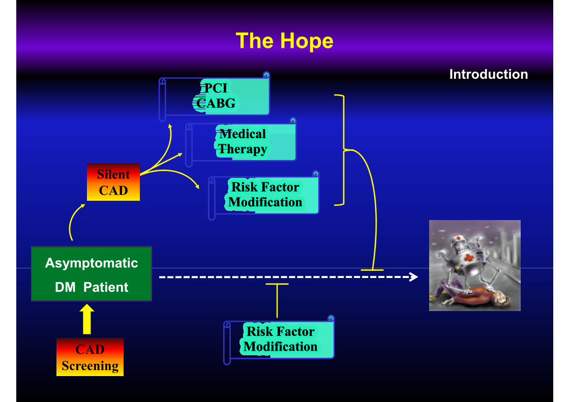
The Hope
IntroductionPCIPCICABGCABG
Medical Medical
Asymptomatic
Medical Medical TherapyTherapy
Risk FactorRisk FactorModificationModification
Silent
CAD
Asymptomatic
DM Patient
Risk FactorRisk FactorModificationModificationCAD
Screening
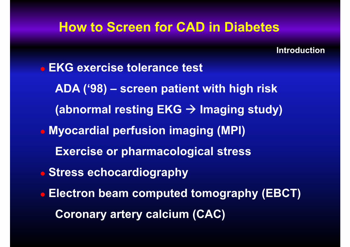
How to Screen for CAD in Diabetes
Introduction
� EKG exercise tolerance test
ADA (‘98) – screen patient with high risk ADA (‘98) – screen patient with high risk
(abnormal resting EKG � Imaging study)
� Myocardial perfusion imaging (MPI)
Exercise or pharmacological stress
� Stress echocardiography
� Electron beam computed tomography (EBCT)
Coronary artery calcium (CAC)
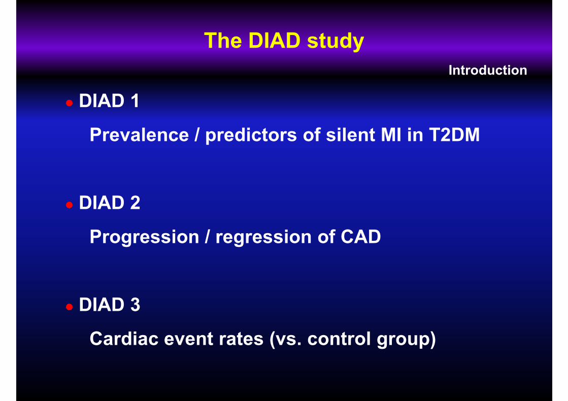
The DIAD study
� DIAD 1
Prevalence / predictors of silent MI in T2DM
Introduction
Prevalence / predictors of silent MI in T2DM
� DIAD 2
Progression / regression of CAD
� DIAD 3
Cardiac event rates (vs. control group)
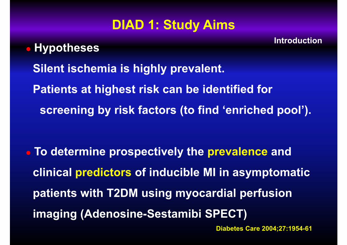
DIAD 1: Study AimsIntroduction
� Hypotheses
Silent ischemia is highly prevalent.
Patients at highest risk can be identified for
screening by risk factors (to find ‘enriched pool’).
� To determine prospectively the prevalence and
clinical predictors of inducible MI in asymptomatic
patients with T2DM using myocardial perfusion
imaging (Adenosine-Sestamibi SPECT)Diabetes Care 2004;27:1954-61
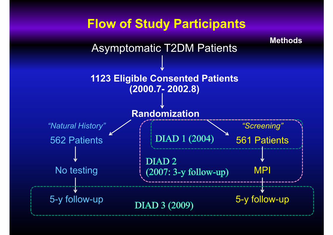
Flow of Study Participants
Asymptomatic T2DM Patients
1123 Eligible Consented Patients
Methods
1123 Eligible Consented Patients (2000.7- 2002.8)
Randomization
562 Patients 561 Patients
“Natural History” “Screening”
MPINo testing
5-y follow-up 5-y follow-up
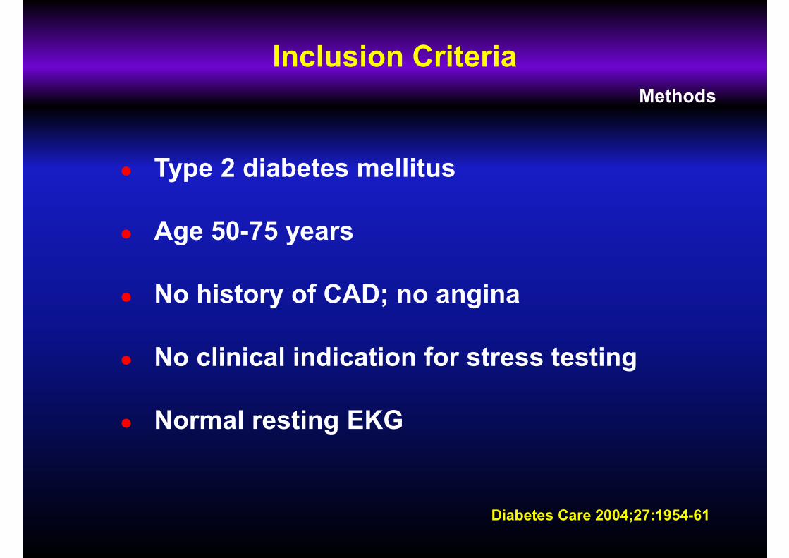
Inclusion Criteria
� Type 2 diabetes mellitus
Methods
� Age 50-75 years
� No history of CAD; no angina
� No clinical indication for stress testing
� Normal resting EKG
Diabetes Care 2004;27:1954-61
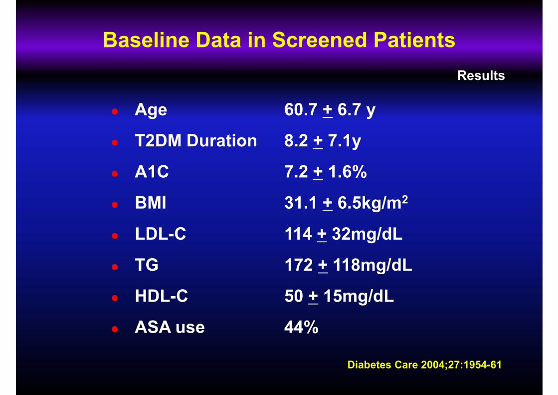
Baseline Data in Screened Patients
� Age
T2DM Duration
60.7 + 6.7 y
8.2 + 7.1y
Results
� T2DM Duration
� A1C
� BMI
� LDL-C
� TG
8.2 + 7.1y
7.2 + 1.6%
31.1 + 6.5kg/m2
114 + 32mg/dL
172 + 118mg/dL� TG
� HDL-C
� ASA use
172 + 118mg/dL
50 + 15mg/dL
44%
Diabetes Care 2004;27:1954-61
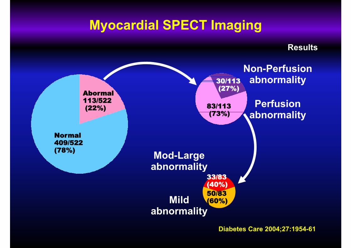
Myocardial SPECT Imaging
30/113
Non-Perfusion abnormality
Results
Normal409/522 (78%)
Abormal113/522(22%)
30/113(27%)
83/113(73%)
Perfusion abnormality
abnormality
Mod-Largeabnormality
50/83(60%)
33/83(40%)
abnormality
Mild abnormality
Diabetes Care 2004;27:1954-61
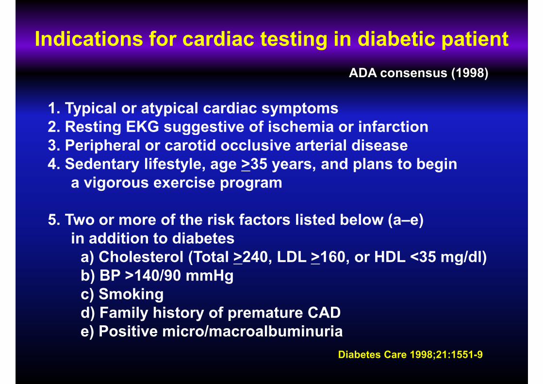
Indications for cardiac testing in diabetic patient
1. Typical or atypical cardiac symptoms
2. Resting EKG suggestive of ischemia or infarction
ADA consensus (1998)
2. Resting EKG suggestive of ischemia or infarction
3. Peripheral or carotid occlusive arterial disease
4. Sedentary lifestyle, age >35 years, and plans to begin
a vigorous exercise program
5. Two or more of the risk factors listed below (a–e)
in addition to diabetes
a) Cholesterol (Total >240, LDL >160, or HDL <35 mg/dl)
Diabetes Care 1998;21:1551-9
a) Cholesterol (Total >240, LDL >160, or HDL <35 mg/dl)
b) BP >140/90 mmHg
c) Smoking
d) Family history of premature CAD
e) Positive micro/macroalbuminuria
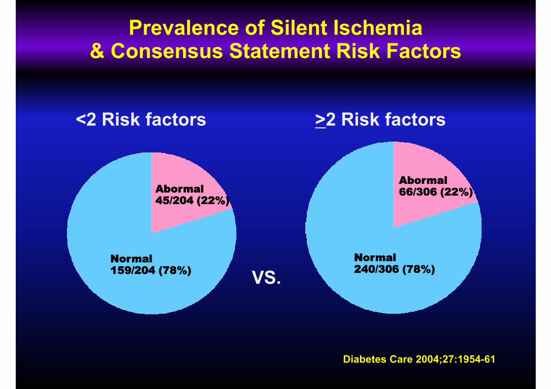
Prevalence of Silent Ischemia & Consensus Statement Risk Factors
<2 Risk factors >2 Risk factors
Normal
Abormal45/204 (22%)
Normal
Abormal66/306 (22%)
Normal159/204 (78%)
Normal240/306 (78%)
VS.
Diabetes Care 2004;27:1954-61
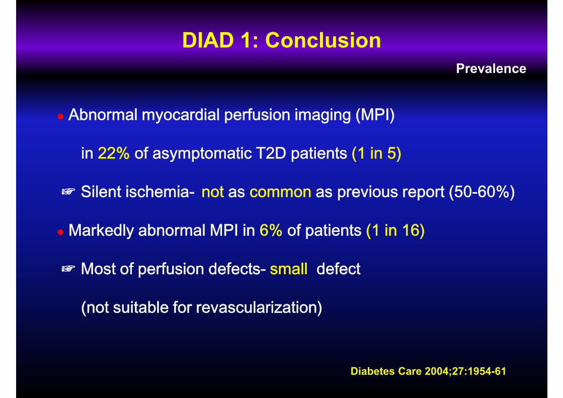
DIAD 1: Conclusion
Prevalence
� Abnormal myocardial perfusion imaging (MPI)
in 22% of asymptomatic T2D patients (1 in 5)
☞ Silent ischemia- not as common as previous report (50-60%)
� Markedly abnormal MPI in 6% of patients (1 in 16)
☞ Most of perfusion defects- small defect
Diabetes Care 2004;27:1954-61
☞ Most of perfusion defects- small defect
(not suitable for revascularization)
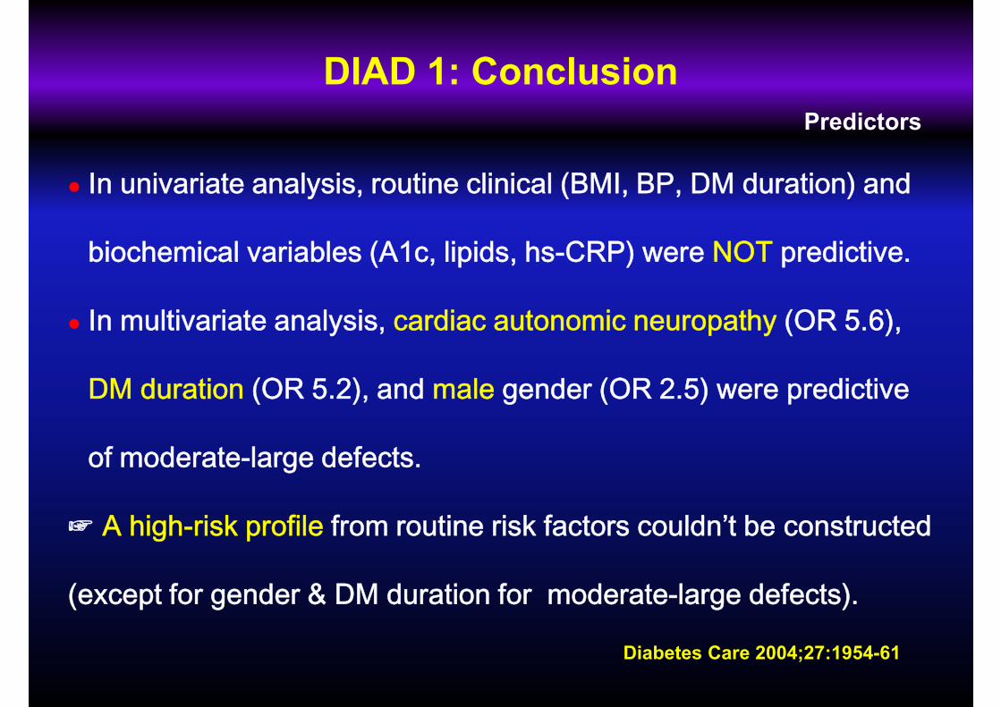
DIAD 1: Conclusion
Predictors
� In univariate analysis, routine clinical (BMI, BP, DM duration) and
biochemical variables (A1c, lipids, hs-CRP) were NOT predictive.biochemical variables (A1c, lipids, hs-CRP) were NOT predictive.
� In multivariate analysis, cardiac autonomic neuropathy (OR 5.6),
DM duration (OR 5.2), and male gender (OR 2.5) were predictive
of moderate-large defects.
Diabetes Care 2004;27:1954-61
of moderate-large defects.
☞ A high-risk profile from routine risk factors couldn’t be constructed
(except for gender & DM duration for moderate-large defects).
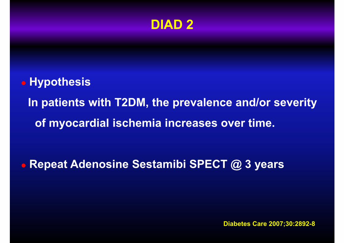
DIAD 2
� Hypothesis� Hypothesis
In patients with T2DM, the prevalence and/or severity
of myocardial ischemia increases over time.
� Repeat Adenosine Sestamibi SPECT @ 3 years� Repeat Adenosine Sestamibi SPECT @ 3 years
Diabetes Care 2007;30:2892-8
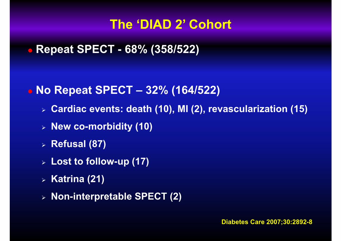
The ‘DIAD 2’ Cohort
� Repeat SPECT - 68% (358/522)
� No Repeat SPECT – 32% (164/522)
� Cardiac events: death (10), MI (2), revascularization (15)
� New co-morbidity (10)
� Refusal (87)
� Lost to follow-up (17)� Lost to follow-up (17)
� Katrina (21)
� Non-interpretable SPECT (2)
Diabetes Care 2007;30:2892-8
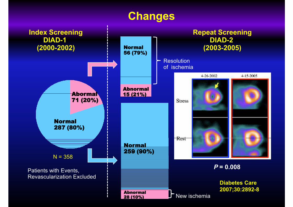
Normal56 (79%)
Index ScreeningDIAD-1
(2000-2002)
Repeat ScreeningDIAD-2
(2003-2005)
Resolution of ischemia
Changes
Normal287 (80%)
Abormal71 (20%)
Normal315 (88%)
Abormal43 (12%)
Abnormal15 (21%)
of ischemia
Abnormal28 (10%)
Normal259 (90%)
Patients with Events,Revascularization Excluded
New ischemia
N = 358 N = 358
P = 0.008
Diabetes Care
2007;30:2892-8
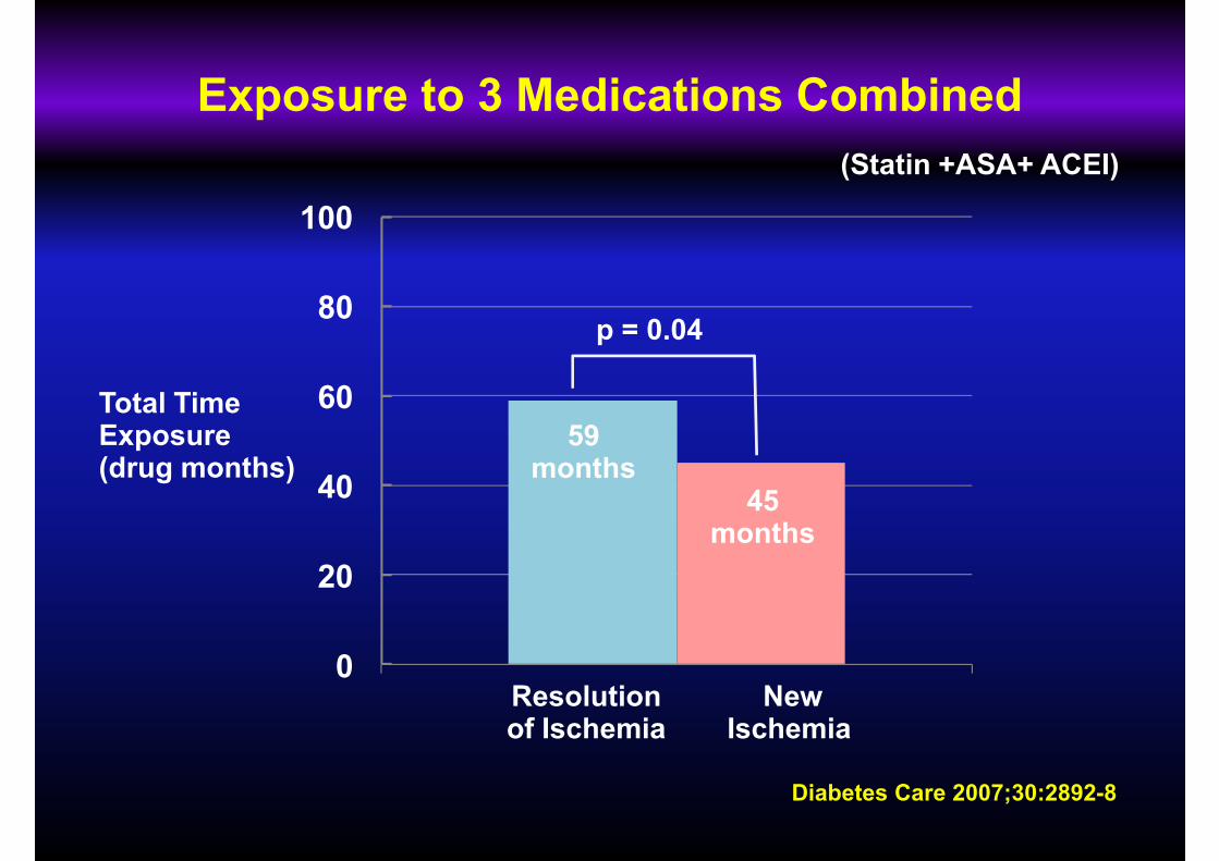
Exposure to 3 Medications Combined
(Statin +ASA+ ACEI)
100
20
40
60
80
Total Time Exposure(drug months)
p = 0.04
59months
45months
Diabetes Care 2007;30:2892-8
0
20
Resolution of Ischemia
NewIschemia
Lessons from DIAD 2
In the context of modern medical therapy,
myocardial perfusion defects can and often do
resolve in patients with T2DM.
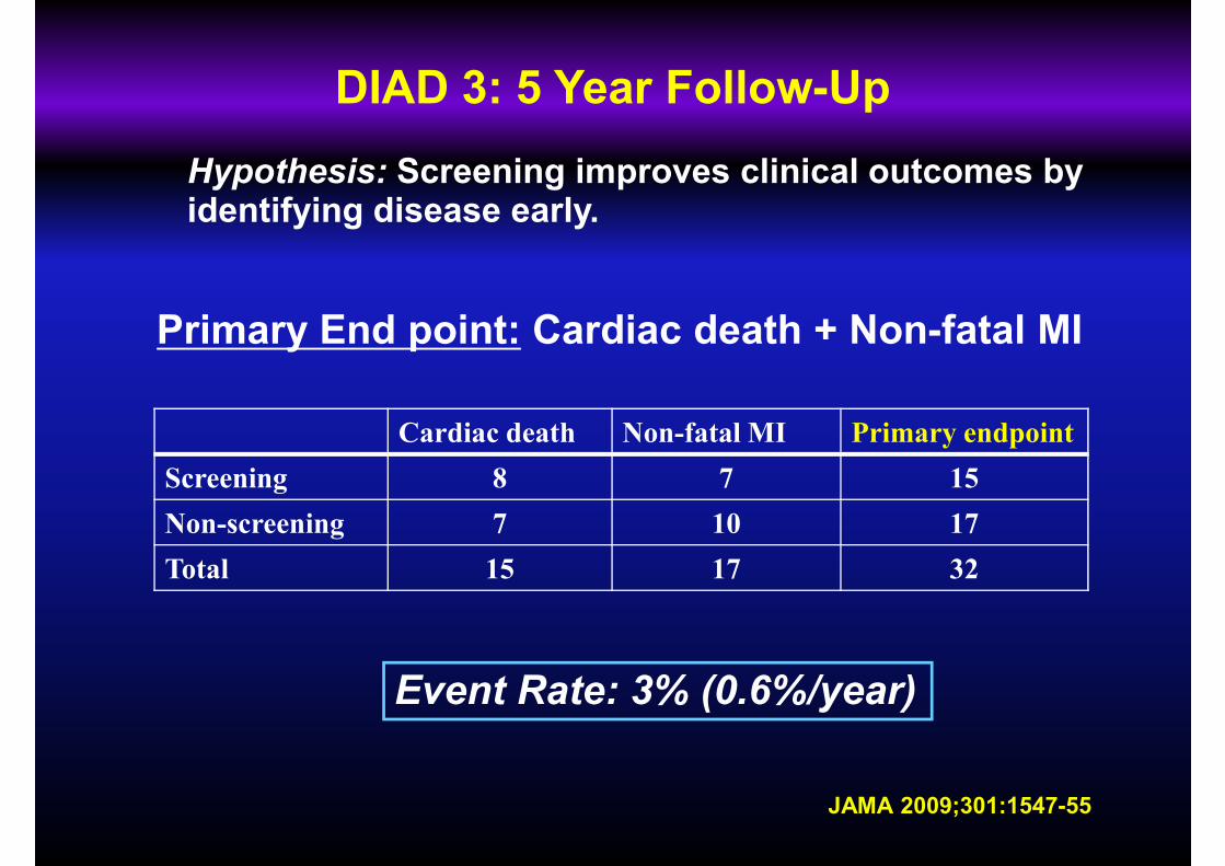
DIAD 3: 5 Year Follow-Up
Hypothesis: Screening improves clinical outcomes by identifying disease early.
Cardiac death Non-fatal MI Primary endpoint
Screening 8 7 15
Non-screening 7 10 17
Total 15 17 32
Primary End point: Cardiac death + Non-fatal MI
JAMA 2009;301:1547-55
Total 15 17 32
Event Rate: 3% (0.6%/year)
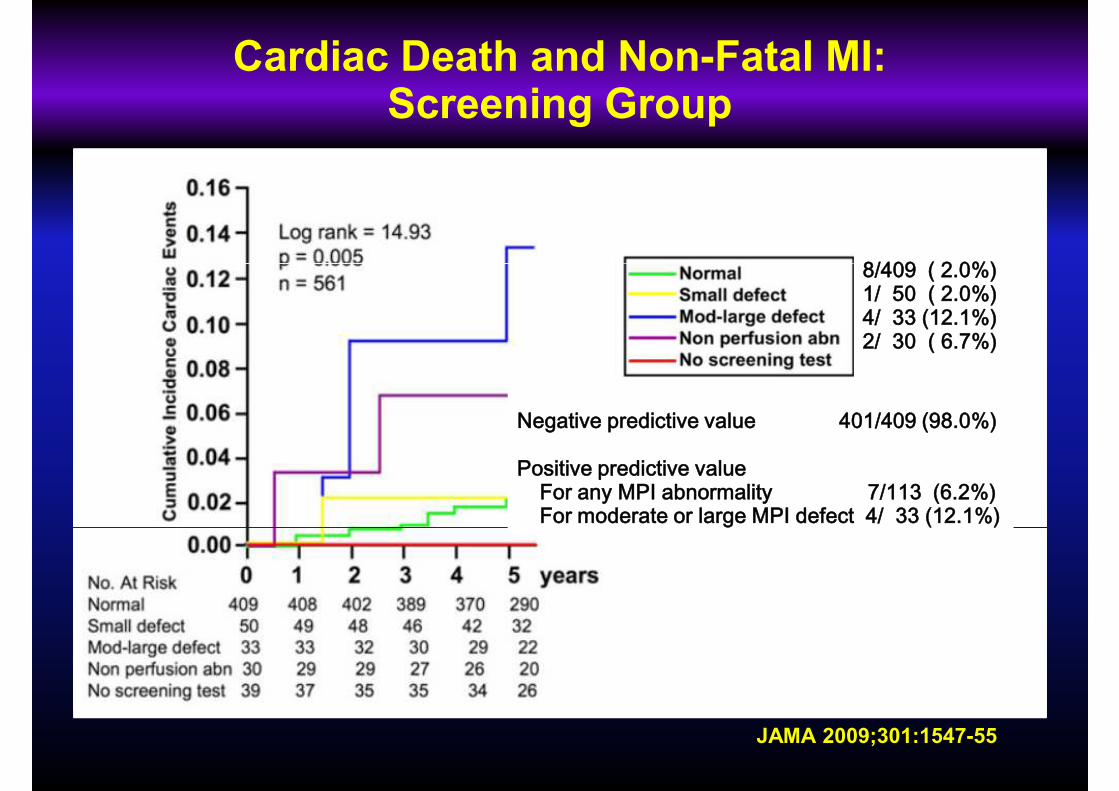
Cardiac Death and Non-Fatal MI:Screening Group
8/409 ( 2.0%)8/409 ( 2.0%)1/ 50 ( 2.0%)4/ 33 (12.1%)2/ 30 ( 6.7%)
Negative predictive value 401/409 (98.0%)
Positive predictive value For any MPI abnormality 7/113 (6.2%)For moderate or large MPI defect 4/ 33 (12.1%)
JAMA 2009;301:1547-55
For moderate or large MPI defect 4/ 33 (12.1%)
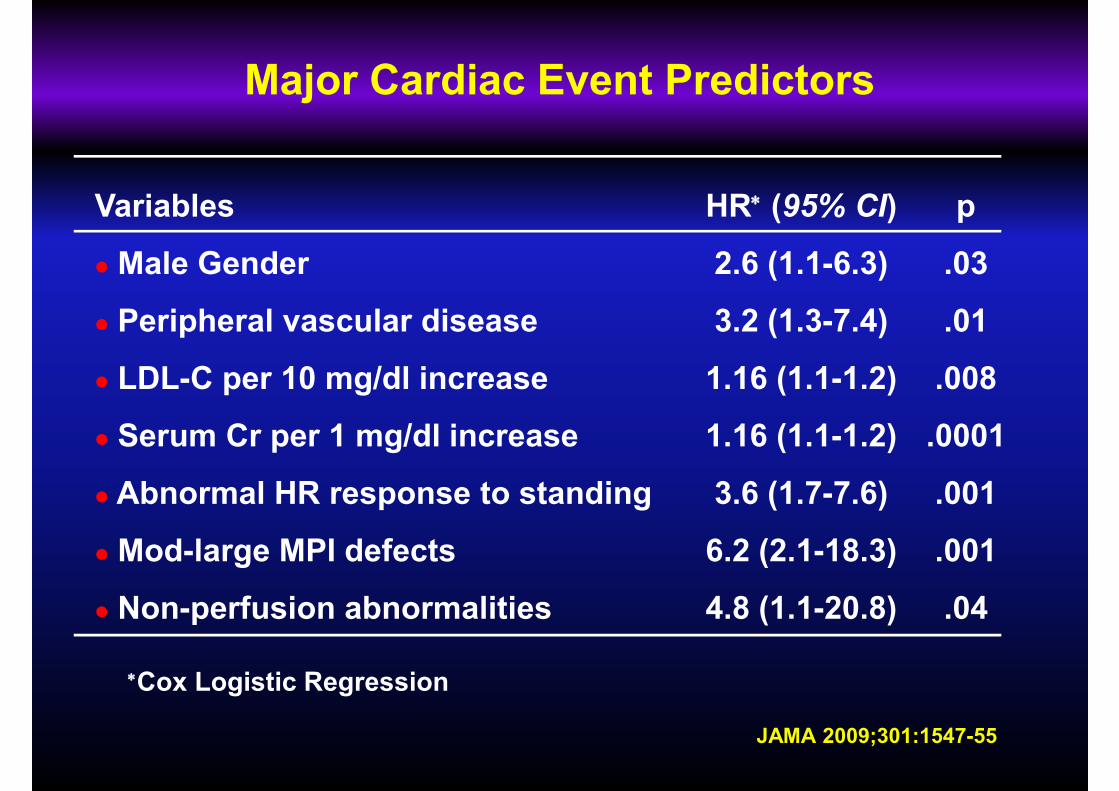
Major Cardiac Event Predictors
Variables
� Male Gender
HR∗ (95% CI)
2.6 (1.1-6.3)
p
.03� Male Gender
� Peripheral vascular disease
� LDL-C per 10 mg/dl increase
� Serum Cr per 1 mg/dl increase
� Abnormal HR response to standing
2.6 (1.1-6.3)
3.2 (1.3-7.4)
1.16 (1.1-1.2)
1.16 (1.1-1.2)
3.6 (1.7-7.6)
.03
.01
.008
.0001
.001
JAMA 2009;301:1547-55
� Mod-large MPI defects
� Non-perfusion abnormalities
6.2 (2.1-18.3)
4.8 (1.1-20.8)
.001
.04
∗Cox Logistic Regression
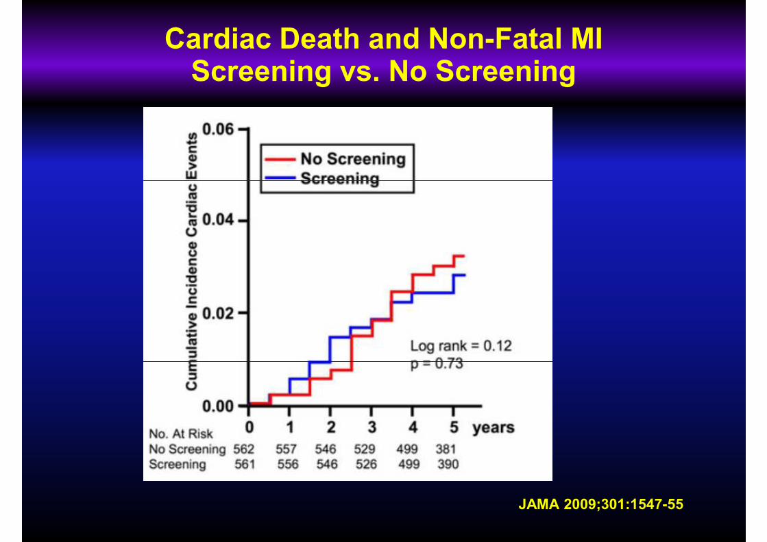
Cardiac Death and Non-Fatal MIScreening vs. No Screening
JAMA 2009;301:1547-55
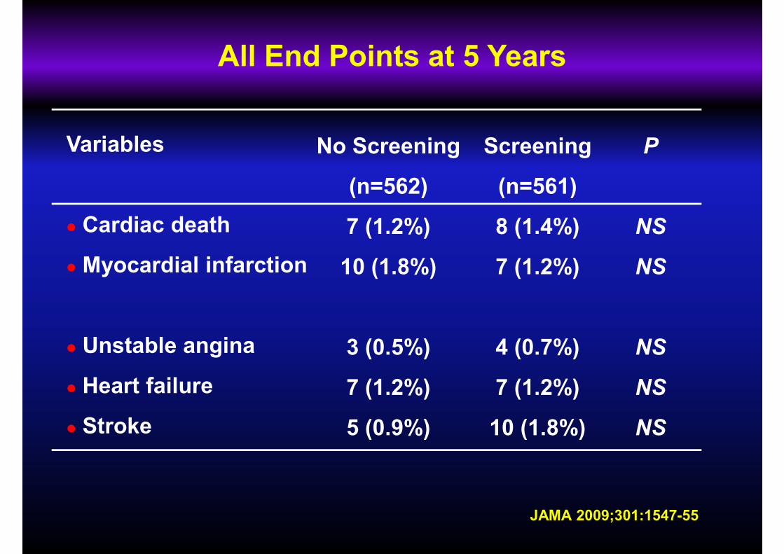
All End Points at 5 Years
Variables Screening
(n=561)
No Screening
(n=562)
P
� Cardiac death
� Myocardial infarction
� Unstable angina
(n=561)
8 (1.4%)
7 (1.2%)
4 (0.7%)
(n=562)
7 (1.2%)
10 (1.8%)
3 (0.5%)
NS
NS
NS
JAMA 2009;301:1547-55
� Heart failure
� Stroke
7 (1.2%)
10 (1.8%)
7 (1.2%)
5 (0.9%)
NS
NS
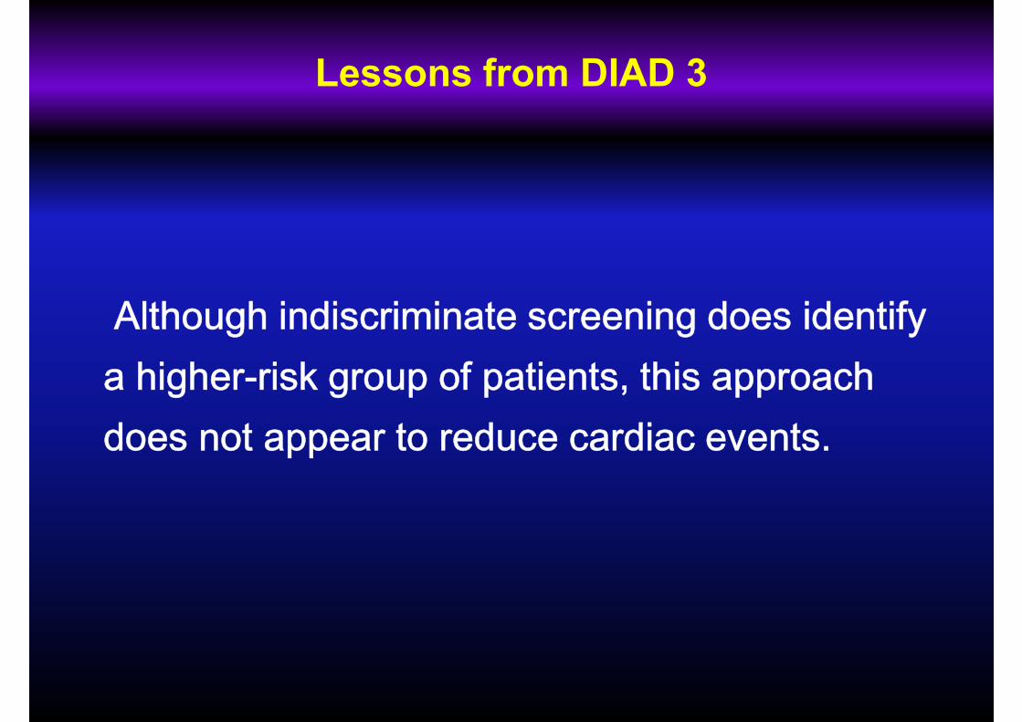
Lessons from DIAD 3
Although indiscriminate screening does identify a higher-risk group of patients, this approach does not appear to reduce cardiac events.
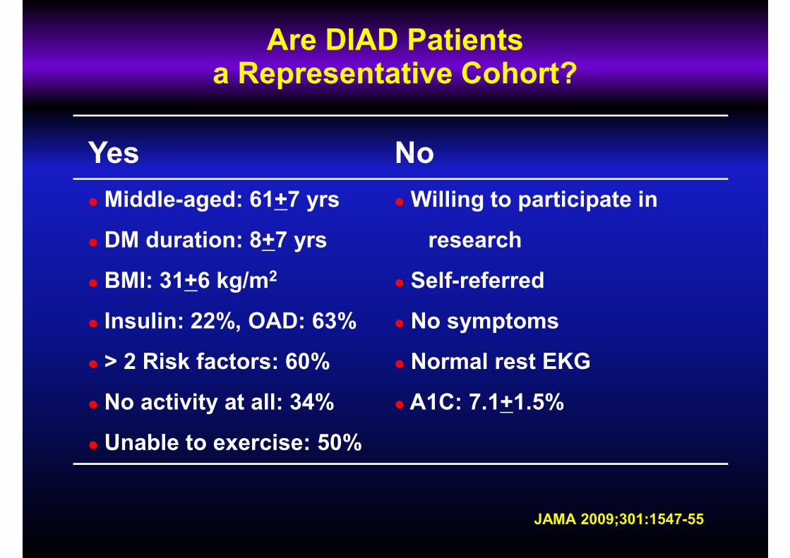
Are DIAD Patients a Representative Cohort?
Yes No
� Middle-aged: 61+7 yrs
� DM duration: 8+7 yrs
� BMI: 31+6 kg/m2
� Insulin: 22%, OAD: 63%
� > 2 Risk factors: 60%
� Willing to participate in
research
� Self-referred
� No symptoms
� Normal rest EKG
JAMA 2009;301:1547-55
> 2 Risk factors: 60%
� No activity at all: 34%
� Unable to exercise: 50%
Normal rest EKG
� A1C: 7.1+1.5%
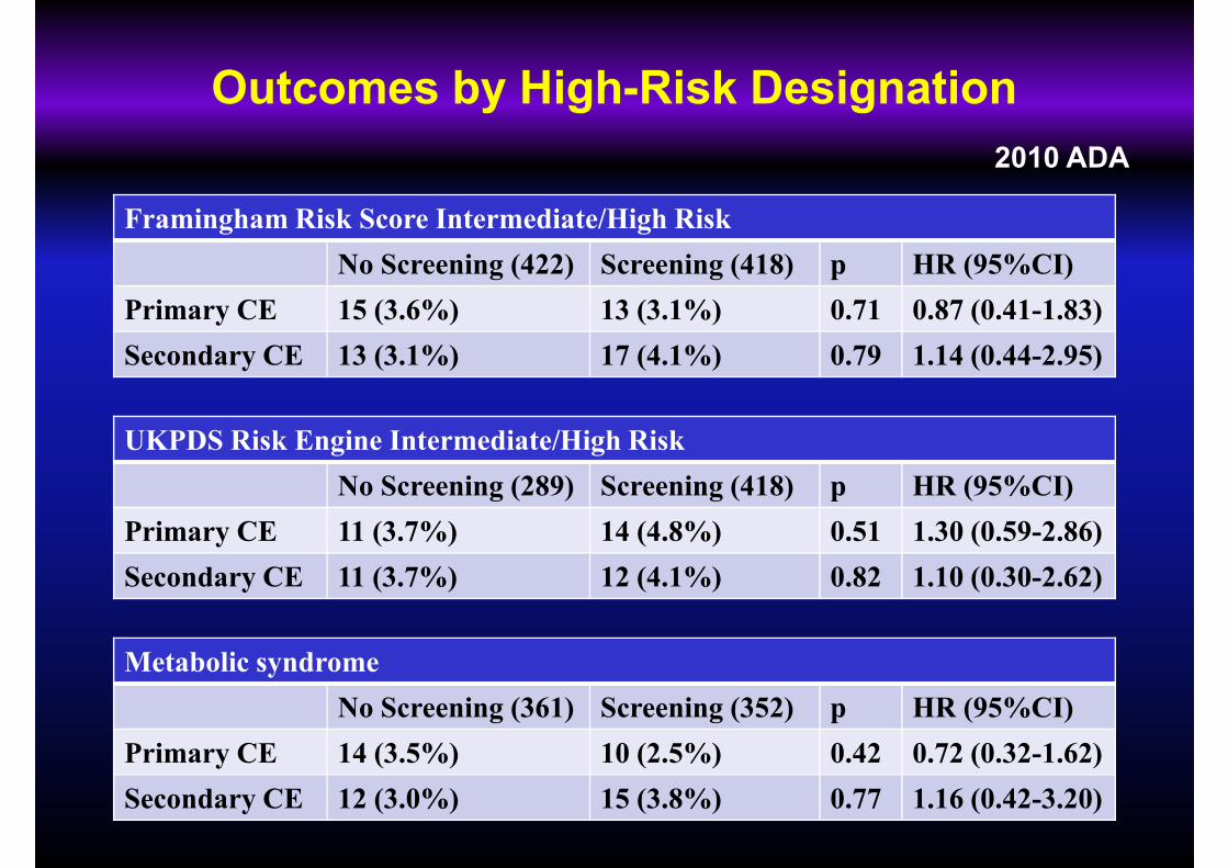
Outcomes by High-Risk Designation
2010 ADA
Framingham Risk Score Intermediate/High Risk
No Screening (422) Screening (418) p HR (95%CI)
Primary CE 15 (3.6%) 13 (3.1%) 0.71 0.87 (0.41-1.83)
Secondary CE 13 (3.1%) 17 (4.1%) 0.79 1.14 (0.44-2.95)
UKPDS Risk Engine Intermediate/High Risk
No Screening (289) Screening (418) p HR (95%CI)
Primary CE 11 (3.7%) 14 (4.8%) 0.51 1.30 (0.59-2.86)
Secondary CE 11 (3.7%) 12 (4.1%) 0.82 1.10 (0.30-2.62)Secondary CE 11 (3.7%) 12 (4.1%) 0.82 1.10 (0.30-2.62)
Metabolic syndrome
No Screening (361) Screening (352) p HR (95%CI)
Primary CE 14 (3.5%) 10 (2.5%) 0.42 0.72 (0.32-1.62)
Secondary CE 12 (3.0%) 15 (3.8%) 0.77 1.16 (0.42-3.20)
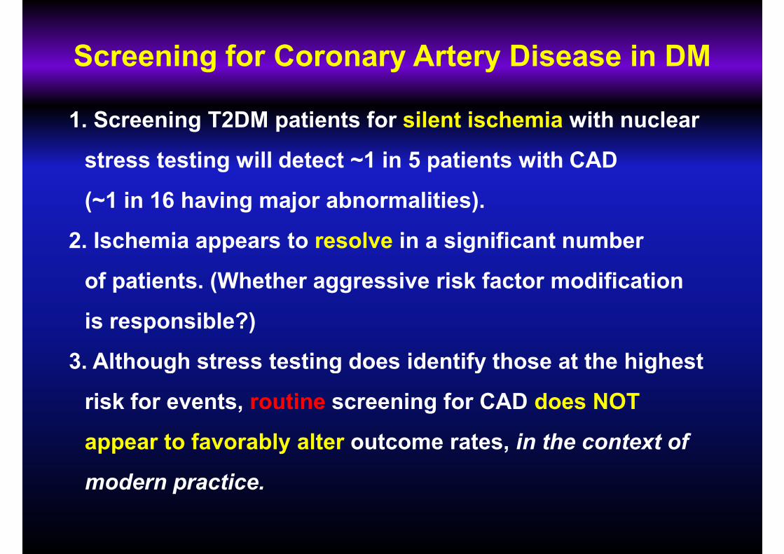
Screening for Coronary Artery Disease in DM
1. Screening T2DM patients for silent ischemia with nuclear
stress testing will detect ~1 in 5 patients with CAD
(~1 in 16 having major abnormalities).
2. Ischemia appears to resolve in a significant number
of patients. (Whether aggressive risk factor modification
is responsible?)
3. Although stress testing does identify those at the highest3. Although stress testing does identify those at the highest
risk for events, routine screening for CAD does NOT
appear to favorably alter outcome rates, in the context of
modern practice.
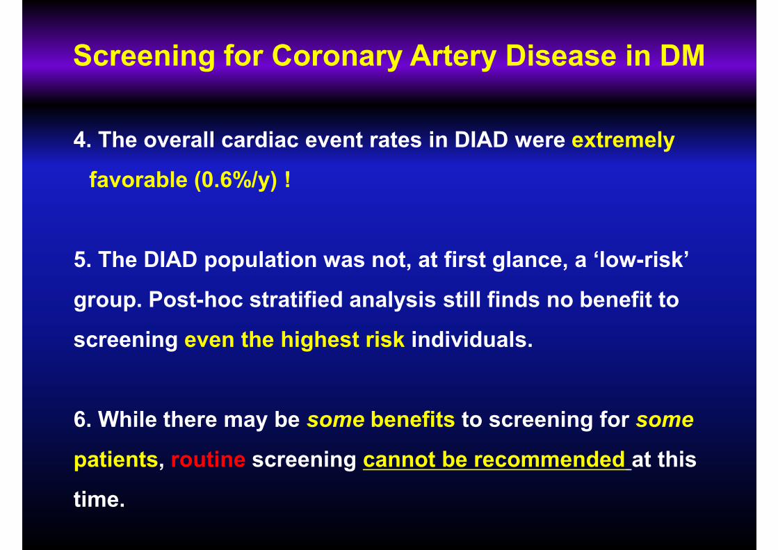
Screening for Coronary Artery Disease in DM
4. The overall cardiac event rates in DIAD were extremely
favorable (0.6%/y) !favorable (0.6%/y) !
5. The DIAD population was not, at first glance, a ‘low-risk’
group. Post-hoc stratified analysis still finds no benefit to
screening even the highest risk individuals.
6. While there may be some benefits to screening for some
patients, routine screening cannot be recommended at this
time.
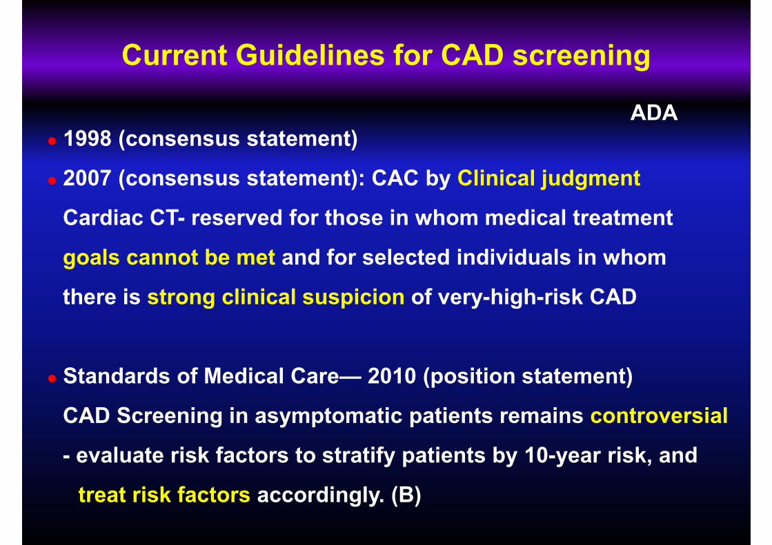
Current Guidelines for CAD screening
� 1998 (consensus statement)
� 2007 (consensus statement): CAC by Clinical judgment
ADA
� 2007 (consensus statement): CAC by Clinical judgment
Cardiac CT- reserved for those in whom medical treatment
goals cannot be met and for selected individuals in whom
there is strong clinical suspicion of very-high-risk CAD
Standards of Medical Care— 2010 (position statement)� Standards of Medical Care— 2010 (position statement)
CAD Screening in asymptomatic patients remains controversial
- evaluate risk factors to stratify patients by 10-year risk, and
treat risk factors accordingly. (B)
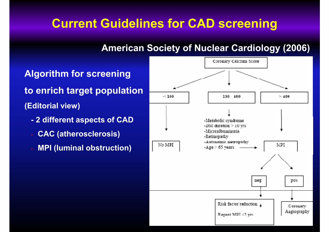
Current Guidelines for CAD screening
Algorithm for screening
American Society of Nuclear Cardiology (2006)
Algorithm for screening
to enrich target population
(Editorial view)
- 2 different aspects of CAD
• CAC (atherosclerosis)
• MPI (luminal obstruction)
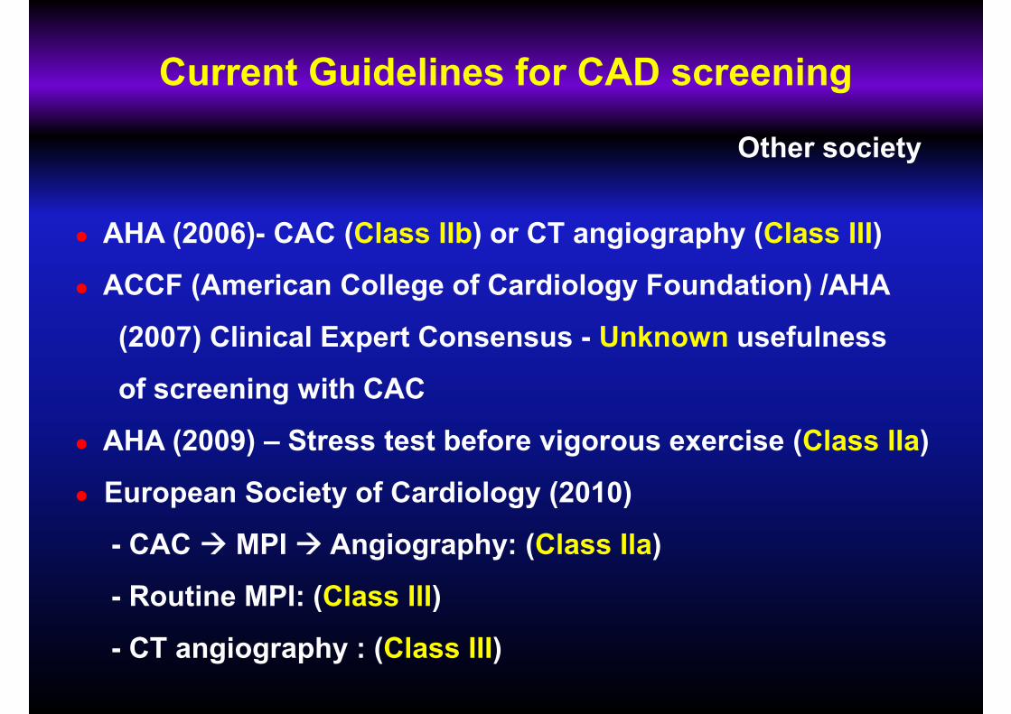
Current Guidelines for CAD screening
� AHA (2006)- CAC (Class IIb) or CT angiography (Class III)
Other society
� AHA (2006)- CAC (Class IIb) or CT angiography (Class III)
� ACCF (American College of Cardiology Foundation) /AHA
(2007) Clinical Expert Consensus - Unknown usefulness
of screening with CAC
� AHA (2009) – Stress test before vigorous exercise (Class IIa)
European Society of Cardiology (2010)� European Society of Cardiology (2010)
- CAC � MPI � Angiography: (Class Ila)
- Routine MPI: (Class IlI)
- CT angiography : (Class III)
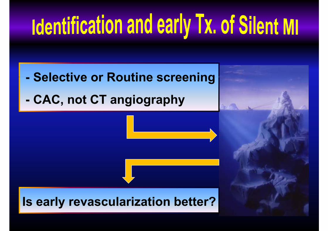
- Selective or Routine screening- Selective or Routine screening
- CAC, not CT angiography
Is early revascularization better?