Screening for CAD: What Test to Order for Which Situation John L. Tan, MD, PhD Presbyterian Hospital...
54
Screening for CAD: What Test to Order for Which Situation John L. Tan, MD, PhD Presbyterian Hospital of Dallas
-
Upload
gregory-lucas -
Category
Documents
-
view
217 -
download
4
Transcript of Screening for CAD: What Test to Order for Which Situation John L. Tan, MD, PhD Presbyterian Hospital...

Screening for CAD: What Test to Order for Which
Situation
John L. Tan, MD, PhD
Presbyterian Hospital of Dallas

Estimated Annual Incidence of CV Disease
Cardiovascular Diseases70 million
Stroke0.5 million
Stroke Deaths150,000
Silent Ischemia? 3 million
AMI Deaths500,000
Chest Pain6 million
Not Admitted2 millionHeart Attack
1.5 million
Unstable Angina1 million Wrongful Discharge
30,000

Available Tests
• Stress ECG
• Stress Imaging Study
• Ultra-fast CT (EBCT)
• CT Angiography
• Stress Cardiac MRI/MRA
• Coronary Angiography

Initial Considerations
• Symptomatic versus Asymptomatic
• Diagnosis versus Prognosis
• Assessment of Risk for CV mortality

Patients with Symptoms

Clinical Classification of Chest Pain
Typical Angina (definite)(1) Substernal chest discomfort with a characteristic quality and duration that is (2) provoked by exertion or emotional stress and (3) relieved by rest or nitroglycerin
Atypical Angina (probable)Meets 2 of the above characteristics
Noncardiac Chest PainMeets one or none of the typical angina characteristics
ACC/AHA ACP-ASIM Guidelines for Chronic Stable Angina, 1999
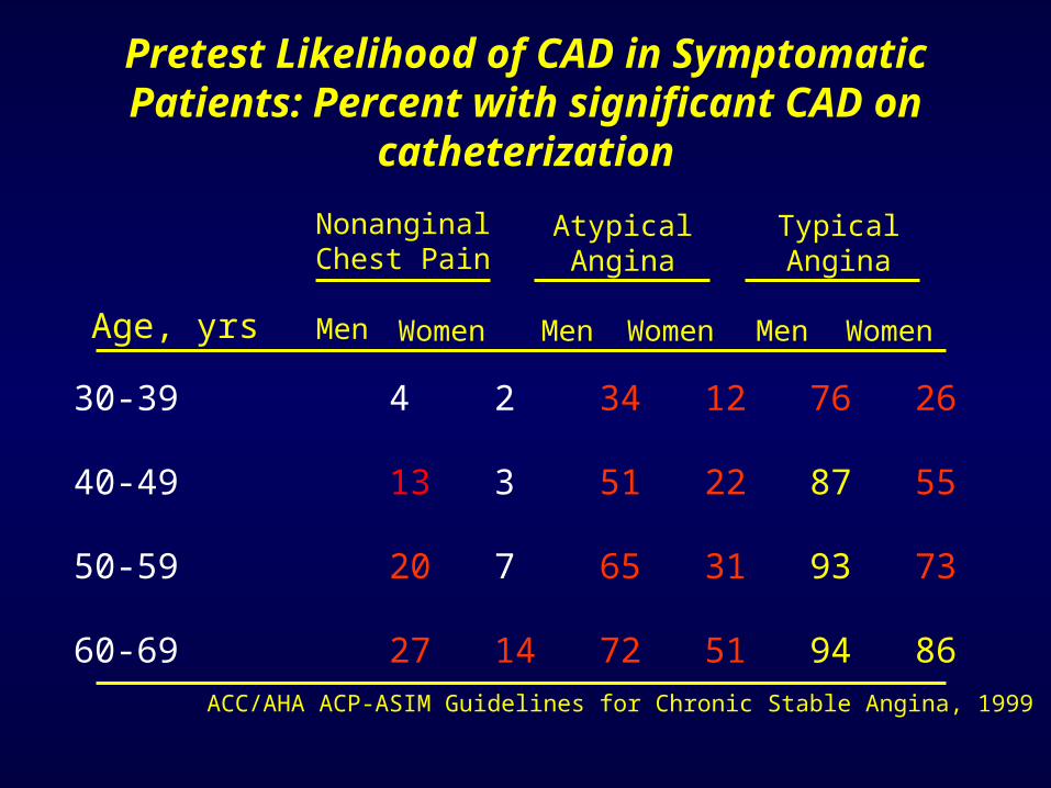
Pretest Likelihood of CAD in Symptomatic Patients: Percent with significant CAD on catheterization
30-39 4 2 34 12 76 26
40-49 13 3 51 22 87 55
50-59 20 7 65 31 93 73
60-69 27 14 72 51 94 86
Age, yrs Men MenWomen Men Women Women
NonanginalChest Pain
AtypicalAngina
TypicalAngina
ACC/AHA ACP-ASIM Guidelines for Chronic Stable Angina, 1999

Kaplan-Meier Survival in Risk Stratified Patients
Shaw, et al, AJC, 2000

Contraindications to stress testing?
Symptoms or clinical findings warranting angiography?
Patient able to exercise?
Resting ECG interpretable?
Pharmacologic imaging study
Previous coronary revascularization?
Perform exercise test
Exercise imaging study
Consider coronary angiography
Diagnosis and Risk Stratification of Patients with Chest Pain
No
No
No
No
Yes
Yes
Yes
Yes
Yes
No
ACC/AHA ACP-ASIM Guidelines for Chronic Stable Angina, 1999

Exercise Testing

Indications for Stress Testing without an Imaging Modality
1. Patients with an intermediate probability of CAD, including those with RBBB or <1 mm resting ST-segment changes (Class I)
2. Patients with suspected vasospastic angina (Class IIa)
3. Patients with a high or low probability of CAD (Class IIb)
4. Annual TMT in asymptomatic patients with estimated annual mortality rate >1%ACC/AHA ACP-ASIM Guidelines for Chronic Stable Angina, 1999

Four-year Mortality Rates with Abnormal ETT: Effects of Severity of CAD
5.5
9.5
13.5
0
2
4
6
8
10
12
14 1-Vessel2-Vessel3-Vessel
4-ye
ar M
orta
lity
Rat
es (
%)
Weiner, et al, JACC, 1984

Four-year Mortality Rates with Abnormal ETT: Effects of Exercise Capacity
0
18 20
47
0
5
1015
20
25
3035
40
45
50STAGE 5STAGE 2-4STAGE 1STAGE <1
4-ye
ar M
orta
lity
Rat
es (
%)
Weiner, et al, JACC, 1984

Clinically Useful Bench Marks of Exercise Capacity
1 MET Basal activity level (3.5 ml O2 comsumed/Kg/min
< 5 METs Associated with a poor prognosis in patients <65 y/o
5 METs Marks the limit of ADLs, usual limit immediate post MI
10 METs Considered average level of fitness
In patients with angina, no mortality benefit CABG vs medical Rx
13 METs Good prognosis in spite of any abnormal exercise test response
18 METs Aerobic master athelete
22 METs Achieved by well-trained competitive atheletes

Exercise Parameters Associated with Advanced CAD or Poor Prognosis
1. Duration of ETT <6.5 METS (<5 METS for women)
2. Exercise HR <120 bpm off -blockers
3. Ischemic ST segment change at HR <120 bpm or <6.5 METS
4. ST segment depression >2 mm, especially in multiple leads
5. ST segment depression for >6 min in recovery
6. Decrease in BP during exercise

Survival According to Risk Groups Based on Duke TM Scores
Low (5 or greater) 62 0.99 0.25
Moderate (-10 to 4) 34 0.95 1.25
High (-10 or less) 4 0.79 5.0
Risk Group, Score % of Total Survival Mortality, %
Duke TM Score = Exercise time - (5 x ST deviation) - (4 x Treadmill angina)
ACC/AHA ACP-ASIM Guidelines for Chronic Stable Angina, 1999

Special Populations
• Elderly Persons(Age > 65 )
• Women

Exercise Testing of the Elderly
• Few elderly persons were included in studies validating the use of exercise testing (mean age in Duke Treadmill Score studies was 49 years old)
• The elderly have – greater prevalence and severity of disease– more co-morbid diseases
– increasingly sedentary lifestyle

Prognostic Value of Treadmill Exercise Testing in the Elderly
• Two variables are associated with cardiac events in the elderly
1. Angina with exercise
2. Workload achieved
• After workload was taken into account, neither abnormal ST-segment changes or exercise-induced angina was independently related to time to cardiac event
Ann Intern Med 132:862-870, June 2000

The Problem with Women . . .
• Almost half the women younger than 65 year old with
anginal symptoms in CASS had normal coronary
arteriograms
• More women with inability to exercise to maximumaerobic capacity

More Problems with Women . . .
• Exercise-induced ST-segment depression is less sensitive
in women than men due to lower prevalence of severe
CAD (22-42% of women vs 13-29% of men with
CAD have one-vessel disease)
• Exercise ECG may also be less specific (72 vs 79%, with
a PPVof 62 vs 85%)

. . .But it may not be that Bad

Probability of Significant Disease Across Duke TM Scores
Alexander, et al, JACC, 1998

Meta-analysis of Exercise Testing
Standard exercise test 147 68 77 73
Without MI 58 67 72 69
Without workup bias 3 50 90 69
With ST depression 22 69 70 69
Without ST depression 3 67 84 75
With digoxin 15 68 74 71
Without digoxin 9 72 69 70
With LVH 15 68 69 68
Without LVH 10 72 77 74
Overall ~70 ~80
Number of Sensitivity Specificity Predictive Grouping Studies (%) (%) Accuracy (%)
ACC/AHA Guidelines for Exercise Testing, 1997

The “Ischemic Ladder”M
VO
2
DiastolicDysfunction
SystolicDysfunction
ECGChanges
Angina
Time

Stress Imaging

Stress Imaging Studies
• Exercise• Dobutamine• Adenosine• (Persantine)
• Echocardiography• Perfusion Imaging
– Nuclear Scan
– Thallium Scan
– Sestamibi Scan
– Hybrid Scan
• MRI
Stress Modalities Imaging Modalities

Indications for Stress Imaging for Diagnosis
1. Abnormal resting ECG
Wolff-Parkinson-White syndrome> 1mm resting ST-segment
depressionLBBBV-paced rhythm
2. Previous non-diagnostic TMT
3. Inability to perform TMT ACC/AHA ACP-ASIM Guidelines for Chronic Stable Angina, 1999

Indications for Stress Imaging for Diagnosis
4. Prior re-vascularization including percutaneous interventions or CABG
5. Increased likelihood of a false-positive TMTDigoxin useLeft ventricular hypertrophy
6. As the initial stress test in patients with a normal resting ECG
ACC/AHA ACP-ASIM Guidelines for Chronic Stable Angina, 1999
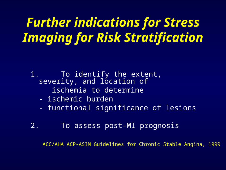
Further indications for Stress Imaging for Risk Stratification
1. To identify the extent, severity, and location of ischemia to determine
- ischemic burden- functional significance of lesions
2. To assess post-MI prognosis
ACC/AHA ACP-ASIM Guidelines for Chronic Stable Angina, 1999

Of Note
• Adenosine/dipyridamole perfusion imaging preferred inpatients able to exercise with a V-paced rhythm orunderlying LBBB (Class I vs IIb for stress echocardiography)
ACC/AHA ACP-ASIM Guidelines for Chronic Stable Angina, 1999

Comparing Stress Echo to Perfusion Imaging
Myocardial Perfusion Imaging
Normal Ischemic Fixed Total
Normal 137 10 7 154
Ischemic 4 47 3 54
Fixed 13 30 38 81
Total 154 87 48 289
Ec h
o car
d io g
r aph
y
137 + 47 + 38 = 222/289 77% AgreementSPECT vs Echo 87 vs 54 Ischemic regions 48 vs 81 Fixed regions Quinones and Zoghbi

Sensitivity and Specificity of Stress Studies
Procedure Sensitivity (%) Specificity (%)
Exercise Test 68 77
Stress Echo 76 88
SPECT 88 77

Advantages of Stress Echocardiography
1. Higher specificity
2. Versatility: more extensive evaluation ofcardiac anatomy and function
3. Greater convenience/efficacy/availability
4. Lower cost

Advantages of Stress Myocardial Perfusion Imaging
1. Higher technical success rate
2. Higher sensitivity, especially for one-vessel disease
3. Better accuracy in evaluating possible ischemia when multiple rest LV wall motion abnormalities are present
4. More extensive published database, especially in evaluation of prognosis

Prognostic Value of a Normal Perfusion Scan
Number Mean Annual of Patients Study Type follow-up mortality (%)
3594 Meta-analysis 29 months 0.9
473 Retrospective 30 +/- 16 months 0.2
5183 Prospective 642 +/- 226 day <0.5
8411 Prospective 2.5 +/- 1.5 years <0.4
In contrast, patients with an abnormal scan have a 5-7% annualized serious adverse event rate

Myocardial Perfusion Imaging
Normal Study

Myocardial Perfusion ImagingAbnormal Study post-CABG

Cardiac Imaging
Echo MRI

Testing in Symptomatic Patients
• Exercise Test
– Probable more than we do
• Stress Echocardiogram
– Lower pre-test probablility population– Valvular or other structural heart disease

Testing in Symptomatic Patients
• Stress Perfusion Scan– Higher pre-test probability population
• Cardiac MRI
– When above unhelpful and expertise is
available

Testing in Symptomatic Patients
• Ultra-fast CT (EBCT)
– No role in symptomatic patients
• CT Angiography– Will play larger role with ability to image
coronaries (Triple Rule Out)
• Coronary Angiography
– When stress testing is potentially dangerous

Patients without Symptoms

Estimated Annual Incidence of CV Disease
Cardiovascular Diseases70 million
Stroke0.5 million
Stroke Deaths150,000
Silent Ischemia? 3 million
AMI Deaths500,000
Chest Pain6 million
Not Admitted2 millionHeart Attack
1.5 million
Unstable Angina1 million Wrongful Discharge
30,000

The Framingham
Score for Risk
Prediction
Greenland and Gaziano, NEJM, 2003

Elevated hs-CRP as an Independent Risk Factor
Ridker et al, NEJM, 2004

Elevated hs-CRP as an Independent Risk Factor
Ridker et al, NEJM, 2004

Available Tests
• Stress ECG
• Stress Imaging Study
• Ultra-fast CT (EBCT)
• CT Angiography
• Stress Cardiac MRI/MRA
• Coronary Angiography

Coronary Calcium Scoring
Greenland and Gaziano, NEJM, 2003

Coronary Calcium Scoring
• Meta-analysis:– Sensitivity of 80-92%– Specificity of 40-51%
• High prevalence of unexpected, incidental noncardiac findings

Sensitivity and Specificity of CAD Studies
Procedure Sensitivity (%) Specificity (%)
Exercise Test 68 77
Stress Echo 76 88
SPECT 88 77
EBCT 80-90 40-50

Incremental Value of Non-
invasive Testing to Risk
Assessment
Low Risk <10%Interm Risk 10-20%High Risk >20%
Greenland and Gaziano, NEJM, 2003

Incremental Value of Coronary Calcium Scoring to Risk Assessment
Greenland et al, JAMA, 2004

Greenland and Gaziano, NEJM, 2003