Sclerotinia Diseases of Crop Plants: Biology, Ecology and ...978-1-4020-8408-9/1.pdf · Sclerotinia...
58
Sclerotinia Diseases of Crop Plants: Biology, Ecology and Disease Management
Transcript of Sclerotinia Diseases of Crop Plants: Biology, Ecology and ...978-1-4020-8408-9/1.pdf · Sclerotinia...

Sclerotinia Diseases of Crop Plants:
Biology, Ecology and Disease Management

G. S. Saharan • Naresh Mehta
Sclerotinia Diseases of Crop Plants: Biology, Ecology and Disease Management

Dr. G. S. Saharan Dr. Naresh Mehta
CCS Haryana Agricultural University CCS Haryana Agricultural University
Hisar, Haryana, India Hisar, Haryana, India
ISBN 978-1-4020-8407-2 e-ISBN 978-1-4020-8408-9
Library of Congress Control Number: 2008924858
© 2008 Springer Science+Business Media B.V.
No part of this work may be reproduced, stored in a retrieval system, or transmitted in any form or by any
means, electronic, mechanical, photocopying, microfilming, recording or otherwise, without written
permission from the Publisher, with the exception of any material supplied specifically for the purpose
of being entered and executed on a computer system, for exclusive use by the purchaser of the work.
Printed on acid-free paper
9 8 7 6 5 4 3 2 1
springer.com

Foreword
The fungus Sclerotinia has always been a fancy and interesting subject of research
both for the mycologists and pathologists. More than 250 species of the fungus
have been reported in different host plants all over the world that cause heavy
economic losses. It was a challenge to discover weak links in the disease cycle to
manage Sclerotinia diseases of large number of crops. For researchers and stu-
dents, it has been a matter of concern, how to access voluminous literature on
Sclerotinia scattered in different journals, reviews, proceedings of symposia,
workshops, books, abstracts etc. to get a comprehensive picture. With the publi-
cation of book on ‘Sclerotinia’, it has now become quite clear that now only three
species of Sclerotinia viz., S. sclerotiorum, S. minor and S. trifoliorum are valid.
The authors have made an excellent attempt to compile all the available informa-
tion on various aspects of the fungus Sclerotinia. The information generated so
far has been presented in different chapters. After introducing the subject various
aspects viz., the diseases, symptomatology, disease assessment, its distribution,
economic importance, the pathogen, its taxonomy, nomenclature, reproduction,
reproductive structures with fine details, variability, perpetuation, infection and
pathogenesis, biochemical, molecular and physiological aspects of host-pathogen
interaction, seed infection, disease cycle, epidemiology and forecasting, host
resistance with sources of resistance, mechanism of resistance and other manage-
ment strategies have been covered. The inclusion of numerous laboratory and
field techniques is additional quality of the book for researchers, teachers and
students. The chapters on Sclerotinia as myco-herbicide, phytotoxin, phytoalexins,
hypo-virulence, resistance to fungicides, volatile compounds of Sclerotinia,
sporigermin from sclerotia and Sclerotinia as health hazard problem will give a
futuristic insight to the book. Outlining of future research priorities and disease
management strategies speaks of the wisdom of the authors.
I congratulate Dr. G.S. Saharan, Ex Professor and Head, Department of Plant
Pathology and Dr. Naresh Mehta, Professor of Plant Pathology, CCS Haryana
Agricultural University, Hisar for their stupendous, incredible and splendor task
of bringing comprehensive treatise on Sclerotinia which will propel fraternity of
Agriculture to get bounty of knowledge at one edifice. I am sure this book will
v

be of immense help to the scientists, teachers, students, extension specialists
and all those who are interested in protecting the plant health from Sclerotinia
diseases.
October 2007 C.D. Mayee
Chairman
Agricultural Service Recruitment Board (ASRB)
Indian Council of Agricultural Research (ICAR)
Krishi Bhawan
New Delhi – 110 001
vi Foreword

Preface
Sclerotinia is one of the most devastating and cosmopolitan plant pathogen. More
than 60 names have been used to refer to diseases caused by this fungal pathogen.
The fungus infects more than 500 species of plants worldwide including important
field crops, fruit crops, ornamental plants, trees, shrubs and numerous weeds.
Annual yield losses due to Sclerotinia diseases exceed over several hundred million
dollars each year world over. Extensive crop damage, lack of high levels of host
resistance and the general difficulty of managing diseases caused by Sclerotinia
have been the impetus for sustainable research on this pathogen. Despite continued
study by phytopathologists and mycologists, the taxonomic delimitation and rela-
tionship of the plant pathogenic species of Sclerotinia have never been resolved
over the years, using traditional morphological and host preference characters.
The fungus Sclerotinia is belonging to phylum Ascomycota, class Discomycetes,
order Helotiales, family Sclerotiniaceae has been redefined to include only those
species that produce tuberoid sclerotia not incorporating host tissue within the
sclerotial medulla developing an apothecial ectal excipulum composed of globose
cells and not producing a disseminative conidial state. The taxonomy and nomen-
clature of 259 epithets previously referred to Sclerotinia have been reviewed with
21 placed in synonymy under the three accepted species and 25 included as imper-
fectly known. Two hundred and ten epithets have been excluded and either assigned
or accepted to other genera. S. homoeocarpa causing “Dollar spot” in turf grasses
now belonging to Lanzia sp. and Moellerodiscus sp. has been briefly covered as
reference for readers. Now recently, with the increased information available on
molecular biology, genetics, variability and epidemiology of these species and with
reexamination in the light of micro-anatomical and cultural characters employed
only three species, i.e., Sclerotinia sclerotiorum, S. minor and S. trifoliorum have
been retained in this genus.
The present monograph on Sclerotinia deals with the aspects on taxonomy,
nomenclature, geographical distribution, economic importance, host range, the
diseases caused, symptomatology, disease assessment, reproduction, ultra structures,
pathogenic variability, perpetuation, infection and pathogenesis, biochemical, molecular
and physiological aspects of host pathogen interaction, seed infection, disease cycle,
epidemiology and forecasting, host resistance and disease management strategies. In
addition, laboratory and field techniques developed so far for Sclerotinia have been
vii

included. Some newly emerging areas of Sclerotinia research which are likely to
have a bearing on its management are Sclerotinia as myco-herbicide, phytotoxin,
phytoalexin elicitors, hypovirulence, volatile compound imitator, sporigermin
from sclerotia, resistance to fungicides and Sclerotinia diseases as health hazard
problem have been discussed.
The subject matter is vividly illustrated with photographs (macroscopic, micro-
scopic, electron micrographs, scanning electron micrographs), drawings, figures,
histograms, graphs, tables and flow charts of techniques to make more interesting
stimulating, effective and easy to understand by the readers. Each chapter is
arranged in chronological order in the form of headings and sub-headings through
numerical series to make the subject contiguous. Inclusion of most of the important
references and websites will be helpful in original consultations by the Sclerotinia
researchers, teachers and students.
We are sure that this comprehensive treatise on Sclerotinia will be of immense
use to the scientists, teachers, students and all others in diagnosis and management
of Sclerotinia diseases of crops worldwide.
G. S. Saharan
Naresh Mehta
viii Preface

ix
Acknowledgements
Authors are indebted and highly grateful to the following persons/scientists/
publishers/societies/journals/institutes/websites and all others whose valuable
materials such as photographs (macroscopic, microscopic, electron micrographs,
scanning electron micrographs), drawings, figures, histograms, graphs, tables,
drawings and flow charts etc. have been used through reproduction in the present
document. Authors are thankful to all the scientists/persons/societies/publishers/
books/journals/institutes and websites etc. whose materials have been used in this
document but have not been acknowledged inadvertently. The address of the author/
source from where material adapted can be obtained from the reference which has been
cited in the reference section of the book.
A Persons/Scientists
Abd-Elrazik, A.A. Hart, I.P.
Adams, P.B. Hartill, W.F.T.
Agrios, G.N. Hawthorne, B.T.
Ayers, W.A. Hoes, J.A.
Boland, G.J. Holliday, P.
Bolton, M.D. Huang, H.C.
Bullock, S. Huang, R.C.
Caesar, A.J. Hughes, K.J.D.
Casale, W.L. Imolehin, E.D.
Casanova, S. Jain, J.P.
Cerkauskas, R.F. Jarvis, W.R.
Chandler, L. Jeferies, P.
Chen, C. Jones, D.
Dickman, M.B. Cummings, K.
Dillard, H.R. Kapil, R.
Dixon, G.R. Kapoor, A.S.
Doodson, J.K. Kemp, W.
Dorrell, D.G. Kerr, E.W.

x Acknowledgements
Ellis, M.B. Kerr, D.
Fravel, D.R. Kohn, L.M.
Gepp, V. Kokko, E.G.
Godika, S. Kora, C.
Dr. Greg, E. Lewis, J.A.
Grogan, R.G. Lorbeer, J.W.
Gutteridge, C.S. Lumsden, R.D.
Hall, R. Luth, P.
Hao, J.J. Spooner, B.M.
Martinez, A. Steadman, J.R.
Mc Donald, M.R. Subbarao, K.V.
Mc Quilken, M.P. Sugha, S.K.
McKenzie, D.L. Swanson, J.
Melzer, M.S. Tariq, V.N.
Millner, P.D. Tewari, J.P.
Mordue, J.E.M. Thomma, B.P.H.J.
Muralia, S. Tricot, D.
Nelson, B.D. Tripathi, N.N.
Nelson, L.A. Tu, J.C.
Pathak, A.K. Underhill, A.P.
Pearson, R.C. Verma, P.R.
Dr. Peter, L. Waller, J.M.
Rashid, K.Y. Weiss, A.
Rowe, D.E. Wharton, P.
Saito, I. Willetts, H.J.
Shukla, A.K. Williams, M.A.J.
Silvera, E. Wong, J.A.L.
Singh, H.B. Wu, B.M.
Singh, P. Young, C.S.
Singh, R.
Singh, S.
Smith, E.A.
B Publishers/Societies/Journals/Institutes/Websites
Academic Press, USA
Blackwell Publishing Co., UK
British Mycological Society, UK
British Society for Plant Pathology, UK
CABI, Commonwealth Agriculture Bureau International, UK
Cambridge University Press, UK
Canadian Journal of Botany, Canada
Canadian Phytopathological Society, Canada

CCS Haryana Agricultural University, Hisar-India
Crop Science
Crop Science Society of America
Department of Scientific and Industrial Research, New Zealand
Elsevier Publishing Co., USA
Euphytica
Hokkaido Central Agricultural Experiment Station, Naganuma, Hokkaido, Japan
Indian Phytopathological Society, India
Indian Society of Mycology and Plant Pathology, India
International Society for Plant Pathology
Journal of General Microbiology
Journal of Phytopathology, Germany
Kluwer Publishers, USA
Michigan State University, USA
Micron
Molecular Microbiology
Molecular Plant Pathology
Mycological Research
Mycotaxon
North Carolina State University, USA
New Zealand Journal of Agricultural Research
Penn Sylvia State University, USA
Phytopathology
Plant Diseases
Scientific Publishers, USA
Springer SBM, The Netherlands
Taylor and Francis Group, FL
The American Phytopathological Society, USA
The National Research Council of Canada, Canada
The Netherlands Study Circles of Plant Breeding
The Royal Society of New Zealand, New Zealand
The Society for General Microbiology
University of Georgia, USA
USDA – Agricultural Research Service, USA
www.broad.mit.edu/annotation/fungi/sclerotinia/sclerotiorum
www.caes.uga.edu
www.ces.ncsu.edu
www.potatodiseases.org
www.sciencedirect.com
www.turfgrassmanagement.psu.edu
www.whitemoldresearch.com
(Authors)
Acknowledgements xi

xiii
Contents
Foreword ...................................................................................................... v
Preface .......................................................................................................... vii
Acknowledgements ..................................................................................... ix
List of Tables ................................................................................................ xxv
List of Figures .............................................................................................. xxix
List of Plates ................................................................................................ xxxv
Color Plates .................................................................................................. xlvii
1 Introduction ........................................................................................... 1
2 Geographical Distribution .................................................................... 13
2.1 Distribution Map ............................................................................ 13
2.1.1 Sclerotinia sclerotiorum ................................................... 13
2.1.2 Sclerotinia minor .............................................................. 14
2.1.3 Sclerotinia trifoliorum ...................................................... 14
2.1.4 Sclerotinia fructigena ....................................................... 15
2.1.5 Sclerotinia laxa ................................................................ 15
2.1.6 Sclerotinia fructicola ........................................................ 15
2.1.7 Sclerotinia squamosa ....................................................... 16
2.1.8 Sclerotinia narcissicola .................................................... 16
2.1.9 Sclerotinia borealis .......................................................... 16
2.1.10 Sclerotinia fuckeliana ....................................................... 16
3 History and Host Range ....................................................................... 19
3.1 History............................................................................................ 19
3.2 Host Range ..................................................................................... 21
3.2.1 Sclerotinia sclerotiorum (Lib.) de Bary ........................... 21
3.2.2 Sclerotinia minor Jagger .................................................. 22
3.2.3 Sclerotinia trifoliorum Erikss ........................................... 22

4 Economic Importance .............................................................................. 41
4.1 General ............................................................................................ 41
4.2 Peanut .............................................................................................. 41
4.3 Beans ............................................................................................... 42
4.4 Sunflower ........................................................................................ 42
4.5 Rapeseed-Mustard........................................................................... 44
4.6 Soybean ........................................................................................... 45
4.7 Tomato ............................................................................................ 45
4.8 Potato .............................................................................................. 45
4.9 Pepper ............................................................................................. 45
4.10 Carrot .............................................................................................. 45
5 The Disease and Symptoms ..................................................................... 47
5.1 The Disease ....................................................................................... 47
5.2 Symptoms ......................................................................................... 48
5.2.1 General ................................................................................ 48
5.2.2 Cabbage............................................................................... 49
5.2.3 Cauliflower ......................................................................... 49
5.2.4 Eggplant .............................................................................. 50
5.2.5 Tomato ................................................................................ 50
5.2.6 Vegetable Crops .................................................................. 50
5.2.7 Rapeseed-Mustard............................................................... 51
5.2.8 Soybean ............................................................................... 53
5.2.9 Sunflower ............................................................................ 53
5.2.10 Safflower ............................................................................. 56
5.2.11 Peanut .................................................................................. 57
5.2.12 Beans ................................................................................... 57
5.2.13 Carrot .................................................................................. 59
5.2.14 Celery .................................................................................. 59
5.2.15 Lettuce................................................................................. 61
5.2.16 Linseed ................................................................................ 61
5.2.17 Potato .................................................................................. 62
5.2.18 Opium Poppy ...................................................................... 62
5.2.19 Lentil ................................................................................... 62
5.2.20 Buckwheat........................................................................... 66
5.2.21 Mungbean and Urdbean ...................................................... 66
5.2.22 Cucumber ............................................................................ 66
5.2.23 Pepper ................................................................................. 66
5.2.24 Chickpea ............................................................................. 66
5.2.25 Dollar Spot of Turf Grass ................................................... 67
5.2.26 Clover .................................................................................. 70
5.2.27 Alfalfa or Lucerne ............................................................... 70
xiv Contents

6 Disease Assessment .................................................................................. 71
6.1 Beans ................................................................................................. 71
6.2 Soybean ............................................................................................. 72
6.3 Sunfl ower .......................................................................................... 73
6.4 Peas ................................................................................................... 73
6.5 Clover ................................................................................................ 73
6.6 Rapeseed-Mustard............................................................................. 75
7 The Pathogen – Sclerotinia ...................................................................... 77
7.1 Taxonomy and Nomenclature ......................................................... 77
7.2 The Correct Name for Sclerotinia ................................................... 78
7.3 Species Characters in Sclerotinia .................................................... 79
7.4 Variability in Species Characters in Sclerotinia ............................. 80
7.4.1 Generic Diagnosis ............................................................. 85
7.4.2 Morphology of Stroma ...................................................... 85
7.4.3 Microconidia ..................................................................... 86
7.4.4 Ascocarp ........................................................................... 88
7.5 Key to the Sclerotium Forming Genera of the Sclerotineaceae
(Kohn, 1979a) ................................................................................. 91
7.6 Key Leading to the Plant Pathogenic Species of Sclerotinia, Based on Sclerotia Producing (Cultures Grown
on PDA at 15–20°C and on Field-Collected Sclerotia
(Kohn, 1979a) ) ............................................................................... 92
7.7 Key Leading to the Sclerotium-Forming Plant Pathogenic
Species of Sclerotinia Based on Apothecia with Sclerotia
Produced In Vitro or in Nature (Kohn, 1979a) ................................ 93
7.8 Key to the Plant Pathogenic Species Included in Sclerotinia
(Kohn, 1979a) ................................................................................. 94
7.9 Accepted Species ............................................................................ 95
7.9.1 Sclerotinia sclerotiorum .................................................... 95
7.9.2 Sclerotinia minor ............................................................... 96
7.9.3 Sclerotinia trifoliorum ....................................................... 97
7.10 Taxa Imperfecti Known .................................................................. 98
7.11 Economically Important or Often Cited Species Excluded
from Sclerotinia .............................................................................. 99
7.12 Description of Species .................................................................... 99
7.12.1 Sclerotinia fuckeliana ........................................................ 99
7.12.2 Sclerotinia sclerotiorum .................................................... 100
7.12.3 Sclerotinia fructicola ......................................................... 101
7.12.4 Sclerotinia fructigena ........................................................ 101
7.12.5 Sclerotinia homoeocarpa .................................................. 102
7.12.6 Sclerotinia laxa ................................................................. 102
7.12.7 Sclerotinia borealis ........................................................... 102
7.12.8 Sclerotinia narcissicola ..................................................... 103
Contents xv

7.12.9 Sclerotinia trifoliorum ....................................................... 103
7.13 New Species of Sclerotinia ............................................................. 104
7.13.1 Sclerotinia nivalis sp. nov. ................................................ 104
7.13.2 Sclerotinia ginseng sp. nov. .............................................. 105
7.13.3 Sclerotinia glacialis sp. nov. ............................................. 105
7.13.4 Sclerotinia trillii sp. nov. ................................................... 105
7.14 Cultural and Biochemical Characteristics for Distinguishing
Sclerotinia Species .......................................................................... 105
7.15 Cytology .......................................................................................... 108
7.16 Genetics and Molecular Aspects ..................................................... 108
7.17 Electron Microscopy ....................................................................... 109
7.18 Identifying New Characters for Sclerotinia Taxonomy .................. 110
7.19 Phylogeny of Sclerotinia and Related Genera ................................ 110
8 Reproduction and Reproductive Structures .......................................... 113
8.1 Sclerotia .......................................................................................... 113
8.2 Sclerotium Formation ..................................................................... 116
8.3 Cytology and Morphology of Sclerotia .......................................... 118
8.4 Composition of Sclerotia ................................................................ 122
8.5 Metabolites Associated with Sclerotium Formation ....................... 122
8.6 Factors Affecting Sclerotium Formation ........................................ 123
8.6.1 Effect of Temperature ....................................................... 123
8.6.2 Effect of Light ................................................................... 124
8.6.3 Effect of Nutrients ............................................................ 125
8.6.4 Effect of pH and Osmotic Potential .................................. 125
8.6.5 Effect of Specific Compounds .......................................... 125
8.6.6 Effect of Inhibitors ............................................................ 126
8.6.7 Effect of Soil and Host Residues ...................................... 126
8.7 Sclerotium Survival ........................................................................ 126
8.7.1 Effect of Soil Moisture, Texture, pH, Temperature,
Nutritional Status and Depth of Sclerotial Burial
in the Soil .......................................................................... 127
8.7.2 Effect of Other Soil Micro-organisms .............................. 130
8.7.3 Effect of Animal Feeding ................................................. 130
8.7.4 Effect of Host Tissues ....................................................... 130
8.7.5 Effect of Soil Atmosphere ................................................ 130
8.7.6 Effect of Mode of Germination ........................................ 131
8.8 Sclerotium Dissemination ............................................................... 131
8.9 Sclerotia as Inoculum ...................................................................... 131
8.10 Sclerotium Germination .................................................................. 132
8.10.1 Carpogenic Germination ................................................... 134
8.10.2 Myceliogenic Germination ............................................... 134
8.11 Regulation of Stipe Production from Sclerotia ............................... 135
xvi Contents

8.11.1 Effect of Nutrition ........................................................... 136
8.11.2 Effect of the Low Temperature Pretreatment ................. 139
8.11.3 Effect of Myceliogenic Germination .............................. 146
8.11.4 Effect of Soil Moisture ................................................... 147
8.11.5 Effect of Temperature ..................................................... 148
8.11.6 Effect of Light ................................................................. 149
8.11.7 Effect of Sclerotium Size and the Depth of Sclerotium
Burial in Soil ................................................................... 151
8.11.8 Effect of Soil pH, Soil Textures, Soil Mixture
and the Nutrient Status of the Soil .................................. 151
8.11.9 Effect of Inhibitors .......................................................... 152
8.11.10 Effect of Growth Regulators ........................................... 152
8.11.11 Effect on Dry Weight ...................................................... 153
8.11.12 Effect of Enzyme Activity .............................................. 154
8.11.13 Effect of Conditioning Medium and Period ................... 155
8.11.14 Effect of Host Exudates and Host Tissues ...................... 155
8.11.15 Effect of Cropping History ............................................. 155
8.11.16 Effect of Crop Canopy .................................................... 156
8.11.17 Effect of Other Micro-organisms .................................... 156
8.11.18 Effect of Fungicides and Herbicides ............................... 156
8.11.19 Influence of Different Irrigation Regimes on Carpogenic
Germination of Sclerotia of Sclerotinia .......................... 156
8.11.20 Effect of Age of Sclerotia ............................................... 157
8.12 Ascospore Discharge and Dispersal ................................................ 157
8.13 Ascospores Survival........................................................................ 158
8.14 Ascospore Germination .................................................................. 159
8.15 Ascospore as Inoculum ................................................................... 159
8.16 Calcineurin for Sclerotial Development and Pathogenicity............ 160
8.17 Effects of Exudates Depletion on Sclerotial Development ............. 160
8.18 Effect of Rind Damage and Regeneration on Permeability
of Sclerotia ...................................................................................... 161
9 Ultrastructures ......................................................................................... 163
9.1 Sclerotial Maturation ........................................................................ 163
9.1.1 Tissue Differentiation of Sclerotia and Ultra-structural
Changes of Component Cells ............................................... 163
9.1.2 Histochemistry of Sclerotia................................................... 172
9.1.3 Histology of Normal and Abnormal Sclerotia ...................... 176
9.2 Sclerotial Germination ...................................................................... 176
9.2.1 Ultra-structures ..................................................................... 178
9.2.2 Histochemistry ...................................................................... 187
9.2.3 Ultra-structure of Stipe and Apothecium .............................. 192
9.2.4 Ultra-structures of Microconidia and Stroma ....................... 195
9.3 The Host-Pathogen Interface ............................................................ 197
Contents xvii

10 Pathogenic Variability ........................................................................... 201
10.1 Genetic Analysis of Isolates .......................................................... 205
10.2 Population Biology ....................................................................... 206
10.3 Agrobacterium-Mediated Transformation of Sclerotinia sclerotiorum .................................................................................. 208
10.4 A Group-I Intron in the Mitochondrial Small Subunit
Ribosomal RNA Gene of Sclerotinia ............................................ 208
11 Perpetuation ........................................................................................... 209
11.1 Biology of Sclerotinia ................................................................... 210
11.1.1 Dormancy ........................................................................ 211
11.1.2 Saprophytism .................................................................. 211
11.1.3 Aerobiology .................................................................... 212
11.1.4 Adaptation ....................................................................... 213
11.1.5 Parasitism ........................................................................ 214
12 Infection and Pathogenesis .................................................................... 215
12.1 Penetration of the Host .................................................................. 215
12.2 Initial Stages of Infection .............................................................. 217
12.3 Advanced Stages of Infection ....................................................... 218
12.3.1 Sunflower ........................................................................ 219
12.3.2 Rapeseed-Mustard ........................................................... 221
12.3.3 Carrot .............................................................................. 222
12.3.4 Alfalfa ............................................................................. 222
12.4 Genes Associated with Fungal Pathogenesis ................................ 222
12.5 Pathogenic and Saprophytic Phases of Sclerotinia ....................... 223
12.6 Seed Infection ............................................................................... 223
13 Biochemistry of Host-Pathogen Interaction ........................................ 225
13.1 Molecular Aspects of Host-Pathogen Interaction ......................... 227
13.2 Cell-Wall Degrading Enzymes ..................................................... 227
13.3 Cloning and Sequence Analysis of A Polygalacturonase-
Encoding Gene from Sclerotinia ................................................... 230
14 Physiology of Host-Pathogen Interaction ............................................ 231
14.1 Colonization of Tissue .................................................................. 231
14.2 Nutrition During Pathogenesis ...................................................... 232
14.3 Permeability Changes and Water Relationships ........................... 233
14.4 Oxalic Acid in the Host-Pathogen Interaction .............................. 234
14.4.1 Role of Oxalic Acid in Host Tissues ............................... 236
14.4.2 Response of Oxalic Acid in Tolerant and
Susceptible Hosts ............................................................ 237
xviii Contents

15 Disease Cycle .......................................................................................... 239
16 Epidemiology of Sclerotinia Diseases ................................................... 245
16.1 White Mold of Beans .................................................................... 245
16.1.1 Source of Inoculum ......................................................... 246
16.1.2 Dissemination of Inoculum ............................................. 246
16.1.3 Factors Affecting Production of Ascosporic
Inoculum ......................................................................... 252
16.1.4 Factors Affecting Host Infection and Disease
Development ................................................................... 253
16.2 Lettuce Drop ................................................................................. 256
16.2.1 Source of Inoculum ......................................................... 257
16.2.2 Dissemination of Inoculum ............................................. 258
16.2.3 Factors Affecting Host Infection and Disease
Development ................................................................... 260
16.3 Peanut Rot ..................................................................................... 265
16.4 Sunflower Rot and Wilt ................................................................ 268
16.5 Soybean Stem Rot ......................................................................... 270
16.6 Rapeseed and Mustard .................................................................. 272
16.7 Forage Legume Rot ....................................................................... 273
16.8 Pea White Rot ............................................................................... 274
16.9 Carrot Rot ...................................................................................... 275
16.9.1 The Pre-harvest Epidemic ............................................... 275
16.9.2 The Post-harvest Epidemic ............................................. 277
17 Disease Forecasting ................................................................................ 279
17.1 Sclerotinia Stem Rot of Rapeseed................................................. 280
17.2 Sclerotinia Stem Rot of Soybean .................................................. 281
17.3 Sclerotinia Disease of Lettuce ...................................................... 282
17.4 Sclerotinia Blight of Peanut .......................................................... 282
17.5 White Mold of Snap Bean ............................................................. 283
18 Disease Resistance .................................................................................. 285
18.1 Biotechnology ............................................................................... 285
18.1.1 Development of Transgenics .......................................... 285
18.2 Mechanisms of Host Resistance ................................................... 286
18.2.1 Beans ............................................................................... 287
18.2.2 Clover .............................................................................. 288
18.2.3 Celery .............................................................................. 288
18.2.4 Sunflower ........................................................................ 288
18.2.5 Vegetables ....................................................................... 289
18.2.6 Rapeseed-Mustard ........................................................... 289
18.2.7 Carrot .............................................................................. 289
Contents xix

18.3 Genetics of Host-Pathogen Relationship ...................................... 290
18.3.1 Beans ............................................................................. 290
18.3.2 Cabbage and Cauliflower .............................................. 290
18.3.3 Sunflower ...................................................................... 291
18.3.4 Peanut ............................................................................ 291
18.3.5 Rapeseed-Mustard ......................................................... 291
18.3.6 Soybean ......................................................................... 292
18.3.7 Alfalfa ........................................................................... 292
18.4 Induced Resistance ........................................................................ 292
18.5 Sources of Resistance ................................................................... 293
18.5.1 Beans ............................................................................. 295
18.5.2 Lettuce ........................................................................... 296
18.5.3 Cauliflower ................................................................... 296
18.5.4 Soybean ......................................................................... 296
18.5.5 Safflower ....................................................................... 297
18.5.6 Linseed .......................................................................... 297
18.5.7 Peas ............................................................................... 297
18.5.8 Egg Plants ..................................................................... 297
18.5.9 Alfalfa ........................................................................... 298
18.5.10 Clover ............................................................................ 298
18.5.11 Peanut ............................................................................ 298
18.5.12 Sunflower ...................................................................... 299
18.5.13 Rapeseed–Mustard ........................................................ 299
18.5.14 Sweet Potato .................................................................. 300
18.5.15 Dolichos Bean ............................................................... 300
18.5.16 Cucumber ...................................................................... 300
19 Disease Management ............................................................................. 301
19.1 Cultural Methods .......................................................................... 301
19.1.1 Sanitation ...................................................................... 301
19.1.2 Tillage Operations ......................................................... 302
19.1.3 Mulching of the Soil ..................................................... 303
19.1.4 Host Nutrition ............................................................... 303
19.1.5 Crop Rotation ................................................................ 303
19.1.6 Date of Planting ............................................................ 304
19.1.7 Moisture Regulation ...................................................... 304
19.1.8 Host Row Orientation ................................................... 305
19.1.9 Soil Solarization ............................................................ 306
19.1.10 Microclimate Modification ........................................... 306
19.1.11 Host Growth Habit ........................................................ 307
19.1.12 Host Population and Spacing ........................................ 308
19.1.13 Burning of Stubbles ...................................................... 309
19.2 Seed Treatment ............................................................................. 309
19.3 Soil Treatment ............................................................................... 310
xx Contents
19.4 Soil Amendment ......................................................................... 313
19.5 Herbicides in Disease Control .................................................... 315
19.6 Chemicals Effective Against Various Stages
of the Pathogen ........................................................................... 320
19.7 Foliar Application of Fungicides ................................................ 325
19.7.1 Lettuce ........................................................................ 325
19.7.2 Beans ........................................................................... 326
19.7.3 Rapeseed-Mustard ...................................................... 329
19.7.4 Peanut .......................................................................... 330
19.7.5 Sunflower .................................................................... 331
19.7.6 Soybean ....................................................................... 332
19.7.7 Forage Legumes .......................................................... 333
19.7.8 Cabbage and Cauliflower ............................................ 333
19.7.9 Cucurbits ..................................................................... 333
19.7.10 Tomato ........................................................................ 334
19.7.11 Carrot .......................................................................... 334
19.7.12 Potato .......................................................................... 334
19.8 Post Harvest Disease Control ...................................................... 335
19.9 Biological Control ....................................................................... 336
19.10 Mechanism of Biological Control ............................................... 339
19.10.1 Use of Sporidesmium sclerotivorum as Biological
Control ........................................................................ 354
19.10.2 Biological Control Strategies for Sclerotinia Diseases ....................................................................... 360
19.11 Integrated Disease Management ................................................. 367
19.11.1 Site Selection .............................................................. 369
19.11.2 Crop Rotation and Zero Tillage .................................. 369
19.11.3 Seed Treatment ........................................................... 371
19.11.4 Resistant Cultivars ...................................................... 371
19.11.5 Plant Type ................................................................... 372
19.11.6 Row Width and Plant Density .................................... 372
19.11.7 Chemical Control ........................................................ 372
19.11.8 Biological Control ....................................................... 373
19.12 Resistance to Fungicides in Sclerotinia ...................................... 374
20 Sclerotinia as Mycoherbicide ................................................................ 377
20.1 Resistance to Mycoherbicide ........................................................ 379
20.2 Formulations of Mycoherbicide .................................................... 379
20.3 Constraints in the Development of Mycoherbicides ..................... 380
20.3.1 Biological Constraints ................................................... 380
20.3.2 Environmental Constraints ........................................... 381
20.3.3 Technological Constraints ............................................ 381
20.3.4 Commercial Limitations ............................................... 381
Contents xxi

21 Phytotoxin, Phytoalexin, Fungal Viruses, Hypovirulence, Volatile Compounds of Sclerotinia............................. 383
21.1 Phytotoxin Production and Phytoalexin Elicitation
by Sclerotinia .............................................................................. 383
21.2 Fungal Viruses and Hypovirulence of Sclerotinia ........................ 383
21.3 Volatile Compounds Emitted by Sclerotia of Sclerotinia ............. 384
21.4 Sporigermin from Sclerotia of Sclerotinia .................................... 385
21.5 Sclerotinia Diseases as Health Hazards Problem.......................... 385
22 Laboratory and Field Techniques ........................................................ 387
22.1 A Rapid Screening Technique for Resistance ............................ 387
22.2 Germplasm Screening and Evaluation ........................................ 387
22.2.1 Pea ............................................................................... 387
22.2.2 Cauliflower ................................................................. 388
22.2.3 Rapeseed-Mustard ...................................................... 388
22.2.4 Sunflower .................................................................... 389
22.2.5 Field Peas .................................................................... 391
22.2.6 Lettuce ........................................................................ 392
22.2.7 Beans ........................................................................... 392
22.2.8 Soybean ....................................................................... 393
22.2.9 Forage Legumes .......................................................... 396
22.2.10 Alfalfa ......................................................................... 396
22.3 Field Inoculation of Sclerotinia .................................................. 397
22.4 Separation of Sclerotinia sclerotia from Soil .............................. 397
22.5 Apothecial Production ................................................................ 399
22.6 Ascospore Collection .................................................................. 401
22.7 Single Ascospore Isolation from Apothecium ............................ 401
22.8 Preservation of Ascospores ......................................................... 402
22.8.1 Collection of Ascospores in Water ............................... 402
22.8.2 Collection of Dry Ascospores ....................................... 403
22.9 Selective Medium ....................................................................... 403
22.10 Purification of Seeds from Sclerotia ........................................... 403
22.11 Detection of Sclerotinia by ELISA ............................................. 404
22.12 Medium for Production of Oxalic Acid ...................................... 405
22.13 Medium for Growth and Sporulation
of Sporidesmium sclerotivorum .................................................. 406
22.14 Use of Aerial Photography .......................................................... 407
22.15 Detection of Seed-Borne Infection ............................................. 407
22.15.1 Semi-selective Media for Detection
of Sclerotinia on Bean and Soybean Seeds ................. 408
22.15.2 Isolation and Determination of Incidence
of Sclerotinia in Peanut Seed ...................................... 408
22.16 Assessment of Losses Through Remote Sensing ........................ 409
22.17 RAPD-Based Molecular Diagnosis of Mixed Infections ............ 410
xxii Contents
22.18 Cultivation of Coniothyrium minitans ........................................ 410
22.19 Immunoassay for Early Detection
of Sclerotinia sclerotiorum .......................................................... 411
22.20 A Rapid Viability Test for Sclerotia ........................................... 411
22.21 Artificial Incubation Method of Sclerotia ................................... 412
22.22 A Polymerase Chain Reaction (PCR) Assay for the Detection
of Inoculum of Sclerotinia sclerotiorum ..................................... 412
22.23 Honeybee-Dispersed Biocontrol Agent to Manage
Sunflower Head Rot .................................................................... 412
22.24 Assay of Bacterial Antagonistic Activity ................................... 413
22.25 Use of Digital Imagery to Evaluate Disease Incidence
and Yield Loss of Soybean ......................................................... 413
22.26 Obtaining Pure Sclerotinia sclerotiorum Isolates
from Contaminated Sclerotia ...................................................... 413
22.27 A PCR Assay for Detection of Carbendazim Resistance
in Sclerotinia sclerotiorum .......................................................... 414
22.28 Development of a Web-Based Forecasting Scheme ................... 414
22.29 Transformation of Coniothyrium minitans
with Agrobacterium tumefaciens ................................................ 415
23 Future Strategies and Priorities............................................................ 417
23.1 Future Strategies and Priorities
for Sclerotinia Disease Management .......................................... 417
References ....................................................................................................... 419
Subject Index .................................................................................................. 481
Contents xxiii

List of Tables
Table 1.1 Potential biocontrol agents to control Sclerotinia
species ................................................................................ 9
Table 3.2.1.1 Host range of Sclerotinia sclerotiorum .............................. 23
Table 3.2.1.2 Additions in host range of Sclerotinia sclerotiorum since
1990 .................................................................................... 36
Table 3.2.2.1 Additions in host range of Sclerotinia minor since
1990 .................................................................................... 38
Table 4.3.1 Seed yield, weight of 100 seeds and number of seeds
and pods of healthy and Sclerotinia sclerotiorum
infected dry bean plants Kerr et al., 1978. ........................ 43
Table 7.14.1 Summary of gross mycelial characteristics ......................... 107
Table 7.14.2 Summary of sclerotial characteristics (After three
weeks on 15 ml PDA, at 25°C in the dark) ........................ 107
Table 8.11.1.1 Effects of various nitrogen sources on the production of
sclerotia – amino acids Saito, 1977. ................................. 140
Table 8.11.1.2 Effects of various nitrogen sources on the production of
sclerotia- Ammonium salts and nitrates ............................ 140
Table 8.11.1.3 Difference in the germinability of sclerotia produced
utilizing various nitrogen sources – amino acids ............... 141
Table 8.11.1.4 Difference in the germinability of sclerotia produced
utilizing various nitrogen sources – ammonium salts and
nitrates ................................................................................ 141
Table 8.11.1.5 Effect of amino acid nitrogen on initiation and externally
visible maturation of sclerotia ............................................ 142
Table 8.11.1.6 Difference in the germinability of sclerotia produced on
agar plates utilizing various nitrogen sources .................. 142
xxv

Table 8.11.1.7 Effects of various carbon sources on the initiation, the
number, the dry weight and the externally visible
maturation of sclerotia – monosaccharides ........................ 142
Table 8.11.1.8 Effects of various carbon sources on the initiation, the
number, the dry weight and the externally visible
maturation of sclerotia – di and polysaccharides ............... 143
Table 8.11.1.9 Effects of various carbon sources on the initiation, the
number, the dry weight and the externally visible
maturation of sclerotia – polyols ........................................ 143
Table 8.11.1.10 Difference in the germinability of sclerotia produced on
agar plates utilizing various carbon sources ...................... 143
Table 8.11.1.11 Effect of vitamins on the production of
sclerotia .............................................................................. 144
Table 8.11.1.12 Germination of sclerotia produced on the vitamin-added
basal medium...................................................................... 144
Table 8.11.1.13 Effect of vitamins on the mycelial growth ........................ 144
Table 8.11.3.1 Inhibition of apothecial production (carpogenic
germination) by mycelial growth from sclerotia
(myceliogenic germination) .............................................. 146
Table 8.11.3.2 Percentage of myceliogenic and carpogenic germination
of sclerotia in sterilized sand, sterilized and non-sterilized
soil with organic amendments............................................ 146
Table 8.11.3.3 Percentage of myceliogenic and carpogenic germination
of sclerotia in non-sterilized soil with organic
amendments ........................................................................ 147
Table 8.11.5.1 Time required to kill 50per cent of the propagules
(LD50
) of three soil borne fungi in soil at various
temperatures ....................................................................... 149
Table 8.11.5.2 Survival of sclerotia (based on inoculum density) of
Sclerotinia minor and Sclerotium cepivorum in
moist soil (−0.2 bar) six weeks after infested .................... 149
Table 8.11.5.3 Survival of sclerotia of Sclerotinia minor in the field
at various depths in the soil profile during the summer
of 1985................................................................................ 150
Table 8.11.10.1 Effect of plant growth regulators on the germination
of sclerotia .......................................................................... 153
Table 9.1.3.1 Chemical components of normal and abnormal sclerotia
of Sclerotinia sclerotiorum from sunflower heads ............... 178
xxvi List of Tables

Table 13.2.1 Genes encoding cell wall degrading enzymes (CWDEs)
in Sclerotinia sclerotiorum ................................................. 228
Table 16.2.1.1 Indices of dispersion and best fit probability distribution
for the sclerotial populations of Sclerotinia minor in 15
naturally infested field plots ............................................... 262
Table 16.2.1.2 Results of ordinary runs analysis to determine the
pattern of lettuce plants infected by Sclerotinia minor ...... 262
Table 16.4.1 Effect of plant spacing on time and efficiency of
Sclerotinia sclerotiorum to spread from primary
infection locus (PIL) and cause wilt in sunflower ............. 269
Table 16.4.2 Effect of vertical distance between seed and sclerotia
of Sclerotinia sclerotiorum on incidence of wilt in
sun flower ............................................................................ 269
Table 16.4.3 Effect of horizontal distance between seed and sclerotia
of Sclerotinia sclerotiorum on incidence of wilt in
sunflower ............................................................................ 269
Table 16.6.1 Sclerotinia rot incidence (mean of infected plants/pot) of
mustard crop in various sequential cropping systems ....... 273
Table 18.5.1 Sources of resistance in different crops against
Sclerotinia ........................................................................... 294
Table 19.1.9.1 Effect of solarization on incidence of lettuce drop
(Sclerotinia sp.) in the three experiments .......................... 307
Table 19.3.1 Effect of different soil incorporations on apothecial
production and percentage recovery of sclerotia of
S. sclerotiorum .................................................................... 312
Table 19.3.2 Effect of single and combined applications of soil and
foliar applied fungicides on S. sclerotiorum diseased
lettuce plants ....................................................................... 312
Table 19.5.1 Rate of mycelial growth of Sclerotinia sclerotiorum
on potato-dextrose agar amended with various
concentrations of pre-post emergence herbicides .............. 318
Table 19.5.2 Total weight of sclerotia of Sclerotinia sclerotiorum
per plate of potato dextrose agar amended with various
concentrations of pre-or post-emergence herbicides ......... 318
Table 19.5.3 Effect of EPTC, triallate and trifluralin on incidence of
carpogenic germination and rotting of sclerotia of
Sclerotinia sclerotiorum after incubation in a Sutherland
clay loam soil for 120 days ................................................ 319
List of Tables xxvii

Table 19.6.1 Fungicidal-fungistatic activity of fungicides against
ascospores of Sclerotinia minor (isolate H10) .................. 321
Table 19.6.2 Effect of fungicides formation of stipes from sclerotia of
Sclerotinia minor and Sclerotinia sclerotiorum ................. 322
Table 19.9.1 Antagonists of Sclerotinia ................................................... 355
Table 19.11.1 Integrated management of Sclerotinia rot of sunflower
under screen house and field conditions ............................ 368
Table 19.11.2 Effect of integration of soil application of carbendazim
granules, seed treatment with Bavistin + Thiram and
foliar sprays of Bavistin on the incidence of white rot
of pea .................................................................................. 370
Table 19.11.3 An IDM module for the management of Sclerotinia rot
of mustard ........................................................................... 370
Table 19.11.3.1 Effects of seed treatment in sunflower on early
infections by Sclerotinia sclerotiorum and on yield .......... 371
Table 19.11.8.1 Reduction in disease caused by Sclerotinia sclerotiorum
due to the use of Contans WG (C. minitans) in different
countries and crops ............................................................ 373
Table 20.1 Hosts on which Sclerotinia used as mycoherbicide ........... 378
xxviii List of Tables

List of Figures
Fig. 7.14.1 Growth curves for Sclerotinia isolates (●) S. sclerotiorum
(Ss1–Ss18); (❍) Ss 19 & Ss 20; (▲) S. minor (Sm 25–Sm
27); (■) S. trifoliorum (St 21–St 24) .................................. 106
Fig. 8.1.1 Model of Rasp-1 ................................................................ 115
Fig. 8.2.1 Comparative time requirement for sclerotial germination
and for stipe primordium formation in sclerotial tissue;
(A) Germination rates of sclerotia (solid line) and for-
mation of stage IV primordia in sclerotia (dotted line);
(B) Number of the stipe primordia in the developmental
stages .................................................................................. 116
Fig. 8.7.1.1 Effect of depth of burial and soil moisture tension on
survival and germination of sclerotia of Sclerotinia minor .................................................................................. 129
Fig. 8.11.1 Repeated stipe recovery from sclerotia after the periodic
removal of stipes. (°) Number of stipes removed at each
time (arrows); (▲) total number of stipes removed;
(●) number of stipes and apothecia on the control
sclerotia ............................................................................. 136
Fig. 8.11.1.1 Germination rates of sclerotia produced on storage media
soaked with different nutrient solutions. Fresh weight of
sclerotia (A) above 150 mg; (B) 150–100 mg; (C) below
150 mg ................................................................................ 137
Fig. 8.11.2.1 Effect of pre-temperature treatments to sclerotia on the
germination at 15°C. Temperature treatments: (°) 4°C
moistened; (●) 4°C drying; (×) room temperature drying;
(∆) −10°C; (▲) −20°C ......................................................... 145
xxix

Fig. 8.11.2.2 Relation between the duration of low temperature
treatments to sclerotia and the germination rate at 15°C.
Duration: (°) 5 days; (●) 10 days; (o) 15 days; (n) 20
days; (▲) 30 days; (—-) control. Inset: Relation
between the duration of chilling period and the velocity
of sclerotial germination .................................................... 145
Fig. 8.11.5.1 Survival of sclerotia of Sclerotinia minor in the soil after
soil was dried to the indicated matric potential for seven
days and remoistened to −0.2 bar for six weeks .............. 151
Fig. 8.11.12.1 Comparison between carbohydrase activities of
germinating sclerotia, immature and mature
apothecia ............................................................................ 154
Fig. 8.11.12.2 Activities of glucose-6-phosphate dehydrogenase in
ungerminating and germinating sclerotia and apothecia ........ 155
Fig. 9.1.1.2.1 Changes in the respiration rate of sclerotia during
maturation (M: Mycelium; W: White sclerotium;
SP: Slightly pigmented sclerotium; FP: fully
pigmented sclerotium ......................................................... 173
Fig. 14.4.1.1 Inhibition of seedling caused by oxalic acid and HCL
expressed as cumulative proportions of the inhibition
caused by fungal exudates of Sclerotinia trifoliorum and
S. sclerotiorum on three forage legume species ............... 238
Fig. 15.1 Pre-harvest and post-harvest disease cycle of Sclerotinia
rot of carrot caused by Sclerotinia sclerotiorum in a
cropping system typical for temperate regions .............. 242
Fig. 16.1.2.1 Effect of temperature and relative humidity on the sur-
vival of ascospores of S. sclerotiorum ejected onto glass
cover slips and held over saturated salt solutions with
different equilibrium humidities. Each line represents
one relative humidity treatment .......................................... 248
Fig. 16.1.2.2 Survival of ascospores of S. sclerotiorum on the
topmost bean leaves in the field under three
temperature regimes ........................................................... 248
Fig. 16.1.2.3 Mortality of ascospores of S. sclerotiorum in the field on
the topmost bean leaves ...................................................... 249
Fig. 16.1.2.4 Mortality of ascospores of S. sclerotiorum in the field on
the topmost bean leaves ...................................................... 249
Fig. 16.1.2.5 Survival of ascospores of S. sclerotiorum on bean leaves
at the top of the plant canopy and leaves deep in the
canopy. (A) Mean daily maximum temperature 29.9°C;
(B) Mean daily maximum temperature 24.3°C ................. 250
xxx List of Figures

Fig. 16.1.2.6 Recording of air temperature under the topmost leaves
and at the base of the plant in a dense bean canopy .......... 251
Fig. 16.1.2.7 Effect of solar radiation on survival of ascospores of
S. sclerotiorum in the field under various plastic films
with different ultraviolet transmission properties.
(A) Ascospores on topmost leaves of bean plants
unsheltered or sheltered with type A Mylar;
(B) ascospores on topmost leaves of bean plants
unsheltered or sheltered with type S Mylar or
type A Mylar ...................................................................... 251
Fig. 16.1.2.8 Survival of ascospores of S. sclerotiorum after exposure
to ultraviolet (UV) radiation (3.2 × 1053/m2 estimated
dosage per 32 h exposure period at 250–320 nm) from
two FS-40 sunlamp fluorescent tubes differentially
filtered with three plastic films; 0.27-mm cellulose
acetate; 0.0254-mm type S Mylar and 0.127-mm
type A Mylar ..................................................................... 252
Fig. 16.1.4.1 Percentage of leaf area affected by white mold (S. scle-rotiorum) of dry edible bean plants as a function of time
after inoculation and temperature ...................................... 254
Fig. 16.1.4.2 Influence of a step change in temperature of limited
duration on percentage of leaf area affected by white
mold (S. sclerotiorum) of dry edible bean plants ................ 255
Fig. 16.1.4.3 Distribution of hourly average air temperatures (in 5°C
intervals) at 10 cm above ground in Great Northern
cultivar (Adapted from the publication of Weiss
et al., 1980. With permission) ............................................ 255
Fig. 16.2.1.1 Relationship between initial mean inoculum density of
sclerotia of S. minor in 15 field plots at planting and
disease incidence of lettuce drop at harvest ....................... 259
Fig. 16.2.1.2 Relationship between the percentage of soil samples
with seven or more sclerotia of Sclerotinia minor at
planting from 15 fields plots and disease incidence of
lettuce drop at harvest ......................................................... 259
Fig. 16.2.1.3 Representative disease progress curve for lettuce drop at
three initial inoculum levels of Sclerotinia minor at
planting. (▲) A field with mean of 10.48 sclerotia per
100 cm2 of soil; (■) a field with a mean of 6.36 sclerotia
per 100 cm2 of of soil; (●) a field with amean of
1.84 sclerotia per 100 cm2 of soil ........................................ 260
Fig. 16.2.1.4 Incidence of lettuce drop disease (Disease %), crop
growth stage (Grwth stg.), rainfall (Rain mm) and
List of Figures xxxi

maximum and minimum daily temperature (Temp. °C) in
crops 1(a), 4 (b), 5 (c) and 7(d) ........................................ 261
Fig. 16.2.1.5 Aggregation of Sclerotinia minor sclerotia under
subsurface drip with minimum tillage (SDMT) and
furrow irrigation with conventional tillage (FRCT) ........ 263
Fig. 16.2.3.1 Distribution of lettuce drop incidence (%) caused by
Sclerotinia minor in two commercial lettuce fields,
representing type 1 infection, in California. Each small
square represents incidence in a 2-by-2 m quadrate, with
about 24 plants each. The different pattern represents
incidence classes shown in the legend (Adapted from
the publication of Hao and Subbarao, 2005. With
permission) ......................................................................... 266
Fig. 16.2.3.2 Distribution of lettuce drop incidence (%) caused by
S. sclerotiorum in two commercial lettuce fields,
representing type II infection, in California. (A) Data
from field HUR02 and (B) Data from field HUR 13.
Each small square represents incidence in a 2-by-2 m
quadrate, with about 24 plants each. The different
pattern represents incidence classes shown in the legend
(Adapted from the publication of Hao and Subbarao, 2005.
With permission) ................................................................ 267
Fig. 16.4.1 Effect of plant density on incidence of sunflower wilt
caused by Sclerotinia sclerotiorum. Data based on 912
plants occurring singly; the number of plants belonging
to clumps varied from 132 in clumps of six plants to
780 in clumps of two .......................................................... 270
Fig. 19.2.1 Effect of antagonistic fungi and seed dressing fungicides
on the germination and plant growth parameters in
mustard ............................................................................... 310
Fig. 19.4.1 Per cent lettuce drop caused by Sclerotinia minor in soil
amended with composted sewage sludge or in
nonamended soil in spring and fall plantings over a four
years period ........................................................................ 314
Fig. 19.5.1 Colony diameter of Sclerotinia sclerotiorum grown on
herbicide amended water agar for three days as against
percentage of unamended control ....................................... 316
Fig. 19.5.2 Carpogenic germination of sclerotia (number of
sclerotia with at least one stipe per 20 sclerotia) of
S. sclerotiorum incubated in herbicide amended soil for
27 days in the dark; (B) Stipes produced by 20 sclerotia
incubated in herbicide amended soil for 27 days in the
xxxii List of Figures

dark. Atrazine bars represent total number of stipes to
that treatment; (C) Apothecia produced by 20 sclerotia
incubated in herbicide amended soil for 28 days in the
dark then for 18 days under fluorescent light ................... 317
Fig. 19.6.1 Effect of fungicides on germination of ascospores of
Sclerotinia sclerotiorum. Germination of ascospores in
distilled water was 78 per cent .......................................... 322
Fig. 19.6.2 Effect of a four days exposure to fungicide on sclerotial
viability of S. minor (■) and S. sclerotiorum (®). Viability
of sclerotia after four days in distilled water was 96 per cent
for both S. minor and S. sclerotiorum (Adapted from the
publication of Hawthorne and Jarvis,1973. With
permission) ......................................................................... 323
Fig. 19.6.3 Inhibition of sclerotial germination of S. minor (■) and
S. sclerotiorum (®) after seven days in cornmeal agar
containing fungicide. Sclerotial germination in control
(no fungicide) was 94 and 98 per cent for S. minor and
S. sclerotiorum respectively ................................................. 324
Fig. 19.6.4 Inhibition of mycelial growth of S. minor (■) and
S. sclerotiorum (®) in liquid media containing fungi-
cide. Dry weight of mycelium produced in control
(no fungicide) was 88 mg for S. minor and 127 mg for
S. sclerotiorum .................................................................... 324
Fig. 19.7.2.1 Quantities of benomyl detected by bioassay in great
northern bean blossoms until 23 days after single or
double spray applications .................................................. 327
Fig. 19.11.1 An integrated model for managing Sclerotinia rot of
carrot that incorporates three disease management
principles and selected disease control strategies (outer
circle), that target particular stages in the life cycle of
Sclerotinia sclerotiorum (middle circle) or development
of carrot crop (inner circle). Control strategies
corresponding to respective stages are indicated by
positional overlap ............................................................... 369
Fig. 19.11.8.1 Population dynamics of Trichoderma viride under field
conditions ........................................................................... 375
Fig. 19.11.8.2 Population dynamics of Trichoderma viride under field
conditions ........................................................................... 375
Fig. 22.4.1 Schematic diagram of the wet-sieving flotation
procedure used for the separation of sclerotia of
Sclerotinia minor from artificially or naturally infected
organic soil ......................................................................... 398
List of Figures xxxiii

List of Plates
Plate 5.2.7.1 White stem rot of rapeseed-mustard. (L–R) A: Initial
growth at the lower portion of the stem; B: White
mycelium growth acquires more areas; C, D: Infection
on the stem caused drying of the branches; E: Drying of
the crop visible in the field; F: Black hard sclerotia in
side the pith of the stem ...................................................... 52
Plate 5.2.8.1 Sclerotinia disease of soybean. A: Infected field showing
dried plants; B: Infected stem at the basel portion of
the stem .............................................................................. 54
Plate 5.2.9.1 Sclerotinia disease of sunflower. (L–R) A: Mycelium
growth at the middle of the stem; B: Basel canker
formation at the base of the stem; C: White mould
near the soil level; D: Sudden wilting of the plants in
the field; E: Head rot due to Sclerotinia; F: Apothecia
formation on the soil ......................................................... 55
Plate 5.2.12.1 Sclerotinia disease of pea/beans. A: white mold infection
on peas; B: white mold infection on pods; C: Sclerotinia
infection at basel stem portion............................................ 58
Plate 5.2.13.1 Sclerotinia disease of carrot. A. Leaves and petioles
showing symptoms of Sclerotinia; B: Lesions advancing
on carrot petioles; C: Collapsed leaves and petioles due to
severe field infection; D: Sclerotia production on diseased
leaves and soil surface; E: Secondary spread of foliar
infection in the field; F: Mycelium of S. Sclerotiorum
erupting from the crown of a stored carrot originally
infected in the field ............................................................ 60
Plate 5.2.17.1 Sclerotinia stem rot of potato. A, B: Sclerotinia infection
at the base; C: Sclerotinia causing drying of the stem;
D: Drying and breaking of the stem; E: Breaking and
production of black sclerotia .............................................. 63
xxxv

Plate 5.2.18.1 Sclerotinia disease of poppy; A: Basal rot of poppy;
Abundant apothecial production under field conditions;
B: Healthy (left) and infected (right) stem and mummified
capsule of poppy; C: Flower buds heavily infected with
pathogen showing white colony growth intermingled
with sclerotia; D: Capsule of poppy showing sclerotia in
side; E: Capsule of opium poppy showing infection of
Sclerotinia, Black sclerotia on capsule; F: L.S. of
infected (left) capsule showing fungal growth and
sclerotia with healthy capsule (right) ............................... 64
Plate 5.2.19.1 Sclerotinia rot in lentil. A: Sclerotinia disease infection
on lentil stem; B: Severe infection at the base;
C: Apothecia production at the soil level (Adapted
from http:// www. whitemoldresearch.com. With
permission) ..................................................................... 65
Plate 5.2.24.1 Sclerotinia rot of chickpea. A: drying of leaves at the
initial infection; B: Severe infection cause drying of the
stem; C: Mycelium and sclerotia formation at soil level;
D: Sclerotia sticking to stem ............................................... 67
Plate 5.2.25.1 Dollar spot of turfgrass. A: Dollar spot initiation on bent
grass; B: Tan shaped lesions on the bent grass; C:
Infected area on the leaf blade; D: Dense white
mycelium on seedling turf; E: Large masses of hyphae/
mycelium on the lawns ...................................................... 69
Plate 6.5.1 Disease Intensity key for the clover ................................... 74
Plate 7.4.1 General cytological characteristics of Sclerotinia spe-
cies. (a) Vegetative mycelium of isolate S3 (S. minor)
showing the multinucleate condition of hyphal cells (n,
nucleus); (b) Nucleus at hyphal tips of isolate S8 (S.sclerotiorum); (c): Large main vegetative hypha of iso-
late S8 showing large number of nuclei; the cell on the
left shows synchronous mitosis of nuclei (m, mitotic
fungus); (d): Different sizes of nuclei in a large hypha of
isolate S8; Note the large nuclei (in); (e): Micro-conidia
of isolate S7 (S. trifoliorum); each micro-conidium
contain one nucleus ............................................................ 84
Plate 7.4.2.1 Sclerotinia sclerotiorum (a) Ascus with J + pore channel
wall × 1,500; (b) Ascospores × 1,500; (c) Young asci aris-
ing from crosiers × 1,500; (d) Ascus and paraphyses × 500;
(e) Cross section of sclerotial rind and medulla ................... 86
Plate 7.4.2.2 Sclerotinia trifoliorum (a) Germinating ascospores ×
1,500; (b) Ascospores × 1,500; (c) Ascus with j + pore
xxxvi List of Plates

channel wall × 1,500; (d) Ascus and paraphyses × 500;
(e) Myrioconium microconidial state, young conidio-
phore in developing sporodochium produced on aerial
hyphae in culture × 1,500; (f) Cross section of sclerotial
rind and medulla ................................................................. 87
Plate 7.4.4.1 Growth pattern of Sclerotinia sclerotiorum (a) Mycelial
growth; (b) Initaition of sclerotia formation; (c) Sclerotia
formation at the outer periphery ........................................ 89
Plate. 7.4.4.2 Apothecia formation in Sclerotinia sclerotiorum .............. 90
Plate 7.4.4.3 Sclerotinia minor (a) Ascus and paraphyses × 500;
(b) Young asci arising from crosiers × 1,500;
(c) Ascospores × 1,500; (d) Ascus with J + pore
channel wall × 1,500 .......................................................... 90
Plate 7.4.4.4 Cross section of the margin of the apothecia × 500; S.minor the ectal excipulum at the margin is composed of
globose cells (Adapted from the publication of Kohn,
1979a. With permission) .................................................... 91
Plate 7.4.4.5 Cross section of the margin of the apothecia × 500;
Sclerotinia sclerotiorum the ectal excipulum at the mar-
gin is composed of prosenchyma “turning out” perpen-
dicularly to the apothecial surface (Adapted from the
publication of Kohn, 1979a. With permission) ................. 91
Plate 8.2.1 Diagram illustrating a developmental sequence of
apothecial stipe primordium ............................................... 117
Plate 8.3.1 Large type sclerotium Sclerotinia sclerotiorum and
S. trifoliorum. (a) Normal mycelium growth; (b) Early
stage in development of sclerotial initials, apical growth
has been arrested and numerous dichotomous branches
have been developed; (c) Anastomoses of adjacent
hyphae; (d) Numerous protuberances of the type
associated with anastomoses; (e) The tufted appearance
of a developing sclerotium. At this stage the hyphae are
white; (f) Three small pigmented initials with
interweaving of hyphae in the area between them;
(g) Two large pigmented initials joined by hyphae.
The space between the initials becomes filled with
mycelium and possibly the initials will coalesce to form
a large sclerotium; (h) Part of transverse section of
periphery of mature sclerotium of S. sclerotiorum to
show rind; (i) Of thick walled cells, cortex; c: of hyphae
pseudoparenchymatous cells and medulla; m: of
irregularly arranged hyphae ................................................ 118
List of Plates xxxvii

Plate 8.3.2 Small type sclerotium of Sclerotinia minor and
Sclerotinia libertiana. (a) Normal mycelium growth;
(b) Very early stage in development of sclerotial initials,
several branches have anastomosed; (c) Later stage in
formation of initials; (d) Small differentiating
sclerotium ........................................................................... 120
Plate 8.3.3 (a–c) Stages in the development of a small hyphal
aggregate of Sclerotinia minor by dichotomous branch-
ing and septation. (d) Final stage in development. Most
of the cells have become pigmented. (e) Small spore like
masses. Vegetative hyphae have grown out from some
of the cells. (f) A hyphal mass that formed on the sur-
face
of cellophane placed over the culture medium .................. 121
Plate 9.1.1.1 Light micrographs of vertical sections of sclerotia. (1) A
sclerotium at earlier stage of development; (2) A white
sclerotium; (3) A slightly pigmented sclerotium; (4) A
mature sclerotium; (5) A sclerotium germinated to form
mycelium. Rind is partially destroyed (arrows) ............... 164
Plate 9.1.1.1.1 Ultra-thin sections of vegetative hyphae; (1) A part of
hyphal cell; (2) A septum (S) associated with Woronin
bodies (WB); (3) Lomasome (LO) between the cell wall
(CW) and plasma membrane (PM); (4) Nucleus (N) with
double nuclear membrane (NM) and mitochondrion
(M) ..................................................................................... 165
Plate 9.1.1.1.2 (1) Ultra-thin section of vegetative hyphae showing a
septum (S) with simple pore and electron dense
deposition (DED) on the pore rim. (2) A typical
ultra-thin section of a white sclerotium. Note the
outgrowth of fibrous layer (arrows) .................................. 166
Plate 9.1.1.1.3 Ultra-thin sections of medullary cells of slightly
pigmented sclerotium; (1) A large vacuole (V) including
a concentric membranous structure (CMS) and
electron-dense amorphous materials (AM); (2) Cell in
process of thickening of wall. Note the significant
invaginations of the plasma membrane and the
deposition of electron dense, amorphous materials
within vacuoles (V); (3) Cell including elongated
mitochondria (M) and amorphous material containing
vacuoles (V) ...................................................................... 168
Plate 9.1.1.1.4 (1) Ultra-thin section of cell of slightly pigmented
sclerotium showing a well-developed fibrous layer (FL)
xxxviii List of Plates

enveloped with a electron-dense thin layer (EDL).
(2) Ultra-thin section of medullary cell of mature
sclerotium showing degenerated mitochondria (M),
vacuoles (V) filled with electron dense amorphous
materials and a micro-body like inclusion having a
crystalline structure (CR); (3) Enlarged inset of a
micro-body like inclusion in 2 ........................................... 169
Plate 9.1.1.1.5 Ultra thin sections of mature sclerotium; (1) Portion of
medullary cell wall showing the relative thickness of
different layers; (2) A part of medulla showing intercel-
lular space (ICS); (3) Rind layer; (4) Portion of rind cell
wall showing large middle zone containing micro-fibrill
and inner and outermost electron-dense layer .................. 170
Plate 9.1.1.2.1 Ultra-thin sections of medullary cells of white
sclerotium. (1) Invaginations (INV) of plasma
membrane; (2) A transverse section of cell; (3) The in
growth (arrows) of lateral wall to form a new septum;
(4) Lomasomes (LO) located near the septum (S) ........... 171
Plate 9.1.1.2.2 Ultra-thin sections of medullary cells of mature
sclerotium varying fixation; (1) Cell having a septum,
Glutaraldehyde + acrolein and O8SO
4; (2) Cell having a
septum, KMnO
4 ................................................................... 172
Plate 9.1.2.1 (1) Untreated section of mature sclerotium stained with
PAS reagent, Arrows indicate PAS negative granules;
(2) Section of the same tissue subjected to the α-amylase
digestion; (3) HPMA section of white sclerotium stained
with aniline blue, pH 4.4; (4) Same section of slightly
pigmented sclerotium; (5) Same section of mature
sclerotium. Note the metachromatic granules (arrows);
(6) Chitosan reaction in cell wall and septa (arrows) of
medullary cell of mature sclerotium; (7) A fluorescence
micrograph of medullary cells stained with diluted aniline
blue, pH 9.0; (8) Same micrograph as (7) Except for
ordinary illumination; (9) A fluorescence micrograph of
isolated β-1, 3 glucans stained with diluted aniline blue ........ 174
Plate 9.1.2.2 Enzyme treated sections of mature sclerotium; 1–3
stained with PAS; 4–6, unstained, phase contract;
(1) Untreated section; (2) Section treated with β-1, 3
glucanase; (3) Section treated with β-1, 3 glucanase +
papain; (4) untreated section; (5) section treated with
β-1, 3 glucanase; (6) section treated with β-1, 3 glucanase
+ papain .............................................................................. 175
List of Plates xxxix

Plate 9.2.1 Vertical sections of sclerotium showing successive stages
of apothecial stipe development; (1) Stage I – primordium
showing deeply stained meristematic structure; (2) Stage I
– primordium increased in size, but pigmentation not yet
occurs; (3) Stage II – primordium. Dark pigments occur
around the primordium; (4) Stage-II – Primordium infil-
trated with pigmentation; (5) Stage III – primordium;
(6) Stage IV – primordium .................................................. 177
Plate 9.2.2 (1) Apothecial stipe development from the cut surface of
sclerotium; (2) Section of a cubed medullary tissue with
regenerated rind producing apothecial stipe primordia
(arrows); (3) Section of a cubed medullary tissue
showing regenerated rind and its germination;
(4) Development of mature apothecia from cubed
medullary tissues ................................................................ 179
Plate 9.2.1.1 Ultra-thin section of germinating sclerotium showing the
cellular condition of non-primordial region in medulla.
Vacuoles (V) filled with electron-dense, amorphous
materials are predominating in cytoplasm, but well-
developed endoplasmic reticulum (ER) zonation of cell
wall (CW) are seen ............................................................. 180
Plate 9.2.1.2 (1) Degenerated cell in medulla of germinating sclerotia;
(2) Almost completely degenerated cell adjacent to
healthy one, A pore is plugged with a Woronin body
(WB); (3) A primordial cell cluster in medulla. Note the
difference of size between primordial cells (PC) and
medullary cells (MC) ........................................................ 181
Plate 9.2.1.3 Ultra-thin section of medullary tissue showing a con-
trasted appearance of primordial cells (PC) and medul-
lary cells (MC). Note the decrease of contents in
vacuoles (V) in medullary cells adjoining to primordial
and deposition of electron-dense materials among
primordial cells ................................................................... 182
Plate 9.2.1.4 (1) A part of primordium; (2) Cytoplasmic appearance of a
primordial cell showing many ribosomes and
mitochondria ....................................................................... 183
Plate 9.2.1.5 (1) Ultra-thin section of a primordium. Note deposition
of electron dense materials (DED), and thin wall,
irregular shape and size of primordial cells; (2) Peripheral
part of primordium showing deposition of electron dense
materials un fibrous layer of adjoining medullary
cells ..................................................................................... 184
xl List of Plates

Plate 9.2.1.6 (1) Endo-hyphae like cells in medulla. KMnO4; (2) Ultra-
thin section of a medullary cell of which different layers
in a cell wall are separated each other. Note many ribos-
ome and endoplasmic reticulum; (3) Ultra-thin section
of medullary cell at the same state of (2). Note highly
electron-dense zone between the separating layers;
(4) Endo-hyphae like cell in the medulla of germinating
sclerotium of Sclerotinia borealis ........................................ 185
Plate 9.2.1.7 (1) Distorted cells of stipe fundament in medullary cells.
Zonation occurs in medullary cell wall (arrow) KMnO4.;
(2) A part of medullary tissue of a decayed sclerotium
from which many apothecial have been produced. Cell
walls disappear and fibrous layers mostly lose the struc-
ture, KMnO4; (3) Fibrous layers holding their structure
in decayed medullary tissue, KMnO4.; (4) A transverse
section of secondarily formed hyphal cell in decayed
medullary tissue, KMnO4 ................................................... 186
Plate 9.2.1.8 Ultra thin sections of cells composing stipe tissue; (1) A
cell located in basel region of stipe. Note the zonation
(arrow) of cell wall (CW) many mitochondria (M) and
ribosomes in cytoplasm ....................................................... 187
Plate 9.2.2.1 Light micrographs of histochemical reaction in sections
of sclerotium; (1) PAS reaction in a primordium and the
surrounding medullary tissue; (2) Proteins stained with
mercuric bromophenol blue in a primordium and sur-
rounding medullary tissue; (3) The same staining as (2);
(4) The same as (2). A primordium at later phase of
stage III ............................................................................... 188
Plate 9.2.2.2 Light micrographs of histochemical reaction in sections
of sclerotium; (1) Millon reaction in non-primordial
region of medullary tissue; (2) Millon reaction in
a primordium and the surrounding medullary tissue;
(3) Millon reaction in a state IV-primordium; (4) Non-
primordial region of medullary tissue stained with
pyronin; (5) Pyronin staining of a primordium and
surrounding medullary tissue. An intense staining in
primordium (arrow); (6) Stage IV– primordium stained
with toluidine blue; (7) HPMA section stained with
toluidine blue. A primordium (P) is stained blue and
lacking polyphosphate like granules. An adjoining
medullary cell lacking such granules is seen (arrow) .... 189
Plate 9.2.2.3 (1) Section of young apothecial stipe stained with
pyronin. Intense staining is seen in apical region and
List of Plates xli

some cells distributing in middle zone (arrow); (2) Same
section treated with ribonucrease and stained as (1);
(3) Section of sclerotia including a stage II-primordium
stained with toluidine blue; (4) Same section treated with
ribonucrease and stained as (3); (5) Section stained with
Masson’s ammonical silver nitrate. Intense staining is
seen in primordial (arrows) as well as rind ......................... 190
Plate 9.2.2.4 (1) Section of medullary tissue of germinating sclerotia
showing basophilic, intercellular matrix in pyronin staining
(arrows); (2) Basophilic, intercellular matrix in toluidine
blue staining (arrows); (3) Distribution of succinate
dehydrogenises activities in longitudinal section of
young stipe. Note high activities in the cells of inner part
(right); (4) The same reaction as (3) in the non-primordial
region of medulla of a germinating sclerotium ................... 191
Plate 9.2.3.1 (1) Germinating sclerotia on soil, in pots, with stipes and
apothecia in different stages of development; (2–6)
scanning electron micrographs of severed stipe (Fig. 2),
Immature apothecium (Figs. 3 and 4) and mature
apothecium (Figs. 5 and 6). A: Asci; Al: Immature
apothecium; AM: mature apothecium; H: hyphal
strands ................................................................................. 193
Plate 9.2.3.2 (7): L.S. apothecium showing mature asci with and
without ascospores; (8) L.S. empty ascus illustrating
aperture at tip; (9) L.S. immature ascus, ascospore wall
has not yet formed. Inset: detail of nuclear membrane;
(10) L.S. mature ascus; (11) part of the ascospore with
various organelles, inset; inner surface of plasmalemma
with ridges; (12) cross fractured nucleus in ascospore,
Inset: detail of two layered nuclear membrane .................. 194
Plate 9.2.3.3 Semi diagrammatic drawing of a freeze fractured ascus
containing ascospores ......................................................... 195
Plate 9.3.1 Scanning electron micrographs of stem and leaf surface
of Pisum sativum infected with Sclerotinia sclerotiorum.
(Figs. 1–2) Parchment like tissue of stem lesions show-
ing profusion of wax like rodlets; (Fig. 3) Healthy grren
parts of stem surrounding white area ahowing few cutic-
ular wax protuberances; (Figs. 4, 5) Stem lesions after
immersion in petroleum ether. Note stomatal opening;
(Fig. 6) Ribbon like cuticular wax lower leaf surface ....... 196
Plate 9.3.2 (Figs. 7–10). Scanning electron micrpgraphs of
Sclerotinia sclerotiorum hyphae penetrating stomatal
openings of the adaxial surface of a potato leaf ................ 197
xlii List of Plates

Plate 19.10.1 Scanning electron micrographs of the parasitization of
Sclerotinia sclerotiorum by Gliocladium virens. (Figs.
5, 6) Various shapes and size of appressoria (arrow)
formed by G. virens on the mycelia of S. sclerotiorum;
(Fig. 7) shrinkage of of appressoria after penetration into
the host hyphae; (Fig. 8) shrinkage of host hyphae due
to intercellular parasitism of the mycoparasite ................. 341
Plate 19.10.2 Scanning and transmission electron micrographs of extra
and intracellular parasitization of Sclerotinia sclerotio-rum by Gliocladium virens. (Fig. 9A) Scanning view of
broken sclerotium showing many extracellular and
intracellular hyphae (arrow); (Fig. 10) spores of
G. virens were found exclusively on the surface of the
parasitized sclerotia; (Fig. 11) micrograph of thin section
showing both extracellular [between cell walls of
sclerotial cells (arrow)]; (Fig. 12) extensive intracellular
invasion of sclerotia by mycoparasitic hyphae (asterisks)
as observed in microgrphs of thin section ....................... 342
Plate 19.10.3 Transmission of electron micrographs showing hyphae
of Trichoderma roseum (TR) penetrating (Fig. 1 arrow),
the melanized rind cell walls of a sclerotium of
Sclerotinia sclerotiorum; (Figs. 2, 3) are serial sections,
13 sections apart taken from the area outlined in Fig. 1.
Note lysis of the melanized cell walls (CWS) at the site
of penetrating by hypha of T. roseum (TR). CWT cell wall of
T. roseum. CWS Cell wall of S. sclerotiorum ...................... 343
Plate 19.10.4 (Figs. 4, 5) Transmission electron micrographs showing
hyphae of T. roseum (TR) penetrating intercellular junc-
tions between rind cells (R) of S. sclerotiorum. (Figs.
4A–5A) Lower magnification showing penetration sites.
(Figs. 4B–5B) Higher magnification of Fig. 4A (area
outlined) and Fig. 5A, showing array of fibrous net like
structures (*) connected to cell walls of T. roseum and
etching of the melanized cell walls (CWS). CWT-cell wall
of T. roseum. CWS-cell wall of S. sclerotiorum ................... 344
Plate 19.10.5 A transmission electron micrograph showing T. roseum
(TR) hyphae ramifying throughout the cortical (C) and
medullary (M) tissues of sclerotium of Sclerotinia scle-rotiorum. The cell walls of the rind layer (R) remain
intact but the cell walls of the cortical and medullary
tissues are disintegrated....................................................... 345
Plate 19.10.6 (Figs. 7–10) Transmission electron micrographs showing
cytoplasmic changes of cortical cells of a sclerotium of
List of Plates xliii
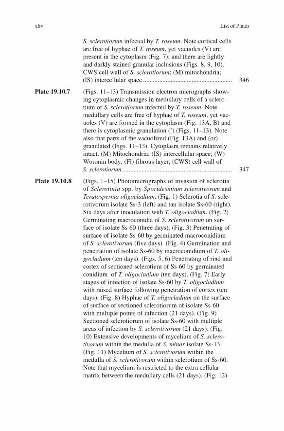
S. sclerotiorum infected by T. roseum. Note cortical cells
are free of hyphae of T. roseum, yet vacuoles (V) are
present in the cytoplasm (Fig. 7); and there are lightly
and darkly stained granular inclusions (Figs. 8, 9, 10).
CWS cell wall of S. sclerotiorum; (M) mitochondria;
(IS) intercellular space ....................................................... 346
Plate 19.10.7 (Figs. 11–13) Transmission electron micrographs show-
ing cytoplasmic changes in medullary cells of a sclero-
tium of S. sclerotiorum infected by T. roseum. Note
medullary cells are free of hyphae of T. roseum, yet vac-
uoles (V) are formed in the cytoplasm (Fig. 13A, B) and
there is cytoplasmic granulation (*) (Figs. 11–13). Note
also that parts of the vacuolized (Fig. 13A) and (or)
granulated (Figs. 11–13). Cytoplasm remains relatively
intact. (M) Mitochondria; (IS) intercellular space; (W)
Woronin body, (Fl) fibrous layer, (CWS) cell wall of
S. sclerotiorum .................................................................... 347
Plate 19.10.8 (Figs. 1–15) Photomicrographs of invasion of sclerotia
of Sclerotinia spp. by Sporidesmium sclerotivorum and
Teratosperma oligocladium. (Fig. 1) Sclerotia of S. scle-
rotivorum isolate Ss-3 (left) and tan isolate Ss-60 (right).
Six days after inoculation with T. oligocladium. (Fig. 2)
Germinating macroconidia of S. sclerotivorum on sur-
face of isolate Ss 60 (three days). (Fig. 3) Penetrating of
surface of isolate Ss-60 by germinated macroconidium
of S. sclerotivorum (five days). (Fig. 4) Germination and
penetration of isolate Ss-60 by macroconidium of T. oli-gocladium (ten days). (Figs. 5, 6) Penetrating of rind and
cortex of sectioned sclerotium of Ss-60 by germinated
conidium of T. oligocladium (ten days). (Fig. 7) Early
stages of infection of isolate Ss-60 by T. oligocladium
with raised surface following penetration of cortex (ten
days). (Fig. 8) Hyphae of T. oligocladium on the surface
of surface of sectioned sclerotiorum of isolate Ss-60
with multiple points of infection (21 days). (Fig. 9)
Sectioned sclerotiorum of isolate Ss-60 with multiple
areas of infection by S. sclerotivorum (21 days). (Fig.
10) Extensive developments of mycelium of S. sclero-tivorum within the medulla of S. minor isolate Ss-13.
(Fig. 11) Mycelium of S. sclerotivorum within the
medulla of S. sclerotivorum within sclerotium of Ss-60.
Note that mycelium is restricted to the extra cellular
matrix between the medullary cells (21 days). (Fig. 12)
xliv List of Plates

Abundant development of S. sclerotivorum within scle-
rotium of Ss-60 and exit hyphae of the mycoparasite (29
days). (Fig. 13) Macroconidium of S. sclerotivorum
sporulating on a sclerotium of S. minor naturally
infected in the field. (Fig. 14) Sparse colonization of an
autoclaved sclerotium of isolate Ss-60 by S. sclerotivo-rum. Compare with heavily invaded live sclerotium in
Fig. 12 inoculated at the same time (29 days). (Fig. 15)
Surface development of T. oligocladium on invaded
sclerotium of isolate Ss-3 and Ss-60. Sclerotia are the
same as in Fig. 1 (16 days) ............................................... 348
Plate 19.10.9 Light (LM) and transmission electron micrographs
(TEM) of Sporidesmium sclerotivorum in sclerotial
tissue of S. minor. (Fig. 1) LM; showing hyphae of
S. sclerotivorum (arrow) in the cortex © and medulla
(M), 20 days after inoculation. Toluidine blue O stain.
(Fig. 2) TEM; showing hyphae of S. sclerotivorum (S)
growing in the extra cellular matrix (EM) of the medulla.
Their walls are thinner and more electron opaque (arrow)
than those of the medullary hyphae (W). 30 days (Fig. 3)
TEM showing thin walled hyphae of S. sclerotivorum (S)
in an empty cortical cell © 15 days (Figs. 4–8) Light
micrographs of haustoria of S. sclerotivorum in medul-
lary cells. The long penetrating hyphae (P) are branched
at their distal ends. There is a deposit (arrow) on the scle-
rotial cell walls at the point of penetration. (Figs. 4–6)
15 days calcoflour white M2R stain. (Figs. 7, 8)
20 days, PAS stain. (Fig. 9) TEM of hypha of
S. sclerotivorum (S) that penetrating the walls of cortical
cell (C) showing the many branches of a hautorium (H)
in section. An electron translucent region surrounds each
branch (arrow). 30 days (Fig. 10) TEM of cortical cell
(C) showing the many branching of a hautorium (H) in
section. An electron translucent region surrounds each
branch 9 arrow). 30 days. (Fig. 11) TEM showing details
of haustoria branches (H) with dense cytoplasm in a
medullary cell (M). The surrounding sheath is delimited
by a unit membrane (arrow) 20 days. (Fig. 12) TEM of
degenerate haustorial branches (arrow) in a cortical cell
(C). The cytoplasm of the branches is disrupted 40 days.
(Fig. 13) TEM showing hyphae of S. sclerotivorum (S) in
the outer medulla (M). The cytoplasm of both
S. sclerotivorum and sclerotial cells has degenerated
40 days ................................................................................ 349
List of Plates xlv

Plate 19.10.10 Cross section of healthy sclerotium of Sclerotinia sclero-tiorum showing three distinctive layers of tissue; rind
(R) (Fig. 1a); Cortex (C) and medulla (M), (Fig. 1b).
(Fig. 2) Cross section of sclerotium infected with C.minitans showing complete destruction and disintegra-
tion of cortical and medullary tissues of mycelia of
hyperparasite (CM). The rind is infected but remains
intact ................................................................................... 350
Plate 19.10.11 Invasion of rind by C. minitans. Note the hyphae of
C. minitans (CM) in the amorphous layer (AL) (Figs. 3,
5) and the thick melanized wall (CW). Note the sign of
wall etching by the hyperparasite (CM) (Fig. 4) and the
loose melanin particles (mel) near the affected cell wall
(Figs. 4, 6). (Figs. 7, 8) Destruction of outer rind cells
by C. minitans .................................................................... 351
Plate 19.10.12 (Fig. 9) Early stage of penetration of the melanized wall
of a rind cell (CW) by C. minitans (CM) showing a
small, well defined gap (Fig. 9a) (arrows) created by the
narrow penetration peg. Invagination of the host cell
wall at the penetration site is not evident. (Fig. 10)
Invagination of the melanized wall (CW) at the penetra-
tion site is evident when penetration is complete. Note
the narrow hyphal neck of the hyperparasite with
Woronin bodies (W) in it (Fig. 10b) ................................. 352
Plate 19.10.13 (Figs. 11, –12) Lysis and death of young cells of
C. minitans. (Figs. 11a–12a) in infected rind tissue.
The wall of the dead cell is either partially disintegrated
(Fig. 11b) or completely disintegration with its
membranous structure (PM) in the host cell (Fig. 12) ........ 353
xlvi List of Plates

Color Plates
Plate 5.2.7.1 White stem rot of rapeseed-mustard. (L–R) A: Initial growth at the lower portion of
the stem; B: White mycelium growth acquires more areas; C, D: Infection on the stem caused drying
of the branches; E: Drying of the crop visible in the field; F: Black hard sclerotia in side the pith of
the stem (Photos A, B & F. Adapted from http: //www. whitemoldresearch.com. With permission)
xlvii

xlviii Color Plates
Plate 5.2.8.1 Sclerotinia disease of soybean. A: Infected field showing dried plants; B: Infected
stem at the basel portion of the stem (Adapted from http://www.whitemoldresearch.com. With
permission)

Plate 5.2.9.1 Sclerotinia disease of sunflower. (L–R) A: Mycelium growth at the middle of the
stem; B: Basel canker formation at the base of the stem; C: White mould near the soil level;
D: Sudden wilting of the plants in the field; E: Head rot due to Sclerotinia F: Apothecia formation
on the soil (Adapted from http://www.whitemoldresearch.com. With permission)

l Color Plates
Plate 5.2.12.1 Sclerotinia disease of pea/beans. A: White mold infection on peas; B: White mold
infection on pods; C: Sclerotinia infection at basel stem portion (Adapted from http: //www.
white- moldresearch.com. With permission)
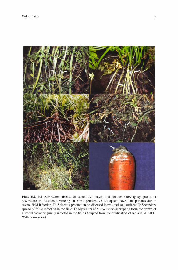
Color Plates li
Plate 5.2.13.1 Sclerotinia disease of carrot. A. Leaves and petioles showing symptoms of
Sclerotinia; B: Lesions advancing on carrot petioles; C: Collapsed leaves and petioles due to
severe field infection; D: Sclerotia production on diseased leaves and soil surface; E: Secondary
spread of foliar infection in the field; F: Mycelium of S. sclerotiorum erupting from the crown of
a stored carrot originally infected in the field (Adapted from the publication of Kora et al., 2003.
With permission)

lii Color Plates
Plate 5.2.17.1 Sclerotinia stem rot of potato. A, B: Sclerotinia infection at the base; C: Sclerotinia
causing drying of the stem; D: Drying and breaking of the stem; E: Breaking and production of
black sclerotia (Adapted from http://www.potatodiseases.org. With permission)

Color Plates liii
Plate 5.2.18.1 Sclerotinia disease of poppy; A: Basal rot of poppy; Abundant apothecial produc-
tion under field conditions: B: Healthy (left) and infected (right) stem and mummified capsule of
poppy; C: Flower buds heavily infected with pathogen showing white colony growth intermingled
with sclerotia; D: Capsule of poppy showing sclerotia in side. E: Capsule of opium poppy showing
infection of Sclerotinia, Black sclerotia on capsule; F: L.S. of infected (left) capsule showing
fungal growth and sclerotia with healthy capsule (right) (Adapted from the publication of Singh
and Singh, 2003. With permission)

liv Color Plates
Plate 5.2.19.1 Sclerotinia rot in lentil. A: Sclerotinia disease infection on lentil stem; B:
Severe infection at the base; C: Apothecia production at the soil level (Adapted from http://
www. whitemoldresearch.com. With permission)

Color Plates lv
Plate 5.2.24.1 Sclerotinia rot of chickpea. A: Drying of leaves at the initial infection; B: Severe
infection cause drying of the stem; C: Mycelium and sclerotia formation at soil level; D: Sclerotia
sticking to stem (Adapted from http:// www. whitemoldresearch.com. With permission)

lvi Color Plates
Plate 5.2.25.1 Dollar spot of turfgrass. A: Dollar spot initiation on bent grass; B: Tan shaped
lesions on the bent grass; C: Infected area on the leaf blade; D: Dense white mycelium on seedling
turf; E: Large masses of hyphae/ mycelium on the lawns (Adapted from http://www.turf – grass
management.psu.edu; http://www.caes.uga.edu; http://www.ces. ncsu.edu. With permission).

Color Plates lvii
Plate 7.4.4.1 Growth pattern of Sclerotinia sclerotiorum (a) Mycelial growth; (b) Initaition of
sclerotia formation; (c) Sclerotia formation at the outer periphery

lviii Color Plates
Plate. 7.4.4.2 Apothecia formation in Sclerotinia sclerotiorum
Fig. 19.11.1 An integrated model for managing Sclerotinia rot of carrot that incorporates three
disease management principles and selected disease control strategies (outer circle), that target
particular stages in the life cycle of Sclerotinia sclerotiorum (middle circle) or development of
carrot crop (inner circle). Control strategies corresponding to respective stages are indicated by
positional overlap (Adapted from the publication of Kora et al., 2003. With permission)
Biodata of G.S. Saharan
lix
Dr. Govind Singh Saharan, Ex Professor and Head (Plant Pathology) retired from
the active service during the year 2002. He did his B.Sc. Agriculture (1965),
M.Sc. Agriculture (1967) from S.K.N. College of Agriculture, Jobner, University
of Udaipur and Ph.D. from Himachal Pradesh University, Palampur (1977). He
served as Lecturer (1967–1976) and Assistant Professor 1976–1980 at HPKVV,
Palampur. Joined as Associate Professor (1980–1988), Professor (1988–2002)
and Professor and Head (2002) of Plant Pathology at CCS, Haryana Agricultural
University, Hisar.
Dr. Saharan has been visiting Professor to the University of Alberta, Edmonton,
Canada (1991 and 1994), Agriculture, Canada, Saskatoon (1991, 1994, 1997) and
Rothamsted, IACR, Harpenden, UK (1994 and 1997).
Dr. Saharan has more than 200 research publications in Journals of National and
International repute. He has been editor of one book Diseases of Oilseed Crops and
two other books. He is author of 25 review articles in books, monographs on White
rust, Alternaria blight, Downy mildew diseases of rapeseed-mustard, and three bib-
liographies. He is on the panel of Experts of State Agricultural Universities, ICAR,

CSIR, UGC and DBT. He is one of the specialists to contribute in the release of
Crop Protection Compendium, 2002, CAB International, UK. He has guided three
M.Sc. and eight Ph.D. students who are well established scientists at different
organizations in India and abroad.
Dr. Saharan has conducted research in diverse fields of Plant Pathology covering
standardization of artificial inoculation technique, identification of sources of
resistance, determination of pathogenic variability, genetics of host-parasite inter-
action, epidemiology and management of different diseases.
Dr. Saharan has been president of Indian Phytopathological Society (NZ) during
2001 and Editor-in-Chief of Indian Society of Mycology and Plant Pathology
(1999–2002).
Dr. Saharan has been deeply associated with the organization of Global and
Asian Congresses organized by the leading Phytopathological societies of India.
lx Biodata of G.S. Saharan

Biodata of Naresh Mehta
lxi
Dr. Naresh Kumar Mehta, Professor (Plant Pathology) did his B.Sc. Agriculture
(Hons) from Haryana Agricultural University, Hisar in 1978 and attained first posi-
tion in elective Plant Pathology. During his M.Sc. Agriculture (Plant Pathology),
he was awarded Excel Industries Ltd. Bombay, India, research fellowship. He
joined Haryana Agricultural University, Hisar as Assistant Scientist (Plant
Pathology) in 1981 and completed Ph.D. degree as in-service candidate in 1993 in
Plant Pathology with Dr. G.S. Saharan. He was awarded Ms. Manju Utereja
Memorial Gold Medal for best Ph.D. thesis for the year 1993/94. He was also
awarded Senior Research Fellowship (SRF) for Ph.D. programme by Council of
Scientific and Industrial Research (CSIR), New Delhi.
Dr. Mehta has been admitted as Fellow of Indian Phytopathological Society,
New Delhi, (FPSI), Indian Society of Plant Pathologist, Ludhiana (FSPP) and
Indian Society of Mycology and Plant Pathology, Udaipur (FISMPP). He has been
nominated as councilor (North Zone) of the society ISMPP for the year 2005/06.
Dr. Mehta was elevated to the post of Associate Professor (Plant Pathology) in
March 1994 and Professor (Plant Pathology) in 2002.

He has conducted research in diverse field of Plant Pathology covering patho-
genic variability, genetics of host pathogen interaction, epidemiological studies,
identification of resistant sources, biochemical/genetical basis for resistance, resid-
ual analysis of fungicides and disease management. He has been Co-Principal
Investigator in the scheme: “Pathogenic variability and epidemiology of Alternariabrassicae” funded by ICAR, New Delhi, from December 1998 to May 2003. He
has guided three M.Sc. (Plant Pathology) students.
He has published more than 75 research papers in the journals of National and
International repute. He has one edited book (Diseases of Oilseed Crops), four
review articles, 14 book chapters, six practical manuals, four lead lectures in the
international and national conferences, 55 research papers presentations in the
International/National conferences and about 35 popular articles to his credit.
Dr. Mehta has been a visiting scientist to University of Alberta, Edmonton,
Canada in 1999 as a FAO fellow and presented a research paper in 8th International
Congress of Plant Pathology at Christchurch, New Zealand, 2002. He has been
invited to deliver a lecture in the 9th International Congress of Plant Pathology at
Torino, Italy, 2008.
lxii Biodata of Naresh Mehta