Science Webinar Series · Science Webinar Series Between Thought and Therapy: Translating...
68
Change the size of any window by dragging the lower right corner. Use controls in top right corner to close or maximize each window. Webinar Series Science Between Thought and Therapy: Translating Neurobiology Research into Treatments 13 February, 2013 Facebook login if you need help shows speaker bios download slides and more info LinkedIn login shows slide window What each widget does: shows the video screen Twitter login (#ScienceWebinar) search Wikipedia view closed captioning
Transcript of Science Webinar Series · Science Webinar Series Between Thought and Therapy: Translating...

Change the size of any window by dragging the lower right corner. Use controls in top right corner to close or maximize each window.
Webinar Series Science Between Thought and Therapy: Translating Neurobiology Research into
Treatments 13 February, 2013
Facebook login
if you need help
shows speaker bios
download slides and more info
LinkedIn login
shows slide window
What each widget does:
shows the video screen
Twitter login (#ScienceWebinar)
search Wikipedia
view closed captioning

Sponsored by:
Participating Experts:
Brought to you by the Science/AAAS Custom Publishing Office
Webinar Series Science Between Thought and Therapy: Translating Neurobiology Research into
Treatments 13 February, 2013
Ellen Sidransky, M.D.
National Human Genome Research Institute, NIH
Bethesda, MD
Anand Swaroop, Ph.D.
National Eye Institute, NIH
Bethesda, MD
Carlos A. Zarate, M.D.
National Institute of Mental Health, NIH
Bethesda, MD

Development of Rapid-Acting Antidepressants and Biomarkers of
Response
Carlos A. Zarate, Jr., M.D
Experimental Therapeutics & Pathophysiology Branch (ETPB)
Division of Intramural Research Program
National Institute of Mental Health

Major depressive disorders: a major cause of disability
>10% of the American population suffer from a mood disorder each year
Depression is one of THE leading causes of disability worldwide, ranking ahead of ischemic heart disease, cerebrovascular disease, cancers, infectious diseases, etc.
An increase in the death rate at any age, independent of suicide, smoking, or other risk factors
> 30,000 deaths from suicide/yr (cf ~ 18,000 homicides)
Individuals with major depression sometimes describe an emotional pain much worse than any physical pain that they have experienced

Drug Development in the past 50 years
Insel and Skolnick Mol Psychiatry 2006;11:11-17
# o
f M
ech
an
isti
call
y D
isti
nct
Dru
gs
Lithium
Anticonvulsants
Divalproex
Carbamazepine
Lamotrigine
Topiramate
Oxcarbazepine
Levetiracetam
Antipsychotics
Clozapine
Risperidone
Olanzapine
Quetiapine
Ziprasidone
Aripiprazole
Except for Li all available FDA approved treatments for BPD are anticonvulsants or Antipsychotic drugs
Antidepressants ONLY serotonin and
norepinephrine based (‘me too drugs’)

•Disruption to:
•Personal life
•Familial life
•Occupation
•Social life
•Increased economic
costs
•Risk for suicidal behavior
Natural course
Standard
antidepressant Treated course
Euthymic
Depressed
Manic Next generation antidepressant
6-12 months
10-14 weeks
33% remission
Hours/day
Identify a problem: Lag of onset of antidepressant effects
Course of
Illness

Targeting the Glutamatergic System to Develop Improved Therapeutics for Mood Disorders
Target
receptor
Brain region Effect
NMDA Hippocampus Reduced [3H]CGP-39653 and
MK-801 binding and reduced
levels of NR1 and NR2A
transcript
NMDA Sup temporal
cortex
NMDA receptor binding and NR1
subunit expression
NMDA ACC NR2A subunit decreased in
GABA interneurons
NMDA DLPFC Polymorphisms in genes coding
NR2B, no difference in mRNA
expression
AMPA Striatum Increased AMPA binding is
associated with decreased
GLUR1 subunit expression
AMPA DLPFC Decreased levels of GLUR2
Sanacora, Zarate, Krystal, Manji. Nat Rev Drug Discov 2008

Ketamine is a widely used
disassociate anesthetic mainly
for ambulatory surgery and
chronic pain
Ketamine is 10-50 times less
potent than PCP in blocking
Non-competitive NMDA receptor
antagonist
Psychotomimetic properties (5-
20%)
Abused as “club drug”
Modification in synaptic AMPA & NMDA receptors
Can the Onset of Action of Antidepressants be Accelerated?
Ketamine i.v. (0.5 mg/kg over 40 min)
Drug Free Period
2 weeks
Placebo Placebo
Ketamine Ketamine
1 week
Ratings: min: 0, 40, 80, 110, 230; Days: 1,2,3, 7, 14 (bipolar)
Study design 3 studies Unipolar and Bipolar Depression
Ketamine
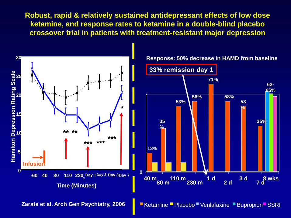
Robust, rapid & relatively sustained antidepressant effects of low dose
ketamine, and response rates to ketamine in a double-blind placebo
crossover trial in patients with treatment-resistant major depression
Zarate et al. Arch Gen Psychiatry, 2006
13%
35
%
53% 56%
71%
58% 53
%
Response: 50% decrease in HAMD from baseline
62-
65%
35%
40 m 80 m
110 m 230 m
1 d 2 d
3 d 7 d
8 wks
Infusion
0
5
10
15
20
25
30
-60 40 80 110 230 Day 1 Day 2 Day 3 Day 7
Time (Minutes)
Ha
mil
ton
De
pre
ss
ion
Ra
tin
g S
ca
le
** **
*** *** ***
*
//
Ketamine Placebo Venlafaxine SSRI Bupropion
Infusion
33% remission day 1

5
10
15
20
25
30
-60 40 80 110 230 D1 D2 D3
HDRS
† ‡
// //
†
Minutes Days
D7
†
‡ ‡ Infusion
Unipolar depression
5
10
15
20
25
- 60 40 80 110 230 D1 D2 D3 D7 D10 D14
HDRS
†
† ‡
‡ ‡ ‡
‡
‡
Minutes Days
Bipolar depression
Rapid antidepressant response to ketamine in two different disorders
High SSI (>4) (n=10)
Low SSI (<3) (n=23)
-60 0 60 120 180 240 Minutes
0
2
4
6
8
10
12
]
]
]
]
]
]
]
]
]
]
d= 1.3
* * *
*
d= 2.4
Zarate et al. 2006; Diazgranados et al. 2010a; Diazgranados et al. 2010b
33% remission
Genes Gene expression Cellular Circuits Rapid reversal of complex
behavioral phenotype
Metabolome Neurochemical

Ketamine mGluR I gln
glu
glutaminase
glu
glu
mGluR I
EAAT3
Glu AMPA R
NMDA R (-)
KA R
Postsynapti
c neuron
Presynaptic neuron
gln
aketo
glutarate
TCA
Astrocyte
? AD
response
Moghaddam J Neurosci 1997
Time (min) Ket.
Inj.
-50
50
150
250
350
0 20 40 60 80 100
Saline Ketamine (10 mg/kg) Ketamine (20 mg/kg) Ketamine (30 mg/kg)
* *
*
* * * * * *
Ketamine increases glutamate release, thereby facilitating AMPA/NMDA throughput ---->mTOR activation----> Antidepressant effects
* p<0.05, ** p<0.01
*
*
**
* *
dose (mg/kg) 0 1 3 10
imm
ob
ile
tim
e (
se
c)
0
20
40
60
80
100
120
140
160
180 p=0.02
*
**
*p<0.05, **p<0.01
Ro 25-6981 in animal model of depression
0
20
40
60
80
100
120
140
160
180 p=0.01
Maeng et al. Biol Psych 2008
NR2B antagonist
(Ro256981)
24hs Antidepressant-like
Li et al. Science 2010
Genes Gene expression Cellular Circuits Rapid reversal of complex
behavioral phenotype
Metabolome Neurochemical
**
SAL KET NBQX KET+
NBQX
AMPA Antagonists block the
antidepressant effects of Ketamine
0
20
40
60
80
100
120
140
160
180

Glutamate Modulation in TRD: Pipeline (Partial List)
Compound
(Manufacturer)
Mechanism of
Action
Phase Route
MK-0657* (Merck)
NR2B Antagonist IIa p.o. Completed
CP101,606
(Pfizer)
NR2B antagonist IIa IV + response;
discontinued
RO4917523
(Roche)
mGluR5 allosteric
antagonist
IIa p.o. Ongoing study
AZD6765* (AstraZeneca)
NR2AB Antagonist IIb IV Completed
EVT 101
(Evotec)
NR2B antagonist IIa p.o. Study halted

AZD6765: A low-affinity NMDA channel blocker
AZD6765 was developed in Europe as an intravenous txt for
stroke but was not further pursued because of a lack of efficacy
AZD6765 is a low-trapping NMDA channel blocker, NMDA receptor
antagonist. It is blood-brain penetrant
AZD6765 well tolerated with dizziness, nausea, and vomiting,
being the most common AEs. No psychotomimetic effects up to
160 mg
Antidepressant effects in learned helplessness, FST
Anxiolytic activity in the rat punished responding model

0
5
10
15
20
25
30
35
40
-60 60 80 110 230Day1
Day2
Day3
MA
DR
S
Time (Minutes)
Placebo
AZD150
** *
0
5
10
15
20
25
HA
MD
(1
7 Ite
m)
Time (Minutes)
Placebo
AZD150
** * * * *
VEGF
Zarate et al. Biol Psych 2012
A double-blind placebo-controlled study of NMDA antagonist (AZD6765) in treatment-resistant depression (N=22)

Study of ketamine’s mechanism of action from synapses through a range of systems
Synaptic Plasticity
(mTOR, eEF2, GSK-3B inh)
•Glucose changes
(glutamate signal)
•Circuits/connectivity Cortical excitability
Genes Gene expression Cellular Circuits Rapid reversal of complex
behavioral phenotype
Polysomnograph
y MEG PET & MEG MRS
Neurochemicals
(Glx DM/DA-PFC
Glx/Glu ratio)
Synaptic Dysfunction

Medial prefrontal cortex layer V pyramidal
cells in brain-derived neurotrophic factor
Val66Met knock-in mice have both apical and
basilar dendritic atrophy
Antidepressant response to Ket in the
forced swim test is attenuated in brain-
derived neurotrophic factor Val66Met
knock-in mice
Liu et al. Biol Psychiatry 2012
Val66Met and Antidepressant Response to Ketamine
P = .025
Laje et al. Biol Psychiatry 2012

Salvadore et al., Biol Psychiatry 2010
Experiment 1 (an affective task):
rostral ACC activity is positively
correlated with AD response to
ketamine
MA
DR
S c
ha
ng
e s
co
re (
%)
R
L
Linear change in ACC activity
x=5
Rostral ACC
pretreatment
activity (Emotion)
Experiment 2 (a cognitive task): rostral
ACC activity is negatively correlated with
AD response to ketamine
Salvadore et al. Neuropsychopharmacology 2010
+1
-
+1
-
Rostral ACC
pretreatment
activity
(Cognition)
-20
0
20
40
60
80
100
-1.5 -0.5 0.5 1.5 2.5 3.5
-20
0
20
40
60
80
100
-1.5 -0.5 0.5 1.5 2.5 3.5
MA
DR
S c
han
ge s
co
re (
%))
1 1
+

MEG: Grand Averaged (N=20) Time-frequency plots
Baseline
40 Hz
RH stimulation
Left sensors LH stimulation
Right sensors
40
Hz
0 100 200 300 -100 100 200 300 -100 0
10
0
8
2
6
4
10
0
8
2
6
4
No
rma
lize
d P
ow
er
No
rma
lize
d P
ow
er
Baseline
stimulation
1.5
0.0
Lo
g1
0 No
rmaliz
ed
Po
wer
30-5
0H
z
Grand Averaged (N=20) Evoked Gamma-Band Source Analyses
Somatosensory
stimuli
Central sulcus
MEG: A sensory cortical signature of ketamine’s rapid antidepressant effects
Gamma rhythms are involved
in many aspects of cognitive
function from 1• sensory
representation through
selective attention and short-
term memory.
They possess the ability to
facilitate synchrony of neuronal
activity occurring in many,
anatomically distant areas at
the same time.
Using ketamine in rodents
produces an increase in gamma
rhythm generation
(frontoparietal, hippocampus).
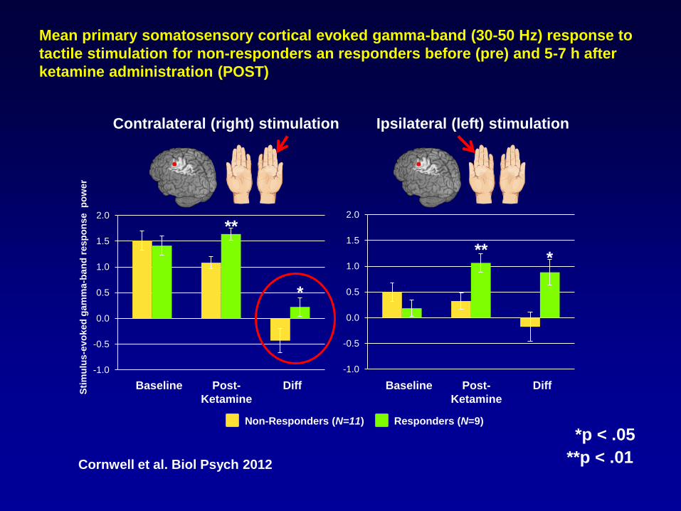
Mean primary somatosensory cortical evoked gamma-band (30-50 Hz) response to
tactile stimulation for non-responders an responders before (pre) and 5-7 h after
ketamine administration (POST)
Contralateral (right) stimulation Ipsilateral (left) stimulation
Non-Responders (N=11) Responders (N=9)
Sti
mu
lus
-ev
oked
gam
ma
-ban
d r
esp
on
se p
ow
er
-1.0
-0.5
0.0
0.5
1.0
1.5
2.0
-1.0
-0.5
0.0
0.5
1.0
1.5
2.0
Baseline Post-
Ketamine
Diff Baseline Post-
Ketamine
Diff
*
*
**
**
*p < .05
**p < .01 Cornwell et al. Biol Psych 2012

Cells:
Ketamine enhances
AMPA:NMDA
throughput in critical
neuronal circuits and
activates the mTOR
pathway: increased
synaptic signaling
proteins and increased
number and function of
new spine synapses in
the PFC of rats
Systems:
ACC desynchronization
and functional
connectivity with the
amygdala during a
working memory task
predict rapid
antidepressant response
to ketamine
Behavior:
Rapid reversal
of Complex
Phenotype
Genes
Multiple susceptibility
alleles each of small
effect
? Ketamine effects
Cellular
Programming
Gene and
Protein
Expression
? Ketamine effects
The Neurobiology of Mood Disorders
Understanding the Mechanism of Ketamine Across Different Systems
5
10
15
20
25
30
-60 40 80 110 230 D1 D2 D3
HDRS
† ‡
// //
†
Minutes Days
D7
†
‡ ‡ Infusion
Depression
Genes Gene expression Cellular Circuits Rapid reversal of complex
behavioral phenotype
Metabolome Neurochemical
VAL/
VAL
MET+
BDNF SNP response

Conclusions
• The new paradigm of rapid antidepressant action is to develop antidepressant drugs that work in hours or a few days instead of weeks
• Scopolamine and ketamine are tools/models that can be used in understand the mechanisms involved & develop treatments with a rapid onset of action
• Directly targeting the muscarinic receptors and NMDA receptor complex may bring about rapid antidepressant effects in both unipolar and bipolar depression
• A more precise understanding of the downstream changes are relevant to the mechanism of rapid antidepressant action
• Anterior cingulate cortex activity modulation is associated with antidepressant response to ketamine
• Several bio-signatures appear promising and are associated with significant improvement in depressive sxs in patients treatment with scopolamine and ketamine

Research studies: http://patientinfo.nimh.nih.gov
1-877-MIND-NIH (1-877-646-3644)
email [email protected]
Research = Hope

Sponsored by:
Participating Experts:
Brought to you by the Science/AAAS Custom Publishing Office
Webinar Series Science Between Thought and Therapy: Translating Neurobiology Research into
Treatments 13 February, 2013
Ellen Sidransky, M.D.
National Human Genome Research Institute, NIH
Bethesda, MD
Anand Swaroop, Ph.D.
National Eye Institute, NIH
Bethesda, MD
Carlos A. Zarate, M.D.
National Institute of Mental Health, NIH
Bethesda, MD


Detailed examination of
a single gene disorder Gaucher
Studies of rare recessive diseases may
provide a window into complex disorders
Ultimately help to unravel
complex disorders. Parkinson
Disease
Apply insights to other
monogenic disorders
PKU CF

Gaucher Disease
(GD) • Deficiency of enzyme
glucocerebrosidase,
accumulation of
glucosylceramide
• Variable age of onset
• Rare recessive, single gene
disorder
• Multi-organ involvement
• Symptoms include enlarged
spleens and livers, low platelet
counts, bone and brain
involvement
Parkinson Disease
(PD) • Loss of dopaminergic neurons
in the brain, accumulation of
Lewy bodies - aggregates of
proteins including α-synuclein
• Late onset
• Common disorder affecting 1.5%
of population over age 65,
complex multi-gene disorder
• Symptoms include bradykinesia,
rigidity and tremor, and
sometimes dementia
• Mainly affects substantia nigra
and brainstem

Clinical Studies
Cell Biology
Gaucher disease and parkinsonism
Gaucher disease Parkinsonism Integrated
translational
approach

Glucocerebroside + H20 Ceramide + Glucose
Glucocerebrosidase
Gaucher disease: A Mendelian lysosomal
storage disorder
C C CH2 O
C O
H H
OH
OH
N
CH2OH
OH
O
H
OH
O

Vast clinical heterogeneity is encountered
in this disorder
Type 1 Type 2 Type 3
Hydrops
fetalis
Asymptomatic
Congenital
ichthyosis Progressive
neurologic
degeneration Myoclonic
epilepsy
Parkinsonian
manifestations
Eye movement
disorder
2° neurologic
involvement
Skeletal
disease Visceral
disease
Neurologic
manifestations
Hydrocephalus, cardiac
valve calcifications
Gaucher disease: a spectrum of phenotypes

The association of glucocerebrosidase (GBA)
mutations and parkinsonism
This story began in the clinic, with rare patients with Gaucher disease who developed parkinsonism.
GD
PD PD also seen in relatives of Gaucher probands.
We and others noted an increased incidence of GBA mutations in patients with PD and LBD.
However many of the initial studies were greeted with skepticism due to limitations in power or controls and because GWAS did not identify GBA.
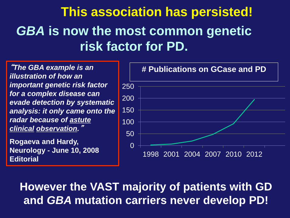
“The GBA example is an
illustration of how an
important genetic risk factor
for a complex disease can
evade detection by systematic
analysis: it only came onto the
radar because of astute
clinical observation.”
Rogaeva and Hardy,
Neurology - June 10, 2008
Editorial
This association has persisted!
0
50
100
150
200
250
1998 2001 2004 2007 2010 2012
# Publications on GCase and PD
GBA is now the most common genetic
risk factor for PD.
However the VAST majority of patients with GD
and GBA mutation carriers never develop PD!

International multi-center study of GBA
mutations in PD
Subjects with PD are >5 times
more likely to have a mutation
in GBA (Overall OR=5.43;
95%Cl=3.89-7.57)
GBA carriers had earlier PD
onset and more cognitive
deficits.
Oct 2009
16 centers joined,
5691 genotypes from
patients with PD and
>5000 from controls
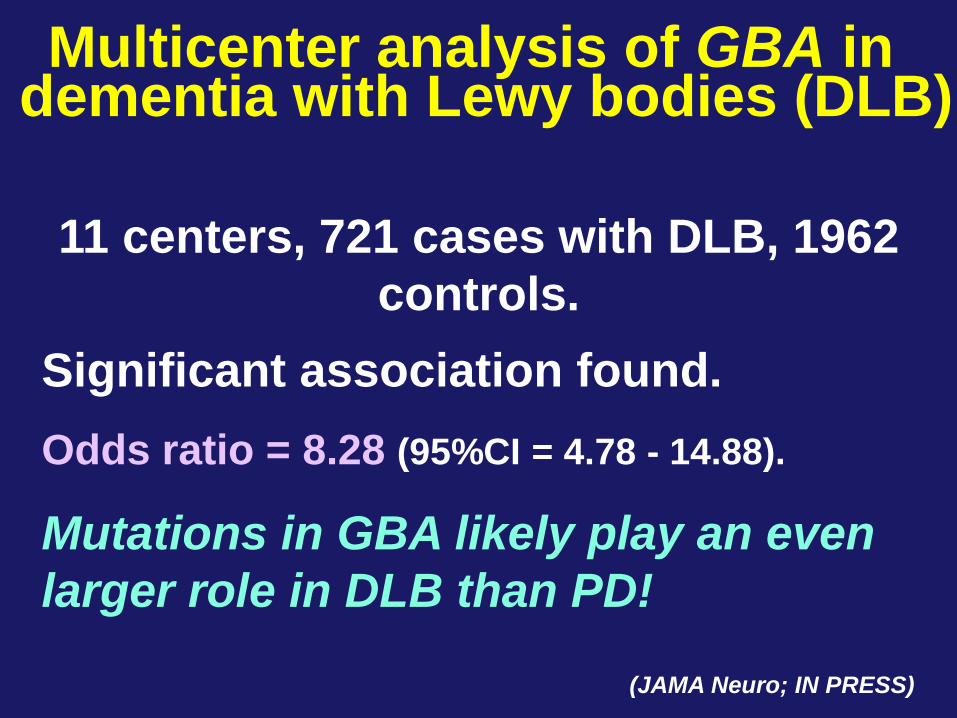
Significant association found.
Odds ratio = 8.28 (95%CI = 4.78 - 14.88).
Mutations in GBA likely play an even
larger role in DLB than PD!
(JAMA Neuro; IN PRESS)
Multicenter analysis of GBA in dementia with Lewy bodies (DLB)
11 centers, 721 cases with DLB, 1962
controls.
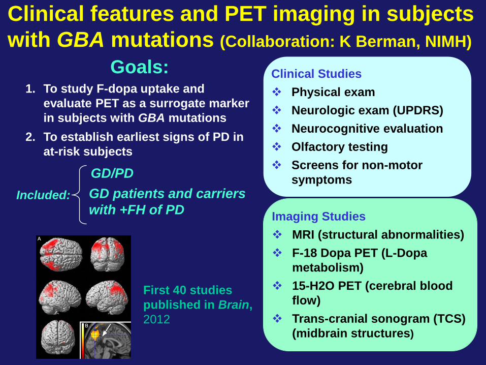
1. To study F-dopa uptake and
evaluate PET as a surrogate marker
in subjects with GBA mutations
2. To establish earliest signs of PD in
at-risk subjects
Clinical features and PET imaging in subjects
with GBA mutations (Collaboration: K Berman, NIMH)
Clinical Studies
Physical exam
Neurologic exam (UPDRS)
Neurocognitive evaluation
Olfactory testing
Screens for non-motor
symptoms
Imaging Studies
MRI (structural abnormalities)
F-18 Dopa PET (L-Dopa
metabolism)
15-H2O PET (cerebral blood
flow)
Trans-cranial sonogram (TCS)
(midbrain structures)
Goals:
Included: GD patients and carriers
with +FH of PD
GD/PD
First 40 studies
published in Brain,
2012

Formation of insoluble a-
synuclein aggregates
Neuronal
cell death
Mutant
glucocerebrosidase
may lead to…
Increased
aggregate
formation
Organelle
dysfunction:
decreased
aggregate
clearance
How can mutations in a metabolic enzyme
lead to parkinsonism?
Gain-of-function:

a-synuclein
aggregates
Neuronal
cell death
Mutant
glucocerebrosidase
may lead to…
How can mutations in a metabolic enzyme
lead to parkinsonism?
Unstable or deficient
protein
Degradation
Insufficient enzyme
GlcCer accumulation
Neuronal cell death
Loss-of-function:
soluble α-syn
oligomers and fibrils
α-syn oligomers block ER-
Golgi trafficking of GCase (Mazzulli et al. Cell, 2011)
“Bidirectional
feedback loop”

Shown by fluorescence spectroscopy, NMR and co-
immunoprecipitation studies, but only at pH 5.5.
Interaction occurs at C-terminus of α-synuclein.
A molecular link between α-syn and glucocerebrosidase Collaboration with J. Lee, NHLBI (JBC 2011)
A B
This binding at lysosomal pH could facilitate α-syn
degradation or prevent aggregation. The GBA story
implicates the lysosome in PD pathogenesis.

Chemical chaperone therapy for Gaucher disease
Chemical chaperones may partially correct the enzyme deficiency.

High Throughput Screening Collaboration: W. Zheng, J. Maragun and C. Austin
Small molecule therapy may stabilize mutant
GCase and be used to treat GD as well as PD
GCase chaperones as therapy for GD
New screening approach: (Goldin et al.PLoS One 2012)
Patient spleen sample = source of mutant GCase.
HT screen - 250K compounds.
30 new non-inhibitor chaperones identified.
Leads improved translocation in patient fibroblasts.
In macrophages: compounds reverses storage!

•Embryoid bodies generated from Gaucher fibroblasts
•Cells show appropriate markers,karyotype; form teratomas .
•Differentiated into monocytes and then macrophages.
•iPSC derived Gaucher macrophages show storage!
•Model useful for drug development and understanding
pathophysiology.
iPS cells from
type 2 GD DAPI Bodipy-GlcCer
Control Gaucher type 2
Progress with Gaucher iPS cells
hiPSC –derived macrophages
(fed with labeleds erythrocyte ghosts)

Lead chaperone decreases storage in both
N370S/N370S macrophages and iPSC-
macrophages (IVS2+1/L444P)
Cultured
control
macrophages
Gaucher iPSc-
macrophages
Cultured
Gaucher
macrophages
Testing of lead compound in macrophage
models of Gaucher disease

Understanding the links between the two
disorders
Gaucher
Disease Parkinsonism
Pathogenesis of
both disorders
Lysosome
Genetic
counseling
Therapeutic
strategies

Acknowledgements Section on Molecular Neurogenetics
Arash Velayati Ozlem Goker-Alpan
Behafta Berhe Barbara Stubblefield
Wendy Westbroek Rafi Tamargo
Elma Aflaki Grisel Lopez
Nahid Tayebi Ann Marie Gustafson
Emerson Maniwang Ehud Goldin
Collaborators
Ari Zimran-Jerusalem Mark Cookson- NIA
Jennifer Lee- NHLBI Andy Singleton- NIA
Wei Zheng- NCAT Mark Hallett- NINDS
Benoit Giasson- U. Florida Ricardo Feldman UMD
Raphael Schiffmann- Baylor Edward Ginns- U. Mass.
Richard Youle- NINDS Karen Berman- NIMH
Joe Masdeu- NIMH Glenda Halliday- Australia
Juan Marugan -NCAT Scott Martin-NCAT
Dimitri Krainc-Harvard
With special thanks to the patients, family members and referring
physicians who have contributed greatly to these studies.

Sponsored by:
Participating Experts:
Brought to you by the Science/AAAS Custom Publishing Office
Webinar Series Science Between Thought and Therapy: Translating Neurobiology Research into
Treatments 13 February, 2013
Ellen Sidransky, M.D.
National Human Genome Research Institute, NIH
Bethesda, MD
Anand Swaroop, Ph.D.
National Eye Institute, NIH
Bethesda, MD
Carlos A. Zarate, M.D.
National Institute of Mental Health, NIH
Bethesda, MD

Anand Swaroop, Ph.D. Senior Investigator and Chief,
Neurobiology-Neurodegeneration & Repair Laboratory National Eye Institute, National Institutes of Health

Capture, Integrate, Process, Submit visual information Over 30% of our brain devoted to visual processing, even larger devoted to follow up

A major cause of untreatable blindness
Clinically and genetically heterogeneous
Over 200 genetic loci for Mendelian
diseases; 150+ genes
Many genes – same phenotype; Same
gene/mutation – different phenotypes
Numerous syndromic diseases
Multifactorial diseases – age-related
macular degeneration, diabetic
retinopathy, glaucoma: susceptibility
genes, environment

The dysfunction or death of photoreceptors leads to vision loss
Rods (dim light) and Cones (bright light, color vision) – 20:1 in humans
S, M, L cones in humans. Only S and M cones in mice
Highly metabolically active. Outer segments regenerated in 10 days

Aging Disease
Treatment
Development

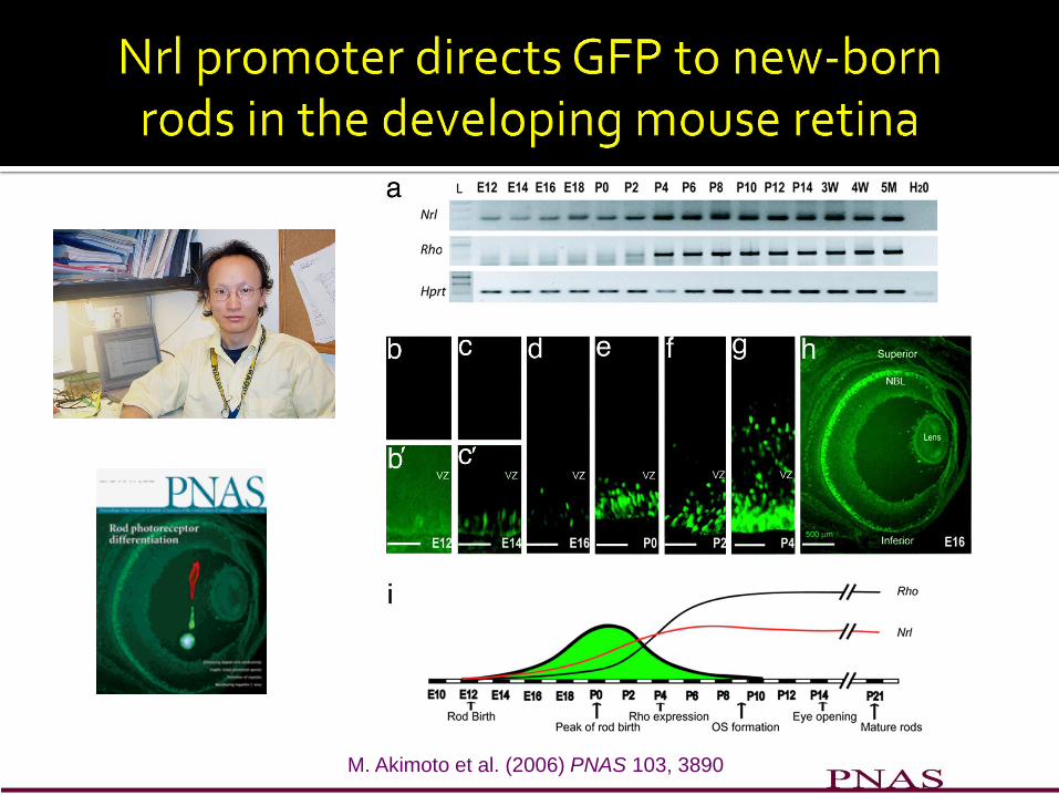
birthdating in mouse retina (Cepko 1996)
All cells generated from common pool(s) of retinal progenitor cells (RPCs)
Conserved order of birth in all vertebrates. Rods are almost 70% in mouse,
born over an extended period. Most cones born prenatally. Peak of rod birth P0-P2
Swaroop et al. Nat Rev Neurosci 2010

No rods differentiate in the absence of Nrl – cone only
retina, S-cone characteristics (Mears et al. Nat. Genet. 2001)
ko wild type
Dark-adapted ERG
-2.4
-1.4
0.6
0.1
Light-adapted ERG
500 µV
100 ms
Sti
mu
lus
In
ten
sit
y (
log
cd
-s/m
2)
Wild-type Nrl-ko
Window of developmental plasticity
Crx promoter – Nrl: all rods
S-opsin promoter – Nrl: 40% rods Oh, E et al. (2007) PNAS 104, 1679-1684

Ng L et al. J. Neurosci. 2011;31:11118-11125

Swaroop et al. Nat Rev Neurosci 2010
Combinatorial action of multiple factors
S-cone “default”
NRL and TRb2 dictate the generation of three photoreceptor types
NRL action is dominant over TRb2
M. Akimoto et al. (2006) PNAS 103, 3890

Gene Regulatory Networks underlying development and disease Cell Replacement Therapy Drug Discovery

Cell-type specific gene expression
maturation
functional photoreceptor
immature photoreceptor
Elaboration of outer segments and synapse formation
immature photoreceptor
post-mitotic precursors
E12-E18 P0-P2 P4-P6 P8-P12 P21-P28
mRNA profiling of flow-sorted rods – Affy GeneChips and RNA-Seq
(Matthew Brooks, Linn Gieser, Jerome Roger, Norimoto Gotoh, Tiziana Cogliati)
Transcription factors – ChIP-Seq (Hong Hao et al. PLoS Genet 2012) and Protein
complexes (Sharda Yadav)
Histone modifications – ChIP-Seq (Hyun-Jin Yang)
DNA methylation (Jung-Woong Kim)

Differentiation and integration of GFP-tagged rod precursors
only immature developing rods
MacLaren et al. Nature 2006

Yao, Zacks et al., IOVS 2009
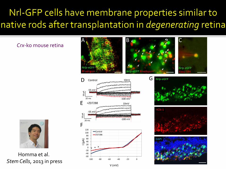
Homma et al. Stem Cells, 2013 in press
Crx-ko mouse retina

Immature Photoreceptors
Cell replacement Disease mechanisms
Small molecule screening
hES cells
Induced pluripotent stem (iPS) cells
iPS cells from genetically abnormal cells – patients

Eiraku et al. 2011 Y. Sasai group
Optic Vesicle
Kohei Homma and Jessica Cooke

Dnmt1 knockdown in early eye development Nasonkin et al. Development 2013 in press

Photoreceptors from stem cells – more work is needed on
function, quality, growth characteristics, epigenome
No cell surface markers yet for distinct stages of differentiation
Cell transplantation methods – good
Cell spreading – scaffold may be needed
Cell integration – poor
Synapse formation?
Cell survival – for several months
Better assessment of efficacy in animal models – Optical
coherence tomography, optokinetic response……
Integration with RPE: 3-D reconstruction

Douglas Forrest (NIDDK), Mahendra Rao (NCRM),
Robert Fariss, Lijin Dong, Kapil Bharti and Sheldon Miller (NEI)
N-NRL 2012


Sponsored by:
Participating Experts:
Brought to you by the Science/AAAS Custom Publishing Office
Webinar Series Science Between Thought and Therapy: Translating Neurobiology Research into
Treatments 13 February, 2013
Ellen Sidransky, M.D.
National Human Genome Research Institute, NIH
Bethesda, MD
Anand Swaroop, Ph.D.
National Eye Institute, NIH
Bethesda, MD
Carlos A. Zarate, M.D.
National Institute of Mental Health, NIH
Bethesda, MD

Look out for more webinars in the series at:
webinar.sciencemag.org
For related information about the Intramural Research Program:
irp.nih.gov
To provide feedback on this webinar, please e-mail
your comments to [email protected]
Sponsored by:
Brought to you by the Science/AAAS Custom Publishing Office
Webinar Series Science Between Thought and Therapy: Translating Neurobiology Research into
Treatments 13 February, 2013