SCI Adv Drug Deliv 2008
24
Cell Therapy for Spinal Cord Regeneration Stephanie M. Willerth 1 and Shelly E. Sakiyama-Elbert 1,2,* 1 Department of Biomedical Engineering, Washington University in St. Louis 2Center for Materials Innovation, Washington University in St. Louis Abstract This review presents a summary of the various types of cellular therapy used to treat spinal cord injury. The inhibitory environment and loss of axonal connections after spinal cord injury pose many obstacles to regenerating the lost tissue. Cellular therapy provides a means of restoring the cells lost to the injury and could potentially promote functional recovery after such injuries. A wide range of cell types have been investigated for such uses and the advantages and disadvantages of each cell type are discussed along with the research studying each cell type. Additionally, methods of delivering cells to the injury site are evaluated. Based on the current research, suggestions are given for future investigation of cellular therapies for spinal cord regeneration. Keywords embryonic stem cells; neural stem cells; spinal cord injury; regenerative medicine; bone marrow stromal cells; biomaterials 1. Introduction Less than 1% of people who sustain significant injury to their spinal cord recover complete neurological function and many of these injuries result in partial or complete paralysis. The three most common causes of spinal cord injury (SCI) include motor vehicle accidents, falls, and gunshot wounds. The number of people in the United States who currently live with SCI is estimated to be around 253,000 with 11,000 new cases occurring each year. The cost of lifetime care for one patient with SCI ranges from $700,000 to $3,000,000 [1]. Thus, a need exists for the development of therapies for treatment to ease both the physical and financial burdens of people who are afflicted with SCI. Many of the current strategies for treatment of SCI involve replacing the cells lost to injury with cells derived from an alternative source. The end goal of such treatments is to help restore function that was lost to the injury. In preclinical studies, such recovery is usually evaluated in the context of rodent models of SCI with the most promising strategies being translated to primate models. Such studies lay the groundwork for future clinical trials. This review seeks to describe the present state of cell therapy for spinal cord injury. First, the obstacles to regeneration present in the injury site as well as the issues involved in cell sourcing are detailed. Next, the different types of cell therapy are defined with the benefits and * To whom correspondence should be addressed: Shelly Sakiyama-Elbert Department of Biomedical Engineering Washington University Campus Box 1097 One Brookings Drive St. Louis, MO 63130 Telephone: (314) 935-7556 Fax: (314) 935-7448 sakiyama@w ustl.edu Publisher's Disclaimer: This is a PDF file of an unedited manuscript that has been a ccepted for publication. As a service t o our customers we are providing this early version of the manuscript. The manuscript will undergo copyediting, typesetting, and review of the resulting proof before it is published in its final citable form. Please note that during the production process errors may be discovered which could affect the content, and all legal disclaimers that apply to the journal pertain. NIH Public Access Author Manuscript Adv Drug Deliv Rev. Author manuscript; available in PMC 2009 January 14. Published in final edited form as: Adv Drug Deliv Rev. 2008 January 14; 60(2): 263–276. N I H - P A A u t h o r M a u s c r i p t N I H - P A A u t h o r a u s c r i p t N I H P A A u t h o r a u s c r i p t
-
Upload
amaiaferrero -
Category
Documents
-
view
224 -
download
0
Transcript of SCI Adv Drug Deliv 2008
8/8/2019 SCI Adv Drug Deliv 2008
http://slidepdf.com/reader/full/sci-adv-drug-deliv-2008 1/24
Cell Therapy for Spinal Cord Regeneration
Stephanie M. Willerth 1 and Shelly E. Sakiyama-Elbert 1,2,*1 Department of Biomedical Engineering, Washington University in St. Louis
2 Center for Materials Innovation, Washington University in St. Louis
AbstractThis review presents a summary of the various types of cellular therapy used to treat spinal cordinjury. The inhibitory environment and loss of axonal connections after spinal cord injury pose manyobstacles to regenerating the lost tissue. Cellular therapy provides a means of restoring the cells lostto the injury and could potentially promote functional recovery after such injuries. A wide range of cell types have been investigated for such uses and the advantages and disadvantages of each celltype are discussed along with the research studying each cell type. Additionally, methods of
delivering cells to the injury site are evaluated. Based on the current research, suggestions are givenfor future investigation of cellular therapies for spinal cord regeneration.
Keywords
embryonic stem cells; neural stem cells; spinal cord injury; regenerative medicine; bone marrowstromal cells; biomaterials
1. IntroductionLess than 1% of people who sustain significant injury to their spinal cord recover completeneurological function and many of these injuries result in partial or complete paralysis. The
three most common causes of spinal cord injury (SCI) include motor vehicle accidents, falls,and gunshot wounds. The number of people in the United States who currently live with SCIis estimated to be around 253,000 with 11,000 new cases occurring each year. The cost of lifetime care for one patient with SCI ranges from $700,000 to $3,000,000 [1]. Thus, a needexists for the development of therapies for treatment to ease both the physical and financialburdens of people who are afflicted with SCI. Many of the current strategies for treatment of SCI involve replacing the cells lost to injury with cells derived from an alternative source. Theend goal of such treatments is to help restore function that was lost to the injury. In preclinicalstudies, such recovery is usually evaluated in the context of rodent models of SCI with themost promising strategies being translated to primate models. Such studies lay the groundwork for future clinical trials.
This review seeks to describe the present state of cell therapy for spinal cord injury. First, the
obstacles to regeneration present in the injury site as well as the issues involved in cell sourcingare detailed. Next, the different types of cell therapy are defined with the benefits and
*To whom correspondence should be addressed: Shelly Sakiyama-Elbert Department of Biomedical Engineering Washington UniversityCampus Box 1097 One Brookings Drive St. Louis, MO 63130 Telephone: (314) 935-7556 Fax: (314) 935-7448 [email protected]
Publisher's Disclaimer: This is a PDF file of an unedited manuscript that has been accepted for publication. As a service to our customerswe are providing this early version of the manuscript. The manuscript will undergo copyediting, typesetting, and review of the resultingproof before it is published in its final citable form. Please note that during the production process errors may be discovered which couldaffect the content, and all legal disclaimers that apply to the journal pertain.
NIH Public AccessAuthor Manuscript
Adv Drug Deliv Rev . Author manuscript; available in PMC 2009 January 14.Published in final edited form as: Adv Drug Deliv Rev . 2008 January 14; 60(2): 263–276.N
I H -P A A u
t h or Manus c r i pt
N I H -P A A ut h or Manus c r i pt
N I H -P A A ut h or M
anus c r i pt

8/8/2019 SCI Adv Drug Deliv 2008
http://slidepdf.com/reader/full/sci-adv-drug-deliv-2008 2/24
drawbacks of each type given. Methods for delivering cells are also discussed. Finally,suggestions are made for future work in developing therapies for SCI.
2. Considerations for developing cell therapy for spinal cord regenerationTo successfully treat SCI by promoting functional recovery, cellular therapies must integrateinto the injury site and restore the lost neuronal circuitry or promote plasticity of the sparedneurons. To achieve this goal, cellular therapies should be designed considering both theobstacles posed by the injury site as well as sourcing and reproducibility issues associated withdifferent cell culture systems.
2.1 Obstacles to regeneration presented by the injured spinal cord
SCI initiates a chain of events that lead to cell death, scarring and the loss of function. Theinitial trauma injures cells and induces swelling. The damaged cells release toxins that causenecrosis of the cells above and below the injury site. The lesion site that results from SCI posesmany obstacles to promoting regeneration. Subsequent events include the formation of a cysticcavity at the injury site, which becomes surrounded by a glial scar, composed of mainly reactiveastrocytes [2]. The demyelination that occurs after injury produces several inhibitory moleculesthat contribute to the lack of regeneration [3-5]. This section will address these obstacles inmore depth and offer suggestions for creating a more permissive environment for regeneration
following SCI.
2.1.1 Cavity formation— After the initial injury and following necrosis, a fluid filled cavitydevelops, resulting in the loss of grey and white matter [6]. The cavity can expand to additionalspinal cord segments preceding and following the injury site, causing additional cell death andincreased loss of function. Formation of the cystic cavity generates a physical barrier tospontaneous regeneration. Cell therapy can help overcome this barrier through a variety of methods. Implantation of neurons or cells that can differentiate into neurons could potentiallybridge the lesion and restore signaling in the spinal cord. Other types of cells can secrete factorsthat promote regeneration of the damaged axons into the cavity while also providing the trophicsupport necessary for cell migration. Such strategies have demonstrated that reduction of thecavity size correlates with an increase in functional recovery [7,8].
2.1.2 Glial scar formation and its inhibitory nature— Many different types of cells,such as macrophages, oligodendrocyte precursors, and meningeal cells, migrate to the injurysite at different time points following injury. However, the resulting glial scar that formsconsists of predominantly reactive astrocytes [2]. The astrocytes are densely packed and secreteinhibitory molecules into their extracellular matrix, making it difficult for injured neurons toregenerate across the injury site. The main class of inhibitory molecules produced by reactiveastrocytes is chondroitin sulfate proteoglycans (CSPGs) [9]. These molecules have been shownto inhibit axonal regeneration in 3-D settings similar to those found in glial scarring [10]. Oneof the CSPGs responsible for inhibition of axonal migration is N2, which blocks the neuriteextension promoting effects of laminin found in the extracellular matrix [10,11].
Cell therapy addresses some of these barriers produced by the glial scar. Transplanted cellscan secrete extracellular matrix and cytokines that promote cell migration to help providecounterbalance to the inhibitory effects of the glial scar. Such strategies can also lessen theamount of scarring present. Additional strategies for eliminating inhibitory CSPGs, whichcould also affect the migration of transplanted cells, include the use of molecules that preventCSPG synthesis and chondroitinases which degrade the CSPGs in vivo [10,12]. These strategiescan be combined with cell transplantation to create effective regeneration strategies.
Willerth and Sakiyama-Elbert Page 2
Adv Drug Deliv Rev . Author manuscript; available in PMC 2009 January 14.
N I H -P A A
ut h or Manus c r i pt
N I H -P A A ut h or Manus c r i pt
N I H -P A A ut h or
Manus c r i pt

8/8/2019 SCI Adv Drug Deliv 2008
http://slidepdf.com/reader/full/sci-adv-drug-deliv-2008 3/24
2.1.3 Myelin Based Inhibitors— Other major obstacles present after SCI include myelinbased inhibitors such as Nogo, myelin based glycoprotein (MAG), oligodendrocyte myelinglycoprotein (OMgp) and tenascin, which are released by damaged oligodendrocytes [3,5,13,14]. Nogo-A, the predominant isoform expressed in the CNS, consists of two transmembranedomains and one extracellular domain and inhibits neurite outgrowth [15]. This extracellulardomain contains 66 amino acids and by itself can be used as an agonist referred to as Nogo-66[3]. Other inhibitors present after SCI include MAG and OMgp. These three proteins (Nogo-
A, OMgp, and MAG) all bind to the Nogo receptor, which mediates the inhibition of neuriteoutgrowth by signaling through Rho kinase. These proteins can be neutralized through the useof Nogo specific antibodies, such as the monoclonal antibody IN-1 [16], agonists such asNogo-66 [3], using enzymatic removal of the receptor [17] or by knocking down expressionof the Nogo receptor that binds these proteins [18]. Sialidase, which removes the glycan bindingdomains of MAG, has also shown promise for enhancing axonal outgrowth after injury [19].Tenascin-R, an extracellular matrix glycoprotein, contributes to the inhibitory environmentpresent after SCI. Multiple studies have demonstrated its effects on preventing neuriteoutgrowth and restricting motor neuron innervations after injury [20,21]. Tenascin-R deficientmice showed improved functional recovery after SCI compared to wild type mice, suggestingthat knocking down its expression could be used as a therapeutic intervention. Thus, acombination of genetic manipulation and antibodies can be used to address the inhibitoryproteins produced by the damaged oligodendrocytes.
2.2 Challenges in developing cellular therapies to treat spinal cord injury
The spinal cord possesses a complex architecture, composed of neurons, oligodendrocytes, andastrocytes. The careful arrangement of the different tracts of the spinal cord allow for a varietyof information to be transmitted throughout the body. The different cell types and spatialarrangement of the spinal cord present challenges when trying to replicate the structure throughthe use of transplanted cells. This section will address the issues associated with developingcellular therapies for the treatment of SCI.
2.2.1 Cell Sourcing Issues— To successfully develop cell based therapies, many cellsourcing issues need to be considered. When developing such a strategy, the first considerationis how the cells will be obtained and the quantity of cells needed for transplantation. If the cells
are obtained from an animal source for preclinical studies, thought should be given into howto translate such work to humans for clinical studies. For cells isolated from tissue, care shouldbe taken to characterize the cells obtained to determine which specific cell phenotypes arebeing implanted. For studies examining the effects of implanting subpopulations of isolatedcells, it is important to ensure the purity of the cells for implantation to later allow determinationof how specific cell types contribute to regeneration. For many stem cell lines, issues arisewhen the cells are continuously cultured, including the reproducibility of the cell lines andnumber of times a cell line can be passaged. In particular, the properties of stem cells can varywith passage number. Other issues associated with human embryonic stem cell lines includethe use of culture systems that rely on serum or feeder cell layers. There is a need fordevelopment of culture methods for human stem cells that involve chemically defined media.Such advances will allow for human stem cells to be transplanted in clinical trials with fewerregulatory issues.
2.2.2 Transplantation Issues— Once appropriate cell sourcing has been obtained, manydifferent issues associated with cell transplantation must be considered. The first issue is howthe cells will be delivered and common methods of transplantation will be discussed later inthis review. The cell transplantation method should ensure the integrity and viability of thecells. For progenitor cells, care should be taken to ensure that the delivery method does notinduce unwanted differentiation or proliferation, which can lead to formation of tetratomas.
Willerth and Sakiyama-Elbert Page 3
Adv Drug Deliv Rev . Author manuscript; available in PMC 2009 January 14.
N I H -P A A
ut h or Manus c r i pt
N I H -P A A ut h or Manus c r i pt
N I H -P A A ut h or
Manus c r i pt

8/8/2019 SCI Adv Drug Deliv 2008
http://slidepdf.com/reader/full/sci-adv-drug-deliv-2008 4/24
For cross species transplantation, such as mouse cells or human cells being transplanted intorat injury models, the issues of immune response should be addressed by treating the animalswith appropriate immunosuppressants. The end goal for cellular therapies would be to createcell lines for transplantation that do not require immune suppression of the patient. Other issuesto consider are the location of the cells after transplantation and how to control celldifferentiation, proliferation and migration in the injury sites. Uncontrolled proliferation canlead to the formation of tetratomas after implantation, which is undesirable. One delivery
method that can address the issue of controlling cell differentiation and migration is throughthe use of biomaterial scaffolds which are discussed later in this article [22-26].
3. Types of Cellular TherapyA variety of cell types have been evaluated in the context of SCI. Each section will summarizethe advantages and disadvantages of each cell type and discuss the studies performed. TableOne lists the cell types discussed in this review along with the references associated with eachcell type. Unless specifically mentioned, these cellular therapies are evaluated in the contextof rat models of SCI. The most common assessment of functional recovery in these models isthe Basso, Beattie, and Bresnahan (BBB) scale for assessing locomotor behavior [27].Additional methods of assessing functional recovery include having the animals performspecific tasks, such as tape removal or rope walking and sensory recovery, such as response
to thermal and mechanical stimuli.
3.1 Embryonic stem cells
Embryonic stem cells (ESCs), which are derived from the blastocyst, possess two importantproperties: pluripotency and the ability to self renew. Their ability to differentiate into cellsfrom all three germ layers make them an attractive source for a variety of applications[28-30]. Some of the challenges in using ESCs include determining the correct cues to directdifferentiation in the specific desired cell types in vitro and in vivo and preventing tetratomaformation [31]. Many of these differentiation protocols involve formation of aggregates of cellsknown as embryoid bodies (EBs) and treatment with growth factors [23,32-34].
3.1.1 Mouse embryonic stem cells— Mouse ESCs have been investigated as a startingpoint for developing potential therapies for SCI. These cells are attractive for preclinical studiesbecause they are easy to culture and readily differentiate into the cells found in the CNS. Manydifferent mouse ES cell lines exist and these lines can be studied with federal funding in theUnited States. Often, mouse ESCs are tested in the context of rat models of SCI, allowing theimplanted cells to be distinguished from the host cells. However, immunosupression isnecessary to prevent rejection of these cells by the host immune system.
One of the first studies that looked at using mouse ESCs for treatment of spinal cord injuryinvolved a multistep process that promoted the formation of oligodendrocytes and astrocytesfor transplantation [35]. Once transplanted into the spinal cord, these cells demonstrated theability to restore myelination in myelin-deficient shiverer rats, indicating the potential of mouseESCs to treat SCI.
This work was followed by a pair of studies performed by the McDonald lab that looked at theability of mouse ESCs treated with retinoic acid to promote recovery in SCI models [34,36].In the first study, the cells were grown as 4 − /4+ retinoic acid treated EBs [33], partiallydissociated and injected nine days after contusion injury [36]. These cells differentiated intomainly oligodendrocytes and astrocytes and were able to promote an increase in BBB scores.The second study focused on producing oligodendrocytes from mouse ESCs for remyelinationtherapy [34]. The first part of the study determined a set of culture conditions to produce mainlyoligodendrocytes from the mouse ESCs. These oligodendrocytes were demonstrated to
Willerth and Sakiyama-Elbert Page 4
Adv Drug Deliv Rev . Author manuscript; available in PMC 2009 January 14.
N I H -P A A
ut h or Manus c r i pt
N I H -P A A ut h or Manus c r i pt
N I H -P A A ut h or
Manus c r i pt
8/8/2019 SCI Adv Drug Deliv 2008
http://slidepdf.com/reader/full/sci-adv-drug-deliv-2008 5/24
remyelinate axons both in vitro and in an in vivo model consisting of chemically induceddemyelination in the spinal cord.
Other studies have demonstrated that mouse ESCs can survive in the spinal cord for more than50 days, suggesting their potential as a long term treatment for SCI [37]. A more recent studyshowed that mouse ESCs pretreated with a lecithinized brain derived neurotrophic factor(BDNF) implanted 9 days after T9/10 contusion injury showed better differentiation and
promoted increased functional recovery afterwards, as assessed by BBB compared to cellstreated with normal BDNF, demonstrating the influence of growth factor treatments on ESCdifferentiation [38]. Mouse ESCs predifferentiated using the 4 − /4+ protocol were implantedafter 7 days into a chronic pain SCI model that consisted of repeated microinjections of quisqualic acid between T13 and L1 [39]. These cells helped relieve chronic pain as indicatedby reduction in excessive grooming behavior and increased sensitivity to other thermal andmechanical stimuli.
Recent work has looked at different types of genetically modified mouse ESCs and their impacton SCI. Mouse ESCs were modified to overexpress the bcl-2 protein, which blocks apoptosis,and implanted 9 days after contusion injury [40]. However, these cells did not produce anincrease in functional recovery and resulted in tumor-like growths. This study illustrates theimportance of selecting the appropriate proteins when genetically modifying cells for
transplantation. Another study examined the effect of transfecting mouse ESCs with the L1adhesion molecule, which promotes neuronal outgrowth, to determine the effect on cellbehavior [41]. These cells were injected both rostrally and caudaully to the injury site 7 daysafter a T7-9 compression injury. After one month, the cells transfected with L1 showedincreased survival and migration compared to untransfected ESCs. Overall, mouse ESCs servea model cell culture system for determining effective strategies for treatment of SCI due to theease of culture and ability to be genetically modified. These strategies provide a starting placefor translating such methods to human ESCs.
3.1.2 Human embryonic stem cells— Development of methods and strategies for usinghuman ESCs for treatment of SCI allows for translation to clinical studies. One of thechallenges of developing human ESC therapies is determining the necessary cues to promotedifferentiation in the desired cell types, such as oligodendrocytes and motor neurons. Although
the work done with mouse ESCs provides a good starting point, the end goal is to determinehuman ESC differentiation protocols. Similar to the mouse ESCs, testing the ability of humanESCs to promote recover in rat injury models requires immunosupression. Many of the humanESC lines that currently exist have been cultured in the presence of mouse feeder cell layers,making them undesirable for use in clinical trials. Other major challenges include developingperson specific ESC lines to allow transplantation without immunosupression in a clinicalsetting.
Work from the Keirstead lab has focused on developing protocols that produce large numbersof oligodendrocytes from human ESCs [42-45]. Their time intensive differentiation protocolinvolves multiple selection steps with serum free media to produce oligodendrocyte progenitorcells (OPCs) that can differentiate into mature oligodendrocytes in vitro and in vivo [42]. Theyimplanted these cells into myelin deficient shiverer mice where they integrated into the spinalcord, differentiated into oligodendrocytes and restored myelination. Based on these results,they investigated transplanting these human ESC-derived OPCs 7 days after T10 contusioninjury [43]. They demonstrated that these cells differentiated into mature oligodendrocytes invivo that could remyelinate damaged axons. An increase in BBB scores for animals thatreceived the OPCs was observed compared to animals that received media only and injuredcontrol animals. Further studies indicated that such a strategy for treatment of SCI is onlysuccessful when extensive demyelination induced by a 200 kD contusion injury is present, but
Willerth and Sakiyama-Elbert Page 5
Adv Drug Deliv Rev . Author manuscript; available in PMC 2009 January 14.
N I H -P A A
ut h or Manus c r i pt
N I H -P A A ut h or Manus c r i pt
N I H -P A A ut h or
Manus c r i pt
8/8/2019 SCI Adv Drug Deliv 2008
http://slidepdf.com/reader/full/sci-adv-drug-deliv-2008 6/24
not for a 50 kD injury that does not induce demyelination [45]. Another study has investigateddifferentiating human ESCs into neurons for use as a therapy for SCI [32]. This studydetermined a protocol for producing large numbers of motor neurons from human ESCs. Thesecells were able to maintain their phenotype after in vivo transplantation into the adult rat spinalcord. This study demonstrate the potential to create human ESC derived neurons, but thesecells still need to be further characterized to determine how they will integrate and contributeto functional recovery after SCI.
3.2 Neural stem cells
Neural stem cells (NSCs) refer to the multipotent stem cells that give rise to the cells of thenervous system. These cells are self renewing and are often cultured as neurospheres. They arefound in both embryonic and adult tissues. NSCs isolated from the central nervous system(CNS) also have been explored as a potential therapy for SCI. Figure 1 shows a schematic of how NSCs could potentially promote recovery after SCI through different mechanisms. Themajority of the studies use NSCs derived from embryonic animal tissue, usually mouse or rats.Unless specifically mentioned otherwise, embryonic NSCs were isolated from the CNS of ratson day 14 of embryonic development. Unlike ESCs, these cells are limited to becomingneurons, oligodendrocytes, and astrocytes, making it easier to achieve differentiation into thedesired cell types. For many of the studies, the NSCs were harvested from the same species of animal that they were then transplanted back into to eliminate the possibility of rejection. Thismethod of harvesting embryonic NSCs poses a problem for clinical studies since these methodscannot be replicated ethically in humans. Adult NSCs provide an important alternative toembryonic NSCs and could potentially be obtained from a patient or cadaver to be transplantedinto their injured spinal cord, making them attractive from a clinical perspective.
In one of the first studies using rat embryonic NSCs, Cao et al . investigated implanting NSCsinto both normal and lesioned (T8 contusion) spinal cord [46]. They found the majority of theimplanted cells differentiated into astrocytes, suggesting that these cells may need to be pre-differentiated before implantation [47]. Further work demonstrated that factors present afterSCI restrict differentiation of these cells. To try and overcome some of these signals,specifically bone morphogenetic protein (BMP), NSCs were engineered to express noggin, aBMP agonist, to attempt to obtain better differentiation into neurons and oligodendrocytes[48]. This strategy was unsuccessful at preventing differentiation and these cells actuallyproduced an increase in lesion size. However, a different study successfully implemented thisstrategy and achieved differentiation of NSCs expressing noggin into neurons andoligodendrocytes after being implanted into a T8 contusion injury as well as promoted anincrease in BBB scores 3 weeks after injury [49]. These studies illustrate the importance of controlling protein expression levels when performing genetic manipulation.
Other studies have used NSCs in combination with other strategies to treat SCI. The Langerlab seeded mouse NSCs into polymer scaffolds [25]. These scaffolds were then implanted intothe lesion site resulting from a T9/10 lateral hemisection. This approach produced functionalrecovery as indicated by an increase in BBB scores after 3 weeks compared to animals receivingonly cells and the lesion only animals. This increase in recovery was observed throughout therest of the 10 week study even though the transplanted cells did not stain positive for mature
cell markers, indicating that they remained undifferentiated. Tuszynski and colleaguesconfirmed that mouse NSCs constitutively secrete a variety of growth factors, including nervegrowth factor (NGF), BDNF, and glial-derived neurotrophic factor (GDNF), in vitro and invivo [50]. In vivo , the transplanted NSCs promoted axonal sprouting in a C3 hemisection injury.They then genetically modified these NSCs to produce neurotrophin-3 (NT-3), which whentested in the same in vivo model, enhanced the axonal sprouting that was previously observed.Another approach combined NSC transplantation with antibodies that neutralize the effects of
Willerth and Sakiyama-Elbert Page 6
Adv Drug Deliv Rev . Author manuscript; available in PMC 2009 January 14.
N I H -P A A
ut h or Manus c r i pt
N I H -P A A ut h or Manus c r i pt
N I H -P A A ut h or
Manus c r i pt

8/8/2019 SCI Adv Drug Deliv 2008
http://slidepdf.com/reader/full/sci-adv-drug-deliv-2008 7/24
ciliary neurotrophic factor (CNTF) [51]. This study showed that neutralizing CNTF reducedthe amount of NSCs that differentiated into astrocytes while promoting regeneration of thecorticospinal tract. No behavioral analysis was reported for this study. A different study by theSchwartz lab looked at transplanting adult NSCs 7 day post injury in combination with myelinspecific T cells to determine the effect on SCI in a mouse model (T12 contusion injury) [52].The combination of cells was able to produce functional recovery as evidenced by an increasein the Basso mouse scale (BMS) [53].
The Weidner group has studied the use of rat adult neural progenitor cells (NPCs) as a therapyfor treating SCI. One of their studies looked at the differentiation of these cells both in vitroand in vivo [54]. In vitro , the adult NPCs differentiated into mostly neurons andoligodendrocytes with few astrocytes present. These NPCs were then tested in vivo bytransplanting them immediately following a C3 dorsal column transaction injury. These cellsdid not have any effect on the lesion size, but did colocalize with the axons of the corticospinaltract. A follow up study showed that transplanting these NPCs along with fibroblasts producedan increase in axonal regeneration compared to transplanting only fibroblasts into the injurysite [55].
Several studies have investigated the potential of NSCs derived from humans to promoterecovery in animal models of SCI. One of the first studies looked at the ability of human NSCs
derived from fetal brain cultured as neurospheres to promote recovery after T9 contusion injuryin both severe combined immunodeficiency (SCID) and myelin-deficient shiverer mice [56].These cells differentiated into functional neurons and oligodendrocytes while promoting anincrease in functional recovery as assessed by BBB scores. Additional work confirmed theincrease in recovery was due to the NSCs [57]. Another study looked at priming human NSCsderived from fetal tissue with a combination of growth factors to induce preferentialdifferentiation towards cholinergic neurons [58]. After transplantation into the spinal cord,these cells were able to differentiate into cholinergic neurons whereas the same cells that werenot treated with the growth factors differentiated into astrocytes. A different study looked atthe effect of implanting human NSCs into a C5 contusion injury in marmosets [59]. The NSCsdifferentiated into neurons, oligodendrocytes, and astrocytes while promoting an increase inhand grip and motor activity compared to control animals. Finally, Tarasenko and colleaguestested the effects of priming human NSCs with a combination of heparin, laminin and FGF to
determine if it would affect in vivo differentiation [60]. The cells that were primed demonstratedincreased viability after transplantation into rats compared unprimed cells. Although nodifferences were observed in BBB scores between groups, the animals receiving the primedcells showed an increase in trunk stability as assessed by rearing activity.
3.3 Bone marrow stromal stem cells
Bone marrow stromal cells (BMSCs) are multipotent progenitor cells obtained by taking theheterogeneous cell population found in bone marrow and selecting for the adherentsubpopulation. Rat and human BMSCs can differentiate into cells that express markers formature neuronal cells [61-63], but later studies indicate that these cells do not express voltagegated ion channels [64]. From a clinical perspective, BMSCs are attractive for transplantationbecause they are easily obtained from bone marrow and can be transplanted back into the
original donor, eliminating the risk of rejection. Hofstetter et al. transplanted BMSCs into T7contusion injury model of SCI both immediately following injury and 7 days post injury [64].The cells transplanted 7 days post injury showed better rates of survival and formed bundlesthat bridged the lesion. Implantation of BMSCs also led to an increase in BBB scores comparedto controls. Another study showed coculture of BMSCs along with NSCs obtained from thespinal cord promoted differentiation of the NSCs into neurons and glia in vitro [8]. In vivotransplantation of BMSCs immediately after T8/9 contusion injury reduced cavity formation
Willerth and Sakiyama-Elbert Page 7
Adv Drug Deliv Rev . Author manuscript; available in PMC 2009 January 14.
N I H -P A A
ut h or Manus c r i pt
N I H -P A A ut h or Manus c r i pt
N I H -P A A ut h or
Manus c r i pt
8/8/2019 SCI Adv Drug Deliv 2008
http://slidepdf.com/reader/full/sci-adv-drug-deliv-2008 8/24
and also promoted an increase in BBB scores. The authors suggest that the BMSCs providedtrophic factors and support for the cells present in the injured spinal cord, allowing them tomigrate into the injury site. Similar results were obtained by a different group when BMSCswere infused in the cerebrospinal fluid (CSF), providing an alternate method of delivery [7].Other studies have also looked at the feasibility of different methods of intravenous injectionfor BMSC delivery [65-67], but a study by Vaquero et al. showed that direct injection of BMSCs into the site of a T6-8 contusion injury promoted superior functional recovery when
compared to intravenous injections of BMSCs [68]. A long term study by the Vaquero lablooked at the long term effects of transplanting BMSCs 3 months after a crush injury [69]. Thisstudy showed that functional recovery steadily increased over the course of a year as indicatedby BBB scores. Finally, human BMSCs transplanted into a T9 contusion model of SCI alsopromoted functional recovery as indicated by BBB scores, suggesting that this therapy haspotential to work in humans [70].
Additional work has been done to clarify the mechanisms by which BMSCs promote functionalrecovery. One study demonstrated that BMSCs help guide regenerating axons across the injurysite when implanted 2 days after a T8 contusion injury and can help promote recovery byrestoring the stepping control circuitry [71]. A more recent study showed that BMSCs expressthe gamma aminobutyric acid (GABA receptor) [72]. They implanted BMSCs rostrally to thelesion 7 days after a T10/11 impact injury. However, in this study, functional recovery as
assessed by the BBB scale was not observed 4 weeks after injury. BMSCs stimulatephosphoinositide-3-kinase and mitogen-activated protein kinase signaling in neurons, whichpromotes their survival [73].. All of these mechanisms contribute to the success of BMSCtransplantation as a treatment for SCI.
A pair of studies from the Tuszynski lab investigated the use of BMSCs that were geneticallymodified to express growth factors to treat a dorsal column transaction injury at C3 [74,75].The first study looked at the effects of transplanting BMSCs that secreted BDNF immediatelyfollowing injury. The BDNF expressing cells induced more robust axonal growth into the lesionsite compared to normal BMSCs [74]. Functional recovery, judged by a tape removal task andrope walking, was not observed. The second study examined the ability of BMSCs modifiedto express NT-3 to induce axonal growth through chronic glial scars [75]. These cells wereimplanted 6 weeks post injury at the C3 level and the scar was not resected. These cells were
able to promote regeneration of axons through the scars and into the lesion site, demonstratingthat the glial scar can be penetrated. These studies illustrate the additional benefits of geneticmodification when used in combination with BMSCs.
3.4 Other cell types
In addition to progenitor cells, researchers have also investigated the use of terminallydifferentiated cells, such as Schwann cells, olfactory ensheathing cells (OECs), and fibroblasts,for the treatment of SCI. These cells are easily obtained and cultured, making them attractivefor translational studies. Schwann cells could potentially be harvested autologously fromperipheral nerves in manner similar to how nerves are harvested for isograft use in repair largegaps produced by injury to the PNS [76]. OECs can be obtained for autologous transplantationthrough nasal biopsies and grown in cell culture until needed [77]. Finally, fibroblasts can be
isolated from skin and cultured in vitro to allow for genetic manipulation before transplantation[78]. These methods for harvesting and culture allow for transplantation without the issue of rejection. However, it often takes 2-4 weeks of in vitro cell culture to generate enough cellsfor transplantation.
3.4.1 Schwann cells— Schwann cells produce the myelin sheaths that surround the neuronsof the peripheral nervous system (PNS) [79]. They play a key role in regenerating axons of the
Willerth and Sakiyama-Elbert Page 8
Adv Drug Deliv Rev . Author manuscript; available in PMC 2009 January 14.
N I H -P A A
ut h or Manus c r i pt
N I H -P A A ut h or Manus c r i pt
N I H -P A A ut h or
Manus c r i pt

8/8/2019 SCI Adv Drug Deliv 2008
http://slidepdf.com/reader/full/sci-adv-drug-deliv-2008 9/24
PNS after injury by converting to non-myelinating Schwann cells that secrete a variety of growth factors, including NT-3, NGF, BDNF, CNTF, and fibroblast growth factor (FGF).These cells also migrate to the injury site after SCI despite being part of the PNS [80]. Thesecells can be easily expanded in vitro , allowing for autologous cell transplantation to avoidimmune response. Thus, Schwann cells have been studied for their potential to promote axonalregeneration and myelination after SCI [81,82].
Bunge and colleagues investigated using Schwann cells seeded in Matrigel inside of nerveguidance conduits (NGCs) with the distal end capped to determine their effect on the injuredspinal cord [83]. In this study, they used a complete transaction injury model, removing thespinal cord between T9-11 and replacing it with the NGCs containing Schwann cells.Compared to control conduits, the NGCs containing Schwann cells showed increasedremyelination as well as propriospinal and sensory axonal regeneration. A follow up studycombined this approach with delivery of BDNF and NT-3 from osmotic pumps [84]. Thisapproach resulted in the regeneration of the supraspinal axons, but no assessment of functionalrecovery was reported. A third study indicated that using open ended channels with such anapproach resulted in axonal regeneration from both the rostral and caudal ends of the injury[85]. Follow up studies using similar approaches demonstrated that the injured animalsregained impulse conduction only after receiving the Schwann cells [86,87]. Finally, Schwanncells showed the ability to promote functional recovery as indicated by the BBB scores when
used to treat a less severe injury (T8 contusion), showing their promise as a therapy for mildSCI [88].
Other studies have looked at genetically modifying Schwann cells to secrete increased amountsof growth factors, specifically NGF and BDNF [89-91]. Although the genetically modifiedcells promoted more axonal regeneration than unmodified Schwann cells, no functionalrecovery was observed with either cell line. Additionally, several studies have used Schwanncells in combination with other strategies for treatment of SCI. Chondroitinase ABC enhancesSchwann cell-induced axonal regeneration, but it does not lead to functional recovery in T9dorsal hemisection injury model [92]. Combining cyclic adenosine monophosphate (cAMP)with Schwann cells as therapy for treatment of a T8 mild contusion injury resulted in functionalrecovery as assessed by the BBB [93]. This study used an inhibitor against thephosphodiesterase that hydrolyzes cAMP to extend its signaling, which allows the neurons to
extend into otherwise inhibitory substrates. These results suggest that Schwann cells work bestwhen used in combination with other therapeutic approaches that minimize the inhibitorynature of the lesion site.
3.4.2 Olfactory ensheathing cells— Unlike the rest of the adult CNS, the olfactory bulbretains its regenerative capacity. Neurons found in the olfactory system last approximately 4weeks before they die and are replaced by newly formed neurons. This difference is attributedto the presence of OECs, which are glial cells that direct the differentiation of progenitor cellspresent in the olfactory system [94]. These cells have been extensively investigated to see if they have the same capacity to promote regeneration following SCI. These cells can beharvested and then transplanted back into the original human donor, eliminating rejection.These cells can are easily obtained, making them ideal for translational research.
Raisman et al. demonstrated that OECs isolated from rats could be used to repair thecorticospinal tract after injury at C1/2 as demonstrated by recovery of reaching behavior onthe side of the lesion [95]. Follow up studies showed that these cells induced myelination,which allowed for this recovery [96]. They later repeated this work in a chronic SCI model. Inthat study, the cells were implanted 8 weeks after injury and 1 to 3 weeks later, the animalsreceiving the cells demonstrated functional recovery [97].
Willerth and Sakiyama-Elbert Page 9
Adv Drug Deliv Rev . Author manuscript; available in PMC 2009 January 14.
N I H -P A A
ut h or Manus c r i pt
N I H -P A A ut h or Manus c r i pt
N I H -P A A ut h or
Manus c r i pt

8/8/2019 SCI Adv Drug Deliv 2008
http://slidepdf.com/reader/full/sci-adv-drug-deliv-2008 10/24
Other groups confirmed similar findings when OECs were implanted in other types of injurymodels. After complete spinal cord transaction at T8/9, rats receiving OECs immediately afterinjury showed an increased ability to climb on grids 3 months following the surgery comparedto untreated injured rats [98]. Similar results were obtained by other groups [99,100]. Adifferent study investigated the use of human OECs obtained during surgery to remove frontalbase tumors. These cells demonstrated that they could also remyelinate myelin-deficientimmunosupressed rats, indicating that human OECs possess similar regenerative properties as
the rat OECs [101].More recent studies have investigated if other mechanisms, in addition to remyelination, werecontributing to the ability of OECs to promote regeneration. One study showed that thepresence of OECs helps mitigate the loss of tissue after photochemical (Rose Bengal) injuryat T8 [102]. This study also observed functional recovery as indicated by BBB scores andmeasurement of sensory-evoked potentials. Similar results were observed by Bunge andcolleagues when OECs were grafted 7 days post T8 moderate contusion injury [103]. Theyalso saw regeneration of supraspinal axons.
Studies have also investigated the use of genetically modified OECs to see if additional benefitand recovery could be obtained. One study compared the effects of modifying OECs to secreteeither NT-3 or BDNF [104]. The cells that expressed BDNF promoted recovery of the
rubrospinal tract after C3 unilateral transaction injury compared to the cells that expressedNT-3 and control cells. The growth factor secreting OECs also promoted functional recoveryas assessed by a rope walking task as compared to animals receiving unmodified OECs andcontrol animals. A follow up study confirmed that the NT-3 secreting cells promoted tissuesparing in the same injury model, but no differences in functional recovery were observedbetween experimental and control groups [105]. OECs modified to secrete GDNF have alsobeen shown to promote functional recovery after SCI [106]. In this study, OECs that secretedGDNF were implanted immediately following T8 transection injury. The animals receivingthe GDNF secreting cells promoted increased BBB scores after 4 weeks and increased abilityto walk up planes after 5 weeks compared to both animals that only received OECs anduntreated injured animals. Currently, Phase I clinical trials using autologous OECtransplantation are underway in Australia where these cells have been demonstrated to be safefor up to one year after treatment [77].
3.4.3 Fibroblasts— Fibroblasts are cells that make up connective tissue and secreteextracellular matrix molecules, such as collagen and glycoproteins. These cells are also easyto obtain and expand in culture, making them attractive for use as a cellular therapy. Fibroblastsare also easy to genetically modify, allowing for additional functionality. However, the methodof transfection should be carefully considered. Some transfection agents only produce transientexpression of the target protein while other methods, such as lentiviral transfection, producestable protein expression, but may cause unwanted mutations upon integrating into thechromosomal DNA of the fibroblasts. It is also important to characterize the rate at which thesecells produce the target proteins as often there is a target range of therapeutic concentrations.Too little or too much protein expression can result in seeing no effect or negative effects onfunctional recovery.
Many studies have been performed to determine the effect of transplanting fibroblasts thatoverexpress neurotrophins, including NT-3, BDNF, and NGF. The Tuszynski lab and theircolleagues have done extensive research into the use of genetically modified fibroblasts fortreatment of SCI. Initially, they examined the effect of grafting fibroblasts modified to secreteNT-3 acutely into a T7 dorsal hemisection model. These cells produced increases in functionalrecovery as assessed by a grid walk test compared to unmodified fibroblasts and fibroblastsmodified to secrete NGF [107]. They also evaluated the effects of grafting the NGF secreting
Willerth and Sakiyama-Elbert Page 10
Adv Drug Deliv Rev . Author manuscript; available in PMC 2009 January 14.
N I H -P A A
ut h or Manus c r i pt
N I H -P A A ut h or Manus c r i pt
N I H -P A A ut h or
Manus c r i pt

8/8/2019 SCI Adv Drug Deliv 2008
http://slidepdf.com/reader/full/sci-adv-drug-deliv-2008 11/24
fibroblasts into a primate model of SCI. In that study, the spinal cord was injured throughmultiple needle penetrations from T7- T9 followed by injections of the fibroblasts. The graftspromoted secretion of extracellular matrix molecules, such as L1, and increased cell migrationinto the injury sites, but no data about the effect on functional recovery was reported [108].They also studied the effects of implanting these cells into a variety of lesion types anddetermined that they could promote axonal regeneration through the CSPG-rich areas of theglial scar [11].
A pair of studies looked at the effects of delayed transplantation of two types of fibroblasts,one set that expressed NT-3 and one set that expressed BDNF [109,110]. The first study lookedat the physiological effects of implanting these cells 6 weeks after complete transection of thespinal cord between the C3 and C4 segments [109]. They observed that the growth factor-secreting grafts promoted axonal growth into the injury site compared to unmodified fibroblastsand rescued red nucleus neurons. The second study looked at the effects of transplanting thesecells on functional recovery through a variety of assessments including BBB scoring, grid walk,mechanical and heat sensitivity tests [110]. The BBB scores actually decreased followingimplantation of the cells, suggesting that the additional surgery reinjured the spinal cord. Theexperimental group receiving the growth factor-secreting cells did showed increased sensitivityto heat compared to the control animals.
Many other groups have also investigated the use of fibroblasts as a potent treatment for SCI.McTigue et al . investigated the effects of several growth factors, including NT-3, BDNF,CNTF, NGF and basic FGF, secreted by genetically modified fibroblasts on regeneration afterSCI [111]. The cells modified to secrete NT-3 and BDNF promoted proliferation of endogenousoligodendrocytes and remyelination of damaged axons.
Other studies have looked at the effect of modifying fibroblasts to express other proteins thatmay have therapeutic benefits for the treatment of SCI. Blesch and Tuszynski investigated theeffects of implanting cells that secrete GDNF immediately after T7 dorsal hemisection injury.These cells promoted regeneration of the dorsal sensory column, propriopsinal and motoraxons, but functional recovery was not observed [112]. Another study looked at graftingfibroblasts that secreted neurotrophin-4/5 into hemisection and complete transaction injuriesat T8 [113]. These cells promoted robust axonal regeneration and Schwann cells were able to
infiltrate the grafts. However, no functional recovery as assessed using the BBB scale wasobserved compared to control animals that only received green fluorescent protein expressingcells. A third study looked at the effects of transplanting fibroblasts that produce matrixmetalloproteinase-3 into T7 contusion injury [114]. They hypothesized that these cells woulddegrade the inhibitory CSPGs present at the injury site, allowing for enhanced regeneration.These cells promoted increased serotonergic fibers distal to the injury site and functionalrecovery compared to unmodified fibroblasts but not to control animals that did not receivecells. This study suggests that the transplantation process or the cells themselves used causeda decrease in functional recovery.
4. Delivery methods for cellular therapyOnce a decision has been made regarding what type of cells to use, the next consideration is
how to deliver the cells to the injury site. Most of the studies discussed previously involveddirectly injecting the cells into and around the injury site . Other minimally invasive injectionmethods have been studied, such as intravenous injection, infusion into CSF, and lumbarpuncture [7,65,67,68]. An alternative method of delivery involves seeding cells into scaffoldsand then implanting the scaffolds into the injury site. This section will review these methodsand their advantages and disadvantages will be discussed.
Willerth and Sakiyama-Elbert Page 11
Adv Drug Deliv Rev . Author manuscript; available in PMC 2009 January 14.
N I H -P A A
ut h or Manus c r i pt
N I H -P A A ut h or Manus c r i pt
N I H -P A A ut h or
Manus c r i pt
8/8/2019 SCI Adv Drug Deliv 2008
http://slidepdf.com/reader/full/sci-adv-drug-deliv-2008 12/24
4.1 Injection
The majority of studies use a variety of injection methods for delivery of cellular therapeutics.The most common method of cell grafting involves injecting cells to deliver them into andaround the site of injury [68]. Many studies inject cells rostrally or caudally from the injurysite to achieve better levels of survival. Direct injection into the injury site often produces poorresults in terms of cell survival due to the harsh environment in the lesion. One of the mainissues with using direct injection in the surviving tissues is minimizing the risk of further injurycaused by performing a laminectomy which to allow access for injection. To overcome thisissue, some studies have explored alternative methods of delivery. Systemic administration of BMSCs by injection into the tail veins of rats has been studied as a minimally invasive injectiontechnique. Although cells delivered using this method were found at the injury site, directinjection of BMSCs into the injury site produced superior functional recovery [68]. Otherinjection methods include using lumbar puncture to deliver cells and injecting the cells intothe CSF containing cavities found in the brain [65,66]. The first study investigated injectingBMSCs at L4-5 using a lumbar puncture method [65]. Administering this cellular therapywithin 14 days of T8 contusion injury produced the best results including a reduction in cystvolume and injury size. Additionally, this method allows for multiple injections, which couldbe useful for treatment of chronic injuries. The second study examined injecting cells into thefourth ventricle of the brain, which consists of a cavity containing CSF [7]. These cells wereinjected at the time of T8 contusion injury and the presence of BMSCs promoted an increasein BBB scores 7 days after surgery compared to untreated, injury only control animals. Thisincrease in scores was observed up to 35 days following the surgery. These minimally invasivemethods show promise, but more research needs to be done to fully characterize the efficacyof these strategies compared to the direct injection method.
4.2 Implantable scaffolds
An alternative method for delivering cells to the injury site involves seeding cells into scaffoldsand then implanting the scaffold into the injury. This method has several advantages over directinjection. One advantage is that scaffolds can provide a more hospitable environment for cellsurvival, as well as trophic support for cells. In some cases, these scaffolds can be modified tocontain cues to promote cell survival and promote progenitor cell differentiation. A variety of materials have been evaluated for their use as scaffolds for treatment of SCI [22,26]. The Langer
lab seeded NSCs into poly(lactic-co-glycolic) acid (PLGA) scaffolds before implanting theminto a hemisection model of SCI where they promoted functional recovery as indicated by anincrease of 4 on the BBB scale compared to animals that received cells only and untreatedinjury only animals [25]. The details of this scaffolding system are shown in Figure 2. Thescaffold architecture was designed to mimic the arrangement of white and grey matter foundin the uninjured spinal cord. More recent in vitro work from the Langer lab has investigatedthe use of scaffolds consisting of 50/50 blends of PLGA and poly L-lactic acid (PLLA) forpromoting the differentiation of human ESCs into neuronal phenotypes [23,24]. Thesescaffolds have been used to promote neuronal differentiation by presenting such cues as retinoicacid and NT-3. Other groups have investigated using poly (ethylene glycol) (PEG) scaffoldsseeded with rat NSCs to engineer neural tissue [115]. A follow up study indicated that bFGFenhanced NSC survival while scaffolds containing collagen did not have an observable effecton NSC behavior [116]. Our lab has evaluated fibrin scaffolds as a method for differentiatingand delivering mouse ESCs [22]. Previous in vivo work has shown that controlled growth factorrelease from such scaffolds provides beneficial short term effects as a treatment for SCI[117-119]. Our most recent work evaluated the effect of different doses and combinations of growth factors on the differentiation of mouse ESCs seeded inside of 3D fibrin scaffolds[120]. Ongoing studies are currently being performed to test this approach in vivo . Additionally,Ma and colleagues have shown that rat embryonic NSCs form functional neuronal circuitswhen cultured inside of collagen scaffolds, showing promise an alternative scaffold material
Willerth and Sakiyama-Elbert Page 12
Adv Drug Deliv Rev . Author manuscript; available in PMC 2009 January 14.
N I H -P A A
ut h or Manus c r i pt
N I H -P A A ut h or Manus c r i pt
N I H -P A A ut h or
Manus c r i pt
8/8/2019 SCI Adv Drug Deliv 2008
http://slidepdf.com/reader/full/sci-adv-drug-deliv-2008 13/24
[121]. This research area, which combines scaffolds with cell transplantation, holds promiseas an alternative to injection methods. Such scaffolds may enhance cell survival aftertransplantation and promote differentiation into desired phenotypes based on the scaffoldproperties.
5. Conclusions and future directions
This review has discussed the major issues associated with cell therapy for spinal cordregeneration, including obstacles presented by SCI, the different cell types available, andmethods for cell transplantation. SCI presents many obstacles to regeneration and many of thestrategies that achieve functional recovery take a multi-component approach. One of themulticomponent approaches involves transplanting two or more types of cells as therapy forSCI. These strategies can be successful because one cell line can provide secreted factors andtrophic support for the second cell line. In addition to cells, protein therapy, such as the use of growth factors, enzymes, and neutralizing antibodies, helps promote regeneration after SCI.Of the growth factors tested experimentally, NT-3, BDNF, and GDNF show the most promisefor SCI treatment. These growth factors can be delivered through genetic modification, osmoticpumps, or by drug eluting scaffolds. The last two methods (osmotic pumps and drug elutingscaffolds) can also be used to deliver enzymes, such as chondroitinase ABC, and neutralizingantibodies, such as α -CNTF and α -NogoR.
Cells genetically modified to secrete growth factors have the advantage of continuouslyproviding an influx of protein. However, this benefit can turn into a disadvantage once healthytissue has been restored. The presence of additional growth factor could lead to unwanted tocell proliferation. The Tuszynski lab has investigated the use of cells that secrete growth factorin response to tetracycline ingestion, thus controlling the rate of secretion [122]. Drug elutingscaffolds can only release the amount of growth factor that was loaded initially into the scaffold.The rate of release can be modulated through a variety of methods and the time course of releasecan be tailored to the time course of regeneration. This process eliminates the issue of unwantedgrowth factor present after regeneration has occurred.
Overall, cell therapy combined with other regeneration promoting strategies holds the mostpromise for restoring function after traumatic SCI. Many of these strategies have demonstrated
efficacy in preclinical trials. For successful translation to the clinic, these strategies should betested in chronic models of SCI and functional recovery should be assessed using a variety of tests, not just the BBB scale. Additionally, the development of embryonic and neural stem celltechnology will require further research to produce both the cell lines and culture methodsnecessary for clinical trials as well as for producing patient specific stem cells. Recent studieshave suggested that patient specific ESCs could be produced by dedifferentiating mature cells[123]. This research provides one potential avenue for translation of ESC therapies to clinicaltrials. Overall, cellular therapies hold great potential for the treatment of SCI.
Acknowledgments
Financial support for this work was provided by NIH R01 NS051454.
AbbreviationsBBB, Basso, Beattie, and BresnahanBMS, Basso mouse scaleBMSCs, Bone marrow stromal cellsBMP, Bone morphogenetic proteinBDNF, Brain derived neurotrophic factorCNS, Central nervous system
Willerth and Sakiyama-Elbert Page 13
Adv Drug Deliv Rev . Author manuscript; available in PMC 2009 January 14.
N I H -P A A
ut h or Manus c r i pt
N I H -P A A ut h or Manus c r i pt
N I H -P A A ut h or
Manus c r i pt
8/8/2019 SCI Adv Drug Deliv 2008
http://slidepdf.com/reader/full/sci-adv-drug-deliv-2008 14/24
CSF, Cerebrospinal fluidCSPGs, Chondroitin sulfate proteoglycansCNTF, Ciliary neurotrophic factorcAMP, Cyclic adenosine monophosphateEBs, Embryoid bodiesESCs, Embryonic stem cellsFGF, Fibroblast growth factor
GDNF, Glial-derived neurotrophic factorMAG, Myelin associated glycoproteinNGF, Nerve growth factorNGCs, Nerve guidance conduitsNPCs, Neural progenitor cellsNSCs, Neural stem cellsNT-3, Neurotrophin-3OECs, Olfactory ensheathing cellsOMgp, Oligodendrocyte myelin glycoproteinOPCs, Oligodendrocyte progenitor cellsPNS, Peripheral nervous systemPEG, Poly (ethylene glycol)PLGA, Poly (lactic-co-glycolic) acid
PLLA, Poly (Llactic) acidSCID, Severe combined immunodeficiencySCI, Spinal cord injury
References1. National Spinal Cord Injury Statistical Center Fact Sheet. National Spinal Cord Injury Center; Jun.
20062. Fawcett JW, Asher RA. The glial scar and central nervous system repair. Brain Res Bull 1999;49:377–
91. [PubMed: 10483914]3. McGee AW, Strittmatter SM. The Nogo-66 receptor: focusing myelin inhibition of axon regeneration.
Trends Neurosci 2003;26:193–8. [PubMed: 12689770]4. Myckatyn TM, Mackinnon SE, McDonald JW. Stem cell transplantation and other novel techniques
for promoting recovery from spinal cord injury. Transpl Immunol 2004;12:343–58. [PubMed:15157926]
5. Busch SA, Silver J. The role of extracellular matrix in CNS regeneration. Curr Opin Neurobiol2007;17:120–7. [PubMed: 17223033]
6. Greitz D. Unraveling the riddle of syringomyelia. Neurosurgical review 2006;29:251–63. [PubMed:16752160]discussion 64
7. Ohta M, Suzuki Y, Noda T, Ejiri Y, Dezawa M, Kataoka K, et al. Bone marrow stromal cells infusedinto the cerebrospinal fluid promote functional recovery of the injured rat spinal cord with reducedcavity formation. Experimental neurology 2004;187:266–78. [PubMed: 15144853]
8. Wu S, Suzuki Y, Ejiri Y, Noda T, Bai H, Kitada M, et al. Bone marrow stromal cells enhancedifferentiation of cocultured neurosphere cells and promote regeneration of injured spinal cord. Journalof neuroscience research 2003;72:343–51. [PubMed: 12692901]
9. Fok-Seang J, Smith-Thomas LC, Meiners S, Muir E, Du JS, Housden E, et al. An analysis of astrocytic
cell lines with different abilities to promote axon growth. Brain Res 1995;689:207–23. [PubMed:7583324]
10. Smith-Thomas LC, Stevens J, Fok-Seang J, Faissner A, Rogers JH, Fawcett JW. Increased axonregeneration in astrocytes grown in the presence of proteoglycan synthesis inhibitors. J Cell Sci1995;108(Pt 3):1307–15. [PubMed: 7622613]
Willerth and Sakiyama-Elbert Page 14
Adv Drug Deliv Rev . Author manuscript; available in PMC 2009 January 14.
N I H -P A A
ut h or Manus c r i pt
N I H -P A A ut h or Manus c r i pt
N I H -P A A ut h or
Manus c r i pt

8/8/2019 SCI Adv Drug Deliv 2008
http://slidepdf.com/reader/full/sci-adv-drug-deliv-2008 15/24
11. Jones LL, Sajed D, Tuszynski MH. Axonal regeneration through regions of chondroitin sulfateproteoglycan deposition after spinal cord injury: a balance of permissiveness and inhibition. JNeurosci 2003;23:9276–88. [PubMed: 14561854]
12. Curinga GM, Snow DM, Mashburn C, Kohler K, Thobaben R, Caggiano AO, et al. Mammalian-produced chondroitinase AC mitigates axon inhibition by chondroitin sulfate proteoglycans. JNeurochem. 2007
13. Wang KC, Koprivica V, Kim JA, Sivasankaran R, Guo Y, Neve RL, et al. Oligodendrocyte-myelinglycoprotein is a Nogo receptor ligand that inhibits neurite outgrowth. Nature 2002;417:941–4.[PubMed: 12068310]
14. Kottis V, Thibault P, Mikol D, Xiao ZC, Zhang R, Dergham P, et al. Oligodendrocyte-myelinglycoprotein (OMgp) is an inhibitor of neurite outgrowth. J Neurochem 2002;82:1566–9. [PubMed:12354307]
15. Huber AB, Schwab ME. Nogo-A, a potent inhibitor of neurite outgrowth and regeneration. Biologicalchemistry 2000;381:407–19. [PubMed: 10937871]
16. Chen MS, Huber AB, van der Haar ME, Frank M, Schnell L, Spillmann AA, et al. Nogo-A is a myelin-associated neurite outgrowth inhibitor and an antigen for monoclonal antibody IN-1. Nature2000;403:434–9. [PubMed: 10667796]
17. Liu, BP.; Fournier, A.; GrandPre, T.; Strittmatter, SM. Science. 297. New York, NY: 2002. Myelin-associated glycoprotein as a functional ligand for the Nogo-66 receptor; p. 1190-3.
18. Li S, Kim JE, Budel S, Hampton TG, Strittmatter SM. Transgenic inhibition of Nogo-66 receptorfunction allows axonal sprouting and improved locomotion after spinal injury. Molecular and cellularneurosciences 2005;29:26–39. [PubMed: 15866044]
19. Yang LJ, Lorenzini I, Vajn K, Mountney A, Schramm LP, Schnaar RL. Sialidase enhances spinalaxon outgrowth in vivo. Proceedings of the National Academy of Sciences of the United States of America 2006;103:11057–62. [PubMed: 16847268]
20. Apostolova I, Irintchev A, Schachner M. Tenascin-R restricts posttraumatic remodeling of motoneuron innervation and functional recovery after spinal cord injury in adult mice. J Neurosci2006;26:7849–59. [PubMed: 16870730]
21. Becker CG, Becker T, Meyer RL, Schachner M. Tenascin-R inhibits the growth of optic fibers invitro but is rapidly eliminated during nerve regeneration in the salamander Pleurodeles waltl. JNeurosci 1999;19:813–27. [PubMed: 9880601]
22. Willerth SM, Arendas KJ, Gottlieb DI, Sakiyama-Elbert SE. Optimization of fibrin scaffolds fordifferentiation of murine embryonic stem cells into neural lineage cells. Biomaterials 2006;27:5990–6003. [PubMed: 16919326]
23. Levenberg S, Burdick JA, Kraehenbuehl T, Langer R. Neurotrophin-induced differentiation of humanembryonic stem cells on three-dimensional polymeric scaffolds. Tissue engineering 2005;11:506–12. [PubMed: 15869429]
24. Levenberg S, Huang NF, Lavik E, Rogers AB, Itskovitz-Eldor J, Langer R. Differentiation of humanembryonic stem cells on three-dimensional polymer scaffolds. Proceedings of the National Academyof Sciences of the United States of America 2003;100:12741–6. [PubMed: 14561891]
25. Teng YD, Lavik EB, Qu X, Park KI, Ourednik J, Zurakowski D, et al. Functional recovery followingtraumatic spinal cord injury mediated by a unique polymer scaffold seeded with neural stem cells.Proceedings of the National Academy of Sciences of the United States of America 2002;99:3024–9. [PubMed: 11867737]
26. Nomura H, Tator CH, Shoichet MS. Bioengineered strategies for spinal cord repair. J Neurotrauma2006;23:496–507. [PubMed: 16629632]
27. Basso DM, Beattie MS, Bresnahan JC. A sensitive and reliable locomotor rating scale for open fieldtesting in rats. J Neurotrauma 1995;12:1–21. [PubMed: 7783230]
28. Elisseeff JH. Embryonic stem cells: potential for more impact. Trends in biotechnology 2004;22:155–6. [PubMed: 15104108]
29. Polak JM, Bishop AE. Stem cells and tissue engineering: past, present, and future. Annals of the NewYork Academy of Sciences 2006;1068:352–66. [PubMed: 16831937]
30. Wobus AM, Holzhausen H, Jakel P, Schoneich J. Characterization of a pluripotent stem cell linederived from a mouse embryo. Experimental cell research 1984;152:212–9. [PubMed: 6714319]
Willerth and Sakiyama-Elbert Page 15
Adv Drug Deliv Rev . Author manuscript; available in PMC 2009 January 14.
N I H -P A A
ut h or Manus c r i pt
N I H -P A A ut h or Manus c r i pt
N I H -P A A ut h or
Manus c r i pt
8/8/2019 SCI Adv Drug Deliv 2008
http://slidepdf.com/reader/full/sci-adv-drug-deliv-2008 16/24
31. Keirstead HS. Stem cell transplantation into the central nervous system and the control of differentiation. Journal of neuroscience research 2001;63:233–6. [PubMed: 11170172]
32. Lee, H.; Al Shamy, G.; Elkabetz, Y.; Schoefield, CM.; Harrsion, NL.; Panagiotakos, G., et al. Stemcells. Dayton, Ohio: 2007. Directed Differentiation And Transplantation of Human Embryonic StemCell Derived Motoneurons.
33. Bain G, Kitchens D, Yao M, Huettner JE, Gottlieb DI. Embryonic stem cells express neuronalproperties in vitro. Developmental biology 1995;168:342–57. [PubMed: 7729574]
34. Liu S, Qu Y, Stewart TJ, Howard MJ, Chakrabortty S, Holekamp TF, et al. Embryonic stem cellsdifferentiate into oligodendrocytes and myelinate in culture and after spinal cord transplantation.Proceedings of the National Academy of Sciences of the United States of America 2000;97:6126–31. [PubMed: 10823956]
35. Brustle, O.; Jones, KN.; Learish, RD.; Karram, K.; Choudhary, K.; Wiestler, OD., et al. Science. 285.New York, NY: 1999. Embryonic stem cell-derived glial precursors: a source of myelinatingtransplants; p. 754-6.
36. McDonald JW, Liu XZ, Qu Y, Liu S, Mickey SK, Turetsky D, et al. Transplanted embryonic stemcells survive, differentiate and promote recovery in injured rat spinal cord. Nature medicine1999;5:1410–2.
37. Jendelova P, Herynek V, Urdzikova L, Glogarova K, Kroupova J, Andersson B, et al. Magneticresonance tracking of transplanted bone marrow and embryonic stem cells labeled by iron oxidenanoparticles in rat brain and spinal cord. Journal of neuroscience research 2004;76:232–43.[PubMed: 15048921]
38. Kitagawa A, Nakayama T, Takenaga M, Matsumoto K, Tokura Y, Ohta Y, et al. Lecithinized brain-derived neurotrophic factor promotes the differentiation of embryonic stem cells in vitro and in vivo.Biochemical and biophysical research communications 2005;328:1051–7. [PubMed: 15707984]
39. Hendricks, WA.; Pak, ES.; Owensby, JP.; Menta, KJ.; Glazova, M.; Moretto, J., et al. Molecularmedicine. 12. Cambridge, Mass: 2006. Predifferentiated embryonic stem cells prevent chronic painbehaviors and restore sensory function following spinal cord injury in mice; p. 34-46.
40. Howard MJ, Liu S, Schottler F, Joy Snider B, Jacquin MF. Transplantation of apoptosis-resistantembryonic stem cells into the injured rat spinal cord. Somatosensory & motor research 2005;22:37–44. [PubMed: 16191756]
41. Chen J, Bernreuther C, Dihne M, Schachner M. Cell adhesion molecule l1-transfected embryonicstem cells with enhanced survival support regrowth of corticospinal tract axons in mice after spinalcord injury. J Neurotrauma 2005;22:896–906. [PubMed: 16083356]
42. Nistor GI, Totoiu MO, Haque N, Carpenter MK, Keirstead HS. Human embryonic stem cellsdifferentiate into oligodendrocytes in high purity and myelinate after spinal cord transplantation. Glia2005;49:385–96. [PubMed: 15538751]
43. Keirstead HS, Nistor G, Bernal G, Totoiu M, Cloutier F, Sharp K, et al. Human embryonic stem cell-derived oligodendrocyte progenitor cell transplants remyelinate and restore locomotion after spinalcord injury. J Neurosci 2005;25:4694–705. [PubMed: 15888645]
44. Faulkner J, Keirstead HS. Human embryonic stem cell-derived oligodendrocyte progenitors for thetreatment of spinal cord injury. Transpl Immunol 2005;15:131–42. [PubMed: 16412957]
45. Cloutier F, Siegenthaler MM, Nistor G, Keirstead HS. Transplantation of human embryonic stemcell-derived oligodendrocyte progenitors into rat spinal cord injuries does not cause harm.Regenerative medicine 2006;1:469–79. [PubMed: 17465839]
46. Cao QL, Zhang YP, Howard RM, Walters WM, Tsoulfas P, Whittemore SR. Pluripotent stem cellsengrafted into the normal or lesioned adult rat spinal cord are restricted to a glial lineage. Experimentalneurology 2001;167:48–58. [PubMed: 11161592]
47. Cao QL, Howard RM, Dennison JB, Whittemore SR. Differentiation of engrafted neuronal-restrictedprecursor cells is inhibited in the traumatically injured spinal cord. Experimental neurology2002;177:349–59. [PubMed: 12429182]
48. Enzmann GU, Benton RL, Woock JP, Howard RM, Tsoulfas P, Whittemore SR. Consequences of noggin expression by neural stem, glial, and neuronal precursor cells engrafted into the injured spinalcord. Experimental neurology 2005;195:293–304. [PubMed: 16087174]
Willerth and Sakiyama-Elbert Page 16
Adv Drug Deliv Rev . Author manuscript; available in PMC 2009 January 14.
N I H -P A A
ut h or Manus c r i pt
N I H -P A A ut h or Manus c r i pt
N I H -P A A ut h or
Manus c r i pt
8/8/2019 SCI Adv Drug Deliv 2008
http://slidepdf.com/reader/full/sci-adv-drug-deliv-2008 17/24
49. Setoguchi T, Nakashima K, Takizawa T, Yanagisawa M, Ochiai W, Okabe M, et al. Treatment of spinal cord injury by transplantation of fetal neural precursor cells engineered to express BMPinhibitor. Experimental neurology 2004;189:33–44. [PubMed: 15296834]
50. Lu P, Jones LL, Snyder EY, Tuszynski MH. Neural stem cells constitutively secrete neurotrophicfactors and promote extensive host axonal growth after spinal cord injury. Experimental neurology2003;181:115–29. [PubMed: 12781986]
51. Ishii K, Nakamura M, Dai H, Finn TP, Okano H, Toyama Y, et al. Neutralization of ciliaryneurotrophic factor reduces astrocyte production from transplanted neural stem cells and promotesregeneration of corticospinal tract fibers in spinal cord injury. Journal of neuroscience research2006;84:1669–81. [PubMed: 17044031]
52. Ziv Y, Avidan H, Pluchino S, Martino G, Schwartz M. Synergy between immune cells and adultneural stem/progenitor cells promotes functional recovery from spinal cord injury. Proceedings of the National Academy of Sciences of the United States of America 2006;103:13174–9. [PubMed:16938843]
53. Engesser-Cesar C, Anderson AJ, Basso DM, Edgerton VR, Cotman CW. Voluntary wheel runningimproves recovery from a moderate spinal cord injury. J Neurotrauma 2005;22:157–71. [PubMed:15665610]
54. Vroemen M, Aigner L, Winkler J, Weidner N. Adult neural progenitor cell grafts survive after acutespinal cord injury and integrate along axonal pathways. The European journal of neuroscience2003;18:743–51. [PubMed: 12925000]
55. Pfeifer K, Vroemen M, Blesch A, Weidner N. Adult neural progenitor cells provide a permissiveguiding substrate for corticospinal axon growth following spinal cord injury. The European journalof neuroscience 2004;20:1695–704. [PubMed: 15379990]
56. Cummings BJ, Uchida N, Tamaki SJ, Salazar DL, Hooshmand M, Summers R, et al. Human neuralstem cells differentiate and promote locomotor recovery in spinal cord-injured mice. Proceedings of the National Academy of Sciences of the United States of America 2005;102:14069–74. [PubMed:16172374]
57. Cummings BJ, Uchida N, Tamaki SJ, Anderson AJ. Human neural stem cell differentiation followingtransplantation into spinal cord injured mice: association with recovery of locomotor function. NeurolRes 2006;28:474–81. [PubMed: 16808875]
58. Wu P, Tarasenko YI, Gu Y, Huang LY, Coggeshall RE, Yu Y. Region-specific generation of cholinergic neurons from fetal human neural stem cells grafted in adult rat. Nature neuroscience2002;5:1271–8.
59. Iwanami A, Kaneko S, Nakamura M, Kanemura Y, Mori H, Kobayashi S, et al. Transplantation of human neural stem cells for spinal cord injury in primates. Journal of neuroscience research2005;80:182–90. [PubMed: 15772979]
60. Tarasenko YI, Gao J, Nie L, Johnson KM, Grady JJ, Hulsebosch CE, et al. Human fetal neural stemcells grafted into contusion-injured rat spinal cords improve behavior. Journal of neuroscienceresearch 2007;85:47–57. [PubMed: 17075895]
61. Woodbury D, Schwarz EJ, Prockop DJ, Black IB. Adult rat and human bone marrow stromal cellsdifferentiate into neurons. Journal of neuroscience research 2000;61:364–70. [PubMed: 10931522]
62. Sanchez-Ramos J, Song S, Cardozo-Pelaez F, Hazzi C, Stedeford T, Willing A, et al. Adult bonemarrow stromal cells differentiate into neural cells in vitro. Experimental neurology 2000;164:247–56. [PubMed: 10915564]
63. Deng W, Obrocka M, Fischer I, Prockop DJ. In vitro differentiation of human marrow stromal cellsinto early progenitors of neural cells by conditions that increase intracellular cyclic AMP.Biochemical and biophysical research communications 2001;282:148–52. [PubMed: 11263984]
64. Hofstetter CP, Schwarz EJ, Hess D, Widenfalk J, El Manira A, Prockop DJ, et al. Marrow stromalcells form guiding strands in the injured spinal cord and promote recovery. Proceedings of theNational Academy of Sciences of the United States of America 2002;99:2199–204. [PubMed:11854516]
65. Bakshi A, Barshinger AL, Swanger SA, Madhavani V, Shumsky JS, Neuhuber B, et al. Lumbarpuncture delivery of bone marrow stromal cells in spinal cord contusion: a novel method forminimally invasive cell transplantation. J Neurotrauma 2006;23:55–65. [PubMed: 16430372]
Willerth and Sakiyama-Elbert Page 17
Adv Drug Deliv Rev . Author manuscript; available in PMC 2009 January 14.
N I H -P A A
ut h or Manus c r i pt
N I H -P A A ut h or Manus c r i pt
N I H -P A A ut h or
Manus c r i pt
8/8/2019 SCI Adv Drug Deliv 2008
http://slidepdf.com/reader/full/sci-adv-drug-deliv-2008 18/24
66. Shi E, Kazui T, Jiang X, Washiyama N, Yamashita K, Terada H, et al. Therapeutic benefit of intrathecal injection of marrow stromal cells on ischemia-injured spinal cord. The Annals of thoracicsurgery 2007;83:1484–90. [PubMed: 17383362]
67. Khalatbary AR, Tiraihi T. Localization of bone marrow stromal cells in injured spinal cord treatedby intravenous route depends on the hemorrhagic lesions in traumatized spinal tissues. Neurol Res2007;29:21–6. [PubMed: 17427270]
68. Vaquero J, Zurita M, Oya S, Santos M. Cell therapy using bone marrow stromal cells in chronicparaplegic rats: systemic or local administration? Neuroscience letters 2006;398:129–34. [PubMed:16423458]
69. Zurita M, Vaquero J. Bone marrow stromal cells can achieve cure of chronic paraplegic rats: functionaland morphological outcome one year after transplantation. Neuroscience letters 2006;402:51–6.[PubMed: 16713677]
70. Himes BT, Neuhuber B, Coleman C, Kushner R, Swanger SA, Kopen GC, et al. Recovery of functionfollowing grafting of human bone marrow-derived stromal cells into the injured spinal cord.Neurorehabilitation and neural repair 2006;20:278–96. [PubMed: 16679505]
71. Ankeny DP, McTigue DM, Jakeman LB. Bone marrow transplants provide tissue protection anddirectional guidance for axons after contusive spinal cord injury in rats. Experimental neurology2004;190:17–31. [PubMed: 15473977]
72. Yano S, Kuroda S, Shichinohe H, Seki T, Ohnishi T, Tamagami H, et al. Bone marrow stromal celltransplantation preserves gammaaminobutyric acid receptor function in the injured spinal cord. JNeurotrauma 2006;23:1682–92. [PubMed: 17115913]
73. Isele NB, Lee HS, Landshamer S, Straube A, Padovan CS, Plesnila N, et al. Bone marrow stromalcells mediate protection through stimulation of PI3-K/Akt and MAPK signaling in neurons.Neurochemistry international 2007;50:243–50. [PubMed: 17050038]
74. Lu P, Jones LL, Tuszynski MH. BDNF-expressing marrow stromal cells support extensive axonalgrowth at sites of spinal cord injury. Experimental neurology 2005;191:344–60. [PubMed:15649491]
75. Lu P, Jones LL, Tuszynski MH. Axon regeneration through scars and into sites of chronic spinal cordinjury. Experimental neurology 2007;203:8–21. [PubMed: 17014846]
76. Pannucci C, Myckatyn TM, Mackinnon SE, Hayashi A. End-to-side nerve repair: Review of theliterature. Restorative neurology and neuroscience 2007;25:45–63. [PubMed: 17473395]
77. Feron F, Perry C, Cochrane J, Licina P, Nowitzke A, Urquhart S, et al. Autologous olfactoryensheathing cell transplantation in human spinal cord injury. Brain 2005;128:2951–60. [PubMed:16219671]
78. Current Protocols in Cell Biology. John Wiley & Sons; 1998.79. Oudega M, Xu XM. Schwann cell transplantation for repair of the adult spinal cord. J Neurotrauma
2006;23:453–67. [PubMed: 16629629]80. Beattie MS, Bresnahan JC, Komon J, Tovar CA, Van Meter M, Anderson DK, et al. Endogenous
repair after spinal cord contusion injuries in the rat. Experimental neurology 1997;148:453–63.[PubMed: 9417825]
81. Blakemore WF. Remyelination of CNS axons by Schwann cells transplanted from the sciatic nerve.Nature 1977;266:68–9. [PubMed: 840300]
82. David, S.; Aguayo, AJ. Science. 214. New York, NY: 1981. Axonal elongation into peripheral nervoussystem “bridges” after central nervous system injury in adult rats; p. 931-3.
83. Xu XM, Guenard V, Kleitman N, Bunge MB. Axonal regeneration into Schwann cell-seeded guidancechannels grafted into transected adult rat spinal cord. The Journal of comparative neurology1995;351:145–60. [PubMed: 7896937]
84. Xu XM, Guenard V, Kleitman N, Aebischer P, Bunge MB. A combination of BDNF and NT-3promotes supraspinal axonal regeneration into Schwann cell grafts in adult rat thoracic spinal cord.Experimental neurology 1995;134:261–72. [PubMed: 7556546]
85. Xu XM, Chen A, Guenard V, Kleitman N, Bunge MB. Bridging Schwann cell transplants promoteaxonal regeneration from both the rostral and caudal stumps of transected adult rat spinal cord. Journalof neurocytology 1997;26:1–16. [PubMed: 9154524]
Willerth and Sakiyama-Elbert Page 18
Adv Drug Deliv Rev . Author manuscript; available in PMC 2009 January 14.
N I H -P A A
ut h or Manus c r i pt
N I H -P A A ut h or Manus c r i pt
N I H -P A A ut h or
Manus c r i pt
8/8/2019 SCI Adv Drug Deliv 2008
http://slidepdf.com/reader/full/sci-adv-drug-deliv-2008 19/24
86. Imaizumi T, Lankford KL, Kocsis JD. Transplantation of olfactory ensheathing cells or Schwanncells restores rapid and secure conduction across the transected spinal cord. Brain Res 2000;854:70–8. [PubMed: 10784108]
87. Pinzon A, Calancie B, Oudega M, Noga BR. Conduction of impulses by axons regenerated in aSchwann cell graft in the transected adult rat thoracic spinal cord. Journal of neuroscience research2001;64:533–41. [PubMed: 11391708]
88. Takami T, Oudega M, Bates ML, Wood PM, Kleitman N, Bunge MB. Schwann cell but not olfactoryensheathing glia transplants improve hindlimb locomotor performance in the moderately contusedadult rat thoracic spinal cord. J Neurosci 2002;22:6670–81. [PubMed: 12151546]
89. Menei P, Montero-Menei C, Whittemore SR, Bunge RP, Bunge MB. Schwann cells geneticallymodified to secrete human BDNF promote enhanced axonal regrowth across transected adult ratspinal cord. The European journal of neuroscience 1998;10:607–21. [PubMed: 9749723]
90. Tuszynski MH, Weidner N, McCormack M, Miller I, Powell H, Conner J. Grafts of geneticallymodified Schwann cells to the spinal cord: survival, axon growth, and myelination. Celltransplantation 1998;7:187–96. [PubMed: 9588600]
91. Hurtado A, Moon LD, Maquet V, Blits B, Jerome R, Oudega M. Poly (D,L-lactic acid) macroporousguidance scaffolds seeded with Schwann cells genetically modified to secrete a bi-functionalneurotrophin implanted in the completely transected adult rat thoracic spinal cord. Biomaterials2006;27:430–42. [PubMed: 16102815]
92. Chau CH, Shum DK, Li H, Pei J, Lui YY, Wirthlin L, et al. Chondroitinase ABC enhances axonalregrowth through Schwann cell-seeded guidance channels after spinal cord injury. Faseb J2004;18:194–6. [PubMed: 14630702]
93. Pearse DD, Pereira FC, Marcillo AE, Bates ML, Berrocal YA, Filbin MT, et al. cAMP and Schwanncells promote axonal growth and functional recovery after spinal cord injury. Nature medicine2004;10:610–6.
94. Ruitenberg MJ, Vukovic J, Sarich J, Busfield SJ, Plant GW. Olfactory ensheathing cells:characteristics, genetic engineering, and therapeutic potential. J Neurotrauma 2006;23:468–78.[PubMed: 16629630]
95. Li, Y.; Field, PM.; Raisman, G. Science. 277. New York, NY: 1997. Repair of adult rat corticospinaltract by transplants of olfactory ensheathing cells; p. 2000-2.
96. Li Y, Field PM, Raisman G. Regeneration of adult rat corticospinal axons induced by transplantedolfactory ensheathing cells. J Neurosci 1998;18:10514–24. [PubMed: 9852589]
97. Keyvan-Fouladi N, Raisman G, Li Y. Functional repair of the corticospinal tract by delayedtransplantation of olfactory ensheathing cells in adult rats. J Neurosci 2003;23:9428–34. [PubMed:14561871]
98. Ramon-Cueto A, Cordero MI, Santos-Benito FF, Avila J. Functional recovery of paraplegic rats andmotor axon regeneration in their spinal cords by olfactory ensheathing glia. Neuron 2000;25:425–35. [PubMed: 10719896]
99. Lu J, Feron F, Ho SM, Mackay-Sim A, Waite PM. Transplantation of nasal olfactory tissue promotespartial recovery in paraplegic adult rats. Brain Res 2001;889:344–57. [PubMed: 11166728]
100. Garcia-Alias G, Lopez-Vales R, Fores J, Navarro X, Verdu E. Acute transplantation of olfactoryensheathing cells or Schwann cells promotes recovery after spinal cord injury in the rat. Journal of neuroscience research 2004;75:632–41. [PubMed: 14991839]
101. Kato T, Honmou O, Uede T, Hashi K, Kocsis JD. Transplantation of human olfactory ensheathingcells elicits remyelination of demyelinated rat spinal cord. Glia 2000;30:209–18. [PubMed:10756071]
102. Verdu E, Garcia-Alias G, Fores J, Lopez-Vales R, Navarro X. Olfactory ensheathing cellstransplanted in lesioned spinal cord prevent loss of spinal cord parenchyma and promote functionalrecovery. Glia 2003;42:275–86. [PubMed: 12673833]
103. Plant GW, Christensen CL, Oudega M, Bunge MB. Delayed transplantation of olfactory ensheathingglia promotes sparing/regeneration of supraspinal axons in the contused adult rat spinal cord. JNeurotrauma 2003;20:1–16. [PubMed: 12614584]
104. Ruitenberg MJ, Plant GW, Hamers FP, Wortel J, Blits B, Dijkhuizen PA, et al. Ex vivo adenoviralvector-mediated neurotrophin gene transfer to olfactory ensheathing glia: effects on rubrospinal
Willerth and Sakiyama-Elbert Page 19
Adv Drug Deliv Rev . Author manuscript; available in PMC 2009 January 14.
N I H -P A A
ut h or Manus c r i pt
N I H -P A A ut h or Manus c r i pt
N I H -P A A ut h or
Manus c r i pt

8/8/2019 SCI Adv Drug Deliv 2008
http://slidepdf.com/reader/full/sci-adv-drug-deliv-2008 20/24
tract regeneration, lesion size, and functional recovery after implantation in the injured rat spinalcord. J Neurosci 2003;23:7045–58. [PubMed: 12904465]
105. Ruitenberg MJ, Levison DB, Lee SV, Verhaagen J, Harvey AR, Plant GW. NT-3 expression fromengineered olfactory ensheathing glia promotes spinal sparing and regeneration. Brain2005;128:839–53. [PubMed: 15716305]
106. Cao L, Liu L, Chen ZY, Wang LM, Ye JL, Qiu HY, et al. Olfactory ensheathing cells geneticallymodified to secrete GDNF to promote spinal cord repair. Brain 2004;127:535–49. [PubMed:14691064]
107. Grill R, Murai K, Blesch A, Gage FH, Tuszynski MH. Cellular delivery of neurotrophin-3 promotescorticospinal axonal growth and partial functional recovery after spinal cord injury. J Neurosci1997;17:5560–72. [PubMed: 9204937]
108. Tuszynski MH, Grill R, Jones LL, McKay HM, Blesch A. Spontaneous and augmented growth of axons in the primate spinal cord: effects of local injury and nerve growth factor-secreting cell grafts.The Journal of comparative neurology 2002;449:88–101. [PubMed: 12115695]
109. Tobias CA, Shumsky JS, Shibata M, Tuszynski MH, Fischer I, Tessler A, et al. Delayed grafting of BDNF and NT-3 producing fibroblasts into the injured spinal cord stimulates sprouting, partiallyrescues axotomized red nucleus neurons from loss and atrophy, and provides limited regeneration.Experimental neurology 2003;184:97–113. [PubMed: 14637084]
110. Shumsky JS, Tobias CA, Tumolo M, Long WD, Giszter SF, Murray M. Delayed transplantation of fibroblasts genetically modified to secrete BDNF and NT-3 into a spinal cord injury site is associatedwith limited recovery of function. Experimental neurology 2003;184:114–30. [PubMed: 14637085]
111. McTigue DM, Horner PJ, Stokes BT, Gage FH. Neurotrophin-3 and brain-derived neurotrophicfactor induce oligodendrocyte proliferation and myelination of regenerating axons in the contusedadult rat spinal cord. J Neurosci 1998;18:5354–65. [PubMed: 9651218]
112. Blesch A, Tuszynski MH. Cellular GDNF delivery promotes growth of motor and dorsal columnsensory axons after partial and complete spinal cord transections and induces remyelination. TheJournal of comparative neurology 2003;467:403–17. [PubMed: 14608602]
113. Blesch A, Yang H, Weidner N, Hoang A, Otero D. Axonal responses to cellularly delivered NT-4/5after spinal cord injury. Molecular and cellular neurosciences 2004;27:190–201. [PubMed:15485774]
114. Pizzi MA, Crowe MJ. Transplantation of fibroblasts that overexpress matrix metalloproteinase-3into the site of spinal cord injury in rats. J Neurotrauma 2006;23:1750–65. [PubMed: 17184186]
115. Mahoney MJ, Anseth KS. Three-dimensional growth and function of neural tissue in degradablepolyethylene glycol hydrogels. Biomaterials 2006;27:2265–74. [PubMed: 16318872]
116. Mahoney MJ, Anseth KS. Contrasting effects of collagen and bFGF-2 on neural cell function indegradable synthetic PEG hydrogels. Journal of biomedical materials research 2007;81:269–78.[PubMed: 17120204]
117. Taylor SJ, McDonald JW 3rd, Sakiyama-Elbert SE. Controlled release of neurotrophin-3 from fibringels for spinal cord injury. J Control Release 2004;98:281–94. [PubMed: 15262419]
118. Taylor SJ, Rosenzweig ES, McDonald JW 3rd, Sakiyama-Elbert SE. Delivery of neurotrophin-3from fibrin enhances neuronal fiber sprouting after spinal cord injury. J Control Release2006;113:226–35. [PubMed: 16797770]
119. Taylor SJ, Sakiyama-Elbert SE. Effect of controlled delivery of neurotrophin-3 from fibrin on spinalcord injury in a long term model. J Control Release 2006;116:204–10. [PubMed: 16919351]
120. Willerth, SM.; Faxel, TE.; Gottlieb, DI.; Sakiyama-Elbert, SE. Stem cells. Dayton, Ohio: 2007. TheEffects of Soluble Growth Factors on Embryonic Stem Cell Differentiation Inside of FibrinScaffolds.
121. Ma W, Fitzgerald W, Liu QY, O'Shaughnessy TJ, Maric D, Lin HJ, et al. CNS stem and progenitorcell differentiation into functional neuronal circuits in three-dimensional collagen gels.Experimental neurology 2004;190:276–88. [PubMed: 15530869]
122. Blesch A, Conner JM, Tuszynski MH. Modulation of neuronal survival and axonal growth in vivoby tetracycline-regulated neurotrophin expression. Gene therapy 2001;8:954–60. [PubMed:11426336]
Willerth and Sakiyama-Elbert Page 20
Adv Drug Deliv Rev . Author manuscript; available in PMC 2009 January 14.
N I H -P A A
ut h or Manus c r i pt
N I H -P A A ut h or Manus c r i pt
N I H -P A A ut h or
Manus c r i pt

8/8/2019 SCI Adv Drug Deliv 2008
http://slidepdf.com/reader/full/sci-adv-drug-deliv-2008 21/24
123. Wernig M, Meissner A, Foreman R, Brambrink T, Ku M, Hochedlinger K, et al. In vitroreprogramming of fibroblasts into a pluripotent ES-cell-like state. Nature. 2007
124. Lowry NA, Temple S. Making human neurons from stem cells after spinal cord injury. PLoSmedicine 2007;4:e48. [PubMed: 17298166]
Willerth and Sakiyama-Elbert Page 21
Adv Drug Deliv Rev . Author manuscript; available in PMC 2009 January 14.
N I H -P A A
ut h or Manus c r i pt
N I H -P A A ut h or Manus c r i pt
N I H -P A A ut h or
Manus c r i pt
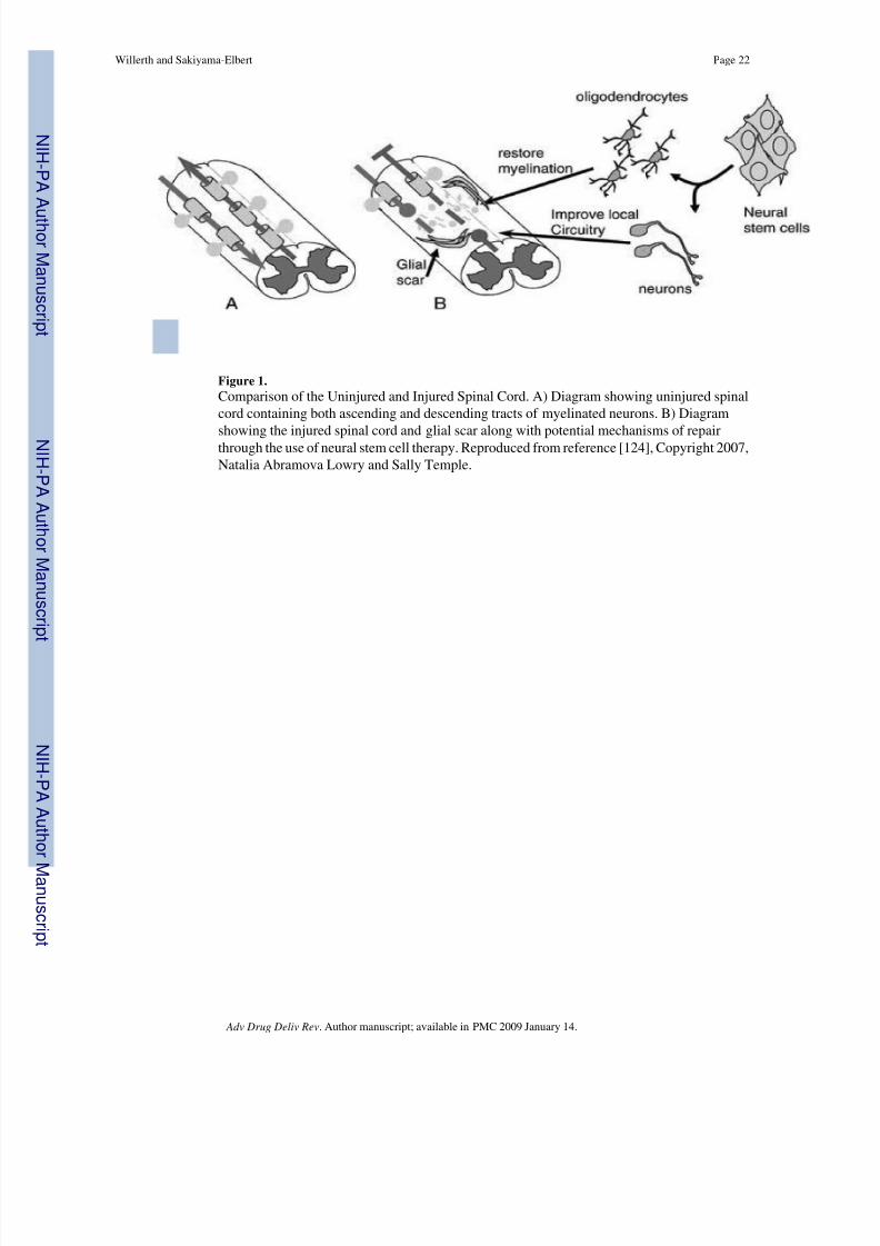
8/8/2019 SCI Adv Drug Deliv 2008
http://slidepdf.com/reader/full/sci-adv-drug-deliv-2008 22/24
Figure 1.Comparison of the Uninjured and Injured Spinal Cord. A) Diagram showing uninjured spinalcord containing both ascending and descending tracts of myelinated neurons. B) Diagram
showing the injured spinal cord and glial scar along with potential mechanisms of repairthrough the use of neural stem cell therapy. Reproduced from reference [124], Copyright 2007,Natalia Abramova Lowry and Sally Temple.
Willerth and Sakiyama-Elbert Page 22
Adv Drug Deliv Rev . Author manuscript; available in PMC 2009 January 14.
N I H -P A A
ut h or Manus c r i pt
N I H -P A A ut h or Manus c r i pt
N I H -P A A ut h or
Manus c r i pt
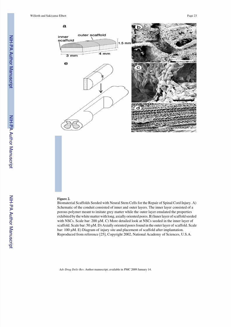
8/8/2019 SCI Adv Drug Deliv 2008
http://slidepdf.com/reader/full/sci-adv-drug-deliv-2008 23/24
Figure 2.Biomaterial Scaffolds Seeded with Neural Stem Cells for the Repair of Spinal Cord Injury. A)Schematic of the conduit consisted of inner and outer layers. The inner layer consisted of aporous polymer meant to imitate grey matter while the outer layer emulated the propertiesexhibited by the white matter with long, axially oriented pores. B) Inner layer of scaffold seededwith NSCs. Scale bar: 200 μ M. C) More detailed look at NSCs seeded in the inner layer of scaffold. Scale bar: 50 μ M. D) Axially oriented pores found in the outer layer of scaffold. Scalebar: 100 μ M. E) Diagram of injury site and placement of scaffold after implantation.Reproduced from reference [25], Copyright 2002, National Academy of Sciences, U.S.A.
Willerth and Sakiyama-Elbert Page 23
Adv Drug Deliv Rev . Author manuscript; available in PMC 2009 January 14.
N I H -P A A
ut h or Manus c r i pt
N I H -P A A ut h or Manus c r i pt
N I H -P A A ut h or
Manus c r i pt

8/8/2019 SCI Adv Drug Deliv 2008
http://slidepdf.com/reader/full/sci-adv-drug-deliv-2008 24/24
N I H -P A A ut h or Manus c r i pt
N I H -P A A ut h or Manus c r i pt
N I H -P A A ut h or Manus c r i pt
Willerth and Sakiyama-Elbert Page 24
Table OneCell Types Used As Therapy For Spinal Cord Injury
Cell Type References
Embryonic Stem CellsMouse [22,33-41,120]Human [23,24,32,42-45]
Neural Stem Cells [25,46-52,54-57,59,60,115,116,121]
Bone Marrow Stromal Cells [7,8,61-75]Mature CellsSchwann Cells [79-93]
Olfactory Ensheathing Cells [77,94-106]Fibroblasts [11,107-114,122]
Adv Drug Deliv Rev . Author manuscript; available in PMC 2009 January 14.


![Compiled By Adv. Prashant Mali [MSc(Comp Sci),LLB] By Adv. Prashant Mali [MSc(Comp Sci),LLB] Page 3 of 96 cyberlawconsulting@gmail.com create it; and the Digital Signature shall be](https://static.fdocuments.net/doc/165x107/5afd52f57f8b9a256b8b6d70/compiled-by-adv-prashant-mali-msccomp-scillb-by-adv-prashant-mali-msccomp.jpg)