SCENIC international consensus statement on surveillance ... · GIE CONSENSUS STATEMENT SCENIC...
39
GIE Ò CONSENSUS STATEMENT SCENIC international consensus statement on surveillance and management of dysplasia in inflammatory bowel disease INTRODUCTION Patients with ulcerative colitis or Crohn’s colitis have an increased risk of colorectal cancer (CRC). Most cases are believed to arise from dysplasia, and surveillance colonos- copy therefore is recommended to detect dysplasia. Detec- tion of dysplasia traditionally has relied on both examination of the mucosa with targeted biopsies of visible lesions and extensive random biopsies to identify invisible dysplasia. Current U.S. guidelines recommend obtaining at least 32 random biopsy specimens from all segments of the colon as the foundation of endoscopic surveillance. 1-4 However, much of the evidence that provides a basis for these recommendations is from older literature, when most dysplasia was diagnosed on random biopsies of colon mucosa. 5 With the advent of video endoscopy and newer endoscopic technologies, investigators now report that most dysplasia discovered in patients with inflammatory bowel disease (IBD) is visible. 6,7 Such a paradigm shift may have important implications for the surveillance and management of dysplasia. The evolving evidence regarding newer endoscopic methods to detect dysplasia has resulted in variation among guideline recommendations from organizations around the world. 1-4,8-10 We therefore sought to develop unifying consensus recommendations addressing 2 issues: (1) How should surveillance colonoscopy for detec- tion of dysplasia be performed? (2) How should dysplasia identified at colonoscopy be managed? DEVELOPMENT PROCESS An international multidisciplinary group representing a wide spectrum of stakeholders and attitudes regarding IBD surveillance (Appendix 1, available online at www. giejournal.org) developed these recommendations following a process that adhered to suggested standards for guideline development from the Institute of Medicine and others and that incorporated the GRADE methodol- ogy. 11-14 Details regarding the development process are provided in Figure 1 and Appendix 2. A systematic review was performed for each focused clinical question. The search strategy is shown in Appendix 3, and the full synthe- sis of evidence reviewed by panelists is presented in Ap- pendix 4. All appendices are available online at www. giejournal.org. The strength of recommendation, provided for each recommendation, reflects the level of confidence that desirable effects of an intervention outweigh undesirable effects. Strong recommendations mean panelists are confi- dent that the desirable effects outweigh the undesirable ef- fects; therefore, most informed patients would choose the recommended management, and clinicians would provide the intervention to most patients. Conditional recommen- dations mean the desirable and undesirable effects of the intervention are closely balanced or appreciable uncer- tainty exists regarding the balance; therefore, informed patients’ choices will vary according to their values and preferences, with many not wanting the intervention, and clinicians must ensure that patients’ care is in keeping with their values and preferences. 13 TERMINOLOGY A subgroup of panelists developed a set of terms for co- lonoscopic findings in IBD surveillance to establish unifor- mity in communication. Descriptive phrases, modified from the Paris Classification, 15 were recommended for adoption (Table 1). Modifications included the addition of terms for ulceration and border of the lesion. It was agreed that the terms dysplasia-associated lesion or mass (DALM), adenoma-like, and non-adenoma-like should be aban- doned. The term endoscopically resectable indicates that (1) distinct margins of the lesion could be identified, (2) the lesion appears to be completely removed on visual inspection after endoscopic resection, (3) histologic exam- ination of the resected specimen is consistent with com- plete removal, and (4) biopsy specimens taken from mucosa immediately adjacent to the resection site are free of dysplasia on histologic examination. CONSENSUS RECOMMENDATIONS AND SUMMARY OF SUPPORTING EVIDENCE Detection of dysplasia on surveillance colonoscopy The goal of this section is to define the optimal method(s) of detecting colon dysplasia in patients Copyright ª 2015 by the American Society for Gastrointestinal Endoscopy and American Gastroenterological Association 0016-5107/$36.00 http://dx.doi.org/10.1016/j.gie.2014.12.009 www.giejournal.org Volume 81, No. 3 : 2015 GASTROINTESTINAL ENDOSCOPY 489
Transcript of SCENIC international consensus statement on surveillance ... · GIE CONSENSUS STATEMENT SCENIC...

Can00h
w
GIE�
CONSENSUS STATEMENT
opyright ª 2015 by thed American Gastroente16-5107/$36.00ttp://dx.doi.org/10.1016
ww.giejournal.org
SCENIC international consensus statement on surveillance andmanagement of dysplasia in inflammatory bowel disease
INTRODUCTION
Patients with ulcerative colitis or Crohn’s colitis have anincreased risk of colorectal cancer (CRC). Most cases arebelieved to arise from dysplasia, and surveillance colonos-copy therefore is recommended to detect dysplasia. Detec-tion of dysplasia traditionally has relied on bothexamination of the mucosa with targeted biopsies of visiblelesions and extensive random biopsies to identify invisibledysplasia. Current U.S. guidelines recommend obtaining atleast 32 random biopsy specimens from all segments of thecolon as the foundation of endoscopic surveillance.1-4
However, much of the evidence that provides a basis forthese recommendations is from older literature, whenmost dysplasia was diagnosed on random biopsies of colonmucosa.5 With the advent of video endoscopy and newerendoscopic technologies, investigators now report thatmost dysplasia discovered in patients with inflammatorybowel disease (IBD) is visible.6,7 Such a paradigm shiftmay have important implications for the surveillance andmanagement of dysplasia.
The evolving evidence regarding newer endoscopicmethods to detect dysplasia has resulted in variationamong guideline recommendations from organizationsaround the world.1-4,8-10 We therefore sought to developunifying consensus recommendations addressing 2issues: (1) How should surveillance colonoscopy for detec-tion of dysplasia be performed? (2) How should dysplasiaidentified at colonoscopy be managed?
DEVELOPMENT PROCESS
An international multidisciplinary group representing awide spectrum of stakeholders and attitudes regardingIBD surveillance (Appendix 1, available online at www.giejournal.org) developed these recommendationsfollowing a process that adhered to suggested standardsfor guideline development from the Institute of Medicineand others and that incorporated the GRADE methodol-ogy.11-14 Details regarding the development process areprovided in Figure 1 and Appendix 2. A systematic reviewwas performed for each focused clinical question. The
American Society for Gastrointestinal Endoscopyrological Association
/j.gie.2014.12.009
search strategy is shown in Appendix 3, and the full synthe-sis of evidence reviewed by panelists is presented in Ap-pendix 4. All appendices are available online at www.giejournal.org.
The strength of recommendation, provided for eachrecommendation, reflects the level of confidence thatdesirable effects of an intervention outweigh undesirableeffects. Strong recommendations mean panelists are confi-dent that the desirable effects outweigh the undesirable ef-fects; therefore, most informed patients would choose therecommended management, and clinicians would providethe intervention to most patients. Conditional recommen-dations mean the desirable and undesirable effects of theintervention are closely balanced or appreciable uncer-tainty exists regarding the balance; therefore, informedpatients’ choices will vary according to their values andpreferences, with many not wanting the intervention, andclinicians must ensure that patients’ care is in keepingwith their values and preferences.13
TERMINOLOGY
A subgroup of panelists developed a set of terms for co-lonoscopic findings in IBD surveillance to establish unifor-mity in communication. Descriptive phrases, modifiedfrom the Paris Classification,15 were recommended foradoption (Table 1). Modifications included the addition ofterms for ulceration and border of the lesion. It was agreedthat the terms dysplasia-associated lesion or mass (DALM),adenoma-like, and non-adenoma-like should be aban-doned. The term endoscopically resectable indicates that(1) distinct margins of the lesion could be identified, (2)the lesion appears to be completely removed on visualinspection after endoscopic resection, (3) histologic exam-ination of the resected specimen is consistent with com-plete removal, and (4) biopsy specimens taken frommucosa immediately adjacent to the resection site arefree of dysplasia on histologic examination.
CONSENSUS RECOMMENDATIONS ANDSUMMARY OF SUPPORTING EVIDENCE
Detection of dysplasia on surveillancecolonoscopy
The goal of this section is to define the optimalmethod(s) of detecting colon dysplasia in patients
Volume 81, No. 3 : 2015 GASTROINTESTINAL ENDOSCOPY 489
Identification of executive committee and development panelConflict of interest disclosures
Formulate questions(Population, Intervention, Comparator, Outcome)
Systematic literature search
Synthesis of evidence and estimates of effect size for outcomes
Initial statements posted on web portalNarrative summary, data, and relevant literature linked to each statement
First round of voting and commenting by panel
First revision of statements by executive committee
Revised statements posted on web portal GRADE assessment of evidence linked to each statementNarrative summary, data, and relevant literature linked to each statement Second round of voting and commenting by panel
Second revision of statements by executive committee
Statements finalized and report prepared
Face-to-face meeting
Abbreviated review of literature and data synthesis linked to each statement Revised statements and comments presented to panelistsFinal voting, including GRADE assessment of strength of recommendation
Figure 1. Development process.
Consensus statement on dysplasia in IBD
with IBD. Detection of dysplasia, which is the immedi-ate goal of surveillance colonoscopy, was chosen as theprimary endpoint, with the understanding that detec-tion of dysplasia is not clearly documented to improveclinical outcomes such as CRC incidence or mortality.Only histologic diagnoses of low-grade or high-gradedysplasia were considered; diagnoses of indefinitefor dysplasia were excluded. Current guideline recom-mendations regarding the need for serial surveillancecolonoscopy in patients with IBD were accepted, andother issues such as the appropriate surveillance inter-val or risk stratification1-4,8-10 were not addressed.
Recommendations are listed in Table 2 and appearindividually hereafter with the proportion of panelistsin agreement, the strength of the recommendation, andthe quality of evidence. A summary of the evidence anddiscussion regarding the recommendation follows eachstatement.
490 GASTROINTESTINAL ENDOSCOPY Volume 81, No. 3 : 2015
Statement 1: When performing surveillance withwhite-light colonoscopy, high definition is recom-mended rather than standard definition.
(80% agreement; strong recommendation; low-quality evidence)
Summary of evidence and discussion. High-definition (1080 system) endoscopy provides image signalsof higher pixel density than standard definition (480 sys-tem), with faster line scanning on high-definition monitors,leading to sharper images with fewer artifacts.16 A high-definition system includes a high-definition endoscope,processor, cabling, and monitor. A retrospective observa-tional study found that dysplasia was discovered in approx-imately twice as many patients undergoing high-definitioncolonoscopy (n Z 203) compared with a cohort undergo-ing standard-definition colonoscopy (n Z 154): adjustedprevalence ratio Z 2.2 (95% confidence interval [CI],1.1-4.5).17
www.giejournal.org
TABLE 1. Terminology for reporting findings on colonoscopic surveillance of patients with inflammatory bowel disease (modified from ParisClassification15)
Term Definition
Visible dysplasia Dysplasia identified on targeted biopsies from a lesion visualized at colonoscopy
Polypoid Lesion protruding from the mucosa into the lumen R2.5 mm
Pedunculated Lesion attached to the mucosa by a stalk
Sessile Lesion not attached to the mucosa by a stalk: entire base is contiguous with the mucosa
Nonpolypoid Lesion with little (!2.5 mm) or no protrusion above the mucosa
Superficial elevated Lesion with protrusion but!2.5 mm above the lumen (less than the height of the closed cup of a biopsy forceps)
Flat Lesion without protrusion above the mucosa
Depressed Lesion with at least a portion depressed below the level of the mucosa
General descriptors
Ulcerated Ulceration (fibrinous-appearing base with depth) within the lesion
Border
Distinct border Lesion’s border is discrete and can be distinguished from surrounding mucosa
Indistinct border Lesion’s border is not discrete and cannot be distinguished from surrounding mucosa
Invisible dysplasia Dysplasia identified on random (non-targeted) biopsies of colon mucosa without a visible lesion
Consensus statement on dysplasia in IBD
Given that most dysplastic lesions are visible,6,7 theimproved visualization and lack of negative effects withhigh-definition endoscopy justified a strong recommenda-tion for its use. In addition, patients likely would stronglydesire high-definition colonoscopy because of the beliefthat visualization and examination are improved. Thecost of purchasing new high-definition endoscopic equip-ment is a consideration. However, high-definition colonos-copy already is widely used in endoscopic units.
Statement 2: When performing surveillance withstandard-definition colonoscopy, chromoendo-scopy is recommended rather than white-lightcolonoscopy.
(85% agreement; strong recommendation;moderate-quality evidence)
Summary of evidence and discussion. Chromoen-doscopy involves the application of dye to the colonmucosa, thereby providing contrast enhancement toimprove visualization of epithelial surface detail. Methyleneblue and indigo carmine, the agents most commonly used,are applied to the colon mucosa via a catheter or thecolonoscope biopsy or water jet channel,18 and accentuatethe changes in epithelial surface topography.19
We identified 8 trials that used standard-definition colo-noscopy and compared chromoendoscopy with white-lightcolonoscopy alone (Table 3).20-27 The proportion ofpatients with dysplasia was 0% to 10% greater with chro-moendoscopy in the individual studies, but the differencewas not significant in any study. Meta-analysis revealed asignificantly greater proportion of patients with dysplasiaby using chromoendoscopy (relative risk [RR] Z 1.8[1.2-2.6] and absolute risk increase Z 6% [3%-9%]).Meta-analysis of the 2 randomized, parallel-group trials
www.giejournal.org
also confirmed a significant increase with chromoendo-scopy in the proportion of patients with dysplasia(RR Z 2.3 [1.1-4.6], absolute increase Z 8% [2%-15%]).The number of dysplastic lesions identified was greaterwith chromoendoscopy in all studies (Table 3), and inthe 4 tandem studies in which all patients had both chro-moendoscopy and white-light examination, the numberof dysplastic areas discovered increased almost 2-fold(RR Z 1.9, 1.4-2.7) with chromoendoscopy. Chromoendo-scopy significantly increased the duration of colonoscopyby a mean of 11 minutes (range 9-12 minutes).
An economic analysis concluded that chromoendoscopywith targeted biopsies was less costly and more effectivethan white-light colonoscopy with random biopsies,28 sug-gesting that chromoendoscopy should be used in place ofwhite-light endoscopy when surveillance colonoscopy isperformed. The cost-effectiveness of chromoendoscopyincreased with increasing surveillance interval, suggestingthat varying the surveillance interval based on the risk ofCRC may be appropriate and could increase the cost effec-tiveness of surveillance colonoscopy. However, whensurveillance is performed, even if performed lessfrequently than currently recommended in lower-riskpatients, the best technique should be used.
Although chromoendoscopy increases the yield ofdysplasia compared with standard-definition white-lightcolonoscopy, whether the additional lesions identifiedwith chromoendoscopy are associated with the sameincreased risk for CRC as the visible and invisible dysplasiaidentified in older studies is not known. Data from the Sur-veillance, Epidemiology and End-Results Medicare-linkeddatabase of patients R67 years old revealed that intervalcancers 6 to 36 months after colonoscopy occurred in a
Volume 81, No. 3 : 2015 GASTROINTESTINAL ENDOSCOPY 491
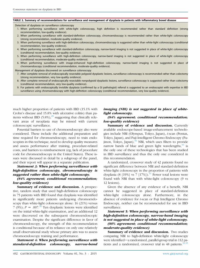
TABLE 2. Summary of recommendations for surveillance and management of dysplasia in patients with inflammatory bowel disease
Detection of dysplasia on surveillance colonoscopy1. When performing surveillance with white-light colonoscopy, high definition is recommended rather than standard definition (strong
recommendation, low-quality evidence).2. When performing surveillance with standard-definition colonoscopy, chromoendoscopy is recommended rather than white-light colonoscopy
(strong recommendation, moderate-quality evidence).3. When performing surveillance with high-definition colonoscopy, chromoendoscopy is suggested rather than white-light colonoscopy (conditional
recommendation, low-quality evidence).4. When performing surveillance with standard-definition colonoscopy, narrow-band imaging is not suggested in place of white-light colonoscopy
(conditional recommendation, low-quality evidence).5. When performing surveillance with high-definition colonoscopy, narrow-band imaging is not suggested in place of white-light colonoscopy
(conditional recommendation, moderate-quality evidence).6. When performing surveillance with image-enhanced high-definition colonoscopy, narrow-band imaging is not suggested in place of
chromoendoscopy (conditional recommendation, moderate-quality evidence).
Management of dysplasia discovered on surveillance colonoscopy7. After complete removal of endoscopically resectable polypoid dysplastic lesions, surveillance colonoscopy is recommended rather than colectomy
(strong recommendation, very low-quality evidence).8. After complete removal of endoscopically resectable nonpolypoid dysplastic lesions, surveillance colonoscopy is suggested rather than colectomy
(conditional recommendation, very low-quality evidence).9. For patients with endoscopically invisible dysplasia (confirmed by a GI pathologist) referral is suggested to an endoscopist with expertise in IBD
surveillance using chromoendoscopy with high-definition colonoscopy (conditional recommendation, very low-quality evidence).
Consensus statement on dysplasia in IBD
much higher proportion of patients with IBD (15.1% withCrohn’s disease and 15.8% with ulcerative colitis) than pa-tients without IBD (5.8%),29 suggesting that clinically rele-vant areas of neoplasia may be missed with currentcolonoscopic surveillance.
Potential barriers to use of chromoendoscopy also wereconsidered. These include the additional preparation andtime required for chromoendoscopy, need to train endo-scopists in this technique, need to develop quality measuresand assess performance after training, procedure-relatedcosts, and barriers to reimbursement (eg, lack of procedurecode for chromoendoscopy in the United States). These is-sues were discussed in detail by a subgroup of the panel,and their report will appear in a separate publication.
Statement 3: When performing surveillance withhigh-definition colonoscopy, chromoendoscopy issuggested rather than white-light colonoscopy.
(84% agreement; conditional recommendation;low-quality evidence)
Summary of evidence and discussion. A prospec-tive, tandem study that used high-definition colonoscopyin 75 patients with IBD found that dysplasia was identifiedin significantly more patients undergoing chromoendo-scopy than white-light colonoscopy alone: 16 (21%) versus7 (9%); P Z .007.30 Ten dysplastic lesions were identifiedon the initial white-light examination, and an additional 12were discovered on the subsequent chromoendoscopicexamination. Despite the significant difference in favor ofchromoendoscopy, the strength of this recommendationis conditional because of its reliance on only one relativelysmall observational study whose primary aim was to assesschromoendoscopy training and performance.
Statement 4: When performing surveillance withstandard-definition colonoscopy, narrow-band
492 GASTROINTESTINAL ENDOSCOPY Volume 81, No. 3 : 2015
imaging (NBI) is not suggested in place of white-light colonoscopy.
(84% agreement; conditional recommendation;low-quality evidence)
Summary of evidence and discussion. Currentlyavailable endoscope-based image-enhancement technolo-gies include NBI (Olympus, Tokyo, Japan), i-scan (Pentax,Tokyo, Japan), and Fuji Intelligent Chromo Endoscopy (Fu-jinon, Tokyo, Japan).16 NBI, which uses filters to providenarrow bands of blue and green light wavelengths,16 isthe only one of these technologies that has been studiedin IBD surveillance and thus the only one considered inthis recommendation.
A randomized, crossover study of 42 patients found nosignificant difference between NBI and standard-definitionwhite-light colonoscopy in the proportion of patients withdysplasia (8 [19%] vs 7 [17%]).31 Fewer total lesions werefound with NBI than with white-light colonoscopy (9 vs12 lesions).
Given the absence of any evidence of a benefit, NBIcannot be suggested in place of standard-definitionwhite-light colonoscopy alone. Furthermore, in theabsence of evidence for i-scan or Fuji Intelligent ChromoEndoscopy, neither can be recommended for use in IBDsurveillance.
Statement 5: When performing surveillance withhigh-definition colonoscopy, narrow-band imagingis not suggested in place of white-light colonoscopy.
(80% agreement; conditional recommendation;moderate-quality evidence)
Summary of evidence and discussion. Two studiescomparing NBI to high-definition white-light colonoscopywere identifiedda randomized, parallel-group trial in 112 pa-tients and a randomized, crossover trial in 48 patients.32,33
www.giejournal.org

TABLE 3. Proportion of patients with dysplasia and number of visible dysplastic lesions identified in studies comparing chromoendoscopy versuswhite-light colonoscopy
Study Study type
Patients withdysplasia/all patients RR (95% CI)
Absolute riskincrease (95% CI) No. of visible dysplastic lesions
Chromoendoscopy White-light Chromoendoscopy White-light
Kiesslich20 Randomizedparallel-group
13/84 6/81 2.1 (0.8-5.2) 8% (-2% to 18%) 32 10
Kiesslich21 Randomizedparallel-group
11/80 4/73 2.5 (0.8-7.5) 8% (-1% to 17%) 19 2
Marion24 Prospectivetandem
22/102 12/102 1.8 (0.96-3.5) 10% (0% to 20%) 35 13
Rutter23 Prospectivetandem
7/100 2/100 3.5 (0.8-16.4) 5% (-1% to 11%) 9 2
Matsumoto25 Prospectivetandem
12/57 12/57 1.0 (0.5-2.0) 0% (-2% to 2%) 18 8
Hlvaty26 Prospectivetandem andadditional cohort
4/30 2/45 3.0 (0.6-15.4) 9% (-5% to 23%) 6 2
Gunther27 Retrospective two-group
2/50 0/50 5.0 (0.3-101.6) 4% (-3% to 11%) 2 0
Chiorean22 Prospectivetandem
No per-patientdata given (N Z 63)
41 18
SCENICmeta-analysis
1.8 (1.2-2.6) 6% (3%-9%)
RR, Relative risk; CI, confidence interval; SCENIC, Surveillance for Colorectal Endoscopic Neoplasia Detection and Management in Inflammatory Bowel Disease Patients:International Consensus Recommendations.
Consensus statement on dysplasia in IBD
Neither study suggested a benefit for NBI, with the pro-portion of patients having dysplasia identified with NBIversus white-light colonoscopy of 5 of 56 (9%) versus 5of 56 (9%) and 9 of 48 (19%) versus 13 of 48 (27%). Inaddition, NBI identified slightly fewer dysplastic lesionsthan white-light colonoscopy (5 vs 7 and 14 vs 16). Again,in the absence of any evidence of a benefit, NBI cannot besuggested in place of high-definition white-light colonos-copy alone.
Statement 6: When performing surveillancewith image-enhanced high-definition colonoscopy,narrow-band imaging is not suggested in place ofchromoendoscopy.
(90% agreement; conditional recommendation;moderate-quality evidence)
Summary of evidence and discussion. Four studieswere identified comparing chromoendoscopy with NBI:two randomized, parallel-group trials; a randomized cross-over trial; and a prospective, tandem study.34-37 The pro-portion of patients with dysplasia was 0.1% to 22%greater with chromoendoscopy than with NBI in the indi-vidual studies, but none of the differences were significant.Meta-analysis also failed to show a significant difference:RR Z 1.3 (0.8-2.1) and absolute risk difference Z 6%(-1% to 14%). The mean withdrawal times were identicalin one study,36 whereas the mean procedure or withdrawaltimes in the other studies were 11 to 12 minutes longerwith chromoendoscopy.
www.giejournal.org
The results of the studies indicate that a meaningfulbenefit of NBI over chromoendoscopy is unlikely.Nonetheless, they do not document a benefit of chro-moendoscopy over NBI.
Additional topics considered for detection ofdysplasia
Random biopsies with high-definition white-lightcolonoscopy or chromoendoscopy. Given that high-definition white-light colonoscopy and chromoendoscopywere considered superior to standard-definition white-lightcolonoscopy, the panelists considered the question ofwhether random biopsies should be performed whenendoscopists use high-definition white-light colonoscopyor chromoendoscopy. Table 4 shows the yield of targetedand random biopsies for dysplasia from pooled analyses;the evidence was graded as low quality.
Among patients with dysplasia undergoing high-defini-tion white-light colonoscopy17,30,32,33,36 or chromoendo-scopy,20-27,36,38 dysplasia is detected only on randombiopsies in approximately 10% of patients and on targetedbiopsies in the other 90%. About 1% to 1.5% of all patientsundergoing surveillance would not have dysplasiadetected if random biopsies were not performed. Onlyabout one in a thousand random biopsies revealsdysplasia. Pooled results also were determined for detec-tion of dysplasia by using standard-definition white-lightcolonoscopy.6,17,20-27,31,33,39-41 The proportion of patients
Volume 81, No. 3 : 2015 GASTROINTESTINAL ENDOSCOPY 493
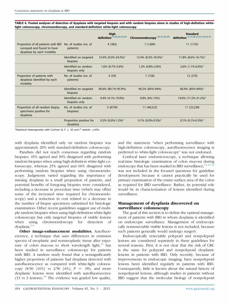
TABLE 4. Pooled analyses of detection of dysplasia with targeted biopsies and with random biopsies alone in studies of high-definition white-light colonoscopy, chromoendoscopy, and standard-definition white-light colonoscopy
Highdefinition17,30,32,33,36 Chromoendoscopy 20-27,36,38
Standarddefinition 6,17,20-27,31,33,39-41
Proportion of all patients with IBDsurveyed and found to havedysplasia by each modality
No. of studies (no. ofpatients)
4 (382) 7 (1289) 11 (1735)
Identified on targetedbiopsies
15.4% (9.3%-24.5%)* 12.4% (8.3%-18.3%)* 11.8% (8.6%-16.1%)*
Identified on randombiopsies only
1.6% (0.7%-3.6%) 1.2% (0.8%-2.0%) 2.6% (1.1%-6.0%)*
Proportion of patients withdysplasia identified by eachmodality
No. of studies (no. ofpatients)
4 (59) 7 (158) 12 (270)
Identified on targetedbiopsies
90.6% (80.1%-95.9%) 90.2% (85%-94%) 80.4% (85%-94%)*
Identified on randombiopsies only
9.4% (4.1%-19.9%) 9.8% (6%-15%) 19.6% (11.5%-31.2%)*
Proportion of all random biopsyspecimens positive fordysplasia
No. of studies (no. ofbiopsies)
5 (8739) 11 (48,522) 11 (25,238)
Proportion positive fordysplasia
0.2% (0.0%-1.2%)* 0.1% (0.0%-0.3%)* 0.1% (0.1%-0.3%)*
*Statistical heterogeneity with Cochran Q; P % .02 and I2 statistic R65%.
Consensus statement on dysplasia in IBD
with dysplasia identified only on random biopsies wasapproximately 20% with standard-definition colonoscopy.
Panelists did not reach consensus regarding randombiopsies: 45% agreed and 30% disagreed with performingrandom biopsies when using high-definition white-light co-lonoscopy, whereas 25% agreed and 60% disagreed withperforming random biopsies when using chromoendo-scopy. Judgments varied regarding the importance ofmissing dysplasia in a small proportion of patients, andpotential benefits of foregoing biopsies were considered,including a decrease in procedure time (which may offsetsome of the increased time required for chromoendo-scopy) and a reduction in cost related to a decrease inthe number of biopsy specimens submitted for histologicexamination. Other recent guidelines suggest use of multi-ple random biopsies when using high-definition white-lightcolonoscopy but only targeted biopsies of visible lesionswhen using chromoendoscopy for detection ofdysplasia.2,8
Other image-enhancement modalities. Autofluor-escence, a technique that uses differences in emissionspectra of neoplastic and nonneoplastic tissue after expo-sure of colon mucosa to short wavelength light,42 hasbeen studied in surveillance colonoscopy for patientswith IBD. A tandem study found that a nonsignificantlyhigher proportion of patients had dysplasia detected withautofluorescence as compared with white-light colonos-copy (8/50 [16%] vs 2/50 [4%]; P Z .09), and moredysplastic lesions were identified with autofluorescence(13 vs 3 lesions).39 The evidence was graded low quality,
494 GASTROINTESTINAL ENDOSCOPY Volume 81, No. 3 : 2015
and the statement “when performing surveillance withhigh-definition colonoscopy, autofluorescence imaging ispreferred to white-light colonoscopy” was not endorsed.
Confocal laser endomicroscopy, a technique allowingreal-time histologic examination of colon mucosa duringendoscopy that has been studied in IBD surveillance,21,27,42
was not included in the focused questions for guidelinedevelopment because it cannot practically be used forprimary examination of the entire surface area of the colonas required for IBD surveillance. Rather, its potential rolewould be in characterization of lesions identified duringsurveillance.
Management of dysplasia discovered onsurveillance colonoscopy
The goal of this section is to define the optimal manage-ment of patients with IBD in whom dysplasia is identifiedon endoscopic surveillance. Management of endoscopi-cally nonresectable visible lesions is not included, becausesuch patients generally would undergo surgery.
Endoscopically resectable polypoid and nonpolypoidlesions are considered separately in these guidelines forseveral reasons. First, it is not clear that the risk of CRCis the same for polypoid and nonpolypoid dysplasticlesions in patients with IBD. Only recently, because ofimprovements in endoscopic imaging, have nonpolypoidlesions been identified regularly in patients with IBD.Consequently, little is known about the natural history ofnonpolypoid lesions, although studies in patients withoutIBD suggest that the molecular biology of nonpolypoid
www.giejournal.org

Consensus statement on dysplasia in IBD
colorectal neoplasms may differ from that of polypoid colo-rectal neoplasms.43 Second, the methods for endoscopicresection of polypoid and nonpolypoid lesions differ,with endoscopic resection of nonpolypoid lesions typicallymore difficult and often requiring advanced endoscopicskills that many endoscopists may lack. Third, confidencethat the lesion has been completely removed may be lowerfor nonpolypoid than for polypoid lesions.
Statement 7: After complete removal of endoscop-ically resectable polypoid dysplastic lesions, sur-veillance colonoscopy is recommended ratherthan colectomy.
(100% agreement; strong recommendation; verylow-quality evidence)
Summary of evidence and discussion. No studycomparing surveillance colonoscopy and colectomy afterendoscopic resection of dysplastic lesions was identified.However, 6 studies from the video-endoscopic era (1990onward) were identified that reported CRC incidence afterendoscopic removal of polypoid dysplastic lesions in O15patients with IBD.6,44-48 Among studies that reported theproportion of patients with low-grade versus high-gradedysplasia, most patients had low-grade dysplasia. Overmean follow-up periods of 36 to 82 months, the incidenceof CRC in these studies was 19 of 311 (6%, range 2%-13%).A single study focused only on polypoid lesions with high-grade dysplasia49 found that 0 of 9 patients followed for amean of 76.5 months (range 52-99 months) after endo-scopic resection developed CRC or flat dysplasia.
A recent systematic review of 10 studies, which followed376 patients with IBD with resected polypoid dysplasia fora mean of 54 months, reported an annualized incidence forCRC of 0.5%.50 The definition of an “acceptable” incidenceof synchronous and metachronous CRC for physiciansdand, more importantly, for patientsdneeds to be consid-ered when determining management strategies.
The strength of this recommendation was consideredstrong despite the lack of evidence comparing the manage-ment strategies, largely based on views regarding patientpreference. Stakeholders indicated that patients diagnosedwith dysplasia were much more likely to refuse or delaycolectomy and choose surveillance colonoscopy. Theysuggested that patients might accept colectomy at a laterdate, depending on results of subsequent surveillanceprocedures and further information and education aboutcolectomy and CRC risk provided by physicians, nurses,other patients, and patient advocacy groups. These viewswere supported by a survey that assessed the managementpreferences of 199 patients with ulcerative colitis who weretold that dysplasia was detected.51 On average, patientswould agree to immediate colectomy only when the riskof synchronous CRC rose to R73%.51
More intensive surveillance for patients with endoscop-ically resectable dysplasia than for those without dysplasiaseems reasonable, and subsequent surveillance may varybased on the size and appearance of the dysplastic lesion.
www.giejournal.org
For example, current multi-society guidelines on colorectalpolyps in patients without IBD suggest a short interval of!1 year for flat and sessile adenomatous and serrated polypsO15 mm that are removed by using injection-assisted pol-ypectomy and piecemeal resection if there is any questionabout completeness of resection.52 Thus, patients with IBDwho have larger sessile lesions removed in piecemealfashion or via endoscopic mucosal resection or endoscopicsubmucosal dissection probably should return at approxi-mately 3 to 6 months, with longer subsequent intervals(eg, yearly) if the initial repeat colonoscopy result is nega-tive. Patients with smaller polypoid lesions resected enbloc may return at 1-year intervals.
Statement 8: After complete removal of endoscop-ically resectable nonpolypoid dysplastic lesions,surveillance colonoscopy is suggested rather thancolectomy.
(80% agreement; conditional recommendation;very low-quality evidence)
Summary of evidence and discussion. No studycomparing surveillance colonoscopy to colectomy orproviding the natural history for nonpolypoid dysplasticlesions after endoscopic resection was identified.
Analogous to the polypoid lesion discussed previously,if a nonpolypoid lesion is removed completely at endos-copy, it is acceptable to follow the patient with regular sur-veillance colonoscopy, because most dysplasia is visible,and careful follow-up with high-definition chromoendo-scopy likely would identify new or recurrent dysplasticlesions. Nonetheless, this recommendation is conditional,given the possibility that nonpolypoid lesions could confera higher CRC risk and the greater endoscopic difficulty inassuring complete removal of these lesions. In addition,because many of the larger nonpolypoid lesions must beremoved with endoscopic mucosal resection or endo-scopic submucosal dissection and/or in piecemeal fashion,patients with such lesions should undergo initial follow-upsurveillance colonoscopy in approximately 3 to 6 monthsas outlined previously for larger sessile polypoid lesions.
In contrast to the recommendation from this and otherpublications,10 some recent guidelines have suggestedcolectomy for nonpolypoid dysplastic lesions because theyconsidered such lesionsgenerally not amenable toendoscopicresection.2,8 However, variation in terminology for dysplasticlesions across publications makes comparisons difficult.
Statement 9: For patients with endoscopically-invisible dysplasia (confirmed by a GI pathologist)referral is suggested to an endoscopist with expertisein IBD surveillance using chromoendoscopy withhigh-definition colonoscopy.
(100% agreement; conditional recommendation;very-low-quality evidence)
Summary of evidence and discussion. No studycomparing surveillance colonoscopy and colectomy forendoscopically invisible dysplasia was identified. However, 4studies fromthevideo-endoscopicera reportedCRC incidence
Volume 81, No. 3 : 2015 GASTROINTESTINAL ENDOSCOPY 495

Consensus statement on dysplasia in IBD
after invisible dysplasia was diagnosed in O15 patients withIBD.45,48,53,54 Over a mean follow-up of 15 to 50 months,CRC developed in 7 of 122 patients (6%, range 3%-9%).
The proportion of patients with synchronous CRC at thetime invisible dysplasia is detected also is important whenconsidering management strategies. A systematic review55
of 20 surveillance studies and 477 patients with invisiblelow-grade dysplasia (which included patients from beforethe video-endoscope era) found that 18 of 81 patients(22%) with invisible low-grade dysplasia who had colectomyhad CRC. It is uncertain what characteristics led the minorityof patients with low-grade dysplasia to undergo colectomydother unknown or unreported factors that increase the riskof CRC may have been present in some of these patients.
Colectomy has been performed more commonly wheninvisible high-grade dysplasia is discovered because of thereported higher risk of CRC. A 1994 systematic reviewfound that 10 of 24 patients (42%) with non-DALM high-grade dysplasia had CRC on colectomy, whereas 15 of 47patients (32%) who had high-grade dysplasia on subse-quent surveillance examinations developed CRC.5 Otherindividual studies of patients with invisible high-gradedysplasia undergoing colectomy reported since 1994show rates of CRC ranging from 45% to 67%.56-59
The findings reported in older studies may be of limitedrelevance in the current video-endoscopic era. A 1994review of 10 prospective studies with 1225 patients under-going surveillance colonoscopy found that dysplasia that isnot associated with a lesion accounted for 272 of 312patients (87%) found to have dysplasia.5 In contrast,more recent studies of chromoendoscopy or high-definition white-light colonoscopy report that invisibledysplasia accounts for about 10% of patients with dysplasia(Table 4). Thus, random biopsy specimens showing invis-ible dysplasia in older studies may have been taken frompreviously unrecognizable lesions that can now be visual-ized with modern endoscopic techniques.
Based on this information, general statements that theinitial management step for patients with invisible low-grade or high-grade dysplasia be surveillance colonoscopyor colectomy2,8 were not endorsed. Rather, referral to anendoscopist with expertise in IBD surveillance andimage-enhanced examination using chromoendoscopywith high-definition endoscopy was considered an appro-priate next step to better inform subsequent decisionsregarding surveillance colonoscopy versus colectomy. If avisible dysplastic lesion is identified in the same regionof the colon as the invisible dysplasia, and the lesion canbe resected endoscopically, then such patients may remainin a surveillance program, as recommended previously instatements 7 and 8. Alternatively, if dysplasia is not discov-ered, management of such patients would be individual-ized after discussion of the risks and benefits ofsurveillance colonoscopy and colectomy. Continued inten-sive surveillance is an acceptable strategy if, after carefuldiscussion, patients prefer this course.
496 GASTROINTESTINAL ENDOSCOPY Volume 81, No. 3 : 2015
Histologic distinctions may play a role in managementdecisions for patients with invisible dysplasia and no visiblelesions on follow-up chromoendoscopy. Physicians may becomfortable having patients with invisible low-gradedysplasia remain in intensive surveillance while morestrongly suggesting colectomy for those with invisiblehigh-grade dysplasia. In addition, some physicians believethat multifocal invisible low-grade dysplasia is associatedwith higher CRC risk than unifocal low-grade dysplasia,leading to a greater likelihood of recommending colec-tomy, although a single study assessing this issue54 failedto show an increased risk.
Confirmation of dysplasia by a pathologist with exper-tise in IBD is suggested before making management deci-sions. Even expert GI pathologists show no better than fairor moderate interobserver agreement on the histologicdiagnosis of dysplasia, low-grade dysplasia, or high-gradedysplasia.60-62 Diagnosis of low-grade dysplasia in Barrett’sesophagus by one pathologist does not predict progres-sion to high-grade dysplasia or cancer, but agreementamong 2 or 3 pathologists significantly increases the riskof progression.63 Similar studies are not available for IBD,but confirmation of dysplasia by a second pathologistseems appropriate before embarking on major diagnosticand therapeutic interventions.
IMPLEMENTATION OF HIGH-QUALITYENDOSCOPIC SURVEILLANCE
Widespread implementation of high-quality endoscopicsurveillance in patients with IBD will require a variety of ini-tiatives, which will be discussed in a separate publication.Resources will be needed to train endoscopists in endo-scopic surveillance and recognition of visible dysplasiawith both white-light endoscopy and chromoendoscopy.These may include training courses, photographic at-lases,64-66 and video repositories.67 Quality metrics andmethods to document acceptable performance qualityalso should be developed. In addition, techniques such aschromoendoscopy should be standardized to allow imple-mentation in endoscopy units, and endoscopic resectiontechniques for nonpolypoid lesions should be taught anddisseminated.68-70 Development of a procedure code forchromoendoscopy and reimbursement for the increasedtime and intensity required for chromoendoscopy wouldincrease implementation, at least in the United States.
Performance of chromoendoscopy forsurveillance of patients with IBD
Description of the technique. Surveillance colonos-copy should be performed when the disease is in remis-sion in order to minimize potential misdiagnosis betweeninflammatory changes and dysplasia.18,71,72 Clean bowelpreparation is a prerequisitedthe entire mucosa shouldbe free from pus, mucus, blood, or stool. Small amounts
www.giejournal.org
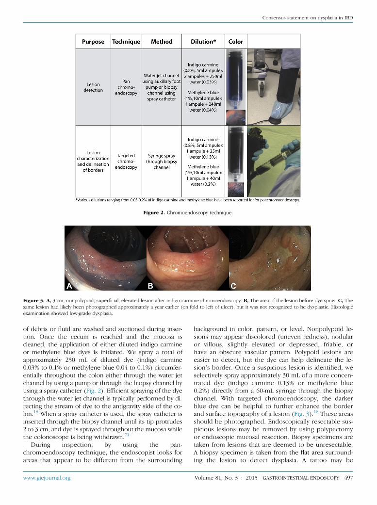
Figure 2. Chromoendoscopy technique.
Figure 3. A, 3-cm, nonpolypoid, superficial, elevated lesion after indigo carmine chromoendoscopy. B, The area of the lesion before dye spray. C, Thesame lesion had likely been photographed approximately a year earlier (on fold to left of ulcer), but it was not recognized to be dysplastic. Histologicexamination showed low-grade dysplasia.
Consensus statement on dysplasia in IBD
of debris or fluid are washed and suctioned during inser-tion. Once the cecum is reached and the mucosa iscleaned, the application of either diluted indigo carmineor methylene blue dyes is initiated. We spray a total ofapproximately 250 mL of diluted dye (indigo carmine0.03% to 0.1% or methylene blue 0.04 to 0.1%) circumfer-entially throughout the colon either through the water jetchannel by using a pump or through the biopsy channel byusing a spray catheter (Fig. 2). Efficient spraying of the dyethrough the water jet channel is typically performed by di-recting the stream of dye to the antigravity side of the co-lon.18 When a spray catheter is used, the spray catheter isinserted through the biopsy channel until its tip protrudes2 to 3 cm, and dye is sprayed throughout the mucosa whilethe colonoscope is being withdrawn.71
During inspection, by using the pan-chromoendoscopy technique, the endoscopist looks forareas that appear to be different from the surrounding
www.giejournal.org
background in color, pattern, or level. Nonpolypoid le-sions may appear discolored (uneven redness), nodularor villous, slightly elevated or depressed, friable, orhave an obscure vascular pattern. Polypoid lesions areeasier to detect, but the dye can help delineate the le-sion’s border. Once a suspicious lesion is identified, weselectively spray approximately 30 mL of a more concen-trated dye (indigo carmine 0.13% or methylene blue0.2%) directly from a 60-mL syringe through the biopsychannel. With targeted chromoendoscopy, the darkerblue dye can be helpful to further enhance the borderand surface topography of a lesion (Fig. 3).18 These areasshould be photographed. Endoscopically resectable sus-picious lesions may be removed by using polypectomyor endoscopic mucosal resection. Biopsy specimens aretaken from lesions that are deemed to be unresectable.A biopsy specimen is taken from the flat area surround-ing the lesion to detect dysplasia. A tattoo may be
Volume 81, No. 3 : 2015 GASTROINTESTINAL ENDOSCOPY 497

TABLE 5. Suggested steps for implementation of chromoendoscopy into endoscopic practice
Equipment
Colonoscope High-definition colonoscope, monitor, and cables
Accessories Apply dye via:Water jet channel by using water pump attached to the endoscope activated via foot pedal orSpray catheter: length 240 cm, endoscope accessory channel 2.8 mm
Contrast agent Indigo carmine, 5-mL ampule (0.8%)Methylene blue, 10-mL ampule (1%)
Procedure and protocol
Time allotment Consider doubling colonoscopy time slot initially during the learning curve period.
Standard operatingprocedure
Complete colonoscopy to cecum.Lavage with water and suction during intubation.
Prepare dye solution during insertion for application via the foot pump or spray.Indigo carmine (0.03%): mix 2 5-mL ampules of 0.8% indigo carmine with 250 mL water.Methylene blue (0.04%): mix one 10-mL ampule of 1% methylene blue with 240 mL water.
If using a foot pump: once the cecum is intubated, the water irrigation can be exchanged with the contrast solution.Apply the dye solution in a circumferential technique while withdrawing the colonoscope. Direct spray to the anti-gravity side.
If using a spray catheter: the dye spray catheter is inserted into the biopsy channel; the catheter tip should protrude2-3 cm from the endoscope. Apply dye solution segmentally by using a rotational technique while withdrawing thecolonoscope to cover the surface mucosa with dye.
Suction any excess solution after approximately 1 minute to aid mucosal visualization.
Focus on 20-30–cm segments sequentially with reinsertion of the endoscope to the proximal extent of each segmentbefore slow withdrawal and mucosal visualization.
Targeted dye spray for suspicious lesions:Prepare more concentrated dye solution for application.Indigo carmine (0.13%): mix one 5-mL ampule of 0.8% indigo carmine with 25 mL water.Methylene blue (0.2%): mix one 10-mL ampule of 1% methylene blue with 40 mL water.Spray about 30 mL directly from a 60-mL syringe through the biopsy channel.
Remove endoscopically resectable suspicious lesions by using polypectomy or endoscopic mucosal resection.
Do targeted biopsies of any unresectable abnormality visualized through chromoendoscopy to diagnose dysplasia.
Do biopsies of flat area surrounding lesions to assess for dysplasia.
Consider tattoo of suspicious dysplastic lesions arising from flat mucosa or not amenable to complete removal.
Recommendations regarding the need to perform random, non-targeted biopsies for detection of dysplasia vary.
If biopsies for dysplasia are not done, 2 random biopsies in every bowel segment are commonly recommended todocument microscopic disease activity.
Consensus statement on dysplasia in IBD
necessary to mark the location of resection or suspiciouslesion. Biopsies to document disease activity may be per-formed during the procedure.
Available resources for self-learning. Descriptionsof a systematic approach to performance of pancolonicchromoendoscopy by using either indigo carmine or meth-ylene blue dyes with targeted biopsy for surveillance of pa-tients with IBD are available.18,71 In addition, endoscopicvideos,18,67 atlases,64-66 and books73,74 have been pub-lished recently to provide readers with information onthe techniques and findings related to endoscopy in IBD.Open access of several of the materials serves to facilitatelearning (http://www.youtube.com/watch?vZOARkbgwlObI,http://www.giendo.theclinics.com/issues/?elsca1Zetoc&elsca2Zemail&elsca3Z1052-5157_201407_24_3&elsca4Zgastroenterology&issue_keyZS1052-5157%2814%29X0003-6). Key steps for the implementation of chromoendo-
498 GASTROINTESTINAL ENDOSCOPY Volume 81, No. 3 : 2015
scopy technique into endoscopic practice are provided inTable 5.
FUTURE RESEARCH
The evidence currently available to inform decisions onappropriate colonoscopic surveillance methods to detectand manage dysplasia in patients with IBD is limited.Thus, further research would be of value for most of theissues addressed in this guideline. Suggested research in-cludes the following: larger trials of chromoendoscopyusing high-definition colonoscopy, comparison of differentchromoendoscopy techniques (eg, indigo carmine vsmethylene blue, concentration of dye, delivery of dyevia spray catheter vs endoscopy water jet channel), a regis-try of endoscopists performing chromoendoscopy to
www.giejournal.org

Consensus statement on dysplasia in IBD
determine detection rates and learning curves, evaluationof new generations of equipment-based modalities, deter-mination of appropriate surveillance intervals with high-definition chromoendoscopy, the natural history of visibledysplastic lesions after endoscopic resection (especiallynonpolypoid lesions), and the natural history of patientswith endoscopically invisible dysplasia, even after expertchromoendoscopy.
DISCLOSURE
Two non-profit charitable foundations, the Maxineand Jack Zarrow Family Foundation and the WilliamK. Warren Foundation, provided unrestricted gifts sup-porting the guideline development process. The fundingsources had no involvement at any stage of the develop-ment process, no representation at the consensusmeeting, and no role in the drafting or approval of themanuscript. T. Kaltenbach is a consultant and hasreceived research support from Olympus. A. Barkun isa consultant for Cook, is on the advisory board for Pen-dopharm and Olympus, and has received research sup-port from Boston Scientific and Cook. He is a memberof the speakers’ bureau for AstraZeneca, Pendopharm,and Takeda. R. Soetikno is a consultant and has receivedresearch support from Olympus. Honoraria were pro-vided to panel members as compensation for the timeinvolved in the development process and meeting. Fulldisclosures for all panel members are provided in Appen-dix 1 (available online at www.giejournal.org). The viewsexpressed herein are those of the authors and do notreflect the official policy or position of Brooke Army Med-ical Center, the U.S. Army Medical Department, U.S. ArmyOffice of the Surgeon General, Department of the Army,Department of the Air Force, Department of Defense,Department of Veterans Affairs, or U.S. Government.
Abbreviations: CRC, colorectal cancer; IBD, inflammatory boweldisease; NBI, narrow-band imaging; SCENIC, Surveillance forColorectal Endoscopic Neoplasia Detection and Management inInflammatory Bowel Disease Patients: International ConsensusRecommendations.
REFERENCES
1. Farraye FA, Odze RD, Eaden J, et al. AGA Medical Position Statementon the Diagnosis and Management of Neoplasia in InflammatoryBowel Disease. Gastroenterology 2010;138:738-45.
2. Farraye FA, Odze RD, Eaden J, et al. AGA Technical Review on the diag-nosis and management of colorectal dysplasia in inflammatory boweldisease. Gastroenterology 2010;138:746-74.
3. Leighton JA, Shen B, Baron TH, et al. ASGE Guidelines: endoscopy inthe diagnosis and treatment of inflammatory bowel disease. Gastroint-est Endosc 2006;63:558-65.
4. Itzkowitz SH, Present DH; Crohn’s and Colitis Foundation of AmericaColon Cancer in IBD Study Group. Consensus conference: colorectal
www.giejournal.org
cancer screening and surveillance in inflammatory bowel disease. In-flamm Bowel Dis 2005;11:314-21.
5. Bernstein CN, Shanahan F, Weinstein WM. Are we telling patients thetruth about surveillance colonoscopy in ulcerative colitis? Lancet1994;343:71-4.
6. Rutter MD, Saunders BP, Wilkinson KH, et al. Most dysplasia in ulcer-ative colitis is visible at colonoscopy. Gastrointest Endosc 2004;6:334-9.
7. Rubin DT, Rothe JA, Hetzel JT, et al. Are dysplasia and colorectal cancerendoscopically visible in patients with ulcerative colitis? GastrointestEndosc 2007;65:998-1004.
8. Van Assche G, Dignass A, Bokemeyer B, et al. Second Europeanevidence-based consensus on the diagnosis and management of ul-cerative colitis part 3: special situations. J Crohns Colitis 2013;7:1-33.
9. Cairns SR, Scholefield JH, Steele RJ, et al. Guidelines for colorectal can-cer screening and surveillance in moderate and high risk groups (up-date from 2002). Gut 2010;59:666-89.
10. Cancer Council Australia Colonoscopy Surveillance Working Party. Clin-ical Practice Guidelines for Surveillance Colonoscopydin adenomafollow-up; following curative resection of colorectal cancer; and forcancer surveillance in inflammatory bowel disease. Sydney: CancerCouncil Australia; 2011.
11. Graham R, Mancher M, Wolman DM, et al. Clinical Practice GuidelinesWe Can Trust. Washington, DC: National Academy of Sciences, 2011.
12. Woolf SH, Schunemann HJ, Eccles MP, et al. Developing Clinical Prac-tice Guidelines: types of evidence and outcomes; values and eco-nomics, synthesis, grading, and presentation and derivingrecommendations. Implementation Sci 2012;7:1-12.
13. Guyatt GH, Oxman AD, Vist GE, et al. GRADE: an emerging consensuson rating quality of evidence and strength of recommendations. BMJ2008;336:924-6.
14. Schunemann HJ, Wiercioch W, Etxeandia I, et al. Guidelines 2.0: sys-tematic development of a comprehensive checklist for a successfulguideline enterprise. CMAJ 2014;186:E123-42.
15. The Paris classification of superficial neoplastic lesions: esophagus,stomach, and colon. Gastrointest Endosc 2003;58(suppl):S3-43.
16. Subramanian V, Ragunath K. Advanced endoscopic imaging: a reviewof commercially available technologies. Clin Gastroenterol Hepatol2014;12:368-76.
17. Subramanian V, Ramappa V, Telakis E, et al. Comparison of high defi-nition with standard white light endoscopy for detection of dysplasticlesions during surveillance colonoscopy in patients with colonic in-flammatory bowel disease. Inflamm Bowel Dis 2013;19:350-5.
18. Soetikno R, Subramanian V, Kaltenbach T, et al. The detection ofnonpolypoid (flat and depressed) colorectal neoplasms in patientswith inflammatory bowel disease. Gastroenterology 2013;144:1349-52.e6.
19. Kaltenbach T, Sano Y, Friedland S, et al. American GastroenterologicalAssociation (AGA) Institute Technology Assessment on ImageEnhanced Endoscopy. Gastroenterology 2008;134:327-40.
20. Kiesslich R, Fritsch J, Holtmann M, et al. Methylene blue-aided chro-moendoscopy for the detection of intraepithelial neoplasia and coloncancer in ulcerative colitis. Gastroenterology 2003;124:880-8.
21. Kiesslich R, Goetz M, Lammersdorf K, et al. Chromoscopy-guided endo-microscopy increases the diagnostic yield of intraepithelial neoplasiain ulcerative colitis. Gastroenterology 2007;132:874-82.
22. Chiorean MV, Helper DJ, Saxena R, et al. Targeted biopsies using chro-moendoscopy can replace random biopsies in patients with IBD athigh risk for colorectal neoplasia. Gastroenterology 2012;142(suppl 1):S339.
23. Rutter MD, Saunders BP, Schofield G, et al. Pancolonic indigo carminedye spraying for the detection of dysplasia in ulcerative colitis. Gut2004;53:256-60.
24. Marion JF, Waye JD, Present DH, et al. Chromoendoscopy-targeted bi-opsies are superior to standard colonoscopic surveillance for detectingdysplasia in inflammatory bowel disease patients: a prospective endo-scopic trial. Am J Gastroenterol 2008;103:2342-9.
Volume 81, No. 3 : 2015 GASTROINTESTINAL ENDOSCOPY 499
Consensus statement on dysplasia in IBD
25. Matsumoto T, Nakamura S, Jo Y, et al. Chromoscopy might improvediagnostic accuracy in cancer surveillance for ulcerative colitis. Am JGastroenterol 2003;98:1827-33.
26. Hlavaty T, Huorka M, Koller T, et al. Colorectal cancer screening in pa-tients with ulcerative and Crohn’s colitis with use of colonoscopy, chro-moendoscopy and confocal endomicroscopy. Eur J GastroenterolHepatol 2011;23:680-9.
27. Gunther U, Kusch D, Heller F, et al. Surveillance colonoscopy in patientswith inflammatory bowel disease: Comparison of random biopsy vs.targeted biopsy protocols International. J Colorectal Dis 2011;26:667-72.
28. Konijeti GG, Shrime MG, Ananthakrishnan A, et al. Cost-effectivenessanalysis of chromoendoscopy for colorectal cancer surveillance in pa-tients with ulcerative colitis. Gastrointest Endosc. Epub 2013 Nov 18.
29. Wang YR, Cangemi JR, Loftus EV Jr, et al. Rate of early/missed colo-rectal cancers after colonoscopy in older patients with or without in-flammatory bowel disease in the United States. Am J Gastroenterol2013;108:444-9.
30. Picco MF, Pasha S, Leighton JA, et al. Procedure time and the determi-nation of polypoid abnormalities with experience: implementation of achromoendoscopy program for surveillance colonoscopy for ulcerativecolitis. Inflamm Bowel Dis 2013;19:1913-20.
31. Dekker E, van den Broek FJ, Reitsma JB, et al. Narrow-band imagingcompared with conventional colonoscopy for the detection ofdysplasia in patients with longstanding ulcerative colitis. Endoscopy2007;39:216-21.
32. Ignjatovic A, East JE, Subramanian V, et al. Narrow band imaging fordetection of dysplasia in colitis: a randomized controlled trial. AmerJ Gastroenterol 2012;107:885-90.
33. van den Broek FJC, Fockens P, Van ES, et al. Narrow-band imagingversus high-definition endoscopy for the diagnosis of neoplasia in ul-cerative colitis. Endoscopy 2011;43:108-15.
34. Bisschops R, Bessissow T, Baert FJ, et al. Chromo-endoscopy versusnarrow band imaging in ulcerative colitis: a prospective randomizedcontrolled trial [abstract]. Gastrointest Endosc 2012;75(suppl 1):AB148.
35. Feitosa F, Carlos A, Guilherme NJ, et al. Narrow-band imaging andchromoendoscopy for the detection of colonic dysplasia in inflamma-tory bowel disease: a prospective and randomized study. InflammBowel Dis 2011;17:S14-5.
36. Efthymiou M, Allen PB, Taylor ACF, et al. Chromoendoscopy versus nar-row band imaging for colonic surveillance in inflammatory bowel dis-ease. Inflamm Bowel Dis 2013;19:2132-8.
37. Pellise M, Lopez-Ceron M, Rodriguez de Miguel C, et al. Narrow-bandimaging as an alternative to chromoendoscopy for the detection ofdysplasia in long-standing inflammatory bowel disease: a prospec-tive, randomized, crossover study. Gastrointest Endosc 2011;74:840-8.
38. Moussata D, Allez M, Cazals-Hatem M, et al. Are random biopsies stilluseful for the detection of intraepithelial neoplasia in IBD patients un-dergoing surveillance colonoscopy with chromoendoscopy? Gut2012;61(suppl 3):A24.
39. van den Broek FJC, Fockens P, Van ES, et al. Endoscopic tri-modal im-aging for surveillance in ulcerative colitis: randomised comparison ofhigh-resolution endoscopy and autofluorescence imaging forneoplasia detection; and evaluation of narrow-band imaging for clas-sification of lesions. Gut 2008;57:1083-9.
40. Jaramillo E, Watanabe M, Befritis R, et al. Small, flat colorectal neo-plasias in long-standing ulcerative colitis detected by high-resolution electronic video endoscopy. Gastrointest Endosc1996;44:15-22.
41. Blonksi W, Kundu R, Lewis J, et al. Is dysplasia visible during surveil-lance colonoscopy in patients with ulcerative colitis? Scand J Gastro-nterol 2008;43:698-703.
42. Naymagon S, Marion JF. Surveillance in inflammatory bowel disease:chromoendoscopy and digital mucosal enhancement. Gastrointest En-dosc Clin N Am 2013;23:679-94.
500 GASTROINTESTINAL ENDOSCOPY Volume 81, No. 3 : 2015
43. Voorham QJ, Rondagh EJ, Knol DL, et al. Tracking the molecular fea-tures of nonpolypoid colorectal neoplasms: a systematic review andmeta-analysis. Am J Gastroenterol 2013;108:1042-56.
44. Odze RD, Farraye FA, Hecht JL, et al. Long-term follow-up after poly-pectomy treatment for adenoma-like dysplastic lesions in ulcerativecolitis. Clin Gastroenterol Hepatol 2004;2:534-41.
45. Goldstone R, Itzkowitz S, Harpaz N, et al. Progression of low-gradedysplasia in ulcerative colitis: effect of colonic location. Gastrointest En-dosc 2011;74:1087-93.
46. Van Shaik F, Mooiweer E, van der Have M, et al. Adenomas in patientswith inflammatory bowel disease are associated with an increased riskof advanced neoplasia. Inflamm Bowel Dis 2013;19:342-9.
47. Kisiel JB, Loftus EV Jr, Harmsen WS, et al. Outcome of sporadicadenomas and adenoma-like dysplasia in patients with ulcerativecolitis undergoing polypectomy. Inflamm Bowel Dis 2012;18:226-35.
48. Navaneethan U, Jegadeesan R, Gutierrez NG, et al. Progression of low-grade dysplasia to advanced neoplasia based on the location andmorphology of dysplasia in ulcerative colitis patients with extensivecolitis under colonoscopic surveillance. J Crohn’s Colitis 2013;7:e684-91.
49. Blonksi W, Kundu R, Furth DF, et al. High-grade dysplastic adenoma-like mass lesions are not an indication for colectomy in patients withulcerative colitis. Scand J Gastronterol 2008;43:817-20.
50. Wanders LK, Dekker E, Pullens B, et al. Cancer risk after resectionof polypoid dysplasia in patients with longstanding ulcerativecolitis: a meta-analysis. Clin Gastroenterol Hepatol 2014;12:756-64.
51. Siegel CA, Schwartz LM, Woloshin S, et al. When should ulcerative co-litis patients undergo colectomy for dysplasia? Mismatch between pa-tient preferences and physician recommendations. Inflamm Bowel Dis2010;16:1658-62.
52. Lieberman DA, Rex DK, Winawer SJ, et al. Guidelines for colonoscopysurveillance after screening and polypectomy: a consensus updateby the US Multi-Society Task Force on Colorectal Cancer. Gastroenter-ology 2012;143:844-57.
53. Ullman TA, Loftus EV Jr, Kakar S, et al. The fate of low grade dysplasiain ulcerative colitis. Am J Gastroenterol 2002;97:922-7.
54. Ullman TA, Croog V, Harpaz N, et al. Progression of flat low-gradedysplasia to advanced neoplasia in patients with ulcerative colitis.Gastroenterology 2003;125:1311-9.
55. Thomas T, Abrams KA, Robinson RJ, et al. Meta-analysis: cancer risk oflow-grade dysplasia in chronic ulcerative colitis. Aliment PharmacolTher 2007;25:657-68.
56. Rutter MD, Saunders BP, Wilkinson KH, et al. Thirty-year analysis of acolonoscopic surveillance program for neoplasia in ulcerative colitis.Gastroenterology 2006;130:1030-8.
57. Connell WR, Lennard-Jones JE, Williams CB, et al. Factors affecting theoutcome of endoscopic surveillance for cancer in ulcerative colitis.Gastroenterology 1994;107:934-44.
58. Hata K, Watanabe T, Kazama S, et al. Earlier surveillance colonoscopyprogramme improves survival in patients with ulcerative colitis associ-ated colorectal cancer: results of a 23-year surveillance programme inthe Japanese population. Br J Cancer 2003;89:1232-6.
59. Friedman S, Rubin PH, Bodian C, et al. Screening and surveillance co-lonoscopy in chronic Crohn’s colitis: results of a surveillance programspanning 25 years. Clin Gastroenterol Hepatol 2008;6:993-8.
60. Odze RD, Goldblum J, Noffsinger A, et al. Interobserver variability inthe diagnosis of ulcerative colitis-associated dysplasia by telepathol-ogy. Mod Pathol 2002;15:379-86.
61. Eaden J, Abrams K, McKay H, et al. Inter-observer variation betweengeneral and specialist gastrointestinal pathologists when gradingdysplasia in ulcerative colitis. J Pathol 2001;194:152-7.
62. Dixon MF, Brown LJR, Gilmour HM, et al. Observer variation in theassessment of dysplasia in ulcerative colitis. Histopathology 1988;13:385-97.
www.giejournal.org
ACKNOWLEDGMENTS
The SCENIC Consensus Group would like to thankSarah McGill, MD, for taking notes during the face-to-face meeting, Paul Sinclair, MSc, for technical supportrelated to the electronic platform used to provideevidence to panelists and for voting on statements, andMyriam Martel for her assistance in the additional litera-ture searches from major scientific meetings for thesystematic review.
This document is a product of the Surveillance for Colo-rectal Endoscopic Neoplasia Detection and Managementin Inflammatory Bowel Disease Patients: InternationalConsensus Recommendations. This document was re-viewed and approved by the Governing Boards of theAmerican Society for Gastrointestinal Endoscopy andthe American Gastroenterological Association. It appearssimultaneously in Gastrointestinal Endoscopy andGastroenterology.
The SCENIC international consensus statement also wasreviewed and endorsed by the Asian Pacific Associationof Gastroenterology, British Society of Gastroenterology,Canadian Association of Gastroenterology, EuropeanSociety of Gastrointestinal Endoscopy, and Japan Gastro-enterological Endoscopy Society.
Consensus statement on dysplasia in IBD
63. Skacel M, Petras RE, Gramlich TL, et al. The diagnosis of low-gradedysplasia in Barrett’s esophagus and its implications for disease pro-gression. Am J Gastroenterol 2000;95:3383-7.
64. Soetikno R, Sanduleanu S, Kaltenbach T. An atlas of the nonpolypoidcolorectal neoplasms in inflammatory bowel disease. Gastrointest En-dosc Clin N Am 2014;24:483-520.
65. Matsumoto T, Iwao Y, Igarashi M, et al. Endoscopic and chromoendo-scopic atlas featuring dysplastic lesions in surveillance colonoscopy forpatients with long-standing ulcerative colitis. Inflamm Bowel Dis2008;14:259-64.
66. Ueno Y, Tanaka S, Chayama K. Non-polypoid colorectal neoplasms inulcerative colitis. Gastrointest Endosc Clin N Am 2010;20:525-42.
67. Kaltenbach T, McQuaid K, Sanchez-Yague A, et al. Chromoendoscopywith targeted biopsy to detect nonpolypoid colorectal neoplasms ininflammatory bowel disease. ASGE Educational Products and CME Pro-gram Audiovisual Award, 2014. Available at: http://www.youtube.com/watch?vZOARkbgwlObI. Accessed January 7, 2015.
68. Soetikno RM, Gotoda T, Nakanishi Y, et al. Endoscopic mucosal resec-tion. Gastrointest Endosc 2003;57:567-79.
69. Soetikno R, Kaltenbach T. Dynamic submucosal injection technique.Gastrointest Endosc Clin N Am 2010;20:497-502.
70. East JE, Toyonaga T, Suzuki N. Endoscopic management of nonpoly-poid colorectal lesions in colonic IBD. Gastrointest Endosc Clin N Am2014;24:435-45.
71. Rutter M, Bernstein C, Matsumoto T, et al. Endoscopic appearance ofdysplasia in ulcerative colitis and the role of staining. Endoscopy2004;36:1109-14.
72. Nett A, Velayos F, McQuaid K. Quality bowel preparation for surveil-lance colonoscopy in patients with inflammatory bowel disease is amust. Gastrointest Endosc Clin N Am 2014;24:379-92.
73. Kozarek R, Chiorean MV, Wallace M. Endoscopy in inflammatory boweldisease. Switzerland: Springer; 2015.
74. Kaltenbach T, Soetikno R. Nonpolypoid colorectal neoplasms in inflam-matory bowel disease. Philadelphia: Elsevier; 2014.
Loren Laine, MD1,2
Tonya Kaltenbach, MD3
Alan Barkun, MD4
Kenneth R. McQuaid, MD5
Venkataraman Subramanian, MD6
Roy Soetikno, MD3
for the SCENIC Guideline Development Panel
Current affiliations: Section of Digestive Diseases, Yale School of Medicine,New Haven, Connecticut (1), Veterans Affairs Connecticut Healthcare Sys-tem, West Haven, Connecticut (2), Veterans Affairs Palo Alto HealthcareSystem and Stanford University School of Medicine (affiliate), Palo Alto,California (3), Division of Gastroenterology, McGill University, Montreal,Quebec, Canada (4), University of California at San Francisco, Veterans Af-fairs Medical Center, San Francisco, California (5), University of Leeds,Leeds, United Kingdom (6).
Reprint requests: Loren Laine, MD, Section of Digestive Diseases, YaleSchool of Medicine, PO Box 208019, New Haven, CT 06520.
Additional SCENIC (Surveillance for Colorectal Endoscopic NeoplasiaDetection and Management in Inflammatory Bowel Disease Patients:International Consensus Recommendations) Development PanelMembers: James E. East, BSc, MBChB, MD (Res), FRCP (John RadcliffeHospital, University of Oxford, Oxford, United Kingdom) Francis A. Farraye,MD, MSc (Boston University, Boston, Massachusetts, United States) BrianFeagan, MD, MSc (Roberts Research Institute, University of WesternOntario, Canada) John Ioannidis, MD, DSc (Stanford University, Palo Alto,
www.giejournal.org
California, United States) Ralf Kiesslich, MD, PhD (Johannes GutenbergUniversity, Mainz, Germany) Michael Krier, MD (Brooke Army MedicalCenter/San Antonio Military Medical Center, San Antonio, Texas, UnitedStates) Takayuki Matsumoto, MD (Iwate Medical University, Morioka,Japan) Robert P. McCabe, MD (Minnesota Gastroenterology, Minneapolis,Minnesota, United States) Klaus Mönkemüller, MD, PhD (University ofAlabama, Birmingham, Alabama, United States) Robert Odze, MD (Brigham& Women’s, Boston, Massachusetts, United States) Michael Picco, MD, PhD(Mayo Clinic, Jacksonville, Florida, United States) David T. Rubin, MD(University of Chicago Medicine, Chicago, Illinois, United States) MicheleRubin, RN (Society of Gastroenterology Nurses and Associates, Universityof Chicago Medicine, Chicago, Illinois, United States) Carlos A. Rubio, MD,PhD (Karolinska Institute and University Hospital, Stockholm, Sweden)Matthew D. Rutter, MBBS, MD, FRCP (University Hospital of North Tees,Teesside, United Kingdom) Andres Sanchez-Yague, MD (Hospital Costa delSol, Marbella, Spain) Silvia Sanduleanu, MD, PhD (University of Maastricht,Maastricht, Netherlands) Amandeep Shergill, MD (Veterans Affairs SanFrancisco; University of California, San Francisco; San Francisco California,United States) Thomas Ullman, MD (Mount Sinai, New York, New York,United States) Fernando Velayos, MD (University of California, SanFrancisco; San Francisco, California, United States) Douglas Yakich (Crohn’sand Colitis Foundation of America) Yu-Xiao Yang, MD, MSCE (University ofPennsylvania, Philadelphia, Pennsylvania, United States)
Volume 81, No. 3 : 2015 GASTROINTESTINAL ENDOSCOPY 501

Consensus statement on dysplasia in IBD
APPENDIX 1. PARTICIPANTS, AFFILIATIONS,ROLES/AREAS OF FOCUS, AND POTENTIALCONFLICTS
Participant affiliations and roles/areas of focusVoting chair and co-chair: Loren Laine, United States,
and Alan Barkun, CanadaVoting executive committee members:Loren Laine, Yale School of Medicine, New Haven, CT
and Veterans Affairs Connecticut Healthcare System,West Haven, CT, United States. Methodology (guidelines,systematic reviews), general gastroenterology
Alan Barkun, McGill University, Montreal, Quebec, Can-ada. Methodology (guidelines, systematic reviews), generalgastroenterology
Tonya Kaltenbach, Veterans Affairs Palo Alto Health CareSystem, Stanford University School of Medicine (affiliate),Palo Alto, CA, United States. Methodology (systematic re-views), endoscopic imaging and resection
Kenneth R. McQuaid, Veterans Affairs San Francisco,University of California San Francisco, San Francisco, CA,United States. Inflammatory bowel disease (IBD)
Nonvoting executive committee members:Roy Soetikno, Veterans Affairs Palo Alto Health Care Sys-
tem, Stanford University School of Medicine (affiliate), PaloAlto, CA, United States. IBD (endoscopy imaging andresection)
Voting participants:James E. East, John Radcliffe Hospital, University of Ox-
ford, Oxford, United Kingdom. IBD endoscopy imagingand resection
Francis A. Farraye, Boston University, Boston, MA,United States, IBD
Brian Feagan, Roberts Research Institute, University ofWestern Ontario, Canada. IBD
John Ioannidis, Stanford University, Palo Alto, CA,United States. Methodology (systematic review), internalmedicine
Michael Krier, Brooke Army Medical Center/San AntonioMilitary Medical Center, San Antonio, TX, United States.General gastroenterology (military medicine)
Takayuki Matsumoto, Iwate Medical University, Mor-ioka, Japan. IBD endoscopy imaging and resection
Robert P. McCabe, Minnesota Gastroenterology, Minne-apolis, MN, United States. IBD and general gastroenter-ology (community practice)
Fabrizio Michelassi, Cornell Medical Center, New York,NY, United States. Colorectal surgeon (first vote only;scheduling conflicts prevented subsequent participation)
Klaus Mönkemüller, University of Alabama, Birmingham,AL, United States. IBD endoscopy imaging and resection
Robert Odze, Brigham & Women’s, Boston, MA, UnitedStates. GI pathologist
David T. Rubin, University of Chicago Medicine, Chi-cago, IL, United States. IBD
501.e1 GASTROINTESTINAL ENDOSCOPY Volume 81, No. 3 : 2015
Michele Rubin, Society of Gastroenterology Nurses andAssociates, University of Chicago Medicine, Chicago, IL,United States. Advance practice GI nurse
Carlos A. Rubio, Karolinska Institute and UniversityHospital, Stockholm, Sweden. GI pathologist
Mathew D. Rutter, University Hospital of North Tees,Teesside, United Kingdom. IBD endoscopy imaging
Thomas Ullman, Mount Sinai, New York, NY, UnitedStates. IBD
Douglas Yakich, Los Angeles, CA, United States.Patient representation, Crohn’s and Colitis Foundation ofAmerica
Yu-Xiao Yang, University of Pennsylvania, Philadelphia,PA, United States. Methodology (epidemiology, guidelines)
Nonvoting participants:Ralf Kiesslich, Johannes Gutenberg University, Mainz,
Germany. IBD endoscopy imaging and resectionMichael Picco, Mayo Clinic, Jacksonville, FL, United
States. IBD, education/training/implementationAndres Sanchez-Yague, Hospital Costa del Sol, Marbella,
Spain. Guideline dissemination, endoscopy imaging andresection
Silvia Sanduleanu, University of Maastricht, Maastricht,Netherlands. education/training/implementation, endos-copy imaging
Amandeep Shergill, Veterans Affairs San Francisco; Uni-versity of California, San Francisco; San Francisco CA,United States. Guideline dissemination
Venkataraman Subramanian, University of Leeds, Leeds,United Kingdom. Methodology (systematic review), endo-scopic imaging
Fernando Velayos, University of California, San Fran-cisco; San Francisco, CA, United States. IBD
Nonvoting ethics expert: Derek J. Jones, Montreal,Quebec, Canada, and Yidan Lu, McGill University, Mon-treal, Canada.
Nonvoting scribe: Sarah K. McGill, Veterans Affairs PaloAlto Health Care System, Stanford University School ofMedicine, Palo Alto, CA, United States.
Participants potential conflicts:Declaration of personal interests:The following is a summary of the financial disclosure
provided by the following participants: Alan Barkun (AB),James E. East (JE), Francis A. Farraye (FF), Brian Feagan(BF), John Ioannidis (JI), Tonya Kaltenbach (TK), Ralf Kies-slich (RK), Michael Krier (MK), Loren Laine (LL), TakayukiMatsumoto (TM), Robert P. McCabe (RM), Kenneth R.McQuaid (KMc), Fabrizio Michelassi (FM), Klaus Mönke-müller (KM), Robert Odze (RO), Michael Picco (MP), DavidT. Rubin (DR), Michele Rubin (MR), Carlos A. Rubio (CR),Matt D. Rutter (MRu), Andres Sanchez-Yague (AS-Y), SilviaSanduleanu (SS), Amandeep Shergill (ASh), Roy Soetikno(RS), Venkataraman Subramanian (VS), Thomas Ullman(TU), Fernando Velayos (FV), Douglas Yakich (DY),Yu-Xiao Yang (Y-XY).
www.giejournal.org

Consensus statement on dysplasia in IBD
No significant financial interest to report:JI, RK, MK, LL, TM, RM, KMc, FM, RO, MP, MR, CR, AS-Y,
SS, ASh, VS, FV, DY, Y-XY.Yes, a significant financial interest as follows:Advisory Board: Abbvie (JE, BF), Amgen (BF), AstraZe-
neca (BF), Avaxia Biologics (BF), Braintree (FF), BristolMyers Squibb (BF), Celgene (BF), Centocor (BF), CosmoPharmaceuticals (JE), Elan/Biogen (BF), Entera Health(FF), Ferring (BF), Genentech (TU), Janssen (FF, BF), Merck(BF), Novartis (BF), NovoNordisk (BF), Olympus (AB), Pen-dopharm (AB), Pfizer (BF), Prometheus (BF), Salix Pharma-ceuticals (FF, BF), Takeda (BF), Teva Pharmaceuticals (BF),Tillotts Pharmaceuticals (BF), UCB Pharmaceuticals (BF).
Honorarium: Abbvie (JE, BF), Actogenix (BF), AlbireoPharmaceuticals (BF), Amgen (BF), AstraZeneca (AB, BF),Avaxia Biologics (BF), Axcan (BF), Baxter (BF),Boehringer-Ingelheim (BF), Boston Scientific (AB), BristolMyers Squibb (BF), Calypso Biotech (BF), CDX (TU), Cel-gene (BF), Centocor (BF), Cook (AB, KM), Cosmo Pharma-ceuticals (JE), Elan/Biogen (BF),EnGene (BF), Ferring (BF),Genentech (TU), GiCare Pharmaceuticals (BF), Gilead(BF), Given Imaging (BF), GSK (BF), Ironwood Pharma-ceuticals (BF), Janssen Biotech (Centocor) (BF), Janssen(TU, BF), Kyowa Kakko Kirin (BF), Lexicon (BF), Lilly(BF), Merck (BF), Millennium (BF), Nektar (BF), Novartis(BF), NovoNordisk (BF), Olympus, (AB, MRu), Ovesco(KM), Pendopharm (AB), Prometheus Laboratories (BF),Prometheus Therapeutics and Diagnostics (BF), Pfizer(BF, TU), Receptos (BF), Roche/Genentech (BF), SalixPharmaceuticals (BF), Serono (BF), Shire (BF), SigmoidPharmaceuticals (BF), Synergy Pharmaceuticals (BF),Takeda (AB, BF), Teva Pharmaceuticals (BF), Tillotts Phar-maceuticals (BF), UCB Pharmaceuticals (BF), Vertex Phar-maceuticals (BF), Warner-Chilcott (BF), Wyeth (BF),Zealand (BF), Zyngenia (BF).
Research Support: Abbott (BF), Abbvie (BF, DR), Am-gen (BF), AstraZeneca (BF), Boston Scientific (AB), BristolMyers Squibb (BF), Cook (AB), Cosmo Pharmaceuticals(JE), Cubist (FF), Elan Pharmaceuticals (DR), Genentech(BF, TU), Janssen Biotech (Centocor) (BF), Janssen (BF),Millennium (BF), Olympus (JE, RS, TK), Pfizer (BF), Prome-theus Pharmaceuticals (FF, DR), Receptos (BF), Santarus(BF), Sanofi (BF), Shire (DR), Tillotts (BF), UCB Pharma-ceuticals (BF), Warner Chilcott (DR).
Speaker’s Bureau: Abbvie (JE, BF), AstraZeneca (AB),Cook (AB, KM), Cosmo Pharmaceuticals (JE), Janssen(BF, TU), Ovesco (KM), Olympus (RS, MRu), Pendopharm(AB), Takeda (AB, BF), UCB Pharmaceuticals (BF), Warner-Chilcott (BF).
Consultant: Abbvie (BF, DR), Actogenix (BF), AlbireoPharmaceuticals (BF), Amgen (BF), AstraZeneca (BF), AvaxiaBiologics (BF), Axcan (BF), Baxter (BF), Boehringer-Ingelheim (BF), Bristol Myers Squibb (BF, DR), CalypsoBiotech (BF), CDX (TU), Celgene (BF), Cook (AB), Elan/Bio-gen (BF, DR), EnGene (BF), Emmi (DR), Ferring (BF), Gen-entech (BF, TU), GiCare Pharmaceuticals (BF), Gilead (BF),
www.giejournal.org Vo
Given Imaging (BF, DR), GSK (BF), Ironwood Pharmaceuti-cals (BF, DR), Janssen Biotech (Centocor) (FF, BF, DR, TU),Janssen (BF), Kyowa Kakko Kirin (BF), Lexicon (BF), Life-core Biomedical (DR), Lilly (BF), Merck (BF), Nektar (BF),NovoNordisk (BF), Olympus (TK), Olympus (RS), Pfizer(BF, TU), Prometheus Pharmaceuticals (DR), PrometheusTherapeutics and Diagnostics (BF), Receptos (BF), Roche/Genentech (BF), Salix Pharmaceuticals (BF), Santarus (FF,DR), Serono (BF), Shire (BF), Sigmoid Pharmaceuticals(BF), Synergy Pharmaceuticals (BF), Takeda (BF), TevaPharmaceuticals (BF), Tillotts (BF), Takeda-Millennium(BF, DR), Telsar Pharmaceuticals (DR), UCB Pharmaceuti-cals (BF, DR), Vertex Pharmaceuticals (BF, DR), Warner-Chilcott (BF), Wyeth (BF), Zealand (BF), Zyngenia (BF).
Investor: Genentech (TU)Equipment loan: Olympus (JE), Pentax (JE).Other:Celgene (FF) Data Safety Monitoring CommitteeCornerstones Health, Inc (DR) Co-founder, non-profit
medical education entityGenentech (TU) investigatorIdentified potential companies with interests:
1. When performing surveillance with white-light colonos-copy, high definition is recommended rather than stan-dard definition.
EndoChoice, Fujifilm, Fujinon, Olympus, Pentax2. When performing surveillance with standard-definition
colonoscopy, chromoendoscopy is recommendedrather than white-light colonoscopy.
EndoChoice, Fujifilm, Fujinon, Mauna Kea TechnologydCellvizio, Medivator, Olympus, Pentax, Akorn, AmendChemical Company, American Regent, Baker JT, Lex Phar-maceuticals, Medsica, Professional Compounding Centers,Akorn, Amend Chemical Company, American Regent,Baker JT, Lex Pharmaceuticals, Medsica, Professional Com-pounding Centers3. When performing surveillance with high-definition colo-
noscopy, chromoendoscopy is suggested rather thanwhite-light colonoscopy.
EndoChoice, Fujifilm, Fujinon, Mauna Kea TechnologydCellvizio, Medivator, Olympus, Pentax, Akorn, AmendChemical Company, American Regent, Baker JT, Lex Phar-maceuticals, Medsica, Professional Compounding Centers,Akorn, Amend Chemical Company, American Regent,Baker JT, Lex Pharmaceuticals, Medsica, Professional Com-pounding Centers4. When performing surveillance with standard-definition
colonoscopy, narrow band imaging is not suggested inplace of white-light colonoscopy.
EndoChoice, Fujifilm, Fujinon, Mauna Kea TechnologydCellvizio, Olympus, Pentax5. When performing surveillance with high-definition colo-
noscopy, narrow-band imaging is not suggested in placeof white-light colonoscopy.
EndoChoice, Fujifilm, Fujinon, Mauna Kea TechnologydCellvizio, Olympus, Pentax
lume 81, No. 3 : 2015 GASTROINTESTINAL ENDOSCOPY 501.e2

Consensus statement on dysplasia in IBD
6. When performing surveillance with image-enhancedhigh-definition colonoscopy, narrow-band imaging isnot suggested in place of chromoendoscopy.
EndoChoice, Fujifilm, Fujinon, Mauna Kea TechnologydCellvizio, Olympus, Pentax7. After complete removal of endoscopically-resectable
polypoid dysplastic lesions, surveillance colonoscopy isrecommended rather than colectomy.
Boston Scientific, Conmed, Cook, Covidien, EndoChoice,Erbe, Johnson and Johnson, LifeCore Biomedical, Mediva-tors, Fuji, Olympus, Pentax, Seikakagu Co, TOP, US Endos-copy, Valley Lab8. After removal of endoscopically-resectable nonpolypoid
dysplastic lesions, surveillance colonoscopy is preferredto colectomy.
Boston Scientific, Conmed, Cook, Covidien, EndoChoice,Erbe, Johnson and Johnson, LifeCore Biomedical, Mediva-tors, Fuji, Olympus, Pentax, Seikakagu Co, TOP, US Endos-copy, Valley Lab9. For patients with endoscopically-invisible dysplasia
(confirmed by a GI pathologist) referral is suggested toan endoscopist with expertise in IBD surveillance usingchromoendoscopy with high-definition colonoscopy.
Boston Scientific, Conmed, Cook, Covidien, EndoChoice,Erbe, Johnson and Johnson, LifeCore Biomedical, Mediva-tors, Fuji, Olympus, Pentax, Seikakagu Co, TOP, US Endos-copy, Valley Lab
APPENDIX 2. DEVELOPMENT PROCESS
Development panelA 5-member executive committee of content experts,
general gastroenterologists, and methodologists oversawthe development process. The executive committeeselected a multidisciplinary panel to represent a wide spec-trum of stakeholders in the diagnosis and management ofdysplasia in patients with inflammatory bowel disease(IBD) and to provide international viewpoints. This21-member panel included IBD experts, general gastroen-terologists, advanced endoscopists, methodologists, pa-thologists, a surgeon, an advanced practice IBD nurse,and a patient representative from an IBD non-profit orga-nization. We emphasized representation from a wide spec-trum of stakeholders and attitudes toward the detectionand management of dysplasia in IBD. An additional 8non-voting physicians, chosen for their expertise in areassuch as endoscopic techniques or guideline dissemina-tion/implementation, attended the meeting to provide in-formation as requested by voting panelists. The list ofparticipants is provided in Appendix 1.
Formulation of focused clinical questionsThe participants formulated clinically pertinent focused
statements related to the detection and management ofdysplasia in IBD and framed each statement in terms of
501.e3 GASTROINTESTINAL ENDOSCOPY Volume 81, No. 3 : 2015
population, intervention, comparator, and outcome(PICO).
Systematic literature search and meta-analysesA systematic literature search of multiple bibliographic
databases (EMBASE 1980 to 2013 Week 38; Cochrane Cen-tral Register of Controlled Trials 1898 to August 2013; OvidMEDLINE, 1946 to present, in-process and other non-indexed citations, and daily update September 24, 2013)was performed for each focused statement by the Co-chrane Upper Gastrointestinal Pancreatic Diseases ReviewGroup. Additional searches from major gastroenterologyscientific meetings (eg, Digestive Disease Week, AmericanCollege of Gastroenterology, United European Gastroen-terology Week) for 2009-2013 and of reference lists fromselected articles were also performed (Figure). The searchstrategy keywords were framed for the PICO-formattedfocused clinical statements (Appendix 3). The search waslimited to human studies without any language restriction.Two reviewers (T.K., V.S.) performed the initial title andabstract review, review of full-text articles for inclusion,and data extraction independently. Following full text re-view and article selection, a third person (L.L.) adjudicatedany discrepancies.
By using pre-specified criteria, we excluded abstracts/ar-ticles when (1) the population did not include colonic in-flammatory bowel disease; (2) the intervention orcomparator did not include sigmoidoscopy or colonoscopyfor the detection, diagnosis or management of colorectalneoplasia, dysplasia or early cancer; (3) the outcome didnot include colorectal neoplasia, dysplasia or cancer-related detection, incidence or mortality; (4) the articletype was a case report or series; (5) the article containedduplicate data; (6) the article had relevant missing datathat could not be obtained despite attempts to contact cor-responding authors; (7) the author had articles on thetopic retracted from the literature; and (8) the studiesincluded data from the fiberoptic endoscope era (pre-dating 1990).
Risk of bias for individual studies was assessed indepen-dently by two reviewers (T.K., V.S.) with the QUADAS-2tool for observational diagnostic studies and a modified Ja-dad score (one point added if allocation was concealed) forrandomized trials; a third person (L.L.) adjudicated any dis-crepancies. The quality of the evidence for each statementwas rated by two reviewers (L.L., A.B.) independently asvery low quality, low quality, moderate quality, and highquality based on the GRADE methodology; disagreementswere resolved by discussion. Quality of evidence defini-tions were: (1) very low qualitydany estimate of effect isvery uncertain; (2) low qualitydfurther research is verylikely to have an important impact on our confidencein the estimate of effect and is likely to change the esti-mate; (3) moderate qualitydfurther research is likely tohave an important impact on our confidence in the esti-mate of effect and may change the estimate; and (4)
www.giejournal.org

Records identified through database searching
(n = 5736)
Additional records identifiedthrough other sources
(n = 6)
Records after duplicates removed(n = 4917)
Records screened(n = 4917)
Full-text articles assessedfor eligibility
(n = 102)
Studies included inqualitative synthesis
(n = 33)
Studies included inquantitative synthesis
(meta-analysis)(n = 12)
Records excluded(n = 4815)
Full-text articles excluded, n = 69Reasons:1. Intervention/comparator, n = 26;2. Outcome, n = 5;3. Article type, n = 2;4. Duplicate date, n = 18;5. Missing date; n = 86. Author’ s research retracted, n = 27. Pre-videoendoscope era, n = 8
Iden
tific
atio
nSc
reen
ing
Elig
ibili
tyIn
clud
ed
Figure. Flow diagram for systematic review and meta-analysis
Consensus statement on dysplasia in IBD
high qualitydfurther research is very unlikely to changeour confidence in the estimate of effect.
Meta-analyses were performed when multiple studiesrelevant to a focused question were found and couldbe appropriately pooled. We used a fixed effect model,except in cases of significant heterogeneity when weused a DerSimonian-Laird random effects model. Weused the Cochran Q test and I2 statistic to assess heteroge-neity. Significant heterogeneity was defined as a P! .10for the Cochran Q test or I2 statistic O50%. We performedthe data analysis by using the Comprehensive Meta Anal-ysis version 2.2 (Biostat, Englewood, NJ) statisticalpackage.
We identified 4917 abstracts and selected 102 for fullarticle retrieval based on the pre-defined inclusion criteria.We ultimately included 33 articles for qualitative synthesisfor the statements (see flow diagram). We performedmeta-analysis of articles for statements 2 and 6.
Consensus process for development ofrecommendations
We deployed an online consensus platform to facilitatemost aspects of the consensus process. The panel received
www.giejournal.org Vo
evidence reports for each statement. Two rounds of votingon level of agreement with the statements were conductedby using the online platform prior to a face-to-face meetingof all participants to determine consensus on the recom-mendations. Modifications to the wording of the state-ments were made as needed in response to theparticipants’ comments after each round of voting.
We held a one and a half–day consensus conference inMarch 2014, where data were presented, wording of thestatements was discussed and finalized, and participantsvoted on their level of agreement by using a 5-point scale(1 Z strongly disagree, 2 Z disagree, 3 Z neutral, 4 Zagree, 5 Z strongly agree). We defined the criterion for ac-cepting a statement as a recommendation as R80% of par-ticipants voting 4 (agree) or 5 (strongly agree). If a panelmember was absent or did not vote at the time of avote, the denominator of panelists who were present andvoted was used. Once a recommendation was accepted,panelists voted on whether to label the recommendationas strong or conditional according to GRADE criteria.Wording of recommendations was based on the strengthof recommendation: recommend was used for strong rec-ommendations, and suggest was used for conditional
lume 81, No. 3 : 2015 GASTROINTESTINAL ENDOSCOPY 501.e4
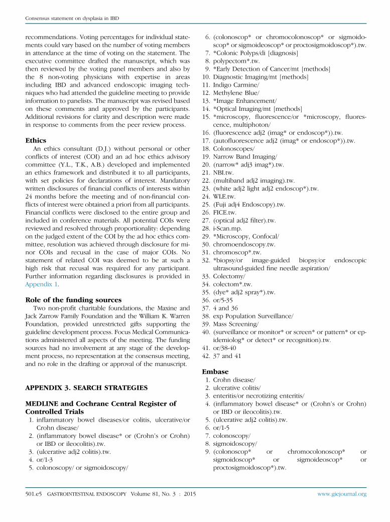
Consensus statement on dysplasia in IBD
recommendations. Voting percentages for individual state-ments could vary based on the number of voting membersin attendance at the time of voting on the statement. Theexecutive committee drafted the manuscript, which wasthen reviewed by the voting panel members and also bythe 8 non-voting physicians with expertise in areasincluding IBD and advanced endoscopic imaging tech-niques who had attended the guideline meeting to provideinformation to panelists. The manuscript was revised basedon these comments and approved by the participants.Additional revisions for clarity and description were madein response to comments from the peer review process.
EthicsAn ethics consultant (D.J.) without personal or other
conflicts of interest (COI) and an ad hoc ethics advisorycommittee (Y.L., T.K., A.B.) developed and implementedan ethics framework and distributed it to all participants,with set policies for declarations of interest. Mandatorywritten disclosures of financial conflicts of interests within24 months before the meeting and of non-financial con-flicts of interest were obtained a priori from all participants.Financial conflicts were disclosed to the entire group andincluded in conference materials. All potential COIs werereviewed and resolved through proportionality: dependingon the judged extent of the COI by the ad hoc ethics com-mittee, resolution was achieved through disclosure for mi-nor COIs and recusal in the case of major COIs. Nostatement of related COI was deemed to be at such ahigh risk that recusal was required for any participant.Further information regarding disclosures is provided inAppendix 1.
Role of the funding sourcesTwo non-profit charitable foundations, the Maxine and
Jack Zarrow Family Foundation and the William K. WarrenFoundation, provided unrestricted gifts supporting theguideline development process. Focus Medical Communica-tions administered all aspects of the meeting. The fundingsources had no involvement at any stage of the develop-ment process, no representation at the consensus meeting,and no role in the drafting or approval of the manuscript.
APPENDIX 3. SEARCH STRATEGIES
MEDLINE and Cochrane Central Register ofControlled Trials1. inflammatory bowel diseases/or colitis, ulcerative/or
Crohn disease/2. (inflammatory bowel disease* or (Crohn’s or Crohn)
or IBD or ileocolitis).tw.3. (ulcerative adj2 colitis).tw.4. or/1-35. colonoscopy/ or sigmoidoscopy/
501.e5 GASTROINTESTINAL ENDOSCOPY Volume 81, No. 3 : 2015
6. (colonoscop* or chromocolonoscop* or sigmoido-scop* or sigmoideoscop* or proctosigmoidoscop*).tw.
7. *Colonic Polyps/di [diagnosis]8. polypectom*.tw.9. *Early Detection of Cancer/mt [methods]
10. Diagnostic Imaging/mt [methods]11. Indigo Carmine/12. Methylene Blue/13. *Image Enhancement/14. *Optical Imaging/mt [methods]15. *microscopy, fluorescence/or *microscopy, fluores-
cence, multiphoton/16. (fluorescence adj2 (imag* or endoscop*)).tw.17. (autofluorescence adj2 (imag* or endoscop*)).tw.18. Colonoscopes/19. Narrow Band Imaging/20. (narrow* adj3 imag*).tw.21. NBI.tw.22. (multiband adj2 imaging).tw.23. (white adj2 light adj2 endoscop*).tw.24. WLE.tw.25. (Fuji adj4 Endoscopy).tw.26. FICE.tw.27. (optical adj2 filter).tw.28. i-Scan.mp.29. *Microscopy, Confocal/30. chromoendoscopy.tw.31. chromoscop*.tw.32. *biopsy/or image-guided biopsy/or endoscopic
ultrasound-guided fine needle aspiration/33. Colectomy/34. colectom*.tw.35. (dye* adj2 spray*).tw.36. or/5-3537. 4 and 3638. exp Population Surveillance/39. Mass Screening/40. (surveillance or monitor* or screen* or pattern* or ep-
idemiolog* or detect* or recognition).tw.41. or/38-4042. 37 and 41
Embase1. Crohn disease/2. ulcerative colitis/3. enteritis/or necrotizing enteritis/4. (inflammatory bowel disease* or (Crohn’s or Crohn)
or IBD or ileocolitis).tw.5. (ulcerative adj2 colitis).tw.6. or/1-57. colonoscopy/8. sigmoidoscopy/9. (colonoscop* or chromocolonoscop* or
sigmoidoscop* or sigmoideoscop* orproctosigmoidoscop*).tw.
www.giejournal.org

Consensus statement on dysplasia in IBD
10. colon polyp/di, dm, pc [diagnosis, disease manage-ment, prevention]
11. polypectomy/12. *early diagnosis/13. *diagnostic imaging/14. indigo carmine/15. methylene blue/16. image enhancement/17. fluorescence imaging/or autofluorescence imaging/or
voltage sensitive dye imaging/18. (fluorescence adj2 (imag* or endoscop*)).tw.19. (autofluorescence adj2 (imag* or endoscop*)).tw.20. exp colonoscope/21. narrow band imaging/22. (narrow* adj3 imag*).tw.23. NBI.tw.24. (multiband adj2 imaging).tw.25. white light endoscopy/26. (white adj2 light adj2 endoscop*).tw.27. WLE.tw.28. (Fuji adj4 Endoscopy).tw.29. FICE.tw.30. optical filter/31. (optical adj2 filter).tw.32. i-Scan.mp.33. *confocal microscopy/34. chromoendoscopy/
SUPPLEMENTAL TABLE 1. Summary characteristics of study
Study Country Year Enrollment period Stud
Subramanian1 UK 2013 2008-2010 Retroco
*Study included 353 patients with 369 colonoscopies. The data are reported on the 203 pain the standard definition group. Four patients had one high definition and one standard
SUPPLEMENTAL TABLE 2. Summary results, surveillance colonoscopy withdefinition white light colonoscopy in inflammatory bowel disease patient
Outcome
High definitionwhite lightN [ 209*
Standarddefinitionwhite lightN [ 160*
Prevaor risk(95%
No. of patients withdysplasia
24 (11.5%) 8 (5.0%) 2.3 (1.0-
No. of patients withendoscopicallyvisible dysplasia
22 5 3.37 (1.2
No. of dysplasticlesions/areas
32 11 –
CI, Confidence interval.*Study included 353 patients with 369 colonoscopies. The data are reported on the 209 c–, ratio cannot be calculated.
www.giejournal.org Vo
35. chromoendoscop*.tw.36. chromoscop*.tw.37. colon biopsy/or rectum biopsy/38. image guided biopsy/39. *colon resection/or *sigmoidectomy/40. colectom*.tw.41. (dye* adj2 spray*).tw.42. or/7-4143. 6 and 4244. cancer epidemiology/45. *health survey/46. mass screening/or cancer screening/47. (surveillance or monitor* or screen* or pattern* or ep-
idemiolog* or detect* or recognition).tw.48. or/44-4749. 43 and 4850. (animal$ not human$).sh,hw.51. 49 not 50
APPENDIX 4. SUMMARY OF EVIDENCEREVIEWED IN DEVELOPMENT OF CONSENSUSRECOMMENDATIONS
Statement 1. When performing surveillance with
Detection of Dysplasiawhite-light colonoscopy, high definition is recom-mended rather than standard definition (Supple-mental Tables 1-3).
y design Type Patient no.*No. withdysplasia
spectivehorts
High definition vsstandard definition
353 32
tients with 209 colonoscopies in the high definition group and 154 patients with 160definition colonoscopy during the study period and were included in both arms.
high definition white light colonoscopy compared to standards
lenceratioCI) Summary
5.1) Surveillance colonoscopy using high definition white lightdetected 2.3 times more patients with dysplasiacompared to standard white light.
8-8.89) Targeted biopsy strategy during high definitioncolonoscopy was 3 times more likely to detect patientswith dysplastic lesions than targeted biopsy strategyduring standard white light colonoscopy.
Surveillance colonoscopy using high definition white lightdetected more dysplasia compared to standard whitelight.
olonoscopies n the high definition group and 160 in the standard definition group.
lume 81, No. 3 : 2015 GASTROINTESTINAL ENDOSCOPY 501.e6
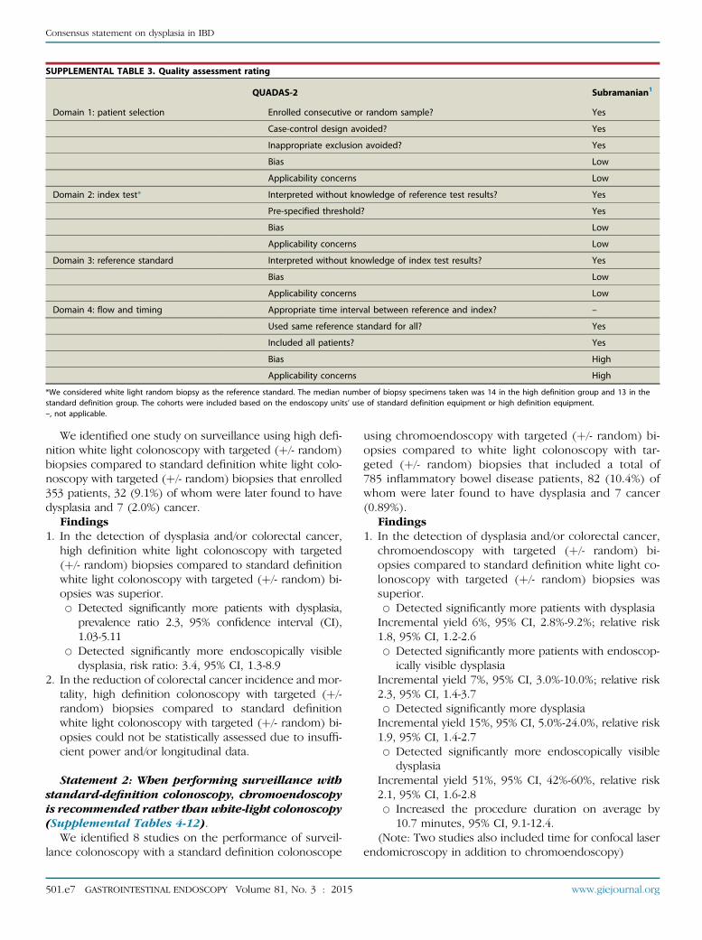
SUPPLEMENTAL TABLE 3. Quality assessment rating
QUADAS-2 Subramanian1
Domain 1: patient selection Enrolled consecutive or random sample? Yes
Case-control design avoided? Yes
Inappropriate exclusion avoided? Yes
Bias Low
Applicability concerns Low
Domain 2: index test* Interpreted without knowledge of reference test results? Yes
Pre-specified threshold? Yes
Bias Low
Applicability concerns Low
Domain 3: reference standard Interpreted without knowledge of index test results? Yes
Bias Low
Applicability concerns Low
Domain 4: flow and timing Appropriate time interval between reference and index? –
Used same reference standard for all? Yes
Included all patients? Yes
Bias High
Applicability concerns High
*We considered white light random biopsy as the reference standard. The median number of biopsy specimens taken was 14 in the high definition group and 13 in thestandard definition group. The cohorts were included based on the endoscopy units’ use of standard definition equipment or high definition equipment.–, not applicable.
Consensus statement on dysplasia in IBD
We identified one study on surveillance using high defi-nition white light colonoscopy with targeted (þ/- random)biopsies compared to standard definition white light colo-noscopy with targeted (þ/- random) biopsies that enrolled353 patients, 32 (9.1%) of whom were later found to havedysplasia and 7 (2.0%) cancer.
Findings1. In the detection of dysplasia and/or colorectal cancer,
high definition white light colonoscopy with targeted(þ/- random) biopsies compared to standard definitionwhite light colonoscopy with targeted (þ/- random) bi-opsies was superior.B Detected significantly more patients with dysplasia,
prevalence ratio 2.3, 95% confidence interval (CI),1.03-5.11
B Detected significantly more endoscopically visibledysplasia, risk ratio: 3.4, 95% CI, 1.3-8.9
2. In the reduction of colorectal cancer incidence and mor-tality, high definition colonoscopy with targeted (þ/-random) biopsies compared to standard definitionwhite light colonoscopy with targeted (þ/- random) bi-opsies could not be statistically assessed due to insuffi-cient power and/or longitudinal data.
Statement 2: When performing surveillance withstandard-definition colonoscopy, chromoendoscopyis recommended rather than white-light colonoscopy(Supplemental Tables 4-12).
We identified 8 studies on the performance of surveil-lance colonoscopy with a standard definition colonoscope
501.e7 GASTROINTESTINAL ENDOSCOPY Volume 81, No. 3 : 2015
using chromoendoscopy with targeted (þ/- random) bi-opsies compared to white light colonoscopy with tar-geted (þ/- random) biopsies that included a total of785 inflammatory bowel disease patients, 82 (10.4%) ofwhom were later found to have dysplasia and 7 cancer(0.89%).
Findings1. In the detection of dysplasia and/or colorectal cancer,
chromoendoscopy with targeted (þ/- random) bi-opsies compared to standard definition white light co-lonoscopy with targeted (þ/- random) biopsies wassuperior.B Detected significantly more patients with dysplasiaIncremental yield 6%, 95% CI, 2.8%-9.2%; relative risk1.8, 95% CI, 1.2-2.6B Detected significantly more patients with endoscop-
ically visible dysplasiaIncremental yield 7%, 95% CI, 3.0%-10.0%; relative risk2.3, 95% CI, 1.4-3.7B Detected significantly more dysplasiaIncremental yield 15%, 95% CI, 5.0%-24.0%, relative risk1.9, 95% CI, 1.4-2.7B Detected significantly more endoscopically visible
dysplasiaIncremental yield 51%, 95% CI, 42%-60%, relative risk2.1, 95% CI, 1.6-2.8B Increased the procedure duration on average by
10.7 minutes, 95% CI, 9.1-12.4.(Note: Two studies also included time for confocal laser
endomicroscopy in addition to chromoendoscopy)
www.giejournal.org
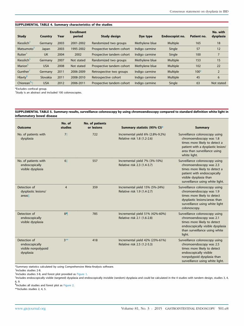
SUPPLEMENTAL TABLE 4. Summary characteristics of the studies
Study Country YearEnrollmentperiod Study design Dye type Endoscopist no. Patient no.
No. withdysplasia
Kiesslich2 Germany 2003 2001-2002 Randomized two groups Methylene blue Multiple 165 18
Matsumoto3 Japan 2003 1995-2002 Prospective tandem cohort Indigo carmine Single 57 12
Rutter4 UK 2004 2002 Prospective tandem cohort Indigo carmine Single 100 7
Kiesslich5 Germany 2007 Not stated Randomized two groups Methylene blue Multiple 153 15
Marion6 USA 2008 Not stated Prospective tandem cohort Methylene blue Multiple 102 22
Gunther7 Germany 2011 2006-2009 Retrospective two groups Indigo carmine Multiple 100* 2
Hlavty8 Slovakia 2011 2008-2010 Retrospective cohort Indigo carmine Multiple 45 6
Chiorean9,y USA 2012 2006-2011 Prospective tandem cohort Indigo carmine Single 63 Not stated
*Excludes confocal group.yStudy is an abstract and included 100 colonoscopies.
SUPPLEMENTAL TABLE 5. Summary results, surveillance colonoscopy by using chromoendoscopy compared to standard definition white light ininflammatory bowel disease
OutcomeNo. ofstudies
No. of patientsor lesions Summary statistic (95% CI)* Summary
No. of patients withdysplasia
7y 722 Incremental yield 6% (2.8%-9.2%)Relative risk 1.8 (1.2-2.6)
Surveillance colonoscopy usingchromoendoscopy was 1.8times more likely to detect apatient with a dysplastic lesion/area than surveillance usingwhite light.
No. of patients withendoscopicallyvisible dysplasia
6z 557 Incremental yield 7% (3%-10%)Relative risk 2.3 (1.4-3.7)
Surveillance colonoscopy usingchromoendoscopy was 2.3times more likely to detect apatient with endoscopicallyvisible dysplasia thansurveillance using white light.
Detection ofdysplastic lesions/areasx
4 359 Incremental yield 15% (5%-24%)Relative risk 1.9 (1.4-2.7)
Surveillance colonoscopy usingchromoendoscopy was 1.9times more likely to detectdysplastic lesions/areas thansurveillance using white lightcolonoscopy.
Detection ofendoscopicallyvisible dysplasia
8{ 785 Incremental yield 51% (42%-60%)Relative risk 2.1 (1.6-2.8)
Surveillance colonoscopy usingchromoendoscopy was 2.1times more likely to detectendoscopically visible dysplasiathan surveillance using whitelight.
Detection ofendoscopicallyvisible nonpolypoiddysplasia
3** 418 Incremental yield 42% (23%-61%)Relative risk 2.5 (1.2-5.3)
Surveillance colonoscopy usingchromoendoscopy was 2.5times more likely to detectendoscopically visiblenonpolypoid dysplasia thansurveillance using white light.
*Summary statistics calculated by using Comprehensive Meta-Analysis software.yIncludes studies 2-8.zIncludes studies 3-8, and forest plot provided as Figure 1.xIncludes endoscopically visible (targeted) dysplasia and endoscopically invisible (random) dysplasia and could be calculated in the 4 studies with tandem design, studies 3, 4,6, 9.{Includes all studies and forest plot as Figure 2.**Includes studies 2, 4, 5.
www.giejournal.org Volume 81, No. 3 : 2015 GASTROINTESTINAL ENDOSCOPY 501.e8
Consensus statement on dysplasia in IBD

SUPPLEMENTAL TABLE 6. Individual study outcomes, incremental yield in the number of patients with dysplasia during surveillance colonoscopycomparing chromoendoscopy to white light
Type of studyNo. ofpatients
Chromoendoscopy(No. of patients with
dysplasia/no. of patients)
White light (No.of patients with
dysplasia/no. of patients)
Summary statistics (95% CI)*
Incremental yield Relative risk
All 722 71/503 (14.1%) 38/508 (7.5%) 6% (3% to 9%) 1.78 (1.23-2.58)
Randomized, two groups2 165 13/84 6/81 8% (-2% to 18%) 2.09 (0.83-5.23)
Prospective, tandem cohort3 57 12/57 12/57 0% (-2% to 2%) 1.0 (0.49-2.04)
Prospective, tandem cohort4 100 7/100 2/100 5% (-1% to 11%) 3.5 (0.75-16.44)
Randomized, two groups5 153 11/80 4/73 8% (-1% to 17%) 2.51 (0.84-7.53)
Prospective, tandem cohort6 102 22/102 12/102 10% (0% to 20%) 1.83 (0.96-3.50)
Retrospective, two groups7 100* 2/50 0/50 4% (-3% to 11%) 5 (0.25-101.58)
Retrospective, cohort8 45 4/30 2/45 9% (-5% to 23%) 3 (0.59-15.36)
Prospective, tandem cohort9,y – – – – –
CI, Confidence interval; –, not stated.*Summary statistics calculated by using Comprehensive Meta-Analysis software, by using fixed-effects model.yStudy 9 did not provide per-patient level data.
Hlavaty 2011
Gunther 2011
Marion 2008
Kiesslich 2007
Rutter 2004
Matsumoto 2003
Summary
-15% -10% -5% 0% 5% 10% 15% 20% 25%
Incremental Yield, % (95% CI)
9% (-5 to 22%)
4% (-3 to 11%)
8% (-1 to 18%)
11% (3 to 19%)
5% (-1 to 11%)
9% (-5 to 22%)
7% (3 to 10%)
Favors WLE Favors CE
Supplemental Figure 1. Forest plot of incremental yield in the numberof patients with dysplasia during surveillance colonoscopy comparingchromoendoscopy to white light. CI, confidence interval; WLE, white-light endoscopy; CE, chromoendoscopy.
SUPPLEMENTAL TABLE 7. Incremental yield in the number of patients with endoscopically visible dysplasia during surveillance colonoscopycomparing chromoendoscopy to white light
Type of study No. of patients
Chromoendoscopy(No. of patients with
endoscopicallyvisible dysplasia/no.
of patients)
White light (No.of patients withendoscopically
visible dysplasia/no.of patients)
Incrementalyield* (95% CI)
Relativerisk* (95% CI)
All 557 53/419 (12.6%) 22/427 (5.2%) 7% (3% to 10%) 2.32 (1.44-3.72)
Randomized, two groups 2,y – – – – –
Prospective, tandem cohort3 57 12/57 7/57 9% (-5% to 22%) 1.71 (0.73-4.04)
Prospective, tandem cohort4 100 7/100 2/100 5% (-1% to 11%) 3.50 (0.75-16.44)
Randomized, two groups5 153 11/80 2/73 11% (3% to 19%) 5.05 (1.15-21.89)
Prospective, tandem cohort6 102 17/102 9/102 8% (-1% to 18%) 1.95 (0.91-4.16)
Retrospective, two groups7 100 2/50 0/50 4%(-3% to 11%) 5(0.25-1.58)
Retrospective, cohort8 45 4/30 2/45 9%(-5% to 22%) 3(0.59-15.36)
Prospective, tandem cohort9,y not stated not stated not stated not stated not stated
CI, Confidence interval.*Summary statistics calculated by using Comprehensive Meta-Analysis software, by using fixed-effects model.yStudies 2, 9 did not have per-patient data available.
Efthymiou 2013
Feitosa 2013
Bisschops 2012
Pellise 2011
Summary
-30% -20% -10% 0% 10% 20% 30% 40% 50%
Incremental Yield, % (95% CI)
2% (-16 to 20%)
22% (1 to 43%)
0% (-15 to 16%)
7% (-5 to 19%)
6% (-1 to 14%)
Favors NBI Favors CE
Supplemental Figure 2. Forest plot of incremental yield in the numberof patients with endoscopically visible dysplasia during surveillance colo-noscopy comparing chromoendoscopy to white light. CI, confidence in-terval; WLE, white-light endoscopy; CE, chromoendoscopy.
501.e9 GASTROINTESTINAL ENDOSCOPY Volume 81, No. 3 : 2015 www.giejournal.org
Consensus statement on dysplasia in IBD
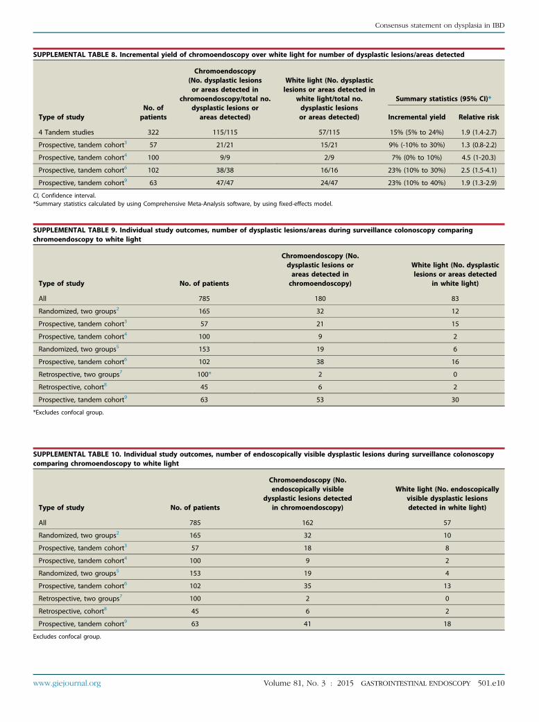
SUPPLEMENTAL TABLE 8. Incremental yield of chromoendoscopy over white light for number of dysplastic lesions/areas detected
Type of studyNo. ofpatients
Chromoendoscopy(No. dysplastic lesionsor areas detected in
chromoendoscopy/total no.dysplastic lesions or
areas detected)
White light (No. dysplasticlesions or areas detected in
white light/total no.dysplastic lesionsor areas detected)
Summary statistics (95% CI)*
Incremental yield Relative risk
4 Tandem studies 322 115/115 57/115 15% (5% to 24%) 1.9 (1.4-2.7)
Prospective, tandem cohort3 57 21/21 15/21 9% (-10% to 30%) 1.3 (0.8-2.2)
Prospective, tandem cohort4 100 9/9 2/9 7% (0% to 10%) 4.5 (1-20.3)
Prospective, tandem cohort6 102 38/38 16/16 23% (10% to 30%) 2.5 (1.5-4.1)
Prospective, tandem cohort9 63 47/47 24/47 23% (10% to 40%) 1.9 (1.3-2.9)
CI, Confidence interval.*Summary statistics calculated by using Comprehensive Meta-Analysis software, by using fixed-effects model.
SUPPLEMENTAL TABLE 9. Individual study outcomes, number of dysplastic lesions/areas during surveillance colonoscopy comparingchromoendoscopy to white light
Type of study No. of patients
Chromoendoscopy (No.dysplastic lesions orareas detected inchromoendoscopy)
White light (No. dysplasticlesions or areas detected
in white light)
All 785 180 83
Randomized, two groups2 165 32 12
Prospective, tandem cohort3 57 21 15
Prospective, tandem cohort4 100 9 2
Randomized, two groups5 153 19 6
Prospective, tandem cohort6 102 38 16
Retrospective, two groups7 100* 2 0
Retrospective, cohort8 45 6 2
Prospective, tandem cohort9 63 53 30
*Excludes confocal group.
SUPPLEMENTAL TABLE 10. Individual study outcomes, number of endoscopically visible dysplastic lesions during surveillance colonoscopycomparing chromoendoscopy to white light
Type of study No. of patients
Chromoendoscopy (No.endoscopically visible
dysplastic lesions detectedin chromoendoscopy)
White light (No. endoscopicallyvisible dysplastic lesionsdetected in white light)
All 785 162 57
Randomized, two groups2 165 32 10
Prospective, tandem cohort3 57 18 8
Prospective, tandem cohort4 100 9 2
Randomized, two groups5 153 19 4
Prospective, tandem cohort6 102 35 13
Retrospective, two groups7 100 2 0
Retrospective, cohort8 45 6 2
Prospective, tandem cohort9 63 41 18
Excludes confocal group.
www.giejournal.org Volume 81, No. 3 : 2015 GASTROINTESTINAL ENDOSCOPY 501.e10
Consensus statement on dysplasia in IBD
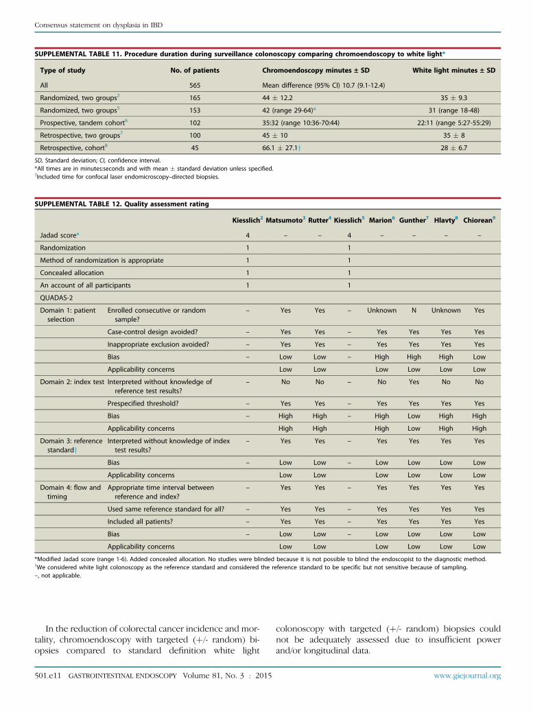
SUPPLEMENTAL TABLE 11. Procedure duration during surveillance colonoscopy comparing chromoendoscopy to white light*
Type of study No. of patients Chromoendoscopy minutes ± SD White light minutes ± SD
All 565 Mean difference (95% CI) 10.7 (9.1-12.4)
Randomized, two groups2 165 44 � 12.2 35 � 9.3
Randomized, two groups5 153 42 (range 29-64)* 31 (range 18-48)
Prospective, tandem cohort6 102 35:32 (range 10:36-70:44) 22:11 (range 5:27-55:29)
Retrospective, two groups7 100 45 � 10 35 � 8
Retrospective, cohort8 45 66.1 � 27.1y 28 � 6.7
SD, Standard deviation; CI, confidence interval.*All times are in minutes:seconds and with mean � standard deviation unless specified.yIncluded time for confocal laser endomicroscopy–directed biopsies.
SUPPLEMENTAL TABLE 12. Quality assessment rating
Kiesslich2 Matsumoto3 Rutter4 Kiesslich5 Marion6 Gunther7 Hlavty8 Chiorean9
Jadad score* 4 – – 4 – – – –
Randomization 1 1
Method of randomization is appropriate 1 1
Concealed allocation 1 1
An account of all participants 1 1
QUADAS-2
Domain 1: patientselection
Enrolled consecutive or randomsample?
– Yes Yes – Unknown N Unknown Yes
Case-control design avoided? – Yes Yes – Yes Yes Yes Yes
Inappropriate exclusion avoided? – Yes Yes – Yes Yes Yes Yes
Bias – Low Low – High High High Low
Applicability concerns Low Low Low Low Low Low
Domain 2: index test Interpreted without knowledge ofreference test results?
– No No – No Yes No No
Prespecified threshold? – Yes Yes – Yes Yes Yes Yes
Bias – High High – High Low High High
Applicability concerns High High High Low High High
Domain 3: referencestandardy
Interpreted without knowledge of indextest results?
– Yes Yes – Yes Yes Yes Yes
Bias – Low Low – Low Low Low Low
Applicability concerns Low Low Low Low Low Low
Domain 4: flow andtiming
Appropriate time interval betweenreference and index?
– Yes Yes – Yes Yes Yes Yes
Used same reference standard for all? – Yes Yes – Yes Yes Yes Yes
Included all patients? – Yes Yes – Yes Yes Yes Yes
Bias – Low Low – Low Low Low Low
Applicability concerns Low Low Low Low Low Low
*Modified Jadad score (range 1-6). Added concealed allocation. No studies were blinded because it is not possible to blind the endoscopist to the diagnostic method.yWe considered white light colonoscopy as the reference standard and considered the reference standard to be specific but not sensitive because of sampling.–, not applicable.
Consensus statement on dysplasia in IBD
In the reduction of colorectal cancer incidence and mor-tality, chromoendoscopy with targeted (þ/- random) bi-opsies compared to standard definition white light
501.e11 GASTROINTESTINAL ENDOSCOPY Volume 81, No. 3 : 2015
colonoscopy with targeted (þ/- random) biopsies couldnot be adequately assessed due to insufficient powerand/or longitudinal data.
www.giejournal.org

Consensus statement on dysplasia in IBD
Statement 3: When performing surveillance withhigh-definition colonoscopy, chromoendoscopy issuggested rather than white-light colonoscopy(Supplemental Tables 13-14).
We identified one study on the performance of surveil-lance colonoscopy with a high definition colonoscopeby using chromoendoscopy with targeted (þ/- random) bi-opsies compared to white light colonoscopy with targeted(þ/- random) biopsies that enrolled 75 patients, 16 (21.3%)of whomwere later found to have dysplasia and none cancer.
Findings1. In the detection of dysplasia and/or colorectal cancer,
chromoendoscopy with targeted (þ/- random) biopsiescompared to high definition white light colonoscopywith targeted (þ/- random) biopsies was superior.B Detected significantly more patients with dysplasia,
21.3% (16/75) vs 9.3% (7/75)Incremental yield 12% (P Z .007)B Detected significantly more endoscopically visible
dysplasia, 100% (22/22) vs 45.4% (10/22)
SUPPLEMENTAL TABLE 13. Summary characteristics of the study
Study Country Year Enrollment period Study design
Picco10,* USA 2013 2009-2013 Prospective tandem I
*Personal communication with author confirmed use of high definition colonoscopies at
SUPPLEMENTAL TABLE 14. Quality assessment rating
QUADAS-2
Domain 1: patient selection Enrolled consecutive
Case-control design
Inappropriate exclus
Bias
Applicability concern
Domain 2: index test * Interpreted without
Prespecified thresho
Bias
Applicability concern
Domain 3: reference standardy Interpreted without
Bias
Applicability concern
Domain 4: flow and timing Appropriate time in
Used same referenc
Included all patients
Bias
Applicability concern
*Personal communication with author confirmed use of high definition colonoscopies atyWe considered white light colonoscopy as the reference standard and considered the r
www.giejournal.org Vol
Incremental yield 16% (P Z .004)B Detected significantly more patients with nonpoly-
poid dysplastic lesions, 9.3% vs 1.3%Incremental yield 8% (P Z .011)
2. In the reduction of colorectal cancer incidence and mor-tality, chromoendoscopy with targeted (þ/- random) bi-opsies compared to high definition white lightcolonoscopy with targeted (þ/- random) biopsies couldnot be adequately assessed due to insufficient powerand/or longitudinal data.
3. The authors aimed to study the interobserver variabilityin the detection of dysplastic lesions and dysplasiadetection rates as well as the procedure time by usingchromoendoscopy for ulcerative colitis surveillanceamong non-expert endoscopists.
Procedure withdrawal time was reported based on the en-doscopist procedure volume:
B !5 procedures: median 31 minutes, range 15-36B 5-14 procedures: median 18 minutes, range 13-27B O14 procedures: median 19 minutes, range 18-22
Dye type Endoscopist no. Patient no. No. with dysplasia
ndigo carmine Multiple 75 16
the 3 sites.
Picco10
or random sample? Unknown
avoided? Yes
ion avoided? Yes
High
s Low
knowledge of reference test results? No
ld? Yes
High
s High
knowledge of index test results? Yes
Low
s Low
terval between reference and index? Yes
e standard for all? Yes
? Yes
Low
s Low
the 3 sites.eference standard to be specific but not sensitive because of sampling.
ume 81, No. 3 : 2015 GASTROINTESTINAL ENDOSCOPY 501.e12

SUPPLEMENTAL TABLE 15. Summary characteristics of the studies
Study Country YearEnrollmentperiod Study design
Image enhancedendoscopy type Endoscopist no. Patient no.
No. withdysplasia
Dekker11 Netherlands 2007 2003-2004 Randomized Narrow band imaging Multiple 42 11
Consensus statement on dysplasia in IBD
Statement 4. When performing surveillance withstandard-definition colonoscopy, narrow bandimaging is not suggested in place of white-light co-lonoscopy (Supplemental Tables 15-16).
We identified one study on the performance of surveil-lance colonoscopy with a standard definition colonoscopethat compared narrow band imaging with targeted (þ/-random) biopsies to white light colonoscopy with targeted(þ/- random) biopsies in which they randomized 42 pa-tients, 11 (26.2%) of whom were later found to havedysplasia and 3 (7.1%) cancer.
Findings1. In the detection of dysplasia and/or colorectal cancer,
standard definition colonoscopy using narrow band im-aging with targeted (þ/- random) biopsies compared towhite light with targeted (þ/- random) biopsies wassimilar.B Showed no differences in detection rates, 8 patients
with dysplasia compared to 7; P Z .705B Showed no difference in procedure time, 50 � 14.4
minutes compared to 47 � 12.1 minutes; P Z .132. In the reduction of colorectal cancer incidence and mor-
tality, narrow band imaging with targeted (þ/- random)biopsies compared to standard definition white light co-lonoscopy with targeted (þ/- random) biopsies could
SUPPLEMENTAL TABLE 16. Quality assessment rating
Dekker11
Jadad score* 3
Randomization 1
Method of randomization is appropriate 0
Concealed allocation 1
An account of all participants 1
*Modified Jadad score (range 1-6). Added concealed allocation.
SUPPLEMENTAL TABLE 17. Summary characteristics of the studies
Study Country YearEnrollmentperiod Study des
van denBroek12
Netherlands 2011 2006-2009 Randomized cover
Ignjatovic13 UK 2012 2006-2010 Randomized
NBI, Narrow band imaging.
501.e13 GASTROINTESTINAL ENDOSCOPY Volume 81, No. 3 : 2015
not be adequately assessed due to insufficient powerand/or longitudinal data.
Statement 5. When performing surveillance withhigh-definition colonoscopy, narrow band imagingis not suggested in place of white-light colonoscopy(Supplemental Tables 17-19).
For equipment-based image enhanced endoscopy:B 2 studies compared narrow band imaging and white
light colonoscopy.B 1 study compared auto fluorescence imaging endos-
copy and white light colonoscopy.B No studies compared other equipment-based image
enhanced endoscopy methods (eg, i-scan [Pentax, To-kyo, Japan]; Fuji Intelligent Chromo Endoscopy [Fuji-non, Tokyo, Japan]16) and white light.
B Multiple studies reported on the diagnostic accuracy ofequipment-based image enhanced endoscopy methodssuch as confocal endomicroscopy, fluorescein, or opti-cal coherence for dysplasia but not on the detection ofdysplasia.We identified 2 studies on the performance of surveil-
lance colonoscopy with a high definition colonoscopethat compared equipment-based image enhanced endos-copy by using narrow band imaging to white light. Thestudies included a total of 160 IBD patients, 21 (13.1%)of whom were later found to have dysplasia and nonecancer.
Findings1. In the detection of dysplasia and/or colorectal cancer,
by using a high definition colonoscope, equipment-based image enhanced endoscopy with targeted (þ/-random) biopsies by using narrow band imagingcompared to white light colonoscopy with targeted(þ/- random) biopsies showed no significant differ-ences. Due to the small study numbers, pooled anal-ysis was not performed.B Detected similar number of patients with any grade
of dysplasia
ignImage
enhancementEndoscopist
no.Patientno.
No. withdysplasia
ross- NBI Multiple 48 11
NBI Multiple 112 10
www.giejournal.org

SUPPLEMENTAL TABLE 19. Quality assessment rating
van den Broek12 Ignjatovic13
Jadad score* 4 4
Randomization 1 1
Method of randomization isappropriate
0 1
Concealed allocation 1 1
An account of all participants 1 1
*Modified Jadad score (range 1-6). Added concealed allocation. No studies wereblinded as it not possible to blind the endoscopist to the diagnostic method used.
SUPPLEMENTAL TABLE 18. Summary results, surveillance colonoscopy with a high definition colonoscope, by using equipment-based imageenhanced endoscopy with NBI compared to white light in IBD
OutcomeNo. ofstudies
No. ofpatients
Results
SummaryStudy 12 Study 13
No. of patientswith dysplasia*
2 160 NBI 9 vs WL 13 NBI 5 vs WL 5 Surveillance colonoscopy with a highdefinition colonoscope showed nodifference in the no. of patients found tohave dysplasia when NBI was usedcompared to white light.
Detection of dysplasticlesions/areas*
2 160 NBI 15 vs WL 17 NBI 6 vs WL 7 Surveillance colonoscopy with a highdefinition colonoscope showed nodifference in the detection of dysplasiawhen NBI was used compared to whitelight.
Detection of endoscopicallyvisible dysplasia
2 160 NBI 14 vs WL 16 NBI 5 vs WL 7 Surveillance colonoscopy with a highdefinition colonoscope showed nodifference in the detection ofendoscopically visible dysplasia when NBIwas used compared to white light.
NBI, Narrow band imaging; IBD, inflammatory bowel disease; WL, white light.*This includes endoscopically visible (targeted) dysplasia and endoscopically invisible (random) dysplasia. It is notable that a random biopsy identified dysplasia and due to thecross-over study design is counted in both groups.
Consensus statement on dysplasia in IBD
B Detected fewer dysplastic lesions2. In the reduction of colorectal cancer incidence and
mortality, by using a high definition colonoscope,equipment based image enhanced endoscopy with tar-geted (þ/- random) biopsies by using narrow band im-aging compared to white light colonoscopy withtargeted (þ/-) random biopsies could not be
SUPPLEMENTAL TABLE 20. Summary characteristics of the studies
Study Country YearEnrollmentperiod Study design
Pellise14 Spain 2011 2006-2007 Randomized cross-over
Bisschops15,* Belgium 2012 Not stated Randomized two groups
Feitosa16,y Brazil 2013 Not stated Randomized two groups
Efthymiou17,z Australia 2013 2009-2010 Prospective tandem
NBI, Narrow band imaging.*Abstract.yData from final document (in Portuguese).zOnly study to perform targeted and random biopsy. 3408, 468, 1220 did not perform ra
www.giejournal.org Vol
adequately assessed due to insufficient power and/orlongitudinal data.
Statement 6. When performing surveillancewith image-enhanced high-definition colonoscopy,narrow band imaging is not suggested in place ofchromoendoscopy (Supplemental Tables 20-24).
We identified 4 studies on surveillance colonoscopywith a high definition colonoscopy that comparedchromoendoscopy to equipment-based image enhancedendoscopy. They included a total of 231 inflammatorybowel disease patients, 54 (23.4%) of whom were laterfound to have dysplasia and one cancer (0.43%). All studiescompared chromoendoscopy to narrow band imagingcolonoscopy.
Findings1. In the detection of dysplasia and/or colorectal cancer, by
using a high definition colonoscope, chromoendoscopywith targeted biopsies compared to equipment based
Imageenhancement type Endoscopist no. Patient no.
No. withdysplasia
Indigo carmine vs NBI Multiple 60 13
Methylene blue vs NBI Multiple 93 16
Indigo carmine vs NBI Multiple 34 4
Methylene blue vs NBI Multiple 44 21
ndom biopsies in either group.
ume 81, No. 3 : 2015 GASTROINTESTINAL ENDOSCOPY 501.e14

Hlavaty 2011
Gunther 2011
Marion 2008
Kiesslich 2007
Rutter 2004
Matsumoto 2003
Kiesslich 2003
Summary
Incremental Yield, % (95% CI)
-15% -10% -5% 0% 5% 10% 15% 20% 25%
9% (-5 to 23%)
4% (-3 to 11%)
10% (0 to 20%)
8% (-1 to 17%)
5% (-1 to 11%)
0% (-2 to 2%)
8% (-2 to 18%)
6% (3 to 9%)
Favors WLE Favors CE
Supplemental Figure 3. Forest plot of incremental yield in the numberof patients with endoscopically visible dysplasia during surveillance colo-noscopy comparing narrow band imaging to chromoendoscopy. CI, con-fidence interval; WLE, white-light endoscopy; CE, chromoendoscopy.
SUPPLEMENTAL TABLE 23. Procedure duration during surveillance colono
Type of studyTotal no. of patients
(chromoendoscopy/NBI)
All 231
Randomized, cross-over14 60 (60/60)
Randomized, two groups15 93 (52/42)
Randomized, two groups16 34 (18/16)
Prospective, tandem17 44 (44/44)
NBI, Narrow band imaging.*Average procedure time.yMedian and interquartile range.
SUPPLEMENTAL TABLE 21. Individual study outcomes, incremental yield in the number of patients with dysplasia during surveillancecolonoscopy comparing chromoendoscopy to NBI
Type of studyNo. ofpatients
ChromoendoscopyNo. of patients with
dysplasia/no. of patients
NBI No. of patientswith dysplasia/no.
of patientsIncremental yield
(95% CI)*Relative risk(95% CI)
All 231 34/174 23/161 6% (-1.4 to 14.2) 1.27 (0.78-2.06)
Randomized, cross-over14 60 10/60 6/60 7% (-5.4 to 18.8) 1.67 (0.65-4.30)
Randomized, two groups15 93 9/52 7/41 0% (-15.2 to 15.7) 1.01 (0.41-2.49)
Randomized, two groups16 34 4/18 0/16 22% (-1.5 to 43) 8.05 (0.47-138.87)
Prospective, tandem17 44 11/44 10/44 2% (-15.5 to 20.1) 1.10 (0.52-2.32)
NBI, Narrow band imaging.
SUPPLEMENTAL TABLE 22. Individual study outcomes, incremental yield in the number of endoscopically visible dysplastic lesions duringsurveillance colonoscopy comparing chromoendoscopy to NBI
Type of study No. of patientsChromoendoscopy
No. endoscopically visible dysplasiaNBI No. endoscopically
visible dysplasia
All 231 61 37
Randomized, cross-over14 60 12 10
Randomized, two groups15 93 25 10
Randomized, two groups16 34 4 0
Prospective, tandem17 44 20 17
NBI, Narrow band imaging.
501.e15 GASTROINTESTINAL ENDOSCOPY Volume 81, No. 3 : 2015
Consensus statement on dysplasia in IBD
image enhanced endoscopy by using NBI with targetedbiopsies showed no significant differences.B Showed no significant difference in the detection of
patients with dysplasiaIncremental yield of 6%, 95% CI, -1.4%-14.2%B Showed no significant difference in the detection of
dysplastic lesions, chromoendoscopy 61 vs NBI 37.B Increased the procedure time overall, but pooled
analysis is not available.2. In the reduction of colorectal cancer incidence and
mortality, by using a high definition colonoscope,chromoendoscopy with targeted biopsies comparedto equipment-based image enhanced endoscopywith targeted biopsies could not be adequately as-sessed due to insufficient power and/or longitudinaldata.
scopy comparing chromoendoscopy to NBI
Chromoendoscopy,minutes, mean ± SD
NBI, minutes,mean ± SD
– –
26 17
26.87 � 9.89 15.74 � 5.62
43.6* 34.1*
13 (12-24)y 13 (12-22)y
www.giejournal.org

SUPPLEMENTAL TABLE 24. Quality assessment rating
Pellise14 Bisschops15 Feitosa16 Efthymiou17
Jadad score* 3 4 4 –
Randomization 1 1 1 –
Method of randomization is appropriate 1 1 1 –
Concealed allocation 0 1 1 –
An account of all participants 1 1 1 –
QUADAS-2
Domain 1: patient selection Enrolled consecutive or random sample? – – – Yes
Case-control design avoided? – – – Yes
Inappropriate exclusion avoided? – – – Yes
Bias – – – Low
Applicability concern HighyDomain 2: index test Interpreted without knowledge of reference test results? – – – Yes
Prespecified threshold? – – – Yes
Bias – – – Low
Applicability concern Low
Domain 3: reference standardz Interpreted without knowledge of index test results? – – – Yes
Bias – – – Low
Applicability concern Low
Domain 4: flow and timing Appropriate time interval between reference and index? – – – Yes
Used same reference standard for all? – – – Yes
Included all patients? – – – Yes
Bias – – – Low
Applicability concern Low
*Modified Jadad score (range 1-6). Added concealed allocation. No studies were blinded as it is not possible to blind the endoscopist to the diagnostic method used.yStudy included left-sided colitis O8 years and Crohn’s disease of any duration.zWe considered chromoendoscopy as the reference standard, and consider the reference standard to be specific but not sensitive due to sampling.–, not applicable.
Consensus statement on dysplasia in IBD
chromoendoscopy surveillance colonoscopy
Additional topic: random biopsy with(Supplemental Tables 25-26).We identified 11 studies on the performance of surveil-
lance colonoscopy by using chromoendoscopy that re-ported data on random biopsy. The studies included atotal of 48,522 random biopsies in 1635 IBD patients. Weevaluated the chromoendoscopy arms within the studies.1. In the detection of dysplasia and/or colorectal cancer on
surveillance colonoscopy using chromoendoscopy:B Proportion of patients surveyed who had dysplasia
identified by targeted biopsies or by random bi-opsies alone (7 studies, 1289 patients)
www.g
� Chromoendoscopy with targeted biopsy:12.4% (95% CI, 8.3%-18.3%) of all patients sur-veyed had dysplasia identified with targetedbiopsies by using chromoendoscopy; Hetero-geneity: Cochran’s Q 24.9 (P ! .001) andI2 Z 75%
� Random biopsy alone: 1.2% (95% CI, 0.8%-2.0%)of all patients surveyed had dysplasia identified
iejournal.org Volume 81
on random biopsies only; Heterogeneity: Co-chran’s Q 3.04 (P Z .80) and I2 Z 0%
B Proportion of patients with dysplasia who haddysplasia identified by targeted biopsies or byrandom biopsies alone (7 studies, 158 patientswith dysplasia):
� Chromoendoscopy with targeted biopsy: 90.2%(95% CI, 85%-94%) of the patients with dysplasiahad their dysplasia identified with targeted biopsyusing chromoendoscopy; Heterogeneity: Co-chran’s Q 0.6 (P Z .97) and I2 Z 0%� Random biopsy alone: 9.8% (95% CI, 6%-15%)of the patients with dysplasia had theirdysplasia identified on random biopsies only;Heterogeneity: Cochran’s Q 0.6 (P Z .97)and I2 Z 0%
B Proportion of random biopsies positive for dysplasia(11 studies, 48522 random biopsies):
� Dysplasia was identified in 0.1% (95% CI, 0.0%-0.3%)of all random biopsy specimens taken; Heterogene-ity: Cochran’s Q 115.3 (P! .001) and I2 Z 91%, No. 3 : 2015 GASTROINTESTINAL ENDOSCOPY 501.e16

SUPPLEMENTAL TABLE 26. Quality assessment rating
Matsumoto3 Rutter4 Marion6 Gunther7 Hlavty8 Mousatta18 Picco10
Jadad score – – – – – – –
Randomization – – – – – – –
Method of randomization is appropriate – – – – – – –
Concealed allocation – – – – – – –
An account of all participants – – – – – – –
QUADAS-2
Domain 1: patientselection
Enrolled consecutive or random sample? Yes Yes Unknown No Unknown Yes Unknown
Case-control design avoided? Yes Yes Yes Yes Yes Yes Yes
Inappropriate exclusion avoided? Yes Yes Yes Yes Yes Yes Yes
Bias Low Low High High High Low High
Applicability concerns Low Low Low Low Low Low Low
Domain 2: index test Interpreted without knowledge of referencetest results?
No No No Yes No No No
Prespecified threshold? Yes Yes Yes Yes Yes Yes Yes
Bias High High High Low High High High
Applicability concerns High High High Low High High High
Domain 3: referencestandard*
Interpreted without knowledge of index testresults?
Yes Yes Yes Yes Yes Yes Yes
Bias Low Low Low Low Low Low Low
Applicability concerns Low Low Low Low Low Low Low
Domain 4: flow andtiming
Appropriate time interval between referenceand index?
Yes Yes Yes Yes Yes Yes Yes
Used same reference standard for all? Yes Yes Yes Yes Yes Yes Yes
Included all patients? Yes Yes Yes Yes Yes Yes Yes
Bias Low Low Low Low Low Low Low
Applicability concerns Low Low Low Low Low Low Low
(continued on next page)
SUPPLEMENTAL TABLE 25. Study outcomes*, random biopsies in patients who underwent chromoendoscopy surveillance
Study Type of study*
No. ofpatientstotal
No. of patientswith dysplasia
Random biopsy
Total no. ofrandom biopsyspecimens taken
No. of randombiopsy specimenswith dysplasia
No. of patients withdysplasia on random
biopsy alone
Matsumoto3 Prospective tandem cohort 57 12 702 3 1
Rutter4 Prospective tandem cohort 100 9 2904 0 0
Marion6 Prospective tandem cohort 102 22 3264 3 2
Gunther7 Retrospective two groups 100 2 1811 0 0
Hlavaty8 Prospective cohort 30 4 1576 0 0
Mousatta18 Prospective cohort 900 93 27596 18 9
Picco10 Prospective tandem cohort 75 16 2400 3 2
Efthymiou17 Prospective tandem cohort 44 12 474 12 Not stated
Kiesslich2 Randomized two groups 84 13 2352 0 Not stated
Kiesslich5 Randomized two groups 80 11 1376 0 Not stated
Chiorean9 Prospective tandem cohort 63 Not stated 4067 6 Not stated
*Data provided are single arm or subset chromoendoscopy cohorts of the studies.
501.e17 GASTROINTESTINAL ENDOSCOPY Volume 81, No. 3 : 2015 www.giejournal.org
Consensus statement on dysplasia in IBD

SUPPLEMENTAL TABLE 26. Continued
Efthymiou17 Kiesslich2 Kiesslich5 Chiorean9
Jadad score* – 4 4 –
Randomization – 1 1 –
Method of randomization is appropriate – 1 1 –
Concealed allocation – 1 1 –
An account of all participants – 1 1 –
QUADAS-2
Domain 1: patient selection Enrolled consecutive or random sample? Yes – – Yes
Case-control design avoided? Yes – – Yes
Inappropriate exclusion avoided? Yes – – Yes
Bias Low – – Low
Applicability concerns Highy – Low
Domain 2: index test Interpreted without knowledge of reference test results? Yes – – No
Prespecified threshold? Yes – – Yes
Bias Low – – High
Applicability concerns Low – High
Domain 3: reference standardy Interpreted without knowledge of index test results? Yes – – Yes
Bias Low – – Low
Applicability concerns Low – Low
Domain 4: flow and timing Appropriate time interval between reference and index? Yes – – Yes
Used same reference standard for all? Yes – – Yes
Included all patients? Yes – – Yes
Bias Low – – Low
Applicability concerns Low – – Low
*We considered white light colonoscopy as the reference standard and consider the reference standard to be specific but not sensitive due to sampling.*Modified Jadad score (range 1-6). Added concealed allocation. No studies were blinded as it is not possible to blind the endoscopist to the diagnostic method used.yStudy included left-sided colitis O8 years and Crohn’s disease of any duration.–, not applicable.
Consensus statement on dysplasia in IBD
2. In the reduction of colorectal cancer incidence and mor-tality, random biopsy specimens could not beadequately assessed due to insufficient power and/orlongitudinal data.
SUPPLEMENTAL TABLE 27. Study outcomes, random biopsies in patients
Study Type of study*Total no. ofpatients
No. of patiewith dyspla
van den Broek12 Randomized cross over 48 14
Ignjatovic13 Randomized two groups 56 5
Subramanian1 Retrospective cohort 203 24
Picco10 Prospective tandem cohort 75 16
Efthymiou17 Prospective tandem cohort 44 12
*Data are provided from single arm or subset high definition white light cohorts of the s
www.giejournal.org Vol
Additional topic: random biopsy with high-definition white-light surveillance colonoscopy(Supplemental Tables 27-28)
We identified 5 studies on the performance of surveillancecolonoscopy by using high definition white light that
who underwent high-definition white-light surveillance
ntssia
Random biopsy
Total no.of random biopsyspecimens taken
No. of randombiopsy specimenswith dysplasia
No. of patients withdysplasia on random
biopsy alone
1580 3 1
1359 0 0
2926 5 2
2400 3 2
474 12 Not stated
tudies.
ume 81, No. 3 : 2015 GASTROINTESTINAL ENDOSCOPY 501.e18
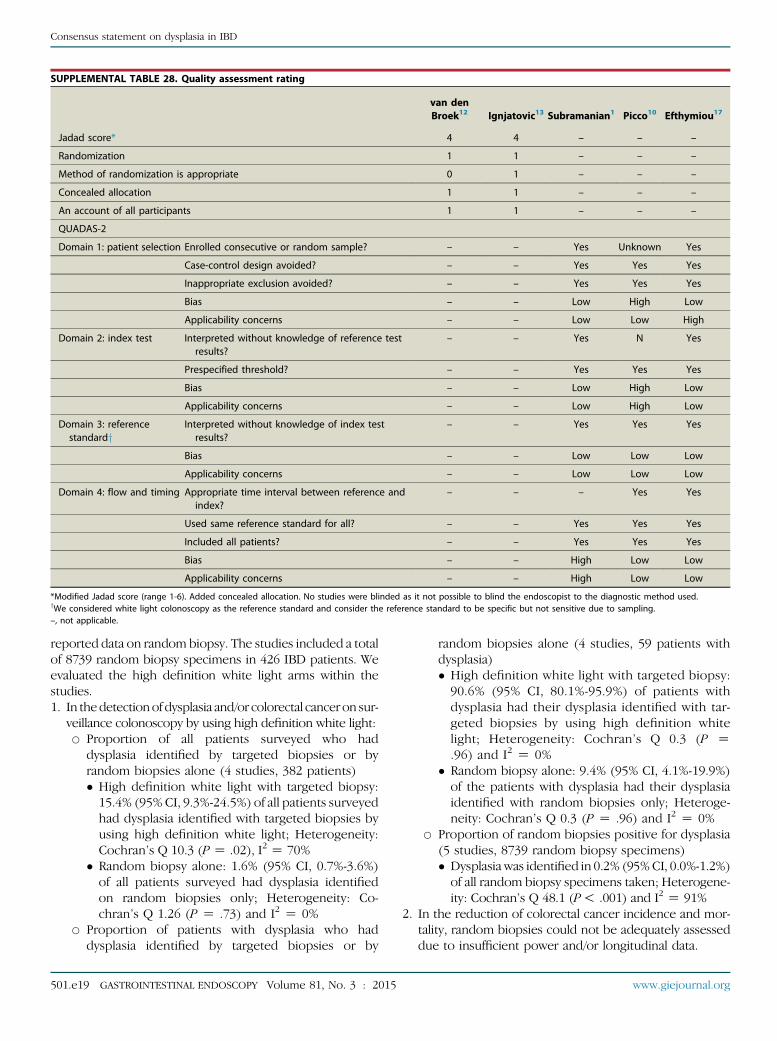
SUPPLEMENTAL TABLE 28. Quality assessment rating
van denBroek12 Ignjatovic13 Subramanian1 Picco10 Efthymiou17
Jadad score* 4 4 – – –
Randomization 1 1 – – –
Method of randomization is appropriate 0 1 – – –
Concealed allocation 1 1 – – –
An account of all participants 1 1 – – –
QUADAS-2
Domain 1: patient selection Enrolled consecutive or random sample? – – Yes Unknown Yes
Case-control design avoided? – – Yes Yes Yes
Inappropriate exclusion avoided? – – Yes Yes Yes
Bias – – Low High Low
Applicability concerns – – Low Low High
Domain 2: index test Interpreted without knowledge of reference testresults?
– – Yes N Yes
Prespecified threshold? – – Yes Yes Yes
Bias – – Low High Low
Applicability concerns – – Low High Low
Domain 3: referencestandardy
Interpreted without knowledge of index testresults?
– – Yes Yes Yes
Bias – – Low Low Low
Applicability concerns – – Low Low Low
Domain 4: flow and timing Appropriate time interval between reference andindex?
– – – Yes Yes
Used same reference standard for all? – – Yes Yes Yes
Included all patients? – – Yes Yes Yes
Bias – – High Low Low
Applicability concerns – – High Low Low
*Modified Jadad score (range 1-6). Added concealed allocation. No studies were blinded as it not possible to blind the endoscopist to the diagnostic method used.yWe considered white light colonoscopy as the reference standard and consider the reference standard to be specific but not sensitive due to sampling.–, not applicable.
Consensus statement on dysplasia in IBD
reported data on random biopsy. The studies included a totalof 8739 random biopsy specimens in 426 IBD patients. Weevaluated the high definition white light arms within thestudies.1. In thedetectionof dysplasia and/or colorectal cancer on sur-
veillance colonoscopy by using high definition white light:B Proportion of all patients surveyed who had
dysplasia identified by targeted biopsies or byrandom biopsies alone (4 studies, 382 patients)
501.e1
� High definition white light with targeted biopsy:15.4% (95%CI, 9.3%-24.5%) of all patients surveyedhad dysplasia identified with targeted biopsies byusing high definition white light; Heterogeneity:Cochran’s Q 10.3 (P Z .02), I2 Z 70%
� Random biopsy alone: 1.6% (95% CI, 0.7%-3.6%)of all patients surveyed had dysplasia identifiedon random biopsies only; Heterogeneity: Co-chran’s Q 1.26 (P Z .73) and I2 Z 0%
B Proportion of patients with dysplasia who haddysplasia identified by targeted biopsies or by
9 GASTROINTESTINAL ENDOSCOPY Volume 81, No. 3 : 2015
random biopsies alone (4 studies, 59 patients withdysplasia)
� High definition white light with targeted biopsy:90.6% (95% CI, 80.1%-95.9%) of patients withdysplasia had their dysplasia identified with tar-geted biopsies by using high definition whitelight; Heterogeneity: Cochran’s Q 0.3 (P Z.96) and I2 Z 0%� Random biopsy alone: 9.4% (95% CI, 4.1%-19.9%)of the patients with dysplasia had their dysplasiaidentified with random biopsies only; Heteroge-neity: Cochran’s Q 0.3 (P Z .96) and I2 Z 0%
B Proportion of random biopsies positive for dysplasia(5 studies, 8739 random biopsy specimens)
� Dysplasia was identified in 0.2% (95%CI, 0.0%-1.2%)of all random biopsy specimens taken; Heterogene-ity: Cochran’s Q 48.1 (P! .001) and I2 Z 91%2. In the reduction of colorectal cancer incidence and mor-tality, random biopsies could not be adequately assesseddue to insufficient power and/or longitudinal data.
www.giejournal.org

SUPPLEMENTAL TABLE 29. Individual study outcomes of random biopsies in standard definition white light surveillance
Study Type of study*Total no. ofpatients
No. of patientswith dysplasia
Random biopsy
Total no.of random biopsyspecimens taken
No. of randombiopsy specimenswith dysplasia
No. of patientswith dysplasia on
random biopsy alone
Matsumoto3 Prospective tandem cohort 57 12 702 7 5
Rutter4 Prospective tandem cohort 100 9 2904 0 0
Marion6 Prospective tandem cohort 102 22 3264 3 3
Gunther7 Retrospective two groups 100 2 1531 0 0
Hlavaty8 Prospective cohort 45 6 1576 0 0
Dekker11 Randomized cross-over 42 11 1522 9 1
van den Broek19 Randomized cross-over 50 10 1992 2 0
Subramanian1 Retrospective cohort 154 8 2080 5 3
Kiesslich2 Randomized two groups 81 18 2746 2 Not stated
Kiesslich5 Randomized two groups 73 15 2854 2 Not stated
Chiorean9 Prospective tandem cohort 63 Not stated 4067 6 Not stated
Jaramillo20 Prospective cohort 85 32 Not stated 19 16
Blonski21 Retrospective cohort Not stated 49 Not stated Not stated 7
Rutter22 Retrospective cohort 525 56 Not stated Not stated 13
van den Broek23 Retrospective cohort 475 53 Not stated Not stated 4
*Data are provided from single arm or subset high definition white light cohorts of the studies.
Consensus statement on dysplasia in IBD
Additional topic: random biopsy with standard-definition white-light surveillance colonoscopy(Supplemental Table 29)
(We provide the following narrative summary and tablefor standard-definition white-light colonoscopy in order toplace the chromoendoscopy and high-definition white-light results in context.)
We identified 15 studies on the performance of surveillancecolonoscopy by using standard definition white light that re-ported data on random biopsy. The studies included over25,238 random biopsies in 1952 IBD patients. We evaluatedthe standard definition white light arms within the studies.
In thedetectionof dysplasia and/or colorectal cancer on sur-veillance colonoscopy by using standard definition white light:B Proportion of all patients surveyed who had dysplasia
identified by targeted biopsies or by random biopsiesalone (11 studies, 1735 patients)
ww
� Standard definition white light with targeted biopsy:11.8% (95% CI, 8.6%-16.1%) of all patients surveyedhad dysplasia identified with targeted biopsies by usingstandard definition white light; Heterogeneity: Co-chran’s Q 39.1 (P! .001) and I2 Z 74%
� Random biopsy alone: 2.6% (95% CI, 1.1%-6.0%) ofall patients surveyed had dysplasia identified onrandom biopsies only; Heterogeneity: Cochran’s Q60.1 (P! .001) and I2 Z 83%
B Proportion of patients with dysplasia who had dysplasiaidentified by targeted biopsies or by random biopsiesalone (12 studies, 270 patients with dysplasia)
w.giejournal.org Volume
� Standard definition white light with targeted biopsy:80.4% (95% CI, 85%-94%) of the patients with dysplasiahad their dysplasia identified with targeted biopsy byusing standard definition white light; Heterogeneity:Cochran’s Q 28.7 (P Z .001) and I2 Z 65%
� Random biopsy alone: 19.6% (95% CI, 11.5%-31.2%)of the patients with dysplasia had their dysplasiaidentified on random biopsies only; Heterogeneity:Cochran’s Q 28.7 (P Z .001) and I2 Z 65%
B Proportion of random biopsies positive for dysplasia(11 studies, 25,238 random biopsies)
� Dysplasia was identified in 0.1% (95% CI, 0.1%-0.3%)of all random biopsies taken; Heterogeneity: Co-chran’s Q 38.3 (P! .001) and I2 Z 76%Additional topic: other image-enhancedendoscopy detection modalities (SupplementalTables 30-32)
We identified one study on the performance of surveil-lance colonoscopy with a high definition colonoscope thatcompared equipment-based image enhanced endoscopyby using autofluorescence to white light. The studiesincluded a total of 50 IBD patients, 10 (20%) of whomwere later found to have dysplasia and none cancer.
Findings1. In the detection of dysplasia and/or colorectal cancer, by
using a high definition colonoscope, equipment basedimage enhanced endoscopy with targeted (þ/- random)biopsies by using auto fluorescence imaging compared
81, No. 3 : 2015 GASTROINTESTINAL ENDOSCOPY 501.e20
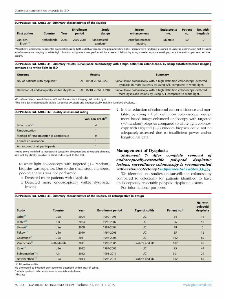
SUPPLEMENTAL TABLE 31. Summary results, surveillance colonoscopy with a high definition colonoscope, by using autofluorescence imagingcompared to white light in IBD
Outcome Results Summary
No. of patients with dysplasia* AFI 10/50 vs WL 6/50 Surveillance colonoscopy with a high definition colonoscope detecteddysplasia in more patients by using AFI, compared to white light.
Detection of endoscopically visible dysplasia AFI 16/18 vs WL 13/18 Surveillance colonoscopy with a high definition colonoscope detectedmore dysplastic lesions by using AFI, compared to white light.
IBD, Inflammatory bowel disease; AFI, autofluorescence imaging; WL, white light.*This includes endoscopically visible (targeted) dysplasia and endoscopically invisible (random) dysplasia.
SUPPLEMENTAL TABLE 32. Quality assessment rating
van den Broek19
Jadad score* 4
Randomization 1
Method of randomization is appropriate 0
Concealed allocation 1
An account of all participants 1
*Jadad score modified to incorporate concealed allocation, and to exclude blinding,as it not logistically possible to blind endoscopist to the test.
SUPPLEMENTAL TABLE 30. Summary characteristics of the studies
First author Country YearEnrollmentperiod
Studydesign
Imageenhancement
Endoscopistno.
Patientno.
No. withdysplasia
van denBroek19
Netherlands 2008 2005-2006 Randomizedtandem*
Autofluorescenceimaging
Multiple 50 10
*All patients underwent segmental examination using both autofluorescence imaging and white light. Patients were randomly assigned to undergo examination first by usingautofluorescence imaging or white light. Random assignment was performed by a research fellow, by using a sealed opaque envelope, once the endoscopist reached thececum.
Consensus statement on dysplasia in IBD
to white light colonoscopy with targeted (þ/- random)biopsies was superior. Due to the small study numbers,pooled analysis was not performed.B Detected more patients with dysplasiaB Detected more endoscopically visible dysplastic
lesions
SUPPLEMENTAL TABLE 33. Summary characteristics of the studies, all ret
Study Country Year Enrollment pe
Odze24 USA 2004 1990-1995
Rutter22 UK 2004 1998-2002
Blonski21 USA 2008 1997-2004
Pekow25 USA 2010 1994-2008
Goldstone26 USA 2011 1994-2006
Van Schaik27 Netherlands 2011 1990-2006
Kisiel28 USA 2012 1994-2003
Subramanian29,y UK 2012 1991-2011
Navaneethan 30 USA 2013 1998-2011
UC, Ulcerative colitis.We attempted to included only adenoma described within area of colitis.*Excludes patients who underwent immediate colectomy.yAbstract.
501.e21 GASTROINTESTINAL ENDOSCOPY Volume 81, No. 3 : 2015
2. In the reduction of colorectal cancer incidence and mor-tality, by using a high definition colonoscope, equip-ment based image enhanced endoscopy with targeted(þ/- random) biopsies compared to white light colonos-copy with targeted (þ/-) random biopsies could not beadequately assessed due to insufficient power and/orlongitudinal data.
Statement 7: After complete removal of
Management of Dysplasiaendoscopically-resectable polypoid dysplasticlesions, surveillance colonoscopy is recommendedrather than colectomy (Supplemental Tables 33-35).
We identified no studies on surveillance colonoscopycompared to colectomy for patients identified to haveendoscopically resectable polypoid dysplastic lesions.
For informational purposes
rospective in design
riod Type of colitis Patient no.*
No. withpolypoiddysplasia
UC 34 18
UC 56 50
UC 49 6
UC 35 12
UC 162 89
Crohn’s and UC 617 45
UC 95 44
UC 301 29
Crohn’s and UC 102 65
www.giejournal.org
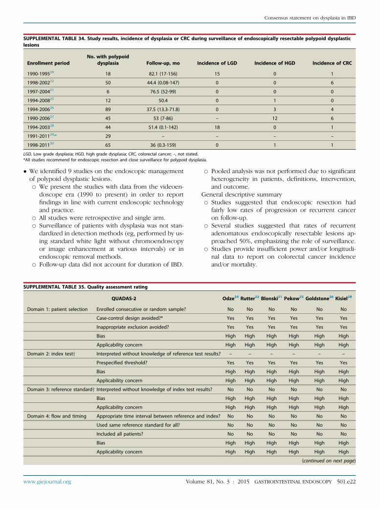
SUPPLEMENTAL TABLE 34. Study results, incidence of dysplasia or CRC during surveillance of endoscopically resectable polypoid dysplasticlesions
Enrollment periodNo. with polypoid
dysplasia Follow-up, mo Incidence of LGD Incidence of HGD Incidence of CRC
1990-199524 18 82.1 (17-156) 15 0 1
1998-200222 50 44.4 (0.08-147) 0 0 6
1997-200421 6 76.5 (52-99) 0 0 0
1994-200825 12 50.4 0 1 0
1994-200626 89 37.5 (13.3-71.8) 0 3 4
1990-200627 45 53 (7-86) – 12 6
1994-200328 44 51.4 (0.1-142) 18 0 1
1991-201129,* 29 – – – –
1998-201130 65 36 (0.3-159) 0 1 1
LGD, Low grade dysplasia; HGD, high grade dysplasia; CRC, colorectal cancer; –, not stated.*All studies recommend for endoscopic resection and close surveillance for polypoid dysplasia.
Consensus statement on dysplasia in IBD
� We identified 9 studies on the endoscopic managementof polypoid dysplastic lesions.B We present the studies with data from the videoen-
doscope era (1990 to present) in order to reportfindings in line with current endoscopic technologyand practice.
B All studies were retrospective and single arm.B Surveillance of patients with dysplasia was not stan-
dardized in detection methods (eg, performed by us-ing standard white light without chromoendoscopyor image enhancement at various intervals) or inendoscopic removal methods.
B Follow-up data did not account for duration of IBD.
SUPPLEMENTAL TABLE 35. Quality assessment rating
QUADAS-2
Domain 1: patient selection Enrolled consecutive or random sample?
Case-control design avoided?*
Inappropriate exclusion avoided?
Bias
Applicability concern
Domain 2: index testy Interpreted without knowledge of reference tes
Prespecified threshold?
Bias
Applicability concern
Domain 3: reference standardy Interpreted without knowledge of index test re
Bias
Applicability concern
Domain 4: flow and timing Appropriate time interval between reference an
Used same reference standard for all?
Included all patients?
Bias
Applicability concern
www.giejournal.org Volum
B Pooled analysis was not performed due to significantheterogeneity in patients, definitions, intervention,and outcome.
General descriptive summaryB Studies suggested that endoscopic resection had
fairly low rates of progression or recurrent canceron follow-up.
B Several studies suggested that rates of recurrentadenomatous endoscopically resectable lesions ap-proached 50%, emphasizing the role of surveillance.
B Studies provide insufficient power and/or longitudi-nal data to report on colorectal cancer incidenceand/or mortality.
Odze24 Rutter22 Blonski21 Pekow25 Goldstone26 Kisiel28
No No No No No No
Yes Yes Yes Yes Yes Yes
Yes Yes Yes Yes Yes Yes
High High High High High High
High High High High High High
t results? – – – – – –
Yes Yes Yes Yes Yes Yes
High High High High High High
High High High High High High
sults? No No No No No No
High High High High High High
High High High High High High
d index? No No No No No No
No No No No No No
No No No No No No
High High High High High High
High High High High High High
(continued on next page)
e 81, No. 3 : 2015 GASTROINTESTINAL ENDOSCOPY 501.e22

TABLE 35. Continued
QUADAS-2 Subramanian29 Van Schaik27 Navaneethan30
Domain 1: patient selection Enrolled consecutive or random sample? No No No
Case-control design avoided?* Yes Yes Yes
Inappropriate exclusion avoided? Yes Yes Yes
Bias High High High
Applicability concern High High High
Domain 2: index test Interpreted without knowledge of reference test results? – – –
Pre-specified threshold? Yes Yes Yes
Bias High High High
Applicability concern High High High
Domain 3: reference standardy Interpreted without knowledge of index test results? No No No
Bias High High High
Applicability concern High High High
Domain 4: flow and timing Appropriate time interval between reference and index? No No No
Used same reference standard for all? No No No
Included all patients? No No No
Bias High High High
Applicability concern High High High
*Population included IBD patients with known dysplasia.yWe considered the index test to be endoscopic removal and surveillance and reference standard to be colectomy.–, not applicable.
Consensus statement on dysplasia in IBD
� A recent meta-analysis on the cancer risk after resectionof polypoid dysplasia in patients with longstanding ulcer-ative colitis includes polypoid dysplasia lesions withinand outside areas of colitis (Wanders LK, Dekker E, Pul-lens B, et al. Clin Gastroenterol Hepatol 2014;12:756-64).
Statement 8: After complete removal ofendoscopically-resectable nonpolypoid dysplasticlesions, surveillance colonoscopy is suggestedrather than colectomy.
We identified 0 studies on surveillance colonoscopycompared to colectomy for patients identified to haveendoscopically resectable nonpolypoid dysplastic lesions.
Note: There is a growing consensus to use similar termi-nology to describe IBD-related superficial colon neoplasia/dysplasia and non-IBD neoplasia/dysplasia. Historic guide-lines and literature have used the term flat dysplasia to referto endoscopically undetectable lesions, and the term raiseddysplasia to refer to endoscopically detectable lesions. Theyhave used the term flat dysplasia to describe endoscopically
SUPPLEMENTAL TABLE 36. Summary characteristics of the studies: all US
Study Year Enrollment period
Ullman31 2002 1990-1993
Ullman32 2003 1994-2001
Pekow25 2010 1994-2008
Goldstone26 2011 1994-2006
Navaneethan30 2013 1998-2011
*Excludes patients who underwent immediate colectomy.
501.e23 GASTROINTESTINAL ENDOSCOPY Volume 81, No. 3 : 2015
detectable but only slightly raised lesions, and the acronymDALM-dysplastic lesions ormasses to refer tomore raised le-sions. All of these terms resulted in inconsistent criteria inthe published literature. Future studies in IBD should usethe Paris Classification system for describing lesionmorphology, which includes polypoid and nonpolypoid.
We did not include Hurlstone DP, Sanders DS, Atkinson R,et al. Endoscopic mucosal resection for flat neoplasia inchronic ulcerative colitis: Can we change the endoscopicmanagement paradigm? Gut 2007;56:838-846. Otherresearch works by this author were formally retracted.
Statement 9: For patients with endoscopicallyinvisible dysplasia (confirmed by a GI pathologist)referral to an endoscopist with expertise in IBDsurveillance by using chromoendoscopy with highdefinition colonoscopy is suggested (SupplementalTables 36-38).
We identified no studies on colectomy compared to sur-veillance colonoscopy for patients identified as havingendoscopically invisible dysplasia.
A based, retrospective, and regarding ulcerative colitis
Patient no.* No. with invisible dysplasia
18 18
137 35
35 13
162 32
102 37
www.giejournal.org

SUPPLEMENTAL TABLE 37. Study results, surveillance of patients with endoscopically invisible dysplasia
Enrollmentperiod
No. with invisibledysplasia
Follow-up, mo
Incidenceof HGD
Incidenceof CRC
Progression to dysplasia inpatients with endoscopically
invisible dysplasia
1990-199331 18 32 (2-117) 3 1 Cumulative incidences13% (0-29) at 1 y26% (4-48) at 2 y33% (9-56) at 5 y
1994-200132 35 15 (4.5-50.5) 3 2 Cumulative incidence53% (29-79) at 5 y*
Progression-free survivalUnifocal 71.4 mo (47-96)Multifocal 54.6 mo (35-74)
1994-200825 13 50.4 1 0 Cumulative incidence 4.3 cases per 100 person years
1994-200626 32 37.5 5 3 Progression-free survival 59.1 � 12.6%y1998-201130 37 36 (0.3-159) 2 1 Higher progression to advanced neoplasia in
flat compared to raised dysplasiahazard ratio 3.6 (1.3-10.6)*,y
HGD, high grade dysplasia; CRC, colorectal cancer.Findings also suggest multifocal* and distal locationy to be more strongly associated with progression to advanced neoplasia and cancer.Studies overall suggest total proctocolectomy for patients with endoscopically invisible dysplasia.
Consensus statement on dysplasia in IBD
Low grade dysplasia� We identified 5 studies on the natural history of endo-scopically invisible low grade dysplasia followed withsurveillance.B We present the studies with data from the videoen-
doscope era (1990 to present) in order to report
SUPPLEMENTAL TABLE 38. Quality assessment rating
QUADAS-2
Domain 1: patient selection Enrolled consecutive or random sample?
Case-control design avoided?
Inappropriate exclusion avoided?
Bias
Applicability concern
Domain 2: index test Interpreted without knowledge of referencetest results?
Prespecified threshold?
Bias
Applicability concern
Domain 3: reference standardy Interpreted without knowledge of indextest results?
Bias
Applicability concern
Domain 4: flow and timing Appropriate time interval between referenceand index?
Used same reference standard for all?
Included all patients?
Bias
Applicability concern
–, not applicable.
www.giejournal.org Volum
findings in line with current endoscopic technologyand practice.
B All (with exception of one) studies were retrospec-tive, single arm, with small numbers and with limitedfollow up.
Ullman31 Ullman32 Pekow25 Goldstone26 Navaneethan30
No No No No No
Yes Yes Yes Yes Yes
Yes Yes Yes Yes Yes
High High High High High
High High High High High
– – – – –
Yes Yes Yes Yes Yes
High High High High High
High High High High High
No No No No No
High High High High High
High High High High High
No No No No No
No No No No No
No No No No No
High High High High High
High High High High High
e 81, No. 3 : 2015 GASTROINTESTINAL ENDOSCOPY 501.e24
REFERENCES
1. Subramanian V, Ramappa V, Telakis E, et al. Comparison of high defi-nition with standard white light endoscopy for detection ofdysplastic lesions during surveillance colonoscopy in patients withcolonic inflammatory bowel disease. Inflamm Bowel Dis 2013;19:350-5.
2. Kiesslich R, Fritsch J, Holtmann M, et al. Methylene blue-aided chro-moendoscopy for the detection of intraepithelial neoplasia and coloncancer in ulcerative colitis. Gastroenterology 2003;124:880-8.
3. Matsumoto T, Nakamura S, Jo Y, et al. Chromoscopy might improvediagnostic accuracy in cancer surveillance for ulcerative colitis. Am JGastroenterol 2003;98:1827-33.
4. Rutter MD, Saunders BP, Schofield G, et al. Pancolonic indigo carminedye spraying for the detection of dysplasia in ulcerative colitis. Gut2004;53:256-60.
5. Kiesslich R, Goetz M, Lammersdorf K, et al. Chromoscopy-guided endo-microscopy increases the diagnostic yield of intraepithelial neoplasia inulcerative colitis. Gastroenterology 2007;132:874-82.
6. Marion JF, Waye JD, Present DH, et al. Chromoendoscopy-targeted bi-opsies are superior to standard colonoscopic surveillance for detectingdysplasia in inflammatory bowel disease patients: a prospective endo-scopic trial. Am J Gastroenterol 2008;103:2342-9.
7. Gunther U, Kusch D, Heller F, et al. Surveillance colonoscopy in pa-tients with inflammatory bowel disease: comparison of random bi-opsy vs. targeted biopsy protocols. Int J Colorectal Dis 2011;26:667-72.
8. Hlavaty T, Huorka M, Koller T, et al. Colorectal cancer screening in pa-tients with ulcerative and Crohn’s colitis with use of colonoscopy, chro-moendoscopy and confocal endomicroscopy. Eur J GastroenterolHepatol 2011;23:680-9.
9. Chiorean MV, Helper DJ, Saxena R, et al. Targeted biopsies using chro-moendoscopy can replace random biopsies in patients with IBD athigh risk for colorectal neoplasia. Gastroenterol 2012;142(suppl 1):S-339.
10. Picco MF, Pasha S, Leighton JA, et al. Procedure time and the determi-nation of polypoid abnormalities with experience: implementation of achromoendoscopy program for surveillance colonoscopy for ulcerativecolitis. Inflamm Bowel Dis 2013;19:1913-20.
11. Dekker E, van den Broek FJ, Reitsma JB, et al. Narrow-band imagingcompared with conventional colonoscopy for the detection ofdysplasia in patients with longstanding ulcerative colitis. Endoscopy2007;39:216-21.
12. van den Broek FJ, Fockens P, van Eeden S, et al. Narrow-band imagingversus high-definition endoscopy for the diagnosis of neoplasia in ul-cerative colitis. Endoscopy 2011;43:108-15.
13. Ignjatovic A, East JE, Subramanian V, et al. Narrow band imaging fordetection of dysplasia in colitis: a randomized controlled trial. Am JGastroenterol 2012;107:885-90.
Consensus statement on dysplasia in IBD
B Surveillance of patients with dysplasia was not stan-dardized in detection methods (eg, performed by us-ing standard white light without chromoendoscopyor image enhancement at various intervals) or inendoscopic removal methods.
B Follow-up data did not account for duration of IBD.� A pooled analysis was thought inappropriate due to sig-nificant heterogeneity in patients, definitions, interven-tion, and outcome.General descriptive summaryB The majority of studies did not recommend procto-
colectomy for patients with endoscopically invisiblelow-grade dysplasia.
B All studies emphasized the cumulative incidence ofcancer and important role of vigilant surveillance.
B Some studies reported increased cumulative inci-dence of cancer when endoscopically invisibledysplasia is multifocal or when it is located in thedistal colon.
B Studies provide insufficient power and/or longitudi-nal data to report on colorectal cancer incidenceand/or mortality.
� For the articles that we identified for this statement,we interpreted the report of “flat dysplasia” as endo-scopically invisible dysplasia unless there were clearmorphologic features described. We used thisapproach based on the historic guidelines and litera-ture that have used the term flat dysplasia to referto endoscopically undetectable lesions and the termraised dysplasia to refer to endoscopically detectablelesions.
� A recent meta-analysis on the cancer risk of low-gradedysplasia in chronic ulcerative colitis that included 20surveillance studies totaling 508 flat low gradedysplasia or low grade dysplasia with dysplasia-associated lesions or masses. The studies predate theuse of current technology of image enhanced endos-copy or even high resolution endoscopy. The authorsreported a 9-fold risk of developing cancer (odds ratio[OR] 9.0, 95% CI, 4.0-20.5) and a 12-fold risk of devel-oping any advanced lesion (OR 11.9, 95% CI, 5.2-27).The absolute risk of cancer in this meta-analysis was14 (95% CI, 5-34) cancers/100 years patient follow-up. Meta-analysis: cancer risk of low-grade dysplasiain chronic ulcerative colitis. Thomas T, Abrams KA,Robinson RJ, et al. Aliment Pharmacol Ther 2007;25,657-68.
High-grade dysplasia� We identified no studies on the natural history of endo-scopically invisible high-grade dysplasia followed withsurveillance that reported findings during the videoen-doscope era (1990 to present).
A systematic review of findings from the fiberoptic erareported a probability of finding cancer in a patientwith high-grade dysplasia of 42% (10/24) if colectomywas done immediately and 32% (15/47) if colectomy
501.e25 GASTROINTESTINAL ENDOSCOPY Volume 81, No. 3 : 2015
was done after some follow-up: Bernstein CN, Shana-han F, Weinstein WM. Are we telling patients the truthabout surveillance colonoscopy in ulcerative colitis?Lancet 1994;343,71-4. Importantly, interpretation ofthe data should be with caution due to significant lim-itations in the sensitivity of the fiberoptic technologyto detect dysplasia or cancer at index colonoscopy.Furthermore, surveillance of patients with dysplasiawas not standardized (eg, performed without chro-moendoscopy or image enhancement at various inter-vals or in endoscopic removal methods). Thus, thetrue incidence of synchronous colorectal cancer inthe setting of high grade dysplasia as well as thetrue natural history of endoscopically invisible highgrade dysplasia is not known.
www.giejournal.org

14. Pellise M, Lopez-Ceron M, Rodriguez de Miguel C, et al. Narrow-band imaging as an alternative to chromoendoscopy for the detec-tion of dysplasia in long-standing inflammatory bowel disease: aprospective, randomized, crossover study. Gastrointest Endosc2011;74:840-8.
15. Bisschops R, Bessissow T, Baert FJ, et al. Chromo-endoscopy versus nar-row band imaging in ulcerative colitis: a prospective randomizedcontrolled trial [abstract]. Gastrointest Endosc 2012;75:AB148.
16. Feitosa F, Carlos A, Nogueira JG, et al. Narrow-band imaging and chro-moendoscopy for the detecion of colonic dysplasia in inflammatorybowel disease: a prospective and randomized study. Inflamm BowelDis 2011;17:S14-15
17. Efthymiou M, Allen PB, Taylor AC, et al. Chromoendoscopy versus nar-row band imaging for colonic surveillance in inflammatory bowel dis-ease. Inflamm Bowel Dis 2013;19:2132-8.
18. Moussata D, Allez M, Cazals-Hatem D, et al. Are random biopsies stilluseful for the detection of intraepithelial neoplasia in IBD patients un-dergoing surveillance colonoscopy with chromoendoscopy? Gut2012;61:A24.
19. van den Broek FJ, Fockens P, van Eeden S, et al. Endoscopic tri-modalimaging for surveillance in ulcerative colitis: randomised comparisonof high-resolution endoscopy and autofluorescence imaging forneoplasia detection; and evaluation of narrow-band imaging for clas-sification of lesions. Gut 2008;57:1083-9.
20. Jaramillo E, Watanabe M, Befrits R, et al. Small, flat colorectal neoplasiasin long-standing ulcerative colitis detected by high-resolution elec-tronic video endoscopy. Gastrointest Endosc 1996;44:15-22.
21. Blonski W, Kundu R, Lewis J, et al. Is dysplasia visible during surveil-lance colonoscopy in patients with ulcerative colitis? Scand J Gastroen-terol 2008;43:698-703.
22. Rutter MD, Saunders BP, Wilkinson KH, et al. Most dysplasia in ul-cerative colitis is visible at colonoscopy. Gastrointest Endosc2004;60:334-9.
23. van den Broek FJ, Stokkers PC, Reitsma JB, et al. Random biopsies takenduring colonoscopic surveillance of patients with longstanding ulcera-tive colitis: low yield and absence of clinical consequences. Am J Gas-troenterol 2014;109:715-22.
24. Odze RD, Farraye FA, Hecht JL, et al. Long-term follow-up after polypec-tomy treatment for adenoma-like dysplasitc lesions in ulcerative colitis.Clin Gastroenterol Hepatol 2004;2:534-41.
25. Pekow JR, Hetzel JT, Rothe JA, et al. Outcome after surveillance of low-grade and indefinite dysplasia in patients with ulcerative colitis. In-flamm Bowel Dis 2010;16:1352-6.
26. Goldstone R, Itzkowitz S, Harpaz N, et al. Progression of low-gradedysplasia in ulcerative colitis: effect of colonic location. Gastrointest En-dosc 2011;74:1087-93.
27. van Schaik FD, Mooiweer E, van der Have M, et al. Adenomas in patientswith inflammatory bowel disease are associated with an increased risk ofadvanced neoplasia. Inflamm Bowel Dis 2013;19:342-9.
28. Kisiel JB, Loftus EV Jr, Harmsen WS, et al. Outcome of sporadic ade-nomas and adenoma-like dysplasia in patients with ulcerative colitisundergoing polypectomy. Inflamm Bowel Dis 2012;18:226-35.
29. Subramanian V, Chatu S, Echterdiek F. Patients with endoscopicallyvisible polypoid adenomatous lesions within the extent of ulcerativecolitis have an increased risk of colorectal cancer despite endoscopicresection. 2012.
30. Navaneethan U, Jegadeesan R, Gutierrez NG, et al. Progression oflow-grade dysplasia to advanced neoplasia based on the locationand morphology of dysplasia in ulcerative colitis patients withextensive colitis under colonoscopic surveillance. J Crohns Colitis2013;7:e684-91.
31. Ullman TA, Loftus EV Jr, Kakar S, et al. The fate of low grade dysplasia inulcerative colitis. Am J Gastroenterol 2002;97:922-7.
32. Ullman T, Croog V, Harpaz N, et al. Progression of flat low-gradedysplasia to advanced neoplasia in patients with ulcerative colitis.Gastroenterology 2003;125:1311-9.
www.giejournal.org Volume 81, No. 3 : 2015 GASTROINTESTINAL ENDOSCOPY 501.e26
Consensus statement on dysplasia in IBD