Scanning transmission mucosa - Gutgut.bmj.com/content/gutjnl/11/6/471.full.pdfGut, 1970, 11, 471-481...
11
Gut, 1970, 11, 471-481 Scanning and transmission electron microscopic studies of human intestinal mucosa PETER G. TONER, KATHARINE E. CARR1, ANNE FERGUSON, AND COLIN MACKAY From the Departments of Pathology and Surgery, University of Glasgow Western Infirmary, Glasgow, the Department of Medicine, Royal Infirmary Glasgow, and the Bio-Engineering Unit, University of Strathclyde, Glasgow SUMMARY The scanning electron microscope is capable of 25 m, resolution combined with a great depth of focus, features which make the technique of value in the study of the intestinal mucosa. Surgical biopsies and postmortem specimens of small intestine have been examined using scanning and transmission electron microscopy. In biopsies, at low magnification, the villous pattern is seen while at higher magnification details of cell surfaces may be observed. Some features are best observed using scanning microscopy to examine dewaxed thick histo- logical sections. In necropsy specimens villous architecture can still be satisfactorily assessed and details of the villous cores are seen, their collagen skeletons apparently less robust than the villi of the fresh biopsy. Scanning microscopy can extend the three-dimensional study of the small intestinal mucosa beyond the limits imposed by the resolution of the dissecting microscope. The technique of peroral intestinal biopsy intro- duced by Shiner (1956 and 1957) has made avail- able fresh human tissues for examination not only by conventional histological and ultra- structural methods but also by binocular dis- secting light microscopy (Rubin and Dobbins, 1965; Trier and Rubin, 1965). Two further methods have recently been applied to the inves- tigation of intestinal morphology. First, Loehry and Creamer (1966) have shown that the pattern of the mucosa is retained, despite autolysis, in specimens obtained at necropsy. A surprisingly detailed assessment of mucosal architecture is thus possible using the dissecting microscope (Creamer and Leppard, 1965; Ferguson, Maxwell, and Carr, 1969; Loehry and Creamer, 1969). Second the range of dissecting light microscopy has been extended by the use of the scanning electron microscope, allowing examination of the surface contours of individual villi as well as the overall pattern of the mucosa (Carr and Toner, 1968; Marsh, Swift, and Williams, 1968; Toner and Carr, 1969; Demling, Received for publication 22 October 1969. 'Present address: Department of Anatomy, University of Glasgow. Becker, and Classen, 1969). The present paper describes further applications of scanning electron microscopy to the study of human intestine, including its use in the examination of post- mortem specimens and of thick sections. Material and Methods The preparation of intestinal biopsies for scan- ning electron microscopy has been described in previous papers (Carr and Toner, 1968; Toner and Carr, 1969). Specimens of human jejunal and duodenal mucosa were obtained at laparo- tomy during the surgical treatment of patients with duodenal ulcers. Twenty-five such samples have now been examined as a basis for this study. In addition, specimens of small bowel obtained at necropsy were stored at room temperature for several days in saline, allowing further autolysis. They were then fixed in formalin and examined by dissecting microscopy (Ferguson et al, 1969). Pieces of tissue up to 10 mm wide were then washed, postfixed in osmium tetroxide, and pre- on 8 July 2018 by guest. Protected by copyright. http://gut.bmj.com/ Gut: first published as 10.1136/gut.11.6.471 on 1 June 1970. Downloaded from
Transcript of Scanning transmission mucosa - Gutgut.bmj.com/content/gutjnl/11/6/471.full.pdfGut, 1970, 11, 471-481...

Gut, 1970, 11, 471-481
Scanning and transmission electron microscopicstudies of human intestinal mucosa
PETER G. TONER, KATHARINE E. CARR1, ANNE FERGUSON, ANDCOLIN MACKAYFrom the Departments of Pathology and Surgery, University of Glasgow Western Infirmary, Glasgow,the Department of Medicine, Royal Infirmary Glasgow, and the Bio-Engineering Unit, University ofStrathclyde, Glasgow
SUMMARY The scanning electron microscope is capable of 25 m, resolution combined witha great depth of focus, features which make the technique of value in the study of the intestinalmucosa. Surgical biopsies and postmortem specimens of small intestine have been examinedusing scanning and transmission electron microscopy. In biopsies, at low magnification, thevillous pattern is seen while at higher magnification details of cell surfaces may be observed.Some features are best observed using scanning microscopy to examine dewaxed thick histo-logical sections. In necropsy specimens villous architecture can still be satisfactorily assessedand details of the villous cores are seen, their collagen skeletons apparently less robust thanthe villi of the fresh biopsy. Scanning microscopy can extend the three-dimensional study ofthe small intestinal mucosa beyond the limits imposed by the resolution of the dissectingmicroscope.
The technique of peroral intestinal biopsy intro-duced by Shiner (1956 and 1957) has made avail-able fresh human tissues for examination notonly by conventional histological and ultra-structural methods but also by binocular dis-secting light microscopy (Rubin and Dobbins,1965; Trier and Rubin, 1965). Two furthermethods have recently been applied to the inves-tigation of intestinal morphology. First, Loehryand Creamer (1966) have shown that the patternof the mucosa is retained, despite autolysis, inspecimens obtained at necropsy. A surprisinglydetailed assessment of mucosal architecture isthus possible using the dissecting microscope(Creamer and Leppard, 1965; Ferguson,Maxwell, and Carr, 1969; Loehry andCreamer, 1969). Second the range of dissectinglight microscopy has been extended by the use ofthe scanning electron microscope, allowingexamination of the surface contours of individualvilli as well as the overall pattern of the mucosa(Carr and Toner, 1968; Marsh, Swift, andWilliams, 1968; Toner and Carr, 1969; Demling,Received for publication 22 October 1969.'Present address: Department of Anatomy, University of Glasgow.
Becker, and Classen, 1969). The present paperdescribes further applications of scanning electronmicroscopy to the study of human intestine,including its use in the examination of post-mortem specimens and of thick sections.
Material and Methods
The preparation of intestinal biopsies for scan-ning electron microscopy has been described inprevious papers (Carr and Toner, 1968; Tonerand Carr, 1969). Specimens of human jejunaland duodenal mucosa were obtained at laparo-tomy during the surgical treatment of patientswith duodenal ulcers. Twenty-five such sampleshave now been examined as a basis for this study.In addition, specimens of small bowel obtainedat necropsy were stored at room temperature forseveral days in saline, allowing further autolysis.They were then fixed in formalin and examinedby dissecting microscopy (Ferguson et al, 1969).Pieces of tissue up to 10 mm wide were thenwashed, postfixed in osmium tetroxide, and pre-
on 8 July 2018 by guest. Protected by copyright.
http://gut.bmj.com
/G
ut: first published as 10.1136/gut.11.6.471 on 1 June 1970. Dow
nloaded from

Peter G. Toner, Katharine E. Carr, Anne Ferguson, and Colin Mackay
Fig. 1 Low magnification scanning electronmicrograph ofhuman jejunal mucosa obtained atsurgical biopsy. The majority of villi are finger-shaped, but occasional bifid and leaf-shaped villican be easily recognized. The characteristic resolutionand depth offocus are seen. x 100.
pared for scanning electron microscopy in thesame way as fresh specimens.
Blocks were also prepared for conventionalhistology. Sections 30, thick were cut from theseblocks and were mounted and prepared forscanning electron microscopy.' After dewaxingand drying, these sections were coated withcarbon-platinum and gold-palladium or withgold-palladium alone, in a vacuum evaporator.'The scanning microscope used was the Cambridge StereoscanMk 2a, operated at accelerating voltages of from 2 to 30 ky.
Parallel investigations were also undertakenusing transmission electron microscopy. Speci-mens postfixed in osmium tetroxide were em-bedded in Araldite, then sectioned and examinedin the Siemens Elmiskop la.
Results
SCANNING ELECTRON MICROSCOPY OFINTESTINAL BIOPSIESA typical low magnification scanning electronmicrograph of the mucosal surface of humanjejunum is shown in Fig. 1, which displays theresolution and depth of focus which are charac-teristic of the scanning image. The features seenin these specimens (Carr and Toner, 1968; Marsh
472 on 8 July 2018 by guest. P
rotected by copyright.http://gut.bm
j.com/
Gut: first published as 10.1136/gut.11.6.471 on 1 June 1970. D
ownloaded from

Scanning and transmission electron microscopic studies of human intestinal mucosa
Fig. 2 Slightly higher magnification scanningelectron micrograph ofhuman jejunal mucosashowing the surface creases of the villi and occasionalcrypt mouths (arrows). x 260.
473 on 8 July 2018 by guest. P
rotected by copyright.http://gut.bm
j.com/
Gut: first published as 10.1136/gut.11.6.471 on 1 June 1970. D
ownloaded from

Peter G. Toner, Katharine E. Carr, Anne Ferguson, and Colin Mackay
et al, 1968; Toner and Carr, 1969), include sur-face creases on villi, goblet cell mouths, andoccasional intestinal crypt mouths opening be-tween villi (Figs. 1 and 2). At high magnification(Marsh et al, 1968; Toner and Carr, 1969) hexa-gonal patterns formed by the close-packed apicalsurfaces of the columnar cells may be seen. Inthe fixed and air-dried biopsies examined, micro-villi are not readily demonstrated although theyhave been seen in freeze-dried specimens (Marshet al, 1968).Dewaxed paraffin sections, 30,u of intestinal
biopsies show some aspects of internal tissuestructure which cannot be visualized in wholemucosal specimens. Among the features observedin these sections are the interconnecting inter-cellular spaces between the columnar cell bases(Figs. 3 and 4). At the free border of the epithelialcells there is a zone which corresponds to thestriated border (Fig. 5). This can be resolved intoits component microvilli when cut in longitudinalsection. Limited areas of the surfaces of the villiare still available for examination in 30u sections;a simultaneous display of the cut surface of thesection and, at right angles, the natural freesurface of the villus, is easily achieved (Fig. 3).In these cases, in addition to the surface featuresalready described, the general pattern of arrange-ment of the microvilli can now often be seen(Fig. 6), due perhaps to minor structural altera-tions produced by wax embedding. Large intra-cellular features can also be observed in thicksections. Paneth cell granules, for example, areclearly seen at the bases of intestinal crypts(Fig. 7). Within the core of the villus and in thelamina propria, blood vessels are recognized bytheir contained red blood corpuscles (Fig. 8) andthe connective tissue framework of collagen fibrebundles can be seen.
SCANNING ELECTRON MICROSCOPY OFNECROPSY SPECIMENSThe mucosal surface of autolysed small intestine,which retains the general structural featurespresent at death, is recorded with clarity usingthe scanning electron microscope (Fig. 9). As inbiopsies examined at low magnification, in-dividual villi of different shapes can be identified,but in autolysed tissues the villous cores are moreangular and often appear twisted. In some casesthe villi may even appear collapsed or flattened,but are still easily recognizable. Figure 10 demon-strates the appearance of the connective tissue'skeleton' of the mucosa in a case found to havea 'flat' intestinal mucosa. The absence of villiemphasizes the width of the crypt mouths whichare much more prominent than in biopsy speci-mens owing to shedding of the autolysed cryptcells. In specimens with a more normal villouspattern the relationship between crypts and villican be seen.The surface of autolysed villi is surprisingly
Fig. 3 Scanning electron micrograph ofdewaxed30g thick section ofhuman jejunum showing boththe cut surface and the natural free surface of thevillus. Intercellular spaces are seen between the lowerparts of the epithelial cells on the cut surface, whilethe hexagonal pattern of adjacent cell boundariescan be made out on the free surface. A pit corre-sponding to a goblet cell mouth is present (arrow).x 1,750.
Fig. 4 Scanning electron micrograph ofdewaxed30,u thick section ofhuman jejunum showing theepithelial cells cut slightly obliquely, revealing theintercellular spaces. The 'basement membrane' regionof the villus is displayed (B). x 1,925.
Fig. 5 Scanning electron micrograph ofdewaxed30,u thick section ofhuman jejunum showing theapical parts of the epithelium of two adjacent villi.Although little intracellular detail is seen, the regionof the striated border is readily seen (arrows).A 'microcrypt' is present (C), corresponding to thegrooves seen in Figs. 1 and 2 in the whole specimen.x 2,500.
474 on 8 July 2018 by guest. P
rotected by copyright.http://gut.bm
j.com/
Gut: first published as 10.1136/gut.11.6.471 on 1 June 1970. D
ownloaded from

Scanning and transmission electron microscopic studies ofhuman intestinal mucosa
Fig. 4. Fig. 5.
Fig. 6 Scanning electron micrograph of microvilliin the columnar cell surface ofa human jejunalvillus. The material was a dewaxed 30. section.Only the surface pattern ofarrangement of thesemicrovilli can be seen. x 40,000.
l
t:.
475 on 8 July 2018 by guest. P
rotected by copyright.http://gut.bm
j.com/
Gut: first published as 10.1136/gut.11.6.471 on 1 June 1970. D
ownloaded from
Peter G. Toner, Katharine E. Carr, Anne Ferguson, and Colin Mackay
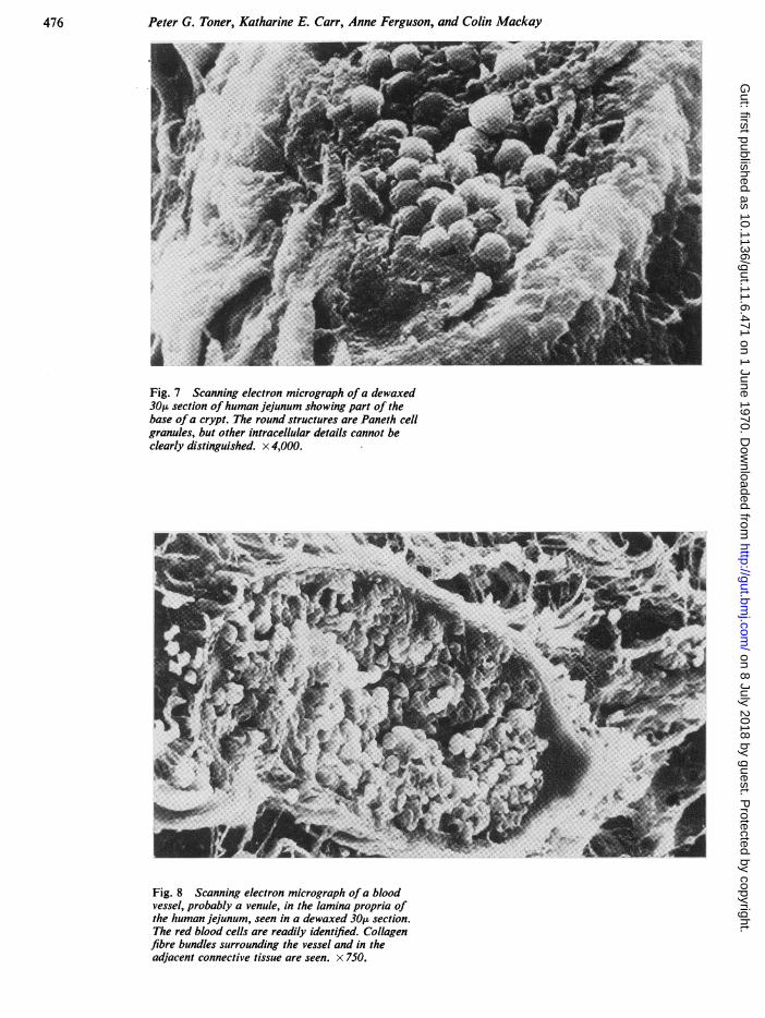
Fig. 7 Scanning electron micrograph ofa dewaxed30gt section ofhuman jejunum showing part of thebase ofa crypt. The round structures are Paneth cellgranules, but other intracellular details cannot beclearly distinguished. x 4,000.
Fig. 8 Scanning electron micrograph of a bloodvessel, probably a venule, in the lamina propria ofthe human jejunum, seen in a dewaxed 30 section.The red blood cells are readily identified. Collagenfibre bundles surrounding the vessel and in theadjacent connective tissue are seen. x 750.
476 on 8 July 2018 by guest. P
rotected by copyright.http://gut.bm
j.com/
Gut: first published as 10.1136/gut.11.6.471 on 1 June 1970. D
ownloaded from

Scanning and transmission electron microscopic studies ofhuman intestinal mucosa
Fig. 9 Scanning electron micrograph of survivingvillous cores from an autolysed necropsy specimen ofjejunum from a human foetus. Predominantlyfinger-like villi are seen, although they are moredistorted than in a fresh biopsy. x 500.
477 on 8 July 2018 by guest. P
rotected by copyright.http://gut.bm
j.com/
Gut: first published as 10.1136/gut.11.6.471 on 1 June 1970. D
ownloaded from

Peter G. Toner, Katharine E. Carr, Anne Ferguson, and Colin Mackay
Fig. 10 Scanning electron micrograph ofa flatmucosa. Autolysed necropsy specimen showingabsence of villi and disproportionately wide cryptmouths, due to the loss of the lining epithelium.Moderate amounts of surface debris can be seen.x 325.
Fig. 11 Scanning electron micrograph ofpart of avillus from an autolysed necropsy specimen ofjejunum from a human foetus. The irregular skeletonprovided by the collagen framework may be seen,but the surface is surprisingly coherent. x 1,250.
478 on 8 July 2018 by guest. P
rotected by copyright.http://gut.bm
j.com/
Gut: first published as 10.1136/gut.11.6.471 on 1 June 1970. D
ownloaded from

Scanning and transmission electron microscopic studies ofhuman intestinal mucosa
Fig. 12 Scanning electron micrograph ofpart ofavillus from an autolysed necropsy specimen ofjejunum from a human foetus. Two circular gaps arepresent in an otherwise coherent surface layer. Theunderlying collagen strands are sen. x 7,500.
Fig. 13 Transmission electron micrograph of a thinsection from the specimen illustrated in Figure 9.No cellular elements remain but the collagen frame-work is recognizable. Granular and amorphous debris isadherent to the fibre bundles, but there is no evidenceof survival ofa coherent basal lamina. x 35,000.
479
21
on 8 July 2018 by guest. Protected by copyright.
http://gut.bmj.com
/G
ut: first published as 10.1136/gut.11.6.471 on 1 June 1970. Dow
nloaded from
Peter G. Toner, Katharine E. Carr, Anne Ferguson, and Colin Mackay
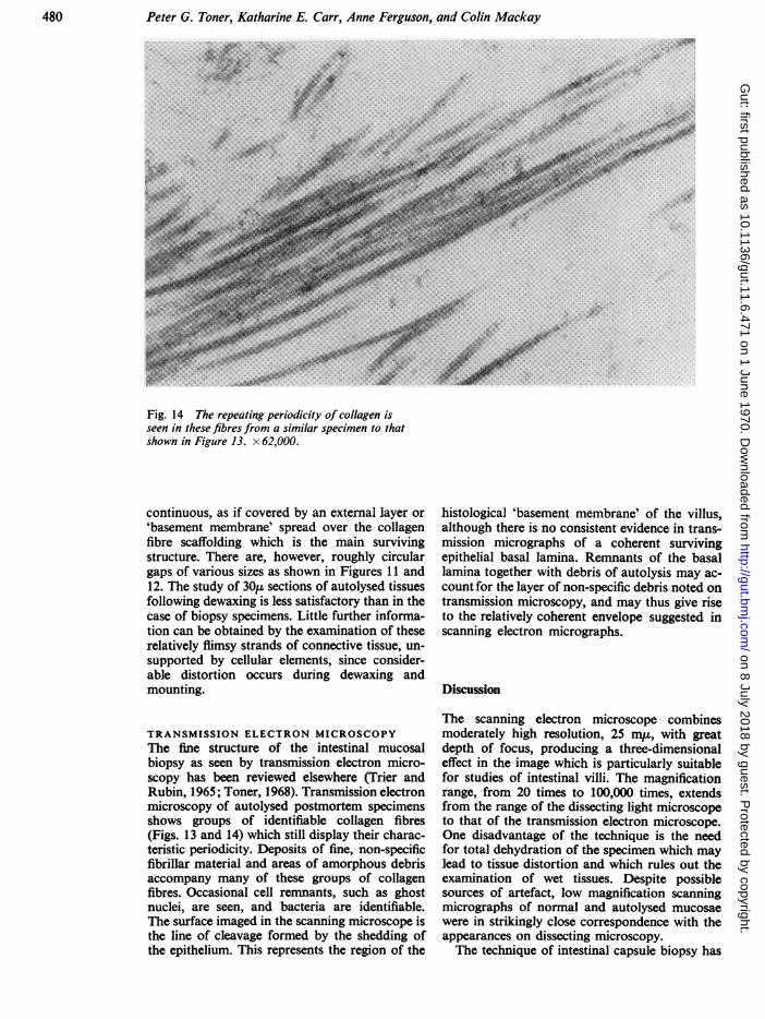
Fig. 14 The repeating periodicity of collagen isseen in these fibres from a similar specimen to thatshown in Figure 13. x 62,000.
continuous, as if covered by an external layer or'basement membrane' spread over the collagenfibre scaffolding which is the main survivingstructure. There are, however, roughly circulargaps of various sizes as shown in Figures 11 and12. The study of 30,u sections of autolysed tissuesfollowing dewaxing is less satisfactory than in thecase of biopsy specimens. Little further informa-tion can be obtained by the examination of theserelatively flimsy strands of connective tissue, un-supported by cellular elements, since consider-able distortion occurs during dewaxing andmounting.
TRANSMISSION ELECTRON MICROSCOPYThe fine structure of the intestinal mucosalbiopsy as seen by transmission electron micro-scopy has been reviewed elsewhere (Trier andRubin, 1965; Toner, 1968). Transmission electronmicroscopy of autolysed postmortem specimensshows groups of identifiable collagen fibres(Figs. 13 and 14) which still display their charac-teristic periodicity. Deposits of fine, non-specificfibrillar material and areas of amorphous debrisaccompany many of these groups of collagenfibres. Occasional cell remnants, such as ghostnuclei, are seen, and bacteria are identifiable.The surface imaged in the scanning microscope isthe line of cleavage formed by the shedding ofthe epithelium. This represents the region of the
histological 'basement membrane' of the villus,although there is no consistent evidence in trans-mission micrographs of a coherent survivingepithelial basal lamina. Remnants of the basallamina together with debris of autolysis may ac-count for the layer of non-specific debris noted ontransmission microscopy, and may thus give riseto the relatively coherent envelope suggested inscanning electron micrographs.
Discussion
The scanning electron microscope combinesmoderately high resolution, 25 m,, with greatdepth of focus, producing a three-dimensionaleffect in the image which is particularly suitablefor studies of intestinal villi. The magnificationrange, from 20 times to 100,000 times, extendsfrom the range of the dissecting light microscopeto that of the transmission electron microscope.One disadvantage of the technique is the needfor total dehydration of the specimen which maylead to tissue distortion and which rules out theexamination of wet tissues. Despite possiblesources of artefact, low magnification scanningmicrographs of normal and autolysed mucosaewere in strikingly close correspondence with theappearances on dissecting microscopy.The technique of intestinal capsule biopsy has
480 on 8 July 2018 by guest. P
rotected by copyright.http://gut.bm
j.com/
Gut: first published as 10.1136/gut.11.6.471 on 1 June 1970. D
ownloaded from

481 Scanning and transmission electron microscopic studies of human intestinal mucosa
provided the means for recording qualitativechanges of intestinal structure in disease (Rubinand Dobbins, 1965). The success of peroralbiopsy has perhaps led to some neglect of altern-ative sources of tissue for investigation, eventhough some problems which concern the extentand significance of variations in villous form arenot readily resolved by capsule biopsy. Suchproblems include the variations which occurfrom proximal to distal small intestine, and varia-tions with age, from intrauterine to late adultlife. In cases such as these, necropsy studies ofthe intestine are of particular value since muchmore material is available than is the case withcapsule biopsies (Creamer and Leppard, 1965;Loehry and Creamer, 1966; Ferguson et al,1969).Despite the autolysis of the cellular components
of the mucosa, the connective tissue fibre frame-work of the villi and the crypts survives to a con-siderable extent, preserving the form of themucosa when examined by dissecting and trans-mission microscopy. These villous cores, how-ever, lacking the moderate degree of rigidityimposed by an organized epithelium, lose muchof their former mechanical stability and tend tofold, bend, and twist. The persistence of theseoutlines of villous architecture is related, in finestructural terms, to the survival of a frameworkof recognizable collagen fibres which show typicalperiodicity in transmission electron micrographs.Although, as shown by thin sections, the epithelialbasal lamina does not survive intact, remnantsof this structure associated with a loose surfacecoagulum of fine debris must account for thecoherence of the villous surface as seen on scan-ning microscopy. The occasional gaps which canbe seen seem more likely to be chance discon-tinuities produced during autolysis than meaning-ful structural features, but might represent dis-continuities in the basal lamina and its collagenreinforcement caused by the transmigration oflymphocytes.
Significant variations in detail can probablyresult from the use of different techniques ofpreparing specimens for scanning electronmicroscopy. Microvilli, for example, were notclearly seen in whole mucosal specimens fixed asfor transmission electron microscopy and airdried, but were easily shown in dewaxed 30,sections and have been demonstrated in tissuesfreeze-dried after fixation (Marsh et al, 1968).Technique-dependent variations in detail in speci-mens may therefore require to be considered ininterpretation. In general it is preferable to
transfer to scanning electron microscopy thosetechniques known to give satisfactory preserva-tion of fine structure at the level of transmissionelectron microscopy. An attempt must be madeto retain the maximum comparability betweenthese two techniques.
There are other problems of interpretationpeculiar to scanning electron microscopy, suchas the reliance mainly on surface-contour effectsfor image contrast. This raises particular difficul-ties of recognition and interpretation of detailsin high-resolution images. The relatively crudesectioning techniques used in this study are un-able to show much significant intracellular detailand methods such as freeze fracturing will bemore satisfactory for such work. Thick sectionsdo, however, permit an unconventional view indepth of the larger structural features of thevilli, including in particular the connective tissuecomponents. Despite these problems, the limita-tions of the technique do not reduce the value ofscanning electron microscopy in the study of thenatural surface contours of the intestinal mucosa,whether obtained at biopsy or at necropsy.
The authors are grateful for research facilities pro-vided by their respective departments.
References
Carr, K. E., and Toner, P. G. (1968). Scanning electron micro-scopy of rat intestinal villi. Lancet, 2, 570-571.
Creamer, B., and Leppard, P. (1965). Post-mortem examinationof a small intestine in the coeliac syndrome. Gut, 6, 466-471.
Demling, L., Becker, V., and Classen, M. (1969). Examinationsof the mucosa of the small intestine with the scanningelectron microscope. Digestion, 2, 51-60.
Ferguson, A., Maxwell, J. D., and Carr, K. E. (1969). Progressivechanges in the small intestinal villous pattern with increas-ing length of gestation. J. Path., 99, 87-91.
Loehry, C. A., and Creamer, B. (1966). Post-mortem study ofsmall intestinal mucosa. Brit. med. J., 1, 827-829.
Loehry, C. A., and Creamer, B. (1969). Three-dimensional struc-ture of rat small intestinal mucosa related to mucosal dyn-amics. Gut, 10, 112-120.
Marsh, M. N., Swift, J. A., and Williams, E. D. (1968). Studies ofsmall intestinal mucosa with the scanning electron micro-scope. Brit. med. J., 4, 95-96.
Rubin, C. E., and Dobbins, W. 0. (1965). Peroral biopsy of thesmall intestine. A review of its diagnostic usefulness.Gastroenterology, 49, 676-697.
Shiner, M. (1956). Jejunal-biopsy tube. Lancet, 1, 85.Shiner, M. (1957). Duodenal and jejunal biopsies. I. A discussionof the method, its difficulties and applications. Gastro-
enterology, 33, 64-70.Toner, P. G. (1968). Cytology of intestinal epithelial cells. Int.
Rev. Cytol., 24, 233-343.Toner, P. G., and Carr, K. E. (1969). The use of scanning electron
microscopy in the study of the intestinal villi. J. Path., 97,611-617.
Trier, J. S., and Rubin, C. E. (1965). Electron microscopy of thesmall intestine: a review. Gastroenterology, 49, 574-603.
on 8 July 2018 by guest. Protected by copyright.
http://gut.bmj.com
/G
ut: first published as 10.1136/gut.11.6.471 on 1 June 1970. Dow
nloaded from