Scanning electron microscope studies on the genus ...
11
Scanningelectron microscope studies on the genus Hemicycliophora de Man, 1921 sensu lato (Nematoda : Criconematoidea) Pieter A. A. LOOF Department of Nematology, Agricultural University, Binnenhaven 10, P.O. Box 8123, 6700 ES Wageningen, Netherlands SUMMARK SEM photos of ‘about thirty species of Hemicycliophora sensu lato showed that, apart from some aberrant species, three main types of head structure cari be distinguished : 1) amphid apertures open, oral disc protruding; 2) amphid apertures closed, oral disc protruding; 3) amphid apertures closed, oral disc not protruding. These three types do not coincide with the three genera recognized by Siddiqi (1980); consequently the generic names Aulosphora Siddiqi, 1980 and Loofia Siddiqi, 1980 are synonymized with Hemicycliophora. H. nyanzae Wolff Schoemaker, 1968 is reinstated as a valid species; H. salicisSofrygina, 1972 is synomymized with H. thienemanni (Schneider, 1925). Apparently, in the genus Hemicycliophom there is no correlation between head structure and other characters like e.g. tail shape and organization of lateral fïeld. Several species do not conform to any of the three main types. Especially H. megalodiscus, H. tesselata and H. vitiensis are very aberrant. RÉSUME Étude au microscope e7ectronique à balayage du genre Hemicycliophora de Man, 1921 sensu lato (Nematoda : Criconematoidea) Les photographies effectuées au microscope électronique à balayage montrent que, parmi environ trente espèces du genre Hemicycliophora sensu lato, il existe trois types de structure céphalique : 1) ouvertures amphidiennes ouvertes, disque oral saillant; 2) ouvertures amphidiennes closes, disque oral saillant; 3) ouvertures amphidiennes closes, disque oral non saillant. Ces trois types ne coïncident pas avec les trois genres dans lesquels Siddiqi (1980) a réparti les espèces de ce groupe. L’auteur rejette donc cette division et synonymise les genres Aulosphora Siddiqi, 1980 et Loofia Siddiqi, 1980 avec Hemicycliophora, bien que Loofïa puisse être ultérieurement revalidé, mais avec en ce cas une définition différente. Dans le genre Hemicychophom il n’y a apparemment pas de corrélation entre la structure céphalique et d’autres caractères tels la forme de la queue, la structure du champ latéral, etc. Dans plusieurs cas la structure céphalique offre des caractères spécifiques, notamment chez le type 1. En particulier, le disque oral de H. nyanzae Wolff Schoemaker, 1968 est nettement différent de celui de H. typica de Man, 1921 espèce avec laquelle H. nyanzae avait été synonymisé par Brzeski (1974). Il faut donc considérer H. nyanzae et H. typica comme des espèces différentes. H. sali& Sofrygina, 1972 est synomisé avec H. thienemanni (Schneider, 1925). Plusieurs espèces ne correspondent à aucun des trois types définis : c’est le cas en particulier de H. megalodiscus, H. tesselata et H. vitiensis qui paraissent très aberrants. The genus Hemicycliophora sensu lato contains at the moment about one hundred species which are consi- dered valid. These species show much morphological divers@ and it is difficult to detect pattems of similarity. In 1976 Andrassy took out H. truncata Colbran, 1956 and made it type of a new genus Colbranium. In 1980 Siddiqi divided the remaining species over three genera : Hemicycliophora sensu stricto, Aulosphora Siddiqi, 1980, herewith emended to Adophorti and Loofia Siddiqi, 1980. The differentiating characters were : shape of vulva lips, length of vulval sleeve, shape of spicules and length of bursa. For reasons to be given hereafter 1 do not consider this division very happy. It is striking that structure of the head end of females does not play any * In accordance with the International Code of Zoologid Nomenclature, Appendix D, VI, 29 (c). Revue Nématol., 8 (2) : 113-123 (1985) part in Siddiqi’s system. In related criconematids study of head structure has greatly helped in elucidating relationships, and anyhow head structure is considered very important in nematode systematics. Therefore 1 started a study of the structure of the head end with the help of the scanning electron microscope. Al1 observa- tions refer to females. Material and methods The specimens used in this study partly were kindly sent by colleagues, partly they were taken from slides of the Wageningen nematode collection; these latter were transferred from glycerin to water first. The specimens were then transferred through an ethanol series to absolute ethanol, critical-point-dried, mounted on a stub and coated with gold. The electron microscope 113
Transcript of Scanning electron microscope studies on the genus ...

Scanning electron microscope studies on the genus Hemicycliophora de Man, 1921 sensu lato
(Nematoda : Criconematoidea) Pieter A. A. LOOF
Department of Nematology, Agricultural University, Binnenhaven 10, P.O. Box 8123, 6700 ES Wageningen, Netherlands
SUMMARK
SEM photos of ‘about thirty species of Hemicycliophora sensu lato showed that, apart from some aberrant species, three main types of head structure cari be distinguished : 1) amphid apertures open, oral disc protruding; 2) amphid apertures closed, oral disc protruding; 3) amphid apertures closed, oral disc not protruding. These three types do not coincide with the three genera recognized by Siddiqi (1980); consequently the generic names Aulosphora Siddiqi, 1980 and Loofia Siddiqi, 1980 are synonymized with Hemicycliophora. H. nyanzae Wolff Schoemaker, 1968 is reinstated as a valid species; H. salicisSofrygina, 1972 is synomymized with H. thienemanni (Schneider, 1925). Apparently, in the genus Hemicycliophom there is no correlation between head structure and other characters like e.g. tail shape and organization of lateral fïeld. Several species do not conform to any of the three main types. Especially H. megalodiscus, H. tesselata and H. vitiensis are very aberrant.
RÉSUME
Étude au microscope e7ectronique à balayage du genre Hemicycliophora de Man, 1921 sensu lato (Nematoda : Criconematoidea)
Les photographies effectuées au microscope électronique à balayage montrent que, parmi environ trente espèces du genre Hemicycliophora sensu lato, il existe trois types de structure céphalique : 1) ouvertures amphidiennes ouvertes, disque oral saillant; 2) ouvertures amphidiennes closes, disque oral saillant; 3) ouvertures amphidiennes closes, disque oral non saillant. Ces trois types ne coïncident pas avec les trois genres dans lesquels Siddiqi (1980) a réparti les espèces de ce groupe. L’auteur rejette donc cette division et synonymise les genres Aulosphora Siddiqi, 1980 et Loofia Siddiqi, 1980 avec Hemicycliophora, bien que Loofïa puisse être ultérieurement revalidé, mais avec en ce cas une définition différente. Dans le genre Hemicychophom il n’y a apparemment pas de corrélation entre la structure céphalique et d’autres caractères tels la forme de la queue, la structure du champ latéral, etc. Dans plusieurs cas la structure céphalique offre des caractères spécifiques, notamment chez le type 1. En particulier, le disque oral de H. nyanzae Wolff Schoemaker, 1968 est nettement différent de celui de H. typica de Man, 1921 espèce avec laquelle H. nyanzae avait été synonymisé par Brzeski (1974). Il faut donc considérer H. nyanzae et H. typica comme des espèces différentes. H. sali& Sofrygina, 1972 est synomisé avec H. thienemanni (Schneider, 1925). Plusieurs espèces ne correspondent à aucun des trois types définis : c’est le cas en particulier de H. megalodiscus, H. tesselata et H. vitiensis qui paraissent très aberrants.
The genus Hemicycliophora sensu lato contains at the moment about one hundred species which are consi- dered valid. These species show much morphological divers@ and it is difficult to detect pattems of similarity. In 1976 Andrassy took out H. truncata Colbran, 1956 and made it type of a new genus Colbranium. In 1980 Siddiqi divided the remaining species over three genera : Hemicycliophora sensu stricto, Aulosphora Siddiqi, 1980, herewith emended to Adophorti and Loofia Siddiqi, 1980. The differentiating characters were : shape of vulva lips, length of vulval sleeve, shape of spicules and length of bursa. For reasons to be given hereafter 1 do not consider this division very happy. It is striking that structure of the head end of females does not play any
* In accordance with the International Code of Zoologid Nomenclature, Appendix D, VI, 29 (c).
Revue Nématol., 8 (2) : 113-123 (1985)
part in Siddiqi’s system. In related criconematids study of head structure has greatly helped in elucidating relationships, and anyhow head structure is considered very important in nematode systematics. Therefore 1 started a study of the structure of the head end with the help of the scanning electron microscope. Al1 observa- tions refer to females.
Material and methods
The specimens used in this study partly were kindly sent by colleagues, partly they were taken from slides of the Wageningen nematode collection; these latter were transferred from glycerin to water first. The specimens were then transferred through an ethanol series to absolute ethanol, critical-point-dried, mounted on a stub and coated with gold. The electron microscope
113

l? A. A. Loof
used was initially a Jeol J.§.M. U 3, later a Jeol J.§.M. 35, both located at the T.F.D.L., Wageningen.
Current opinions Whereas most species have a truncate head end,
several have a more conoid one. In my 1968 paper 1 distinguished them as follows :
- truncate, lips separate, oral disc separate and often protruding; lateral lips lower than submedian ones (hereafter to be referred to as type A);
- conoid, lips connate, the lateral ones higher than the submedian ones; oral disc not separate (he- reafter to be referred to as type B).
Fig. 1. Hemicycliophora species. SEM photos of head ends. A : H. triangulum, from Lauwerszeepolder, Netherlands; B : H. Zabiata, from Donald Golf Club, Australia (no locality given); C : H. typica, from Oegstgeest, Netherlands; D : H. nyanzae, from Nairobi, Kenya; E-F : H. conida, from Wageningen, Netherlands. (C and D from Loof (1984); by courtesy of E. J. Brill, Leiden).
114 Revue Nématol., 8 (2) : 113-123 (1985)

SEM studies on Hemicycliophora
Several older and newer species descriptions include drawings of the end-on view of the head as seen in the light microscope. Most of these indicate two lateral semicircular structures currently considered the amphid apertures. Several so-called « end-on views », however, ratber represent sections through the lip region. Most older descriptions do not enter upon the detailed structure of the head end. An exception is De Grisse (1977), who presented a thorough study on the structure of the lip region of H. triangulum Loof, 1968 and also gave some SEM photos of this species [Fig. 130-132 and 264-265; his figures 133 and 266 represent a different species, possibly H. thienemanni (Schneider, 1925)]. His work showed that the real lip region is represented by the oral disc followed by head annules; the amphidial apertures lie between the lip region and the first head annule. These observations show that my interpretation of 1968 was incorrect.
Results
H. triangulum (Fig. 1 A). These observations confirm the results of De Grisse. The oral disc is broadly ovate, with the slit-like prestoma aperture in the centre. The disc is differentiated into a central part and a rather narrow collar. The amphidial apertures are rectangular, wide open, and are surrounded on the outside by the first head annule which is slightly indented medially; the lateral diameter is slightly larger than the media1 one. In the amphidial apertures there is a kind of « plug » which leaves a slit.
A number of species show essentially the same structure : H. Zabiata Colbran, 1960 (Fig. 1 B). Oral disc oval, with rather broad collar. SEM photos of this species were also given by van den Berg (1981). H. typica de Man, 1921 (Fig. 1 C). Oral disc raised only slightly above the first annule, as in H. triangulum; it is more rectangular in shape and has a lower strip dorsally and ventrally. H. nyanzae Wolff Schoemaker, 1968 (Fig. 1 D). The oral disc has a very wide collar which laterally is bent anteriad. H. conida Thorne, 1955 (Fig. 1 E and F). The oral disc protrudes more strongly and is larger than in H. tn’angulum, H. typica and H. Zubiata; it has a much broader collar. The first annule is lower laterally than medially. H. parvana Tazjan, 1952 (Fig. 2 A and B). Oral disc broadly oval, collar narrower than in H. conida. First annule hardly lower laterally. H. spinituberculata Loof, 1984 (Fig. 2 C). Oral disc rectan- gular, not protruding beyond first annule, with narrow collar. H. sculpturuta Loof, 1984 (Fig. 2 D). Resembles H. typica. Basa1 head annule conspicuously incised, with numerous small lobes. H. Zutosoides Loof, 1984 (Fig. 2 E). Oral disc rectangular, with broad collar, not pru- truding beyond first annule. H. ircznica Loof, 1984 (Fig. 2 F). Oral disc almost rectangular, with narrow collar. H. sturhani Loof, 1984 (Fig. 3 A). Oral disc almost as wide as long, with very broad collar.
Revue Nématol., 8 (2) : 113-123 (1985)
SEM photos by other authors indicate the same structure in the following species : H. rionegrensis Doucet, 1982. Oral disc oval, with rather broad collar; protruding distinctly beyond first annule. H. ripa van den Berg, 1981. Oral disc broadly oval, but no details visible. H. transvaalensis Heyns, 1962 : Van den Berg (1981) gave a SEM photo of the head end which shows this species to be rather like H. typica (with which van den Berg synonymized it) although the lower median strips are not SO apparent. H. quercea Mehta & Raski, 1985. Oral disc rectangular, with narrow, lowered collar. H. pinocheti Mehta & Ra&i, 1985. No details of oral disc visible owing to dirt. H. chilensis Brzeski, 1974 : Mehta and Raski (1985) gave a SEM photo which shows the oral disc being almost circular. Finally H. amchit- kaensis Bernard, 1982 has open amphidial apertures as indicated by the light microscope drawing (Fig. 21 in the original description).
A second type is exemplified by H. epicharoides Loof, 1968 (Fig. 3 C). Under the light microscope the lip region resembles that of the triangzJz~zn-group, but the amphidial apertures are closed by lateral plates. The oral disc is raised above these lateral plates and there is an indication of a six-radiate symmetry in the tïrst head annule.
The following species also show this type : H. robusta Loof, j968 (Fig. 3 D); H. penetruns Thorne, 1955 (Fig. 3 B); H. vidua Ra&i, 1958 (see Mehta & Raski, 1985); possibly also H. fragilis Doucet, 1982 and H. tenuistriata Doucet, 1982.
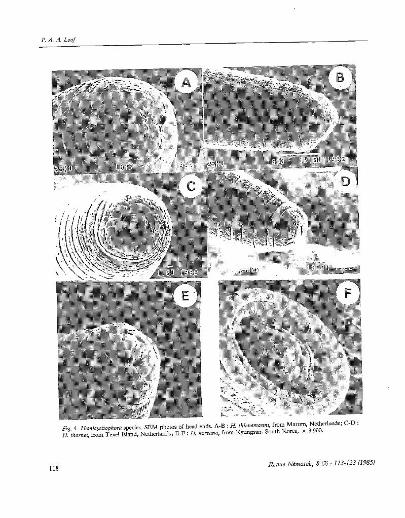
A third type is exemplified by H. thienemanni (Schneider, 1925)* (Fig. 4 A and B). The flrst annule is slightly indented dorsally and ventrally. The oral disc is narrow-ovate, convex anteriorly, without distinct collar, medially hardly or not detached from the first head annule. On both its lateral sides there is a large rectangu- lar shield completely covering the ampbidial apertures. These shields lie in the same plane as the oral disc. The dorsoventral diameter of the shields is longer than that of the oral disc.
Two other species show this type : H. nucleata Loof, 1968 (SEM photos not of publicable quality) : first annule and lateral shields more rectangular, first annule strongly crenate; H. thornei Goodey, 1963 (Fig. 4 C and D) : first annule crenate as in H. nucleatu; lateral shields dorsoventrally no longer than oral disc.
Finally there are some species which do not clearly fit into any of these three groups : H. koreana Choi & Geraert, 1971 (Fig. 4 E and F). Amphidial apertures closed, apparently separated from oral disc by « trans-
* 1 regard H. salicis Sofrygina, 1972 identical with H. thienemanni. The measurements and general morphology (head, vulva lips, tail) agree wholly; the c value (11-17) is evidently incorrect, the anus being indicated as lying at the level of the phasmids (!) near the tail contraction.
115

l? A. A. Loqf
Fig. 2. Hemicycliophora species. SEM photos of head ends. A-B : H. parvana, from Sanford, Florida, U.S.A.; C : H. spinituberculata, from Bandar Pahlavi, Iran; D : H. sculpturata, from Iraq; E : H. lutosoides, from Zavareh, Iran, x 3.900; F : H. iranica, from Zibakenar, Iran.
116 Revue Nématol., 8 (2) : 113-123 (1985)

SEM studies on Hemicycliophora
Fig. 3. Hemicycliophora species. SEM photos of head ends. A : H. sturhani, from Zandjan, Iran; B : H. penetrans, syntype specimen from Bogor, Java, Indonesia; C : H. epicharoides, from Scheveningen, Netherlands, D : H. robusta, from Terschelling Island, Netherlands; E : H. megalodiscus, from Iran, x 3.120; F : H. arenaria, from Califomia, U.S.A., x 4.860. (A and E from Loof (1984); by courtesy of E. J. Brill, Leiden.)
Revue Nématol., 8 (2) : 113-123 (1985) 117
F’. A. A. Loof
118
Fig. 4. Hemicycliophora species. SEM photos of head ends. A-B : H. thienemanni, from Marum, Netherlands; C-D : H. thornei, from Texel Island, Netherlands; E-F : H. koreanu, from Kyungsan, South Korea, x 3.900.
Revue Nématol., 8 (2) : 113-123 (198.5)
SEM studies on Hemicycliophora
verse bars » noted already by Choi and Geraert. How- ever, in another specimen these bars were joined to form a ring around the oral disc. and the shape of the first annule is reminiscent of H. epicharoides. H. megalodisczts Loof, 1984 (Fig. 3 E). Oral disc oval, with very large collar which is bent anteriad and protrudes strongly. First head annule four-lobed. Amphid apertures uncer- tain (a11 specimens dirty) but probably closed. H. vi- tiensis Orton Williams, 1978 (SEM photo in original descripnon). The oral disc lies in a groove between two very atrongly bulging plates; below these two small (amphid?) apertures are present. H. loofi Maas, 1970 (Fig. 5 A). Amphid apertures closed, oral disc raised as in the epicharoides-group, but shape of first annule is more reminiscent of the thienemanni-group. H. arenaria Ra&i, 1958 (Fig. 3 F). Amphid apertures closed by plates which appear thicker than in the epicharoides- group; oral disc not raised. Indication of six-radiate symmetry in the first head annule, but this has to be confïrmed on fresh, better material. H. tesselata Sauer, 1958 (Fig. 5 B-D). Amphid apertures closed. Both the covering plates and the oral disc bulge strongly. Between plates and oral disc are transverse bars, much thinner than those of H. koreana. H. saueri Brzeski, 1974 (Fig. 5 E-F). The first head annule appears four-lobed, inter- rupted laterally. Amphid apertures closed by oval plates. H. sheri Brzeski, 1974 (SEM photo by Mehta and Raski, 1985). Oral disc almost round, with broad, strongly demarcated collar. The first head annule closely sur- rounds the oral disc, leaving only small slits for the amphid apertures. Probably H. hesperis Ra&i, 1958 Will be found exceptional too, judging from the light micros- cope drawings in the original description.
Discussion
Leaving aside Colbranium truncatum, of which no SEM pictures are available, and leaving provisionally also aside the aberrant species, we note that, whereas in light microscopy two types of head structure were distinguished, SEM shows the existence of three types :
Type 1 : Amphid apertures wide open. Oral disc often raised distinctly above first head annule. In the light microscope such a lip region looks truncate.
Type 2 : Amphid apertures covered by plates. Oral disc protruding beyond the plane of these plates. Under the light microscope such a lip region looks truncate; in fact, 1 am not able to distinguish types 1 and 2.
Type 3 : Amphid apertures covered by plates. Oral disc in the same plane as these plates. Under the Sght microscope such a lip region looks conoid.
What was called above type A comprises SEM types 1 and 2, whereas type B is identical with SEM type 3.
It happens that the type species of Siddiqi’s three
Revue Nématol., 8 (2) : 113-123 (1985)
genera each show a different type : Hemicycliophora typica de Man, 1921 belongs in (the) group (showing type) 1: Azdophora penetrans (Thorne, 1955) in group 2 and Loofia thienemanni (W. Schneider, 1925) in group 3. If these three genera coincided with the three types of head structure, this would support Siddiqi’s action. But this is not the case.
Among the species of Hemicycliophora s. str. are species belonging to group 1 (triangulunz, typica, conida, parvana and others), to group 2 (epicharoides), to group 3 (nucleata, thomei) and to the aberrant group (koreana, loofi, tesselata, saueri, vitiensis, arenai-ia).
Among Loofia we tïnd group 2 (robusta) and group 3 (thienenzanni).
Of Azdophora nothing cari be said at present, only one species having been examined. Anyhow it is certain that the groups of head structure tut across the genera as conceived by Siddiqi.
Two species of Loofia were examined, L. thienemanni and L. robusta. The latter shows head structure, not of group 3, but of group 2. In its further morphology robusta does not resemble thienemanni at all, but shows strong resemblance to epicharoides. This throws doubt upon the value of vulva lip shape as generic character. The male diagnostic characters of Loofia are doubtful, because males are known in one species only (thiene- manni) and even there they are extremely rare.
As to Aulophora, Siddiqi diagnosed this genus by : elongate spicules; very long vulval sleeve; long bursa (relation precloacal to postcloacal part 3-4 : 1 against about 1 : 1 in Loofia and Hemicycliophora s. str.). There are, however, species of Aulophora showing intermediate characters : A. oostenbrinki (Luc, 1958) has spicules not differring much from the typical Hemicycliophora shape, though the vulval sleeve is rather long; A. brzeskii (Barbez & Geraert, 1980) has more typical Aulophora- like spicules but the vulval sleeve is short. Moreover there is no Sharp separation between the long bursa of Aulophora and the short one of the two other genera. From published drawings 1 calculated the following ratios :
Aulophora : dahomensis Germani & Luc, 1976 brzeskii Barbez & Geraert, 1980 oostenbrinki Luc, 1958 penetrans Thorne, 1955
Loofia : trzienemanni W. Schneider, 1925
Hemicycliophora : arenaria Ra&i, 1958 belenznis Germani & Luc, 1973 brevicauda Sauer, 1958 brevis Thome, 1955 chathami Yeates, 1978 conida Thorne, 1955 demani Edward & Rai, 1971
3.9 2.4 2.4 4.1
1.0
1.2 1.7 1.2 1.2 3.3 2.0 1.4
119

l? A. A. Loof
Fig. 5. Hemicycliophora species. SEM photos of head ends. A : H. loof, from Un& Brasil; B-D : H. tesselata, from Australia; E-F : H. saueti, from Australia.
dhirendri Husain 81 Khan, 1967 diolaensis Germani & Luc, 1973 epicharis Ra&i, 1958 fragilis Doucet, 1982 koreana Choi & Geraert, 1971 Zabiata Colbran, 1960 lutosa Loof & Heyns, 1969 micoletzkyi Goffan, 1951
1.8 straturata Germani & Luc, 1973 1.1 1.8 tenuistriata Doucet, 1982 1.0 1.6 tesselata Sauer, 1958 0.9 1.4 thomei Goodey, 1963 1.0 1.6 typica de Man, 1921 1.7 2.3 vitiensis Orton Williams, 1978 1.7 1.9 Own observations : 1.9 A. oostenbrinki 2.9-3.1 (n = 2)
120 Revue Nématol., 8 (2) : 113-123 (1985)

SEM studies on Hemicycliophora
L. thienemanni 1.0-1.3 (n = 6) H. typica 1.7-2.2 (n = 7) H. thornei 1.0-1.5 (n = 9) H. conida 1.6-2.0 (n = 5)
Thus there is no difference between Hemicycliophora s. str. and Loofia. Aulophora tends to have a higher ratio, but typical Hemicycliophora species like conida, labiata and chathami bridge the gap. The difference cari hardly be regarded of generic importance.
The next question is : cari the species be re-arranged, SO that Group 1 coincides with Hemicycliophora s. str., Group 2 with Aulophora and Group 3 with Loofia?This would be possible if other morphological characters were consistently correlated with the head structure types. But it must strike everyone who works with these nematodes, how few characters which could be used at the generic level, are present in Hemicycliophora s.l. The species vary in vulva position, annule number, stylet length (there is also some variation in shape of stylet knobs but this is a weak character, cf. Hemicriconemoi- des), organization of lateral field, cuticular sculpture, tail length, position of anus relative to postvulval body part, and tail shape. Most of these do not permit the forma- tion of clear groups and anyhow cari hardly be consi- dered of more than specific importance. The only character that could be considered of generic impor- tance, is Lai1 shape. Whereas the great majority of the species has at least the distal part of the tail tapering to a narrowly rounded or acute terminus (with an almost continuous series from uniformly tapering to strongly spicate tails) there is a group in which the tail is cylindri- cal with hemispheroid terminus (arenaria, biloculata, brevicauda, corbetti, nana, obtusa, rotundicauda, si- gnata, straturata and tesselata) but it is perhaps signifi- tant that this tail shape also occurs in odd specimens of other species (conida, nigeriensis, thienemanni, zucker- mani). Moreover H. aberrans and H. obesa are interme- diate. Only two round-tailed species were examined : arenaria (perhaps Group 2 but better material is nee- ded) and tesselata (which is strongly aberrant).
Group 1 contains species with low R (descending to below 200), e.g. Zabiata, typica, conida- (see Loof, 1968), nyanzae, medium R (between 200 and 300), e.g. ripa, conida-1, triangulum, parvana; and high R (rising to above 300), e.g. spinituberculata, lutosoides, pinocheti; with anrerior vulva (parvana, ripa) to posterior vulva (conida, amchitkaensis); lateral fïeld with simple breaks (iranica, spinituberculata, lutosoides, ripa), with one late- ral line (labiata, chilensis, quercea) and with two lateral lines plus breaks (typica, triangulurn, nyanzae, parvana, sturhani);with uniformly tapering tails (conida, paruana, spinituberculata, lutosoides, ripa), tails with distal part somewhat offset. (triangulum, iranica) and spicate tails (typica).
Group 2 shows a similar picture, but Group 3 is more uniform. The three species thienemanni, thornei and nucleata resemble each other closely : the females are
Revue Nématol., 8 (2) : 113-123 (1985)
rather long, slender, body curved more than usually, vulva more anterior, tail cylindroid proximally, spicate distally. One might ask if these similarities do not outweigh the difference in males between thienemanni and thomei (males of nucleata have never been found). If SO, these three species could be united under the generic name of Loofia. However, 1 refrain from car- rying through this action, because our knowledge of the whole group is as yet insufficient; at least the other species of Loofia (L. ferrisae, L. gigas, L. uniformis and L. vaccinium) should be examined fïrst.
The problem is complicated by the aberrant species. If we divided Hemicycliophora on the basis of head structure, then monotypic genera would have to be erected for at least H. tesselata, H. megalodiscus and H. vitiensis, and probably for H. hesperis. Such a disruption of the group does not appear sound. It seems that in this group of nematodes a variety of head structures is accompanied by a remarkable uniformity in other morphological details. There are some vague trends, but many more species Will have to be examined before these cari be evaluated properly.
1 therefore reject Atdophora and herewith return its species to Hemicycliophora. The same procedure is executed with respect to Loofia, though it is possible that this generic name Will be revived in future, with a different definition.
At least within Group 1 the structure of the lip region shows specific differences and cari he used for identifica- tion. E.g. H. nyanzae was considered identical with H. typica by Brzeski (1974), but SEM shows a clear diffe- rente in shape and position of the oral disc. H. nyanzae is therefore reinstated as a valid species. It is also distinguished from H. typica by the absence of func- tional males (spermatheca empty).
Finally a word about cuticular sculpture. Several species have been described as having longitudinal cuticular sculpture outside the lateral field IH. typica, H. nyanzae, H. sculpturata). In others fine longitudinal scratches were mentioned (e.g. H. conida, H. madagas- cariensis, H. fragilis). In still other species no sculpture was reported. SEM shows that fine scratches occur in the majority of species, e.g. H. epicharoides (Fig. 6 A), H. triangulum (Fig. 6 B), H. Zabiata (Fig. 6 E). Real longitudinal grooves are indicated in H. typica (Fig. 6 C), apparently more strongly developed in H. nyanzae (Fig. 6 D) and H. sculpturata (cf. Loof, 1984). H. tesse- lata shows a very aberrant type of longitudinal sculpture (Fig. 6 F).
ACKNOWLEDGMENTS
1 want to thank Dr. A. Morgan Golden, Beltsville, U.S.A.; Dr. D. J. Ra&i, Davis, U.S.A.; Dr. M. R. Sauer, Merbein, Australia; and Dr. A. C. Tarjan, Gainesville, U.S.A. for kindly putting specimens at my disposal. Mrs C. M. Broers-Vendrig is thanked for her skilful technical assistance.
121

I? A. A. Loof
Fig. 6. Hemicycliophora species. SEM photos of cuticle structure, between mid-body and vulva. A : H. epicharoides, from Scheveningen, Netherlands, ( x 1.350); B : H. triangulum, from Overloon, Netherlands ( x 2.700); C : H. typica, from Oegstgeest, Netherlands; D : H. nyanzae, from Nairobi, Kenya; E : H. Zabiata, from Donald Golf Club, Australia; F : H. tesselata, from Australia (A : from Loof, 1984, by courtesy of E. J. Brill, Leiden).
122 Revue Nématol., 8 (2) : 113-123 (1985)
SEM studies on Hemicycliophora
REFERENCES
hDRASSY, 1. (1976). Evolution as a basis for the systematization of nematodes. Akademiai KiadO, Budapest, 288 p.
VAX DEN BERG, E. (1981). Further studies on the genus Hemicycliophora de Man, 1921 in South Africa (Nemato- da : Hemicycliophoroidea) with a description of a new species. Phytophylactica, 13 : 181-194.
BERN.\RD, E. C. (1982). Criconematina (Nematoda : Tylen- chida) from the Aleutian Islands. 3 Nematol., 14 : 323-331.
CHOI, Y. E. & GERAERT, E. (1971). Two new species of Tylenchida from Korea with a list of other nematodes new for this country. Nematoiogica, 17 : 93-106.
DE GRISSE, A. (1977). De uitrastruktuur van het zenuwstelsel in de kop van 22 soorten plantenparasitaire nematoden behorende tot 19 genera (Nematoda : Tylenchidal. Thesis Univ. Gent, 420 p.
DOUCET, M. E. (1982). Quatre nouvelles espèces du genre Hemicycliophora De Man, 1921 (Nematoda : Tylenchida) provenant d’Argentine, Rev. NtGnatol., 5 : 309-320.
LOOF, P. A. A. (1968). Taxonomy of Hemicycliophora species from West and Central Europe. Meded LandbHogesch. Wageningen 68-14, 43 p.
Accepté pour publication le 18 février 1985.
LOOF, P. A. A. (1984). Hemicycliophora species from Iran (Nematoda : Criconematoidea). Nematologica, 30 : 22-41.
DE hb.N, J. G. (1921). Nouvelles recherches sur les nématodes libres terricoles. Capita zooZ., 1 (1) : l-62.
MEHXX, U. K. & R~SKI, D. J. (1985). Two new species of Hemicycliophora de Man, 1921 from Honduras with ob- servations on five other species (Nematoda : Tylenchida). Revue Nématol., 7 (1984) : 347-353.
ORTON WILLI~S, K. J. (1978). Two new species of the genus Hemicycliophora de Man, 1921 (Nematoda : Tylenchida). Revue Nématol., 1 : 197-205.
F&SKI, D. J. (1958). Four new species of Hemicycliophora de Man, 1921, with further observations on H. brevis Thome, 1955. Proc. helminth. Soc. Wash., 25 : 125-131.
SIDDIQI, M. R. (1980). Taxonomy of the plant nematode superfamily Hemicycliophoroidea, with a proposa1 for Criconematina, new suborder. Revue Nématol., 3 : 179-199.
THORNE, G. (1955). Fifteen new species of the genus Hemi- cycliophora with an emended description of H. typica de Man (Tylenchida Criconematidae). Proc. helminth. Soc. Wash, 22 : 1-16.
Revue Nématol., 8 12) : 113-123 11985) 123