Saturated long-chain fatty acid-producing bacteria ...
16
RESEARCH Open Access Saturated long-chain fatty acid-producing bacteria contribute to enhanced colonic motility in rats Ling Zhao 1† , Yufen Huang 2† , Lin Lu 1† , Wei Yang 1 , Tao Huang 1 , Zesi Lin 3 , Chengyuan Lin 1,4 , Hiuyee Kwan 1 , Hoi Leong Xavier Wong 1 , Yang Chen 5 , Silong Sun 2 , Xuefeng Xie 2 , Xiaodong Fang 2,5 , Huanming Yang 6 , Jian Wang 6 , Lixin Zhu 7* and Zhaoxiang Bian 1* Abstract Background: The gut microbiota is closely associated with gastrointestinal (GI) motility disorder, but the mechanism(s) by which bacteria interact with and affect host GI motility remains unclear. In this study, through using metabolomic and metagenomic analyses, an animal model of neonatal maternal separation (NMS) characterized by accelerated colonic motility and gut dysbiosis was used to investigate the mechanism underlying microbiota-driven motility dysfunction. Results: An excess of intracolonic saturated long-chain fatty acids (SLCFAs) was associated with enhanced bowel motility in NMS rats. Heptadecanoic acid (C17:0) and stearic acid (C18:0), as the most abundant odd- and even- numbered carbon SLCFAs in the colon lumen, can promote rat colonic muscle contraction and increase stool frequency. Increase of SLCFAs was positively correlated with elevated abundances of Prevotella, Lactobacillus, and Alistipes. Functional annotation found that the level of bacterial LCFA biosynthesis was highly enriched in NMS group. Essential synthetic genes Fabs were largely identified from the genera Prevotella, Lactobacillus, and Alistipes. Pseudo germ-free (GF) rats receiving fecal microbiota from NMS donors exhibited increased defecation frequency and upregulated bacterial production of intracolonic SLCFAs. Modulation of gut dysbiosis by neomycin effectively attenuated GI motility and reduced bacterial SLCFA generation in the colon lumen of NMS rats. Conclusions: These findings reveal a previously unknown relationship between gut bacteria, intracolonic SLCFAs, and host GI motility, suggesting the importance of SLCFA-producing bacteria in GI motility disorders. Further exploration of this relationship could lead to a precise medication targeting the gut microbiota for treating GI motility disorders. Keywords: Gastrointestinal motility disorder, Gut microbiota, Neonatal maternal separation, Saturated long-chain fatty acids Background Disordered gastrointestinal (GI) motility is one major symptom frequently presented in patients suffering with functional GI disorders (FGIDs) and other bowel diseases [1, 2]. Chronic or recurrent episodes of dysregu- lated GI motility severely impact patients’ quality of life [3]. The underlying mechanisms of disordered GI motil- ity are multifaceted. Previous studies have revealed con- tributions of immune activation, ionic channels and neurohumoral dysregulation to GI motor dysfunction [4–6]. However, current etiological understanding of dis- ordered GI motility is incomplete, which limits the de- velopment of personalized and precisely effective medicine for GI motility disorders. * Correspondence: [email protected]; [email protected] † Ling Zhao, Yufen Huang and Lin Lu contributed equally to this work. 7 Digestive Diseases and Nutrition Center, Department of Pediatrics, The State University of New York at Buffalo, 3435 Main Street, 422BRB, Buffalo, NY 14214, USA 1 Chinese Medicine Clinical Study Center, Jockey Club School of Chinese Medicine, Hong Kong Baptist University, Kowloon Tong, Hong Kong SAR, China Full list of author information is available at the end of the article © The Author(s). 2018 Open Access This article is distributed under the terms of the Creative Commons Attribution 4.0 International License (http://creativecommons.org/licenses/by/4.0/), which permits unrestricted use, distribution, and reproduction in any medium, provided you give appropriate credit to the original author(s) and the source, provide a link to the Creative Commons license, and indicate if changes were made. The Creative Commons Public Domain Dedication waiver (http://creativecommons.org/publicdomain/zero/1.0/) applies to the data made available in this article, unless otherwise stated. Zhao et al. Microbiome (2018) 6:107 https://doi.org/10.1186/s40168-018-0492-6
Transcript of Saturated long-chain fatty acid-producing bacteria ...

RESEARCH Open Access
Saturated long-chain fatty acid-producingbacteria contribute to enhanced colonicmotility in ratsLing Zhao1†, Yufen Huang2†, Lin Lu1†, Wei Yang1, Tao Huang1, Zesi Lin3, Chengyuan Lin1,4, Hiuyee Kwan1,Hoi Leong Xavier Wong1, Yang Chen5, Silong Sun2, Xuefeng Xie2, Xiaodong Fang2,5, Huanming Yang6, Jian Wang6,Lixin Zhu7* and Zhaoxiang Bian1*
Abstract
Background: The gut microbiota is closely associated with gastrointestinal (GI) motility disorder, but themechanism(s) by which bacteria interact with and affect host GI motility remains unclear. In this study, throughusing metabolomic and metagenomic analyses, an animal model of neonatal maternal separation (NMS)characterized by accelerated colonic motility and gut dysbiosis was used to investigate the mechanism underlyingmicrobiota-driven motility dysfunction.
Results: An excess of intracolonic saturated long-chain fatty acids (SLCFAs) was associated with enhanced bowelmotility in NMS rats. Heptadecanoic acid (C17:0) and stearic acid (C18:0), as the most abundant odd- and even-numbered carbon SLCFAs in the colon lumen, can promote rat colonic muscle contraction and increase stoolfrequency. Increase of SLCFAs was positively correlated with elevated abundances of Prevotella, Lactobacillus, andAlistipes. Functional annotation found that the level of bacterial LCFA biosynthesis was highly enriched in NMSgroup. Essential synthetic genes Fabs were largely identified from the genera Prevotella, Lactobacillus, and Alistipes.Pseudo germ-free (GF) rats receiving fecal microbiota from NMS donors exhibited increased defecation frequencyand upregulated bacterial production of intracolonic SLCFAs. Modulation of gut dysbiosis by neomycin effectivelyattenuated GI motility and reduced bacterial SLCFA generation in the colon lumen of NMS rats.
Conclusions: These findings reveal a previously unknown relationship between gut bacteria, intracolonic SLCFAs,and host GI motility, suggesting the importance of SLCFA-producing bacteria in GI motility disorders. Furtherexploration of this relationship could lead to a precise medication targeting the gut microbiota for treating GImotility disorders.
Keywords: Gastrointestinal motility disorder, Gut microbiota, Neonatal maternal separation, Saturated long-chainfatty acids
BackgroundDisordered gastrointestinal (GI) motility is one majorsymptom frequently presented in patients suffering withfunctional GI disorders (FGIDs) and other bowel
diseases [1, 2]. Chronic or recurrent episodes of dysregu-lated GI motility severely impact patients’ quality of life[3]. The underlying mechanisms of disordered GI motil-ity are multifaceted. Previous studies have revealed con-tributions of immune activation, ionic channels andneurohumoral dysregulation to GI motor dysfunction[4–6]. However, current etiological understanding of dis-ordered GI motility is incomplete, which limits the de-velopment of personalized and precisely effectivemedicine for GI motility disorders.
* Correspondence: [email protected]; [email protected]†Ling Zhao, Yufen Huang and Lin Lu contributed equally to this work.7Digestive Diseases and Nutrition Center, Department of Pediatrics, The StateUniversity of New York at Buffalo, 3435 Main Street, 422BRB, Buffalo, NY14214, USA1Chinese Medicine Clinical Study Center, Jockey Club School of ChineseMedicine, Hong Kong Baptist University, Kowloon Tong, Hong Kong SAR,ChinaFull list of author information is available at the end of the article
© The Author(s). 2018 Open Access This article is distributed under the terms of the Creative Commons Attribution 4.0International License (http://creativecommons.org/licenses/by/4.0/), which permits unrestricted use, distribution, andreproduction in any medium, provided you give appropriate credit to the original author(s) and the source, provide a link tothe Creative Commons license, and indicate if changes were made. The Creative Commons Public Domain Dedication waiver(http://creativecommons.org/publicdomain/zero/1.0/) applies to the data made available in this article, unless otherwise stated.
Zhao et al. Microbiome (2018) 6:107 https://doi.org/10.1186/s40168-018-0492-6
The gut microbiota is thought to be an important fac-tor associated with disordered GI motility [7]. For ex-ample, patients with irritable bowel syndrome (IBS),especially for diarrhea-predominant IBS (IBS-D), showeda lower diversity and a higher instability of gut microbialcommunity [8, 9]. Altered composition of the gut micro-biota was also found in children and adults with chronicfunctional constipation [10–12]. Regulation of the gutmicrobiota by probiotics can improve bowel movementfrequency in up to 70% of functionally constipated pa-tients [13]. Transplantation of fecal microbiota fromIBS-D suffers significantly accelerated colonic transit ingerm-free (GF) mice [14]. GF mice colonized with fecalmicrobiota from patients with slow transit constipationexhibit lower fecal frequency, delayed GI transit time,and weaker spontaneous contractions of colonic smoothmuscle [15]. These results point directly to a conclusionthat the gut microbiota serves a pivotal role in GI motil-ity disorder. But the mechanism by which microbiotainteract with and affect host GI motility remains unclear.It has been proposed that the effect of the gut micro-
biota on host GI motility partly derives from release ofbacterial end-products of fermentation or molecules de-rived from host and gut microbiota co-metabolism [16,17]. One study found that microbial products, such as li-popolysaccharides (LPS), can regulate GI motility throughactivating toll-like receptor 4 (TLR4) signaling that in-creases survival of nitrergic neurons [18]. Short-chain fattyacids and lactic acids, produced from bacterial fermenta-tion of dietary fiber and resistant starch, can regulate in-testinal motor action, probably through regulating peptideYY secretion and cholinergic neurons [19]. In anotherstudy, GF mice displayed accelerated GI motility aftercolonization with spore-forming microbes, the metabolitesof which could stimulate enteric serotonin release [20].Therefore, gut microbiota-derived substances appear to bethe link between gut microbiota and host GI motility.A rodent model of neonatal maternal separation
(NMS) is a well-established model characterized bylong-term colonic dysfunction [21]. Our previous studyfound that colon tissue isolated from adult NMS ratsshowed a higher level of muscle amplitude [22]. Suchenhanced colonic motility is involved in upregulation ofL-type calcium channels in colonic smooth muscle cells[22] and increased serotonin production from colonicenterochromaffin cells (ECs) [23]. Meanwhile, gut mi-crobial disturbance is also reported in both mice andrats subjected to NMS [24, 25], but the association ofchanged gut microbiota with colonic dysmotility in anNMS model is yet to be investigated.In this study with an NMS rat model, metabolic profil-
ing of feces and luminal contents of the GI tract of ex-perimental animals was preformed to identify featuredmicrobiota-derived metabolites. A group of saturated
long-chain fatty acids (SLCFAs) was remarkably in-creased in the colon lumen and feces of NMS rats. Afterconfirming their stimulatory effects on colon motility,function-based metagenomic analysis was done and foundhyperactive synthesis of SLCFAs by the gut microbiota ofNMS rats. The relationship between SLCFA-producingbacteria and host colon motility were further clarified bytransplanting fecal microbiota from NMS donors intopseudo germ-free (GF) rats and modulating gut micro-biota in NMS rats by neomycin intervention. Based onthese experiments, we aimed to clarify the linkage be-tween gut microbiota and host colonic motility phenotypeand to determine which bacteria are the primary agentsmanipulating host GI colonic dysmotility.
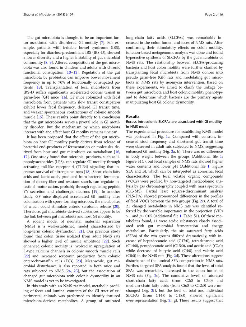
ResultsExcess intracolonic SLCFAs are associated with GI motilityin NMS modelThe experimental procedure for establishing NMS modelwas portrayed in Fig. 1a. Compared with controls, in-creased stool frequency and shortened gut transit timewere observed in adult rats subjected to NMS, suggestingenhanced GI motility (Fig. 2a, b). There was no differencein body weight between the groups (Additional file 1:Figure S1C), but fecal samples of NMS rats showed higherwater contents and lower pH (Additional file 1: FigureS1A and B), which can be interpreted as abnormal fecalcharacteristics. The fecal volatile organic compounds(VOCs) were profiled by non-targeted metabolomic ana-lysis by gas chromatography coupled with mass spectrum(GC-MS). Partial least squares-discriminant analysis(PLS-DA) showed pronounced differences in the profilesof fecal VOCs between the two groups (Fig. 2c). A total of21 changed metabolites in NMS rats was identified re-ferred by the variable importance in the projection (VIP)> 1 and p < 0.05 (Additional file 1: Table S1). Of these me-tabolites found, 11 were acidic substances closely associ-ated with gut microbial fermentation and energymetabolism. Particularly, the six saturated fatty acids(SFAs) of the two groups differed dramatically, with in-crease of heptadecanoic acid (C17:0), tetradecanoic acid(C14:0), pentadecanoic acid (C15:0), and acetic acid (C2:0)while decrease of butyric acid (C4:0) and valeric acid(C5:0) in the NMS rats (Fig. 2d). These alterations suggestdisturbance of the luminal SFA composition in NMS rats.Further, targeted SFA analysis found that the level of totalSFAs was remarkably increased in the colon lumen ofNMS rats (Fig. 2e). The cumulative levels of saturatedshort-chain fatty acids (from C2:0 to C5:0) andmedium-chain fatty acids (from C6:0 to C12:0) were un-changed (Fig. 2f), but the level of total and individualSLCFAs (from C14:0 to C18:0) showed significantover-representation (Fig. 2f, g). These results suggest that
Zhao et al. Microbiome (2018) 6:107 Page 2 of 16

an excess of intracolonic SLCFAs is associated with en-hanced GI motility in NMS rats.
SLCFAs stimulate rat colonic contraction and defecationLipid perfusion has been shown to enhance colon motil-ity in normal subjects and patients with IBS-D [26];however, the effects of SLCFAs on colonic motility havenot been reported. To determine these effects, we stud-ied C17:0 and C18:0, the most abundant even- andodd-numbered carbon SLCFAs in the colon lumen ofNMS rats, using an organ bath system as described inour previous study [27]. Compared with baseline (fattyacid-free BSA), C17:0 (50 and 100 μM) and C18:0 (30,50, and 100 μM) dose-dependently enhanced contrac-tion amplitudes of colonic circular muscles (Fig. 3a, b).Acetylcholine treatment was used as the positive control(Additional file 1: Figure S2A). C2:0, another saturatedFA found to be increased level in fecal VOCs of NMSrats, was tested as a negative control (Additional file 1:Figure S2B). It is well known that GPR40 and GPR120,expressed in the endocrine cells of the colon, can be
activated by free LCFAs [28–30]. To test which freelong-chain fatty acid receptors are involved in suchSLCFA-induced muscle contraction, isolated colonicsegments were separately treated with different doses ofDC260126 (GPR40 antagonist) and AH7614 (GPR120antagonist) prior to introduction of C18:0. We foundthat DC260126 in doses of 5 and 10 μM can effectivelysuppress C18-induced colonic contraction, but AH7614showed no effects (Fig. 3c and Additional file 1: FigureS2B). Furthermore, this effect of SLCFAs on colonic mo-tility was confirmed in normal rats in vivo through orallyadministrating SLCFAs at dosages of 1, 2.5, and 5 mg/kg. Both C17:0 (5 mg/kg) and C18:0 (2.5 and 5 mg/kg)notably increased rat defecation frequency (Fig. 3d, e).The cumulative amount of intracolonic SLCFAs was sig-nificantly increased in rats given 5 mg/kg of C17 or C18(Fig. 3f ), and this amount is close to the total SLCFAlevel in the colonic contents from NMS rats. These find-ings revealed that SLCFAs can accelerate rat colonic mo-tility, and such action is possibly involved in GPR40signaling.
Fig. 1 The detail procedures for animal experiments in neonatal maternal separation (NMS) model (n = 8/group) and pseudo germ-free (GF)model (n = 6/group). a Neonatal pups were separated with mothers for 3 h daily (9 a.m. to 12 a.m) from postnatal days (PD) 1 to 14, while otherpups staying with mother are controls. The GI motility of rats was elevated by defecation frequency and gut transit time at adulthood. Feceswere collected from both groups for metabolomic and metagenomic analysis. In addition, 150 mg/kg of neomycin was twice daily treated toNMS rats from PD 56 to 70 for confirming the relationship between SLCFA-produced bacteria and host bowel motility. b The pseudo GF ratsmodel was induced by intaking water of antibiotic cocktails (ABX) for ten consecutive days. Fecal microbiota from NMS or control donors wereprepared as PBS suspension for oral gavage to GF rats from day 10 to 14. The stool frequency was monitored weekly 1 week after fecal microbialtransplantation. Fecal samples were collected from day 0, 10, 21 and 35 for observing dynamic changes of fecal microbiota at duration ofthe experiment
Zhao et al. Microbiome (2018) 6:107 Page 3 of 16
Changed fecal microbiome is associated with SLCFAgeneration in NMS ratsGenerally, intraluminal SLCFAs are mainly derivedfrom diets or synthesized by host and gut microbiota. Aprevious study screened six genes (Fasn, Scd1, Srebp1,CD36, Fabp2, and Cav1) related to lipogenesis and lip-olysis based on RT-PCR array and found no differencein their levels between adult NMS and control rats[31]. These evidences indicate that NMS rats have nor-mal levels of host fatty acid synthesis, digestion, and
absorption. Another piece of evidence has revealed thatodd-numbered carbon SLCFA C15:0 and C17:0 can beproduced by bacteria only [32]. Hence, we hypothesizedthat increase of SLCFAs is associated with the gutmicrobiota.Gut dysbiosis in NMS rodents has been previously re-
ported based on 16s ribosomal RNA sequencing [24,25]. However, such approach cannot be used to obtaininformation related to metabolic function. To investigatewhether gut dysbiosis results in abnormal SLCFAs, and
Fig. 2 NMS rats are characterized by enhanced gut motility and upregulated intracolonic saturated long-chain fatty acids (SLCFAs) relative tocontrols (n = 8/group). a Accumulation of fecal pellet output within 60 min. b The gut transit time measured by oral administration of carminered marker. c The scatter plot of fecal volatile organic compounds (VOCs) based on partial least squares discriminant analysis (PLS-DA). X and Yaxes (t1 and t2) indicate the first two discriminating vectors, which respectively explain 33.5 and 16.6% of variation in the dataset. d Alteration ofacidic substances derived from microbial metabolism in NMS rats. e The level of total saturated fatty acids (SFAs) per gram of luminal contentscollected from ileum, cecum, proximal colon and feces. f The total levels of saturated short-chain, medium-chain, and long-chain fatty acid pergram of colonic contents in rats. g The level of individual SLCFAs per gram of colonic contents in rats. C2:0, acetic acid; C4:0, butyric acid; C5:0,valeric acid; C14:0, tetradecanoic acid; C15:0, pentadecanoic acid; C16:0, hexadecanoic acid; C17:0, heptadecanoic acid; C18:0, octadecanoic acid.Bar charts are plotted using mean ± SEM value, and statistical significance between both groups is defined as *p < 0.05; **p < 0.01; ***p < 0.005
Zhao et al. Microbiome (2018) 6:107 Page 4 of 16

what the key manipulators are, we performed metage-nomic sequencing analysis of fecal samples collected onpostnatal day (PD) 56 from NMS and control rats (n =8/group). In total, 83.01% of the high-quality sequencingreads (6.6 GB per sample on average) were used to
generate 2.7 million contigs without ambiguous bases(minimum length of 500 bp), which allowed on average74.75% of the reads in each sample to be mapped (Add-itional file 2: Table S2). Metagenomic results showedthat microbial richness and α-diversity was reduced in
Fig. 3 C17:0 and C18:0 dose-dependently stimulate colonic motility ex vivo and in vivo, and such SLCFA-induced muscle contraction can beattenuated by TLR4 inhibitor C34. a, b The effects of C17:0 and C18:0 on the contraction of circular muscles of isolated colon segments throughan organ bath system. The amplitude of contractions was expressed as force/area (g/mm2). c The effects of selective GPR40 antagonist DC260126(DC) on C18:0-induced colonic muscle contraction. d, e The effects of C17:0 and C18:0 on the accumulation of fecal pellet output within 60 minin normal rats (n = 8/group). f The accumulated amounts of SLCFAs per gram of colonic contents in SLCFA-treated and vehicle rats. Statisticaldifferences among individual groups were evaluated using One-way ANOVA, and significance is defined as *p < 0.05; **p < 0.01; ***p < 0.005
Zhao et al. Microbiome (2018) 6:107 Page 5 of 16

NMS rats at gene level (Additional file 1: Figure S3A).Microbial β-diversity was distinct between both groupsbased on either principal coordinate analysis (PCoA)analysis (Fig. 4a) or Bray-Curtis dissimilarity (Add-itional file 1: Figure S3B). Taxonomy of the microbeswas profiled at phylum, genus, and species levels (Add-itional file 2: Tables S3–S5). At the phylum level, Bacter-oidetes, Firmicutes, Proteobacteria, Deferribacteres, andActinobacteria dominated the fecal microbial communi-ties of both groups, but without statistical differences(Additional file 1: Figure S3C). Taxonomic profiles ofboth groups were compared at genus level (Add-itional file 1: Figure S3D) and revealed that relativeabundances of Bacteroides, Blautia, and Parabacteroideswere slightly decreased, while abundances of Prevotella,Lactobacillus, Alistipes, and Ruminiclostridum were sig-nificantly increased in fecal microbiota of NMS rats(Fig. 4b). At species level, 168 species were significantlyaltered, and a majority of them (90 species) were foundsignificantly correlated with at least one of the fecalSLCFAs (Additional file 2: Table S6). More specifically,multiple species from the genera Prevotella, Lactobacil-lus, and Alistipes were positively correlated with fecalC17:0 or/and C14:0 (Spearman’s correlation, adjusted p< 0.05; Fig. 4c and Additional file 2: Table S6). Moreover,pathway analysis of gene and genome (KEGG) orthologs(KO) found that the levels of bacterial fatty acid biosyn-thesis and elongation were highly enriched in the NMSmodel group, whereas fatty acid degradation level ap-peared no difference in NMS rats in comparison to con-trols (Fig. 4d). Ten genes mapped in fatty acid synthesiswere identified in both groups. Of these, five genes, en-coding to synthetases and elongases (FabD, FabF, FabG,FabZ, and FabI) responsible for bacterial LCFA synthesisand elongation [33, 34], showed elevated relative abun-dances in the NMS group (Fig. 4e). Moreover, abun-dances of genes TesA, YciA, and FadL, encoding tothioesterase and long-chain fatty acid transporter thattakes charge of termination of LCFA elongation andLCFA transport across bacterial cytoplasmic membranes[35, 36], were also elevated in the NMS group (Fig. 4e).Notably, the abundant elongating Fab genes that are in-dispensable for LCFA generation were expressed in thegenera Prevotella, Lactobacillus, and Alistipes (Add-itional file 2: Table S7), suggesting that they participateSLCFA formation. In addition, five Fad genes encodingto oxidase enzymes were mapped in fatty acidβ-oxidation, one important pathway for LCFA degrad-ation. The level of FadA was significantly reduced in theNMS group while other genes showed no difference(Fig. 4f ). Taken together, metagenomic results suggesthyperactive bacterial SLCFA synthesis in NMS rats, withcontributions from the genera Prevotella, Lactobacillus,and Alistipes.
Fecal microbiota from NMS donors enhances stoolfrequency and intracolonic SLCFAs in pseudo GF ratsTo investigate whether gut microbiota of NMS rats pro-motes SLCFA production in the colon lumen and en-hances bowel motility, fecal microbiota prepared fromNMS or NH donors were transplanted to pseudo GFrats (n = 6/group). The experimental process is showedin Fig. 1b. The pseudo GF model was induced by provid-ing rats with water spiked with antibiotic cocktail (ABX),according to a published method [37]. During GF modelestablishment, rats from different cages consumed simi-lar volumes of ABX water (Additional file 1: FigureS4A), indicating there would be no difference in bacteri-cidal action for GF modeling. Fecal microbiota extractedfrom donors were prepared as PBS suspensions, and or-ally administrated to pseudo GF rats for 5 consecutivedays. One week after fecal microbial transplant (FMT),accumulation of fecal output within 60 min was foundto be significantly increased in GF rats colonized withmicrobiota of NMS donors (NMS FMT) relative to con-trols that has been colonized with microbiota of controlrats (Control FMT); the change was maintained over thefollowing 2 weeks (Fig. 5a). In addition, the baseline ofdefecation number was higher in GF recipients thanconventional rats, which possibly relates to watery orshapeless stools presumably caused by continuous infu-sion of ABX water. A similar fecal phenotype has beenreported in other antibiotic-induced GF studies [38, 39].Microbial DNA extracts from fecal samples of GF rats
on day 0 (baseline), day 10 (after ABX treatment), day21 (1 week after transplant), and day 35 (3 weeks aftertransplant) were subjected to 16s ribosomal RNA se-quencing analysis to determine the dynamic change offecal enterotypes (Additional file 2: Table S8). Comparedwith the baseline, dramatic loss of microbial richnessand DNA integrity observed in ABX-treated rats indi-cates that the antibiotic-induced pseudo GF model waswell-developed (Additional file 1: Figure S4A-C). Theecological community of feces collected from colonizedGF rats was then compared with that of the donors fromdiversity and taxonomic perspectives. PCoA analysisshowed a certain similarity of bacterial β-diversities be-tween recipients and donors (Additional file 1: FigureS4D). Specifically, changes of 49 genera from NMS do-nors, accounting for 50% of identified bacteria, also ap-peared in GF rats with NMS FMT (Additional file 2:Table S9).Although the bacterial α-diversity from both groups
of recipients was similar (Additional file 1: Figure S4B),both β-diversity and bacterial composition in GF ratwith NMS FMT were certainly distinct from those thathad received control FMT (Additional file 1: FigureS4D and E). Particularly, relative abundances of Prevo-tella, Lactobacillus, and Alistipes were significantly
Zhao et al. Microbiome (2018) 6:107 Page 6 of 16
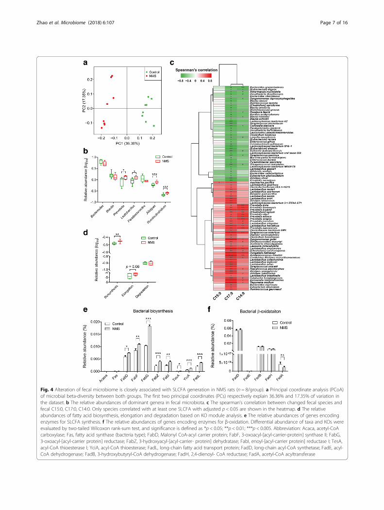
Fig. 4 Alteration of fecal microbiome is closely associated with SLCFA generation in NMS rats (n = 8/group). a Principal coordinate analysis (PCoA)of microbial beta-diversity between both groups. The first two principal coordinates (PCs) respectively explain 36.36% and 17.35% of variation inthe dataset. b The relative abundances of dominant genera in fecal microbiota. c The spearman’s correlation between changed fecal species andfecal C15:0, C17:0, C14:0. Only species correlated with at least one SLCFA with adjusted p < 0.05 are shown in the heatmap. d The relativeabundances of fatty acid biosynthesis, elongation and degradation based on KO module analysis. e The relative abundances of genes encodingenzymes for SLCFA synthesis. f The relative abundances of genes encoding enzymes for β-oxidation. Differential abundance of taxa and KOs wereevaluated by two-tailed Wilcoxon rank-sum test, and significance is defined as *p < 0.05; **p < 0.01; ***p < 0.005. Abbreviation: Acaca, acetyl-CoAcarboxylase; Fas, fatty acid synthase (bacteria type); FabD, Malonyl CoA-acyl carrier protein; FabF, 3-oxoacyl-[acyl-carrier-protein] synthase II; FabG,3-oxoacyl-[acyl-carrier protein] reductase; FabZ, 3-hydroxyacyl-[acyl-carrier- protein] dehydratase; FabI, enoyl-[acyl-carrier protein] reductase I; TesA,acyl-CoA thioesterase I; YciA, acyl-CoA thioesterase; FadL, long-chain fatty acid transport protein; FadD, long-chain acyl-CoA synthetase; FadE, acyl-CoA dehydrogenase; FadB, 3-hydroxybutyryl-CoA dehydrogenase; FadH, 2,4-dienoyl- CoA reductase; FadA, acetyl-CoA acyltransferase
Zhao et al. Microbiome (2018) 6:107 Page 7 of 16

increased after FMT, and such bacterial profile of GFrecipients on day 35 was closer to that of donors(Fig. 5b). Quantitative polymerase chain reaction(qPCR) analysis found that relative levels of two indis-pensable LCFA-synthesized genes FabF and FabG wereincreased in the colon lumens of GF rats receiving fecalmicrobiota of NMS donors on day 35 (Fig. 5c). Consist-ently, the levels of individual SLCFAs (from C14:0 toC18:0) were obviously raised in the colon lumen(Fig. 5d). These results indicate that featured enterotype
of NMS rats could result in elevation of stool frequencyand SLCFA generation.
Microbial modulation effectively reduces GI motility andintracolonic SLCFA production in NMS rats treated withneomycinTo further confirm the relationship betweenSLCFA-producing bacteria and bowel motility, the anti-biotic drug neomycin, previously reported as efficient inthe modulation of gut dysbiosis and improvement of
Fig. 5 Fecal microbiota from NMS donors enhances defecation frequency and intracolonic bacterial SLCFAs production in pseudo germ-free (GF)rats (n = 6/group). a Accumulation of fecal pellets within 60 min in colonized GF rats 1 week (day 21), 2 weeks (day 28), and 3 weeks (day 35)after microbial transplant. b Relative abundances of fecal SLCFA-producing genera in donors and colonized GF rats on days 10, 21, and 35. c Therelative levels of genes required for SLCFA synthesis in colonic contents of colonized GF rats. d The amounts of individual SLCFAs per gram ofcolonic contents. Bar charts are plotted using mean ± SEM value, and statistical significance between both colonized groups is defined as *p <0.05; **p < 0.01; ***p < 0.005
Zhao et al. Microbiome (2018) 6:107 Page 8 of 16

bowel symptoms in patients with IBS [40, 41], waschosen to orally administrate to NMS rats (150 mg/kg),twice daily, from postnatal days (PD) 56 to 70 (Fig. 1a).Control groups were treated with PBS. Stool frequencywas monitored every 3 days during drug intervention.As shown in Fig. 6a, model rats with oral gavage of PBSpresented greater stool pellet output within 60 min. En-hanced defecation number was effectively attenuatedand ultimately returned to normal after 14 days of neo-mycin treatment. The gut transit time became prolongedin neomycin-treated NMS rats, but was shorter thancontrol rats (Fig. 6b). Genus-specific PCR analysis foundthat levels of Prevotella, Lactobacillus, and Alistipeswere downregulated in the colon lumens of theneomycin-treated group (Fig. 6c). Also, the levels ofSLCFA synthetic genes FabF and FabG were reduced(Fig. 6d). In line with changes of SLCFA-producing bac-teria and genes, intracolonic levels of individual SLCFAswere notably attenuated after intervention (Fig. 6e).
DiscussionIn this study, we observed an altered gut microbiota inNMS rats characterized by increased capacity for gener-ation of SLCFAs, which were proved to enhance ratcolonic contraction ex vivo and stool frequency in vivo.Further, with microbiota intervention studies includ-ing FMT and antibiotic treatment, we found thatSLCFA-producing bacteria contribute to the acceler-ation of colonic motility in rats.SLCFAs have been reported to regulate GI motility in
humans, most of studies concerned their effects on theupper GI tract. A study of 10 healthy men showed thatupper intestinal infusion of oils enriched with C18:0 sig-nificantly reduced gastric motility [42]. The similar in-hibitory impact of SLCFAs on the upper gut motility hasalso been reported in patients with FGIDs [43].LCFA-induced slower upper GI motility influences foodintake and energy metabolism, demonstrating the im-portance of SLCFAs in maintenance of body weight aswell as in obesity progression [44]. However, we foundno difference in the body weight and luminal SLCFAs inthe small intestine between NMS rats and controls. Aprevious study found that increase of defecation was notaccompanied by a concomitant change in food consump-tion in NMS model [45]. Thus, the upper GI motility ispossibly not affected by SLCFAs in the NMS model.The effects of LCFAs on the lower GI tract, i.e., the
colon, is another story. Some studies have reported thatlipids can increase colon motility in normal subjects andpatients with IBS-D [26, 43]. Infusion of unsaturatedLCFAs have been shown to accelerate colon transit inhuman [46, 47]. But the effects of SLCFAs specificallyon the colon motor function have not been reported.This study is the first to provide direct evidence for the
promotional action of SLCFAs on colon motility. Com-pared with SLCFAs, short-chain fatty acids (C3:0 andC4:0) show low potency for stimulating the colonicmotor [48]. Also, we found that C2:0, present increasedlevel in fecal VOCs of NMS rats, cannot induce musclecontraction of the isolated colon. This suggests thatLCFAs have distinct and specific effects on the colon.Furthermore, elevated muscle contraction induced byC18:0 can be significantly inhibited by a GRP40 antagon-ist but not a GRP120 antagonist, indicating that GPR40is specifically involved in the SLCFAs-stimulated colonmotility. Briscoe et al. have reported that fatty acids withcarbon chain lengths greater than six are able to activateGPR40, thereby giving rise to elevation of calcium secre-tion and release [49]. Such GPR40-dependent Ca2+ risecan be blocked by inhibition of L-type calcium channelsor opening of the K(ATP) channel [50]. These evidencessuggest that LCFAs stimulate colon motility possiblythrough upregulation of GPR40-dependent Ca2+ influx.We intend to explore this possibility in further study.Generally, luminal SLCFAs are derived from either lip-
olysis of dietary fats or host/bacterial fatty acid synthesis[43, 51, 52]. A previous study reports normal expressionof enzymes for host lipolysis, transport and lipogenesisat the mRNA level in NMS model rats [31]. Moreover,SLCFAs with odd-numbered carbon atoms, such asC15:0 and C17:0, can be produced by bacteria only [32].If this is true then, elevation of individual SLCFAs fromC14 to C18 in the colon lumens of NMS model shouldbe the results of gut bacterial actions. In our study, thecombination of metabolomic and metagenomic resultspoints to a significant correlation between changes ofdominant bacteria and levels of excretive SLCFAs. KOanalysis of fecal metagenome showed obvious elevationin the level of fatty acid synthesis and elongation. Thesefindings also support the hypothesis that gut bacteria en-gage in formation of intracolonic SLCFAs.Bacterial fatty acid synthesis is essential for supplying
the hydrophobic components of the membrane lipidsand for providing components of storage lipids [53]. Thesynthetic process, mainly including initiation and elong-ation, is executed by a cluster of Fab enzymes that arestrongly conserved in bacteria [33, 34, 54]. Of these,FabF and FabG are indispensable for generation of LCFAwith carbon atoms 14 to 18 [55, 56]. In a previous studyof alcoholic liver disease, the gene expression level ofFabF and FabG has been determined to reflect the levelof bacterial LCFA synthesis in humans and mice [57].We observed that genomic levels of FabF and FabGwere highly enriched in fecal microbiota of NMS rats.Such increased levels of FabF and FabG also appeared inGF rats receiving fecal microbiota from NMS donors,but were attenuated upon modulation of NMS-associatedgut dysbiosis. These observations indicate that a higher
Zhao et al. Microbiome (2018) 6:107 Page 9 of 16
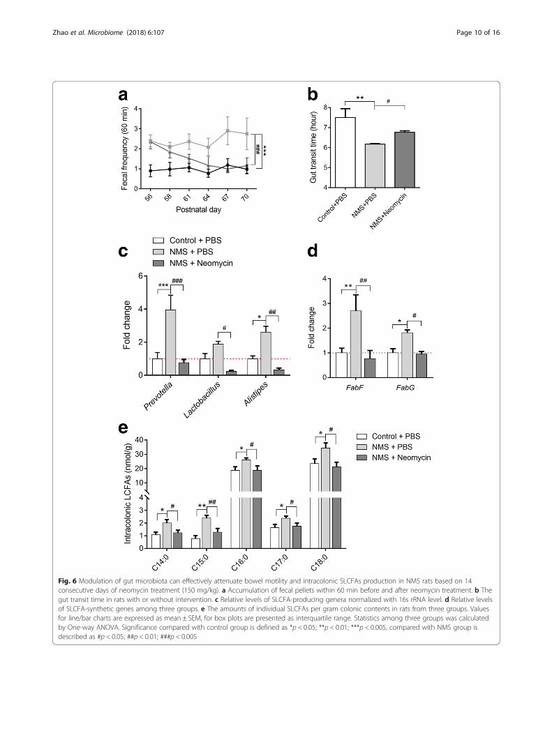
Fig. 6 Modulation of gut microbiota can effectively attenuate bowel motility and intracolonic SLCFAs production in NMS rats based on 14consecutive days of neomycin treatment (150 mg/kg). a Accumulation of fecal pellets within 60 min before and after neomycin treatment. b Thegut transit time in rats with or without intervention. c Relative levels of SLCFA-producing genera normalized with 16s rRNA level. d Relative levelsof SLCFA-synthetic genes among three groups. e The amounts of individual SLCFAs per gram colonic contents in rats from three groups. Valuesfor line/bar charts are expressed as mean ± SEM, for box plots are presented as interquartile range. Statistics among three groups was calculatedby One-way ANOVA. Significance compared with control group is defined as *p < 0.05; **p < 0.01; ***p < 0.005, compared with NMS group isdescribed as #p < 0.05; ##p < 0.01; ###p < 0.005
Zhao et al. Microbiome (2018) 6:107 Page 10 of 16

level of bacterial SLCFA synthesis is characteristic of theNMS-associated enterotype.The genera Prevotella, Lactobacillus and Alistipes ex-
pressing essential Fab genes showed elevated abundancesin NMS rats and GF rats receiving fecal microbiota ofNMS donors, and were significantly reduced in NMS ratsafter neomycin treatment. Probably, these three generaare important manipulators for intracolonic SLCFA dis-ruption. Interestingly, these SLCFA-producing bacteriahave been linked with host GI motility both in clinical andlaboratory studies. For example, abundances of Lactobacil-lus, Prevotella and Alistipes spp. are significantly decreasedin patients with constipation [10–12]. Conversely, increaseof the genus Lactobacillus has been reported in patients ofIBS-D [58]. Higher abundances of Prevotella spp. arefound in children with chronic diarrhea [59]. The generaPrevotella, Lactobacillus or Alistipes showed increasedabundance in chronic stress-induced rodents character-ized by GI dysmotility [24, 25]. Lactobacillus strains accel-erate intestinal transit in GF rats and enhance ilealcontraction in guinea-pig [60, 61]. These lines of evidenceconsistently support a linkage between SLCFA-producingbacteria and host colonic motility; however, further studiesare needed to identify the precise roles of specific speciesor strains in modulating host colonic motility.By profiling fecal VOCs, we noticed that acidic sub-
stances, including SLCFAs and fermented products (e.g.,lactate and acetate) were significantly elevated in NMSgroup, corresponding to the raised acidity in stools.Meanwhile, increase of fecal TCA intermediates citrateand succinate in NMS rats suggest a higher metaboliclevel of citrate cycle. It is supported by fecal metage-nomic results that a significant enrichment of citratecycle module in NMS group relative controls (differentgenes mapping in TCA cycle: 398 genes for control and486 genes for NMS group, adjust p < 0.05). Of them,succinate can stimulate water secretion from intestinalsegments [62], indicating that increased fecal consistencyof NMS rats can be attributed to altered microbial me-tabolites. In contrast, fecal butyrate and valerate showedreduced levels in the NMS group. Previous study hasfound that butyrate is beneficial to maintenance of theintestinal epithelial barrier through facilitating tightjunction assembly [63]. Soderholm et al. revealed thatNMS enhances vulnerability of the colonic mucosal bar-rier to stress [64]. Probably, deficiency of bacterial butyr-ate production is one cause of the weak colonic barrierof the NMS model. This evidence indicates the import-ance of bacterial metabolites in controlling both stoolcharacteristics and colonic barrier function.
ConclusionThis study determined that NMS rats have excessivelevels of intracolonic SLCFAs, and these SLCFAs are
specifically related to accelerated colonic movement andincreased fecal output. Such elevated SLCFAs is contrib-uted by hyperactive synthetic action of gut bacteria, spe-cifically by the genera Prevotella, Lactobacillus andAlistipes. Fecal microbial transplantation and antibioticmodulation revealed a causal relationship betweenSLCFA-producing bacteria and host colonic motility.These findings clarify the stimulatory effects of SLCFAson the colon motility and provide novel insight in gutmicrobiota-driven GI dysmotility, which may lead to atherapeutic intervention targeting specific gut micro-biota for treating GI motility disorders.
MethodsAnimalsAll Sprague-Dawley (SD) rats used for this study wereobtained from the Laboratory Animal Services Centre ofThe Chinese University of Hong Kong. Rats were main-tained on a 12-h light/dark cycle with free access to foodand water under specific pathogen-free (SPF) condition.Materials including cages, diets, water, and litters weresterilized, and stool frequency was tested in a clean hoodwith a UV lamp for pseudo GF rats. All animal experi-ments have followed the Animals Ordinance, Depart-ment of Health, Hong Kong SAR, China.
Neonatal maternal separation modeling and colonicmotility assessmentReferring to our previous study [65], the procedure ofthe NMS model was performed as shown in the timeline(Fig. 1a). Briefly, four pregnant SD rats were housed in-dividually in cages. Randomly, newborn pups from twomothers were assigned to NMS group and pups fromanother two dams were assigned to non-handled controlgroup. NMS pups were separated from their dams dur-ing the period of postnatal days (PD) 2 to 14 for 3 hdaily whereas control pups remained with their mothersconstantly. All pups were weaned on PD 22, and onlymale pups (n = 8/group) with similar body weight wasused for further experiments. GI motility was individu-ally evaluated in rats 8 weeks later. Fecal pellets werecollected to measure pH level and water contents, andwere used for analyses of metagenomics and metabolo-mics. The detail methods for assessment of motor func-tion and stool characteristics were described inAdditional file 1. The luminal contents of ileum (10 cmproximal to the cecum), cecum, and proximal colon(5 cm distal to the cecum) were collected after CO2
anesthesia, and were immediately stored at − 80 °C. Fur-thermore, to investigate the relationship betweenSLCFA-produced bacteria and the colonic motor inNMS rats, the drug neomycin (150 mg/kg) was twicedaily administrated to adult NMS rats for 14 consecutive
Zhao et al. Microbiome (2018) 6:107 Page 11 of 16

days (from PD 56 to 70). Meanwhile, other NMS or con-trol rats were orally gavaged with PBS.
Pseudo germ-free modeling and fecal microbial transferThe experimental procedure is shown in Fig. 1b. Theantibiotic cocktail (ABX), containing ampicillin (1 g/L),neomycin (1 g/L), and metronidazole (0.5 g/L) were pre-pared in drinking water as previously described [37].The ABX-contained water was ad libitum supplied for12 normal SD rats (n = 6/group) to establish the pseudoGF model. No difference in the record of daily and totalwater consumption (Additional file 1: Fig. S4A) indicatesthat rats in different cages ingested similar volumes ofABX. To ensure elimination of the gut microbiome, fecalpellets were frequently collected during experiment forreal-time monitoring of dynamic changes of gut micro-biota. The very low levels of fecal total DNA quality andbacterial diversity (Additional file 1: Fig. S4B and C) in-dicate that the pseudo GF model was successfullyestablished after 10 days of ABX-water intervention. Fur-thermore, donors’ feces from either NMS or controlgroup (n = 8/group) were individually pooled and com-pletely homogenized in pre-reduced PBS at 1 ml per pel-let referring to a published method [20]. One milliliterof the settled suspension was daily administered topseudo GF rats for five consecutive days. The stool fre-quency was weekly determined in colonized GF rats.Fecal samples were collected on Day 0, 10, 21 and 35 forfurther microbial analysis, and colonic contents werecollected on Day 35 for assessment of SLCFA producinggenes and SLCFA amounts.
Preparation of saturated long-chain fatty acids for colonicmotility assessmentsTo evaluate the effects of SLCFAs on host colonic motil-ity, dominant odd- and even-numbered carbon SLCFAsC17:0 and C18:0 were chosen to individually test theirinfluences on the muscle contraction of rat isolatedcolon segments ex vivo and rat stool frequency in vivo.C17:0 and C18:0 (Cat#: 506-12-7 and 57-11-4,Sigma-Aldrich, St. Louis, MO USA) were solubilized withfatty acid free-bovine serum albumin (BSA, Cat#:9048-46-8; Sigma-Aldrich, USA) to generate a series ofconcentrations (10, 30, 50 and 100μM) for organ bathsystem-based colonic contraction test ex vivo [66]. Theconcentration range were calculated in accordance withSLCFA levels found in the colon contents of NMS ratswith 1.1 g/cm3 of density of the colonic contents reportedin a previous study [67]. To test which long-chain fattyacid receptors mediate SLCFA-stimulated colonic contrac-tion, selective GPR40 antagonist DC260126 (DC, Cat#:346692-04-4, Tocris Bioscience, UK) and GPR120 antag-onist AH7614 (AH, Cat#: 6326-06-3, Tocris Bioscience,UK) was prepared at working dosages of 2.5, 5, and 10μM
according to previous studies [68, 69]. Acetylcholine (Ach,Cat#: 60–31-1, Sigma, St. Louis, MO USA) and C2:0(Cat#: 127-09-3, Sigma, St. Louis, MO USA) were appliedas positive and negative control, respectively. The experi-mental procedure was described in Additional file 1. Fur-thermore, both SLCFAs were prepared as differentdosages (1, 2.5 and 5 mg/kg, dissolved in 5% ethanol) forin vivo measurement of stool frequency in normal SD rats(n = 8/group). Each rat orally treated with either 1 mL ofSLCFAs or vehicle solution was located in an individualcage for defecation frequency assessment. Fecal pelletswere collected for determination of intracolonic SLCFAlevels.
Total DNA isolation and metagenomic sequencingTotal bacterial DNA was isolated and purified from fecalpellets (precisely weighted 200 mg) using a stool DNAIsolation Kit (Qiagen, Valencia, CA). All samples weresequenced based on the Illumina Hiseq 4000 platform(paired-end; insert size, 350 bp; read length, 150 bp).After removal of adaptor and low-quality reads, theremaining reads were filtered to eliminate the host DNAgenome based on the genome reference of Rattus norve-gicus by SOAPalign v2.21 [70]. Finally, 105.81 Gbhigh-quality pair-end reads for the 16 rats samples (n =8/group) was acquired with an average of 6.61 Gb persample in groups of NMS and control (Additional file 2:Table S2).
Construction of the gene catalogThe reads were assembled into contigs for all samplesusing the assembly software SOAPdenovo v2.0455. Aver-age 75.67% of the total reads were used to generate 1.6million contigs without ambiguous bases (minimumlength of 500 bp). ORFs were predicted from the assem-bled contigs using the MetaGeneMark v3.26 programme[71]. The 6,116,823 ORFs longer than 100 bp covered89.67% of the total length of the contigs and about half(50.51%) of the ORFs appeared complete. All ORFs wereclustered by CD-hit v4.6.4 to construct a non-redundantgene catalog using a stringent criterion of 95% identityat the nucleotide level over 90% of the length of theshorter ORFs [72]. The final non-redundant gene setcontained 1,462,418 ORFs with an average length of728 bp.
Diversity analysis, taxonomic assignment and functionalcharacterizationThe α-diversity (within-sample diversity) and β-diversity(between-sample diversity) were estimated by the Shan-non index and Bray-Curtis dissimilarity metric, respect-ively [73]. For taxonomic assignment, all predicted geneswere blasted against the reference microbial genomesfrom NCBI (including 4258 microbial genomes) by using
Zhao et al. Microbiome (2018) 6:107 Page 12 of 16

BLAST (v 2.2.26, default parameter) with at least 80%overlap of query. Taxonomic identification was per-formed as 65% identity for phylum, 85% identity forgenus, 95% identity for species [74, 75]. The taxonomicabundance was calculated based on gene abundance,and protein sequences of the predicted genes weresearched using National Center for Biotechnology Infor-mation BLASTP against the KEGG gene database (v79).Each protein was assigned to the KEGG group by thehighest scoring annotated hits. Significance in the rela-tive abundance of genes, KOs, phylum, genera, and spe-cies between both groups were compared by two-tailedWilcoxon rank-sum test (Additional file 2: TablesS3-S7). The relationship between the abundance of eachspecies and SLCFAs contents was assessed by the Spear-man’s correlation [76]. Enrichment in NMS or controlgroup was then determined according to the higherrank-sum.
Determination of bacterial DNA quality and 16s ribosomalRNA amplicon sequencingDNA extracts was prepared from feces (preciselyweighted 200 mg) of donors and colonized GF rats. TheDNA yield and quality were determined spectrophoto-metrically by the NanoDrop™ ND-2000 (Thermo FisherScientific Ltd., Waltham, MA, USA). The DNA integritywas determined through 1% agarose gel (w/v). Further-more, the extracted DNA (30 ng) was amplified withuniversal primers (515F and 806R) to obtain the V4 re-gions of the 16S rRNA gene. The PCR products werepurified and sequenced on the Illumina Hiseq 2500 plat-form. After raw data filtering and merging [77, 78], thenumbers of tag and operational taxonomic unit (OTU)were generated from 50 samples (Additional file 2: TableS8). Bacterial diversity and taxonomy was obtained fromOTU table by using QIIME software package [79].
PCR analysis of genus-specific bacterial 16s rDNA andSLCFA synthetic genesGenus-specific primers for Prevotella, Lactobacillus andAlistipes were obtained from published studies [32, 80].Bacterial DNA extracts (50 ng) of colonic contents wereamplified with pairs of primers and Power SYBR GREENMaster Mix (Applied Biosystems, Foster city, CA, USA)based on an ABI StepOne Plus Sequence Detection Sys-tem (Applied Biosystems, Foster city, CA, USA). Thecycling conditions were as follows: 95 °C for 10 min,followed by 40 cycles of 95 °C for 30 s, 52 °C for 30 s,and 72 °C for 1 min. Moreover, such SYBRGREEN-based PCR detection also applied to testify ex-pression of LCFA synthetic FabF and FabG genes fromcolonic contents. The primers for FabF and FabG weredesigned as previously described [57]. The expression of
each genus and gene was normalized to the level of 16SrRNA.
Metabolite extraction and SLCFA quantificationFecal samples or luminal contents (100 mg) were com-pletely homogenized with five-fold volume of ice-colddistilled water. After high-speed centrifugation(13,000 rpm for 15 min at 4 °C), water extractions weretransferred to a new 2-mL tube. Subsequently, afive-fold volume (500 μL) of methanol was added intothe pellet sample. The mixture was completely homoge-nized and centrifuged again. Methanol extractions werecombined with the previous water extractions. Stronglyvortex and centrifugation again, the resulting superna-tants were obtained for derivatization processing. Mean-while, a quality control (QC) sample pooling all ratsamples was prepared using the same protocol. Forquantification of luminal SFAs in the small and large in-testines, external calibration solution of SFA standardsfrom C2:0 to C18:0 were prepared at a series of concen-trations (from 1 to 20,000 ng/ml). Isotope labeledC13-myristic acid was used as internal standards. Abso-lute quantities of individual SFAs were normalized tothe sample weight.
GC/MS-based metabolomic analysis and data processingFecal metabolites were derivatized by BSTFA with 1%TMCS based on a previously method [81]. A gas chroma-tography coupled with a mass spectrum (GCMS-QP2010systems, Shimaduzu Co., Tokyo, Japan) was applied forfecal metabolome analysis. Fecal derivatives were sepa-rated by a DB-5 MS fused-silica capillary column (30 m ×250 μm i.d.; Agilent J&W Scientific, Folsom, CA), chem-ically bonded with a 5% phenyl-95% methylpolysiloxanecross-linked stationary phase (0.25 μm film thickness).The detail sample preparation and analytical parametersof instrumental conditions were showed in Add-itional file 1. The metabolic signals were deconvoluted,aligned and normalized to final data matrix throughusing R-scipt with xcms package (R version 3.4.2). Theprocessed matrix was introduced into the softwareSIMCA-P (Version 11.0, Umetrics, Umea, Sweden) forprincipal component analysis (PCA) and partial leastsquares-discriminant analysis (PLS-DA). Metabolic fea-tures were selected by combination of the variable im-portance in the projection (VIP) threshold (VIP > 1.0)and the Student’s t-test (p < 0.05). Metabolites wereidentified by the national institute of standards andtechnology (NIST) library (over 95% matched similarity).
Statistical analysisFor sequencing data, statistical analyses were performedin R (v3.4.10) software; differential abundance of genes,taxonomies, and KOs were evaluated by two-tailed
Zhao et al. Microbiome (2018) 6:107 Page 13 of 16
Wilcoxon rank-sum test. For determination of metabo-lites, genes, and colonic motor in animal studies,Mann-Whitney test was used for statistics between twogroups, and one-way ANOVA was used for comparisonamong more than two groups. These statistical analyseswere performed in GraphPad Prism 6 (GraphPad soft-ware lnc., CA, USA). Statistical significance is defined asp < 0.05.
Additional files
Additional file 1 Figure S1. Fecal characteristics and body weight inNMS rats and controls. Figure S2. Muscle amplitudes of rat isolatedcolonic segments with different treatment. Figure S3. Phylogeneticprofiles of fecal microbiomes of NMS and control rats. Figure S4. Fecalmicrobial community in pseudo GF rats at duration of FMT experiment.Table S1. Fecal metabolites with significant difference between NMS ratsand controls. (DOCX 784 kb)
Additional file 2 Table S2. Data production, quality control, assemblyresult and gene prediction resulted from fecal metagenomic sequencinganalysis. Table S3. Taxonomic profiles of fecal microbiota in NMS andcontrol groups at phylum level. Table S4. Taxonomic profiles of fecalmicrobiota in NMS and control groups at genus level. Table S5.Taxonomic profiles of fecal microbiota in NMS and control groups atspecies level. Table S6. Correlation between species and saturated long-chain fatty acids determined by fecal metagenomic and metabolomicanalyses. Table S7. Identified KOs involved in fatty acid synthesis anddegradation. Table S8. Data production and quality control of fecalsamples from 16s rRNA amplicon sequencing analysis. Table S9. Taxonomicchanges at genus level between colonized GF rats and donors. (XLSX 73 kb)
AbbreviationsABX: Antibiotic cocktails; Acaca: Acetyl-CoA carboxylase; C14:0: Tetradecanoicacid; C15:0: Pentadecanoic acid; C16:0: Palmitic acid; C17:0: Heptadecanoicacid; C18:0: Octadecanoic acid; C2:0: Acetic acid; C4:0: Butyric acid;C5:0: Valeric acid; FabD: Malonyl CoA-acyl carrier protein; FabF: 3-Oxoacyl-[acyl-carrier-protein] synthase II; FabG: 3-Oxoacyl-[acyl-carrier protein]reductase; FabI: Enoyl-[acyl-carrier protein] reductase I; FabZ: 3-Hydroxyacyl-[acyl-carrier-protein] dehydratase; FadA: Acetyl-CoA acyltransferase; FadB: 3-Hydroxybutyryl-CoA dehydrogenase; FadD: Long-chain acyl-CoA synthetase;FadE: Acyl-CoA dehydrogenase; FadH: 2,4-Dienoyl-CoA reductase;FadL: Long-chain fatty acid transport protein; Fas: Fatty acid synthase(bacteria type); NMS: Neonatal maternal separation; PCoA: Principalcoordinate analysis; SLCFAs: Saturated long-chain fatty acids; TesA: Acyl-CoAthioesterase I; VIP: Variable importance in the projection; VOCs: Volatileorganic compounds; YciA: Acyl-CoA 12 thioesterase
AcknowledgmentsWe thank the colleagues of Shenzhen Key Laboratory of Human CommensalMicroorganisms and Health Research and Shenzhen Engineering Laboratoryof Detection and Intervention of Human Intestinal Microbiome for theirassistance in microbial sequencing and analysis. We also thank Dr. MarthaDahlen (CA, USA) for her critical appraisal for this manuscript.
FundingThis project was supported by grants from Faculty Research Grant of HongKong Baptist University (FRG2/15-16/001 and FRG2/16-17/003), the ResearchGrants Council of Hong Kong Collaborative Research Fund (C2012-15G), andGuangdong-Hong Kong Technology Cooperation Funding Scheme(2016A050503039).
Availability of data and materialsFecal metagenomic sequencing reads are deposited in the National Centerfor Biotechnology Information (NCBI) database with accession codePRJNA419985. Other data that support the findings of this study are availablefrom the corresponding author upon reasonable request.
Authors’ contributionsZXB, XDF, LXZ, and LZ conceived the idea and designed this study. LZ andLL performed metabolomics, in vivo and ex vivo experiments using animals.YF and YC contributed to metagenomic and 16s rRNA sequencing analysis.WY and ZSL conducted bacterial DNA extraction and relevant PCR analysis.LZ, TH, CYL, and HLW prepared manuscript. HMY, JW, and LXZ providedguidance for both experimental design and manuscript preparation. Allauthors read and approved the final manuscript.
Ethics approvalThe use and care of all animals were performed in accordance with theAnimals Ordinance, and experimental operators obtained licenses issued bythe government of the Hong Kong Special Administrative RegionDepartment of Health, Hong Kong SAR, China.
Competing interestsThe authors declare that they have no competing interests.
Publisher’s NoteSpringer Nature remains neutral with regard to jurisdictional claims inpublished maps and institutional affiliations.
Author details1Chinese Medicine Clinical Study Center, Jockey Club School of ChineseMedicine, Hong Kong Baptist University, Kowloon Tong, Hong Kong SAR,China. 2BGI Genomics, BGI-Shenzhen, Shenzhen, China. 3Preparatory Office ofShenzhen-Melbourne Institute of Life Sciences and Bioengineering,Guangzhou University of Chinese Medicine, Guangzhou, China. 4YMU-HKBUJoint Laboratory of Traditional Natural Medicine, Yunnan Minzu University,Kunming, China. 5The Second Affiliated Hospital of Guangzhou University ofChinese Medicine, Guangzhou, China. 6BGI-Shenzhen, Shenzhen, China.7Digestive Diseases and Nutrition Center, Department of Pediatrics, The StateUniversity of New York at Buffalo, 3435 Main Street, 422BRB, Buffalo, NY14214, USA.
Received: 13 December 2017 Accepted: 1 June 2018
References1. Corazziari E. Definition and epidemiology of functional gastrointestinal
disorders. Best Pract Res Clin Gastroenterol. 2004;18:613–31.2. Bassotti G, Antonelli E, Villanacci V, Salemme M, Coppola M, Annese V.
Gastrointestinal motility disorders in inflammatory bowel diseases. World JGastroenterol. 2014;20:37–44.
3. Lacy BE, Weiser K. Gastrointestinal motility disorders: an update. Dig Dis.2006;24:228–42.
4. Ohama T, Hori M, Ozaki H. Mechanism of abnormal intestinal motility ininflammatory bowel disease: how smooth muscle contraction is reduced? JSmooth Muscle Res. 2007;43:43–54.
5. Beyder A, Farrugia G. Targeting ion channels for the treatment ofgastrointestinal motility disorders. Therap Adv Gastroenterol. 2012;5:5–21.
6. Hansen MB. Neurohumoral control of gastrointestinal motility. Physiol Res.2003;52:1–30.
7. Quigley EM. Microflora modulation of motility. J Neurogastroenterol Motil.2011;17:140–7.
8. Matto J, Maunuksela L, Kajander K, Palva A, Korpela R, Kassinen A, Saarela M.Composition and temporal stability of gastrointestinal microbiota in irritablebowel syndrome–a longitudinal study in IBS and control subjects. FEMSImmunol Med Microbiol. 2005;43:213–22.
9. Pozuelo M, Panda S, Santiago A, Mendez S, Accarino A, Santos J,Guarner F, Azpiroz F, Manichanh C. Reduction of butyrate- andmethane-producing microorganisms in patients with irritable bowelsyndrome. Sci Rep. 2015;5:12693.
10. Khalif IL, Quigley EM, Konovitch EA, Maximova ID. Alterations in the colonicflora and intestinal permeability and evidence of immune activation inchronic constipation. Dig Liver Dis. 2005;37:838–49.
11. Zhu L, Liu W, Alkhouri R, Baker RD, Bard JE, Quigley EM, Baker SS. Structuralchanges in the gut microbiome of constipated patients. Physiol Genomics.2014;46:679–86.
Zhao et al. Microbiome (2018) 6:107 Page 14 of 16
12. de Meij TG, de Groot EF, Eck A, Budding AE, Kneepkens CM, Benninga MA,van Bodegraven AA, Savelkoul PH. Characterization of microbiota inchildren with chronic functional constipation. PLoS One. 2016;11:e0164731.
13. Kim SE, Choi SC, Park KS, Park MI, Shin JE, Lee TH, Jung KW, Koo HS, Myung SJ.Constipation research group of Korean Society of N, motility: change of fecalFlora and Effectiveness of the short-term VSL#3 probiotic treatment in patientswith functional constipation. J Neurogastroenterol Motil. 2015;21:111–20.
14. De Palma G, Lynch MD, Lu J, Dang VT, Deng Y, Jury J, Umeh G,Miranda PM, Pigrau Pastor M, Sidani S, et al. Transplantation of fecalmicrobiota from patients with irritable bowel syndrome alters gutfunction and behavior in recipient mice. Sci Transl Med. 2017;9 https://doi.org/10.1126/scitranslmed.aaf6397.
15. Ge X, Zhao W, Ding C, Tian H, Xu L, Wang H, Ni L, Jiang J, Gong J, Zhu W, et al.Potential role of fecal microbiota from patients with slow transit constipationin the regulation of gastrointestinal motility. Sci Rep. 2017;7:441.
16. Barbara G, Stanghellini V, Brandi G, Cremon C, Di Nardo G, De Giorgio R,Corinaldesi R. Interactions between commensal bacteria and gut sensorimotorfunction in health and disease. Am J Gastroenterol. 2005;100:2560–8.
17. Triantafyllou K, Chang C, Pimentel M. Methanogens, methane andgastrointestinal motility. J Neurogastroenterol Motil. 2014;20:31–40.
18. Anitha M, Vijay-Kumar M, Sitaraman SV, Gewirtz AT, Srinivasan S. Gutmicrobial products regulate murine gastrointestinal motility via toll-likereceptor 4 signaling. Gastroenterology. 2012;143:1006–1016 e1004.
19. Cherbut C. Motor effects of short-chain fatty acids and lactate in thegastrointestinal tract. Proc Nutr Soc. 2003;62:95–9.
20. Yano JM, Yu K, Donaldson GP, Shastri GG, Ann P, Ma L, Nagler CR, IsmagilovRF, Mazmanian SK, Hsiao EY. Indigenous bacteria from the gut microbiotaregulate host serotonin biosynthesis. Cell. 2015;161:264–76.
21. Bulbul M, Babygirija R, Cerjak D, Yoshimoto S, Ludwig K, Takahashi T. Impairedadaptation of gastrointestinal motility following chronic stress in maternallyseparated rats. Am J Physiol Gastrointest Liver Physiol. 2012;302:G702–11.
22. Zhang M, Leung FP, Huang Y, Bian ZX. Increased colonic motility in a ratmodel of irritable bowel syndrome is associated with up-regulation of L-type calcium channels in colonic smooth muscle cells. NeurogastroenterolMotil. 2010;22:e162–70.
23. Bian ZX, Zhang M, Han QB, Xu HX, Sung JJ. Analgesic effects of JCM-16021on neonatal maternal separation-induced visceral pain in rats. World JGastroenterol. 2010;16:837–45.
24. De Palma G, Blennerhassett P, Lu J, Deng Y, Park AJ, Green W, DenouE, Silva MA, Santacruz A, Sanz Y, et al. Microbiota and hostdeterminants of behavioural phenotype in maternally separated mice.Nat Commun. 2015;6:7735.
25. Pusceddu MM, El Aidy S, Crispie F, O'Sullivan O, Cotter P, Stanton C,Kelly P, Cryan JF, Dinan TG. N-3 polyunsaturated fatty acids (PUFAs)reverse the impact of early-life stress on the gut microbiota. PLoS One.2015;10:e0139721.
26. Deiteren A, Camilleri M, Burton D, McKinzie S, Rao A, Zinsmeister AR. Effectof meal ingestion on ileocolonic and colonic transit in health and irritablebowel syndrome. Dig Dis Sci. 2010;55:384–91.
27. Lin CY, Zhang M, Huang T, Yang LL, Fu HB, Zhao L, Zhong LL, Mu HX, ShiXK, Leung CF, et al. Spexin enhances bowel movement through activatingL-type voltage-dependent Calcium Channel via Galanin receptor 2 in mice.Sci Rep. 2015;5:12095.
28. Edfalk S, Steneberg P, Edlund H. Gpr40 is expressed in enteroendocrine cellsand mediates free fatty acid stimulation of incretin secretion. Diabetes.2008;57:2280–7.
29. Paulsen SJ, Larsen LK, Hansen G, Chelur S, Larsen PJ, Vrang N. Expression ofthe fatty acid receptor GPR120 in the gut of diet-induced-obese rats and itsrole in GLP-1 secretion. PLoS One. 2014;9:e88227.
30. Ichimura A, Hirasawa A, Hara T, Tsujimoto G. Free fatty acid receptors act asnutrient sensors to regulate energy homeostasis. Prostaglandins Other LipidMediat. 2009;89:82–8.
31. Paternain L, Martisova E, Milagro FI, Ramirez MJ, Martinez JA, Campion J.Postnatal maternal separation modifies the response to an obesogenic dietin adulthood in rats. Dis Model Mech. 2012;5:691–7.
32. Kumagai H, Maisawa S, Tanaka M, Takahashi M, Takasago Y, Nishijima A,Watanabe S. Intestinal microbiota and secretory immunoglobulin a in fecesof exclusively breast-fed infants with blood-streaked stools. MicrobiolImmunol. 2012;56:657–63.
33. Qiu X, Choudhry AE, Janson CA, Grooms M, Daines RA, Lonsdale JT,Khandekar SS. Crystal structure and substrate specificity of the beta-
ketoacyl-acyl carrier protein synthase III (FabH) from Staphylococcus aureus.Protein Sci. 2005;14:2087–94.
34. Cronan JE, Thomas J. Bacterial fatty acid synthesis and its relationships withpolyketide synthetic pathways. Methods Enzymol. 2009;459:395–433.
35. Schweizer E, Hofmann J. Microbial type I fatty acid synthases (FAS): majorplayers in a network of cellular FAS systems. Microbiol Mol Biol Rev. 2004;68:501–17. table of contents
36. Dellomonaco C, Clomburg JM, Miller EN, Gonzalez R. Engineered reversal ofthe beta-oxidation cycle for the synthesis of fuels and chemicals. Nature.2011;476:355–9.
37. Ijssennagger N, Belzer C, Hooiveld GJ, Dekker J, van Mil SWC, Muller M,Kleerebezem M, van der Meer R. Gut microbiota facilitates dietary heme-induced epithelial hyperproliferation by opening the mucus barrier in colon.Proc Natl Acad Sci U S A. 2015;112:10038–43.
38. Nicklas W, Keubler L, Bleich A. Maintaining and monitoring the definedmicrobiota status of Gnotobiotic rodents. ILAR J. 2015;56:241–9.
39. Ge X, Ding C, Zhao W, Xu L, Tian H, Gong J, Zhu M, Li J, Li N. Antibiotics-induced depletion of mice microbiota induces changes in host serotoninbiosynthesis and intestinal motility. J Transl Med. 2017;15:13.
40. Basseri RJ, Weitsman S, Barlow GM, Pimentel M. Antibiotics for the treatment ofirritable bowel syndrome. Gastroenterol Hepatol (N Y). 2011;7:455–93.
41. Ahmad OF, Akbar A. Microbiome, antibiotics and irritable bowel syndrome.Br Med Bull. 2016;120:91–9.
42. French SJ, Conlon CA, Mutuma ST, Arnold M, Read NW, Meijer G, Francis J.The effects of intestinal infusion of long-chain fatty acids on food intake inhumans. Gastroenterology. 2000;119:943–8.
43. Feinle-Bisset C, Azpiroz F. Dietary lipids and functional gastrointestinaldisorders. Am J Gastroenterol. 2013;108:737–47.
44. Matzinger D, Degen L, Drewe J, Meuli J, Duebendorfer R, Ruckstuhl N, D'AmatoM, Rovati L, Beglinger C. The role of long chain fatty acids in regulating foodintake and cholecystokinin release in humans. Gut. 2000;46:688–93.
45. Coutinho SV, Plotsky PM, Sablad M, Miller JC, Zhou H, Bayati AI, McRobertsJA, Mayer EA. Neonatal maternal separation alters stress-induced responsesto viscerosomatic nociceptive stimuli in rat. Am J Physiol Gastrointest LiverPhysiol. 2002;282:G307–16.
46. Spiller RC, Brown ML, Phillips SF. Decreased fluid tolerance, acceleratedtransit, and abnormal motility of the human colon induced by oleic acid.Gastroenterology. 1986;91:100–7.
47. Kamath PS, Phillips SF, O'Connor MK, Brown ML, Zinsmeister AR. Coloniccapacitance and transit in man: modulation by luminal contents and drugs.Gut. 1990;31:443–9.
48. Hurst NR, Kendig DM, Murthy KS, Grider JR. The short chain fatty acids,butyrate and propionate, have differential effects on the motility of theGuinea pig colon. Neurogastroenterol Motil. 2014;26:1586–96.
49. Briscoe CP, Tadayyon M, Andrews JL, Benson WG, Chambers JK, Eilert MM,Ellis C, Elshourbagy NA, Goetz AS, Minnick DT, et al. The orphan G protein-coupled receptor GPR40 is activated by medium and long chain fatty acids.J Biol Chem. 2003;278:11303–11.
50. Shapiro H, Shachar S, Sekler I, Hershfinkel M, Walker MD. Role of GPR40 infatty acid action on the beta cell line INS-1E. Biochem Biophys ResCommun. 2005;335:97–104.
51. Niot I, Poirier H, Tran TT, Besnard P. Intestinal absorption of long-chain fattyacids: evidence and uncertainties. Prog Lipid Res. 2009;48:101–15.
52. Smith S, Witkowski A, Joshi AK. Structural and functional organization of theanimal fatty acid synthase. Prog Lipid Res. 2003;42:289–317.
53. Parsons JB, Rock CO. Bacterial lipids: metabolism and membranehomeostasis. Prog Lipid Res. 2013;52:249–76.
54. Heath RJ, Rock CO. Enoyl-acyl carrier protein reductase (fabI) plays adeterminant role in completing cycles of fatty acid elongation inEscherichia coli. J Biol Chem. 1995;270:26538–42.
55. Jeon E, Lee S, Lee S, Han SO, Yoon YJ, Lee J. Improved production of long-chain fatty acid in Escherichia coli by an engineering elongation cycleduring fatty acid synthesis (FAS) through genetic manipulation. J MicrobiolBiotechnol. 2012;22:990–9.
56. Garwin JL, Klages AL, Cronan JE Jr. Beta-ketoacyl-acyl carrier proteinsynthase II of Escherichia coli. Evidence for function in the thermalregulation of fatty acid synthesis. J Biol Chem. 1980;255:3263–5.
57. Chen P, Torralba M, Tan J, Embree M, Zengler K, Starkel P, van Pijkeren JP,DePew J, Loomba R, Ho SB, et al: Supplementation of saturated long-chainfatty acids maintains intestinal eubiosis and reduces ethanol-induced liverinjury in mice. Gastroenterology 2015, 148:203–214 e216.
Zhao et al. Microbiome (2018) 6:107 Page 15 of 16

58. Carroll IM, Chang YH, Park J, Sartor RB, Ringel Y. Luminal and mucosal-associated intestinal microbiota in patients with diarrhea-predominantirritable bowel syndrome. Gut Pathog. 2010;2:19.
59. Gilchrist CA, Petri SE, Schneider BN, Reichman DJ, Jiang N, Begum S,Watanabe K, Jansen CS, Elliott KP, Burgess SL, et al. Role of the gutmicrobiota of children in diarrhea due to the protozoan parasiteEntamoeba histolytica. J Infect Dis. 2016;213:1579–85.
60. Husebye E, Hellstrom PM, Sundler F, Chen J, Midtvedt T. Influence ofmicrobial species on small intestinal myoelectric activity and transit ingerm-free rats. Am J Physiol Gastrointest Liver Physiol. 2001;280:G368–80.
61. Massi M, Ioan P, Budriesi R, Chiarini A, Vitali B, Lammers KM, Gionchetti P,Campieri M, Lembo A, Brigidi P. Effects of probiotic bacteria ongastrointestinal motility in Guinea-pig isolated tissue. World J Gastroenterol.2006;12:5987–94.
62. Inagaki A, Ichikawa H, Sakata T. Inhibitory effect of succinic acid onepithelial cell proliferation of colonic mucosa in rats. J Nutr Sci Vitaminol(Tokyo). 2007;53:377–9.
63. Peng L, Li ZR, Green RS, Holzman IR, Lin J. Butyrate enhances the intestinalbarrier by facilitating tight junction assembly via activation of AMP-activatedprotein kinase in Caco-2 cell monolayers. J Nutr. 2009;139:1619–25.
64. Soderholm JD, Yates DA, Gareau MG, Yang PC, MacQueen G, Perdue MH.Neonatal maternal separation predisposes adult rats to colonic barrierdysfunction in response to mild stress. Am J Physiol Gastrointest LiverPhysiol. 2002;283:G1257–63.
65. Chung EK, Zhang XJ, Xu HX, Sung JJ, Bian ZX. Visceral hyperalgesiainduced by neonatal maternal separation is associated with nervegrowth factor-mediated central neuronal plasticity in rat spinal cord.Neuroscience. 2007;149:685–95.
66. Huang S, Rutkowsky JM, Snodgrass RG, Ono-Moore KD, Schneider DA,Newman JW, Adams SH, Hwang DH. Saturated fatty acids activate TLR-mediated proinflammatory signaling pathways. J Lipid Res. 2012;53:2002–13.
67. Lupton JR, Ferrell RG. Using density rather than mass to express theconcentration of gastrointestinal tract constituents. J Nutr. 1986;116:164–8.
68. Hu H, He LY, Gong Z, Li N, Lu YN, Zhai QW, Liu H, Jiang HL, Zhu WL, WangHY. A novel class of antagonists for the FFAs receptor GPR40. BiochemBiophys Res Commun. 2009;390:557–63.
69. Houthuijzen JM, Oosterom I, Hudson BD, Hirasawa A, Daenen LGM, McLeanCM, Hansen SVF, van Jaarsveld MTM, Peeper DS, Jafari Sadatmand S, et al.Fatty acid 16:4(n-3) stimulates a GPR120-induced signaling cascade insplenic macrophages to promote chemotherapy resistance. FASEB J. 2017;31:2195–209.
70. Li R, Yu C, Li Y, Lam TW, Yiu SM, Kristiansen K, Wang J. SOAP2: an improvedultrafast tool for short read alignment. Bioinformatics. 2009;25:1966–7.
71. Zhu W, Lomsadze A, Borodovsky M: Ab initio gene identification inmetagenomic sequences. Nucleic Acids Res 2010, 38:e132.
72. Li W, Godzik A. Cd-hit: a fast program for clustering and comparing largesets of protein or nucleotide sequences. Bioinformatics. 2006;22:1658–9.
73. Liu R, Hong J, Xu X, Feng Q, Zhang D, Gu Y, Shi J, Zhao S, Liu W, Wang X,et al. Gut microbiome and serum metabolome alterations in obesity andafter weight-loss intervention. Nat Med. 2017;23:859–68.
74. Qin J, Li R, Raes J, Arumugam M, Burgdorf KS, Manichanh C, Nielsen T, PonsN, Levenez F, Yamada T, et al. A human gut microbial gene catalogueestablished by metagenomic sequencing. Nature. 2010;464:59–65.
75. Feng Q, Liang S, Jia H, Stadlmayr A, Tang L, Lan Z, Zhang D, Xia H, Xu X, JieZ, et al. Gut microbiome development along the colorectal adenoma-carcinoma sequence. Nat Commun. 2015;6:6528.
76. Karlsson FH, Tremaroli V, Nookaew I, Bergstrom G, Behre CJ, Fagerberg B,Nielsen J, Backhed F. Gut metagenome in European women with normal,impaired and diabetic glucose control. Nature. 2013;498:99–103.
77. Magoc T, Salzberg SL. FLASH: fast length adjustment of short reads toimprove genome assemblies. Bioinformatics. 2011;27:2957–63.
78. Edgar RC. UPARSE: highly accurate OTU sequences from microbial ampliconreads. Nat Methods. 2013;10:996–8.
79. Caporaso JG, Kuczynski J, Stombaugh J, Bittinger K, Bushman FD, CostelloEK, Fierer N, Pena AG, Goodrich JK, Gordon JI, et al. QIIME allows analysis ofhigh-throughput community sequencing data. Nat Methods. 2010;7:335–6.
80. Qiu XY, Li X, Wu Z, Zhang F, Wang N, Wu N, Yang X, Liu YL. Fungal-bacterialinteractions in mice with dextran sulfate sodium (DSS)-induced acute andchronic colitis. RSC Adv. 2016;6:65995–6006.
81. Gao X, Pujos-Guillot E, Sebedio JL. Development of a quantitativemetabolomic approach to study clinical human fecal water metabolome
based on trimethylsilylation derivatization and GC/MS analysis. Anal Chem.2010;82:6447–56.
Zhao et al. Microbiome (2018) 6:107 Page 16 of 16