
Sample Preparation for Light and Electron Microscopy...light microscopy, but electron microscopy...
39
Sample Preparation for Light and Electron Microscopy Hanan Jafar. BDS.MSc.PhD
Transcript of Sample Preparation for Light and Electron Microscopy...light microscopy, but electron microscopy...

Sample Preparation for Light
and Electron MicroscopyHanan Jafar. BDS.MSc.PhD

Sample preparation for light and
electron microscopy
https://www.youtube.com/watch?v=kU3700muLnk
https://www.youtube.com/watch?v=sMPBqKEv6Wo
Preparation of tissues for study
The most common procedure used in histologic research is
the preparation of tissue slices or “sections” that can be
examined visually with transmitted light.
Because most tissues and organs are too thick for light to
pass through, thin translucent sections are cut from them
and placed on glass slides for microscopic examination of
the internal structures.
The ideal microscopic preparation is preserved so that the
tissue on the slide has the same structural features it had
in the body.
Tissue preparation for study
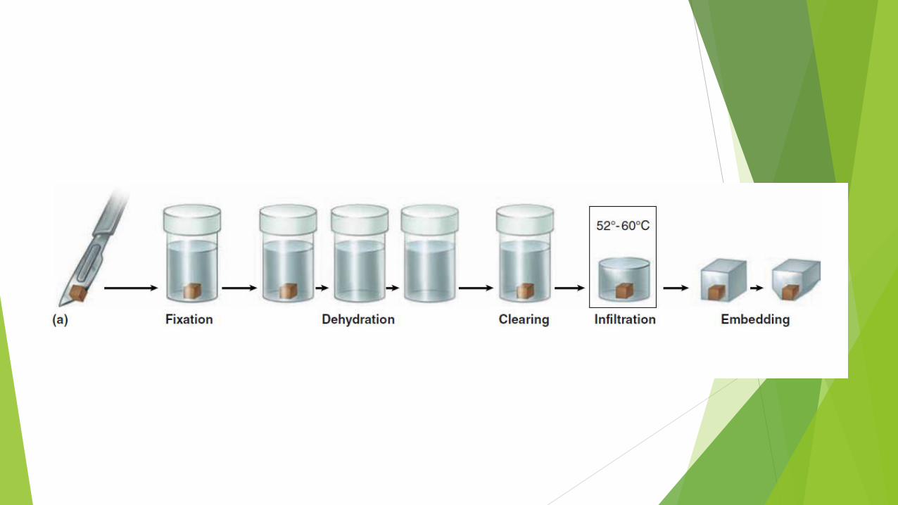
Fixation: Small pieces of tissue are placed in solutions of chemicals
that cross-link proteins and inactivate degradative enzymes, which
preserves cell and tissue structure (e.g. Formaldehyde for LM,
Gluteraldehyde for EM).
Dehydration: The tissue is transferred through a series of increasingly
concentrated alcohol solutions, ending in 100%, which removes all
water.
Clearing: Replacing the dehydrating fluid with a fluid that is totally
miscible with both the dehydrating fluid and the embedding medium
(e.g. Xylene for LM & propylene oxide for EM)
Infiltration: Replacing the clearing agent (inside the cell) with a
material that can harden to support biological tissue (e.g. paraffin
wax for LM & resin for EM)
Together
called tissue
processing
Tissue preparation for study-cont’d
Embedding: The infiltrated tissue is placed in a small mold and
allowed to harden.
Trimming: The resulting block is trimmed to expose the tissue for
sectioning (slicing) on a microtome.
Sectioning: slicing using microtome (for LM, and ultramicrotome for
EM).
Mounting: The process to place (mount) the tissue sections on the
adhesive coated glass slides (for LM, and grids for EM)
Staining: The stain is a chemical substance which reacts with certain
tissue components producing a color
Fixation
To preserve tissue structure and prevent degradation by
enzymes released from the cells or microorganisms,
pieces of organs are placed as soon as possible after
removal from the body in solutions of stabilizing or cross-
linking compounds called fixatives.
Because a fixative must fully diffuse through the tissues to
preserve all cells, tissues are usually cut into small
fragments before fixation to facilitate penetration.
Fixatives
One widely used fixative for light microscopy is formalin, a buffered
isotonic solution of 37% formaldehyde.
Both this compound and glutaraldehyde, a fixative used for electron
microscopy, react with the amine groups (NH2) of proteins, preventing
their degradation by common proteases.
Glutaraldehyde also cross-links adjacent proteins, reinforcing cell and
ECM structures.
Electron microscopy provides much greater magnification and
resolution of very small cellular structures and fixation must be done
very carefully to preserve additional “ultrastructural” detail.
Typically in such studies glutaraldehyde treated tissue is then
immersed in buffered osmium tetroxide, which preserves (and stains)
cellular lipids as well as proteins.

Embedding & Sectioning
To permit thin sectioning fixed tissues are infiltrated and
embedded in a material that imparts a firm consistency.
Embedding materials include paraffin, used routinely for
light microscopy, and plastic resins, which are adapted for
both light and electron microscopy.
Dehydration & Clearing
Before infiltration with such media the fixed tissue must
undergo dehydration by having its water extracted
gradually by transfers through a series of increasing
ethanol solutions, ending in 100% ethanol.
The ethanol is then replaced by an organic solvent
miscible with both alcohol and the embedding medium, a
step referred to as clearing because infiltration with the
reagents used here gives the tissue a translucent
appearance.
Infiltration & Embedding
The fully cleared tissue is then placed in melted paraffin in an oven at 52°-60°C, which evaporates the clearing solvent and promotes infiltration of the tissue with paraffin, and then embedded by allowing it to harden in a small container of paraffin at room temperature.
Tissues to be embedded with plastic resin are also dehydrated in ethanol and then infiltrated with plastic solvents that harden when cross-linking polymerizers are added.
Plastic embedding avoids the higher temperatures needed with paraffin, which helps avoid tissue distortion.
Sectioning
The hardened block with tissue and surrounding embedding
medium is trimmed and placed for sectioning in an instrument
called a microtome
Paraffin sections are typically cut at 3-10 μm thickness for
light microscopy, but electron microscopy requires sections
less than 1 μm thick.
The sections are placed on glass slides and stained for light
microscopy or on metal grids for electron microscopic staining
and examination.

N.B.
One micrometer (1 μm) equals 1/1000 of a millimeter
(mm) or 10–6 m.
Other spatial units commonly used in microscopy are the
nanometer (1 nm = 0.001 μm = 10–6 mm = 10–9 m) and
angstrom (1 A = 0.1 nm or 10–4 μm).
Medical Application
Biopsies are tissue samples removed during surgery or routine medical
procedures. In the operating room, biopsies are fixed in vials of
formalin for processing and microscopic analysis in a pathology
laboratory.
If results of such analyses are required before the medical procedure
is completed, for example to know whether a growth is malignant
before the patient is closed, a much more rapid processing method is
used.
The biopsy is rapidly frozen in liquid nitrogen, preserving cell
structures and making the tissue hard and ready for sectioning. A
microtome called a cryostat in a cabinet at subfreezing temperature
is used to section the block with tissue, and the frozen sections are
placed on slides for rapid staining and microscopic examination by a
pathologist.
Major differences in sample preparation
for TEM
Uses hard epoxy resin for embedding instead of paraffin wax
To improve contrast and resolution in TEM, compounds with
heavy metal ions are often added to the fixative or
dehydrating solutions used for tissue preparation. These include
osmium tetroxide, lead citrate, and uranyl compounds, which
bind cellular macromolecules, increasing their electron density
and visibility.
Sectioning is done through an ultramicrotome producing ultra-
thin sections
Sections are mounted on copper grids instead of glass slides
Staining
Most cells and extracellular material are completely
colorless, and to be studied microscopically tissue
sections must be stained (dyed).
Methods of staining have been devised that make various
tissue components not only conspicuous but also
distinguishable from one another.
Dyes stain material more or less selectively, often
behaving like acidic or basic compounds and forming
electrostatic (salt) linkages with ionizable radicals of
macromolecules in tissues.
Basophilic & acidophilic
Cell components such as nucleic acids with a net negative
charge (anionic) have an affinity for basic dyes and are
termed basophilic; cationic components, such as proteins
with many ionized amino groups, stain more readily with
acidic dyes and are termed acidophilic.
Light microscopy stains
The most commonly used stain in histological preparation
is Hematoxylin and Eosin (H&E) stain (a water soluble
stain).
Other special satins are common, such as:
Periodic Acid Sciff (PAS) stain for carbohydrates
Silver (Ag) for reticular fibers (collagen type III)
Masson’s Trichrome to distinguish collagen (Blue) from
muscle (Red)
Orcein for elastic fibers
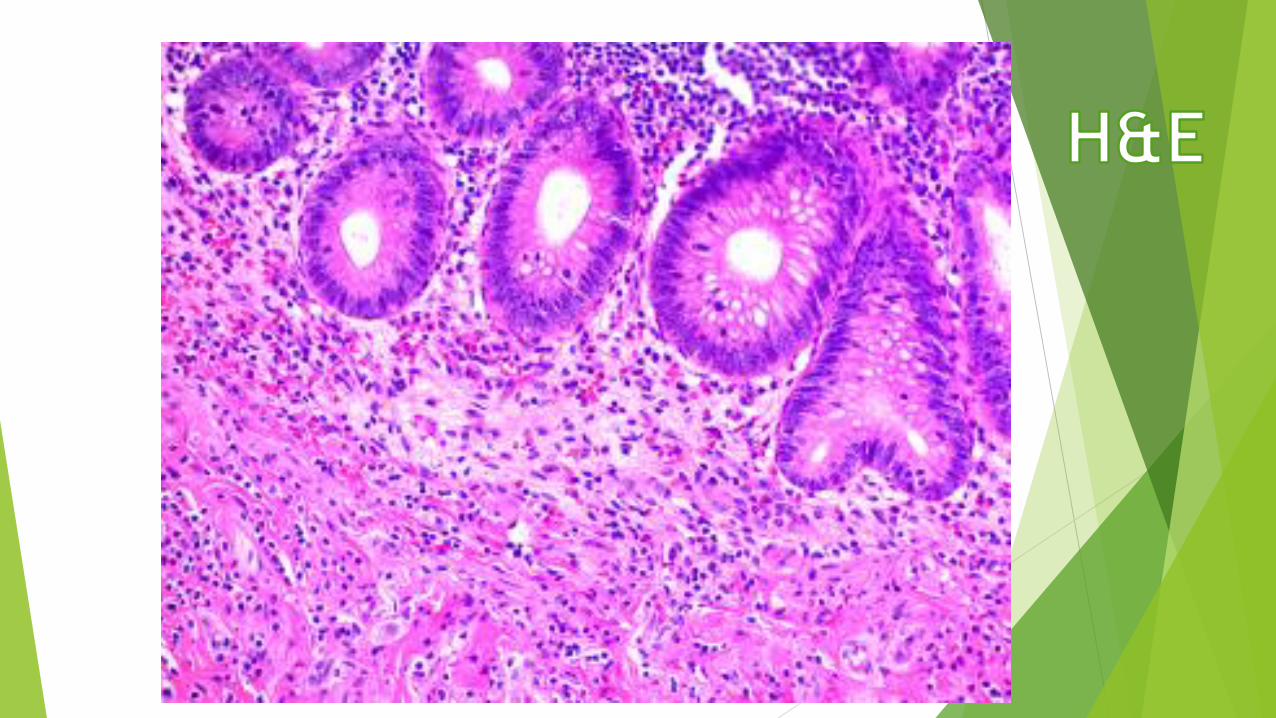
H&E
Hematoxylin
Basic dye
Has positive charge
Will stain negative
(basophilic) structures
BLUE
Examples: DNA, RNA,
ribosomes, rER, GAGs
Eosin
Acidic dye
Has negative charge
Will stain positive
(acidophilic, eosinophilic)
structures PINK
Examples: proteins,
collagen, cytoplasm,
mitochondria, secretory
granules
Lipid-rich compounds
Lipid-rich structures of cells are revealed by avoiding the
processing steps that remove lipids, such as treatment
with heat and organic solvents, and staining with lipid-
soluble dyes such as Sudan black, which can be useful in
diagnosis of metabolic diseases that involve intracellular
accumulations of cholesterol, phospholipids, or
glycolipids.
Visualizing specific molecules
A specific macromolecule present in a tissue section may also
be identified by using tagged compounds or macromolecules
that bind specifically with the molecule of interest. The
compounds that interact with the molecule must be visible
with the light or electron microscope, often by being tagged
with a detectible label.
The most commonly used labels are fluorescent compounds,
molecules of peroxidase or other enzymes that can be
detected with histochemistry, and metal (usually gold)
particles that can be seen with light and electron microscopy.
Examples
Phalloidin, a compound extracted from a mushroom, interacts
strongly with the actin protein of microfilaments.
Protein A, purified from Staphylococcus aureus bacteria, binds
to the Fc region of antibody molecules, and can therefore be
used to localize naturally occurring or applied antibodies bound
to cell structures.
Lectins, glycoproteins derived mainly from plant seeds, bind to
carbohydrates with high affinity and specificity. Different
lectins bind to specific sugars or sequences of sugar residues,
allowing fluorescently labeled lectins to be used to stain
specific glycoproteins or other macromolecules bearing specific
sequences of sugar residues.
Immunohistochemistry
A highly specific interaction between macromolecules is
that between an antigen and its antibody.
For this reason labeled antibodies are routinely used in
immunohistochemistry to identify and localize many
specific proteins
Every immunohistochemical technique requires an
antibody against the protein that is to be detected.
This means that the protein must have been previously
purified using biochemical or molecular methods so that
antibodies against it can be produced.
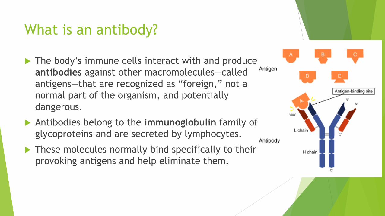
What is an antibody?
The body’s immune cells interact with and produce
antibodies against other macromolecules—called
antigens—that are recognized as “foreign,” not a
normal part of the organism, and potentially
dangerous.
Antibodies belong to the immunoglobulin family of
glycoproteins and are secreted by lymphocytes.
These molecules normally bind specifically to their
provoking antigens and help eliminate them.
How are antibodies produced?
To produce antibodies against protein x of a certain
animal species (eg, a human), the isolated protein is
injected into an animal of another species (eg, a rabbit or
a goat).
If the protein’s amino acid sequence is sufficiently
different for this animal to recognize it as foreign—that is,
as an antigen—the animal will produce antibodies against
the protein.
Polyclonal antibodies
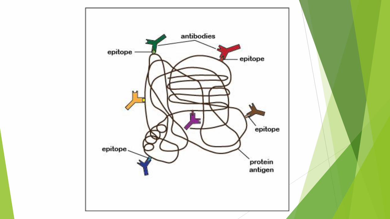
Different groups (clones) of lymphocytes in the injected
animal recognize different parts of protein x and each
clone produces an antibody against that part.
These antibodies are collected from the animal’s plasma
and constitute a mixture of polyclonal antibodies, each
capable of binding a different region of protein x.
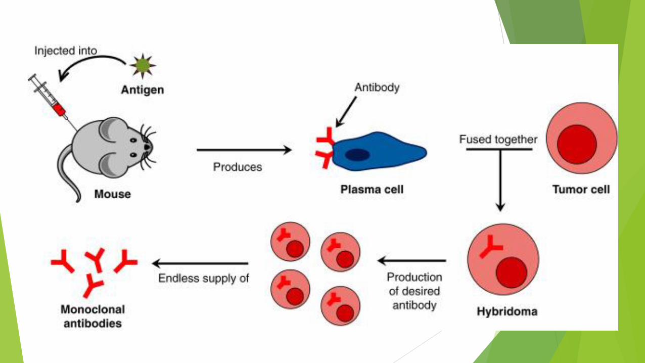
Monoclonal antibodies
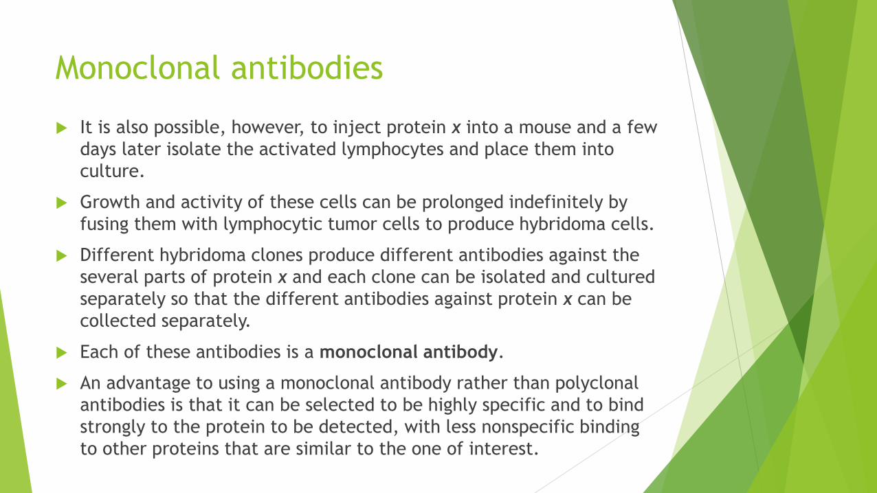
It is also possible, however, to inject protein x into a mouse and a few
days later isolate the activated lymphocytes and place them into
culture.
Growth and activity of these cells can be prolonged indefinitely by
fusing them with lymphocytic tumor cells to produce hybridoma cells.
Different hybridoma clones produce different antibodies against the
several parts of protein x and each clone can be isolated and cultured
separately so that the different antibodies against protein x can be
collected separately.
Each of these antibodies is a monoclonal antibody.
An advantage to using a monoclonal antibody rather than polyclonal
antibodies is that it can be selected to be highly specific and to bind
strongly to the protein to be detected, with less nonspecific binding
to other proteins that are similar to the one of interest.
Immunohistochemistry technique
In immunohistochemistry, a tissue section that one
believes contains the protein of interest is incubated in a
solution containing antibody (either monoclonal or
polyclonal) against this protein.
The antibody binds specifically to the protein and after a
rinse the protein’s location in the tissue or cells can be
seen with either the light or electron microscope by
visualizing the antibody.

Visualizing bound antibodies
Antibodies are commonly tagged with one of the
following:
fluorescent compounds
peroxidase or alkaline phosphatase (enzymes) for
histochemical detection
electron-dense gold particles for TEM.
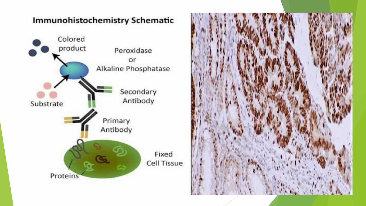
Direct & Indirect methods
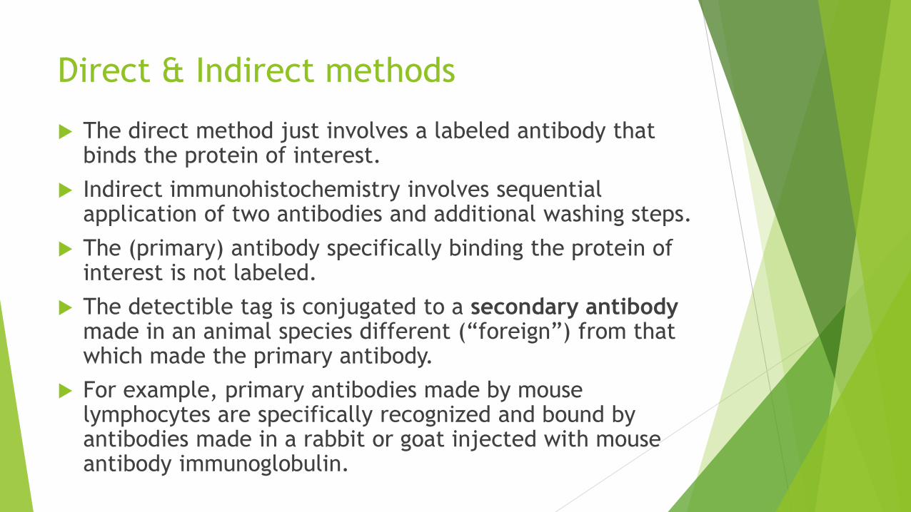
The direct method just involves a labeled antibody that binds the protein of interest.
Indirect immunohistochemistry involves sequential application of two antibodies and additional washing steps.
The (primary) antibody specifically binding the protein of interest is not labeled.
The detectible tag is conjugated to a secondary antibody made in an animal species different (“foreign”) from that which made the primary antibody.
For example, primary antibodies made by mouse lymphocytes are specifically recognized and bound by antibodies made in a rabbit or goat injected with mouse antibody immunoglobulin.
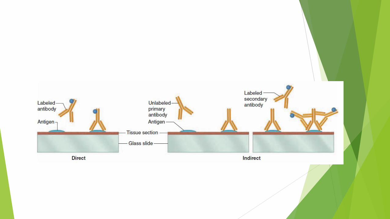
human
mouse
anti-human
goat
anti-mouse
Artifacts
In studying and interpreting stained tissue sections, it is
important to remember that microscopic preparations are
the end result of a series of processes that began with
collecting the tissue and ended with mounting a coverslip
on the slide.
Certain steps in this procedure may distort the tissues
slightly, producing minor structural abnormalities called
artifacts not present in the living tissue.
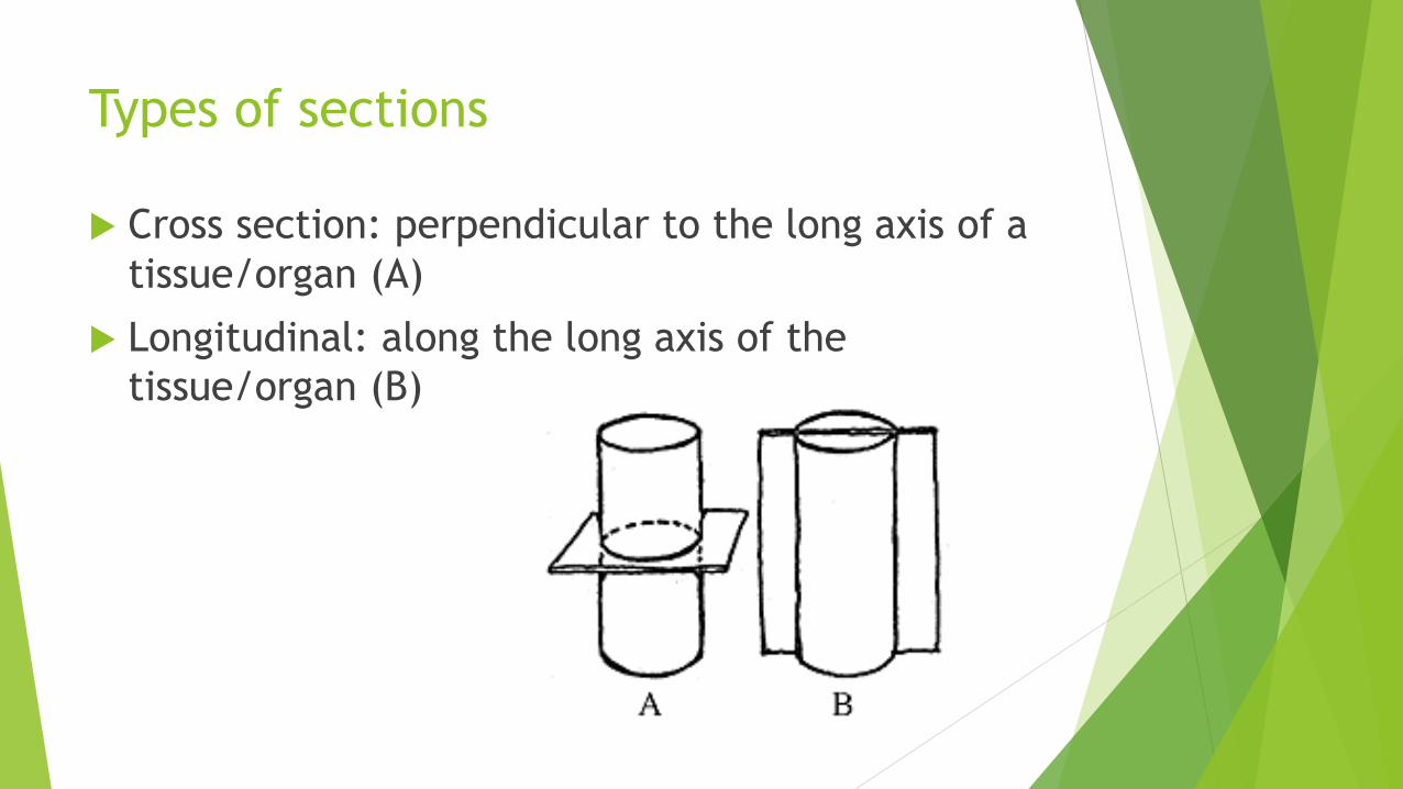
Types of sections
Cross section: perpendicular to the long axis of a
tissue/organ (A)
Longitudinal: along the long axis of the
tissue/organ (B)
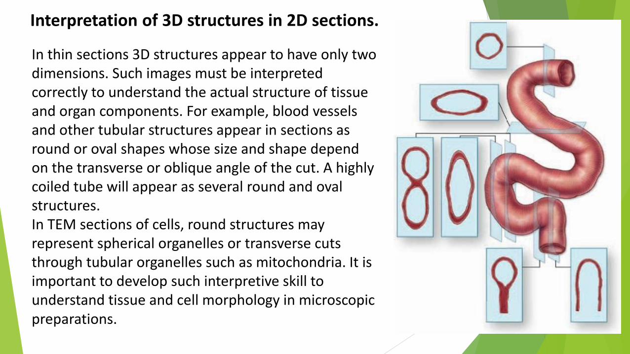
In thin sections 3D structures appear to have only two dimensions. Such images must be interpreted correctly to understand the actual structure of tissue and organ components. For example, blood vessels and other tubular structures appear in sections as round or oval shapes whose size and shape depend on the transverse or oblique angle of the cut. A highly coiled tube will appear as several round and oval structures. In TEM sections of cells, round structures may represent spherical organelles or transverse cuts through tubular organelles such as mitochondria. It is important to develop such interpretive skill to understand tissue and cell morphology in microscopic preparations.
Interpretation of 3D structures in 2D sections.