Salmonella Carriers Among Public Food Handlers In Khartoum ...
93
Salmonella Carriers Among Public Food Handlers In Khartoum Municipality By Hisham Hamedto Seed Ahmed Bachelor of Medical Science Omdurman Islamic University 1998 Supervisor Dr. Awad Elkarim A. Ibrahim B.V. Sc, M.Sc. & Ph.D. A thesis submitted for the partial fulfillment of the requirements for Master Degree In Microbiology Department of Microbiology Faculty of Veterinary Medicine University of Khartoum Feb.2007
Transcript of Salmonella Carriers Among Public Food Handlers In Khartoum ...

Salmonella Carriers Among Public Food Handlers
In Khartoum Municipality
By
Hisham Hamedto Seed Ahmed Bachelor of Medical Science
Omdurman Islamic University
1998
Supervisor
Dr. Awad Elkarim A. Ibrahim B.V. Sc, M.Sc. & Ph.D.
A thesis submitted for the partial fulfillment of the
requirements for Master Degree In Microbiology
Department of Microbiology
Faculty of Veterinary Medicine
University of Khartoum
Feb.2007

2
Dedication
To my parents,
Wonderful Brother and sisters,
Supportive wife,
Sweet heart sons and daughters
And all my teachers throughout my
life
With respect

3
ACKNOWLEDGEMENT
To begin with, my gratitude and praise are due to almighty
Allah, the Beneficent and the Merciful for the precious gift of health
and the capability to accomplish this work.
I am deeply indebted and thankfulness to my supervisor Dr.
Awad Elkarim Abdelghaffar Ibrahim for his priceless guidance, close
supervision, helpful, patience and continuous encouragement.
My special thank, are due to Al-Sahafa Medical Complex staff,
especially Faiza A. Al-Hassan my wife, Wafa A.Mokhtar, Sahar H.
Ali, Ihsan Abass, Prof.Yousif H., Nahid Alhag and my uncle
Mohammed Atta El-Manan for their tremendous support, scientific
discussion and kindness.
I would like to thank the Laboratories Administration staff
especially the Public Health Laboratory staff for their great help.
I am also grateful for all the staff of the Awad Hussein Hospital
and Ibrahim Malik Teaching Hospital laboratories especially Hussam
Mobark, Alfatih Babiker, Hind Osman, Hind Mobark and Eshragh for
endless support.
I am also extended my thanks to my colleagues Mohammed
Alfatih, Hitham Altigani, Moneer Nasir and Jalal for the efforts
exerted to push this work forward.
Finally my thanks extended to everybody who gave me his time,
support and help.

4
ABSTRACT
Enteric fever is a public health problem primarily in endemic
countries, where carriers, convalescent and sick persons spread the
enteric bacilli widely. The development of a permanent carrier state is
an important event in the life cycle of enteric fever bacilli. Our study
was conducted to screen the prevalence of Salmonella carriers among
food handlers working in food services establishments located in
various areas of Khartoum municipality during the period from Feb to
August 2006.
A total of 5493 food handlers were investigated during this
study, regarding their sociodemographic, professional characteristics,
and their hand washing practices were also observed.
Stool cultures for the isolation of Salmonella were carried out by
direct plating or after enrichment in two selective classical media (SS
agar and XLD agar media).
Only Salmonella enterica serotype Paratyphi B was isolated from 17
subjects during February, March & May. Peak of isolation was
registered in March (47.05%). Only two isolates (11.8 %) of
Salmonella Paratyphi B were recovered by direct plating on XLD and
S.S. agar media and 15 isolates (88.2 %) were recovered following
enrichment on Selenite-F broth. All isolates were identified to species
level using culture characteristics and biochemical reactions and
confirmed serologically. S.S. agar and XLD media were found to be
satisfactory for isolation of Salmonella from stool samples of food
handlers with the view we could optimize our routine protocol and to
assess food handlers bacteriologically with the minimum amount of

5
work and cost. Salmonella Paratyphi B was isolated from food
handlers working in various area studies, six carriers were detected at
Al Soug Al Mahali and Al Soug Al Markazi (35.3 %) as well as other
areas including Khartoum-2 and Riyadh areas. Fifteen isolates were
recovered from males (88.2 %) and only two from female subjects
(11.8 %). High prevalence of infection was recorded among
restaurateurs (58.8 %), hence sound Kitchen hygiene is essential for
the prevention of Salmonella infection and this apply equally to
commercially and house-hold kitchens.
This study has revealed a poor knowledge and practices of food
hygiene among food handlers and we recommend a massive health
education campaign directed at both the public and food handlers be
embarked on, to enable people to take necessary steps to prevent food-
borne diseases complementary to strict governmental regulations
empowerment as food handlers play an important role in maintaining
the endemicity of Salmonella infection in the community.

6
بسم اهللا الرحمن الرحيم
ملخص األطروحة
أحد مشاكل الصحة العامة سـيما فـي ) المعوية( تشكل حمى التايفويد
البالد التي يستوطن فيها هذا الداء حيث تنتشر عصيات التايفويد بشكل كبيـر
بواسطة حاملى البكتريا وكذلك مرضي التايفويد بل وأولئك الذين هم في طور
حالة وجود حامل دائم للعدوى واقعة هامة فـي دورة حيـاة النقاهة، وتعتبر
.عصيات حمى التايفويد
جرت هذه الدراسة بغرض مسح مدى انتشار حاملي بكتريا السالمونيال
في أوساط المتعاملين مع األطعمة في المؤسسات التي تقـدم األطعمـة فـي
أغـسطس مناطق مختلفة من محلية الخرطوم خالل الفترة من فبرايـر إلـى
من المتعاملين مع األطعمة خالل هذه الدراسـة 5493وقد تم تقصى .م 2006
فيما يتعلق بوضعهم االجتماعي السكاني وتخصصاتهم المهنية كما جرت ايضا
.مالحظة مدى اعتيادهم علي غسل أيديهم
قد تم أيضاً أستزراع برازهم لعزل السامونيال باالستنبات المباشـر أو
دي . ال . أكـس ( و ) أس أس أغـار ( في وسطين مختارين هما بعد اإلثراء
باستخدام ) ب(من أنماط نظير السالمونيال %) 18.8(وتم عزل اثنين ) . أغار
ــار ــط اغ ــي وس ــتزارع ف ــس ( و ) أس أس( االس 15و ) دي.ال . أك
).ف(بعد عملية اإلثراء في حساء الصفر %) 88.2(حالة
مـن الـذين 17النمطي مـن ) ب(يفويد ولم يتم عزل سوى نظير التا
خضعوا للدراسة خالل فبراير ومارس ومايو وسجلت اعلى نسبه خالل شـهر
من نظير السالمونيال من المتعاملين مع األطعمـة %) 47.05(مارس اذ بلغت
حيث تم اكتشاف ستة حاملين فـي ,في العديد من المناطق التي شملتها الدراسة
وغيرها من المناطق بما %) 35.3( المركزي مناطق السوق المحلي والسوق
مـن %) 88.2( حالـه 15كما تم عـزل . والرياض ) 2(في ذلك الخرطوم
من اإلناث الالئى خضعن للدراسة وقد بلغت نـسبة %) 11.8( الذكور وأثنين
%) 58.8(عمال المطاعم حداً كبيراً حدوث العدوى فـي أوسـاط v
7
ا كافة العناصر التي جرى عزلها وذلـك وقد تم تحديد الفصائل التي تنتمى إليه
باستخدام الخصائص المستنبته والتفاعالت الكيميائية الحيويه كماجرى تأكيدها
( وقد ثبت أن طريقة االستزراع باستخدام وسط األغار .بالفحوصات المصلية
هي طريقة ناجحة لعزل السالمونيال من براز ) دي. ال . أكس ( و ) أس أس
األطعمة لتقييم براز هؤالء بالفحوصات البكترولوجية باقل جهد المتعاملين مع
.وأقل تكلفة
وخلصت الدراسة بذلك إلى أهمية صحة المطبخ في الوقاية من عـدوى
.السالمونيال وينطبق ذلك بالمثل على المطابخ التجارية و المنزلية
وقد كشفت هذه الدراسة تدن مستوى معرفة وممارسة المتعـاملين مـع
ولهذا فأننا نوصى بقيام حملـة للتثقيـف . األطعمة في مجال الصحة الغذائية
الصحي تستهدف المتعاملين مع األطعمة على المستويين العام والخاص وذلـك
لتمكين المواطنين من اتخاذ اإلجراءات الهامة لمنع حدوث األمـراض التـي
كوميـة لمنـع تحملها األطعمة لتتكامل هذه الحملة مـع تنفيـذ الـضوابط الح
.المتعاملين مع األطعمة من نشر واستيطان عدوى السالمونيال في المجتمع

8
LIST OF CONTENTS
Subject Page
Dedication i
Acknowledgment ii
Abstract iii
Arabic Abstract v
List of Contents vii
List of Table xii
Introduction 1
Chapter One: Literature Review 3
1.1 History of Salmonella 3
1.2 Classification of Salmonella 3
1.3 Morphology of Salmonella 6
1.4 Antigenic structure 6
1.5 Epidemiology 8
1.5.1 Normal habitat 10
1.5.2 Source of infection 10
1.5.3 Transmission 10
1.5.4 Effect of Age 11
1.5.5 Chronic Carriers State 12
1.6 Pathology and pathogenesis 13
1.7 Clinical features 14
1.7.1 S. Typhi 14
1.7.2. S. Paratyphi A & B 15
1.7.3 S. Paratyphi C 15
1.7.4. Other Salmonellae 15
1.8. Laboratory diagnosis of Salmonella infections 15
1.8.1 Specimens 16
1.8.1.1. Blood 16

9
1.8.1.2 Faeces 17
1.8.1.3. Urine 17
1.8.2 Isolation and Identification 17
1.8.2.1 Cultivation of micro-organism 17
1.8.2.2. Culture characteristics 18
1.8.2.2.1. Simple and enriched media 18
1.8.2.2.2. Differential and selective media 18
1.8.2.2.3. Enrichment media 21
1.8.2.3. Biochemical reactions 22
1.8.2.3.1 Fermentation tests 22
1.8.2.3.2. Decarboxylase test 24
1.8.2.3.3. KIA test 24
1.8.2.3.4. Other biochemical tests 24
1.8.2.4. Identification 25
1.8.3. Serological tests 25
1.8.3.1. Widal test 25
1.8.3.2. Accu-chek S. typhi test 27
1.8.3.3. Immune fluorescence test 27
1.8.3.4. Polymerase Chain Reaction 28
1.8.4 Diazo urine test 28
1.9. Toxin production 28
1.10. Immunity 29
1.10.1. Antibodies in enteric fever 29

10
1.11. Treatment 31
1.12. Relapse of the disease 31
1.13. Drug Resistance 32
1.14. Prevention and control 33
1.15. Typhoid vaccine 34
Chapter Two: Materials and Methods 36
2.1. Study design 36
2.2. Sample size 36
2.3. Specimens collection 36
2.4. Culture media 36
2.4.1 Solid media 36
2.4.1.1. Salmonella - Shigella agar 36
2.4.1.2. Xylose Lysine Deoxycholate Agar (XLD) 37
2.4.1.3. Nutrient Agar 37
2.4.1.4. Kliger Iron Agar 37
2.4.1.5. Christensen’s Urea Agar 38
2.4.1.6. Simmon’s Citrate agar 38
2.4.1.7. Diagnostic sensitivity test (DST) 38
2.4.2. Semi-solid media 39
2.4.2.1. Motility media 39
2.4.2.2 Oxidation-fermentation (OF) media 39
2.4.3 Liquid media 39
2.4.3.1. Selenite F. broth 39
2.4.3.2. Peptone water 40
2.4.3.3. Peptone water sugar 40
2.4.3.4 Nutrient broth 40
2.4.3.5. Nitrate broth 40
2.4.3.6. Glucose phosphate peptone broth 41
2.5. Chemicals and reagents 41

11
2.6 Antibiotic Discs 43
2.7 Test control organism 43
2.8. Sterilization 43
2.9. Culture Methods 44
2.10 Purification of isolates 45
2.11. Identification of the isolated bacteria 45
2.11.1. Primary tests 45
2.11.2. Secondary tests 47
2.12. Serological tests 49
2.13 Sensitivity test 50
Chapter Three: Results 51
3.1 Culture characteristics. 51
3.1.1 Direct plating on solid media. 51
3.1.1.1 Salmonella Shigella agar. 51
3.1.1.2 Xylose Lysin Deoxycholate (XLD). 51
3.1.2 Enrichment media. 51
3.2. Primary tests. 51
3.2.1. Gram stain. 51
3.2.2 Primary biochemical tests. 52
3.3 Secondary tests. 52
3.4 Serological tests 52
3.5 Prevalence of Salmonella infection among food handlers. 52
3.5.1 The rate of Salmonella infection according to sex and
Nationality. 52
3.5.2 The rate of Salmonella infection according to
occupation 53

12
3.5.3 The rate of Salmonella infection among food handlers
according to areas. 53
3.6 Susceptibility of isolates to antibiotics. 54
3.7 Results of the questionnaire. 54
Chapter Four: Discussion 64
Conclusion 68
Recommendations 69
References 71
Appendix 81
13
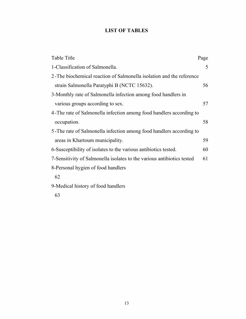
LIST OF TABLES
Table Title Page
1-Classification of Salmonella. 5
2 -The biochemical reaction of Salmonella isolation and the reference
strain Salmonella Paratyphi B (NCTC 15632). 56
3-Monthly rate of Salmonella infection among food handlers in
various groups according to sex. 57
4 -The rate of Salmonella infection among food handlers according to
occupation. 58
5 -The rate of Salmonella infection among food handlers according to
areas in Khartoum municipality. 59
6-Susceptibility of isolates to the various antibiotics tested. 60
7-Sensitivity of Salmonella isolates to the various antibiotics tested 61
8-Personal hygien of food handlers
62
9-Medical history of food handlers
63

14
INTRODUCTION
Enteric fever, a term frequently used to describe the prolonged
febrile state caused by Salmonella enterica serotype Typhi and
serotype Paratyphi A, B, C & Sendai, is the major public health
problem in many parts of the world.
Typhoid fever is a significant cause of morbidity and mortality world
wide, causing an estimated 16 millions cases and 600.000 deaths
annually (Kubota, et al. 2005). In addition, the genus Salmonella
comprises over 2500 other different serotypes, most of these serotypes
are primarily parasites of animals and, when transmitted to man cause
an acute diarrhoeal disease and often few serotypes cause systemic
illness having features characteristic of enteric fever. The typhoid and
paratyphoid bacilli are essentially human parasite and acquired
exclusively from human sources, namely patients and asymptomatic
carriers. Salmonella serotype Paratyphi B that primarily adapted to
man is occasionally isolated from secondary host including cattle,
swine, dog and fowl (Mandel, 1979). Thereby the enteric bacilli
contrast with other Salmonellae that cause food poisoning in man,
which are primarily parasite of other species of animals.
Transmission of the bacilli of enteric fever is through direct or
indirect contact with faeces or urine of a patient or asymptomatic
carrier. The principal vehicles of spread are contaminated water, food,
direct contact with hands, eating utensils and other fomites. Foods are
common vehicle for both typhoid and paratyphoid infection as well as
for food- poisoning Salmonellae.
In patients with enteric fever, positive stool cultures are quite
common during the immediate convalescent period and by the end of
the third month only 4-5% of patients continue to excrete the bacilli.

15
However, one year after infection, only 3% of the patients remains as
faecal excretors and are known as chronic faecal carries and are likely
remain so for the remainder of their lives. In such chronic carriers the
bacilli are almost commonly present in the gallbladder or rarely in the
urinary tract and excreted in faeces or urine. This long duration of
carrier state enables the enteric fever bacilli to infect relatively isolated
communities or families or a large population through food handlers
carriers. This is in contrast to patients who recovered from food-borne
Salmonella infection that usually continue to excrete the organism for,
on average, four to eight weeks (Buchwaled and Blaser, 1984).
In search for typhoid carriers this study was designed to fulfill
the following objectives:
1- To evaluate the prevalence of Salmonella carriers among food
handlers.
2- To isolate Salmonella from stool specimens by:
a- Direct plating.
b- Enrichment and plating.
3- To compare the classical plating media for primary isolation of
Salmonella from stool samples.
4- To evaluate antibiotic susceptibility of the various Salmonella
isolates.

16
CHAPTER ONE
LITERATURE REVIEW
1.1 History of Salmonella:
Before the nineteenth century, human enteric or typhoid fever was
often confused with typhus, a rickettsial disease. The two diseases
were pathologically distinguished by P. Ch. A. Louis in France (1829)
and William Jenner in the United States (1850). The typhoid bacillus
was first isolated in 1884, when the German microbiologist Gaffkey
obtained S.typhi from human spleen. S.choleraesuis was subsequently
isolated from the intestines of pigs infected with hog cholera in 1885
by the veterinary pathologist Daniel Salmon, after whom the
Salmonellae are named (Scherer and Miller, 2001).
Salmonellae are often pathogenic for human or animal when
acquired by the oral route. They are transmitted from animals and
animal products to humans, where they cause enteritis, systemic
infection, and enteric fever (Brook, et al. 2004).
1.2 Classification of Salmonella:
The genus Salmonella is a member of the family enterobacteria-
ceae. The members of the genus Salmonella were originally classified
on the basis of epidemiology, host range, biochemical reaction, and
structures of the O, H, and Vi antigens (Brook, et al. 2004).
1.2.1 Kauffmann-white classification:
Salmonellae Subcommittee (1943) proposed that serology was
the ultimate criterion in the classification of Salmonella (Barrow and
Feltham, 1993). Kauffmann-White Scheme for classification was first
developed in 1934 and it classifies Salmonellae into different O
groups or O serotypes, each of which contains a number of serotypes
possessing a common O antigen not found in other O groups. The O
groups, first defined were designated by capital letters A to Z and

17
those discovered later by the number of the characteristic O antigen
(David, et al. 1989).
In July, 1983 the Centre for Disease Control (CDC) changed the
method for reporting Salmonella serotypes; so that all organisms
identified as Salmonellae were reported as genus and serotype,
omitting reference to species (Koneman, et al. 1997). Within each
group, the differentiation of serotypes is carried out by the
identification of phase 1 and 2 flagellar antigens.
The Kauffmann- White Scheme gave species status to each
serotype, the genus Salmonella is subdivided into more than 2500
serotypes containing different combinations of antigens (Collee, et al.
1996).
Complete antigen analysis is not a routine procedure for clinical
laboratories, but reference laboratories are available in most countries
(Barrow and Feltham, 1993).
1.2.2 DNA-DNA hybridization classification:
DNA-DNA hybridization studies have demonstrated that there
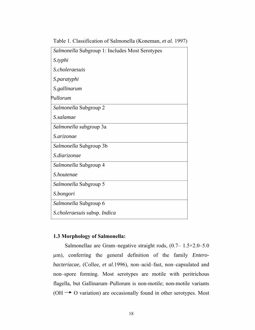
are seven evolutionary groups, as demonstrated in table (1) by
Koneman et al. (1997). Nearly all of the Salmonella serotypes that
infect humans are in DNA hybridization group I; there are rare human
infections with group IIIa and IIIb. There are more than 2500
serotypes of Salmonellae, including more than 1400 in DNA
hybridization group 1 that can infect human. Four serotypes of
Salmonellae that cause enteric fever can be identified in the clinical
laboratory by biochemical and serologic tests. These serotypes should
be routinely identified because of their clinical significance. They are
as follows: Salmonella Paratyphi A (serogroup A), Salmonella
Paratyphi B (serogroup B), Salmonella Choleraesuis (serogroup C1),
and Salmonella Typhi (serogroup D), (Brook, et al. 2004).
18
Table 1. Classification of Salmonella (Koneman, et al. 1997)
Salmonella Subgroup 1: Includes Most Serotypes
S.typhi
S.choleraesuis
S.paratyphi
S.gallinarum
Pullorum
Salmonella Subgroup 2
S.salamae
Salmonella subgroup 3a
S.arizonae
Salmonella Subgroup 3b
S.diarizonae
Salmonella Subgroup 4
S.houtenae
Salmonella Subgroup 5
S.bongori
Salmonella Subgroup 6
S.choleraesuis subsp. Indica
1.3 Morphology of Salmonella:
Salmonellae are Gram–negative straight rods, (0.7– 1.5×2.0–5.0
µm), conferring the general definition of the family Entero-
bacteriacae, (Collee, et al.1996), non–acid–fast, non–capsulated and
non–spore forming. Most serotypes are motile with peritrichous
flagella, but Gallinarum–Pullorum is non-motile; non-motile variants
(OH O variation) are occasionally found in other serotypes. Most

19
strain of most serotypes form type 1 fimbriae (mannose–sensitive,
haemoagglutinating); Gallinarun–Pullorum and a few strains in other
serotypes either form type 2 fimbriae (non– haemoagglutinating) or
are non-immediate; most strains of Paratyphi A are non – fimbriated
(Duguid, et al. 1966).
1.4 Antigenic Structure:
Although the principal varieties of enteric bacilli can be
identified by their reactions in differential media, final identification
of many species, as well as of strains, is based on antigenic structure.
However, strains with the same antigenic pattern may exhibit different
metabolic reactions (fermentative variants or biotypes), three kinds of
surface antigens (H, O, and K or Vi), determine the organisms
reaction with specific antisera (Davis, et al. 1990).
Salmonella, like other Gram-negative bacteria, has somatic (O)
antigens, which are lipopolysaccharide components of the cell wall,
and flagellar (H) antigens, which are proteins (Mandel, et al. 1985;
Collee, et al. 1996 and Brook, et al. 2004).
The designation H (Ger. hauch, breath) was first used to describe
the growth of Proteus bacilli on the surfaces of moist agar plates, the
film produced by the swarming of this highly motile organism
resembles the light mist caused by breathing on glass. The designation
O (Ger. ohne, without), first applied to nonswarming (i.e., non-
flagellated) forms, is now used as a generic term for the
lipopolysaccharide somatic antigens of all enteric bacilli and, more
specifically, for their polysaccharide components. Capsular antigens
are called K Ag (Ger. Kapsel), and a specific capsular Antigen of S.
Typhi is called Vi because of its role in virulence. Fimbrial antigens
previously designated as K Antigens before their structure was

20
recognized, and they now bear descriptive names (CFA I & II:
Colonization Factor Antigen I & II), Davis et al. (1990).
There are about 60 (O) antigens, and many H antigens, each of
which is designated by number and letter (Mandell et al. 1985;
Paniker and Vilma, 1997; Brooks et al. 2004).
Formaldehyde treatment preserves the labile Ags of the
flagella, and the cells are agglutinated by specific antiflagellar (anti-H)
Antibodies, forming a light, fluffy precipitate, the numerous
peritrichous flagella also prevent agglutination by anti-O Abs. On the
other hand, when flagella are absent, or are denatured by heat (100oC,
20 min), acid, or alcohol, the somatic O Ag at the surfaces of adjacent
bacilli can be linked by anti-O Abs, resulting in closely packed,
granular clumps.
The polysaccharide Vi Ag in certain species is usually too thin to
be seen as a capsule. However, it does inhibit O agglutination unless it
is destroyed (along with H-Ag) by boiling for 2 hours. The Vi Ag of S.
typhi is a homopolymer of N-acetylgalacto-saminuronic acid. Other Vi
Ags are found in other Salmonellae.
The O Ags in smooth (S) strains cover the underlying R Ag
which becomes accessible to Abs in rough (R) mutants. The change
from S to R may take place without the loss of flagellar or of Vi Ags.
Rough strains tend to agglutinate spontaneously unless suspended in
media of proper ionic strength ( Davis et al. 1990 ). The smooth-to-
rough variation is associated with change in colony morphology and
loss of the O antigen of virulence. The colony becomes large, rough,
and irregular. Suspension in saline is autoagglutinable. Conversion
into R forms occurs by loss of LPS. R forms may be common in
laboratory strains maintained by serial sub-cultivation (David et al.
1989).

21
Mucoid colonies, associates with the development of a new
mucoid or M antigen, have been described with S. Paratyphi B and
some other species (Ananthanarayan and Paniker, 1997).
Specific antisera, reacting with individual surface antigen in
agglutination tests, are prepared by selective adsorption, for example,
anti-H Abs may be removed by suspensions of homologous flagella
(mechanically removed) or by suspensions of mutant cells flagella but
neither their Vi nor O Ags. Similarly, anti-O or anti-Vi Abs may be
selectively removed by appropriate cell fractions (Davis et al. 1990).
1.5 Epidemiology:
There are two epidemiological types of typhoid fever. The first
is endemic residual typhoid that occurs through the year, though
seasonal variation may sometimes be apparent. The second is
epidemic typhoid, which may occur in endemic or non-endemic areas
irrespective of season.
Typhoid fever has been virtually eliminated from the advanced
countries during the last several decades mainly as a result of
improvements in water supply and sanitation but it continues to be
endemic in the poor nations of the world (Thong et al. 1998).It is
endemic in developing countries, in Africa, South Central America
and the Indian subcontinent (Threlfall and Ward, 2001).
The distribution of paratyphoid bacilli shows marked
geographical differences. S.Paratyphi A is prevalent in India and other
Asian countries, Eastern Europe and South America, S.Paratyphi B in
Western Europe, Britain and North America (Rowe et al. 1997).
The stool of persons who have unsuspected sub-clinical disease
or are carriers are amore important source of contamination than frank
clinical cases that are promptly isolated, e.g., when carriers working as
food handlers are “shedding” organisms. Many animals, including

22
cattle, rodents, and fowl, are naturally infected with a variety of
Salmonellae and have the bacteria in their tissues, excreta, or eggs.
The high incidence of Salmonellae in commercially prepared chickens
has been widely publicized. The incidence of typhoid fever has
decreased, but the incidence of other Salmonella infections has
increased markedly in the United States. The problem probably is
aggravated by the widespread use of animal feeds containing
antimicrobial drugs that favor the proliferation of drug-resistant
Salmonellae and their potential transmission to humans (Brook et al.
2004).
A few Salmonella serotypes are highly host adapted and tend to
be virtually “species specific”. For example, the only known reservoir
for S. typhi is man, and infection with the organism strongly implies
direct or indirect exposure to a human source (Mandell et al. 1985).
Persons with S/S haemoglobin (sickle cell disease) are
exceedingly susceptible to Salmonella infections, particularly
osteomyelitis. Persons with A/S haemoglobin (sickle cell trait) may be
more susceptible than normal individuals (those with A/A
haemoglobin, (Brook et al, 2004).
1.5.1 Normal habitat:
Most Salmonellae are found in the intestines of animals
especially of pigs, cows, goats, sheep, rodents, hens, ducks and other
poultry. S.Typhi and S.Paratyphi, however, are usually found only in
human (Cheesbrough, 2000).
1.5.2 Sources of Infection:
The sources of infection are food and drinks that have been
contaminated with Salmonellae. The following sources are important:
1- Water-contamination with faeces often results in explosive
epidemics.

23
2- Milk and other diary products (ice cream, cheese, custard)-
contamination with faeces and inadequate pasteurization or
improper handing. Some outbreaks are traceable to the source of
supply.
3- Shellfish- from contaminated water.
4- Dried or frozen eggs from infected fowl or contaminated during
processing.
5- Meats and meat products from infected animals (poultry) or
contamination with faeces by rodents or humans.
6- “Recreational drugs“- Marijuana and other drugs.
7- Animal dyes-Dyes (e.g., carmine) used in drugs, foods, and
cosmetics.
8- Household pets-Turtles, dogs, cats, etc. (Brook et al. 2004).
1.5.3 Transmission:
Infection is by ingesting the organisms in contaminated food
and water, or from contaminated hands. S.typhi is spreading mainly by
water and S.paratyphi A and B by food. Salmonellae are not killed by
drying and therefore survive in products such as dried egg or bone
meal fertilizers, (Cheesbrough, 2000).
The minimum infectious dose (ID50) for human pathogenic
serotypes in human volunteers is approximately 106 organisms.
However, single-food source outbreaks indicate that lower doses can
cause disease (Blaser and Newman, 1982; Mintz et al. 1994). In the
stomach Salmonellae are exposed to gastric acid and low pH, which
reduces the number of the viable organism. Most Salmonellae are
killed rapidly at pH 2, which is readily achieved in the normal
stomach (Brook, et al. 2004).
Some of the individuals who presented symptoms after small
inoculi may have impaired gastrointestinal defenses when compared
24
with human volunteers. Conditions that increase susceptibility to
lower inoculi include decreased stomach acidity (e.g. in infants, or due
to achlorhydia or antacid ingestion), chronic gastrointestinal diseases
such as inflammatory bowel disease, gastrointestinal surgery, and
alteration of the intestinal flora by antibiotic administration (Waddell
and Kunz, 1956; Giannella et al. 1972). Immunocompetency also
affects the ID50, as those with immune disorders, such as AIDS, are
approximately 20 times more likely to contract symptomatic
salmonellosis than the general population, even when presented with a
low infectious dose (Scherer and Miller, 2001).
1.5.4 Effect of Age:
Age is an important determinant of the disease produced by
Salmonellae. The influence of age on incidence may be attributed to
the immunity of humoral and cellular immune mechanisms,
diminished antibacterial action of the normal intestinal flora, high
frequency of faecal oral contamination, or other factors. In some
instances, increasing resistance with age is related to immunity
consequent to previous exposure to the organism, even though the
disease has not been imposed (Khan et al. 1998).
1.5.5 Chronic Carriers State:
After manifestation of clinical infection, some individuals
continue to harbour Salmonellae in their tissues for variable length of
time (convalescent carriers or healthy permanent carriers).Three
percent of survivors of typhoid become permanent carriers, harbouring
the organism in the gallbladder, biliary tract, or rarely, the intestine or
urinary tract.(Brook et al. 2004) . Non-typhoidal serotypes on average
persist in the gastrointestinal tract, depending on the serotype, from 6
weeks to 3 months. However, persistence beyond 6 months is rare.
Only about 0.1% of non-typhoidal Salmonella cases are shed in stool
25
samples for periods exceeding 1year, the clinical definition of chronic
carriage. Approximately 2-5% of untreated typhoidal infections result
in a chronic carrier state (WHO, 1997). Bacteria can persist in several
reservoirs, including the urinary tract and bile duct (Kaye et al. 1967).
Factors contributing to the chronic carrier state have not been fully
elucidated, although anatomical abnormalities such as gallstone and
kidney stones have been implicated. Some individuals can harbour
asymptomatic infections without their knowledge, often from low
initial inoculums (Goldberg and Rubin, 1988). The most famous
chronic carrier is Mary Mallon (Typhoid Mary), a New York City
cook, who was held responsible for transmitting typhoid fever to at
least 22 individuals (3 of whom died) between the years 1900 and
1907, (Leavitt, 1996). After being apprehended in 1907 by Public
Health Officials, she was confined to an isolation cottage for 3 years.
Although she was released with the stipulation that she should never
cook again , she was unable to keep this promise, and is thought to be
responsible for at least 25 more cases of typhoid fever at a Manhattan
Maternity Hospital, where she was employed as a cooker in 1915. She
was subsequently confined to the isolation until her death in 1938.
1.6 Pathology and pathogenesis
Ingested bacteria that survive the low pH of the stomach
quickly colonize the lumen of the small intestine, where they
predominantly localize to the Peyer’s patches (Carter and Collins
1974; Kohbata et al. 1986). Passage of Salmonella through the
intestinal epithelial barrier most likely occurs through specialized
microfold-enterocytes, which overlay the Peyer’s patches (Brandtzaeg,
1989). The primary function of M cells is to sample intestinal antigens
(Neutra et al. 1996). This function is aided by the characteristic
“pocket” containing lymphocytes and macrophages that extend from

26
the basolateral layer into the M cell and allow for transcytosis of
phagocytosed antigens and particles to the immune cells (Scherer and
Miller, 2001). Invasive Salmonellae enter M cells via a process that
induces membrane ruffling and endocytosis and gain access to the
underlying lymphatic tissue either by transcytosis across the M cell or
by lysing the M cell (Clark et al. 1994; Penheiter et al. 1997).
The main pathological changes are found in the gastrointestinal
tract. The Peyer's patches show hyperplasia during the first week of
typhoid, necrosis during the second week and ulceration during the
third weak. Healing takes place without scaring during the fourth
week. The ulcer is oval in the long axis of the lower ileum. There are
exudates on the peritoneal surface. Separation of the sloughs may lead
to haemorrhage and perforation. The incidence of ulcer bears no
relationship to the clinical severity of the infection. The liver shows a
cloudy swelling and typhoid nodules, which are small lesions
consisting of collection of macrophages and lymphocytes with or
without central necrosis (El-faki, 1987).
1.7 Clinical features:
Salmonella developed different clinical feature according to their
different species:
1.7.1 S.Typhi
This species causes the following conditions:
a- Typhoid (enteric) fever. The bacteria pass from the small intestine
into the blood by way of the lymphatic system. The
reticuloendothelial system becomes infected and also the gall bladder
and kidneys. From the gall bladder, the organisms invade the intestine
causing inflammation and ulceration. Symptoms of infection include
fever with low pulse rate, headache, toxaemia, enlargement of the
spleen, and apathy or mental confusion. A rash (rose spots) may be

27
seen on light coloured skin. Epistaxis, intestinal haemorrhage and
perforation may also occur. In uncomplicated typhoid, the total white
cell count is low with a relative lymphocytosis. There may also be
anaemia.
b- Nephrotyphoid in those with urinary schistosomiasis. The condition is
an immune complex disorder of the kidneys and is characterized by
fever, oedema, marked albuminuria, and haematuria.
c- Osteomyelitis (inflammation of the bone marrow), especially in
children with sickle cell disease and thalassaemia. Typhoid nodules
can be found in the bone marrow. Inflammation of the joints (typhoid
arthritis) may also occur.
d- Abscesses of the spleen and elsewhere.
e- Meningitis and rarely pneumonia and endocarditis (Cheesbrough,
2000).
1.7.2 S. Paratyphi A and B:
These salmonellae cause paratyphoid (enteric fever). The disease
is generally milder than typhoid, with S.paratyphi A and B being less
invasive than S.typhi. There is usually diarrhoea and vomiting and the
entire intestinal tract may be inflamed especially in S.paratyphi B
infections. In tropical and other developing countries, paratyphoid is
more commonly caused by S. Paratyphi A than S. Paratyphi B
(Scherer and Miller, 2001).
1.7.3 S.Paratyphi C:
This serovar causes mainly septicaemia. Complications of S.
Paratyphi C infections include the formation of abscesses, arthritis,
and inflammation of the gall bladder. S. Paratyphi C occurs mostly in
Eastern Europe (Kohbata et al. 1996).
1.7.4 Other Salmonellae:

28
Several thousands Salmonella serovars are capable of causing
food-poisoning (enterocolitis). Symptoms of Salmonella food-
poisoning occur within 10-30 hours of ingesting the contaminated
food. Some food-poisoning strains can also cause bacteraemia,
inflammation of the gall bladder, osteitis (inflammation of the bone)
especially in children with sickle cell disease, and occasionally
abscesses (Cheesbrough, 2000).
1.8 Laboratory diagnosis of Salmonella infections.
Laboratory diagnosis of Salmonella infections depends mainly
on the isolation and identification of Salmonella from a specimen of
the patient’s blood, faeces, urine or vomit. Testing the patient’s serum
for Salmonella antibodies is useful only in the diagnosis of enteric
fever (Widal reaction) and even for this condition the significance of
the results of the test is often doubtful. The Widal test is not
recommended on a routine basis in areas of low endemicity. For the
diagnosis of pyrexial illnesses, physicians should be advised to submit
blood cultures and faeces to the laboratory and not to rely on the
serological tests.
Isolation of Salmonella by blood culture is a proof that the patient
has a Salmonella septicaemia. Isolation from the faeces is of less
certain significance. In illnesses resembling enteric fever or
gastroenteritis such isolation strongly suggests that Salmonella
infection is the cause, but since Salmonellae may be present in the
faeces of carriers it does not amount to proof of a causal role.
The clinical value of identifying the serotype of a Salmonella
isolate lies in distinguishing the serotypes that cause enteric fever in
man, namely Typhi, Paratyphi A, Paratyphi B and Sendai, from the
other serotypes, which in man commonly cause gastroenteritis (food-
poisoning) but rarely septicaemic infections. The value of identifying

29
serotype of‘non-enteric-fever’ Salmonella, is mainly epidemiological
and knowledge of the serotype helps to define the sources and vehicles
of infection in outbreaks of food poisoning. Phage typing and
biotyping may be used to obtain more precise information for this
purpose (Collee et al. 1996).
1.8.1 Specimens
For the diagnosis of enteric fever, specimens include blood,
faeces, and urine.
1.8.1.1 Blood:
Organisms can usually be detected in 75-90% of patients during the
first ten days of infection, and in about 30% of patients during the
third week. In chronic Salmonellosis, it has been reported that S. typhi
can be more rapidly and successfully isolated from bone marrow than
from blood, especially if the patient had been treated with antibiotics
(Khan et al.1998).
1.8.1.2 Faeces:
Organisms can usually be isolated from 40-50% of patients during
the second week of infection and from about 80% of patients during
the third week (Stokes et al.1993).
1.8.1.3 Urine:
Organisms can usually be isolated form about 15% of patients after
the second week of infection especially from those with urinary
schistosomiasis. The bacteria are not excreted continuously and
therefore several specimens may need to be cultured before the
organisms are isolated (Ochei and Kolhatkar, 2000).
When an abscess or arthritis is suspected, pus or joint fluid is
required for culture.

30
1.8.2 Isolation and identification:
The faeces is cultured on plates of one or more kinds of
selective media, both directly and after preliminary culture in a liquid
enrichment medium, and the plates are observed for the presence of
Salmonella like colonies. A well-separated colony is picked to obtain
a pure culture and the pure culture is identified first by a selection of
biochemical tests and finally by agglutination tests with specific
antisera (Koneman et al. 1997).
1.8.2.1 Cultivation of micro-organism:
Salmonellae are aerobic and facultative anaerobic, grow on simple
laboratory media in the temperature range of 15-45ºC, optimally at
37ºC. Many strains are prototrophic, that is capable of growing in a
glucose–ammonium minimal medium such as that of Davis &
Mingioli, but some strains are auxotrophic and require enrichment of
the minimal medium with one or more amino acids or vitamins e.g.
cysteine or nicotinamide; most Typhi strains require tryptophan
(Collee et al. 1996).
1.8.2.2 Culture characteristics:
1.8.2.2.1 Simple and enriched media:
1.8.2.2.1.1 Nutrient Agar and Blood Agar:
After 24h at 37ºC, colonies of most strains are moderately large
(2-3 mm in diameter), grey–white, moist, circular disc with a smooth
convex surface and entire edge, thus resembling the colonies of many
other enterobacteria. Their size and degree of opacity varies with the
serotype, e.g. those of Paratyphi A, Abortusovis, Pullorum, Sendai and
Typhisuis are relatively small. 'Rough', non-virulent variant strains
(S R variation) form opaque granular colonies with an irregular
surface and indented edge. Many strains of Paratyphi B and few of
other serotypes form large mucoid colonies or colonies surrounded by

31
a thick mucoid "slime wall", when plates are left at room temperature
for a few days after incubation for 24h at 37ºC. The mucoid character
is due to the formation of loose polysaccharide slime (Collee et al.
1996).
1.8.2.2.1.2 Peptone Water and Nutrient Broth.
In liquid media most strains give abundant growth with uniform
turbidity. A thin surface pellicle usually forms on prolonged
incubation. 'Rough' (R) variants, which have hydrophobic surface
and tend to auto-agglutinate, produce a granular deposit and
sometimes a thick pellicle (Murry et al. 2005).
1.8.2.2.2 Differential and selective solid media:
These media are valuable for the isolation of Salmonellae from
faeces and other materials contaminated with many bacteria of other
kinds. They include.
1.8.2.2.2.1 MacConkey Bile-salt Lactose Agar.
After 18-24h at 37ºC the colonies are pale yellow or nearly
colourless, 1-3 mm in diameter, and easily distinguished from the
pink–red colonies of lactose fermenting commensal coliform bacilli,
e.g. Escherichia coli which also grow well on this unselective
differential (indicator) medium (Koneman et al. 1997).
1.8.2.2.2.2 Brilliant Green MacConkey Agar.
The addition to MacConkey agar of brilliant green 0.004g/litre,
which is inhibitory to E.coli, proteus species and the other commensal
enterobacteria likely to outnumber the Salmonellae in faeces, makes
this an excellent selective as well as differential medium for
Salmonellae except Typhi which does not grow well on it.
Salmonellae appear as low convex, pale- green translucent colonies
1-3 mm in diameter. Lactose fermenting bacteria, including rare

32
strains of Salmonella serotypes, produce blue- purple colonies (Murry
et al. 2005).
1.8.2.2.2.3 Leifson's Deoxycholate-Citrate Agar (DCA).
The colonies of Salmonellae on DCA are similar to or slightly
smaller in size than those on MacConkey agar. They are pale, nearly
colourless, smooth, shiny and translucent. Sometimes they have a
black center and sometimes a zone of cleared medium surrounds
them, but these characters may require 48 hours of incubation for their
development. Salmonellae are easily distinguished from the opaque
pink colonies of lactose– fermenting coliform bacilli, which are
largely inhibited on this selective differential medium (Cheesbrough,
2000).
1.8.2.2.2.4 Wilson and Blair's Brilliant-green Bismuth
Sulphite Agar (BBSA).
This medium is particularly valuable for the isolation of Typhi.
Cultures should be examined after 24h, then again after 48h. Crowded
colonies about 1mm in diameter may take up the dye from the
medium and appear green or pale brown. Larger, discrete colonies
have a black centre and a clear edge. All Salmonellae may produce
hydrogen sulphide, which causes the colony to be surrounded by a
metallic sheen (Barrow and Feltham, 1993).
1.8.2.2.2.5 Taylor's Xylose Lysine Deoxycholate (XLD).
It is a popular medium for the primary plating of faeces from
suspected Salmonella and Shigella infection. It gives colony
appearances that distinguish Salmonellae from Shigellae , and these
pathogens from the many non- lactose fermenting strains of non-
pathogenic enterobacteria which form pale colonies similar to theirs
on MacConkey and DCA .Colonies of Salmonellae and Shigellae are
red (alkaline to phenol red ) because Shigellae don’t form acid from

33
the xylose , lactose and sucrose in the medium within 24h and because
Salmonellae neutralize the acid they form from the limited amount
of xylose by decarboxylating the lysine . Most Salmonellae (and
Edwardsiellae) are distinguished from the Shigellae because they
produce hydrogen sulphide, which reacts with ferric ammonium
citrate in the medium to produce black centres in their red colonies
(Cheesbrough, 2000).
1.8.2.2.2.6 Rambach's Agar.
This recently described medium (1990), which contains
propylene glycol (PG) and a novel chromogenic substrate (Merck) to
detect β -galactosidase activity, is claimed to allow detection of 98%
of 'customary' Salmonellae. Non-typhoidal Salmonellae ( β -galacto-
sidase-negative) form acid from the metabolism of PG and, with a
suitable pH indicator, grow as red colonies. (Dusch and Altwegg,
1995).
1.8.2.2.2.7 SM-ID agar.
On this medium (Bio Meraux), colonies of Salmonella, including
Typhi and Paratyphi strains, give red colonies because they form acid
from glucuronate and are β -galactosidase-negative (Collee et al.
1996).
1.8.2.2.2.8 Salmonella-Shigella Agar.
It is a selective and differential medium used for the isolation
and differentiation of Salmonella and Shigella from clinical specimens
and other sources. The nutritive base contains animal and casein
peptones and beef extract. The selective agents are bile salts, citrates,
and brilliant green dye, which inhibit gram-positive organisms.
The high degree of selectivity of the medium results in the
inhibition of some strains of Shigella, and the medium is not
recommended as a primary medium for isolation of this species. The

34
medium contains only lactose and thus differentiates organisms on the
basis of lactose fermentation. The formation of acid on fermentation
of lactose causes the neutral red indicator to make red colonies. Non-
lactose fermenting organisms are clear on the medium. As with
Hektoen enteric agar, sodium thiosulfate and ferric ammonium citrate
allow the differentiation of organisms that produce hydrogen sulphide.
Lactose fermenters, such as E. coli, have colonies, which are pink with
a precipitate, Shigella appears transparent or amber, and Salmonella
appears transparent or amber with black centers. Some strains of
Shigella dysenteriae are inhibited (Murray et al. 2005).
1.8.2.2.3 Enrichment media:
These are liquid media used to assist the isolation of
Salmonellae from faeces, sewage, foodstuffs and other materials
containing a mixed bacterial flora. A larger amount of the material can
be inoculated into an enrichment medium than onto an agar plate, so
facilitating the isolation of Salmonellae when these are present only in
small numbers. During incubation, any Salmonellae multiply rapidly,
while E.coli and most other bacteria are inhibited. After 18-24h the
enriched culture is plated onto a differential agar medium e.g. DCA or
XLD, on which the production of Salmonella-like colonies may be
observed (Barrow and Feltham, 1993).
1.8.2.2.3.1 Selenite broth:
Selenite broth is an enrichment broth medium used for the
isolation of salmonella and shigella species. Casein and meat peptones
provide nutrients. Selenite inhibits enterococci and coliform those are
part of the normal flora if they are subcultured within 12 to 18 hrs.
However, reduction of selenite produces an alkali condition that may
inhibit the recovery of Salmonella. Lactose and phosphate buffers are
added to allow stability of the pH. When fermenting organisms

35
produce acid, the acid neutralizes the effect of the selenite reduction
and subsequent alkalinization. Cystine added to selenite broth
enhances the recovery of Salmonella (Murray et al.2005).
1.8.2.3 Biochemical reactions:
All serotypes of the genus Salmonella and those of the former
genus ‘Arizona’ are now considered to belong to one species for
which the name Salmonella enterica has been proposed (Le Minor, et
al.1985), it comprises seven subspecies which, historically, have been
numbered but now are named. Most (99.5%) of the Salmonellae
isolated from man in developed countries are serotypes of subspecies I
(Le Minor et al. 1985) which also contain serotypes commonly
infecting other mammals.
1.8.2.3.1 Fermentation tests.
Carbohydrates are generally fermented with the production of
acid and gas. Typhi, Gallinarum and rare anaerogenic variants in other
serotypes, e.g. Typhimurium, form only acid. Typically, glucose,
mannitol, arabinose, maltose, dulcitol and sorbitol are fermented, but
not lactose, sucrose, salicin or adonitol; the ONPG test for β -
galactosidase is negative. Among exceptional strains, Choleraesuis
and some strains of Typhi do not ferment arabinose, whereas
Choleraesuis, the Pullorum biotype of Gallinarum, most strains of
Typhi and some strains of Paratyphi A and Paratyphi B do not ferment
dulcitol.
Most strains fermenting a particular sugar give ‘strong’
fermentation; they show acid production in sugar peptone water within
6-10 hrs at 37ºC and can grow on a defined medium with the sugar as
sole source of carbon and energy. Some strains, deficient in the uptake
mechanism, give ‘weak’ fermentation, showing acid production in
sugar peptone water only after 10-20 hrs and fail to grown on a

36
defined medium with the sugar as sole carbon and energy source.
Thus, biotypes 1-16 of Typhimurium give strong fermentation of D-
xylose and biotypes 17-32 weak fermentation (Duguid et al. 1975).
Non-fermenting strains may give rise to fermenting mutants and show
acid production in a proportion of sugar peptone water cultures after
incubation for two or more days; they might be misread as fermenters
unless the definitive reading time of 24 hrs is adhered to.
The fermentation of D-tartrate is observed to distinguish the
Java biotype (positive) of Paratyphi B (negative) and between
biotypes of Typhimurium. The reaction is not demonstrable by acid
production, but by the promotion of growth due to utilization of the
tartrate. The D-tartrate dehydrase of positive strains is oxygen
sensitive and the test should be done under the poorly aerobic
conditions of static culture in a deep tube of D-tartrate peptone water,
in which positive strains give several times as much growth and
turbidity as negative ones ( Duguid et al, 1975; Barker 1985).
Alternatively, positive reactions may be demonstrated by the
appearance of growth on a defined D-tartrate medium in plates
incubated anaerobically for 2 days (Barker, 1985).
1.8.2.3.2 Decarboxylase tests.
Salmonellae decarboxylate the amino acids lysine, ornithine and
arginine, but not glutamic acid. Typhi is exceptional in lacking
ornithine decarboxylase and Paratyphi in lacking lysine decarboxylase
(Collee et al. 1996).
1.8.2.3.3 KIA test.
This medium is used to help identify Salmonellae following
isolation on a primary selective medium (Cheesbrough, 2000),
Salmonellae produce:

37
- Pink-red (alkaline) slope and yellow (acid) butt, indicating
fermentation of glucose but not lactose.
- Cracks in the medium if serotype produces gas from glucose
fermentation (S. typhi does not produce gas).
- Blackening in the medium due to H2S unless serotype does not
produce H2S, e.g. S. Paratyphi A. Only a small amount of blackening
is seen with S. typhi.
1.8.2.3.4 Other biochemical tests.
Most Salmonellae have the following reactions:
Indole not produced. Methyl-red positive. Acetyl methyl carbinol
not produced (i.e. Voges-Proskauer negative). Citrate utilized, except
by Typhi and Paratyphi A. Malonate not utilized. Gluconate not
utilized. Urease not produced. Phenylalanine deaminase not produced.
Hydrogen sulphide produced in ferrous chloride gelatin medium,
except by Paratyphi A, Choleraesuis, Typhisuis and Sendai. No
growth in KCN medium. Gelatin not liquefied.
1.8.2.4 Identification:
The identification of Salmonella species can be carried out
through biochemical investigation. Then the corresponding serotype is
determined by the specific serotyping using the agglutination test. The
recently developed genetic methods using deoxyribonucleic acid
(DNA) provide important tools for accurate specification.
1.8.3 Serological diagnosis:
Tests for the presence of Salmonella antibodies in the patient’s
serum may be of value in the diagnosis of enteric fever, but are of
little help in that of Salmonella food poisoning (Le Minor et al. 1985).
1.8.3.1 Widal test:
Detection of anti S.typhi antibodies in patients is a useful
diagnostic aid. Amongst the various methods developed over the years

38
for this purpose the Widal test, based on bacterial agglutination, has
remained the most widely used, even though it is neither specific nor
sensitive. It is popularly used due to the fact that it is simple to use and
inexpensive (Lim et al. 1998).
This test for the serological diagnosis of enteric fever was
much used in the past when effective selective media were not
available and it was more difficult to isolate the causal, Salmonella
species, from the patient’s blood or faeces (Collee et al. 1996). In
developing countries, where typhoid fever is endemic and where there
are few microbiology laboratories to provide diagnosis by culture, the
search continues forth for non-cultural techniques that provide rapid
and reliable diagnosis.
1. 8.3.1.1 Interpretation of Widal test results:
The value of Widal test in diagnosing enteric fever in endemic
areas remains controversial, some express the view that the test lacks
standardization and adequate sensitivity to be clinically useful, while
others consider the test to have a diagnostic value when judge of
association with clinical findings and knowledge of the normal O and
H agglutinins titre in local population. Of shared concern, however, is
now necessary to avoid misuse and misinterpretation of Widal test
(Cheesbrough, 2000). In typhoid fever the H titre is elevated earlier
and more frequently than the O titre and antibodies rise during the
second week of illness. It appears that H titre is more useful than O
titre.
Antibiotic treatment did not affect the rise of antibody titre in
typhoid fever (Abraham et al. 1981). In Sudan, a titre of 1:160 or
more for O somatic antigen is considered as evidence of typhoid fever
(Elfaki, 1987).

39
The features significantly associated with a final diagnosis of
typhoid fever were: pre-admission duration of fever (longer than or
equal 7 days), hepatomegaly, leucopenia due to absolute neutropenia
with relative lymphocytosis, although the sensitivity, specificity and
predictive value of any of these features can not be used reliably to
distinguish typhoid fever from other non typhoidal febrile illnesses
(Khan et al. 1998).
Causes of raised O and H titres other than active typhoid
These include pervious Salmonella infections, chronic
Salmonellosis associated with schistosomal infection, vaccination with
TAB or typhoid vaccine, current infection with other Salmonella
species, chronic liver disease associated with raised globulin levels,
and disorders such as rheumatoid arthritis, rheumatic fever, multiple
myeloma, nephrotic syndrome, and ulcerative colitis. Following
vaccination, H antibody titre remains elevated for 6 months or longer
(Lim et al. 1998).
1.8.3.2 Accu-chek S.typhi antigen test.
Accu-chek S.typhi is a rapid (10-15 minutes) immuno-
chromatographic (IC) test recently developed by Millennium
biotechnology Inc. to detect S. typhi antigen in faecal specimens. The
test is sensitive and highly specific using two monoclonal antibodies
for S.typhi. It becomes positive as soon as S.typhi becomes present in
the faeces, usually in the first week of the disease. About 0.5 g of
faeces is mixed with extraction reagent. The faecal particles are
allowed settle and the faecal extract is then passed through the sample
well of the testing device which contains an antibody-coloured

40
colloidal gold complex. S.typhi antigen attaches to the complex and
migrates a long the membrane where it becomes captured by a line of
polyclonal antibody, forming a pink/purple band in the test area of the
viewing window. A further pink/purple line, i.e. inbuilt positive
control, is produced above the test line indicating that the test has
performed satisfactorily (Cheesbrough, 2000).
Accu-chek S. typhi does not cross-react with S.paratyphi or other
Salmonella species. The test is highly stable.
1.8.3.3 Immunofluorescence test:
Antisera from rabbits immunized with the synthetic disaccharide
covalently linked to bovine serum albumin (BSA), were used in indirect
immunofluorescence studies for the identification of Salmonella
serogroup A (O-antigen 1, 2, 12) bacteria. This test have a high
specificity of the antiserum elicited by immunization with the synthetic
disaccharide-protein immunogen (Svenungsson and Blinberg ,1978).
1.8.3.4 Polymerase chain reaction (PCR):
A simple and ready-to-go test based on a 5' nuclease (TaqMan)
PCR technique was developed for identification of presumptive
Salmonella enterica isolates. DNA testing methods, such as PCR, can
circumvent the phenotypic variations seen in both biochemical
patterns and the lack of detectable antigens (Mullis et al, 1986).
PCR-based diagnosis is particularly useful for all clinically
suspected cases of typhoid fever. The sensitivity of PCR and its
potential use in routine diagnosis and epidemiological studies of
typhoid fever can be exploited to complement studies by including
bone marrow culture, faeces and bile samples (Kumar et al. 2002).
1.8.4 Diazo urine test:

41
The diazo reaction of urine has been reported as a useful
screening test for typhoid when cultural facilities are not available.
The test is positive in about 80% of patients between the fifth and
fourteenth day of infection, it is based on the reaction of diazo reagent
with a substance formed by the putrefaction of protein in the intestine.
(Cheesbrough, 2000).
1.9 Toxin production:
All members of the family enterobacteriacae possess an
endotoxin. This toxin is responsible for endotoxic shock in Gram-
negative bacterial septicaemia (Ochei and Kolhatkar, 2000).
Salmonella typhi contains gluc-lipo-protein complexes. The endotoxin
is obtained by extracting the bacterial emulsion with trichloroacetic
acid. This endotoxin is thermo stable, survives at a temperature of
120ºC for 30 minutes, and it characterized by a highly specific
precipitin reaction and pronounced toxic and antigenic properties.
Investigations have shown the presence of exotoxic substances in S.
typhi, which are inactivated by light, air and heat (80ºC), as well as
enterotropic toxin phosphatase and pyrogenic substances (Pyatkin and
Krivoshein, 1987).
1.10 Immunity:
The microorganism is an example of facultative intracellular
parasite, surviving well within macrophages and requiring cell-
mediated immunity for control (Youmans et al, 1986). After the onset
of illness, the cell-mediated immunity persists for 16 weeks
(Sarasombath et al. 1987).
Infections with Salmonella Typhi or Salmonella Paratyphi
usually confer a certain degree of immunity. Reinfection may occur
but is often milder than the first infection. Circulating antibodies to O
and Vi are useful for diagnosis. However, relapses may occur in 2-3

42
weeks after recovery in spite of antibodies. Secretary IgA antibodies
may prevent attachment of Salmonellae to intestinal epithelium. It can
persist in the gut for about 48 weeks. Thus, the immunity as a whole
can persist beyond one year after the onset of illness, unless there are
persistent booster stimulation S.typhi bacilli that exist in the
environment, where, then, the immunity may be lifelong (Sarasombath
et al. 1987).
Antibiotics, used as therapeutic agents, inhibit the immunogenic
activity toward the pathogens, which change rapidly and lose their O
and Vi antigens (Pyatkin and Krivoshein, 1987).
1.10.1 Antibodies in enteric fever:
Salmonella causing enteric fever stimulates the formation of
three types of agglutinins, which are of use in diagnosis; they are H, O
and Vi. The anti-H antibody, which is produced in response to
stimulation by flagellar antigens, appears towards the end of the first
week of the disease, and usually reaches the highest titre. It persists
longer than the others after recovery, sometimes for many years, and
its formation may again be stimulated, non-specifically, in subsequent
febrile illnesses. Agglutination is rapid and the agglutinated bacilli
form large fluffy clumps. There is no evidence that anti-H antibody is
protective or helps to combat the disease (Stokes, et al. 1993).
Formation of anti-O antibody is stimulated by somatic antigens
(O). It also appears in the first week of illness and seldom rises above
a titre of about 1:640 (Curtis et al. 1983; Stokes et al. 1993). Anti-O
polysaccharide chain antibody titre is lower at the first week and
increase up to the third week of the infection (Mastroianni et al.
1989). After recovery the titre falls and it is seldom demonstrable a
year later. Production is not easily stimulated, non-specifically, in

43
subsequent illnesses. Agglutination occurs more slowly and the
clumps of the bacilli are small and dense (Stokes et al. 1993).
The Vi antigen, also, a somatic antigen, is possessed by
Salmonella Typhi, S. Paratyphi C and other coliform, including
certain strains of E. coli but not S.Paratyphi B. Typhoid and
paratyphoid bacilli, which possess large quantities of this antigen,
often give rise to severe disease (Stokes et al. 1993).
Occasionally, only Vi antibody can be investigated but that can
happen in rare cases. However, in most cases there is no need to
perform the Vi agglutination test. The antibody is probably of value in
combating infections of Vi strains.
In the majority of patients H and O agglutinins can be easily
demonstrated. Occasionally only one of the agglutinins is found, and
very occasionally no specific antibody is demonstrable in enteric fever
(Stokes et al. 1993).
1.11 Treatment:
While enteric fevers and bacteremias with focal lesions require
antimicrobial treatment, the vast majority of cases of enterocolitis do
not. Antimicrobial treatment of Salmonella enteritis in neonates is
important. In enterocolitis, clinical symptoms and excretion of the
Salmonellae may be prolonged by antimicrobial therapy. In severe
diarrhoea, replacement of fluids and electrolytes is essential.
Antimicrobial therapy of invasive Salmonella infections is with
ampicillin, trimethoprim-sulfamethoxazole, or a third-generation
cephalosporin. Multiple drug resistance transmitted genetically by
plasmids among enteric bacteria is a problem in Salmonella infections.
Susceptibility testing is an important adjunct to selecting a proper
antibiotic.

44
In most carriers, the organisms persist in the gallbladder
(particularly if gallstones are present) and in the biliary tract. Some
chronic carriers have been cured by ampicillin alone, but in most cases
cholecystectomy must be combined with drug treatment (Brook et
al.2004).
1.12 Relapse of the disease:
Apparent recovery can follow by relapse in 5-10% of untreated
cases. Relapse is usually shorter and of milder character than the
initial illness, but can be severe and may end fatally. Severe intestinal
haemorrhage and intestinal perforation are serious complications,
which can occur at any stage of the illness (Greenwood, et al.2004).
Relapse also can occur with antimicrobial therapy, in several
studies, the relapse rate was found to be doubled in patients receiving
chloramphenicol therapy for 2 weeks. Ampicillin probably does not
affect the rate of relapse. Studies of patients given trimethoprim-
sulphamethoxole (co-trimoxazole) indicate that the rate of relapse may
be relatively less than the patients treated with chloramphenicol
(Pyatkin and Krivoshein, 1987).
1.13 Drug Resistant:
The recent emergence of Salmonellae carrying stable
resistance of multiple clinically relevant antibiotics is a significant
health problem worldwide. Antibiotic resistance of S. Typhi has been
an issue since 1950, when strains resistant to chloramphenicol were
isolated in Great Britain, just only 2 years after the successful use of
chloramphenicol in treatment of typhoid fever (Colquhoun and
Weetch, (1950); Anderson and Smith (1972)). Currently, S. Typhi
isolates resistant to six different antimicrobial agents prevail in highly
endemic typhoid areas, particularly China, Pakistan, and India. These
strains of S. Typhi carry a 120-kb plasmid that encodes resistance to
45
ampicillin, chloramphenicol, streptomycin, sulfonamides, tetracycline,
and trimethoprim. In addition, S. Typhi strains isolated from recent
outbreaks in Tadjikistan and Pakistan have also acquired resistance to
ciprofloxacin (a fluoroquinolone), one of the preferred antibiotics for
treatment of typhoid fever (Hampton et al. 1998). As all of the above
antibiotics, except streptomycin and tetracycline, are clinically
relevant for the oral treatment of typhoid fever. The existence of
hepta- resistant strains is a serious health problem. In addition , multi-
antibiotic- resistant ( MAR) typhoid has been a significant cause of
death in children; the mortality rates of children infected with MAR S.
Typhi range form 7 to 16% compared to a rate of 2% for children
infected with antibiotic susceptible strains of Salmonella (Gupta,
1994). Resistance of nontyphoidal Salmonellae is also a growing
health problem. Particularly troubling is the penta-resistant strains of
S.Typhimurium known as DT104 (Definitive phage type 104), which
emerged in Great Britain in 1984 and was reported in 1997 to have
been isolated in the United States (CDC, 1997). This strain has been
isolated from numerous species of animals (wild and farm animals)
and is resistant to ampicillin, chloramphenicol, streptomycin,
sulfonamides, and tetracycline (R type ACSSu T). In addition, there
have been reports of resistance to two other antibiotics, trimethoprim
and fluoroquinolones, in Great Britain (Threlfall et al. 1996). The
veterinary use of antibiotics, such as ciprofloxacin and trimethoprim,
to treat DT104 infections in cattle has been proposed as a factor in the
acquisition of resistance to these (and other) antibiotics by
Salmonellae. Interestingly, resistance to ciprofloxacin has not been
observed in the United States yet, possibly because fluoroquinolones
are only licensed for use in poultry, where S.typhimurium carring DT
104 may not yet be established as a pathogen (Glynn et al. 1998).

46
Reports documenting the emerging resistance to fluoroquinolones by
Salmonellae are worrisome, as ciprofloxacin is the antibiotic of choice
for treating human Salmonellae, resistance to this drug will leave few
available options. In addition to the problematic treatment of DT104
infections, there are reports indicating that DT104 may be more
virulent for humans. In a study performed in the United Kingdom,
41% of patients infected with DT104 were hospitalized, and 3% of
culture- confirmed patient's death, compared to an average death rate
of 0.1% (Wall et al. 1994).
1.14 Prevention and Control:
Sanitary measures must be taken to prevent contamination of
food and water by rodents or other animals that excrete Salmonellae.
Infected poultry, meats, and eggs must be thoroughly cooked. Carriers
must not be allowed to work as food handlers and should observe
strict hygienic precautions.
1.15 Typhoid Vaccines:
As new antibiotics are still in the developmental stage, it is also
important to continue the effort to produce effective vaccines for
typhoid fever. Three vaccines are currently available for typhoid fever,
none of which are 100% effective even when tested on endemic
populations (CDC, 1998). The live attenuated Ty21a strain
(manufactured by the Swiss Serum and Vaccine Institute), which is
orally administered, requires at least four doses to achieve 51-76%
protective efficacy (Levine et al. 1987). The heat and phenol
inactivated typhoid vaccine (manufactured by Wyeth) has similar
efficacy, and requires at least two doses (by injection). However, this
vaccine has a variety of severe local and systemic side effects that
limit its use. Finally, the cell-free, parenteral Vi-antigen vaccine
47
produced by Pasteur Merieux (ViCPS) requires only a single does, but
most likely has a shorter duration of protection.
A number of new live strains have been evaluated in human
volunteers for their vaccine potential. Live attenuated vaccine strains
may be preferable for typhoid fever because they can induce a wide
spectrum of protective immunity, including mucosal, humoral, and
cell- mediated immunity.
Ideal vaccine strains should be genotypically stable (containing at
least two nonreverting deletions) and should offer long-term
protection after one or two initial doses.
Other more promising vaccine strains include aro deletion mutants in
poultry (Hoiseth and Stocker, 1981; Tacket et al. 1992), crplcya
deletion mutants (Tacket et al. 1992; CurtissIII and Kelly, 1987) and
the phoP/phoQ deletion mutants in human (Hohmann et al. 1995;
Hohmann et al. 1996).
The phop/PhoQ null strain is highly immunogenic after a single
does in human volunteers. This strain is also promising because it
does not persist for long periods of time in vivo, produces very few
side effects, and has been shown to be more immunostimulatory
compared to wild type (Wick et al, 1995).
Although there is little requirement for human vaccines for
nontyphoidal Salmonellae, as the infections are usually self-limiting,
various vaccines for use in animals have been developed. Such
vaccines would be quite useful if they cloud eliminate the source of
most Salmonella infections without increasing the potential for
additional antibiotic resistance. Both aro-negative (Hassan and Curtiss
III, 1997) and crplcya mutant (Coloe et al. 1995) S. Typhimurium
strains have been shown to be effective vaccine strains in chickens.

48
These strains offer long-term protection against infection with both
S.Typhimurium and S. Enteritidis. In addition immunization with the
crplcya strain in ova (up to 7 days before hatching) protected chicks
from infection (Coloe et al. 1995), a fact that is particularly important
in light of the ability of S. enteritidis to colonize intact shell eggs.
CHAPTER TWO
MATERIALS AND METHODS
2.1 Study design:
This study was carried out in Khartoum municipality (Riyadh,
Amarat, Doyoum, Arkwit, Soba,Gerief, Emtedad, Sahafa and Soug-
Mahali and Markazi). The period of the study was from Feb to Aug
2006. The target population was food handlers working in food
service establishment located in the Khartoum municipality.
2.2 Sample size:
5493 stool specimens were collected from food handlers
working in the target area of study having different occupations
(Restaurateurs, Shopkeepers, Factotums, Butchers, Veg/Fruit Retailers
and Bakers).
2.3 Specimens collection:
Freshly passed stool samples were collected from food handlers
working in food services in stool containers and cultures were done
immediately after collection.
2.4 Culture media;
Different types of culture media including solid, semi-solid and
liquid media were used to isolate and identify the enteric bacteria. All
culture media were prepared according to the methods described by
the manufacturer.

49
2.4.1 Solid media
2.4.1.1 Salmonella Shigella Agar (SSA) (Oxoid Ltd., England):
It consists of (grams per litre) lab-lemco powder 5.0 grams,
peptone 5.0 grams, bile salts 5.5 grams, sodium thiosulphate 8.5
grams, Lactose 10 grams, sodium citrate 10 grams, ferric citrate 1.0
grams, neutral red 0.025 gram, brilliant green 0.00033 gram and agar
12 grams. It was prepared by dissolving of fifty-seven grams of the
ready dehydrated medium in one litre of distilled water by heating.
The pH was adjusted to 7.1, cooled to 55ºC and distributed aseptically
into sterile Petri-dishes, 20 ml in each dish.
2.4.1.2 Xylose Lysine Deoxycholate Agar (XLD) (Oxoid Ltd.,
England):
It consists of (grams per litre) yeast extract powder 3.0
grams, L-lysine Hcl 5.0 grams, Xylose 3.75 grams, lactose 7.5 grams,
sucrose 7.5 grams, sodium deoxycholate 1.0 gram, sodium chloride
5.0 grams, sodium thiosulphate 6.8 grams, ferric ammonium citrate
0.8 gram, phenol red 0.08 gram and agar (No.1) 12.5 grams. Fifty-
three grams of dehydrated medium were dissolved in one litre of
distilled water by heating, the pH was adjusted to7.2, then cooled
to55ºC and dispensed aseptically in sterile petri-dishes.
2.4.1.3 Nutrient Agar (Oxoid Ltd., England):
It consists of (grams per litre) lab-lemco powder 1.0 grams,
yeast extract 2.0 grams, peptone 5.0 grams, sodium chloride 5.0 grams
and agar 15 grams. Twenty-eight grams of dehydrated medium were
dissolved in one litre of distilled water; the pH was adjusted to 7.4,
then was sterilized by autoclaving (121ºC for 15 minutes), cooled to
50-55ºC and then poured into sterile petri-dishes, 20ml each.
2.4.1.4 Kligler Iron Agar (KIA) (Oxoid Ltd., England):

50
It consists of (grams per litre) lab-Lemco powder 3.0 grams,
yeast extract 3.0 grams, peptone 20 grams, sodium chloride 5.0 grams,
lactose 10 grams, dextrose 1.0 gram, ferric citrate 0.3 gram, sodium
thiosulphate 0.3 gram, phenol red 0.05 gram and agar 12 grams.
According to manufacturer, 55g of dehydrated medium were dissolved
in 1 litre of distilled water by boiling, and then the medium was
poured into test tubes 5 ml each. The tubes were sterilized by
autoclaving (121ºC for 15 minutes), and allowed to cool in a sloped
position to give a butt and slant in each tube.
2.4.1.5: Christensen’s Urea Agar (Oxoid Ltd., England):
The medium was composed of (gram per litre) peptone 1.0 gram,
dextrose 1.0 gram, sodium chloride 5.0 grams, disodium phosphate
1.2 grams, potassium dihydrogen phosphate 0.8 gram, phenol red
0.012 gram and agar 15 grams. According to the manufacturer
instructions, 2.4 grams of dehydrated medium were dissolved in 95 ml
of distilled water by boiling, pH was adjusted to 6.8, sterilized by
autoclaving at 115ºC for 20 minutes, then cooled to 50ºC and
aseptically 5ml of sterile 40% urea solution were added. The medium
was poured into sterile screw-capped bottles 10 ml each, and then
allowed to set in the slope position.
2.4.1.6: Simmon’s Citrate agar (Oxoid Ltd., England):
It consists of (grams per litre) 0.2 gram of magnesium sulphate,
ammonium dihydrogen phosphate 0.2 gram, sodium ammonium
phosphate 1.0 gram, sodium citrate 2.0 grams, sodium chloride 5.0
grams, bromo-thymol blue 0.08 gram and agar 15 grams. Twenty
three grams of dehydrated Simmon's citrate agar were suspended in
one litre of distilled water, boiled to dissolve completely, the pH was
adjusted to 7.0 and sterilized by autoclaving at 121ºC for 15 minutes.

51
It was then poured into sterile screw-capped bottles and allowed to set
in slope position.
2.4.1.7 Diagnostic Sensitivity Test (DST) Agar (Oxoid Ltd., England):
The medium was composed of 10 grams protease peptone, 10
grams veal infusion solid, 2.0 grams disodium phosphate, 2.0 grams
dextrose, 3.0 grams sodium chloride, 1.0 gram sodium acetate, 0.01
gram uracil, 0.01gram adenine sulphate, 0.00002 gram aneurine, 12
grams 'ionagar', 0.01 gram guanine hydrochloride and 0.01 gram
xanthine. Forty grams of dehydrated medium were dissolved in 1 litre
of distilled water by boiling, the pH adjusted to 7.4, the medium was
sterilized by autoclaving (121ºC for 15 minutes), cooled to 50ºC and
then the medium was poured into sterile petri- dishes 20ml each.
2.4.2 Semi-solid media:
2.4.2.1 Motility media (Oxoid Ltd., England):
This media prepared by adding 13 g of Nutrient broth to 7.5 g
of Oxoid agar No. 1 and dissolved in 1 litre of distilled water by
heating to 100ºC. The pH was adjusted to 7.2 and poured into test
tubes (U shape). The tubes were sterilized by autoclaving (121ºC for
15 minutes), then the media were cooled to use for motility test.
2.4.2.2 Oxidation-Fermentation (O-F) medium:
The oxidation fermentation medium was used to test the ability
of organism to break down the carbohydrates either oxidatively or
fermentatively.The medium was composed of 2.0 grams peptone, 5.0
grams sodium chloride, 0.3 gram Di-potassium hydrogen phosphate,
3.0 grams agar and 0.2% aqueous solution of bromocresol purple. The
ingredients were boiled to dissolve completely in one litre of distilled
water, the pH was adjusted to 7.1, filtered and the indicator was added
and then sterilized by autoclaving at 121ºC for 15 minutes. Ten ml of
10% sterile glucose solution was added aseptically to 90 ml of the

52
medium to give a final concentration of 1%, mixed and 10ml amounts
were aseptically distributed into sterile test tubes, (Barrow and
Feltham., 1993).
2.4.3 Liquid media
2.4.3.1 Selenite F- broth (Oxoid Ltd., England):
According to manufacturer, the medium was prepared by
dissolving 5.0 grams peptone, 4.0 grams mannitol, 10 grams di-
sodium hydrogen phosphate and 4.0 grams sodium hydrogen Selenite
in one litre of distilled water, the pH was adjusted to 7.0 and sterilized
by steaming for 20 minutes, mixed well and dispensed into sterile
international containers to depth 5 cm.
2.4.3.2 Peptone water (Oxoid Ltd., England):
This medium was used as base of carbohydrates utilization tests
and for other purposes. It was composed of 10 grams peptone, 5.0
grams sodium chloride. It was prepared by dissolving 15 g of the
powder in 1 litre of distilled water; the pH was adjusted to 7.4. The
media was poured into clean test tubes 5 ml each, and sterilized by
autoclaving 121º C for 15 minutes.
2.4.3.3 Peptone water sugar:
The solution of the specific sugar was prepared by dissolving
10 grams of sugar in 90 ml of distilled water and then in 900 ml of
peptone water. Ten ml of Andrade's indicator was added and mixed
well. The medium was distributed into sterile test tubes with sterile
Durham’s tubes; the pH was adjusted to 7.1 and sterilized by
autoclaving at 115ºC for 10 minutes. The carbohydrates used in this
medium were glucose, lactose, sucrose, maltose and manitol.
2.4.3.4 Nutrient Broth (Oxoid Ltd., England):
It was composed of 1.0 gram lab-lemco powder, 2.0 grams yeast
extract, 5.0 grams peptone and 5.0 grams sodium chloride. It was

53
prepared by dissolving 13 g of the powder into 1 litre of distilled water
and the pH was adjusted to 7.4. The medium was poured into clean
test tubes and then sterilized by autoclaving at 121º C for 15 minutes.
2.4.3.5: Nitrate Broth:
According to Barrow and Feltham (1993), this medium was
prepared by dissolving one gram of potassium nitrate in 1 litre of
nutrient broth, distributed into test tubes and then sterilized by
autoclaving at 121º C for 15 minutes.
2.4.3.6 Glucose Phosphate Peptone Broth:
This medium was prepared according to Barrow and Feltham
(1993). Five grams of peptone and 5 g of potassium phosphate were
added to 1 litre of distilled water and steamed until the solid were
dissolved. The pH was adjusted to 7.5, and then 5 g of glucose were
added and mixed, the medium was poured into test tubes 5 ml each
and sterilized by autoclaving at 115º C of 10 minutes.
2.5 Chemicals and Reagents:
All reagents were prepared according to manufacture and kept
in well-closed glass stoppard bottles and protected from direct sun
light.
2.5.1 Gram's stain solutions:
These reagents were prepared according to Barrow and Feltham
(1993).
2.5.1.1 Crystal violet
This reagent was prepared by dissolving crystal violet powder
in distilled water to give 1% concentration.
2.5.1.2 Lugol’s iodine
This was prepared by dissolving 20 grams of potassium iodide
in 50 ml of distilled water and then 10 grams of iodine were added by
shaking and the volume was adjusted to 100ml.

54
2.5.1.3 Decolourizing reagent:
This was prepared by mixing 475 ml of absolute ethanol, 25 ml
of distilled water in 500 ml of acetone.
2.5.1.4 Counter stain:
This stain was prepared by dissolving 10 grams basic fuchsin,
50 grams phenol and 100 ml absolute ethanol in 1 litre of distilled
water, then the stain was diluted (1:10) in distilled water to use as
counter stain.
2.5.2 Methyl red solution:
This solution was prepared by dissolving 0.04 grams of methyl
red powder in 40 ml of ethanol and the volume was diluted with
distilled water to 100ml (Barrow and Feltham., 1993).
2.5.3 Hydrogen Peroxide (BDH, England):
It was prepared as a 3% aqueous solution and used for catalase
test (Barrow and Feltham., 1993).
2.5.4 Voges- Proskauer (V.P) test reagent:
This reagent was prepared by mixing 40% potassium hydroxide
(KOH) with 5% alpha-naphthol in absolute ethanol.
2.5.5 Nitrate test reagents:
These reagents were composed of two solutions:
Solution A: 0.8% sulphanilic acid in 5N- acetic acid, prepared by
dissolving by gentle heat.
Solution B: 0.6% dimethyl – alphanaphthylamine in 5N-acetic acid.
2.5.6 Oxidase reagent:
This reagent was prepared by dissolving 0.1 gram tetramethyl-
p-phenylene diamine dihydrochloride in 10 ml distilled water,
(Barrow and Feltham, 1993). It’s prepared immediately before use
because it easily oxidized.
2.5.7. Physiological Saline:

55
According to Barrow and Feltham (1993), nine grams of solid
chloride dissolved in 1 litre of distilled water.
2.5.8. Kovac’s reagent:
This reagent was prepared by dissolving 5 grams of para-
dimethylaminobenzaldehyde in 75 ml amyl alcohol by gentle warming
in a water bath, about 50–55ºC. Then cooled and added to 25 ml
concentrated HCL, this reagent stored at 4ºC protected from direct sun
light, (Barrow and Feltham, 1993).
2.5.9 Lead acetate paper
It was prepared from a filter paper cut into strips of 5-10 mm
wide and 50-60 mm long and impregnated with the hot saturated lead
acetate solution, dried at 50-60ºC and stored in screw-capped
containers. It was used for detection of H2S production.
2.5.10 Andrade’s indicator:
This reagent was prepared by dissolving 5 grams of acid fuchsin in
1 litre of distilled water and 150 ml alkali solution (Na OH)was added.
2.5.11 Bromothymol blue:
It was used for citrate medium and (O-F) medium. A total of 0.2
gram of the powder was dissolved in 100 ml of distilled water.
2.5.12 Phenol Red:
It was used for urea agar base medium as 0.2%.
2.5.13 Bromocresol Purple (BDH, England):
It was prepared as 0.2% aqueous solution.
2.5.14 Zinc Powder (BDH, England):
It was used for nitrate reduction test in five mg/ml culture.
2.6 Anti biotic Discs (plasmatic, England):
The antibiotic discs used were Ciprofloxacin (5µg)
Chloramphenicol (30µ g) Ampicillin (10µg), Co-trimoxazole (25µg),
Augmentin (30 µg) and Tetracycline (10µg).

56
2.7 Test control organism:
The control organism from National Collection Type Culture
(NCTC) (UK); was Salmonella Paratyphi B (NCTC 15632).
2.8 Sterilization:
2.8.1Dry heat:
2.8.1.1 Hot air oven:
The glassware (Petri-dishes, Pipettes, Test tubes, Flasks, etc.) was
sterilized in the hot air oven at 160ºC for 1 hr.
2.8.1.2 Flaming:
It was used for wire loops, straight wires and point of tissue
forceps. Sterilization achieved by holding the object as near as
possible to the flame until it became red-hot.
2.8.2 Moist heat:
Glassware such as McCartney, bijou and universal bottles were
sterilized by autoclaving at 15 pounds pressure for 15 minutes (121ºC
for 15 minutes). Media such as motility medium, nutrient broth, and
peptone water, O-F medium, nitrate broth, Nutrient agar, SSA and
XLD also were sterilized at 15 pounds for 15 minutes at 121ºC.
Whereas glucose broth was sterilized by autoclaving at 115ºC for 10
minutes.
2.8.3 Disinfectant and U/V light:
The media room was sterilized by ultra violet (U/V) light. Floor,
walls and roof of the laboratory were disinfected by phenolic
disinfectant. Finally the benches of the laboratory were disinfected by
alcohol (70%) (v/v).
2.9 Culture Methods:
2.9.1 Direct plating:
Suspension of sample in saline was used to inoculate plates of
Salmonella-Shigella agar (SSA) and Xylose Lysine Deoxycholate

57
(XLD) agar by streaking method and were incubated at 37ºC for 18-24
hours.
2.9.2 Enrichment culture:
Bottles of Selenite F. broth were inoculated and incubated at 37ºC
for 18-24 hours.
2.9.3Plating after enrichment culture:
The Selenite-F broths were used to inoculate plates of
Salmonella-Shigella Agar (SSA) and Xylose Lactose Deoxycholate
(XLD) agar by streaking method and were incubated at 37ºC for 18-24
hours.
2.9.4 Examination of Cultures:
All cultures were examined by naked eye for growth and
colonial morphology as well as any changes in the media.
2.10 Purification of isolates:
The primary isolates were sub-cultured on nutrient agar. The
resulting growth was checked for purity by Gram’s stain
microscopically.
2.11 Identification of the isolated bacteria:
2.11.1 Primary tests:
2.11.1.1 Gram stain:
2.11.1.1.1 Smear:
Smear was prepared by emulsifying a colony of the bacteria in
a drop of normal saline and spreading it onto a clean dry glass slide.
Then the smears were allowed to air dry and then fixed by gentle
heating.
2.11.1.1.2 Stain:
According to Barrow and Feltham (1993),The smears were
flooded with crystal violet stain ( primary stain), for two minutes,
then the stain was washed off with distilled water then the smear were

58
covered with mordant (lugol’s iodine) for 1 minute and then washed
off with tap water. After use of mordant, the smears were decolourized
by Acetone for 1-5 seconds and then rinsed in tap water. Finally, the
smears were covered with counter stain (diluted carbol fuchsin) for 30
seconds and then washed with tap water and allowed to dry in air.
The stained films were examined microscopically with objective
oil immersion lens. Bacteria coloured violet were labeled as Gram
positive and red-coloured bacteria were labeled Gram negative.
2.11.1.2 Motility test:
Motility medium (Semi-solid medium in U-shape tube) was
inoculated at the top of one end of the tube with tested organism and
incubated at 37ºC for 24 hours. Positive test was indicated by presence
of growth in the other side of the tube.
2.11.1.3 Catalase test:
The test organism was grown on nutrient agar and one ml of
3% Hydrogen peroxide (H2O2) was added to the culture. Immediate
evolutions of gas bubbles indicate catalase activity.
2.11.1.4 Oxidase test:
A piece of filter paper approximately 2 cm in diameter, 5 cm in
length was placed in a petri-dish and 2-3 drops of the solution of
tetramethyl-p-Phenylene diamine dihydrochloride were added. A
small portion of the tested organism culture was picked with a sterile
bent glass rod and rubbed on the filter paper. A dark purple colour that
developed within 5-10 second was considered as a positive result.
2.11.1.5 Glucose utilization test:
The sugar media were inoculated with the test organism and
incubated at 37ºC overnight. They were examined daily for 7 days.
Acid production was indicted by the development of a pink colour in

59
the medium, Gas production was indicated by air trapped in the
Durham's tube.
2.11.1.6 Oxidation-Fermentation test:
Two tubes containing O-F media were inoculated with test
organism by stabbing with straight wire. The medium in one of the
inoculated tubes was covered with a layer of sterile liquid paraffin oil
to a depth of one cm. The tubes were incubated and examined daily
for up to 14 days. Change in colour to yellow in both opened and oil
sealed tubes, indicated a fermenting organism, yellow colour in the
open tube alone meant that the organism was oxidative.
2.11.2 Secondary tests:
Identification to the species level is performed by additional
biochemical tests for each isolate according to Barrow and Feltham
(1993).
2.11.2.1 Indole test:
The tested organism was inoculated in peptone water and
incubated at 37º C for 48 hours. Then 0.5 ml of Kovac’s reagent was
added and the tube was shaken well and examined after one minutes.
The production of a red colour on the upper layer of the reagent
indicated indole production.
2.11.2.2 Sugar fermentation test:
The sugar media were inoculated with the test organism and
incubated at 37ºC overnight. They were examined daily for 7 days.
Acid production was indicated by the development of a pink colour in
the medium, Gas production was indicated by air trapped in the
Durham's tube. The sugars used in these tests were lactose, salacin,
sucrose, maltose and manitol.
2.11.2.3 Hydrogen sulphide production (H2S):

60
A lead acetate paper was placed into the neck of the tube of the
peptone water after being inoculated with desired isolate and
incubated at 37ºC and examined daily for 7 days. Blackening of the
paper indicated positive reaction (Lead sulphate).
2.11.2.4 Methyl Red Test:
Glucose phosphate peptone medium was inoculated with the
tested organism. The inoculated tubes were incubated at 37ºC for 48
hours. Five to six drops of methyl red solution was added and colour
indicated a positive result i.e. detection of the production of prolonged
acidity from glucose in buffered medium.
2.11.2.5 Voges- proskauer (V.P) test:
This test was used to detect the production of acetyl-methyl
carbinol. Glucose phosphate peptone medium was inoculated with the
test organism and incubated at 37º C for 48 hours. Then 0.6 ml of 5%
alpha naphthol was added to one ml of the culture followed by
addition of 0.2 ml of 40% potassium hydroxide aqueous solution, and
then the tube was shaken well and examined after 15 minutes and one
hour. Bright pink colour indicated positive reaction.
2.11.2.6 Urease test:
Slope of urea agar medium was inoculated with tested organism
and incubated at 37ºC for 24 hour. A change in colour of the medium
to red was considered positive.
2.11.2.7 Nitrate reduction test:
The test organism was grown in nitrate broth and incubated at
37ºC for up to 5 days. One ml of nitrate reagent A was added followed
by I ml of reagent B. A deep red colour within 5 minutes indicated a
positive test of nitrate reduction. Negative test was treated with Zinc
powder and allowed to stand, the formation of a red colour indicated
that the nitrate was present and was not reduced by the organism.

61
2.11.2.8 Citrate utilization test:
Tubes of Simmons's citrate medium were inoculated with the
tested organism by stab inoculation. The inoculated tubes were
incubated at 37ºC for 24 hours. Positive result was indicated by
change in the colour of the medium to blue. Negative result was
indicated by no change in colour of the medium (i.e. green colour).
2.11.2.9 Kligler Iron Agar (KIA):
The KIA medium was inoculated with tested organism by
straight wire, first stabbing the butt and then streaking the slope in a
zig-zag pattern. The test tubes were incubated at 37º C for 24-48 hour.
Interpretation of test result as follows:
- Pink–red (alkaline) slope and yellow (acid) butt, indicating
fermentation of glucose but not lactose.
- Cracks in the medium if the isolate produce gas from glucose
fermentitivly.
- Blackening in the medium was due to H2S unless the isolate don't
produce H2S.
2.12 Serological Tests:
In combination with the result of biochemical reactions, it is
frequently possible to identify Salmonella with serological scheme.
When Salmonella was suspected biochemically, specific antisera can
be used to identify the organism (WHO, 1983).
Diagnostic Salmonella antisera were used (Mast Diagnostic,
U.K.).Polyvalent and monovalent of Salmonella antisera were used for
serological identification of somatic and flagellum antigens by slide
agglutination.

62
Agglutination test was carried out on a clean slide and inoculation
loop used to remove aportion of the growth from surface of Nutrient
agar (Oxoid, U.K.). The growth was emulsified in two separated small
drops of physiological saline and mix thoroughly. Small drop of
antisera was added to one of the suspension usually approximately
equal volume of antisera and growth suspensions were mixed.
In the positive reactions clumping appear within 30 seconds to 1
minute. Initial screening with polyvalent antisera was first performed.
Any positive colonies were tested with specific monovalent antisera.
2.13 Sensitivity Test:
Nutrient broth medium was inoculated with test organism and
incubated at 37ºC for 18 hours. 0.1 ml of over night growth was added
to 9.9ml of normal saline (1:100 Dilutions), from which one ml was
poured onto diagnostic sensitivity test medium, excess fluid was
removed by Pasteur pipette and the plates were allowed to dry. Using
sterile forceps, commercially prepared discs were placed onto the
plates and lightly pressed down to ensure the antibiotic discs were in
contact with the agar. The plates were incubated at 37ºC for 24 hours;
sensitivity to antibiotic was indicated by zone of inhibition around the
disc. Using a ruler on the underside of the plate, the diameter of the
zone of inhibition of growth was measured in mm. Results were
scored according to zone diameter.

63
CHAPTER THREE
RESULTS
Out of 5493 individual stool specimens that were collected from
food handlers, 17 specimens gave growth to Salmonella species. All
salmonella isolates were completely identified biochemically to the
species level as Salmonella Paratyphi B. The isolates were confirmed
serologically in the Laboratories Administration- Ministry of Health-
Khartoum State.
3.1: Culture characteristics:
3.1.1. Direct plating on solid media:
3.1.1.1 Salmonella Shigella Agar (SSA):
After 24 hrs incubation at 37ºC, only one specimen gave
growth to Salmonella. The colonies were non-lactose fermenting with
black centre.
3.1.1.2 Xylose Lysine Deoxycholate (XLD):
After 24 hrs incubation at 37C, only one specimen gave growth
to Salmonella, the colonies were 1-2 mm in diameter in size with
black centre.
3.1.2 Enrichment media:
After 24 hrs incubation at 37ºC, 17 specimens showed growth
which indicated by turbidity, then the specimens were sub- cultured on
SSA and XLD agar and the isolates were labeled according to the area
from which organism was isolated such as RIY1, RIY2, etc.
3.2 Primary tests.
3.2.1 Gram stain.
Isolates of this organism were Gram negative rods, non-
capsulated and non-spore forming.

64
3.2.2 Primary biochemical tests:
Results of primary tests of the Salmonella isolates were as
follows: oxidase negative, motile, utilized glucose with acid and gas,
catalase positive and fermentative in O-F.
3.3 Secondary tests:
The isolates were, indole, VP and urease negative, citrate, MR
and nitrate were positive, ferment Glucose, manitol and salacin, and
did not utilized lactose and sucrose, and produced black colour in
Kligler Iron Agar due to H2S production see table (2).
3.4 Serological test:
Using slide agglutination tests, the isolates were confirmed
serologically as Salmonella enterica Paratyphi B, using polyvalent and
monovalent Salmonella antisera.
3.5 Prevalence of Salmonella infection among food handlers:
The over all rate of infection among 5493 food handlers was 17
(0.31%).
3.5.1 The rate of infection according to sex and nationality.
3.5.1.1 Sudanese food handlers:
The rate of infection among 5325 Sudanese individuals
was 16 (0.30%).From 5083 male specimens only 15 were positive
(0.30%),whereas 242 specimens from female only one isolate was
recovered (0.41%) as indicated in table (3).
3.5.1.2 Foreigners food handlers:
Out of 168 specimens from foreigners who work as food
handlers only one specimen was positive (0.60%). 150 male
specimens showed no growth (0.00%), whereas 18 specimens
from female gave only one isolate (5.8%), as shown in table (3).

65
3.5.2 The rate of infection according to occupation:
Results of the samples collected from different individuals with
various occupations were shown in table (4).
3.5.2.1: The rate of infection among restaurateurs.
Out of 1861 specimens collected from restaurateurs, only 10
specimens were positive for Salmonella (0.54%).
3.5.2.2: The rate of infection among shopkeepers.
Out of 1742 samples, only one sample was reported Salmonella
positive (0.06%).
3.5.2.3: The rate of infection among factotums.
Out of 557 samples collected, only one specimen was positive
for Salmonella (0.18%).
3.5.2.4: The rate of infection among butchers.
From the 367 specimens collected only tow specimens were
positive for Salmonella (0.54%).
3.5.2.5: The rate of infection among vegetable and fruits retailers.
Out of 413 samples that collected, only one specimen was
positive for Salmonella (0.24%).
3.5.2.6: The rate of infection among bakers.
Out of 553 specimens, only two samples were reported positive
for Salmonella (0.36%).
3.5.3 The rate of infection among food handlers according to
areas.
The samples collected from the various areas in Khartoum
municipality were shown in table (5).
3.5.3.1: The rate of infection among food handlers in Riyadh area.
Out of 378 samples collected from food handlers in this area only 3
samples were positive for Salmonella (0.79%).

66
3.5.3.2: The rate of infection among food handlers in Al Soug Al
Mahali and AL-Markazi.
Out of 1520 specimens collected from this area only 6 samples
were positive for Salmonella (0.39%).
3.5.3.3: The rate of infection among food handlers in Al-Emtedad and
Al- Sahafa.
Out of 994 samples collected only two specimens were positive
for Salmonella (0.20%).
3.5.3.4 The rate of infection among food handlers in Amarat and
Doyoum.
Out of 957 samples collected from this area only 2 specimens
were positive for Salmonella (0.21%).
3.5.3.5 The rate of infection among food handlers in Soba and Grief.
Out of 757 specimens collected from food handlers only one
sample was found positive for Salmonella (0.13%).
3.5.3.6The rate of infection among food handlers in Arkwit.
Out of 821 samples collected from food handlers in this area
only two specimens were positive for Salmonella (0.24%).
3.5.3.7The rate of infection among food handlers in Khartoum-2
Out of 66 samples, only one specimen was positive for
Salmonella paratyphi B (1.5%).
3.6 Susceptibility of isolates to antibiotics.
All Salmonella isolates were tested against the various
antibiotics used and results were shown in table (6) and table (7).
3.6.1 Sensitivity of the isolates to tetracycline.
Out of 17 isolates, 11 isolates were sensitive to this drug (65%), 4
isolates shown intermediate sensitivity (24%) and 2 isolates were
resistant (12%).
3.6.2 Sensitivity of the isolates to co-trimoxazole.

67
Out of 17 isolates, only 10 isolates were sensitive (59%), two isolates
showed intermediate sensitive (12%) and five isolates were resistant
(29%).
3.6.3 Sensitivity of the isolates to chloramphenicol :
The 17 Salmonella isolates were sensitive to this drug (100%),
as indicated in table (6).
3.6.4 Sensitivity of the isolates to ciprofloxacin:
All 17 Salmonella isolates were sensitive to ciprofloxacin (100%).
3.6.5 Sensitivity of the isolates to Ampecillin:
Out of 17 Salmonella isolates, five isolates were found to be
moderately sensitive (29%), 12 isolates were resistant (76%) and no
isolate was sensitive to Ampecillin.
3.6.6 Sensitivity of the isolates to Augmentin:
Out of 17 Salmonella isolates, 4 isolates were moderately sensitive
to augmentin (24%) and 13 isolates were resistant (76%).
3.7 Results of the questionnaire:
Results obtained from the questionnaire are shown in table (8)
and table (9).

68
Table (2): The biochemical reactions of Salmonella isolates and the reference strain Salmonella Paratyphi B (NCTC 15632).
K
ligle
r Ir
on
A
gar
Isol
ates
Mot
ility
Indo
le
Citr
ate
VP
MR
Nitr
ate
Oxi
dase
Ura
se
H2S Glucose Lactose Mal
tose
Man
itol
Sala
cin
Sucr
ose
Reference strain
+ - + - + + - - + +AG - - + + - RIY1 + - + - + + - - + +AG - - + + - RIY2 + - + - + + - - + +AG - - + + - RIY3 + - + - + + - - + +AG - - + + - S.M1 + - + - + + - - + +AG - - + + - S.M2 + - + - + + - - + +AG - - + + - S.M.3 + - + - + + - - + +AG - - + + - S.M.4 + - + - + + - - + +AG - - + + - S.M.5 + - + - + + - - + +AG - - + + - S.M.6 + - + - + + - - + +AG - - + + - EM/SAH1 + - + - + + - - + +AG - - + + - EM/SAH2 + - + - + + - - + +AG - - + + - AM/DOY1 + - + - + + - - + +AG - - + + - AM/DOY2 + - + - + + - - + +AG - - + + - SOB/GER1 + - + - + + - - + +AG - - + + - ARK1 + - + - + + - - + +AG - - + + - ARK2 + - + - + + - - + +AG - - + + - KHT-2 + - + - + + - - + +AG - - + + -
NCTC: National collection of type culture (UK) AG: Acid and gas RIY: Riyadh S.M: Al soug Al Mahali and AL Markazi EM/SAH: Emtedad and Al Sahafa Am/DOY: Amarat and Al Doyum ARK: Arkwit KHT-2: Khartoum –2 SOB/GER: Soba and Grief

69
Table (3): The monthly rate of Salmonella infection among food handlers in various groups according to sex.
Sudanese Foreigners Male Female Male Female
Month
Tota
l
Posi
tive
%
Tota
l
Posi
tive
%
Tota
l
Posi
tive
%
Tota
l
Posi
tive
%
Feb 798 3.0 0.38 32.0 0.0 0.00 17.0 0.0 0.00 3.0 0.0 0.00March 775 6.0 0.77 39.0 1.0 2.30 41.0 0.0 0.00 4.0 1.0 25.0
April 584 0.0 0.00 16.0 0.0 0.00 19.0 0.0 0.00 2.0 0.0 0.00May 736 6.0 0.82 39.0 0.0 0.00 19.0 0.0 0.00 3.0 0.0 0.00
June 1166 0.0 0.00 37.0 0.0 0.00 25.0 0.0 0.00 3.0 0.0 0.00
July 724 0.0 0.00 65.0 0.0 0.00 23.0 0.0 0.00 2.0 0.0 0.00
August 300 0.0 0.00 14.0 0.0 0.00 6.0 0.0 0.00 1.0 0.0 0.00
Total 5083 15 0.30 242 1.0 0.41 150 0.0 0.00 18.0 1.0 5.60

70
Table (4): The rate of Salmonella infection among food
handlers according to occupations.
Occupation Total Positive %
Restaurateurs 1861 10.0 0.54
Shop keepers 1742 01.0 0.06
Factotums 0557 01.0 0.18
Butchers 0367 02.0 0.54
Veg/fruit retailers
0413 01.0 0.24
Bakers 0553 02.0 0.36
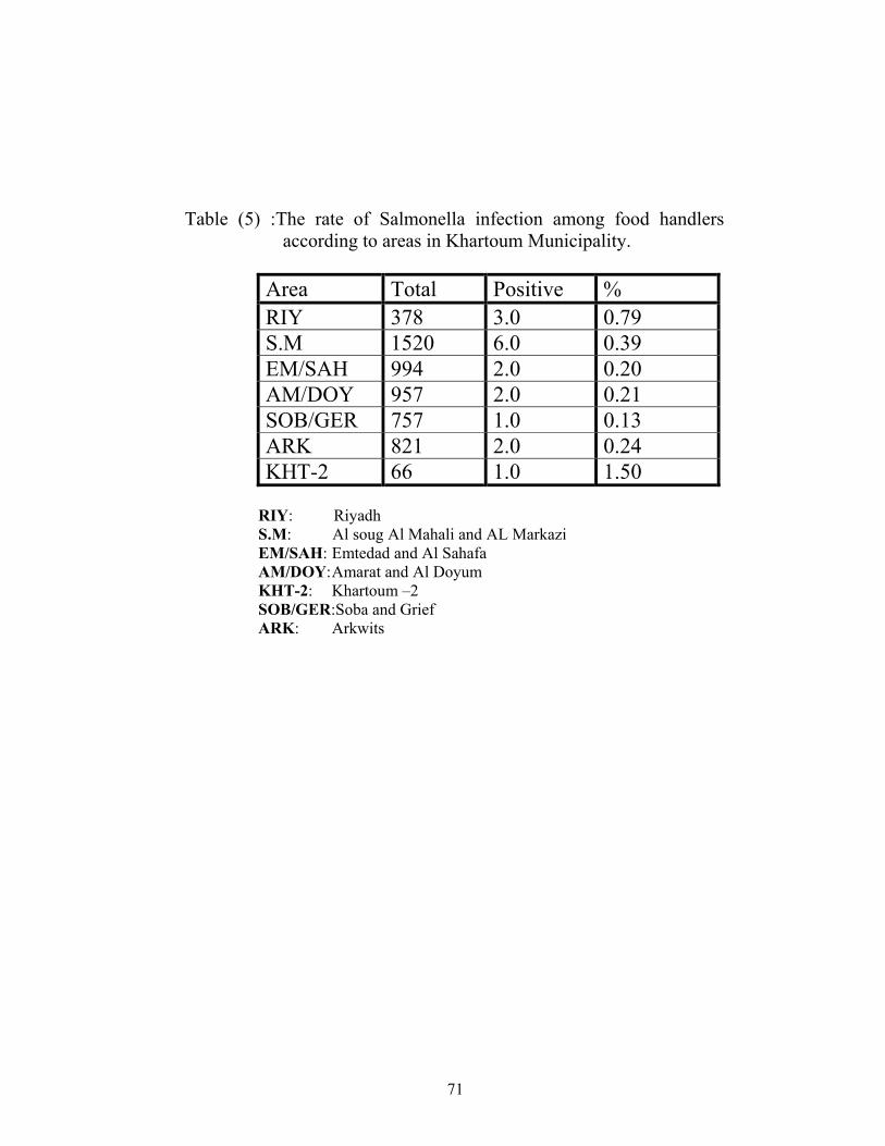
71
Table (5) :The rate of Salmonella infection among food handlers
according to areas in Khartoum Municipality.
Area Total Positive % RIY 378 3.0 0.79 S.M 1520 6.0 0.39 EM/SAH 994 2.0 0.20 AM/DOY 957 2.0 0.21 SOB/GER 757 1.0 0.13 ARK 821 2.0 0.24 KHT-2 66 1.0 1.50
RIY: Riyadh S.M: Al soug Al Mahali and AL Markazi EM/SAH: Emtedad and Al Sahafa AM/DOY: Amarat and Al Doyum KHT-2: Khartoum –2 SOB/GER:Soba and Grief ARK: Arkwits

72
Table (6): Susceptibility of isolates to antibiotics. Sensitive to Isolates
TE SXT C Cip Am Au RIY1 S R S S R R RIY2 S S S S R M RIY3 M S S S M R S.M1 R M S S R R S.M2 S R S S R M S.M3 S R S S M R S.M4 M S S S R R S.M5 M S S S R R S.M6 S R S S R M
EM/SAH1 S R S S M R EM/SAH2 S S S S R R AM/DOY1 S S S S R R AM/DOY2 S S S S M R SOB/GER S S S S R M
ARK1 R M S S R R ARK2 M S S S R R KHT-2 S S S S M R
S : Sensetive M: Moderate sensitive. R: Resistant. TE: tetracycline SXT: Co-trimoxazole C: Chloramphenicol Cip: Ciprofloxacin An: Ampecillin Au: Augmentin RIY: Riyadh S.M: Al soug Al Mahali and AL Markazi EM/SAH: Emtedad and Al Sahafa AM/DOY:Amarat and Al Doyum ARK: Arkwit KHT-2: Khartoum –2
SOB/GER: Soba and Grief

73
Table (7): Sensitivity of Salmonella isolates to the various antibiotics tested.
Sensitive isolates
Intermediate sensitive isolates
Resistant isolates
Type of antibiotics
% % % Tetracycline 11.0 65.0 4.00 24.0 2.00 12.0 Co-trimoxazole 10.0 59.0 2.00 12.0 5.00 29.0 Chloramphenicol 17.0 100.0 0.00 0.00 0.00 0.00 Ciprofloxacin 17.0 100.0 0.00 0.00 0.00 0.00 Ampecillin 0.00 0.00 5.00 29.0 12.0 71.0 Augmentin 0.00 0.00 4.00 24.0 13.0 76.0

74
Table (8) Personal hygiene of food handlers:-
Washing of hand after going to bath room Washing hand
before and after meal
Water only Water and Soap Water and antiseptic
Occupation
Alw
ays
Som
etim
es
Not
at a
ll
Alw
ays
Som
etim
es
Not
at a
ll
Alw
ays
Som
etim
es
Not
at a
ll
Alw
ays
Som
etim
es
Not
at a
ll
Restaurants 1732 118 11 712 510 639 0.00 03 1858 1010 161 290
Shopkeepers 1629 104 09 638 507 597 0.00 02 1740 888 573 281
Factotums 515 037 05 218 170 169 0.00 02 555 263 192 102
Butchers 338 023 06 110 147 110 0.00 00 367 124 159 084
Veg/ fruit retailers
376 030 07 189 106 118 0.00 00 413 118 198 097
Bakers 510 038 05 283 160 110 0.00 00 553 240 187 126
Total 5100 350 43 2150 1600 1742 0.00 07 5486 2643 1870 980

75
Table (9) Medial history of food handlers
Previous disease with enteric fever
Contact with patient with enteric fever
Treatment Occupation
Yes
No
Yes
No
Prev
ious
Rec
ent
No
Restaurateurs 11 1850 08 1853 11 00 185
0
Shopkeepers 02 1740 07 1735 02 00 1740
Factotums 02 555 03 554 02 00 555
Butchers 04 363 02 365 04 00 363
Veg/fruit retailers
03 410 03 410 03 00 410
Bakers 03 550 03 550 03 00 550
Total 25 5468 26 5467 25 00 5468
CHAPTER FOUR

76
DISCUSSION
Enteric fever is a public health problem primarily in endemic
countries where the enteric bacilli are spread widely by carriers,
convalescent and sick persons.
The development of a permanent carrier state is an important
event in the life cycle of enteric fever bacilli. Our present study was
conducted to assess the prevalence of Salmonella carriers among food
handlers working in food services establishments located in various
area of Khartoum municipality.
A totaled of 5493 food handlers were investigated during this
study, regarding their sociodemographic and professional charact-
eristics, their hand washing practices were also observed as shown in
the questionnaire specimen in appendix(1).
High prevalence of infection was recorded among restaurateurs
(0.54%), hence sound Kitchen hygiene is essential to the prevention of
Salmonella infection and this apply equally to commercially and
household kitchens. Moreover, the awareness of the industry of the
public health significance of Salmonella contamination of food and
the need to monitor their employee and products as well as to support
relevant research project is of paramount importance for the control of
Salmonella infection.
From our results isolation of Salmonella on SS agar and XLD
medium was found satisfactory. This is in agreement of the finding of
Dusch and Attwegg (1995) who reported that traditional media may
offer a superior recovery rate for the direct isolation of Salmonella
from stool samples despite an over decade development of
chromogenic media for Salmonella. Furthermore, Rambach agar a
chromogenic medium failed to isolate Salmonella enterica serotype
Typhi (Perry, et al 1999). Generally speaking the isolation of
77
Salmonella species from stool samples requires the use of culture
media which fulfill two important attributes: inhibiting effect on the
growth of the largest possible amount of other saprophytes and
selective and has a discriminating capacity which permit salmonella
species to be recognized among few other enteric Gram negative
bacteria. SS agar and XLD medium both have fulfilled this
requirement. The purpose of this comparative study was to evaluate
these two classical media for their ability to support selective growth
of Salmonella species from stool samples with a view that we could
optimize our routine protocol and to asses food handlers
bacteriologically with the minimum amount of work and cost. In
addition the work described herewith also took in consideration, the
merit of using conventional media, which are relatively cheaper than
chromogenic ones for the isolation of Salmonella from high-risk
population for chronic carrier in Khartoum municipality.
Furthermore from the questionnaire it appears that transmission
depends on various characteristics of the population such as, customs
occupation and personal hygiene. This study has revealed a poor
knowledge and practice of food hygiene among food handlers and we
recommended that a massive health education campaign directed at
both the public and food handlers be embarked on, to enable people to
take necessary steps to prevent food- borne diseases, complementary
to the government regulations. For instance hand-washing practices
were observed to be poor with low use of soap. These practices were
also reported in other endemic countries like Nigeria (Okojie, 2005),
Ghana (Felgo et al, 2004), India (Malhotra et al, 2006), Indonesia
(Vollaard, et al 2004), as well as in USA (Daniels et al, 2002).
Our finding also highlights the importance of periodic stool
examination of Salmonella carriers among food handlers. This is in

78
consistence with conclusion of Feglo, et al. (2004). Nonetheless
routine stool culture of food handler is not cost effective regarding the
volume of work needed to be done and should not be used as a
substitute for health education. At present there is a need to educate
food handlers about the importance of maintaining hand hygiene with
focus on improving their hand washing practices. Moreover,
intervention should be developed and evaluated in compliance with
potential for reducing the risk to human health and cost effectiveness.
Laboratory stool culture may fail to detect intermittently excreting
chronic carrier, however three consecutive culture should be submitted
before food handler resume his work and advised, if necessary
restricted to their employment and the Department of Public Health is
required to maintain on enteric register of food handlers which list all
known chronic carriers .
At present there are increasing numbers of the population eating
in community catering establishment with subsequent occurrence of
more bulk cooking of food with increased sale of ready to eat warm
foods. Therefore, asymptomatic Salmonella carriers among food
handlers remain a threat for the dissemination of Salmonella infection
and probably maintaining the endemicity of the infection in the
community.
Our results highlight the importance of screening of Salmonella
carriers among food handlers with the view to control large-scale
dissemination of Salmonella infection. Our results also indicated that
identification of Salmonella carriers from food handlers is significant
as preventive measure for food hygiene.
Despite the growing trend of antimicrobial resistance of enteric
fever bacilli (Kubota et al, 2005), our 17 isolated tested were found
susceptible to chloramphenicol and ciprofloxacin. This is a very

79
encouraging finding since chloramphenicol seems to remain the drug
of choice in treatment of enteric fever and carriers for over half- a
century worldwide. Also our results has indicated that ciprofloxacin
remain the alternate drug for the treatment of enteric fever. But on the
other hand all strains were found resistant to Ampicillin and augmintin
and only 59% and 65% of the isolates were sensitive to co-
trimoxazole and tetracyclin. Co-trimoxazole emerged in the late 1960s
as the most successful challenger to chloramphenicol as a safe drug of
choice for treatment of enteric fever. From this study an alarming
multidrug resistance to ampicillin, co-trimoxazole and tetracycline has
been demonstrated. This is in agreement of all the 1990s reports of
strains having multiple drug resistance to chloramphenicol, ampicillin,
streptomycin, sulphonamide trimethoprim and tetracyclin that
emerged world wide (Kubota et al, 2005).
On the other hand Vi Ags are only present in serotype Typhi and
serotype Paratyphi A and C but not in serotype Paratyphi B which is
the only isolate recovered form 17 carriers in this study. Paratyphi B
seems to be the dominant enteric typhoid bacilli among food handlers
studied in Khartoum municipality. This is in contrast of the report of
(Mandel, 1979), Ferreceio et al, (1999) in Chilli and Lanta et el,
(1990) in Lima Peru who claimed the Vi titre offers practical and cost
effective way of screening Salmonella enterica serotype Typhi in both
endemic and non endemic typhoid fever areas.
CONCLUSION

80
In search for typhoid carriers as part of routine examination of
food handlers, isolation of Salmonella on classical conversional media
was found satisfactory and only 17 isolate were recovered from the
stool cultures of food handlers. All isolates belong to Salmonella
enterica serotype Paratyphi B. Two isolates were recovered by direct
plating onto both SS and XLD agar media and 15 isolates were
obtained following enrichment in selinit F broth media. SS Agar &
XLD Agar media were found satisfactory for the isolation of
Salmonella from chronic feacal carriers among food handlers. Carriers
of Salmonella Paratyphi B among food handlers might play an
important role in maintaining the endemicity of Salmonella paratyphi
infection. Therefore food handlers should be closely supervised and
ideally should be screened bacteriologically at 6 month intervals and
must be advised and if necessary restricted to their employment and
the Department of Public Health is required to maintaining an enteric
register of food handlers, which list all known chronic carriers. This
study has indicated that recovery of Salmonella carriers from food
handlers is a significant as a preventive measure for food hygiene and
an important step to break the cycle of Salmonella infection.
Despite the growing problem of antimicrobial resistance of
enteric fever bacilli, all our isolates of Salmonella enterica serotype
Paratyphi B were sensitive to chloramphenicol and ciprofloxacin.
However, there was 41% resistance to the alternate drug of choice co-
trimoxazole and all isolates were resistant to ampicillin and
augmentin.
RECOMMENDATION

81
Rapid reduction in the incidence of typhoid fever in the
municipality of Khartoum could be achieved by strengthening food
safety measures.
1- Safe water supply, adequate means for proper disposal of human
excreta are by water- carriage system and food-safety education:
through books media for the commercial control of Salmonella
infection and also infiltrating homes and reducing family-type
outbreaks.
2- Screening for Salmonellae in the regular obligatory six-monthly
examination is required for food handlers to monitor those found to be
infected.
3- Chronic carriers must be advised and if necessary restricted as to
their employment and the Department of Public Health should
maintain an enteric register, which list all known chronic carriers.
Also an epidemiological model has to be constructed to help in
forecasting the effect of various control programs and their cost
effectiveness.
4- Sound kitchen hygiene is essential to prevention of Salmonella and
its principles apply equally to commercial and household kitchen.
5- Awareness of the industry sector of the public health significance
of Salmonella contaminating food and the need to monitor its product
and employees as well as to support relevant research projects.
6- Drug resistance among enteric fever bacilli has emerged
worldwide making antimicrobial susceptibility testing an important
function in Public Health Laboratories and multiple drug resistance
has compromised treatment and we recommend for more prudent uses
of these antimicrobial agents in both the Medical and Veterinary
fields.
7- Development of attenuated oral vaccine.

82
REFERENCES

83
Abraham, G.; Teklu, B.; Gedebu, M. Selassie, G.H. and Azene, G.
(1981). Diagnostic value of Widal test. J. Trop. Geog. Med.
33: 329-933.
Ananthanarayan, R. and Paniker, C. K. J. (1997). Textbook of
Microbiology 5th ed. Orinet Longman Ltd. New Delhi, India.
Anderson, E. S. and Smith, H. R. (1972). Chloramphenicol resistance
in the typhoid bacillus. Brit. Med. J. 3: 329-331.
Barker, R. M. (1985). Utilization of d-tartaric acid by Salmonella
Paratyphi B and Salmonella Java: Comparison of anaerobic
plate test and turbidity test. J. Hygi. 95: 107 –114.
Barrow, G. I. and Feltham, R. K. A. (1993). Cowan and Steel’s
Manual for the Idenfification of Medical Bacteria. 3rd ed,
Cambridge University Press, Cambridge.
Blaser, M. J. and Newman. L. S. (1982). A review of human
Salmonellosis, 1: Infective dose. Rev. Infect. Dis 4: 1096-
1106.
Brandtzaeg, P. (1989). Overview of the mucosal system. Curr. Topics
Microbiol. Immunol. 146: 13-25.
Brook, G. F.; Butel, J. S. and Morse, S. A. (2004). Jawetz, Melnick
and adelbergs Medical Microbiology 23rd ed. Mc Graw Hill
Companies, Inc.
Buchwald, D.S. and Blaser, M.J. (1984). A review of human
salmonellosis: II. Duration of excretion following infection
with nontyphi salmonella. Rev. infect. Dis, 6: 345-356.
Carter, P. B. and Collins, F. M. (1974). The route of enteric infection
in normal mice. J. Exp. Med. 139: 1189-1203.
CDC (1997). Multidrug-resistant Salmonella serotype Typhi-murium.
United States, 1996. MMWR. 46: 308- 310.

84
CDC. (1998). Preventing typhoid fever. A guide for travelers. Centers
for Disease Control.
Chessbrough, M. (2000). District Laboratory practice In Tropical
Countries. Part 2; Cambridge University press, Cambridge.
Clark, M. A.; Jepson, M. A.; Simmons, N. L. and Hirst, B. H. (1994).
Preferential interactions of Salmonella typhimurium with
mouse Peyer’s patch M cells. Res. Microbiol. 145: 543-552.
Collee, J. G.; Fraser, A. G.; Marmiio, B. P.; Simon, A. and Old, D.C.
(1996). Mackie and McCarteny Practical Medical
Microbiology. 14th ed; Longman Singapore publishers (Pte).
Ltd. Singapor.
Coloe, P. J.; alderton, M. R.; Gerraty, N. L.; Christopher, W. and
Smith, S. C. (1995). Aromatic vitamin-dependent Salmonellae
as vaccines in food animals: Efficacy and persistence. Dev.
Biol. Stand. 84: 263-267.
Colquhoun. J. and Weetch. R. S. (1950). Resistance to
chloramphenicol developing during treatment of typhoid
fever. Lancet. 2: 621-623.
Curtis, M.; Edwards, A. and Kapil, A. (1983). Epidemiological
Control of Typhoid Fever and Paratyphoid Fever. Nature.
530: 236-231.
Curtiss III, R. and Kelly, S. M. (1987). Salmonella typhimurium
deletion mutants lacking adenylate cyclase and cyclic AMP
receptor protein are a virulent and immunogenic. Infect.
Immun. 55: 3035-3043.
Daniels, N.A; Mackinnon,L; Rower S.M; Bean, N.H; Griffin, P.M.
and Mead, P.S. (2002). Food borne disease outbreaks in
United States schools. Pediatr. Infect. Dis. J. 21: 623-628.

85
David, B. G.; Bishop, M. L. and Mass, D. (1989). Clinical Laboratory
Science: Strategies for practice. JB Lippicott Company,
Philadelphia.
Davis, B. D.; Dulbecco, R.; Eisen, H. N. and Ginsberg, H. S. (1990).
Microbiology 4th ed. J. B. Lippincott Company.
Dusch, H. and Altwegg,M. (1995). Evaluation of five new plating
media for isolation of salmonella in human faeces. J. Clin.
Microbiol. 33: 802-804.
Duguid, S. P.; Anderson, E. S.; Campbell I. (1966). Fimbriae and
adhesive properties in Salmonella. J. pathol. Bacteriol. 92:
107-138.
Duguid, J. P.; Anderson, E. S; Alfredsson, G. A.; Barker, R. and Old
DC (1975). A new biotyping scheme for Salmonella
typhimurium and its phylogenetic significance. J. Med.
Microbiol. 8: 149-166.
Elfaki, M. E. (1987). Pattern of Typhoid Fever in Sudanese Children.
M.Sc. thesis, University of Khartoum.
Feglo.P.K; Frimpong.E.H and Essel-Ahun-M (2004). Salmonellae
Carrier Status of food vendors in Kumasi, Ghana. East Afr.
Med. J. 81: 358-361.
Ferreccio, C.; leviw, M; Astroza, L; Berrios, G; Solari, V; Misraji, A
and Pefaur,C. (1999). The detection of chronic salmonella
typhi carriers: a practical method applied to food handlers in
Portuguese revista media de chille.
Giannella, R. A.; Broitman, S. A. and Zamcheck, N. (1972). Gastric
acid barrier to ingested microorganisms in man: Studies in
vivo and in vitro. Gut 13: 251-256.
Glynn, M. k.; Bopp, C.; Dewitt, W.; Dabney, P.; Mokhtar, M. and
Angulo, F. J. (1998). Emergence of multidrug-resistant

86
Salmonella enterica serotype Typhimurium DT 104 infections
in the United States. New Engl. J.Med. 338: 1333-1338.
Goldberg, M. B. and Rubin, R. H. (1988). The spectrum of
Salmonella infection. Infect. Dis. Clin. N. Amer.2: 571-598.
Greenwood, D.; Slack, R. and peutherer, S. (2004). Medical
Microbiology. A Guide to Microbial Infection Pathogenesis,
Immunity, Laboratory Diagnosis and Control 15th ed.
Gupta, A. (1994). Multidrug-resistant typhoid fever in children.
Epidemiology and therapeutic approach. Pediatr. Infect. Dis.
J. 13: 134-140.
Hampton, M. D.; Ward, L. R.; Rowe, B. and Threlfal, E. J. (1998).
Molecular fingerprinting of multidrug-resistant Salmonella
enterica serotype Typhi. Emerg. Infect. Dis. 4: 317-320.
Hassan, J. O. and Curtiss III, R. (1997). Efficacy of a live a virulent
Salmonella typhimurium vaccine in preventing colonization
and invasion of laying hens by Salmonella typhimurium and
Salmonella enteritidis. Avian Dis. 41: 783-791.
Hohmann, E. L.; Oletta, C. A. and Miller, S. I. (1995). Evaluation of
phoP/phoQ deleted aroA-detected live oral S. Typhi vaccine
in humans. Vaccine 14: 19 – 24.
Hohmann, E. L.; Oletta, C. A.; Killeen, K. P. and Miller S. I. (1996).
PhoP/phoQ detected salmonella Typhi (TY800) is a safe and
immunogenic single dose typhoid fever vaccine in volunteers.
J. Infect. Dis. 173: 1408-1414.
Hoiseth, S. K. and Stocker, B. A. D. (1981). Aromatic-dependent
Salmonella typhimurium are non-virulent and effective as live
vaccines. Nature. 291: 238-239.

87
Kaye, D.; Merselis Jr. J. G; Connolly, S. and Hook, E.W. (1967).
Treatment of chronic enteric carriers of Salmonella tryphosa
with ampicillin. Ann. N. Y. Acad. Sci. 145: 429-435.
Khan, M.; Coovadia, Y. M.; Cannoly, C.; Stum, A. W. (1998). The
early diagnosis of typhoid fever prior to Widal test and
bacteriological culture results. J. Act. Trop. 69: 165-173.
Kohbata, S.; Yokobata, H. and Yahuuchi, E. (1986). Cytopathogenic
effect of Salmonella Typhi GIFU 10007 on M cells of murine
ileal Peyer’s patches in ligated ileal loops: An ultrastructural
study. Microbiol. Immunol. 30: 1225-1237.
Koneman, W. G.; Allen, D. S.; Janda, M. W; Sclreckenberger, C. P.
and Winn, C. W. (1997). Colour Atlas and Text Book of
Diagnostic Microbiology 5th. Ed.
Kubota, K. T. J.; Barret; M. L.; Ackers, P. S.; Brachman and Mintz, E.
D. (2005). Analysis of Salmonella enterica Serotype Typhi
Pulsed- field gel electrophoresis pattern association with
international travel.J. Clin. Micro. 43: 1205-1209
Kumar,A.; Arora,V.; Bashamboo,A. and Ali,S. (2002). Infection,
Genetics and Evolution. Elserier Science. B.V.
Lanta,C.F; Tafur,C; Benavente,L.; Gotuzzo,E. and Carrillo,C.
(1990).Detection of Salmonella Typhi Carriers in food
handlers by Vi serology in Lima, peru. Bull. Pan. Am. Health.
Organ, 24: 177-182.
Leavitt, J. W. (1996). Typhoid Mary: Captive to the Public’s Health.
Beacon, Boston.
Levine, M. M.; Black, R. E.; Ferreccio, C.; Clements, M. L.; Lanata,
C.; Rooney, J.; Germanier, R. and Chilean Typhoid
Committee ( 1987). Field trails of the efficacy of attenuated
Salmonella Typhi oral vaccine strain Ty21a. “Proceedings of
88
the International Symposium on Bacterial Vaccines” (J.
Robbins, ed.). Praeger. New York.
Le Minor, L.; Le Minor, S.; Grimont, P. A. D. (1985). Rapport
quadriennal du centre national des Salmonella sur I’origine et
la répartition en serotypes souches isolees en france
continentale au cours des annees 1980 a` 1983. Revue
d’Epidemidologic et de santé publique. 33: 13-21.
Lim, P. L.; Tam, F. C.; Cheong, Y. M.; Jegathesan, M. (1998). One
step 2- minutes test to detect typhoid specific antibodies based
on practical separation in tubes. J. Clin. Microbial. 36: 227-
228.
Louis,P.Ch.A. (1829). Cited by Scherer and Muller, (2001).
Malhotra, R.; Lal, P.; Prakash, S.; Daga, M. and kishore, J. (2006).
Study of hand hygiene and enteroparasite infestation among
food handlers working in a medical college of North India.
Indian J. Pathol. Mirobiol. 49: 296-301.
Mandel, K. B. Typhoid & Paratyphoid Fever (1979) in Clinic
Gastroentrology Lambert. (Guet editor). Vol. 8:715-735.
Mandel, G. L.; Hook, E. W. and Douglas, R. G. (1985). Evaluation of
fever in returned travelers. J. Emer. Infec. Dis. 1: 54-57.
Mastroianni, C. M.; Jirillo, E. Desimone, C.; Grassi, P. P.; Maffione,
A. B.; Catino, A. M.; Vollo, V.; Misefari, A. and Delia, S.
(1989). Humoral and cellular immune response to Salmonella
Typhi in patients with typhoid fever. J. Clin. Lab. Anal. 3:
191-195.
Mintz, E. D.; Cartter, M. L.; Hadler, J. L.; Wassell, J. T.; Zingeser, J.
A. and Tauxe, R.V. (1994). Dose-response effects in an
outbreak of Salmonella enteritidis. Epidemiol. Infect. 112:13-
23.

89
Mullis, K. F.; Faloona, S.; Schart, R.; Saiki, G.; Horn, and Erlich, H.
(1986). Specific enzymetic amplification of DNA in vitro: the
polymerase chain reaction. Cold spring Harbor symp. Quant.
Biol. 51: 263-273.
Murray, P. R; Baron, E. J.; Jorgensen, J. H; Pfaller, M. A. and yolken,
R. H. (2005). Manual of Clinical Microbiology 8th ed. ASM
press, Washington. Vol 1.
Neutra, M. R.; Pringault, E. and Kraehenbuhl, L. P. (1996). Antigen
sampling across epithelial barriers and induction of mucosal
immune responses. Ann. Rev. Immunol. 14: 275-300.
Ochei, J. and Kolhatkar, A. (2000). Medical Laboratroy Science
Theory and Pracatice. Tata McGrow-Hill Publishing
Company Ltd. pp: 689-696.
Okojie. H.; wagbatsoma, V. and Ighoroge, A. (2005). An assessment
of food hygiene among food handlers in a Nigerian University
Campus. Niger postgrad Med J. 12: 93-96.
Paniker, C. K. J. and Vilma, K. N. (1997). Transferable
Chloramphenicol resistance in Salmonella Typhi. Nature. 239:
109-110.
Penheiter, K. L.; Mathur, N.; Giles, D.; Fahlen, T. and Jones B. D.
(1997). Non-invasive Salmonella typhimurium mutants are
avirulent because of an inability to enter and destroy M cells
of ileal Peyer’s Patches. Mol. Micobiol. 24: 697-709.
Perry.J.D; Ford. M; Taylor, J; Jones. A.L; Freeman.R and Gould. F.K.
(1999). ABC Medium, a New Chromogenic gar for selective
isolation of salmonella species. J. Clin. Microb. 37: 766-768.
Pyatkin, K. and Krivoshein, Y. U. (1987). Microbiology with
Virology and Immunology, MIR publishers, Moscow.

90
Rowe, B.; Ward, L. R. and Threlfall, E. J. (1997). Multiresistant
Salmonella Typhi, a worldwide epidemic. J. Clin. Infec. Dis.
24: 2106-2109.
Sarasombath, S.; Banchuin, N.; Sukosol. T.; Rungpitarangsi, B. and
Manasatit, S. (1987). Systemic and intestinal immunities after
natural typhoid infection. J. Clin. Mirolobiol. 25:1088-1093.
Scherer, C.A. and Miller, S.I. Molecular pathogenesis of salmonellae
(2001). In: principles of bacteria pathogenesis. Grosimar,S.A.
(editor) pp:265-333. Academic press USA.
Stokes, E. J.; Ridgway, G. L. and Wren, M. W. D. (1993). Clinical
Microbiolgoy, 4th ed. Edward Arnold, London, Britain.
Svenugsson, P. and Blinberg, A. A. (1978). Indirect immuno-
fluorescence studies for the identification of Salmonella
serogroup A bacteria. Acta. Pattiol. Microbiol. Scand B. 86:
35-40.
Tacket, C. O.; Hone, D. M.; Curtiss III, R.; Kelly, S. M.; Losonsky,
G.; Guers, L.; Harris, A. M.; Edelman, R. and Levine, M. M.
(1992). Comparison of the safety and immunogenicity of
aroC, aroD and cya, crp Salmonella Typhi strains in adult
volunteers. Infect. Immune. 60: 536-541.
Thong, K. L.; Nair, S.; Chaudhry, R.; Kapil A. and Chandel D. S.
(1998). Molecular analysis of Salmonella Paratyphi A from an
outbreak in New Delhi, India, by ribotyping and pulsed-field
gel electrophoresis. J. Emer. Infec. Dis. 4: 507-508.
Threlfall, E. J.; Frost. J.A.; Ward L.R. and Rowe, B. (1996).
Increasing spectrum of resistance in multiresistant Salmonella
typhimurium [letter]. Lancet. 347: 1053-1054.

91
Threlfall, E. J. and Ward, L. R. (2001). Decreased Susceptibility to
Ciprofloxacin in Salmonella enterica serotype Typhi, UK. J.
Emer. Infec. Dis. 7: 21-26.
Vollaard.A; Ali.S; Vanasten.H; Widjaja.S; Visser. L.G. Surjadia C.
and Van Dissel. J.T. (2004). Risk Factors for Typhoid and
Paratyphoid fever in Jakarta, Indonesia. JAMA 2; 291: 2607-
2615.
Waddell, W.R. and Kunz, L. J. (1956). Association of Salmonella
enteritis with operation of stomach. New Engl. J. Med. 255:
555-559.
Wall, P.G.; Morgan, D.; Lamden, K.; Ryan M.; Griffin, M.; Threlfall,
E. J.; Ward, L. R. and Rowe. B. (1994). A case control study
of infection with an epidemic strain of multiresistant
Salmonella typhimurium DT 104 in England and Wales.
Commun. Dis. Rep. CDR. Rev. 4: 130-135.
WHO, (1983).Manual of laboratory investigations of acut enteric
infection.WHO/CDD/83.rev.
WHO (1997). Fact sheet no.149. Typhoid fever. World Health
Organization.
Wick, M. J.; Harding, C.V.; Twesten, N. J.; Normark, S. J. and
Pfeifer, J. D. (1995). The phoP locus influences processing
and presentation of Salmonella typhimurium antigens by
activated macrophages. Mol. Microbiol. 16: 465-476.
William Jenner, (1850). Cited by Scherer and Muller, (2001).
Youmans, G. P.; Paterson, P. Y. and Sommers, H. M. (1986). The
biologic and Clinical Basis of Infectious Diseases. 3rd ed.
Saunders Company West; Washington squire Philadelphia.

92
University of Khartoum Faculty of Veterinary Medicine
Department of Microbiology
Questionnaire on Salmonella Carriers among Public Food Handlers

93
Hospital ………………… Date…………………… * Identification data. Pt name …… age ………… sex………...place of work ………. Type of work……….. Residence…………. * Educational level 1- Illiterate ( ) 2- Elementary ( ) 3- Intermediate ( ) 4- Secondary ( ) 5- Graduate ( ) 6- Post graduate ( ) * Personal hygiene: -Washing of hand by water only after going to bathroom 1- Always ( ) 2- Sometime ( ) 3- Not at all ( ) -Washing of hands by water and soap after going to bathroom 1- Always ( ) 2- Sometime ( ) 3- Not at all ( ) - Washing of hands by water and antiseptic after going to bathroom. 1- Always ( ) 2- Sometime ( ) 3- Not at all ( ) - Washing of hand by soap before and after meal. 1- Always ( ) 2- Sometime ( ) 3- Not at all ( ) * Past medical history of Salmonella infection: Yes ( ) No ( ) If yes what is the treatment received ………………….. Recent antibiotic therapy……………………………….
• Previous contact with pt. Infected with enteric fever. Yes ( ) No ( )
• Result 1- Salmonella species isolated ……………………… 2- Antibiotics sensitivity……………………………
a- Sensitive to …………………………………. b- Resistance to ……………………………….
……………………………………………..