Salivary Gland: Oncologic Imagingpatologiabucal.com/index_htm_files/GSimagenoncologica.pdfSALIVARY...
15
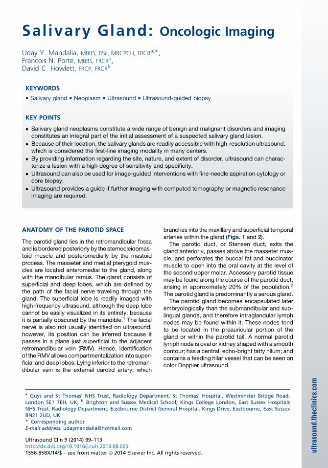
Salivary Gland: Oncologic Imaging Uday Y. Mandalia, MBBS, BSc, MRCPCH, FRCR a, *, Francois N. Porte, MBBS, FRCR a , David C. Howlett, FRCP, FRCR b ANATOMY OF THE PAROTID SPACE The parotid gland lies in the retromandibular fossa and is bordered posteriorly by the sternocleidomas- toid muscle and posteromedially by the mastoid process. The masseter and medial pterygoid mus- cles are located anteromedial to the gland, along with the mandibular ramus. The gland consists of superficial and deep lobes, which are defined by the path of the facial nerve traveling through the gland. The superficial lobe is readily imaged with high-frequency ultrasound, although the deep lobe cannot be easily visualized in its entirety, because it is partially obscured by the mandible. 1 The facial nerve is also not usually identified on ultrasound; however, its position can be inferred because it passes in a plane just superficial to the adjacent retromandibular vein (RMV). Hence, identification of the RMV allows compartmentalization into super- ficial and deep lobes. Lying inferior to the retroman- dibular vein is the external carotid artery, which branches into the maxillary and superficial temporal arteries within the gland (Figs. 1 and 2). The parotid duct, or Stensen duct, exits the gland anteriorly, passes above the masseter mus- cle, and perforates the buccal fat and buccinator muscle to open into the oral cavity at the level of the second upper molar. Accessory parotid tissue may be found along the course of the parotid duct, arising in approximately 20% of the population. 2 The parotid gland is predominantly a serous gland. The parotid gland becomes encapsulated later embryologically than the submandibular and sub- lingual glands, and therefore intraglandular lymph nodes may be found within it. These nodes tend to be located in the preauricular portion of the gland or within the parotid tail. A normal parotid lymph node is oval or kidney shaped with a smooth contour; has a central, echo-bright fatty hilum; and contains a feeding hilar vessel that can be seen on color Doppler ultrasound. a Guys and St Thomas’ NHS Trust, Radiology Department, St Thomas’ Hospital, Westminster Bridge Road, London SE1 7EH, UK; b Brighton and Sussex Medical School, Kings College London, East Sussex Hospitals NHS Trust, Radiology Department, Eastbourne District General Hospital, Kings Drive, Eastbourne, East Sussex BN21 2UD, UK * Corresponding author. E-mail address: [email protected] KEYWORDS Salivary gland Neoplasm Ultrasound Ultrasound-guided biopsy KEY POINTS Salivary gland neoplasms constitute a wide range of benign and malignant disorders and imaging constitutes an integral part of the initial assessment of a suspected salivary gland lesion. Because of their location, the salivary glands are readily accessible with high-resolution ultrasound, which is considered the first-line imaging modality in many centers. By providing information regarding the site, nature, and extent of disorder, ultrasound can charac- terize a lesion with a high degree of sensitivity and specificity. Ultrasound can also be used for image-guided interventions with fine-needle aspiration cytology or core biopsy. Ultrasound provides a guide if further imaging with computed tomography or magnetic resonance imaging are required. Ultrasound Clin 9 (2014) 99–113 http://dx.doi.org/10.1016/j.cult.2013.08.005 1556-858X/14/$ – see front matter Ó 2014 Elsevier Inc. All rights reserved. ultrasound.theclinics.com
Transcript of Salivary Gland: Oncologic Imagingpatologiabucal.com/index_htm_files/GSimagenoncologica.pdfSALIVARY...
Salivary Gland: Oncologic Imaging
Uday Y. Mandalia, MBBS, BSc, MRCPCH, FRCRa,*,Francois N. Porte, MBBS, FRCRa,David C. Howlett, FRCP, FRCRb
KEYWORDS
� Salivary gland � Neoplasm � Ultrasound � Ultrasound-guided biopsy
KEY POINTS
� Salivary gland neoplasms constitute a wide range of benign and malignant disorders and imagingconstitutes an integral part of the initial assessment of a suspected salivary gland lesion.
� Because of their location, the salivary glands are readily accessible with high-resolution ultrasound,which is considered the first-line imaging modality in many centers.
� By providing information regarding the site, nature, and extent of disorder, ultrasound can charac-terize a lesion with a high degree of sensitivity and specificity.
� Ultrasound can also be used for image-guided interventions with fine-needle aspiration cytology orcore biopsy.
� Ultrasound provides a guide if further imaging with computed tomography or magnetic resonanceimaging are required.
ANATOMY OF THE PAROTID SPACE
The parotid gland lies in the retromandibular fossaand is bordered posteriorly by the sternocleidomas-toid muscle and posteromedially by the mastoidprocess. The masseter and medial pterygoid mus-cles are located anteromedial to the gland, alongwith the mandibular ramus. The gland consists ofsuperficial and deep lobes, which are defined bythe path of the facial nerve traveling through thegland. The superficial lobe is readily imaged withhigh-frequency ultrasound, although the deep lobecannot be easily visualized in its entirety, becauseit is partially obscured by the mandible.1 The facialnerve is also not usually identified on ultrasound;however, its position can be inferred because itpasses in a plane just superficial to the adjacentretromandibular vein (RMV). Hence, identificationof the RMV allows compartmentalization into super-ficial and deep lobes. Lying inferior to the retroman-dibular vein is the external carotid artery, which
a Guys and St Thomas’ NHS Trust, Radiology DepartmeLondon SE1 7EH, UK; b Brighton and Sussex Medical SNHS Trust, Radiology Department, Eastbourne District GBN21 2UD, UK* Corresponding author.E-mail address: [email protected]
Ultrasound Clin 9 (2014) 99–113http://dx.doi.org/10.1016/j.cult.2013.08.0051556-858X/14/$ – see front matter � 2014 Elsevier Inc. Al
branches into the maxillary and superficial temporalarteries within the gland (Figs. 1 and 2).
The parotid duct, or Stensen duct, exits thegland anteriorly, passes above the masseter mus-cle, and perforates the buccal fat and buccinatormuscle to open into the oral cavity at the level ofthe second upper molar. Accessory parotid tissuemay be found along the course of the parotid duct,arising in approximately 20% of the population.2
The parotid gland is predominantly a serous gland.The parotid gland becomes encapsulated later
embryologically than the submandibular and sub-lingual glands, and therefore intraglandular lymphnodes may be found within it. These nodes tendto be located in the preauricular portion of thegland or within the parotid tail. A normal parotidlymph node is oval or kidney shaped with a smoothcontour; has a central, echo-bright fatty hilum; andcontains a feeding hilar vessel that can be seen oncolor Doppler ultrasound.
nt, St Thomas’ Hospital, Westminster Bridge Road,chool, Kings College London, East Sussex Hospitalseneral Hospital, Kings Drive, Eastbourne, East Sussex
l rights reserved. ultrasound.th
eclinics.com
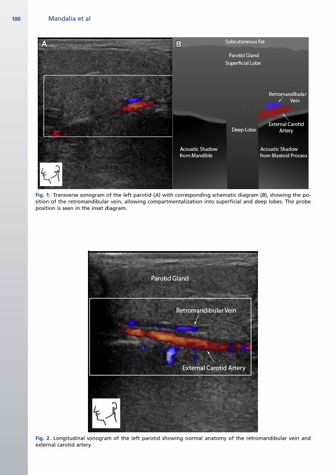
Fig. 1. Transverse sonogram of the left parotid (A) with corresponding schematic diagram (B), showing the po-sition of the retromandibular vein, allowing compartmentalization into superficial and deep lobes. The probeposition is seen in the inset diagram.
Fig. 2. Longitudinal sonogram of the left parotid showing normal anatomy of the retromandibular vein andexternal carotid artery.
Mandalia et al100

Fig. 3. Sonogram of the right parotid mass in the su-perficial lobe. This mass is hypoechoic and inhomoge-neous. The lesion was painful and had enlargedrapidly, and biopsy confirmed an infarcted Warthintumor.
Salivary Gland 101
ANATOMY OF THE SUBMANDIBULAR GLAND
The submandibular gland lies in the space locatedinferior to the body of the mandible and betweenthe anterior and posterior bellies of the digastricmuscle. The gland is roughly triangular in shapeand, like the parotid gland, is made up of superficialand deep lobes, although these are of less clinicalsignificance than theparotid gland. The submandib-ular gland does not contain lymph nodes, althoughlymph nodes are found within the submandibularspace superior and anterior to the gland.3
The facial artery arises from the external carotidartery and passes through a groove on the poster-oinferior aspect the submandibular gland. Thefacial artery may pass through the parenchymaof the gland.3 The facial vein crosses over the su-perficial aspect of the gland. Themarginal mandib-ular nerve also crosses the gland superficially,within the deep cervical fascia. The submandibularduct (or Wharton duct) originates from multipleductal branches situated on the deep aspect ofthe gland, extends anteriorly between the mylo-hyoid and hypoglossus muscles, crosses themedial aspect of the sublingual gland, and drainsinto the mouth at the sublingual caruncle, situatedon the frenulum lingulae. The submandibular glandis a mixed serous/mucinous gland.
ANATOMY OF THE SUBLINGUAL GLAND
The sublingual gland is the smallest of the 3 majorsalivary glands. It is situated posterior to themandible and lies below the mucous membraneof the floor of the mouth. The gland is borderedinferiorly by the mylohyoid muscle and mediallyby the genioglossus and the submandibular duct.The sublingual gland has a variable number ofexcretory ducts, many of which drain directly intothe floor of the mouth, although some ducts formthe sublingual duct of Bartholin, which joins thesubmandibular duct to drain into the sublingualcaruncle. The gland can be visualized on ultra-sound when scanning in the submental regionand appears as an echogenic oval-shaped struc-ture on transverse imaging.3 The sublingual glandis a predominantly mucinous gland.
CLINICAL PRESENTATION OF A SALIVARYGLAND TUMOR
Major salivary gland tumors present as painlessmasses in the region of the affected salivary gland.Benign tumors are usually slow growing, whereasmalignant tumors vary in their rate of growth de-pending on their grade, although a sudden painfulincrease in size may also be related to infarction(Fig. 3). Pain is a poor discriminator between benign
and malignant disease because it is experienced in5.1% of patients with benign tumors and 6.5% ofpatients with malignant disease.4,5 However, inpatients with proven salivary gland carcinoma, thepresence of pain is a poor prognostic indicator,because it signifies perineural spread of disease,which is associated with a 5-year reduction in sur-vival from 68% to 35%. Cranial nerve VII is mostcommonly involved because of its course throughthe parotid, thus symptoms include facial pain andfacial nerve paralysis. With progressive tumor inva-sion other cranial nerves may become involved.6,7
Lymph node metastases from a salivary glandtumor may present as a firm enlarging necklump. The primary drainage for the parotid andsubmandibular glands is to the deep cervicalchain, whereas the sublingual gland drains to sub-mental and submandibular nodes. Nodal metasta-ses are associated with a poorer prognosis, withan associated reduction in 10-year survival from63% to 33%.8
Distant metastasis is also an indicator of poorprognosis and is seen in 20% of the parotid can-cers, most commonly from adenoid cystic carci-noma, followed by undifferentiated carcinoma.6
THE ROLE OF IMAGING
The diagnosis of a salivary gland tumor ideally isbased on the concept of triple assessment:
1. Clinical evaluation2. Imaging3. Histologic/cytologic evaluation
Although salivary gland tumors are rare andusually benign, surgical excision represents the
Box 1The aims of imaging in salivary gland tumors
1. Localization of the lesion
2. Determination of the nature of the lesion
3. Tumor staging for preoperative planning
4. Detection of cervical lymphadenopathy
5. Image-guided biopsy
Mandalia et al102
treatment of choice inmost circumstances. Imagingis needed not only to confirm the presence of alesion but also to determine its spread, intraglandu-lar/extraglandular extent, identification of clinicallyoccult lesions, as well as unsuspected cervicallymphadenopathy. The imaging characteristics ofa lesion can determine whether a tumor is benignor malignant with a high sensitivity and specificity.9
An accurate preoperative diagnosis of a parotidlesion is critical because many nonneoplasticlesions do not require surgery. The need for surgerymay also be avoided in certain benign neoplasms(eg, Warthin tumors) or if the patient is consideredtoo elderly or unfit for surgery. An accurate preoper-ative diagnosis is an important determinant foroperative planning, notably with the increased useof extracapsular, parotid-sparing dissection and toallow appropriate informed patient consent (inparticular pertaining to facial nerve integrity andalso possible nodal dissection in malignancy).10,11
Ultrasound is frequently accepted as the initialimaging modality of choice.12 It has the advan-tages of being portable and allowing multiplanar,noninvasive imaging without the need of ionizingradiation. High-frequency linear array probes arecapable of producing high-resolution images ofthe salivary glands with a spatial resolution sur-passing computed tomography (CT) and magneticresonance (MR) imaging. However, comparedwith these modalities, ultrasound has some disad-vantages: it is operator dependent and is limited invisualization of large lesions or those extendinginto the deep lobe of the parotid because ofobscuration by the mandible.13
Most lesions lie within the superficial lobe andultrasound is usually able to compartmentalize alesion according to its relationship to intraparotidvessels and the inferred position of the facialnerve. For large or suspected malignant lesionsand lesions of the deep lobe of the parotid gland,MR imaging or CT are the modalities of choice;however, even in these cases, ultrasound can actas an indicator for additional investigation.13,14
Following the initial sonographic diagnosis of asalivary gland tumor, ultrasound-guided biopsy(with fine-needle aspiration cytology [FNAC] orultrasound-guided core biopsy [USCB]; discussedlater) is a safe and reliable method of obtaining thehistopathologic confirmation of a lesion necessaryto instruct further surgical management.15–17 Thegoals of imaging salivary gland tumors can beseen in Box 1.
SONOGRAPHIC TECHNIQUE
A high-frequency linear array transducer, typically7 to 12 MHz or greater, should be used in the
assessment of the salivary glands. A lower fre-quency transducer (5–10 MHz) can be used toassess large tumors fully and to visualize lesionslocated in the deeper aspects of the glands.18–20
To facilitate visualization, a pillow or towel canplaced under the patient’s shoulders to extendthe patient’s neck. The salivary glands and anylesion discovered within them should be interro-gated in at least 2 perpendicular planes. An obliqueapproach may be required to navigate around themandible. The contralateral salivary gland needsto be examined for comparison and to look for bilat-eral disease. To complete the study, the entire neckshould be imaged for related disorders and lymphnode enlargement. Color Doppler assessment pro-vides information regarding the vascular resistanceand flow pattern of a lesion, which can improvediagnostic accuracy for malignant tumors.21
SALIVARY GLAND NEOPLASTIC DISEASE
When all salivary gland tumors are considered, theglobal incidence varies from 0.4 to 13.5 cases per100,000 population.22 About 80% of all lesions arebenign, hence salivary malignancies are rare en-tities, comprising less than 0.5% of all malig-nancies and about 5% of cancers of the headand neck.23 They constitute a wide variety of disor-ders (Table 1). They are divided into benign andmalignant neoplasms, which can be epithelial ornonepithelial in origin. The parotid gland contains70% of all salivary gland tumors, with 8% foundin the submandibular glands and 22% in the minorglands.24
There are some general rules that apply to sali-vary gland neoplasms. The smaller the salivarygland, the higher the rate of malignancy. Thus,the rate of malignancy increases from 20% to25% in the parotid gland to 40% to 50% in thesubmandibular gland, and to 50% to 81% in thesublingual glands and minor salivary glands.25–28
Environmental and genetic factors have beenproposed as causes of salivary gland neoplasms.The strongest link seems to be with radiationexposure and smoking, which have been impli-cated in the development of Warthin tumors.29
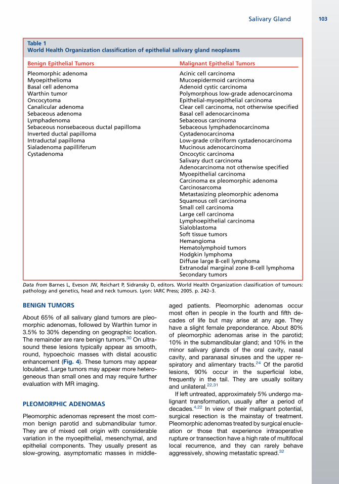
Table 1World Health Organization classification of epithelial salivary gland neoplasms
Benign Epithelial Tumors Malignant Epithelial Tumors
Pleomorphic adenomaMyoepitheliomaBasal cell adenomaWarthin tumorOncocytomaCanalicular adenomaSebaceous adenomaLymphadenomaSebaceous nonsebaceous ductal papillomaInverted ductal papillomaIntraductal papillomaSialadenoma papilliferumCystadenoma
Acinic cell carcinomaMucoepidermoid carcinomaAdenoid cystic carcinomaPolymorphous low-grade adenocarcinomaEpithelial-myoepithelial carcinomaClear cell carcinoma, not otherwise specifiedBasal cell adenocarcinomaSebaceous carcinomaSebaceous lymphadenocarcinomaCystadenocarcinomaLow-grade cribriform cystadenocarcinomaMucinous adenocarcinomaOncocytic carcinomaSalivary duct carcinomaAdenocarcinoma not otherwise specifiedMyoepithelial carcinomaCarcinoma ex pleomorphic adenomaCarcinosarcomaMetastasizing pleomorphic adenomaSquamous cell carcinomaSmall cell carcinomaLarge cell carcinomaLymphoepithelial carcinomaSialoblastomaSoft tissue tumorsHemangiomaHematolymphoid tumorsHodgkin lymphomaDiffuse large B-cell lymphomaExtranodal marginal zone B-cell lymphomaSecondary tumors
Data from Barnes L, Eveson JW, Reichart P, Sidransky D, editors. World Health Organization classification of tumours:pathology and genetics, head and neck tumours. Lyon: IARC Press; 2005. p. 242–3.
Salivary Gland 103
BENIGN TUMORS
About 65% of all salivary gland tumors are pleo-morphic adenomas, followed by Warthin tumor in3.5% to 30% depending on geographic location.The remainder are rare benign tumors.30 On ultra-sound these lesions typically appear as smooth,round, hypoechoic masses with distal acousticenhancement (Fig. 4). These tumors may appearlobulated. Large tumors may appear more hetero-geneous than small ones and may require furtherevaluation with MR imaging.
PLEOMORPHIC ADENOMAS
Pleomorphic adenomas represent the most com-mon benign parotid and submandibular tumor.They are of mixed cell origin with considerablevariation in the myoepithelial, mesenchymal, andepithelial components. They usually present asslow-growing, asymptomatic masses in middle-
aged patients. Pleomorphic adenomas occurmost often in people in the fourth and fifth de-cades of life but may arise at any age. Theyhave a slight female preponderance. About 80%of pleomorphic adenomas arise in the parotid;10% in the submandibular gland; and 10% in theminor salivary glands of the oral cavity, nasalcavity, and paranasal sinuses and the upper re-spiratory and alimentary tracts.24 Of the parotidlesions, 90% occur in the superficial lobe,frequently in the tail. They are usually solitaryand unilateral.22,31
If left untreated, approximately 5% undergo ma-lignant transformation, usually after a period ofdecades.4,22 In view of their malignant potential,surgical resection is the mainstay of treatment.Pleomorphic adenomas treated by surgical enucle-ation or those that experience intraoperativerupture or transection have a high rate of multifocallocal recurrence, and they can rarely behaveaggressively, showing metastatic spread.32

Fig. 5. Sonogram of the right parotid shows a largehypoechoic mass, inhomogeneous and ill defined inparts. This lesion had some sonographically suspiciousfeatures, but biopsy confirmed pleomorphic ade-noma. As these lesions enlarge they can appear atyp-ical/malignant on ultrasound with ill-defined marginsand internal calcification, and cystic change may alsobe apparent.
Fig. 4. Sonogram of a parotid pleomorphic adenoma.The lesion is hypoechoic, reasonably homogeneous,and well circumscribed. There is distal acousticenhancement.Note the relationship to the intraparotidvessels deep to the lesion, locating the lesion to thesuperficial lobe.
Mandalia et al104
On ultrasound, pleomorphic adenomas arecharacteristically hypoechoic, well-defined, lobu-lated tumors with posterior acoustic enhancement(see Fig. 4). Larger tumors may appear poorlydefined with cystic degeneration and internal het-erogeneity and can be mistaken for malignancy(Fig. 5). Multifocal primary lesions have also beenreported.33 The homogeneity of internal echoeshas been regarded as a typical feature of pleomor-phic adenoma; however, it is likely to depend ontumor composition.34–36 Dystrophic calcificationsmay also form in long-standing lesions and arebest visualized with CT.37–39
Fig. 6. Warthin tumor (calipers) of the right parotid.Note the inhomogeneous internal architecture andcystic changes.
WARTHIN TUMOR (CYSTADENOLYMPHOMA)
Warthin tumor is the second most common benignneoplasm of the salivary gland. It is exclusivelyfound in the parotid and accounts for 20% of allepithelial parotid tumors. It arises from heterotopicparotid tissue within parotid lymph nodes. Thesetumors present as slow-growing masses withinthe superficial lobe of the parotid near the angleof the mandible. They are most commonly foundin elderly men in the fifth to sixth decade and areassociated with smoking and ionizing radiation.Warthin tumors can be bilateral or multiple in15% of patients. Tumors are metachronous in75% of multifocal cases.22,31,40
On sonography, these tumors are rounded orlobulatedhypoechoicmasses thatmay showcystic
change with hyperechoic internal septation. Theymay present as entirely cystic structures, requiringdifferentiation from other benign and malignantcystic lesions (Figs. 6–8).3,4,41 Biopsy of these tu-mors can be challenging because of the paucityof solid material; however, core biopsies throughthe tumor wall may allow histologic diagnosis.OnMR imaging these tumors are homogenous to
intermediate signal on T1. On T2-weighted imagingthey are intermediate signal with focal hyperintenseareas corresponding with cystic components. Acharacteristic feature of Warthin tumors is theirlack of enhancement with gadolinium.42,43 Warthintumors show increased tracer uptake on 99mTcscintigraphy.44
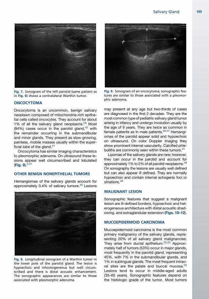
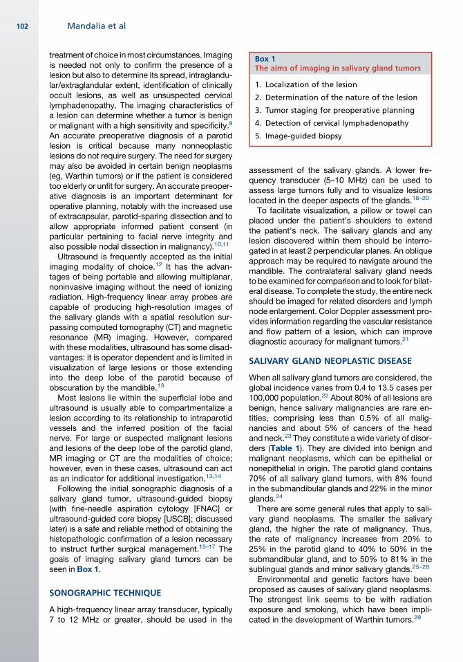
Fig. 7. Sonogram of the left parotid (same patient asin Fig. 6) shows a contralateral Warthin tumor.
Fig. 9. Sonogram of an oncocytoma; sonographic fea-tures are similar to those associated with a pleomor-phic adenoma.
Salivary Gland 105
ONCOCYTOMA
Oncocytoma is an uncommon, benign salivaryneoplasm composed of mitochondria-rich epithe-lial cells called oncocytes. They account for about1% of all the salivary gland neoplasms.26 Most(84%) cases occur in the parotid gland,45 withthe remainder occurring in the submandibularand minor glands. They present as slow-growing,painless, mobile masses usually within the super-ficial lobe of the gland.41
Oncocytoma has similar imaging characteristicsto pleomorphic adenoma. On ultrasound these le-sions appear well circumscribed and lobulated(Fig. 9).4,41
OTHER BENIGN NONEPITHELIAL TUMORS
Hemangiomas of the salivary glands account forapproximately 0.4% of salivary tumors.46 Lesions
Fig. 8. Longitudinal sonogram of a Warthin tumor inthe lower pole of the parotid gland. The lesion ishypoechoic and inhomogeneous but well circum-scribed and there is distal acoustic enhancement.The sonographic appearances are similar to thoseassociated with pleomorphic adenoma.
may present at any age but two-thirds of casesare diagnosed in the first 2 decades. They are themost common typeof pediatric salivary gland tumorarising in infancy and undergo involution usually bythe age of 9 years. They are twice as common infemale patients as in male patients.46,47 Hemangi-omas of the parotid appear solid and hypoechoicon ultrasound. On color Doppler imaging theyshow prominent internal vascularity. Calcified phle-boliths are commonly seen within these tumors.21
Lipomas of the salivary glands are rare; however,they can occur in the parotid and account forapproximately 1% to 2%of all parotid neoplasms.48
On sonography the lesions are usually well definedbut can also appear ill defined. They are normallyhypoechoic and contain internal echogenic foci orstriations.49
MALIGNANT LESION
Sonographic features that suggest a malignantlesion are ill-defined borders, hypoechoic and het-erogeneous architecture with distal acoustic shad-owing, and extraglandular extension (Figs. 10–12).
MUCOEPIDERMOID CARCINOMA
Mucoepidermoid carcinoma is the most commonprimary malignancy of the salivary glands, repre-senting 20% of all salivary gland malignancies.They arise from ductal epithelium.22,25 Approxi-mately half of tumors (53%) occur in major glands,most frequently in the parotid gland, representing45%, with 7% in the submandibular glands, and1% in sublingual glands. The most frequent intrao-ral sites are the palate and buccal mucosa.30
Lesions tend to occur in middle-aged adults(35–65 years). Sonographic features depend onthe histologic grade of the tumor. Most tumors
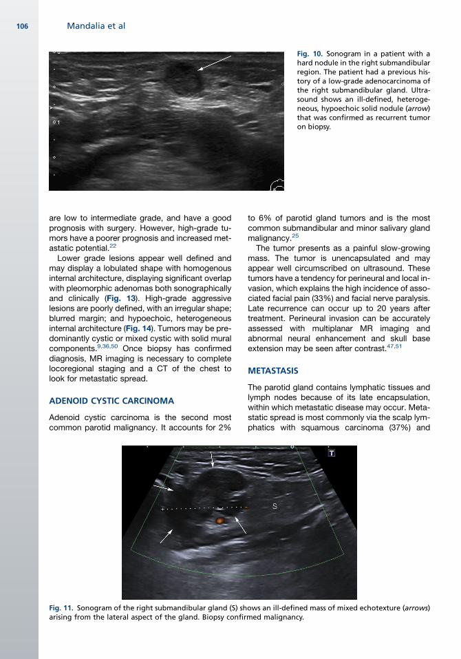
Fig. 10. Sonogram in a patient with ahard nodule in the right submandibularregion. The patient had a previous his-tory of a low-grade adenocarcinoma ofthe right submandibular gland. Ultra-sound shows an ill-defined, heteroge-neous, hypoechoic solid nodule (arrow)that was confirmed as recurrent tumoron biopsy.
Mandalia et al106
are low to intermediate grade, and have a goodprognosis with surgery. However, high-grade tu-mors have a poorer prognosis and increased met-astatic potential.22
Lower grade lesions appear well defined andmay display a lobulated shape with homogenousinternal architecture, displaying significant overlapwith pleomorphic adenomas both sonographicallyand clinically (Fig. 13). High-grade aggressivelesions are poorly defined, with an irregular shape;blurred margin; and hypoechoic, heterogeneousinternal architecture (Fig. 14). Tumors may be pre-dominantly cystic or mixed cystic with solid muralcomponents.9,36,50 Once biopsy has confirmeddiagnosis, MR imaging is necessary to completelocoregional staging and a CT of the chest tolook for metastatic spread.
ADENOID CYSTIC CARCINOMA
Adenoid cystic carcinoma is the second mostcommon parotid malignancy. It accounts for 2%
Fig. 11. Sonogram of the right submandibular gland (S) sharising from the lateral aspect of the gland. Biopsy confir
to 6% of parotid gland tumors and is the mostcommon submandibular and minor salivary glandmalignancy.25
The tumor presents as a painful slow-growingmass. The tumor is unencapsulated and mayappear well circumscribed on ultrasound. Thesetumors have a tendency for perineural and local in-vasion, which explains the high incidence of asso-ciated facial pain (33%) and facial nerve paralysis.Late recurrence can occur up to 20 years aftertreatment. Perineural invasion can be accuratelyassessed with multiplanar MR imaging andabnormal neural enhancement and skull baseextension may be seen after contrast.47,51
METASTASIS
The parotid gland contains lymphatic tissues andlymph nodes because of its late encapsulation,within which metastatic disease may occur. Meta-static spread is most commonly via the scalp lym-phatics with squamous carcinoma (37%) and
ows an ill-defined mass of mixed echotexture (arrows)med malignancy.
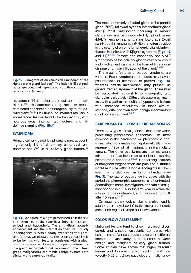
Fig. 12. Sonogram of an acinic cell carcinoma of theright parotid gland (calipers). The lesion is ill defined,heterogeneous, and hypoechoic. Note the extracapsu-lar extension (arrows).
Salivary Gland 107
melanoma (46%) being the most common pri-maries.52 Less commonly lung, renal, or breastcarcinoma can spread hematogenously to the pa-rotid gland.53,54 On ultrasound, metastases vary inappearance: lesions tend to be hypoechoic, withheterogeneous internal architecture and ill-defined margins (Fig. 15).55
LYMPHOMA
Primary salivary gland lymphoma is rare, account-ing for only 5% of all primary extranodal lym-phomas and 2% of all salivary gland tumors.56
Fig. 13. Sonogram of a right parotid nodule (calipers).The lesion lies in the superficial lobe, it is circum-scribed and hypoechoic. There is distal acousticenhancement and the internal architecture is mildlyinhomogeneous, with a poorly hypoechoic focus pre-sent (arrow). On ultrasound, the lesion appears likelyto be benign, with features consistent with a pleo-morphic adenoma. However, biopsy confirmed alow-grade mucoepidermoid carcinoma. Small, low-grade malignancies can mimic benign tumors bothclinically and sonographically.
The most commonly affected gland is the parotidgland (75%), followed by the submandibular gland(20%). Most lymphomas occurring in salivaryglands are mucosa-associated lymphoid tissue(MALT) lymphomas, which are low-grade B-cellnon-Hodgkin lymphomas (NHL) that often developin the setting of chronic lymphoepithelial sialadeni-tis seen in patients with Sjogren syndrome (Figs. 16and 17).57,58 Primary and secondary non-MALTlymphomas of the salivary glands may also occurand involvement can be in the form of focal nodaldisease or diffuse infiltration of the gland.39,59
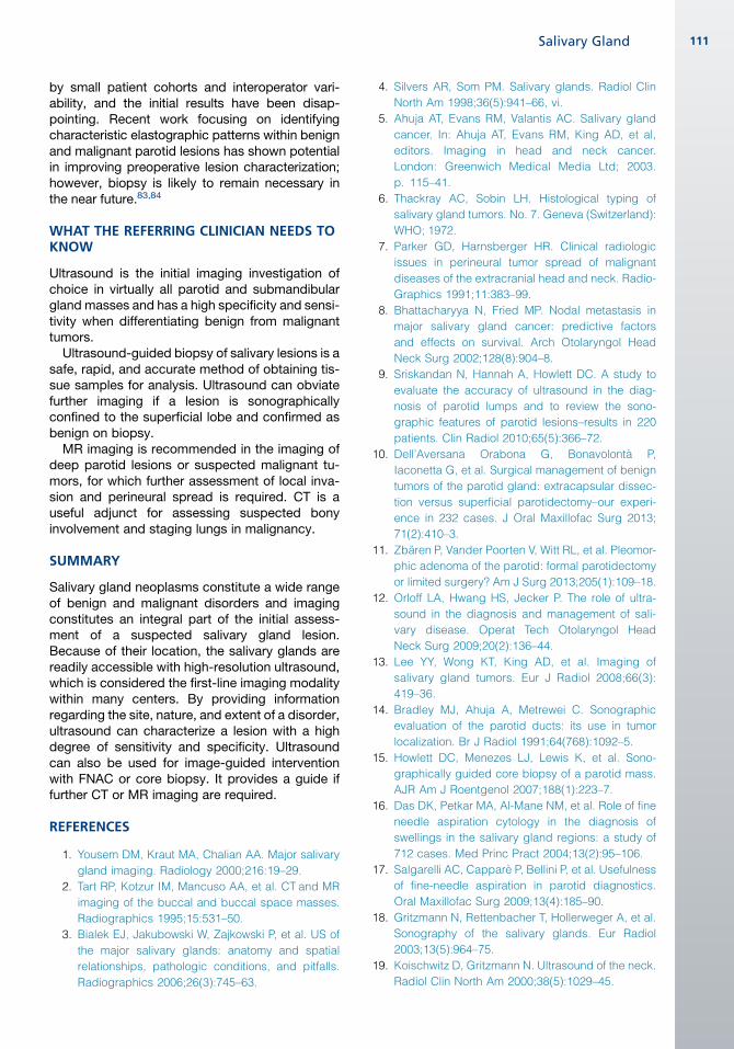
The imaging features of parotid lymphoma arevariable. Focal lymphomatous nodes may have apseudocystic or micronodular pattern (Fig. 18),whereas diffuse involvement may present asgeneralized enlargement of the gland. There maybe associated regional lymphadenopathy andglandular sialectasis. Diffuse disease may mani-fest with a pattern of multiple hypoechoic lesionswith increased vascularity. In these circum-stances, differentiation from benign inflammatoryconditions is required.60,61
CARCINOMA EX PLEOMORPHIC ADENOMAS
There are 3 types of malignancies that occur withinpreexisting pleomorphic adenomas. The mostcommon is the carcinoma ex pleomorphic ade-noma, which originates from epithelial cells; theserepresent 12% of all malignant salivary glandtumors. The other two forms are true malignantmixed tumor (carcinosarcoma) and metastasizingpleomorphic adenoma.62,63 Concerning featuresof malignant degeneration are pain and a suddenincrease in size within a long-standing mass. How-ever, this is also seen in tumor infarction (seeFig. 3). The rate of occurrence increases with theperiod the pleomorphic adenoma is left untreated.According to some investigators, the rate of malig-nant change is 1.5% in the first year in which theadenoma goes untreated, and increases to 9.5%after 15 years.64,65
On imaging they look similar to a pleomorphicadenoma, ormay show infiltrativemargins, necroticareas, and regional lymph node involvement.
COLOR FLOW ASSESSMENT
Malignant lesions tend to show increased, disor-dered, and chaotic vascularity compared withbenign lesion. Various studies have used differentmarkers of vascularity to differentiate betweenbenign and malignant salivary gland tumors.Some studies have shown that highly vascularlesions and those with a high systolic peak flowvelocity (>25 cm/s) are suspicious of malignancy,
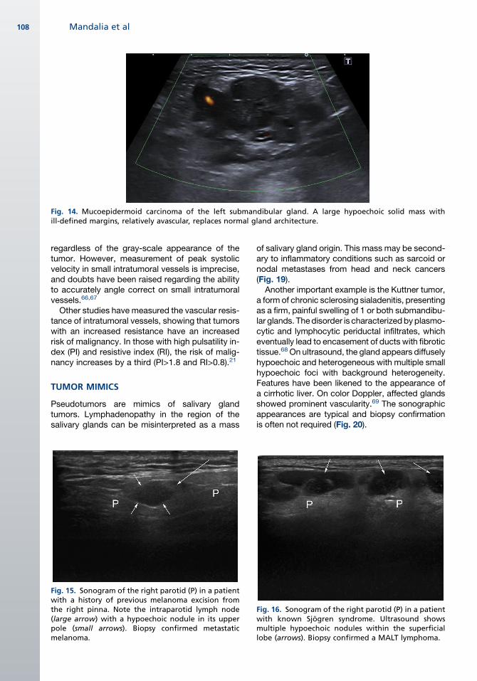
Fig. 14. Mucoepidermoid carcinoma of the left submandibular gland. A large hypoechoic solid mass withill-defined margins, relatively avascular, replaces normal gland architecture.
Mandalia et al108
regardless of the gray-scale appearance of thetumor. However, measurement of peak systolicvelocity in small intratumoral vessels is imprecise,and doubts have been raised regarding the abilityto accurately angle correct on small intratumoralvessels.66,67
Other studies have measured the vascular resis-tance of intratumoral vessels, showing that tumorswith an increased resistance have an increasedrisk of malignancy. In those with high pulsatility in-dex (PI) and resistive index (RI), the risk of malig-nancy increases by a third (PI>1.8 and RI>0.8).21
TUMOR MIMICS
Pseudotumors are mimics of salivary glandtumors. Lymphadenopathy in the region of thesalivary glands can be misinterpreted as a mass
Fig. 15. Sonogram of the right parotid (P) in a patientwith a history of previous melanoma excision fromthe right pinna. Note the intraparotid lymph node(large arrow) with a hypoechoic nodule in its upperpole (small arrows). Biopsy confirmed metastaticmelanoma.
of salivary gland origin. This mass may be second-ary to inflammatory conditions such as sarcoid ornodal metastases from head and neck cancers(Fig. 19).Another important example is the Kuttner tumor,
a form of chronic sclerosing sialadenitis, presentingas a firm, painful swelling of 1 or both submandibu-lar glands. Thedisorder is characterizedbyplasmo-cytic and lymphocytic periductal infiltrates, whicheventually lead to encasement of ducts with fibrotictissue.68 On ultrasound, the gland appears diffuselyhypoechoic and heterogeneous with multiple smallhypoechoic foci with background heterogeneity.Features have been likened to the appearance ofa cirrhotic liver. On color Doppler, affected glandsshowed prominent vascularity.69 The sonographicappearances are typical and biopsy confirmationis often not required (Fig. 20).
Fig. 16. Sonogram of the right parotid (P) in a patientwith known Sjogren syndrome. Ultrasound showsmultiple hypoechoic nodules within the superficiallobe (arrows). Biopsy confirmed a MALT lymphoma.
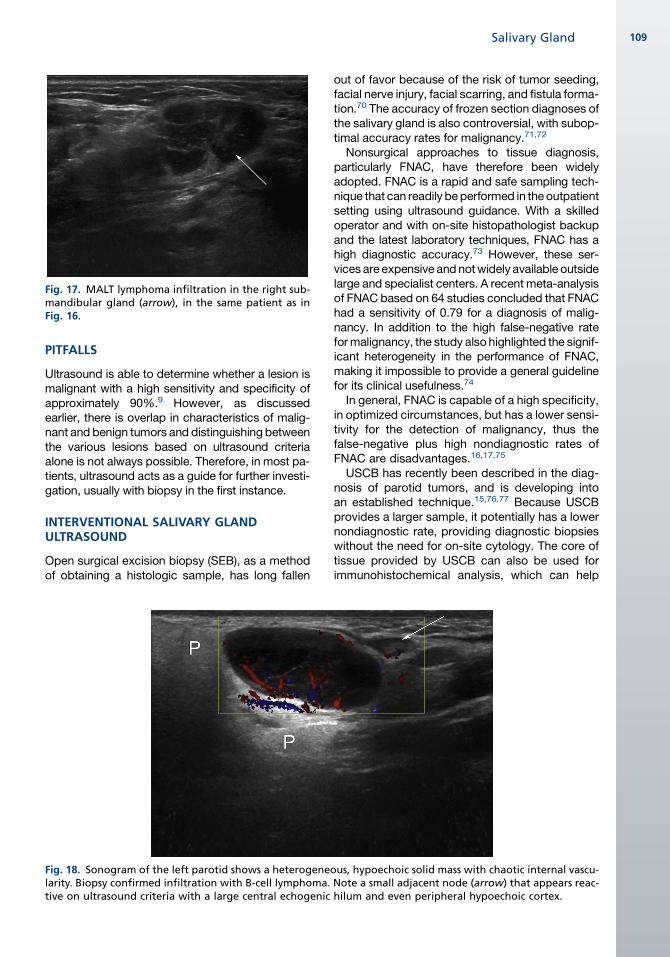
Fig. 17. MALT lymphoma infiltration in the right sub-mandibular gland (arrow), in the same patient as inFig. 16.
Salivary Gland 109
PITFALLS
Ultrasound is able to determine whether a lesion ismalignant with a high sensitivity and specificity ofapproximately 90%.9 However, as discussedearlier, there is overlap in characteristics of malig-nant and benign tumors and distinguishing betweenthe various lesions based on ultrasound criteriaalone is not always possible. Therefore, in most pa-tients, ultrasound acts as a guide for further investi-gation, usually with biopsy in the first instance.
INTERVENTIONAL SALIVARY GLANDULTRASOUND
Open surgical excision biopsy (SEB), as a methodof obtaining a histologic sample, has long fallen
Fig. 18. Sonogram of the left parotid shows a heterogenelarity. Biopsy confirmed infiltration with B-cell lymphoma.tive on ultrasound criteria with a large central echogenic
out of favor because of the risk of tumor seeding,facial nerve injury, facial scarring, and fistula forma-tion.70 The accuracy of frozen section diagnoses ofthe salivary gland is also controversial, with subop-timal accuracy rates for malignancy.71,72
Nonsurgical approaches to tissue diagnosis,particularly FNAC, have therefore been widelyadopted. FNAC is a rapid and safe sampling tech-nique that can readily beperformed in theoutpatientsetting using ultrasound guidance. With a skilledoperator and with on-site histopathologist backupand the latest laboratory techniques, FNAC has ahigh diagnostic accuracy.73 However, these ser-vices are expensive andnotwidely available outsidelarge and specialist centers. A recent meta-analysisof FNAC based on 64 studies concluded that FNAChad a sensitivity of 0.79 for a diagnosis of malig-nancy. In addition to the high false-negative rateformalignancy, the study also highlighted the signif-icant heterogeneity in the performance of FNAC,making it impossible to provide a general guidelinefor its clinical usefulness.74
In general, FNAC is capable of a high specificity,in optimized circumstances, but has a lower sensi-tivity for the detection of malignancy, thus thefalse-negative plus high nondiagnostic rates ofFNAC are disadvantages.16,17,75
USCB has recently been described in the diag-nosis of parotid tumors, and is developing intoan established technique.15,76,77 Because USCBprovides a larger sample, it potentially has a lowernondiagnostic rate, providing diagnostic biopsieswithout the need for on-site cytology. The core oftissue provided by USCB can also be used forimmunohistochemical analysis, which can help
ous, hypoechoic solid mass with chaotic internal vascu-Note a small adjacent node (arrow) that appears reac-hilum and even peripheral hypoechoic cortex.

Fig. 19. Sonogram of the right submandibular space in a patient with a palpablemass. There is a mixed-echotexturemass (arrow) closely abutting and distorting the submandibular gland (S). Biopsy confirmed ametastasis from a squa-mous cell carcinoma, the lesion mimicking a submandibular lesion clinically.
Mandalia et al110
with the grading and typing of parotid malignancythat is crucial in diagnosis of lymphomas. Severalstudies on the performance of USCB have shownthe good diagnostic yields from USCB of the sali-vary glands.75,78,79 A recent meta-analysis of 6studies compiled in 2011, evaluating the accuracyof USCB in the diagnosis of salivary gland lesions,concluded that choice of the test (FNAC vs USCB)to use for an individual patient remains undefined;however, the overall accuracy of core needle bi-opsy is greater than FNAC in some practice set-tings, with less variability in performance.80
NEW DEVELOPMENTS
Sonoelastography is a novel imaging techniquethat canmap the elastic properties of soft tissues.81
A mechanical force, usually manual compressionvia the ultrasound probe, is applied to the region
of interest. The degree and distribution of tissuedeformation is detected and characterized sono-graphically, and is represented visually as an elas-togram of the area of interest. The technique isperformed using a 5-MHz to 12-MHz linear arraytransducer and requires a compatible ultrasoundmachine. Shear wave elastography (SWE) is avariation of sonoelastography.82 In SWE, insteadof the compressive force of the transducer probethe applied mechanical force consists of focusedpulses of ultrasound waves termed push pulses.These induce shear waves that are detected byan ultrafast ultrasound imaging technique. Thistechnique is thought to be more accurate thanstrain elastography because it produces quantita-tive estimates of stiffness and is less operatordependent. There have been several studies ofthe ability of sonoelastography to differentiate pa-rotid neoplasms. These studies have been limited
Fig. 20. Sonogram of the left subman-dibular gland in a patient with a hardleft submandibular gland nodule. Notenormal gland (S) and a geographic areaof reduced echogenicity (calipers). Thisarea is consistent with an area of chronicsialadenitis (Kuttner pseudotumor).Color Doppler assessment can help withthe diagnosis, showing vessels passingfrom the normal to the abnormal glandwith nodeviation. Biopsy is not normallyrequired to confirm this sonographicdiagnosis. A sialogram may be helpfulto exclude a duct stone or stricture.
Salivary Gland 111
by small patient cohorts and interoperator vari-ability, and the initial results have been disap-pointing. Recent work focusing on identifyingcharacteristic elastographic patterns within benignand malignant parotid lesions has shown potentialin improving preoperative lesion characterization;however, biopsy is likely to remain necessary inthe near future.83,84
WHAT THE REFERRING CLINICIAN NEEDS TOKNOW
Ultrasound is the initial imaging investigation ofchoice in virtually all parotid and submandibulargland masses and has a high specificity and sensi-tivity when differentiating benign from malignanttumors.
Ultrasound-guided biopsy of salivary lesions is asafe, rapid, and accurate method of obtaining tis-sue samples for analysis. Ultrasound can obviatefurther imaging if a lesion is sonographicallyconfined to the superficial lobe and confirmed asbenign on biopsy.
MR imaging is recommended in the imaging ofdeep parotid lesions or suspected malignant tu-mors, for which further assessment of local inva-sion and perineural spread is required. CT is auseful adjunct for assessing suspected bonyinvolvement and staging lungs in malignancy.
SUMMARY
Salivary gland neoplasms constitute a wide rangeof benign and malignant disorders and imagingconstitutes an integral part of the initial assess-ment of a suspected salivary gland lesion.Because of their location, the salivary glands arereadily accessible with high-resolution ultrasound,which is considered the first-line imaging modalitywithin many centers. By providing informationregarding the site, nature, and extent of a disorder,ultrasound can characterize a lesion with a highdegree of sensitivity and specificity. Ultrasoundcan also be used for image-guided interventionwith FNAC or core biopsy. It provides a guide iffurther CT or MR imaging are required.
REFERENCES
1. Yousem DM, Kraut MA, Chalian AA. Major salivary
gland imaging. Radiology 2000;216:19–29.
2. Tart RP, Kotzur IM, Mancuso AA, et al. CT and MR
imaging of the buccal and buccal space masses.
Radiographics 1995;15:531–50.
3. Bialek EJ, Jakubowski W, Zajkowski P, et al. US of
the major salivary glands: anatomy and spatial
relationships, pathologic conditions, and pitfalls.
Radiographics 2006;26(3):745–63.
4. Silvers AR, Som PM. Salivary glands. Radiol Clin
North Am 1998;36(5):941–66, vi.
5. Ahuja AT, Evans RM, Valantis AC. Salivary gland
cancer. In: Ahuja AT, Evans RM, King AD, et al,
editors. Imaging in head and neck cancer.
London: Greenwich Medical Media Ltd; 2003.
p. 115–41.
6. Thackray AC, Sobin LH. Histological typing of
salivary gland tumors. No. 7. Geneva (Switzerland):
WHO; 1972.
7. Parker GD, Harnsberger HR. Clinical radiologic
issues in perineural tumor spread of malignant
diseases of the extracranial head and neck. Radio-
Graphics 1991;11:383–99.
8. Bhattacharyya N, Fried MP. Nodal metastasis in
major salivary gland cancer: predictive factors
and effects on survival. Arch Otolaryngol Head
Neck Surg 2002;128(8):904–8.
9. Sriskandan N, Hannah A, Howlett DC. A study to
evaluate the accuracy of ultrasound in the diag-
nosis of parotid lumps and to review the sono-
graphic features of parotid lesions–results in 220
patients. Clin Radiol 2010;65(5):366–72.
10. Dell’Aversana Orabona G, Bonavolonta P,
Iaconetta G, et al. Surgical management of benign
tumors of the parotid gland: extracapsular dissec-
tion versus superficial parotidectomy–our experi-
ence in 232 cases. J Oral Maxillofac Surg 2013;
71(2):410–3.
11. Zbaren P, Vander Poorten V, Witt RL, et al. Pleomor-
phic adenoma of the parotid: formal parotidectomy
or limited surgery? Am J Surg 2013;205(1):109–18.
12. Orloff LA, Hwang HS, Jecker P. The role of ultra-
sound in the diagnosis and management of sali-
vary disease. Operat Tech Otolaryngol Head
Neck Surg 2009;20(2):136–44.
13. Lee YY, Wong KT, King AD, et al. Imaging of
salivary gland tumors. Eur J Radiol 2008;66(3):
419–36.
14. Bradley MJ, Ahuja A, Metrewei C. Sonographic
evaluation of the parotid ducts: its use in tumor
localization. Br J Radiol 1991;64(768):1092–5.
15. Howlett DC, Menezes LJ, Lewis K, et al. Sono-
graphically guided core biopsy of a parotid mass.
AJR Am J Roentgenol 2007;188(1):223–7.
16. Das DK, Petkar MA, Al-Mane NM, et al. Role of fine
needle aspiration cytology in the diagnosis of
swellings in the salivary gland regions: a study of
712 cases. Med Princ Pract 2004;13(2):95–106.
17. Salgarelli AC, Cappare P, Bellini P, et al. Usefulness
of fine-needle aspiration in parotid diagnostics.
Oral Maxillofac Surg 2009;13(4):185–90.
18. Gritzmann N, Rettenbacher T, Hollerweger A, et al.
Sonography of the salivary glands. Eur Radiol
2003;13(5):964–75.
19. Koischwitz D, Gritzmann N. Ultrasound of the neck.
Radiol Clin North Am 2000;38(5):1029–45.
Mandalia et al112
20. Kotecha S, Bhatia P, Rout PG. Diagnostic ultra-
sound in the head and neck region. Dent Update
2008;35(8):529–30, 533–4.
21. Bradley MJ, Durham LH, Lancer JM. The role of
colour flow Doppler in the investigation of the sali-
vary gland tumor. Clin Radiol 2000;55(10):759–62.
22. Auclair PL, Ellis GL, Gnepp DR, et al. Salivary
gland neoplasms. General considerations. In:
Ellis G, Auclair P, Gnepp D, editors. Surgical pa-
thology of the salivary glands. Philadelphia: WB
Saunders; 1991. p. 312–5.
23. WHO. International statistical classification of dis-
eases and related health problems, tenth revision,
vol. 1. Geneva (Switzerland): World Health Organi-
zation; 1992.
24. Eveson JW, Cawson RA. Salivary gland tumors. A
review of 2410 cases with particular reference to
histological types, site, age and sex distribution.
J Pathol 1985;146:51–8.
25. Spiro RH. Salivary neoplasms: overview of a
35-year experience with 2,807 patients. Head
Neck Surg 1986;8:177–84.
26. Pinkston JA,Cole P. Incidence rates of salivary gland
tumors: results from a population-based study.
Otolaryngol Head Neck Surg 1999;120(6):834–40.
27. Kane WJ, McCaffrey TV, Olsen KD, et al. Primary
parotid malignancies: a clinical and pathologic
review. Arch Otolaryngol Head Neck Surg 1991;
117:307–15.
28. Batsakis JG. Tumors of the head and neck: clinical
and pathological considerations. Baltimore (MD):
Williams &Wilkins; 1979.
29. TakeichiN,HiroseF,YamamotoH,etal.Salivarygland
tumors in atomic bomb survivors, Hiroshima, Japan.
II. Pathologic study and supplementary epidemio-
logic observations. Cancer 1983;52:377–85.
30. BarnesL,EvesonJW,ReichartP,SidranskyD,editors.
World Health Organization classification of tumours:
pathology and genetics, head and neck tumours.
Lyon (France): IARC Press; 2005. p. 242–3.
31. Renehan A, Gleave EN, Hancock BD, et al. Long-
term follow-up of over 1000 patients with salivary
gland tumors treated in a single centre. Br J Surg
1996;83:1750–4.
32. Sikorowa L, Meyza JW, Ackerman LW. Salivary
gland tumors. New York: Pergamon; 1982.
33. Tanimoto H, Kumoi K, Otsuki N, et al. Multiple pri-
mary pleomorphic adenomas in a single parotid
gland: report of a new case. Ear Nose Throat J
2002;81(5):341–5.
34. Bia1ek EJ, Jakubowski W, Karpi�nska G. Role of
ultrasonography in diagnosis and differentiation of
pleomorphic adenomas: work in progress. Arch
Otolaryngol Head Neck Surg 2003;129(9):929–33.
35. Zajkowski P, Jakubowski W, Bia1ek EJ, et al. Pleo-
morphic adenoma and adenolymphoma in ultraso-
nography. Eur J Ultrasound 2000;12:23–9.
36. Shimizu M, Ussmuller J, Hartwein J, et al. Statistical
study for sonographic differential diagnosis of
tumorous lesions in the parotid gland. Oral Surg
Oral Med Oral Pathol Oral Radiol Endod 1999;88:
226–33, 74.
37. McGrath MH. Malignant transformation in concur-
rent benign mixed tumors of the parotid and sub-
maxillary glands. Plast Reconstr Surg 1980;65:676.
38. Bryan RN, Mawad ME, Sandlin ME, et al. CT imag-
ing of the salivary glands. Semin Ultrasound CT MR
1986;7:154–65.
39. Pollei SR, Harnsberger HR. The radiologic evalua-
tion of the parotid space. Semin Ultrasound CT
MR 1990;11:486–503.
40. Yoo GH, Eisele DW, Askin FB, et al. Warthin’s tu-
mor: a 40-year experience at the Johns Hopkins
Hospital. Laryngoscope 1994;104:799–803.
41. Som PM, Brandwein MS. Salivary glands: anatomy
and pathology. In: Som PM, Curtin DC, editors.
Head and neck imaging. 4th edition. St Louis
(MO): Mosby; 2003. p. 2005–133, 2.
42. Soler R, Bargiela A, Requejo I, et al. Pictorial re-
view: MR imaging of parotid tumors. Clin Radiol
1997;52(4):269–75.
43. Minami M, Tanioka H, Oyama K, et al. Warthin
tumor of the parotid gland: MR-pathologic correla-
tion. AJNR Am J Neuroradiol 1993;14(1):209–14.
44. CanbayAE, Knorz S, HeimannKD, et al. Sonography
and scintigraphy in the diagnosis of cystadenolym-
phomas (Warthin tumor). Laryngorhinootologie
2002;81:815–9 [in German].
45. BrandweinMS,HuvosAG.Oncocytic tumorsofmajor
salivary glands. A study of 68 cases with follow-up of
44 patients. Am J Surg Pathol 1991;15(6):514–28.
46. Ellis GL, Auclair PL. Tumours of the salivary glands.
3rd edition. Washington, DC: Armed Forces Insti-
tute of Pathology; 1996.
47. Seifert G, Miehlke A, Haubrich J, et al. Diseases of
the salivary glands: pathology, diagnosis, treat-
ment, facial nerve surgery. Stuttgart (Germany):
Thieme Publishing Group; 1986.
48. Ethunandan M, Vura G, Umar T, et al. Lipomatous
lesions of the parotid gland. J Oral Maxillofac
Surg 2006;64(11):1583–6.
49. Chikui T, Yonetsu K, Yoshiura K, et al. Imaging find-
ings of lipomas in the orofacial region with CT, US,
and MRI. Oral Surg Oral Med Oral Pathol Oral Ra-
diol Endod 1997;84(1):88–95.
50. Yoshihara T, Suzuki S, Nagao K. Mucoepidermoid
carcinoma arising in the accessory parotid gland.
Int J Pediatr Otorhinolaryngol 1999;48:47–52.
51. Sigal R, Monnet O, de Baere T, et al. Adenoid
cystic carcinoma of the head and neck: evaluation
with MR imaging and clinical-pathologic correlation
in 27 patients. Radiology 1992;184:95–101.
52. Conley J, Arena S. Parotid gland as a focus of
metastasis. Arch Surg 1963;87:757–64.
Salivary Gland 113
53. Markowski J,GierekT, Zieli�nska-PajakE,et al.Distant
metastases to the parotid gland – review of the litera-
ture and report of own two cases. Otolaryngol Pol
2005;59(4):547–52.
54. Mrena R, Leivo I, Passador-Santos F, et al. Histo-
pathological findings in parotid gland metastases
from renal cell carcinoma. Eur Arch Otorhinolar-
yngol 2008;265(9):1005–9.
55. Howlett DC. High resolution ultrasound assess-
ment of the parotid gland. Br J Radiol 2003;
76(904):271–7.
56. Gleeson MJ, Bennett MH, Cawson RA. Lymphomas
of salivary glands. Cancer 1986;58:699–704.
57. Freeman C, Berg JW, Cutler SJ. Occurrence and
prognosis of extranodal lymphomas. Cancer
1972;29:252–60.
58. Zulman J, Jaffe R, Talal N. Evidence that the malig-
nant lymphoma of Sjogren’s syndrome is a mono-
clonal B-cell neoplasm. N Engl J Med 1978;299:
1215–20.
59. McCurley TL, Collins RD, Ball E, et al. Nodal and
extranodal lymphoproliferative disorders in Sjog-
ren’s syndrome: a clinical and immunopathologic
study. Hum Pathol 1990;21:482–92.
60. Chiou HJ, Chou YH, Chiou SY, et al. High-resolution
ultrasonography of primary peripheral soft tissue
lymphoma. J Ultrasound Med 2005;24:77–86.
61. Lewis K, Vandervelde C, Grace R, et al. Salivary
gland mucosa-associated lymphoid tissue lym-
phoma in 2 patients with Sjogren’s syndrome: clin-
ical and sonographic features with pathological
correlation. J Clin Ultrasound 2007;35(2):97–101.
62. Li Volsi VA, Perzin KH. Malignant mixed tumors
arising in salivary glands. Cancer 1977;39:2209.
63. Antony J, Gopalan V, Smith RA, et al. Carcinoma ex
pleomorphic adenoma: a comprehensive review of
clinical, pathological and molecular data. Head
Neck Pathol 2012;6(1):1–9.
64. Gnepp DR. Malignant mixed tumors of the salivary
glands:a review.PatholAnnu1993;28(Pt1):279–328.
65. Spiro RH, Huvos AG, Strong EW. Malignant mixed
tumor of salivary origin: a clinicopathologic study
of 146 cases. Cancer 1977;39(2):388–96.
66. Schick S, Steiner E, Gahleitner A, et al. Differentia-
tion of benign and malignant tumors of the parotid
gland: value of pulsed Doppler and color Doppler
sonography. Eur Radiol 1998;8(8):1462–7.
67. Martinoli C, Derchi LE, Solbiati L, et al. Color
Doppler sonography of salivary glands. AJR Am
J Roentgenol 1994;163(4):933–41.
68. Beriat GK, Akmansu SH, Kocaturk S, et al. Chronic
sclerosing sialadenitis (Kuttner’s tumour) of the
parotid gland. Malays J Med Sci 2010;17(4):57–61.
69. Ahuja AT, Richards PS, Wong KT, et al. Kuttner
tumour (chronic sclerosing sialadenitis) of the
submandibular gland: sonographic appearances.
Ultrasound Med Biol 2003;29(7):913–9.
70. McGuirt WF, McCabe BF. Significance of node
biopsy before definitive treatment of cervical meta-
static carcinoma. Laryngoscope 1978;88(4):594–7.
71. Carvalho MB, Soares JM, Rapoport A, et al. Periop-
erative frozen section examination in parotid gland
tumors. Sao Paulo Med J 1999;117(6):233–7.
72. Badoual C, Rousseau A, Heudes D, et al. Evalua-
tion of frozen section diagnosis in 721 parotid
gland lesions. Histopathology 2006;49(5):538–40.
73. Robinson IA, Cozens NJ. Does a joint ultrasound–
guided cytology clinic optimize the cytological
evaluation of head and neck masses? Clin Radiol
1999;54:312–6.
74. Schmidt RL, Hall BJ, Wilson AR, et al. A systematic
review and meta-analysis of the diagnostic accu-
racy of fine-needle aspiration cytology for parotid
gland lesions. Am J Clin Pathol 2011;136(1):45–59.
75. Balakrishnan K, Castling B, McMahan J, et al. Fine
needle aspiration cytology in the management of
parotid mass: a two centre retrospective study. Sur-
geon 2005;2:67–72.
76. Wan YL, Chan SC, Chen YL, et al. Ultrasonogra-
phy-guided core-needle biopsy of parotid gland
masses. AJNR Am J Neuroradiol 2004;25(9):
1608–12.
77. Naqvi SQ, Shaikh S, Shah SQ, et al. Ultrasound-
guided core needle biopsy for salivary gland le-
sions. Gomal J Med Sci 2008;6.
78. Breeze J, Andi A, Williams MD, et al. The use of fine
needle core biopsy under ultrasound guidance in
the diagnosis of a parotid mass. Br J Oral Maxillo-
fac Surg 2009;47(1):78–9.
79. Pfeiffer J, Ridder GJ. Diagnostic value of
ultrasound-guided core needle biopsy in patients
with salivary gland masses. Int J Oral Maxillofac
Surg 2012;41(4):437–43.
80. Schmidt RL, Hall BJ, Layfield LJ. A systematic re-
view and meta-analysis of the diagnostic accuracy
of ultrasound-guided core needle biopsy for sali-
vary gland lesions. Am J Clin Pathol 2011;136(4):
516.
81. Lyshchik A, Higashi T, Asato R, et al. Cervical
lymph node metastases: diagnosis at sonoelas-
tography–initial experience. Radiology 2007;243:
258–67.
82. Bhatia KS, Cho CC, Tong CS, et al. Shear wave
elastography of focal salivary gland lesions: prelim-
inary experience in a routine head and neck US
clinic. Eur Radiol 2012;22(5):957–65.
83. Bhatia KS, Rasalkar DD, Lee YP, et al. Evaluation of
real-time qualitative sonoelastography of focal le-
sions in the parotid and submandibular glands: ap-
plications and limitations. Eur Radiol 2010;20:
1958–64.
84. Westerland O, Howlett D. Sonoelastography tech-
niques in the evaluation and diagnosis of parotid
neoplasms. Eur Radiol 2012;22(5):966–9.