Salient pole rotor vs. non salient pole rotor electricaleasy
Review article: Medical intelligence | Published 28 July 2015, doi:10.4414/smw.2015.14154
Cite this as: Swiss Med Wkly. 2015;145:w14154
Salient features of the coronary collateral circulation andits clinical relevance
Michael Stoller, Christian Seiler
Department of Cardiology, Inselspital, Bern University Hospital, and University of Bern, Switzerland
Summary
The coronary collateral circulation provides an alternativesource of blood supply to myocardium jeopardised byischaemia. Collaterals enlarge with obstructive coronaryartery disease to allow bulk flow, but blood flow deliver-able by the native, pre-formed collateral extent can alreadybe sizeable. Genetic determinants contribute significantlyto the wide variability observed in both native collateralextent and its capacity to enlarge, and the severity of thecoronary stenosis is the most significant environmental de-terminant for collateral enlargement. The protective effectof a well-developed coronary collateral circulation trans-lates into relevant improvements in all-cause and cardiacmortality in the acute and chronic phases of coronary arterydisease, as well as into a reduction of future adverse cardi-ovascular events.
Key words: coronary circulation; collateral circulation;myocardial ischaemia; coronary artery disease
Introduction
Coronary collaterals are cross-connections between coron-ary arteries, which are present regardless of coronary arterydisease (CAD) [1]. As natural bypasses, coronary collater-als provide an alternative source of blood flow to a myocar-dial area when normal antegrade supply is compromised.In the presence of arterial stenoses, the process of collateralremodelling leads to a gradual calibre increase of the col-lateral vessels, allowing the effective delivery of bulk flowto jeopardised tissue [2]. Conversely, the collateral networkin the absence of obstructive coronary artery disease rep-resents the native collateral circulation, pre-existing in theabsence of coronary narrowing. Contrary to popular belief,blood flow deliverable by native collaterals can be sizeable[3]. The relevance of the coronary collateral circulation be-comes eminent in CAD, which remains a leading causeof death globally [4]. In acute coronary occlusion, the ex-tent of collateral pathways is a significant determinant ofthe severity of myocardial infarction [5–8]. In the chronicphase of CAD, the collateral circulation variably com-pensates for the flow-limiting features of atherosclerosis,i.e., stenotic lesions.
The purpose of this article is to give a concise review of thesalient features of the coronary collateral circulation andprovide an overview of its assessment and clinical relev-ance.
Coronary collateral dynamics
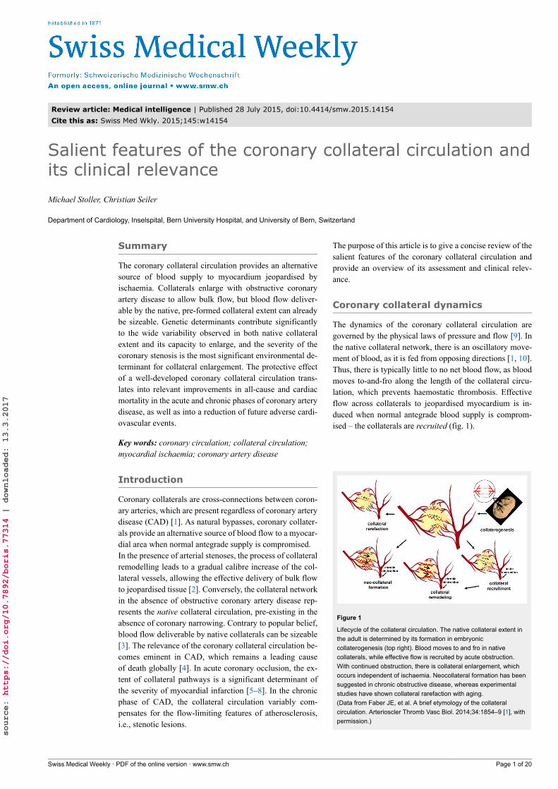
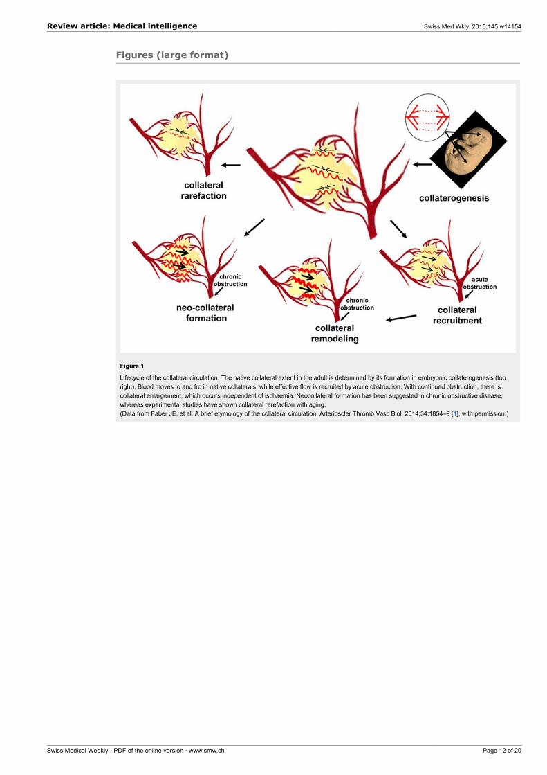
The dynamics of the coronary collateral circulation aregoverned by the physical laws of pressure and flow [9]. Inthe native collateral network, there is an oscillatory move-ment of blood, as it is fed from opposing directions [1, 10].Thus, there is typically little to no net blood flow, as bloodmoves to-and-fro along the length of the collateral circu-lation, which prevents haemostatic thrombosis. Effectiveflow across collaterals to jeopardised myocardium is in-duced when normal antegrade blood supply is comprom-ised – the collaterals are recruited (fig. 1).
Figure 1
Lifecycle of the collateral circulation. The native collateral extent inthe adult is determined by its formation in embryoniccollaterogenesis (top right). Blood moves to and fro in nativecollaterals, while effective flow is recruited by acute obstruction.With continued obstruction, there is collateral enlargement, whichoccurs independent of ischaemia. Neocollateral formation has beensuggested in chronic obstructive disease, whereas experimentalstudies have shown collateral rarefaction with aging.(Data from Faber JE, et al. A brief etymology of the collateralcirculation. Arterioscler Thromb Vasc Biol. 2014;34:1854–9 [1], withpermission.)
Swiss Medical Weekly · PDF of the online version · www.smw.ch Page 1 of 20
source: https://doi.org/10.7892/boris.77314 | downloaded: 13.3.2017
Regulation of collateral growth independent ofischaemiaAlterations in blood flow and blood pressure are the keyregulators of the adaptive process in recruited collaterals,known as remodelling. A haemodynamically significantcoronary stenosis reduces coronary perfusion pressuredistal to the narrowed segment as a result of energy losses.Autoregulation maintains basal flow distal to the stenosisby microvascular vasodilation up to a critical reduction invessel lumen area, beyond which resting coronary bloodflow is also reduced [11]. Concurrently, the pressure dropacross the stenosis leads to a pressure gradient along thepre-existing collaterals connecting to the constricted ves-sel, leading to their recruitment: effective flow is inducedfrom the donor artery across the collateral network to therecipient coronary artery. Heightened flow velocity leads toa proportional increase in fluid shear stress (FSS), whereasthe increased intravascular pressure leads to a proportionalincrease in circumferential wall stress (CWS) [3, 12]. Witha prolonged stimulus, the collateral network reacts by in-creasing the lumen diameter to normalise FSS and by in-creasing the wall thickness to normalise CWS [13].
Range of adaptation of collateralsThe term arteriogenesis was introduced to differentiategrowth of pre-existing collaterals from angiogenesis,which is triggered by ischaemia [12, 14]. Blood flow canincrease maximally 10- to 20-fold by arteriogenesis, butonly 1.5- to 1.7 fold by angiogenesis, which refers to thesprouting of new, minute, high-resistance, low-flow capil-laries [13]. To generate the same lumen area as the arteryby angiogenesis would in essence mean replacing the organwith capillaries – with the end result of a haemangioma[13, 15]. Thus, arteriogenesis has the capacity to com-pensate for an occluded artery, whereas angiogenesis doesnot [12, 16].Even though mature collaterals are able to deliver bulkflow to jeopardised tissue, collateral remodelling stopsshort of completely reconstituting the function of an oc-cluded artery [17, 18]. Primarily responsible for this cir-cumstance is the premature normalisation of the dominantdriving force for continued outward remodelling – flow-in-duced fluid shear stress. Even a small increase in luminaldiameter leads to a much larger fall in FSS, a consequenceof FSS being inversely related to the third power of the ves-sel radius. In physiological circumstances, normal maxim-al blood flow is, therefore, not achievable even in healthycollateral-dependent tissues [19]. Conversely, a completefunctional reconstitution, even surpassing normal maximalfunction, is achieved when FSS is artificially increased [17,18, 20, 21].
Collateral remodelling and its relation to the phases ofcoronary artery diseaseCollateral remodelling is a mitotic process which takes twoto three days before a relevant increase beyond the acutelyrecruitable blood flow begins to occur [2, 3, 16, 22]. Theproliferation of endothelial and smooth muscle cells withperivascular gathering of bone marrow derived cells (not-ably macrophages and T cells) is followed by controlleddestruction of the collateral vessel to build a new scaffold
for the much larger vessel [3]. In the process of many re-modelling collateral connections, a larger diameter in somecollaterals presents an advantage in the ensuing competi-tion for flow. As a consequence, only a few collaterals ul-timately reach maturation, while many others are closed byan overshoot of intimal proliferation – a process known aspruning [2, 23].In patients with chronic total occlusion (CTO), collateralfunction improves over a time period of 12 weeks after oc-clusion [24], which is in line with observations in experi-mental studies [25]. The collateral remodelling in these pa-tients typically relates to chronic stable CAD in a setting ofa more or less gradual progression of coronary obstruction,which allows sufficient time for the growing collaterals topreserve myocardial viability.In patients with acute coronary occlusion, the timeframefor insufficient collaterals to remodel (further) is generallytoo short. Plaque rupture is the most common cause ofacute coronary syndromes, which, when followed by ath-erothrombosis, leads to rapid and insidious thrombotic oc-clusion [26]. In this context, it is of note that most of thecoronary lesions ultimately responsible for major adversecardiovascular events (MACE) have been shown to be an-giographically mild [27]. Therefore, the culprit lesion itselfis unlikely to cause relevant collateral remodelling [27–29].In the absence of stenosis-induced enlargement, the pro-tective effect from acute ischaemia is entirely dependent onthe extent of the native collateral network. Therefore, thenotion of the native collateral circulation is pertinent notonly in the healthy state, but also in coronary artery disease(CAD).Notably, myocardial viability is not a prerequisite for theremodelling of coronary collaterals [31, 32]. Thus, well-de-veloped collaterals can be observed to grow only after itsdependent myocardium has become entirely necrotic, againconsistent with the notion that collateral growth can occurindependently of ischaemia.
Genetic and environmentaldeterminants of the coronarycollateral circulation
The wide interindividual variability in coronary collateralsis evident not only with regard to the preformed extent, butalso with regard to its capacity to remodel. Specifically, theextent of native collaterals relates to the formation and mat-uration of collaterals in early life [30], whereas collateralremodelling takes place generally much later (most oftenunder the influence of coronary obstruction). In experi-mental studies, the use of different animal models permitsthese aspects to be studied also in healthy tissues, which al-lows genetic factors to be separated from environmental in-fluences. Conversely, the protective role of collaterals be-comes eminent chiefly in CAD, the risk of which increaseswith the number and severity of cardiovascular risk factors.Accordingly, the impact of the risk factors predisposing toatherosclerosis has to be considered, too [31].
Severity of coronary obstructionThe presence of a haemodynamically significant stenosis isthe sine qua non for the calibre increase of collaterals in
Review article: Medical intelligence Swiss Med Wkly. 2015;145:w14154
Swiss Medical Weekly · PDF of the online version · www.smw.ch Page 2 of 20
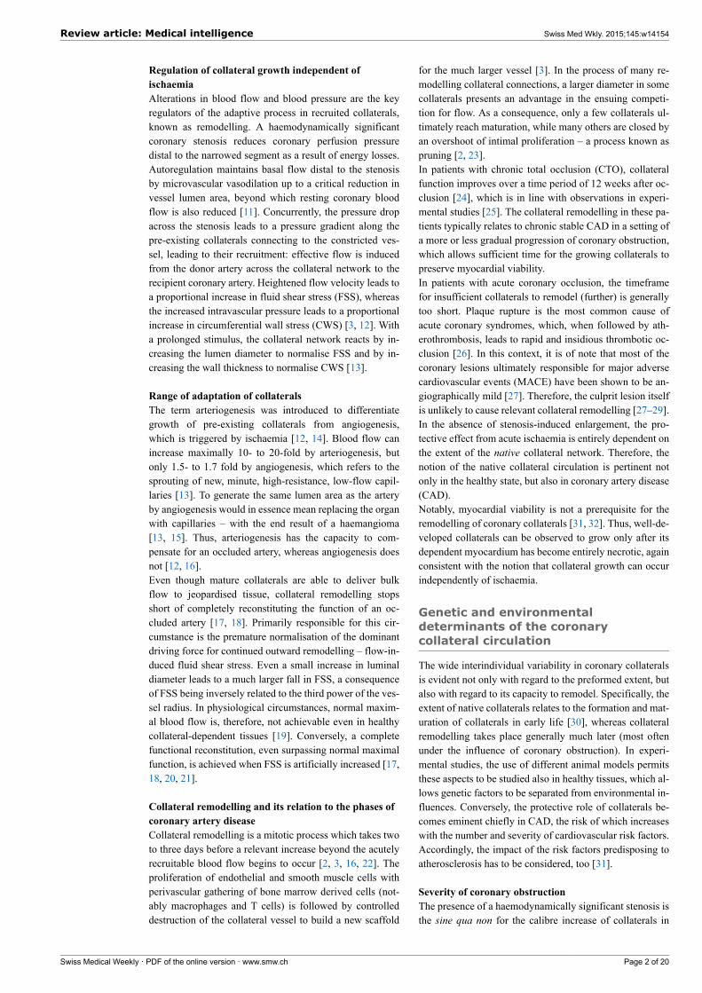
remodelling. The severity of the flow impediment dictatesthe pressure drop, which in turn determines the amount ofshear stress in recruited collaterals, with higher shear stressleading to more pronounced remodelling. Consequently, inpatients with CAD, the severity of the underlying arterialobstruction has been found to be the only independent pre-dictor for good collateral function [32, 33]. Furthermore,collateral flow was found to be higher in CTO than innonocclusive obstructions [24, 34–38].The effect of continued remodelling is well illustrated bythe frequency distributions of collateral function with in-creasing severity of the underlying CAD (fig. 2) [32, 39,40]. Although sufficient collaterals are already present in asignificant minority of patients with angiographically nor-mal coronary arteries [39], with CAD there is a rightward
Figure 2
Frequency distribution (vertical axes in percent) of collateral flowindex (CFI, horizontal axes) in patients without coronary arterydisease (CAD; top left) and with increasing severity of CAD.Sufficient collaterals by CFI are present in a fourth of patients withangiographically normal coronary arteries, whereas this proportionincreases to one third in patients with CAD.(Data from Meier P, et al. Beneficial effect of recruitable collaterals:a 10-year follow-up study in patients with stable coronary arterydisease undergoing quantitative collateral measurements.Circulation. 2007;116:975–83 [40], with permission.)
Figure 3
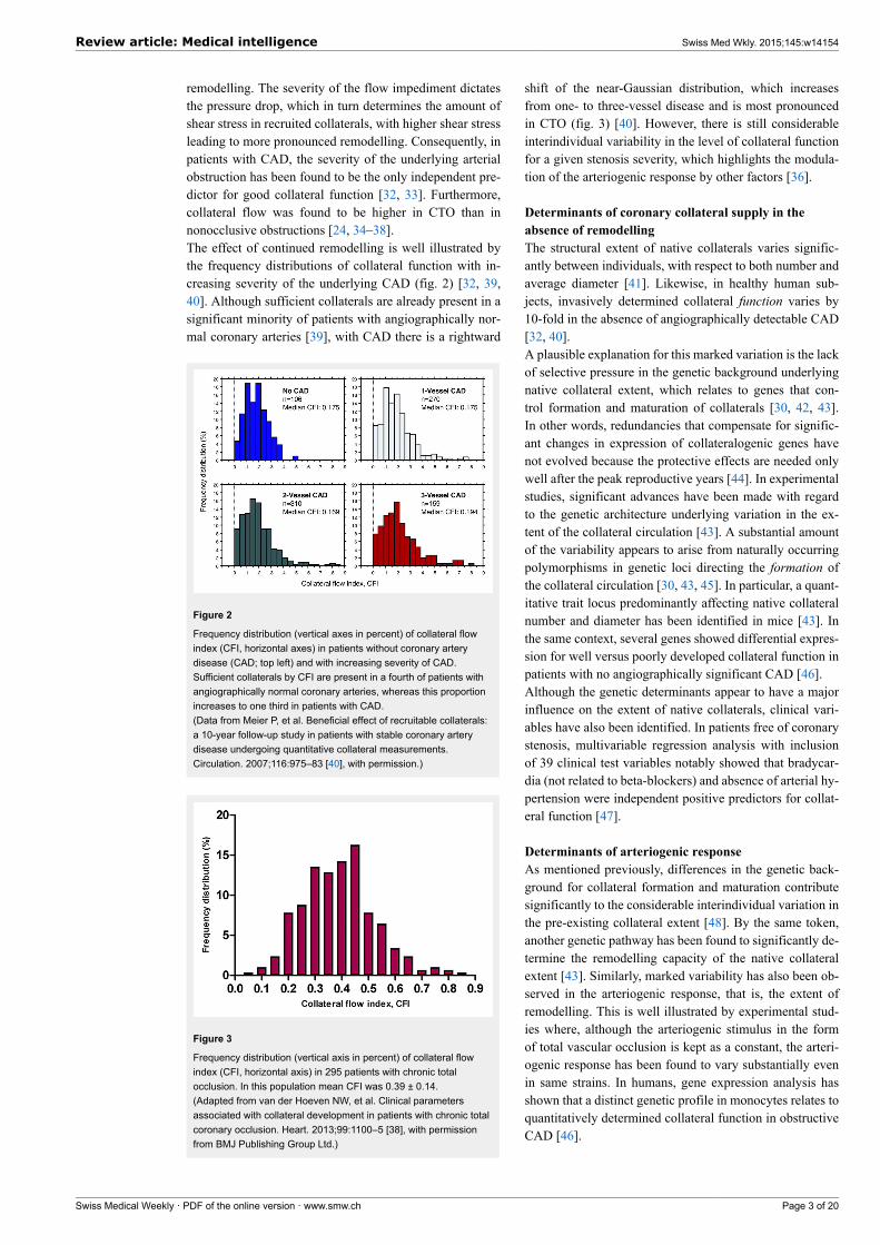
Frequency distribution (vertical axis in percent) of collateral flowindex (CFI, horizontal axis) in 295 patients with chronic totalocclusion. In this population mean CFI was 0.39 ± 0.14.(Adapted from van der Hoeven NW, et al. Clinical parametersassociated with collateral development in patients with chronic totalcoronary occlusion. Heart. 2013;99:1100–5 [38], with permissionfrom BMJ Publishing Group Ltd.)
shift of the near-Gaussian distribution, which increasesfrom one- to three-vessel disease and is most pronouncedin CTO (fig. 3) [40]. However, there is still considerableinterindividual variability in the level of collateral functionfor a given stenosis severity, which highlights the modula-tion of the arteriogenic response by other factors [36].
Determinants of coronary collateral supply in theabsence of remodellingThe structural extent of native collaterals varies signific-antly between individuals, with respect to both number andaverage diameter [41]. Likewise, in healthy human sub-jects, invasively determined collateral function varies by10-fold in the absence of angiographically detectable CAD[32, 40].A plausible explanation for this marked variation is the lackof selective pressure in the genetic background underlyingnative collateral extent, which relates to genes that con-trol formation and maturation of collaterals [30, 42, 43].In other words, redundancies that compensate for signific-ant changes in expression of collateralogenic genes havenot evolved because the protective effects are needed onlywell after the peak reproductive years [44]. In experimentalstudies, significant advances have been made with regardto the genetic architecture underlying variation in the ex-tent of the collateral circulation [43]. A substantial amountof the variability appears to arise from naturally occurringpolymorphisms in genetic loci directing the formation ofthe collateral circulation [30, 43, 45]. In particular, a quant-itative trait locus predominantly affecting native collateralnumber and diameter has been identified in mice [43]. Inthe same context, several genes showed differential expres-sion for well versus poorly developed collateral function inpatients with no angiographically significant CAD [46].Although the genetic determinants appear to have a majorinfluence on the extent of native collaterals, clinical vari-ables have also been identified. In patients free of coronarystenosis, multivariable regression analysis with inclusionof 39 clinical test variables notably showed that bradycar-dia (not related to beta-blockers) and absence of arterial hy-pertension were independent positive predictors for collat-eral function [47].
Determinants of arteriogenic responseAs mentioned previously, differences in the genetic back-ground for collateral formation and maturation contributesignificantly to the considerable interindividual variation inthe pre-existing collateral extent [48]. By the same token,another genetic pathway has been found to significantly de-termine the remodelling capacity of the native collateralextent [43]. Similarly, marked variability has also been ob-served in the arteriogenic response, that is, the extent ofremodelling. This is well illustrated by experimental stud-ies where, although the arteriogenic stimulus in the formof total vascular occlusion is kept as a constant, the arteri-ogenic response has been found to vary substantially evenin same strains. In humans, gene expression analysis hasshown that a distinct genetic profile in monocytes relates toquantitatively determined collateral function in obstructiveCAD [46].
Review article: Medical intelligence Swiss Med Wkly. 2015;145:w14154
Swiss Medical Weekly · PDF of the online version · www.smw.ch Page 3 of 20
Apart from the marked variability in both native collateralextent and capacity to remodel, another consistent findingis that a poor arteriogenic response is also associated withpoor native collateral extent [17, 46, 47]. Indeed, animalswith poor pre-existing collaterals reach significantly lowerlevels of collateral function after arterial occlusion com-pared with animals with good pre-existing collaterals bothwithin, as well as between, strains. Although this obser-vation may be consistent with a genetic link influencingboth maturation and remodelling of collaterals, such an as-sociation has not been found. Then again, on a conceptu-al basis, such an assumption appears superfluous, given theplausibility that the pre-existing collateral extent presents alimiting factor for its enlargement in remodelling. In otherwords, a poor native collateral circulation itself appears tobe a limiting factor for its later positive remodelling, in thatthe extent of flow recovery reaches levels inferior to thatfrom a rich native collateral network [48]. Thus, the pre-existing collateral extent determines critically the degree ofprotection from ischaemic injury not only in the absence,but also in the presence of collateral remodelling, in thatit also governs the capacity for outward remodelling in thelatter [16, 48–51].
Influence of traditional cardiovascular risk factorsIn the majority of cases, coronary atherosclerosis coincideswith one or more cardiovascular risk factors. Given that en-dothelial dysfunction is a well-established response to car-diovascular risk factors, it is conceivable that the collateralcirculation might equally be affected. Notably though, col-lateral arteries and arterioles do not seem prone to athero-sclerosis [6].Dyslipidaemia has been shown to hamper arteriogenesis inhyperlipidaemic experimental models [52, 53]. Contrarily,no evidence exists for such an effect in humans: dyslipid-aemia has not been found to be an independent predictorfor quantitatively determined collateral function in patientswith or without CAD [32]. Furthermore, in a large clinicalstudy in patients with CAD, no effect of statins on coronarycollateral function was found [54].Arterial hypertension is reflected in the tortuosity and en-larged calibre of arteries, secondary to an adaptive responseto increased shear stress [55]. Two clinical studies in pa-tients with obstructive CAD found conflicting results inthis regard, but both were limited in that occlusion of thecollateral-receiving artery was not uniformly present orperformed for appropriate angiographic assessment [56,57]. In patients with CTO with quantitatively assessed col-lateral function, hypertension was a predictor for better de-veloped collaterals in a multivariate analysis. In the humanheart without CAD, the contrary has been found to be true:better collateral function in the absence of arterial hyper-tension [47].Increasing age is a well-known risk factor for atheroscler-osis. Structural alterations with advancing age are accom-panied by a decline in vascular function [58]. However, itsimpact on the collateral circulation is equivocal. Experi-mental studies have documented a decline in structural ex-tent of pre-existing collaterals, which is compounded by re-duced remodelling [51]. A possible mechanism has beensuggested to be the unique haemodynamic environment
with oscillatory shear stress that could predispose the col-lateral circulation to accelerated aging compared with thegeneral circulation. Alternatively, or in addition, impairedendothelial nitric oxide signalling, deemed to be an im-portant factor for maintenance of collateral density, as wellas remodelling in obstructive disease, has been implicatedin the increased propensity of endothelial cells to under-go apoptosis and thereby cause collateral rarefaction [51,59]. Clinically, a causal link between increased mortality ofelderly individuals in acute myocardial infarction and re-duced collateral extent could be supported by the inverserelation between age and the level of angiographic collat-eral extent seen in these patients [60, 61]. However, theclinical association of higher mortality in ischaemic cardi-ovascular disease and poorly developed collaterals has notbeen shown to be independent. In a univariate analysis of alarge database including quantitatively determined collater-al function, only a trend towards the described inverse rela-tion was seen [37]. Furthermore, in a pooled analysis of pa-tients with a CTO and quantitatively determined collateralfunction, an association between higher age and poorly de-veloped collaterals could not be confirmed [38]. In essence,although experimental data are consistent with an age-asso-ciated decline in collateral extent and remodelling, on bal-ance, age is probably not a major determinant of collateralfunction in humans.No differences have been found in quantitatively determin-ed collateral function among matched patients with versuswithout diabetes in angiographically normal or diseasedcoronary arteries [62]. Furthermore, in a patient cohortwith CTO, a multivariate analysis confirmed these findings[38]. Earlier controversies in this context are likely at-tributable to the above-mentioned methodical flaw in an-giographic assessment [63–65].
Assessment of the coronary collateralcirculation
An accurate assessment of the coronary collateral circula-tion requires normal antegrade and collateral blood flowto be distinguished. In the absence of a coronary stenosisthere is essentially no measurable net blood flow acrosspre-formed, but not recruited collaterals. Conversely, in thepresence of nonocclusive disease, the contribution of nor-mal antegrade blood flow cannot be differentiated from therecruited blood flow via the anastomotic pathways. There-fore, for exclusive characterisation of recruitable collater-al blood flow, (transient) coronary occlusion is required,regardless of the method employed [66, 67]. Accordingly,noninvasive imaging techniques are limited to the re-modeled collateral circulation supplying chronic totalcoronary occlusions [66]. Conversely, invasive techniques,i.e., during coronary catheterisation, permit an assessmentof both the native and remodelled collateral circulation.Different modalities allow evaluation of the ischaemia-abating function of coronary collaterals. Abnormalities indistribution and quantity of myocardial blood flow are theearliest pathophysiological events in myocardial ischaemia[67]. Imaging techniques that measure perfusion are there-fore most distinctive in collateral assessment, given thatsufficient collaterals may prevent even stress-induced ab-
Review article: Medical intelligence Swiss Med Wkly. 2015;145:w14154
Swiss Medical Weekly · PDF of the online version · www.smw.ch Page 4 of 20
normalities occurring later in the ischaemic cascade [68].Only a critical reduction in perfusion leads first to animpairment of diastolic, then of systolic function, followedby electrocardiographic (ECG) changes and ultimately bysymptomatic expression (angina pectoris).Commonly employed imaging techniques that delineatecoronary anatomy allow only a very limited structural as-sessment of coronary collaterals, given that most collateralsare below their spatial resolution and therefore go undetec-ted.
Noninvasive assessmentClinically, myocardial perfusion and wall motion functionare measurable notably by (myocardial contrast) echocardi-ography (MCE) [69], positron emission tomography (PET)[66, 70], single-photon emission computed tomography(SPECT) [71], cardiac magnetic resonance imaging (CMR)[72], and cardiac computed tomography (CT). Preservationof perfusion and, consequently, wall motion function [73]vary as a function of the underlying collateral supply andthe extent and degree of coronary lesions. Similarly, fora given lesion severity, there is a continuum from a com-pletely normal ECG under stress conditions to transient,chronic and acute repolarisation abnormalities [74]. It fol-lows from the above considerations that collateral assess-ment by noninvasive techniques is most distinctive in thepresence of total coronary occlusion. In nonocclusive
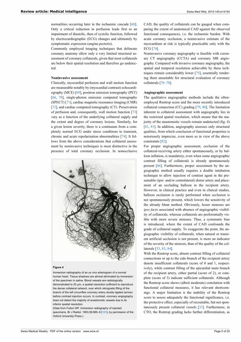
Figure 4
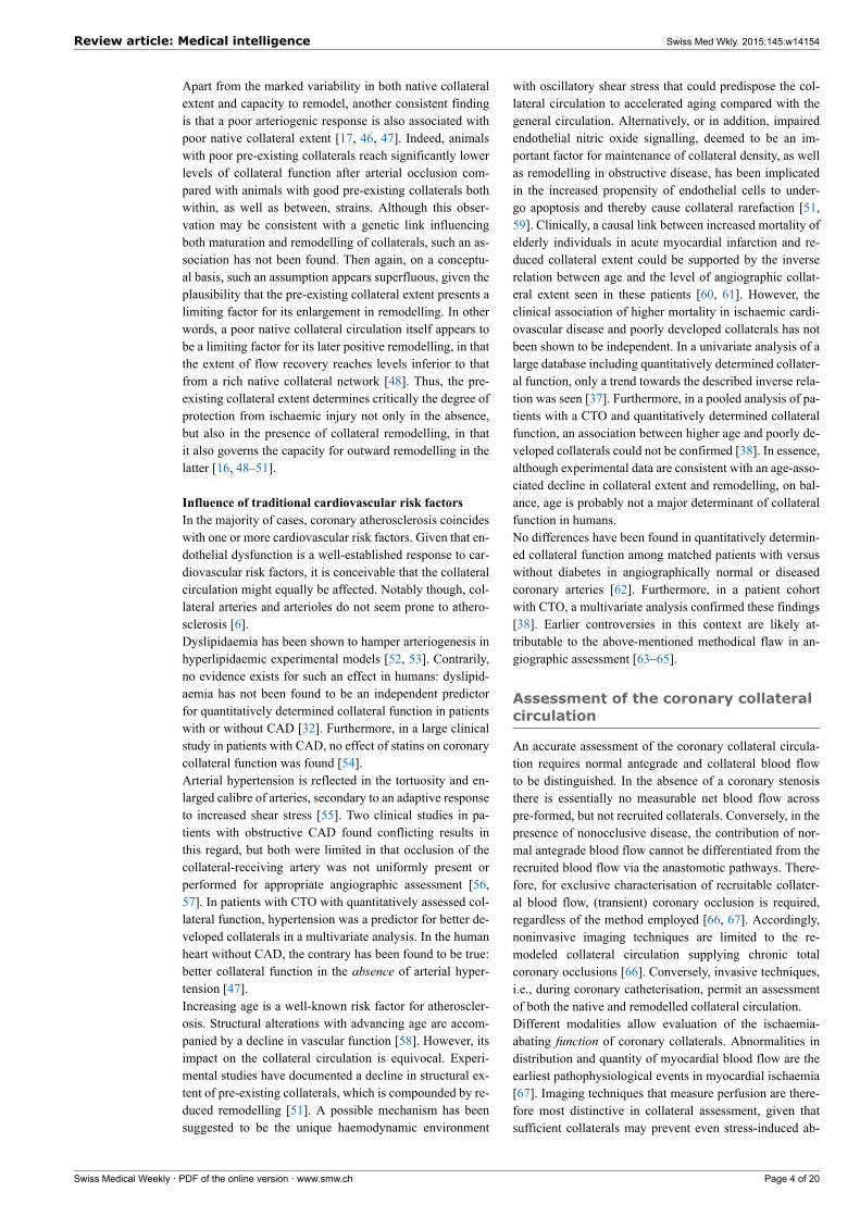
Immersion radiography of an ex vivo arteriogram of a normalhuman heart. Tissue shadows are almost eliminated by immersionof the specimen in saline. Blood vessels are radiologicallydemonstrated to 20 µm, a spatial resolution sufficient to reproducethe dense collateral network, over which retrograde filling of thebranch of the left circumflex coronary artery doubly ligated (arrow)before contrast injection occurs. In contrast, coronary angiographydoes not detect the majority of anastomotic vessels due to itsinferior spatial resolution.(Data from Fulton WF. Immersion radiography of injectedspecimens. Br J Radiol. 1963;36:685–8 [121], by permission of theOxford University Press.)
CAD, the quality of collaterals can be gauged when com-paring the extent of anatomical CAD against the observedfunctional consequences, i.e. the ischaemic burden. Withacute coronary occlusion, a noninvasive estimate of themyocardium at risk is typically practicable only with theECG [74].Noninvasive coronary angiography is feasible with coron-ary CT angiography (CCTA) and coronary MR angio-graphy. Compared with invasive coronary angiography, thespatial and temporal resolution achievable by these tech-niques remain considerably lower [75], essentially render-ing them unsuitable for structural evaluation of coronarycollaterals [76–78].
Angiographic assessmentThe qualitative angiographic methods include the often-employed Rentrop score and the more recently introducedcollateral connection (CC) grading [79, 80]. The limitationinherent to collateral assessment with angiography lies inthe restricted spatial resolution, which means that the ma-jority of the anastomotic vessels remain undetected (fig. 4)[41, 81]. In addition, angiography assesses only structuralqualities, from which conclusion of functional properties isnotoriously imprecise, even more so in view of the aboveconstraints [82].For proper angiographic assessment, occlusion of thecollateral-receiving artery either spontaneously, or by bal-loon inflation, is mandatory, even when some angiographiccontrast filling of collaterals is already spontaneouslypresent [66]. Furthermore, proper assessment by the an-giographic method usually requires a double intubationtechnique to allow injection of contrast agent in the pre-sumable (ipsi- and/or contralateral) donor artery and place-ment of an occluding balloon in the recipient artery.However, in clinical practice and even in clinical studies,balloon occlusion is rarely performed when occlusion isnot spontaneously present, which lowers the sensitivity ofthe already blunt method. Obviously, lesser stenoses areipso facto associated with absence of angiographic visibil-ity of collaterals, whereas collaterals are preferentially vis-ible with more severe stenoses. Thus, a systematic biasis introduced, where the extent of CAD confounds thegrade of collateral supply. To exaggerate the point, the an-giographic visibility of collaterals, when natural or transi-ent artificial occlusion is not present, is more an indicatorof the severity of the stenosis, than of the quality of the col-laterals [33, 83, 84].With the Rentrop score, absent contrast filling of collateralconnections or up to the side-branch of the recipient arterydenote insufficient collaterals (score of 0 and 1, respect-ively), while contrast filling of the epicardial main branchof the recipient artery, either partial (score of 2), or com-plete (score of 3) indicate sufficient collaterals. Althoughthe Rentrop score shows (albeit moderate) correlation withfunctional collateral measures, it has relevant shortcom-ings. A major limitation is the inability of the Rentropscore to assess adequately the functional significance, i.e.the protective effect, especially of recruitable, but not spon-taneously present collateral vessels [33]. Furthermore, inCTO, the Rentrop grading lacks further differentiation, as
Review article: Medical intelligence Swiss Med Wkly. 2015;145:w14154
Swiss Medical Weekly · PDF of the online version · www.smw.ch Page 5 of 20
collaterals are predominantly of Rentrop grade 3 in this set-ting [80].The CC score was evaluated in patients with CTO and itsapplicability in nonocclusive lesions has so far not been es-tablished [80]. Collaterals are evaluated according to thepresence of a continuous connection between donor andrecipient artery: CC0, no continuous connection betweendonor and recipient artery; CC1, continuous, threadlikeconnection; and CC2, continuous, small side branch–likesize of the collateral throughout its course [80]. Regardingthe clinical relevance of the CC grading, in patients withoutprior Q-wave myocardial infarction, the regional wall mo-tion was best preserved with grade CC2 collaterals [80].Furthermore, the CC score was closely associated with in-vasively determined parameters of collateral haemodynam-ics [80].Instead of angiographic grading, assessment of collateralsby means of the semiquantitative washout collaterometryrelies on the time to clearance of contrast medium trappedby balloon occlusion [85]. It correlates well with invasivelydetermined collateral function and distinguishes accuratelybetween sufficient and insufficient collaterals [86].
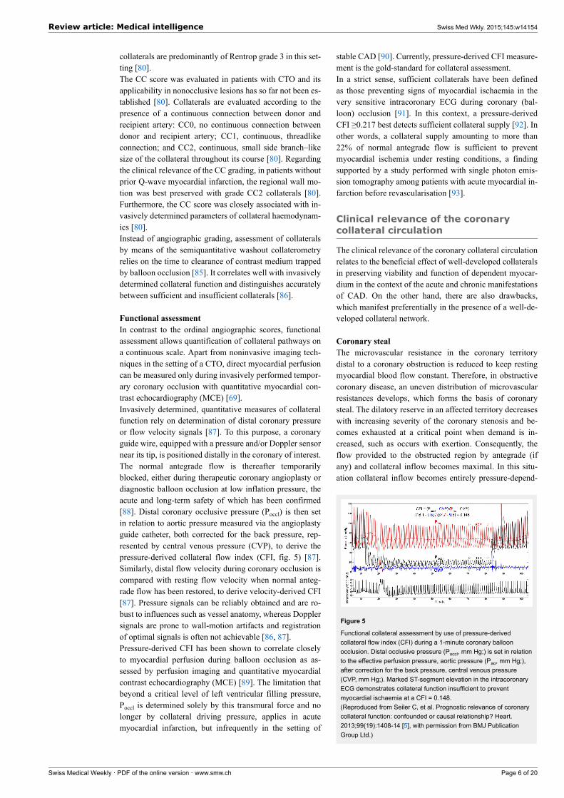
Functional assessmentIn contrast to the ordinal angiographic scores, functionalassessment allows quantification of collateral pathways ona continuous scale. Apart from noninvasive imaging tech-niques in the setting of a CTO, direct myocardial perfusioncan be measured only during invasively performed tempor-ary coronary occlusion with quantitative myocardial con-trast echocardiography (MCE) [69].Invasively determined, quantitative measures of collateralfunction rely on determination of distal coronary pressureor flow velocity signals [87]. To this purpose, a coronaryguide wire, equipped with a pressure and/or Doppler sensornear its tip, is positioned distally in the coronary of interest.The normal antegrade flow is thereafter temporarilyblocked, either during therapeutic coronary angioplasty ordiagnostic balloon occlusion at low inflation pressure, theacute and long-term safety of which has been confirmed[88]. Distal coronary occlusive pressure (Poccl) is then setin relation to aortic pressure measured via the angioplastyguide catheter, both corrected for the back pressure, rep-resented by central venous pressure (CVP), to derive thepressure-derived collateral flow index (CFI, fig. 5) [87].Similarly, distal flow velocity during coronary occlusion iscompared with resting flow velocity when normal anteg-rade flow has been restored, to derive velocity-derived CFI[87]. Pressure signals can be reliably obtained and are ro-bust to influences such as vessel anatomy, whereas Dopplersignals are prone to wall-motion artifacts and registrationof optimal signals is often not achievable [86, 87].Pressure-derived CFI has been shown to correlate closelyto myocardial perfusion during balloon occlusion as as-sessed by perfusion imaging and quantitative myocardialcontrast echocardiography (MCE) [89]. The limitation thatbeyond a critical level of left ventricular filling pressure,Poccl is determined solely by this transmural force and nolonger by collateral driving pressure, applies in acutemyocardial infarction, but infrequently in the setting of
stable CAD [90]. Currently, pressure-derived CFI measure-ment is the gold-standard for collateral assessment.In a strict sense, sufficient collaterals have been definedas those preventing signs of myocardial ischaemia in thevery sensitive intracoronary ECG during coronary (bal-loon) occlusion [91]. In this context, a pressure-derivedCFI ≥0.217 best detects sufficient collateral supply [92]. Inother words, a collateral supply amounting to more than22% of normal antegrade flow is sufficient to preventmyocardial ischemia under resting conditions, a findingsupported by a study performed with single photon emis-sion tomography among patients with acute myocardial in-farction before revascularisation [93].
Clinical relevance of the coronarycollateral circulation
The clinical relevance of the coronary collateral circulationrelates to the beneficial effect of well-developed collateralsin preserving viability and function of dependent myocar-dium in the context of the acute and chronic manifestationsof CAD. On the other hand, there are also drawbacks,which manifest preferentially in the presence of a well-de-veloped collateral network.
Coronary stealThe microvascular resistance in the coronary territorydistal to a coronary obstruction is reduced to keep restingmyocardial blood flow constant. Therefore, in obstructivecoronary disease, an uneven distribution of microvascularresistances develops, which forms the basis of coronarysteal. The dilatory reserve in an affected territory decreaseswith increasing severity of the coronary stenosis and be-comes exhausted at a critical point when demand is in-creased, such as occurs with exertion. Consequently, theflow provided to the obstructed region by antegrade (ifany) and collateral inflow becomes maximal. In this situ-ation collateral inflow becomes entirely pressure-depend-
Figure 5
Functional collateral assessment by use of pressure-derivedcollateral flow index (CFI) during a 1-minute coronary balloonocclusion. Distal occlusive pressure (Poccl, mm Hg;) is set in relationto the effective perfusion pressure, aortic pressure (Pao, mm Hg;),after correction for the back pressure, central venous pressure(CVP, mm Hg;). Marked ST-segment elevation in the intracoronaryECG demonstrates collateral function insufficient to preventmyocardial ischaemia at a CFI = 0.148.(Reproduced from Seiler C, et al. Prognostic relevance of coronarycollateral function: confounded or causal relationship? Heart.2013;99(19):1408-14 [5], with permission from BMJ PublicationGroup Ltd.)
Review article: Medical intelligence Swiss Med Wkly. 2015;145:w14154
Swiss Medical Weekly · PDF of the online version · www.smw.ch Page 6 of 20
ent, as microvascular resistance cannot be further reduced.Any further reduction in the microvascular resistance inthe collateral-donor territory will consequently reduce thepressure gradient driving the collateral inflow. Collateralinflow and with it the net flow is reduced beyond a criticallevel of hyperaemia, and myocardial ischaemia precipit-ated. Coronary flow to a myocardial region during hyper-aemia that decreases below its resting level consequentlyconstitutes coronary steal [94–96].Experimental and clinical studies have shown that coronarysteal is facilitated by a coronary stenosis in the collateraldonor artery and well-developed collaterals. A prevalenceof coronary steal in 10%–20% of patients with nonocclus-ive CAD [97] and a third to a half in patients with a CTOdemonstrates the relevance of this phenomenon [98–100].Clinically, coronary steal can be suspected when ischaem-ic symptoms can be worsened or provoked by vasodilators,such as nifedipine [65, 66].
Regression of collateral flow and risk of restenosisafter percutaneous coronary interventionPercutaneous coronary intervention (PCI) of a coronarystenosis removes its associated resistance to antegradecoronary flow. Concurrently, the pressure gradient acrossthe collateral network driving effective collateral flow isalso diminished. In line with the experimental findings[101–103], clinical studies have accordingly shown a re-gression of collateral function over time after revascular-isation [35, 104–106]. Moreover, it has been shown thatrecanalisation of occlusive lesions was associated with agreater decrease in collateral function immediately after re-vascularisation compared with nonocclusive lesions [104].Consistent with prior findings, collateral function was sig-nificantly higher with occlusive than nonocclusive CAD.Furthermore, in patients with nonocclusive lesions, a col-lateral recruitment could be seen with the second ballooninflation (performed immediately after stent implantation),while patients with a CTO showed acute collateral dere-cruitment [104, 106]. In the months following revascular-isation, a further decrease in collateral function could beobserved in patients without reocclusion [35, 106]. Not-ably, at 6 months follow-up, only 4% of patients with sub-total or total occlusions at baseline showed sufficient col-laterals [35], whereas this was observed in 18% of patientsexclusively with a CTO after a mean of 5 months [106].Incidentally, 10% of patients showed reocclusion after PCIof a CTO; in these patients collateral function was not dif-ferent at follow-up when compared with the baseline value.Also in the context of prior studies [107, 108], an associ-ation between well-developed collateral function and a riskfor restenosis or reocclusion is conceivable: the sizeablecollateral flow especially after PCI for a CTO could com-pete with restored antegrade flow and predispose to stentrestenosis in a process similar to atherosclerosis [100, 101].In view of rather inconsistent findings in prior studies, theimpact of the collateral circulation on the risk for restenosishas recently been determined in a meta-analysis, integrat-ing a total of seven studies with angiographic or function-al collateral assessment [109]. Across studies, good col-laterals were predictive for restenosis, with a relative risk
increase of 40% (95% confidence interval 1.09‒1.80, p =0.009).
Protective effect of the coronary collateral circulationThe partly contradictory results regarding the protective ef-fect of coronary collaterals are chiefly related to the collat-eral circulation being both a marker of CAD severity anda predictor of future cardiac events [5]. In other words, thepositive correlation between the angiographic presence ofcoronary collaterals and an unfavourable prognosis in pa-tients with ischaemic heart disease is confounded by theextent of CAD severity (explaining both) [110]. Therefore,an investigation aimed at the prognostic impact of collat-erals has to correct for the severity of the underling coron-ary disease. On a conceptual basis, the protective effect ofthe collateral circulation can hardly be refuted. A major de-terminant of long-term survival in CAD is left ventricularejection fraction, the preservation of which is associatedwith the coronary collateral circulation both in acute andchronic CAD [81, 82].With acute ischaemia, the outcome is critically dependenton the extent of myocardial infarction, which increaseswith coronary artery occlusion time and with the area atrisk for infarction, but decreases with increasing collateralsupply [7, 111, 112]. Therefore, the beneficial effect of bet-ter developed collaterals is self-evident in acute coronarysyndrome [7]. Moreover, not only is the recovery of leftventricular function after reperfused acute myocardial in-farction significantly determined by the extent of collateralsupply, but it is also less dependent on time to reperfusionin patients with sufficient collaterals [113, 114]. Further-more, in patients with acute myocardial infarction, poorcollaterals are related to the early occurrence of cardio-genic shock, which portends a particularly high mortality[115]. In the context of the arrhythmogenic potential ofmyocardial ischaemia, a clinical study has shown a protect-ive effect of the collateral circulation on ischaemia-inducedQT prolongation, whereas experimental studies primarilyinvestigated the susceptibility to ventricular fibrillation inacute coronary occlusion [116, 117].With chronic ischaemia, a (further) decline in left ventricu-lar function results from hibernating and stunned myocar-dium [118]. Here, the not infrequently encountered case ofcompletely normal ventricular function in the presence of aCTO exemplifies the protective effect of the coronary col-lateral network (fig. 6) [24, 72]. Furthermore, it has beenshown that regional LV function is directly related to theamount of collateral flow during both acute and chron-ic coronary occlusion [24, 119]. Concerning postinfarctionsequelae, the relevant protective role of collaterals has beenshown to result in a reduction of postinfarct ventriculardilatation and less ventricular aneurysm formation [7].Regarding the impact of the coronary collateral circulationon mortality, the majority of studies have relied on an-giographic assessment. A recent meta-analysis included 12angiographic studies, as well as a large study with quantit-atively determined collateral assessment [120]. The pooledstudy population consisted of more than 6,500 patients withstable CAD, or subacute and acute myocardial infarction.With high versus low coronary collateral circulation a sig-nificant, 36% reduced mortality was demonstrated (fig. 7)
Review article: Medical intelligence Swiss Med Wkly. 2015;145:w14154
Swiss Medical Weekly · PDF of the online version · www.smw.ch Page 7 of 20
[120]. Similarly, the latest follow-up (mean 7.3 ± 4.3 years)of a large prospective cohort with chronic stable CAD andquantitatively assessed collateral function again showed areduction in all-cause mortality (fig. 8), and specifically,relevant and significant reductions in cardiac mortality andMACE (fig. 9) with a well-functioning coronary collater-al circulation [5]. Notably, the prediction of all-cause mor-tality by a well-functioning coronary circulation was inde-pendent.
Figure 6
Completely normal left ventricular angiogram (top middle and right)due to extensive collateral supply of the proximally occluded leftanterior descending artery via apical branch and septal collateral bythe right coronary artery (bottom). Functional assessment isperformed by balloon occlusion after recanalization of the chronictotal occlusion (bottom right).(Data from Seiler C. Assessment and impact of the humancoronary collateral circulation on myocardial ischemia andoutcome. Circ Cardiovasc Interv. 2013;6:719–28 [86], withpermission.)
Figure 7
Forest plot of risk ratios for mortality risk, stratified by type ofintervention (PCI, no PCI, and thrombolysis). CAD, coronary arterydisease; CCC, coronary collateral circulation; CI, confidenceinterval; MI, myocardial infarction. Horizontal bars indicate 95%confidence intervals.(Data from Meier P, et al. Beneficial effect of recruitable collaterals:a 10-year follow-up study in patients with stable coronary arterydisease undergoing quantitative collateral measurements.Circulation. 2007;116:975–83 [40], by permission of OxfordUniversity Press.)
Conclusions
The coronary collateral circulation is a network of pre-formed interarterial connections present irrespective ofcoronary artery disease. Genetic determinants contributeconsiderably to the wide variation in the extent of the nat-ive collateral circulation. Stenosis severity is the singlemost important environmental determinant for its enlarge-ment in CAD. Accurate collateral assessment requires aninvasive approach, with pressure-derived CFI measure-ments currently being the gold standard. In patients withCAD, a well-functioning coronary collateral circulation isindependently associated with a reduction in infarct size,left ventricular dysfunction and cardiovascular events,which translates into a relevant improvement in survival.
Disclosures: Supported by a grant from the Swiss NationalScience Foundation (to Dr Seiler), grant-#32003B-141030/1.
Figure 8
Cumulative survival rates in patients with low and with highcollateral flow index (CFI).(Data from Seiler C, et al. Prognostic relevance of coronarycollateral function: confounded or causal relationship? Heart.2013;99(19):1408-14 [5], with permission from BMJ PublicationGroup Ltd.)
Figure 9
Cumulative survival rates related to cardiac mortality (left panel)and to major adverse cardiac events (right panel) in patients withlow and with high collateral flow index (CFI).(Data from Seiler C, et al. Prognostic relevance of coronarycollateral function: confounded or causal relationship? Heart.2013;99(19):1408-14 [5], with permission from BMJ PublicationGroup Ltd.)
Review article: Medical intelligence Swiss Med Wkly. 2015;145:w14154
Swiss Medical Weekly · PDF of the online version · www.smw.ch Page 8 of 20

Correspondence: Professor Christian Seiler, MD, FACC,
FESC, University Hospital, CH-3010 Bern, Switzerland,
christian.seiler[at]insel.ch
References
1 Faber JE, Chilian WM, Deindl E, van Royen N, Simons M. A briefetymology of the collateral circulation. Arterioscler Thromb Vasc Biol.2014;34:1854–9.
2 Schaper W. Collateral circulation: past and present. Basic Res Cardiol.2009;104:5–21.
3 Schaper W and Ito WD. Molecular mechanisms of coronary collateralvessel growth. Circ Res. 1996;79:911–9.
4 Global status report on noncommunicable diseases 2010. Geneva,World Health Organization, 2011.
5 Seiler C, Engler R, Berner L, Stoller M, Meier P, Steck H, TraupeT. Prognostic relevance of coronary collateral function: confounded orcausal relationship? Heart. 2013;99(19):1408-14.
6 Seiler C, Stoller M, Pitt B, Meier P. The human coronary collateral cir-culation: development and clinical importance. Eur Heart J. 2013.
7 Habib GB, Heibig J, Forman SA, Brown BG, Roberts R, Terrin ML,Bolli R. Influence of coronary collateral vessels on myocardial infarctsize in humans. Results of phase I thrombolysis in myocardial infarc-tion (TIMI) trial. The TIMI Investigators. Circulation. 1991;83:739–46.
8 Liebeskind DS, Cotsonis GA, Saver JL, Lynn MJ, Turan TN, Cloft HJ,Chimowitz MI. Collaterals dramatically alter stroke risk in intracranialatherosclerosis. Ann Neurol. 69:963–74.
9 Hoffman JI and Spaan JA. Pressure-flow relations in coronary circula-tion. Physiol Rev. 1990;70:331–90.
10 Toriumi H, Tatarishvili J, Tomita M, Tomita Y, Unekawa M and SuzukiN. Dually supplied T-junctions in arteriolo-arteriolar anastomosis inmice: key to local hemodynamic homeostasis in normal and ischemicstates? Stroke. 2009;40:3378–83.
11 Gould KL. Pressure-flow characteristics of coronary stenoses in un-sedated dogs at rest and during coronary vasodilation. Circ Res.1978;43:242–53.
12 Carmeliet P. Mechanisms of angiogenesis and arteriogenesis. Nat Med.2000;6:389–95.
13 Schaper W, Scholz D. Factors regulating arteriogenesis. ArteriosclerThromb Vasc Biol. 2003;23:1143–51.
14 Helisch A, Schaper W. Arteriogenesis: the development and growth ofcollateral arteries. Microcirculation. 2003;10:83–97.
15 Carmeliet P. VEGF gene therapy: stimulating angiogenesis or angioma-genesis? Nat Med. 2000;6:1102–3.
16 Scholz D, Ziegelhoeffer T, Helisch A, Wagner S, Friedrich C, Podzu-weit T, Schaper W. Contribution of arteriogenesis and angiogenesisto postocclusive hindlimb perfusion in mice. J Mol Cell Cardiol.2002;34:775–87.
17 Eitenmuller I, Volger O, Kluge A, Troidl K, Barancik M, Cai WJ, et al.The range of adaptation by collateral vessels after femoral artery occlu-sion. Circ Res. 2006;99:656–62.
18 Heil M, Schaper W. Influence of mechanical, cellular, and molecularfactors on collateral artery growth (arteriogenesis). Circ Res.2004;95:449-58.
19 Hoefer IE, van Royen N, Buschmann IR, Piek JJ, Schaper W. Timecourse of arteriogenesis following femoral artery occlusion in the rab-bit. Cardiovasc Res. 2001;49:609–17.
20 Pipp F, Boehm S, Cai WJ, Adili F, Ziegler B, Karanovic G, et al. Elev-ated fluid shear stress enhances postocclusive collateral artery growthand gene expression in the pig hind limb. Arterioscler Thromb Vasc Bi-ol. 2004;24:1664–8.
21 Vogel R, Traupe T, Steiger VS, Seiler C. Physical coronary arteriogen-esis: a human “model” of collateral growth promotion. Trends Cardi-ovasc Med. 2010;20:129–33.
22 van Royen N, Piek JJ, Schaper W, Fulton WF. A critical review of clin-ical arteriogenesis research. J Am Coll Cardiol. 2009;55:17–25.
23 Hacking WJ, VanBavel E, Spaan JA. Shear stress is not sufficient tocontrol growth of vascular networks: a model study. Am J Physiol.1996;270:H364–75.
24 Werner GS, Ferrari M, Betge S, Gastmann O, Richartz BM, FigullaHR. Collateral function in chronic total coronary occlusions is relatedto regional myocardial function and duration of occlusion. Circulation.2001;104:2784–90.
25 Schaper W. Pathophysiology of coronary circulation. Prog CardiovascDis. 1971;14:275–96.
26 Xie Y, Mintz GS, Yang J, Doi H, Iniguez A, Dangas GD, et al. Clinicaloutcome of nonculprit plaque ruptures in patients with acute coronarysyndrome in the PROSPECT study. JACC Cardiovasc Imaging.2014;7:397–405.
27 Stone GW, Maehara A, Lansky AJ, de Bruyne B, Cristea E, Mintz GS,et al. A prospective natural-history study of coronary atherosclerosis. NEngl J Med. 2011;364:226–35.
28 Glaser R, Selzer F, Faxon DP, Laskey WK, Cohen HA, Slater J, etal. Clinical progression of incidental, asymptomatic lesions discoveredduring culprit vessel coronary intervention. Circulation.2005;111:143–9.
29 Ambrose JA, Tannenbaum MA, Alexopoulos D, Hjemdahl-MonsenCE, Leavy J, Weiss M, et al. Angiographic progression of coronaryartery disease and the development of myocardial infarction. J Am CollCardiol. 1988;12:56–62.
30 Chalothorn D, Faber JE. Formation and maturation of the native cereb-ral collateral circulation. J Mol Cell Cardiol. 49:251–9.
31 Kinnaird T, Stabile E, Zbinden S, Burnett MS, Epstein SE. Cardiovas-cular risk factors impair native collateral development and may impairefficacy of therapeutic interventions. Cardiovasc Res. 2008;78:257–64.
32 Pohl T, Seiler C, Billinger M, Herren E, Wustmann K, Mehta H, etal. Frequency distribution of collateral flow and factors influencingcollateral channel development. Functional collateral channel measure-ment in 450 patients with coronary artery disease. J Am Coll Cardiol.2001;38:1872–8.
33 Piek JJ, van Liebergen RA, Koch KT, Peters RJ, David GK. Clinical,angiographic and hemodynamic predictors of recruitable collateral flowassessed during balloon angioplasty coronary occlusion. J Am CollCardiol. 1997;29:275–82.
34 Sachdeva R, Agrawal M, Flynn SE, Werner GS, Uretsky BF. Themyocardium supplied by a chronic total occlusion is a persistentlyischemic zone. Catheter Cardiovasc Interv. 2014;83:9–16.
35 Perera D, Kanaganayagam GS, Saha M, Rashid R, Marber MS, Red-wood SR. Coronary collaterals remain recruitable after percutaneousintervention. Circulation. 2007;115:2015–21.
36 Teunissen PF, Horrevoets AJ, van Royen N. The coronary collateral cir-culation: genetic and environmental determinants in experimental mod-els and humans. J Mol Cell Cardiol. 2012;52:897–904.
37 Seiler C. Collateral circulation of the heart. London: Springer 2009.
38 van der Hoeven NW, Teunissen PF, Werner GS, Delewi R, SchirmerSH, Traupe T, et al. Clinical parameters associated with collateral de-velopment in patients with chronic total coronary occlusion. Heart.2013;99:1100–5.
39 Wustmann K, Zbinden S, Windecker S, Meier B, Seiler C. Is there func-tional collateral flow during vascular occlusion in angiographically nor-mal coronary arteries? Circulation. 2003;107:2213–20.
40 Meier P, Gloekler S, Zbinden R, Beckh S, de Marchi SF, Zbinden S, etal. Beneficial effect of recruitable collaterals: a 10-year follow-up studyin patients with stable coronary artery disease undergoing quantitativecollateral measurements. Circulation. 2007;116:975–83.
41 Fulton WF. Arterial Anastomoses in the Coronary Circulation. I. Ana-tomical Features in Normal and Diseased Hearts Demonstrated by Ste-reoarteriography. Scott Med J. 1963;8:420–34.
42 Zhang H, Prabhakar P, Sealock R, Faber JE. Wide genetic variation inthe native pial collateral circulation is a major determinant of variationin severity of stroke. J Cereb Blood Flow Metab. 2010;30:923–34.
43 Wang S, Zhang H, Dai X, Sealock R, Faber JE. Genetic architecture un-derlying variation in extent and remodeling of the collateral circulation.Circ Res. 2010;107:558–68.
Review article: Medical intelligence Swiss Med Wkly. 2015;145:w14154
Swiss Medical Weekly · PDF of the online version · www.smw.ch Page 9 of 20

44 Faber JE, Dai X, Lucitti J. Genetic and environmental mechanisms con-trolling formation and maintenance of the native collateral circulation.in: Arteriogenesis – Molecular Regulation, Pathophysiology and Thera-peutics I E Deindl, W Schaper (eds), Shaker Verlag, Ch 1, pp. 1–22.2011.
45 Chalothorn D, Faber JE. Strain-dependent variation in collateral circu-latory function in mouse hindlimb. Physiol Genomics. 42:469–79.
46 Meier P, Antonov J, Zbinden R, Kuhn A, Zbinden S, Gloekler S, et al.Non-invasive gene-expression-based detection of well-developed col-lateral function in individuals with and without coronary artery disease.Heart. 2009;95:900–8.
47 de Marchi SF, Gloekler S, Meier P, Traupe T, Steck H, Cook S, etal. Determinants of Preformed Collateral Vessels in the Human Heartwithout Coronary Artery Disease. Cardiology. 118:198–206.
48 Helisch A, Wagner S, Khan N, Drinane M, Wolfram S, Heil M, et al.Impact of mouse strain differences in innate hindlimb collateral vascu-lature. Arterioscler Thromb Vasc Biol. 2006;26:520–6.
49 Zbinden S, Clavijo LC, Kantor B, Morsli H, Cortes GA, Andrews JA,et al. Interanimal variability in preexisting collaterals is a major factordetermining outcome in experimental angiogenesis trials. Am J PhysiolHeart Circ Physiol. 2007;292:H1891–7.
50 Dokun AO, Keum S, Hazarika S, Li Y, Lamonte GM, Wheeler F, et al.A quantitative trait locus (LSq-1) on mouse chromosome 7 is linked tothe absence of tissue loss after surgical hindlimb ischemia. Circulation.2008;117:1207–15.
51 Faber JE, Zhang H, Lassance-Soares RM, Prabhakar P, Najafi AH, Bur-nett MS, Epstein SE. Aging causes collateral rarefaction and increasedseverity of ischemic injury in multiple tissues. Arterioscler ThrombVasc Biol. 2011;31:1748–56.
52 van Royen N, Hoefer I, Buschmann I, Kostin S, Voskuil M, Bode C,et al. Effects of local MCP-1 protein therapy on the development ofthe collateral circulation and atherosclerosis in Watanabe hyperlipidem-ic rabbits. Cardiovasc Res. 2003;57:178–85.
53 Boodhwani M, Nakai Y, Mieno S, Voisine P, Bianchi C, Araujo EG, etal. Hypercholesterolemia impairs the myocardial angiogenic responsein a swine model of chronic ischemia: role of endostatin and oxidativestress. Ann Thorac Surg. 2006;81:634–41.
54 Zbinden S, Brunner N, Wustmann K, Billinger M, Meier B, Seiler C.Effect of statin treatment on coronary collateral flow in patients withcoronary artery disease. Heart. 2004;90:448–9.
55 Pries AR, Reglin B, Secomb TW. Remodeling of blood vessels: re-sponses of diameter and wall thickness to hemodynamic and metabolicstimuli. Hypertension. 2005;46:725–31.
56 Koerselman J, de Jaegere PP, Verhaar MC, van der Graaf Y, GrobbeeDE. High blood pressure is inversely related with the presence and ex-tent of coronary collaterals. J Hum Hypertens. 2005;19:809–17.
57 Kyriakides ZS, Kremastinos DT, Michelakakis NA, Matsakas EP, De-movelis T, Toutouzas PK. Coronary collateral circulation in coronaryartery disease and systemic hypertension. Am J Cardiol.1991;67:687–90.
58 Wang M, Monticone RE, Lakatta EG. Arterial aging: a journey into sub-clinical arterial disease. Curr Opin Nephrol Hypertens. 2010;19:201–7.
59 Dai X, Faber JE. Endothelial nitric oxide synthase deficiency causescollateral vessel rarefaction and impairs activation of a cell cycle genenetwork during arteriogenesis. Circ Res. 106:1870–81.
60 Nakae I, Fujita M, Miwa K, Hasegawa K, Kihara Y, Nohara R, et al.Age-dependent impairment of coronary collateral development in hu-mans. Heart Vessels. 2000;15:176–80.
61 Kurotobi T, Sato H, Kinjo K, Nakatani D, Mizuno H, Shimizu M, etal. Reduced collateral circulation to the infarct-related artery in eld-erly patients with acute myocardial infarction. J Am Coll Cardiol.2004;44:28–34.
62 Zbinden R, Zbinden S, Billinger M, Windecker S, Meier B, Seiler C.Influence of diabetes mellitus on coronary collateral flow: an answer toan old controversy. Heart. 2005;91:1289–93.
63 Melidonis A, Tournis S, Kouvaras G, Baltaretsou E, Hadanis S, Hajis-savas I, et al. Comparison of coronary collateral circulation in diabeticand nondiabetic patients suffering from coronary artery disease. ClinCardiol. 1999;22:465–71.
64 Sodha NR, Clements RT, Boodhwani M, Xu SH, Laham RJ, Bianchi C,Sellke FW. Endostatin and angiostatin are increased in diabetic patientswith coronary artery disease and associated with impaired coronary col-lateral formation. Am J Physiol Heart Circ Physiol. 2009;296:H428–34.
65 Abaci A, Oguzhan A, Kahraman S, Eryol NK, Unal S, Arinc H, ErginA. Effect of diabetes mellitus on formation of coronary collateral ves-sels. Circulation. 1999;99:2239–42.
66 Demer LL, Gould KL, Goldstein RA, Kirkeeide RL. Noninvasive as-sessment of coronary collaterals in man by PET perfusion imaging. JNucl Med. 1990;31:259–70.
67 Nesto RW, Kowalchuk GJ. The ischemic cascade: temporal sequenceof hemodynamic, electrocardiographic and symptomatic expressions ofischemia. Am J Cardiol. 1987;59:23C–30C.
68 Bache RJ, Schwartz JS. Myocardial blood flow during exercise aftergradual coronary occlusion in the dog. Am J Physiol.1983;245:H131–8.
69 Vogel R, Indermuhle A, Reinhardt J, Meier P, Siegrist PT, Namdar M,et al. The quantification of absolute myocardial perfusion in humans bycontrast echocardiography: algorithm and validation. J Am Coll Cardi-ol. 2005;45:754–62.
70 McFalls EO, Araujo LI, Lammertsma A, Rhodes CG, Bloomfield P,Pupita G, et al. Vasodilator reserve in collateral-dependent myocardiumas measured by positron emission tomography. Eur Heart J.1993;14:336–43.
71 Aboul-Enein F, Kar S, Hayes SW, Sciammarella M, Abidov A, MakkarR, et al. Influence of angiographic collateral circulation on myocardialperfusion in patients with chronic total occlusion of a single coronaryartery and no prior myocardial infarction. J Nucl Med. 2004;45:950–5.
72 Choi JH, Chang SA, Choi JO, Song YB, Hahn JY, Choi SH, et al. Fre-quency of myocardial infarction and its relationship to angiographiccollateral flow in territories supplied by chronically occluded coronaryarteries. Circulation. 2013;127:703–9.
73 Heyndrickx GR, Millard RW, McRitchie RJ, Maroko PR, Vatner SF.Regional myocardial functional and electrophysiological alterationsafter brief coronary artery occlusion in conscious dogs. J Clin Invest.1975;56:978–85.
74 Christian TF, Gibbons RJ, Clements IP, Berger PB, Selvester RH, Wag-ner GS. Estimates of myocardium at risk and collateral flow in acutemyocardial infarction using electrocardiographic indexes with compar-ison to radionuclide and angiographic measures. J Am Coll Cardiol.1995;26:388–93.
75 Stefanini GG, Windecker S. Can coronary computed tomography an-giography replace invasive angiography? Coronary computed tomo-graphy angiography cannot replace invasive angiography. Circulation.2015;131:418–25; discussion 426.
76 Choi JH, Kim EK, Kim SM, Song YB, Hahn JY, Choi SH, et al.Noninvasive evaluation of coronary collateral arterial flow by coronarycomputed tomographic angiography. Circ Cardiovasc Imaging.2014;7:482–90.
77 Zhang J, Li Y, Li M, Pan J, Lu Z. Collateral vessel opacification withCT in patients with coronary total occlusion and its relationship withdownstream myocardial infarction. Radiology. 2014;271:703–10.
78 Rieber J, Sheth TN, Mooyaart EA, Shapiro MD, Butler J, Ferencik M,et al. Assessment of the presence and extent of coronary collateraliza-tion by coronary computed tomographic angiography in patients withtotal occlusions. Int J Cardiovasc Imaging. 2009;25:331–7.
79 Rentrop KP, Cohen M, Blanke H, Phillips RA. Changes in collateralchannel filling immediately after controlled coronary artery occlusionby an angioplasty balloon in human subjects. J Am Coll Cardiol.1985;5:587–92.
80 Werner GS, Ferrari M, Heinke S, Kuethe F, Surber R, Richartz BM andFigulla HR. Angiographic assessment of collateral connections in com-parison with invasively determined collateral function in chronic coron-ary occlusions. Circulation. 2003;107:1972–7.
81 Fulton WF. Arterial Anastomoses in the Coronary Circulation. Ii. Dis-tribution, Enumeration and Measurement of Coronary ArterialAnastomoses in Health and Disease. Scott Med J. 1963;8:466–74.
82 Rockstroh J, Brown BG. Coronary collateral size, flow capacity, andgrowth: estimates from the angiogram in patients with obstructivecoronary disease. Circulation. 2002;105:168–73.
Review article: Medical intelligence Swiss Med Wkly. 2015;145:w14154
Swiss Medical Weekly · PDF of the online version · www.smw.ch Page 10 of 20

83 Seiler C. The human coronary collateral circulation. Heart.2003;89:1352–7.
84 van Liebergen RA, Piek JJ, Koch KT, de Winter RJ, Schotborgh CE,Lie KI. Quantification of collateral flow in humans: a comparison ofangiographic, electrocardiographic and hemodynamic variables. J AmColl Cardiol. 1999;33:670–7.
85 Seiler C, Billinger M, Fleisch M, Meier B. Washout collaterometry: anew method of assessing collaterals using angiographic contrast clear-ance during coronary occlusion. Heart. 2001;86:540–6.
86 Seiler C. Assessment and impact of the human coronary collateral cir-culation on myocardial ischemia and outcome. Circ Cardiovasc Interv.2013;6:719–28.
87 Seiler C, Fleisch M, Garachemani A, Meier B. Coronary collateralquantitation in patients with coronary artery disease using intravascularflow velocity or pressure measurements. J Am Coll Cardiol.1998;32:1272–9.
88 Gloekler S, Traupe T, Meier P, Steck H, de Marchi SF, Seiler C. Safetyof diagnostic balloon occlusion in normal coronary arteries. Am J Car-diol. 2010;105:1716–22.
89 Matsuo H, Watanabe S, Kadosaki T, Yamaki T, Tanaka S, Miyata S, etal. Validation of collateral fractional flow reserve by myocardial perfu-sion imaging. Circulation. 2002;105:1060–5.
90 de Marchi SF, Oswald P, Windecker S, Meier B, Seiler C. Reciprocalrelationship between left ventricular filling pressure and the recruitablehuman coronary collateral circulation. Eur Heart J. 2005;26:558–66.
91 Friedman PL, Shook TL, Kirshenbaum JM, Selwyn AP, Ganz P. Valueof the intracoronary electrocardiogram to monitor myocardial ischemiaduring percutaneous transluminal coronary angioplasty. Circulation.1986;74:330–9.
92 de Marchi SF, Streuli S, Haefeli P, Gloekler S, Traupe T, Warncke C, etal. Determinants of prognostically relevant intracoronary electrocardio-gram ST-segment shift during coronary balloon occlusion. Am J Cardi-ol. 2012;110:1234–9.
93 Christian TF, Berger PB, O’Connor MK, Hodge DO, Gibbons RJ.Threshold values for preserved viability with a noninvasive measure-ment of collateral blood flow during acute myocardial infarction treatedby direct coronary angioplasty. Circulation. 1999;100:2392–5.
94 Rowe GG. Inequalities of myocardial perfusion in coronary artery dis-ease (“coronary steal”). Circulation. 1970;42:193–4.
95 Stoller M, Seiler C. Pathophysiology of coronary collaterals. Curr Car-diol Rev. 2014;10:38–56.
96 Werner GS, Figulla HR. Direct assessment of coronary steal and associ-ated changes of collateral hemodynamics in chronic total coronary oc-clusions. Circulation. 2002;106:435–40.
97 Seiler C, Fleisch M, Meier B. Direct intracoronary evidence of collater-al steal in humans. Circulation. 1997;96:4261–7.
98 Werner GS, Fritzenwanger M, Prochnau D, Schwarz G, Ferrari M,Aarnoudse W, et al. Determinants of coronary steal in chronic totalcoronary occlusions donor artery, collateral, and microvascular resist-ance. J Am Coll Cardiol. 2006;48:51–8.
99 Werner GS, Surber R, Ferrari M, Fritzenwanger M, Figulla HR. Thefunctional reserve of collaterals supplying long-term chronic totalcoronary occlusions in patients without prior myocardial infarction. EurHeart J. 2006;27:2406–12.
100 Akinboboye OO, Idris O, Chou RL, Sciacca RR, Cannon PJ,Bergmann SR. Absolute quantitation of coronary steal induced by in-travenous dipyridamole. J Am Coll Cardiol. 2001;37:109–16.
101 Khouri EM, Gregg DE, McGranahan GM, Jr. Regression and re-appearance of coronary collaterals. Am J Physiol. 1971;220:655–61.
102 Watanabe N, Yonekura S, Williams AG, Jr., Scheel KW, Downey HF.Regression and recovery of well-developed coronary collateral functionin canine hearts after aorta-coronary bypass. J Thorac Cardiovasc Surg.1989;97:286–96.
103 Fujita M, McKown DP, McKown MD, Franklin D. Coronary collateralregression in conscious dogs. Angiology. 1990;41:621–30.
104 Pohl T, Hochstrasser P, Billinger M, Fleisch M, Meier B, Seiler C. In-fluence on collateral flow of recanalising chronic total coronary occlu-sions: a case-control study. Heart. 2001;86:438–43.
105 Werner GS, Richartz BM, Gastmann O, Ferrari M, Figulla HR. Im-mediate changes of collateral function after successful recanalization ofchronic total coronary occlusions. Circulation. 2000;102:2959–65.
106 Werner GS, Emig U, Mutschke O, Schwarz G, Bahrmann P, FigullaHR. Regression of collateral function after recanalization of chronictotal coronary occlusions: a serial assessment by intracoronary pressureand Doppler recordings. Circulation. 2003;108:2877–82.
107 Lee CW, Hong MK, Choi SW, Kim JH, Kim JJ, Park SW and Park SJ.Influence of coronary collateral flow on restenosis following primaryangioplasty for acute myocardial infarction. Catheter Cardiovasc Interv.2002;55:477–81.
108 Jensen LO, Thayssen P, Lassen JF, Hansen HS, Kelbaek H, JunkerA, et al. Recruitable collateral blood flow index predicts coronary in-stent restenosis after percutaneous coronary intervention. Eur Heart J.2007;28:1820–6.
109 Meier P, Indermuehle A, Pitt B, Traupe T, de Marchi SF, Crake T, et al.Coronary collaterals and risk for restenosis after percutaneous coronaryinterventions: a meta-analysis. BMC medicine. 2012;10:62.
110 Koerselman J, de Jaegere PP, Verhaar MC, Grobbee DE, van derGraaf Y. Prognostic significance of coronary collaterals in patients withcoronary heart disease having percutaneous transluminal coronary an-gioplasty. Am J Cardiol. 2005;96:390–4.
111 Seiler C. The human coronary collateral circulation. Eur J Clin Invest.2010;40:465–76.
112 Reimer KA, Ideker RE, Jennings RB. Effect of coronary occlusion siteon ischaemic bed size and collateral blood flow in dogs. CardiovascRes. 1981;15:668–74.
113 Lee CW, Park SW, Cho GY, Hong MK, Kim JJ, Kang DH, et al.Pressure-derived fractional collateral blood flow: a primary determinantof left ventricular recovery after reperfused acute myocardial infarction.J Am Coll Cardiol. 2000;35:949–55.
114 Rogers WJ, Hood WP, Jr., Mantle JA, Baxley WA, Kirklin JK, ZornGL and Nath HP. Return of left ventricular function after reperfusion inpatients with myocardial infarction: importance of subtotal stenoses orintact collaterals. Circulation. 1984;69:338–49.
115 Waldecker B, Waas W, Haberbosch W, Voss R, Wiecha J, TillmannsH. Prevalence and significance of coronary collateral circulation in pa-tients with acute myocardial infarct. Z Kardiol. 2002;91:243–8.
116 Meier P, Gloekler S, de Marchi SF, Zbinden R, Delacretaz E, SeilerC. An indicator of sudden cardiac death during brief coronary occlu-sion: electrocardiogram QT time and the role of collaterals. Eur HeartJ. 31:1197–204.
117 Garza DA, White FC, Hall RE, Bloor CM. Effect of coronary collater-al development on ventricular fibrillation threshold. Basic Res Cardiol.1974;69:371–8.
118 Chareonthaitawee P, Gersh BJ, Araoz PA, Gibbons RJ. Revasculariza-tion in severe left ventricular dysfunction: the role of viability testing. JAm Coll Cardiol. 2005;46:567–74.
119 Seiler C, Pohl T, Lipp E, Hutter D, Meier B. Regional left ventricularfunction during transient coronary occlusion: relation with coronarycollateral flow. Heart. 2002;88:35–42.
120 Meier P, Hemingway H, Lansky AJ, Knapp G, Pitt B, Seiler C. Theimpact of the coronary collateral circulation on mortality: a meta-ana-lysis. Eur Heart J. 2011.
121 Fulton WF. Immersion Radiography of Injected Specimens. Br J Ra-diol. 1963;36:685–8.
Review article: Medical intelligence Swiss Med Wkly. 2015;145:w14154
Swiss Medical Weekly · PDF of the online version · www.smw.ch Page 11 of 20
Figures (large format)
Figure 1
Lifecycle of the collateral circulation. The native collateral extent in the adult is determined by its formation in embryonic collaterogenesis (topright). Blood moves to and fro in native collaterals, while effective flow is recruited by acute obstruction. With continued obstruction, there iscollateral enlargement, which occurs independent of ischaemia. Neocollateral formation has been suggested in chronic obstructive disease,whereas experimental studies have shown collateral rarefaction with aging.(Data from Faber JE, et al. A brief etymology of the collateral circulation. Arterioscler Thromb Vasc Biol. 2014;34:1854–9 [1], with permission.)
Review article: Medical intelligence Swiss Med Wkly. 2015;145:w14154
Swiss Medical Weekly · PDF of the online version · www.smw.ch Page 12 of 20
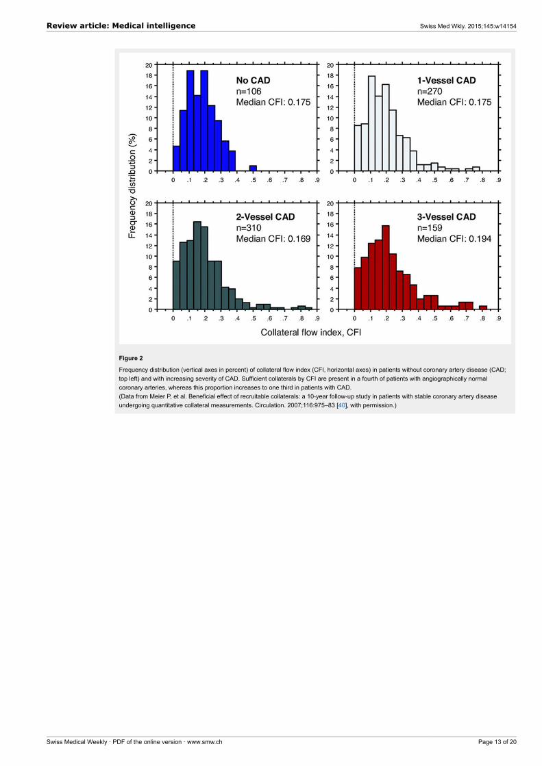
Figure 2
Frequency distribution (vertical axes in percent) of collateral flow index (CFI, horizontal axes) in patients without coronary artery disease (CAD;top left) and with increasing severity of CAD. Sufficient collaterals by CFI are present in a fourth of patients with angiographically normalcoronary arteries, whereas this proportion increases to one third in patients with CAD.(Data from Meier P, et al. Beneficial effect of recruitable collaterals: a 10-year follow-up study in patients with stable coronary artery diseaseundergoing quantitative collateral measurements. Circulation. 2007;116:975–83 [40], with permission.)
Review article: Medical intelligence Swiss Med Wkly. 2015;145:w14154
Swiss Medical Weekly · PDF of the online version · www.smw.ch Page 13 of 20
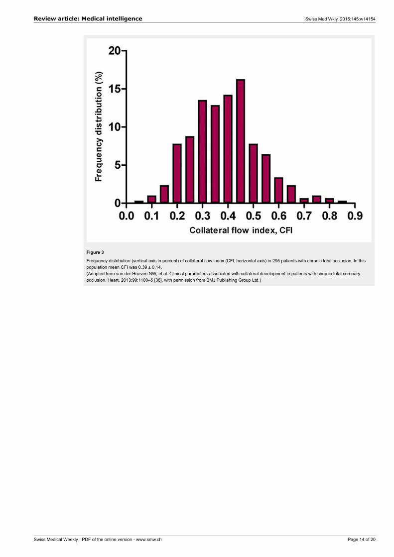
Figure 3
Frequency distribution (vertical axis in percent) of collateral flow index (CFI, horizontal axis) in 295 patients with chronic total occlusion. In thispopulation mean CFI was 0.39 ± 0.14.(Adapted from van der Hoeven NW, et al. Clinical parameters associated with collateral development in patients with chronic total coronaryocclusion. Heart. 2013;99:1100–5 [38], with permission from BMJ Publishing Group Ltd.)
Review article: Medical intelligence Swiss Med Wkly. 2015;145:w14154
Swiss Medical Weekly · PDF of the online version · www.smw.ch Page 14 of 20
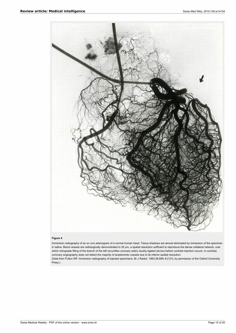
Figure 4
Immersion radiography of an ex vivo arteriogram of a normal human heart. Tissue shadows are almost eliminated by immersion of the specimenin saline. Blood vessels are radiologically demonstrated to 20 µm, a spatial resolution sufficient to reproduce the dense collateral network, overwhich retrograde filling of the branch of the left circumflex coronary artery doubly ligated (arrow) before contrast injection occurs. In contrast,coronary angiography does not detect the majority of anastomotic vessels due to its inferior spatial resolution.(Data from Fulton WF. Immersion radiography of injected specimens. Br J Radiol. 1963;36:685–8 [121], by permission of the Oxford UniversityPress.)
Review article: Medical intelligence Swiss Med Wkly. 2015;145:w14154
Swiss Medical Weekly · PDF of the online version · www.smw.ch Page 15 of 20
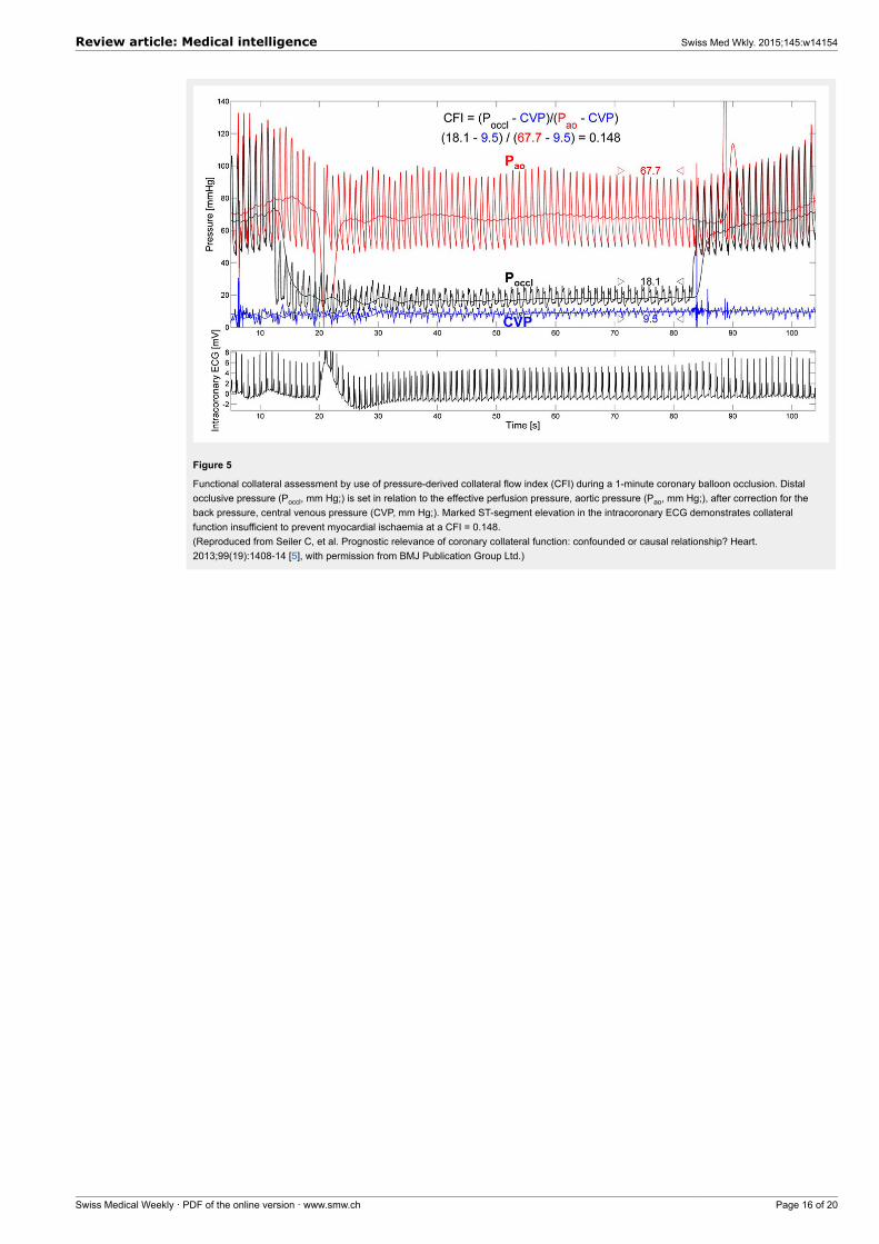
Figure 5
Functional collateral assessment by use of pressure-derived collateral flow index (CFI) during a 1-minute coronary balloon occlusion. Distalocclusive pressure (Poccl, mm Hg;) is set in relation to the effective perfusion pressure, aortic pressure (Pao, mm Hg;), after correction for theback pressure, central venous pressure (CVP, mm Hg;). Marked ST-segment elevation in the intracoronary ECG demonstrates collateralfunction insufficient to prevent myocardial ischaemia at a CFI = 0.148.(Reproduced from Seiler C, et al. Prognostic relevance of coronary collateral function: confounded or causal relationship? Heart.2013;99(19):1408-14 [5], with permission from BMJ Publication Group Ltd.)
Review article: Medical intelligence Swiss Med Wkly. 2015;145:w14154
Swiss Medical Weekly · PDF of the online version · www.smw.ch Page 16 of 20
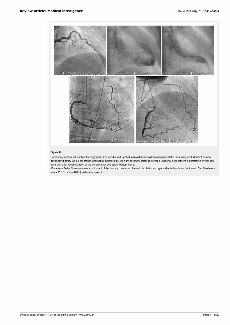
Figure 6
Completely normal left ventricular angiogram (top middle and right) due to extensive collateral supply of the proximally occluded left anteriordescending artery via apical branch and septal collateral by the right coronary artery (bottom). Functional assessment is performed by balloonocclusion after recanalization of the chronic total occlusion (bottom right).(Data from Seiler C. Assessment and impact of the human coronary collateral circulation on myocardial ischemia and outcome. Circ CardiovascInterv. 2013;6:719–28 [86], with permission.)
Review article: Medical intelligence Swiss Med Wkly. 2015;145:w14154
Swiss Medical Weekly · PDF of the online version · www.smw.ch Page 17 of 20
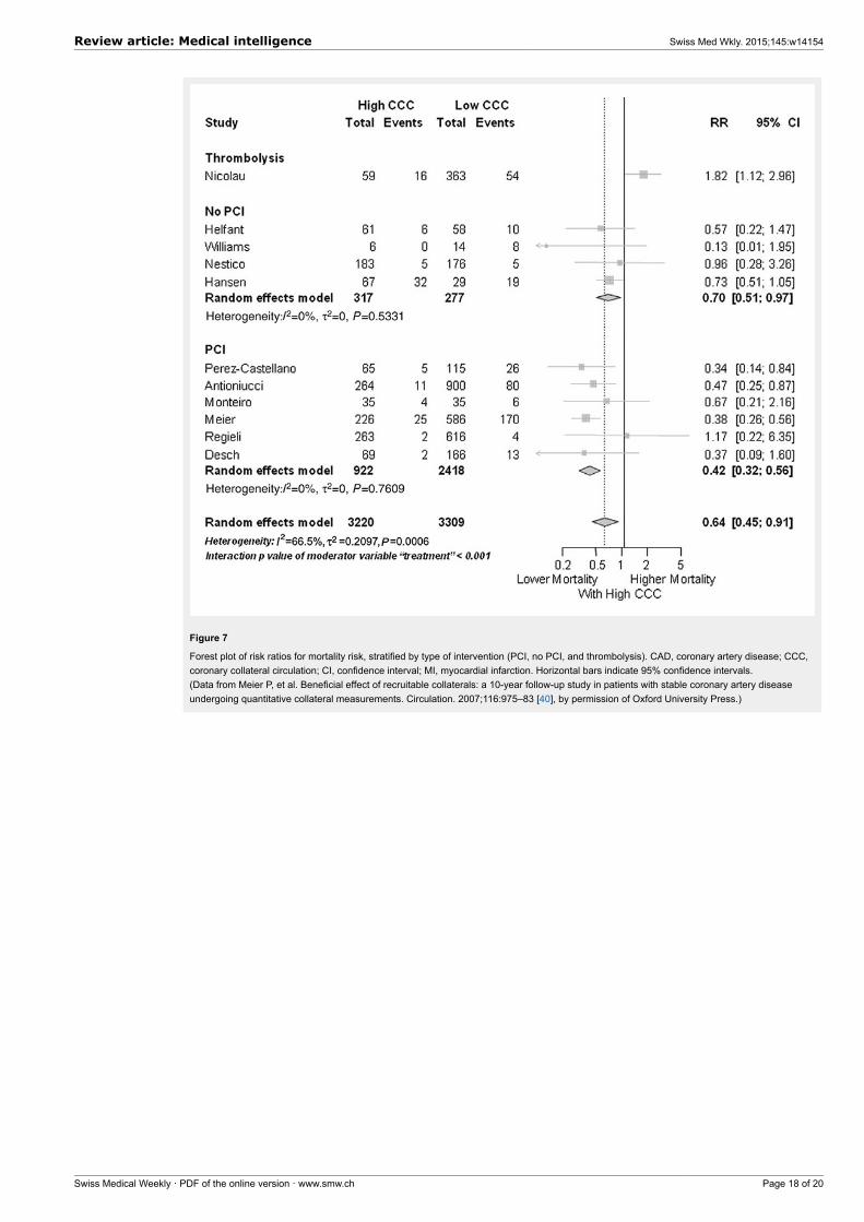
Figure 7
Forest plot of risk ratios for mortality risk, stratified by type of intervention (PCI, no PCI, and thrombolysis). CAD, coronary artery disease; CCC,coronary collateral circulation; CI, confidence interval; MI, myocardial infarction. Horizontal bars indicate 95% confidence intervals.(Data from Meier P, et al. Beneficial effect of recruitable collaterals: a 10-year follow-up study in patients with stable coronary artery diseaseundergoing quantitative collateral measurements. Circulation. 2007;116:975–83 [40], by permission of Oxford University Press.)
Review article: Medical intelligence Swiss Med Wkly. 2015;145:w14154
Swiss Medical Weekly · PDF of the online version · www.smw.ch Page 18 of 20
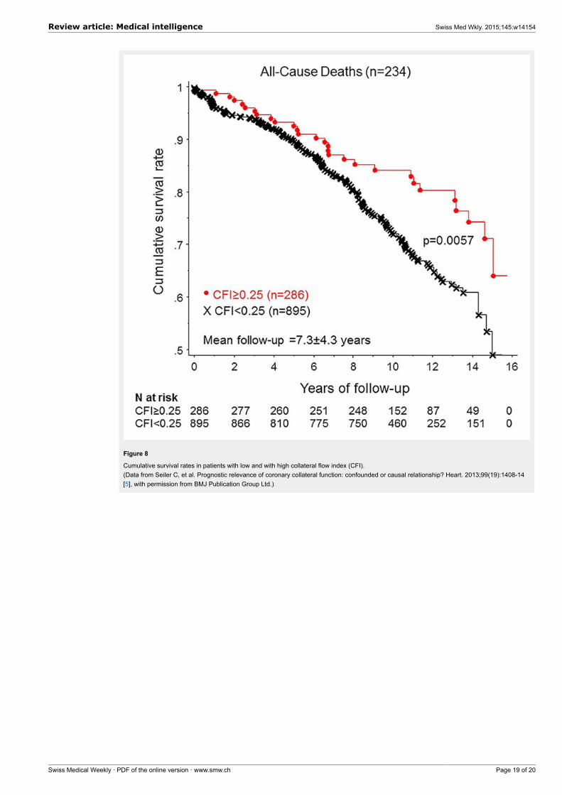
Figure 8
Cumulative survival rates in patients with low and with high collateral flow index (CFI).(Data from Seiler C, et al. Prognostic relevance of coronary collateral function: confounded or causal relationship? Heart. 2013;99(19):1408-14[5], with permission from BMJ Publication Group Ltd.)
Review article: Medical intelligence Swiss Med Wkly. 2015;145:w14154
Swiss Medical Weekly · PDF of the online version · www.smw.ch Page 19 of 20
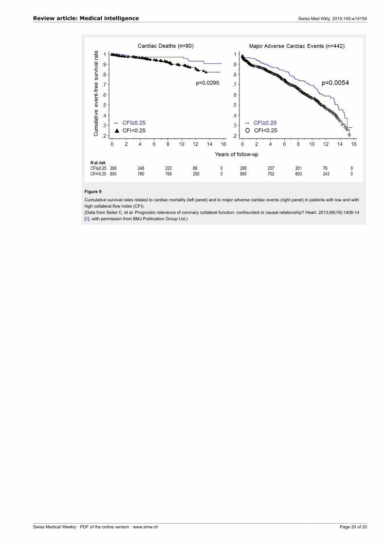
Figure 9
Cumulative survival rates related to cardiac mortality (left panel) and to major adverse cardiac events (right panel) in patients with low and withhigh collateral flow index (CFI).(Data from Seiler C, et al. Prognostic relevance of coronary collateral function: confounded or causal relationship? Heart. 2013;99(19):1408-14[5], with permission from BMJ Publication Group Ltd.)
Review article: Medical intelligence Swiss Med Wkly. 2015;145:w14154
Swiss Medical Weekly · PDF of the online version · www.smw.ch Page 20 of 20