Sahil ultrafest (2)
12
Rare case of Congenital Left Atrial Appendage Aneurysm- Its Multimodality Imaging Presented by:- Sahil Garg (PG Resident) Co Authors- Abhijit Patil, Ashish Ashtekar, V.M Kulkarni Department of Radiodiagnosis Dr DY Patil Medical College , Pimpri,Pune
-
Upload
ultrafest -
Category
Health & Medicine
-
view
63 -
download
3
Transcript of Sahil ultrafest (2)
Rare case of Congenital Left Atrial Appendage
Aneurysm- Its Multimodality Imaging
Presented by:- Sahil Garg (PG Resident)
Co Authors- Abhijit Patil, Ashish Ashtekar,
V.M Kulkarni
Department of Radiodiagnosis
Dr DY Patil Medical College , Pimpri,Pune

Clinical Profile
A 20 year old female from Pune, Maharashtra, presented in medicine OPD with
fever and breathlessness on and off with chest pain since 5 years.
Associated history of headache and weakness with pain in the lower limbs since
5 years
Patient did not delineate any past history of surgery or any hospital admission.
There was no history of joint pain or any chronic illnesses like TB, HTN, DM
and Seizures.
Patient was seronegative for HIV.
Systemic examination revealed split S2 with end systolic murmur at pulmonary
area and systolic murmur at mitral area.

Radiograph – AP and Lateral view
PA View PA View Left Lateral ViewLeft Lateral View
RadiographRadiograph of chest PA and left lateral revealed convex opacity between inferior border of the of chest PA and left lateral revealed convex opacity between inferior border of the
aortic knob upto upper half of left cardiac border with peripheral curvilinear calcification. The aortic knob upto upper half of left cardiac border with peripheral curvilinear calcification. The
lateral projection revealed the hilar location of the lesion below the left main stem bronchus. lateral projection revealed the hilar location of the lesion below the left main stem bronchus.
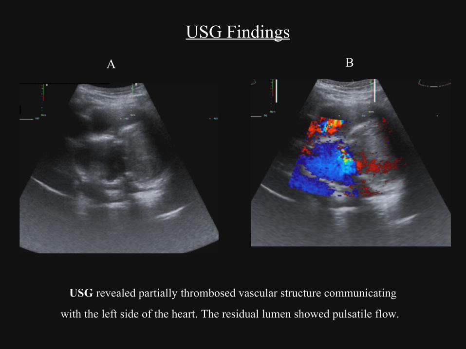
USG Findings
AA BB
USGUSG revealed partially thrombosed vascular structure communicating revealed partially thrombosed vascular structure communicating
with the left side of the heart. The residual lumen showed pulsatile flow. with the left side of the heart. The residual lumen showed pulsatile flow.
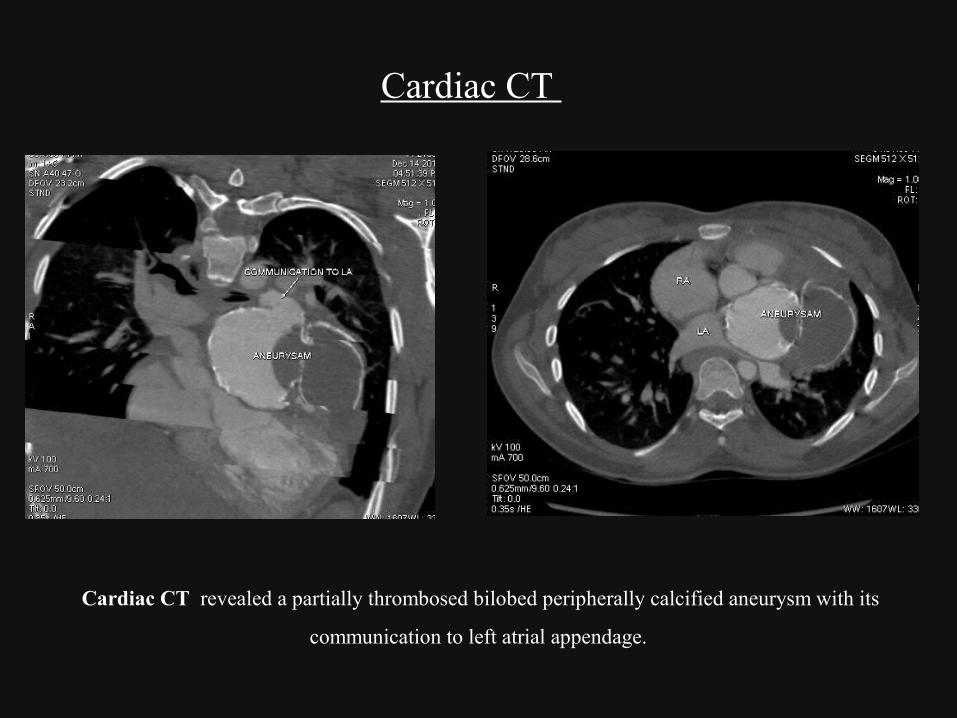
Cardiac CT
Cardiac CT revealed a partially thrombosed bilobed peripherally calcified aneurysm with its
communication to left atrial appendage.

Cardiac MRI
Non thrombosed Non thrombosed aneurysm arising aneurysm arising
from the left from the left atriumatrium
Thrombosed part of Thrombosed part of aneurysmaneurysm
LALA
LVLV
RARA
RVRV
Cardiac MRICardiac MRI revealed a partially thrombosed bilobed revealed a partially thrombosed bilobed
aneurysm of the left atrial appendage. aneurysm of the left atrial appendage.
LALA
RARA
LVLV
ANEUANEU

Diagnosis- Bilobed peripherally calcified Left atrial appendage aneurysm
Differential diagnosis –Sinus of valsalva aneurysm.

Discussion
Left Atrial aneurysms are usually secondary to mitral valve
disease.
Congenital Left Atrial Appendage aneurysm is a rare anomaly
described by Semans and Taussig in 1938.
These aneurysms mimic mediastinal or cardiac tumours on
routine radiography.

Discussion (Contd.)
Sinus of Valsalva aneurysm is a differential diagnosis for left
atrial appendage aneurysm
Cardiac and coronary CT / MR provide a conclusive diagnosis
and helps in differentiating it from sinus of valsalva aneurysm.
The syndrome, if not diagnosed and treated early may lead to
complications like systemic emboli, arrhythmias and eventually
death

Conclusion
Congenital left atrial appendage aneurysm is very rarely
reported entity.
Such cases with unusual clinical presentation may pose a
diagnostic challenge.
USG provides an easily accessible modality for confirming the
location and communication with the mediastinal vascular
structures.
Cardiac and coronary CT / MRI are the gold standard for the
diagnosis
References
Parakh N, Yadav N, Chaturvedi V, GeelaniM:Giant left atrial
appendage: Postgrad Med J 2011;87:436-437.
Gościniak E, Larysz B,Jurczyk K,Kasprzak J:Five-chambered
heart: Eur Heart J (2009).
Victor S and Nayak V: Aneurysm of the Left Atrial Appendage:
Tex Heart Inst J.2001:28(2):111-118.
Hassan M, Said K, Hamamsy I, Abdelsalam S Et al: Giant
Congenital Left Atrial Appendage Aneurysm: J Am Coll
Cardiol. 2013;61(4):478-478.

Thank You