
Sachio Kawai - InTech - Open Science Open Minds | InTechOpen
20
31 Pathology of Takotsubo (Ampulla) Cardiomyopathy Sachio Kawai Department of Cardiology, Juntendo University School of Medicine,Tokyo, Japan 1. Introduction Takotsubo (ampulla) cardiomyopathy is characterized by slight to moderate elevation of cardiac enzymes. It is reasonable to think that the elevated levels are evidence of myocardial damage (cardiac-specific enzymes are released from the cytoplasm of damaged cardiac myocytes into extracellular fluid). Thus, it is problematic to consider takotsubo cardiomyopathy as (neurogenic) stunned myocardium, because stunned myocardium is defined as a state without histopathological changes observable by light microscopy. 1 In the study, I will describe histological findings in autopsied cases in Japan and histopathological findings of endomyocardial biopsy cases with a literature review. 2. Autopsied cases Nine Japanese autopsied cases were studied, with the permission of relevant institutions. We examined their histopathological changes, findings in transverse tissue sections by site (near the left ventricular apex and base) and by layer (relative damage by depth of ventricular myocardium), and differences in lesions. 2 Table 1 shows the age, sex, underlying diseases, trigger event, and symptoms of the cases. 3 Case 2. A 66-year-old woman was admitted to the hospital with paralysis and brain infiltration by Hodgkin’s lymphoma. Her symptoms improved with steroid therapy and radiation therapy. There was no clear trigger event. She developed sudden bradycardia, hypotension, and extensive ST-segment elevation. She underwent intravenous infusion of 2 mg epinephrine, and subsequently developed chest pain. The patient had a normal coronary angiogram, extensive hypokinesis mainly in the apical segment, and hyperkinesis of the basal segment. Creatinine kinase level increased to 169 U/l at a maximum. There was no pulmonary congestion, but severe hypoxemia continued and the patient died on the fourth day of illness. 4 Autopsy showed neither significant coronary artery stenosis nor significant fibrosis in the ventricular transverse sections near the apical or basal segment. The ventricular wall were thickened; 15.2 mm in the basal anterior wall, 13.8 mm in the basal lateral wall, 13.2 mm in the basal posterior wall, 17.5 mm in the basal interventricular septum, 10.0 mm in the apical anterior wall, 13.0 mm in the apical lateral wall, 10.2 mm in the apical posterior wall, and 8.0 mm in the apical interventricular septum. Histologically, there was disseminated or diffuse damage to the cardiac myocytes, and extensive damage was observed from the inner to outer layer. In general, pathological changes were myocyte www.intechopen.com
Transcript of Sachio Kawai - InTech - Open Science Open Minds | InTechOpen
31
Pathology of Takotsubo (Ampulla) Cardiomyopathy
Sachio Kawai Department of Cardiology, Juntendo University School of Medicine,Tokyo,
Japan
1. Introduction
Takotsubo (ampulla) cardiomyopathy is characterized by slight to moderate elevation of cardiac enzymes. It is reasonable to think that the elevated levels are evidence of myocardial damage (cardiac-specific enzymes are released from the cytoplasm of damaged cardiac myocytes into extracellular fluid). Thus, it is problematic to consider takotsubo cardiomyopathy as (neurogenic) stunned myocardium, because stunned myocardium is defined as a state without histopathological changes observable by light microscopy.1 In the study, I will describe histological findings in autopsied cases in Japan and histopathological findings of endomyocardial biopsy cases with a literature review.
2. Autopsied cases
Nine Japanese autopsied cases were studied, with the permission of relevant institutions. We examined their histopathological changes, findings in transverse tissue sections by site (near the left ventricular apex and base) and by layer (relative damage by depth of ventricular myocardium), and differences in lesions.2 Table 1 shows the age, sex, underlying diseases, trigger event, and symptoms of the cases.3
Case 2. A 66-year-old woman was admitted to the hospital with paralysis and brain infiltration by Hodgkin’s lymphoma. Her symptoms improved with steroid therapy and radiation therapy. There was no clear trigger event. She developed sudden bradycardia, hypotension, and extensive ST-segment elevation. She underwent intravenous infusion of 2 mg epinephrine, and subsequently developed chest pain. The patient had a normal coronary angiogram, extensive hypokinesis mainly in the apical segment, and hyperkinesis of the basal segment. Creatinine kinase level increased to 169 U/l at a maximum. There was no pulmonary congestion, but severe hypoxemia continued and the patient died on the fourth day of illness.4 Autopsy showed neither significant coronary artery stenosis nor significant fibrosis in the ventricular transverse sections near the apical or basal segment. The ventricular wall were thickened; 15.2 mm in the basal anterior wall, 13.8 mm in the basal lateral wall, 13.2 mm in the basal posterior wall, 17.5 mm in the basal interventricular septum, 10.0 mm in the apical anterior wall, 13.0 mm in the apical lateral wall, 10.2 mm in the apical posterior wall, and 8.0 mm in the apical interventricular septum. Histologically, there was disseminated or diffuse damage to the cardiac myocytes, and extensive damage was observed from the inner to outer layer. In general, pathological changes were myocyte
www.intechopen.com
Cardiomyopathies – From Basic Research to Clinical Management
710
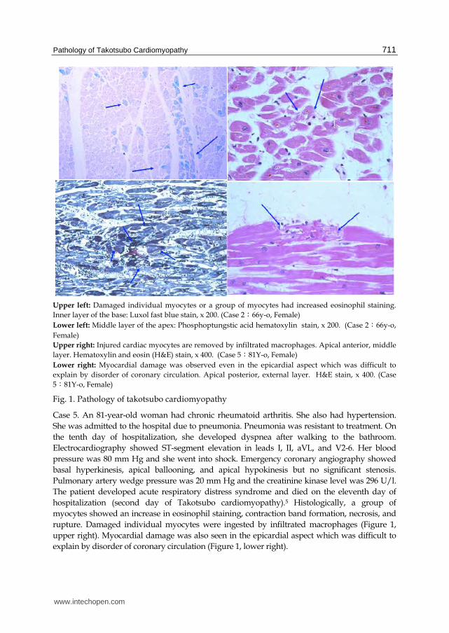
damage, degeneration, lysis, or loss occurring in an individual myocyte or several myocytes. Some cases had lesions concentrated on specific muscle bundles and other cases had disseminated lesions (Figure 1, upper left). In addition, higher degree of cell infiltration was seen at sites with more severely damaged myocardium (Figure 1, lower left).
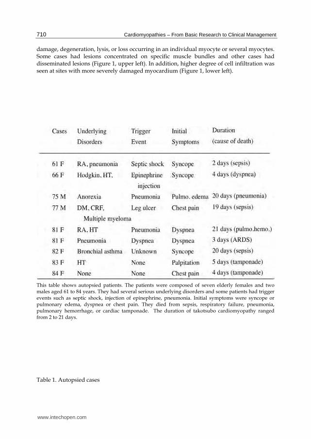
This table shows autopsied patients. The patients were composed of seven elderly females and two males aged 61 to 84 years. They had several serious underlying disorders and some patients had trigger events such as septic shock, injection of epinephrine, pneumonia. Initial symptoms were syncope or pulmonary edema, dyspnea or chest pain. They died from sepsis, respiratory failure, pneumonia, pulmonary hemorrhage, or cardiac tamponade. The duration of takotsubo cardiomyopathy ranged from 2 to 21 days.
Table 1. Autopsied cases
www.intechopen.com
Pathology of Takotsubo Cardiomyopathy
711
Upper left: Damaged individual myocytes or a group of myocytes had increased eosinophil staining.
Inner layer of the base: Luxol fast blue stain, x 200. (Case 2:66y-o, Female)
Lower left: Middle layer of the apex: Phosphoptungstic acid hematoxylin stain, x 200. (Case 2:66y-o,
Female)
Upper right: Injured cardiac myocytes are removed by infiltrated macrophages. Apical anterior, middle
layer. Hematoxylin and eosin (H&E) stain, x 400. (Case 5:81Y-o, Female)
Lower right: Myocardial damage was observed even in the epicardial aspect which was difficult to
explain by disorder of coronary circulation. Apical posterior, external layer. H&E stain, x 400. (Case
5:81Y-o, Female)
Fig. 1. Pathology of takotsubo cardiomyopathy
Case 5. An 81-year-old woman had chronic rheumatoid arthritis. She also had hypertension.
She was admitted to the hospital due to pneumonia. Pneumonia was resistant to treatment. On
the tenth day of hospitalization, she developed dyspnea after walking to the bathroom.
Electrocardiography showed ST-segment elevation in leads I, II, aVL, and V2-6. Her blood
pressure was 80 mm Hg and she went into shock. Emergency coronary angiography showed
basal hyperkinesis, apical ballooning, and apical hypokinesis but no significant stenosis.
Pulmonary artery wedge pressure was 20 mm Hg and the creatinine kinase level was 296 U/l.
The patient developed acute respiratory distress syndrome and died on the eleventh day of
hospitalization (second day of Takotsubo cardiomyopathy).5 Histologically, a group of
myocytes showed an increase in eosinophil staining, contraction band formation, necrosis, and
rupture. Damaged individual myocytes were ingested by infiltrated macrophages (Figure 1,
upper right). Myocardial damage was also seen in the epicardial aspect which was difficult to
explain by disorder of coronary circulation (Figure 1, lower right).
www.intechopen.com
Cardiomyopathies – From Basic Research to Clinical Management
712
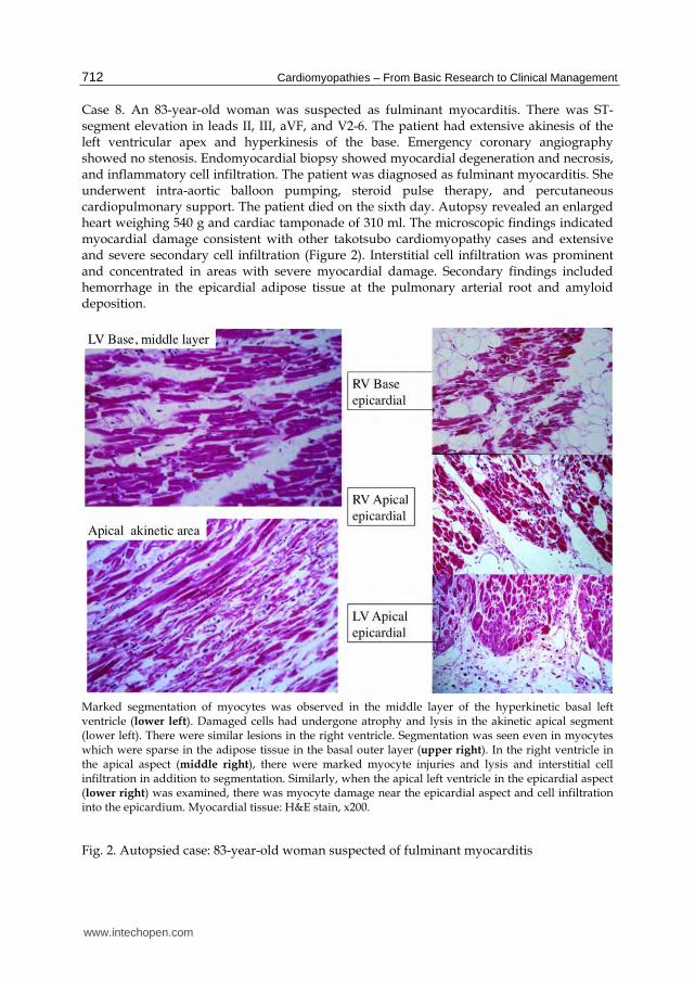
Case 8. An 83-year-old woman was suspected as fulminant myocarditis. There was ST-segment elevation in leads II, III, aVF, and V2-6. The patient had extensive akinesis of the left ventricular apex and hyperkinesis of the base. Emergency coronary angiography showed no stenosis. Endomyocardial biopsy showed myocardial degeneration and necrosis, and inflammatory cell infiltration. The patient was diagnosed as fulminant myocarditis. She underwent intra-aortic balloon pumping, steroid pulse therapy, and percutaneous cardiopulmonary support. The patient died on the sixth day. Autopsy revealed an enlarged heart weighing 540 g and cardiac tamponade of 310 ml. The microscopic findings indicated myocardial damage consistent with other takotsubo cardiomyopathy cases and extensive and severe secondary cell infiltration (Figure 2). Interstitial cell infiltration was prominent and concentrated in areas with severe myocardial damage. Secondary findings included hemorrhage in the epicardial adipose tissue at the pulmonary arterial root and amyloid deposition.
Marked segmentation of myocytes was observed in the middle layer of the hyperkinetic basal left ventricle (lower left). Damaged cells had undergone atrophy and lysis in the akinetic apical segment (lower left). There were similar lesions in the right ventricle. Segmentation was seen even in myocytes which were sparse in the adipose tissue in the basal outer layer (upper right). In the right ventricle in the apical aspect (middle right), there were marked myocyte injuries and lysis and interstitial cell infiltration in addition to segmentation. Similarly, when the apical left ventricle in the epicardial aspect (lower right) was examined, there was myocyte damage near the epicardial aspect and cell infiltration into the epicardium. Myocardial tissue: H&E stain, x200.
Fig. 2. Autopsied case: 83-year-old woman suspected of fulminant myocarditis
www.intechopen.com
Pathology of Takotsubo Cardiomyopathy
713
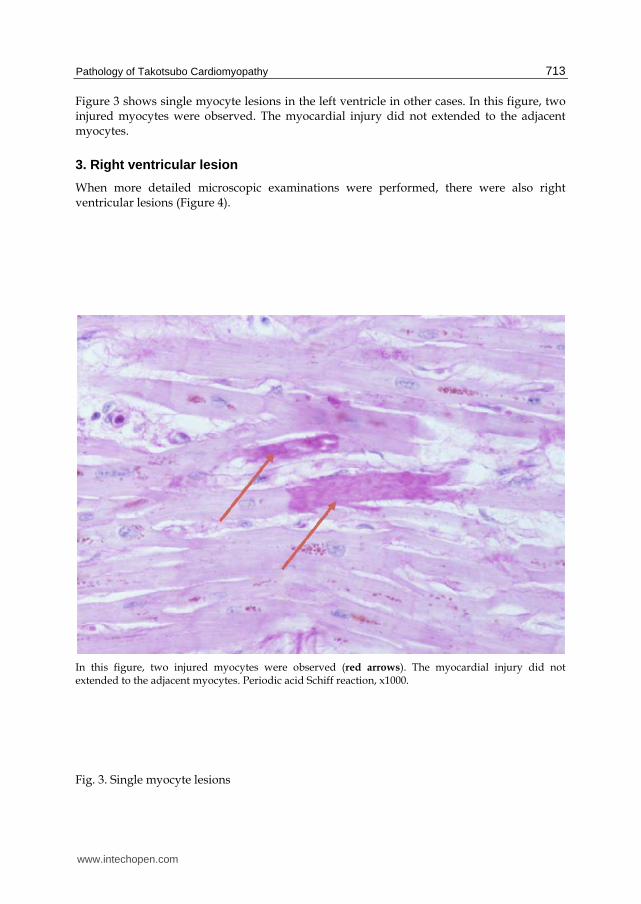
Figure 3 shows single myocyte lesions in the left ventricle in other cases. In this figure, two injured myocytes were observed. The myocardial injury did not extended to the adjacent myocytes.
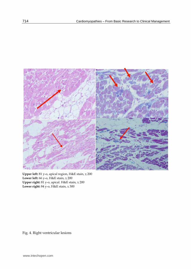
3. Right ventricular lesion
When more detailed microscopic examinations were performed, there were also right ventricular lesions (Figure 4).
In this figure, two injured myocytes were observed (red arrows). The myocardial injury did not extended to the adjacent myocytes. Periodic acid Schiff reaction, x1000.
Fig. 3. Single myocyte lesions
www.intechopen.com
Cardiomyopathies – From Basic Research to Clinical Management
714
Upper left: 81 y-o, apical region, H&E stain,x200
Lower left: 66 y-o, H&E stain,x200
Upper right: 81 y-o, apical. H&E stain,x200
Lower right: 84 y-o, H&E stain,x300
Fig. 4. Right ventricular lesions
www.intechopen.com
Pathology of Takotsubo Cardiomyopathy
715
4. Morphometrical studies
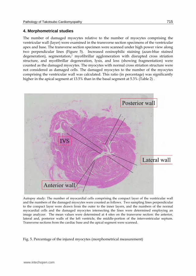
The number of damaged myocytes relative to the number of myocytes comprising the ventricular wall (layer) were examined in the transverse section specimens of the ventricular apex and base. The transverse section specimen were scanned under high power view along two perpendicular lines (Figure 5). Increased eosinophilic staining (azan-blue stained degeneration), segmentation,7 myofibrillar agglomeration with disrupted cross striation structure, and myofibrillar degeneration, lysis, and loss (showing fragmentation) were counted as the damaged myocytes. The myocytes with normal cross striation structure were not considered as damaged cells. The damaged myocytes to the number of the myocytes comprising the ventricular wall was calculated. This ratio (in percentage) was significantly higher in the apical segment at 13.5% than in the basal segment at 5.3% (Table 2) .
Autopsy study: The number of myocardial cells comprising the compact layer of the ventricular wall and the numbers of the damaged myocytes were counted as follows. Two sampling lines perpendicular to the compact layer were drawn from the outer to the inner layers, and the numbers of the normal myocardial cells and the damaged myocytes intersecting the lines were determined employing an image analyzer. The mean values were determined at 4 sites on the transverse section: the anterior, lateral and, posterior walls of the left ventricle, the middle-portion of the interventricular septum. Transverse sections from the cardiac base and the apical segment were scanned.
Fig. 5. Percentage of the injured myocytes (morphometrical measurement)
www.intechopen.com
Cardiomyopathies – From Basic Research to Clinical Management
716
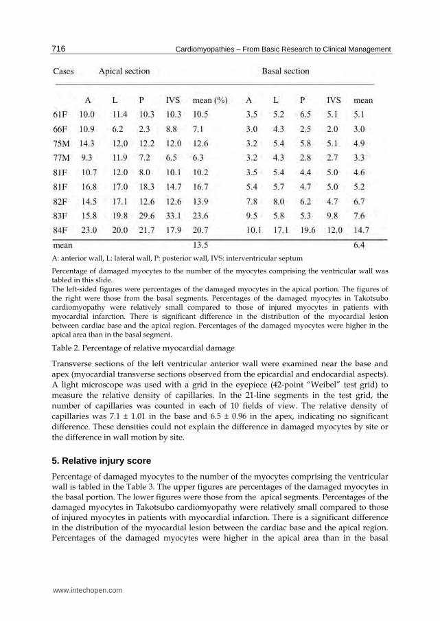
A: anterior wall, L: lateral wall, P: posterior wall, IVS: interventricular septum
Percentage of damaged myocytes to the number of the myocytes comprising the ventricular wall was tabled in this slide. The left-sided figures were percentages of the damaged myocytes in the apical portion. The figures of the right were those from the basal segments. Percentages of the damaged myocytes in Takotsubo cardiomyopathy were relatively small compared to those of injured myocytes in patients with myocardial infarction. There is significant difference in the distribution of the myocardial lesion between cardiac base and the apical region. Percentages of the damaged myocytes were higher in the apical area than in the basal segment.
Table 2. Percentage of relative myocardial damage
Transverse sections of the left ventricular anterior wall were examined near the base and
apex (myocardial transverse sections observed from the epicardial and endocardial aspects).
A light microscope was used with a grid in the eyepiece (42-point “Weibel” test grid) to
measure the relative density of capillaries. In the 21-line segments in the test grid, the
number of capillaries was counted in each of 10 fields of view. The relative density of
capillaries was 7.1 ± 1.01 in the base and 6.5 ± 0.96 in the apex, indicating no significant
difference. These densities could not explain the difference in damaged myocytes by site or
the difference in wall motion by site.
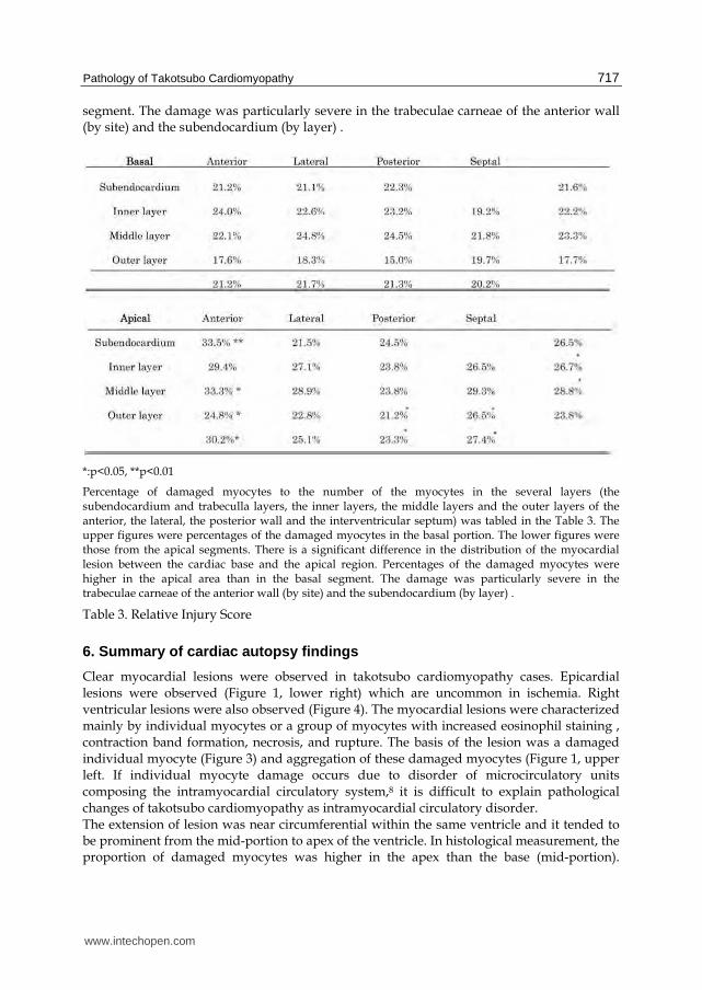
5. Relative injury score
Percentage of damaged myocytes to the number of the myocytes comprising the ventricular wall is tabled in the Table 3. The upper figures are percentages of the damaged myocytes in the basal portion. The lower figures were those from the apical segments. Percentages of the damaged myocytes in Takotsubo cardiomyopathy were relatively small compared to those of injured myocytes in patients with myocardial infarction. There is a significant difference in the distribution of the myocardial lesion between the cardiac base and the apical region. Percentages of the damaged myocytes were higher in the apical area than in the basal
www.intechopen.com
Pathology of Takotsubo Cardiomyopathy
717
segment. The damage was particularly severe in the trabeculae carneae of the anterior wall (by site) and the subendocardium (by layer) .
*:p<0.05, **p<0.01
Percentage of damaged myocytes to the number of the myocytes in the several layers (the subendocardium and trabeculla layers, the inner layers, the middle layers and the outer layers of the anterior, the lateral, the posterior wall and the interventricular septum) was tabled in the Table 3. The upper figures were percentages of the damaged myocytes in the basal portion. The lower figures were those from the apical segments. There is a significant difference in the distribution of the myocardial lesion between the cardiac base and the apical region. Percentages of the damaged myocytes were higher in the apical area than in the basal segment. The damage was particularly severe in the trabeculae carneae of the anterior wall (by site) and the subendocardium (by layer) .
Table 3. Relative Injury Score
6. Summary of cardiac autopsy findings
Clear myocardial lesions were observed in takotsubo cardiomyopathy cases. Epicardial lesions were observed (Figure 1, lower right) which are uncommon in ischemia. Right ventricular lesions were also observed (Figure 4). The myocardial lesions were characterized mainly by individual myocytes or a group of myocytes with increased eosinophil staining , contraction band formation, necrosis, and rupture. The basis of the lesion was a damaged individual myocyte (Figure 3) and aggregation of these damaged myocytes (Figure 1, upper left. If individual myocyte damage occurs due to disorder of microcirculatory units composing the intramyocardial circulatory system,8 it is difficult to explain pathological changes of takotsubo cardiomyopathy as intramyocardial circulatory disorder. The extension of lesion was near circumferential within the same ventricle and it tended to be prominent from the mid-portion to apex of the ventricle. In histological measurement, the proportion of damaged myocytes was higher in the apex than the base (mid-portion).
www.intechopen.com
Cardiomyopathies – From Basic Research to Clinical Management
718
Damage was prominent in the apical inner and middle layers (by ventricular myocardial layer) and in the anterior wall, posterior wall, and septum (by site). The damage was particularly severe in the trabeculae carneae of the anterior wall (by site) and subendocardium (by layer) (Table 3). These results indicate that it might be possible to depict predilection sites by MRI with delayed gadolinium enhancement.9 Autopsy cases with myocardial diseases were reported in the regional meetings of the
Japanese Circulation Society. There have been many recent reports such as on a 77-year-old
woman with myocardial infarction-like electrocardiographic pattern during the course of
malabsorption syndrome10 and on an 84-year-old woman who died from cardiac rupture11
(four days after the onset, cardiac arrest occurred due to cardiac rupture). In the Circulation
Journal, Sacha et al.12 stated the case of Ohara et al.13 in the same journal as the first autopsy
case [of takotsubo cardiomyopathy] and their own case was the second one. However, this
information is not accurate. There were previously reported autopsy cases in the journals
Heart and Respiration & Circulation. There were reports of takotsubo cardiomyopathy itself
by Kumamoto et al.,14 Sasa et al.,15 and Tokioka et al.16 Thus, there were already reports of
takotsubo cardiomyopathy as a phenomenon, and autopsy cases were published in a
journal17 before that of Ohara et al. Thus, Sacha et al. should have been advised to revise the
information in the peer review.
7. Case presentation of endomyocardial biopsy
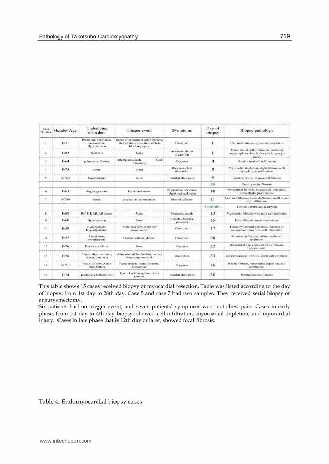
Table 4 shows 15 cases of endomyocardial biopsy for which relevant institutions provided approval for microscopic examination. The cases are listed in the order of number of days from onset to biopsy.
8. Case biopsied on the day of onset (Case 2)
The patient was an 82-year-old woman who had undergone uterine myomectomy at age 48.
She developed sudden pharyngeal discomfort. There was ST-segment elevation in leads II,
III, aVF, and V2-6. Echocardiography showed diffuse hypokinesis except in the base.
Ventriculography revealed hyperkinesis of the base and balloon-like enlargement of the
apex. She was suspected of myocarditis and underwent biopsy. Her condition improved on
the 26th day of illness. Biopsy showed slight increase in young connective tissue and
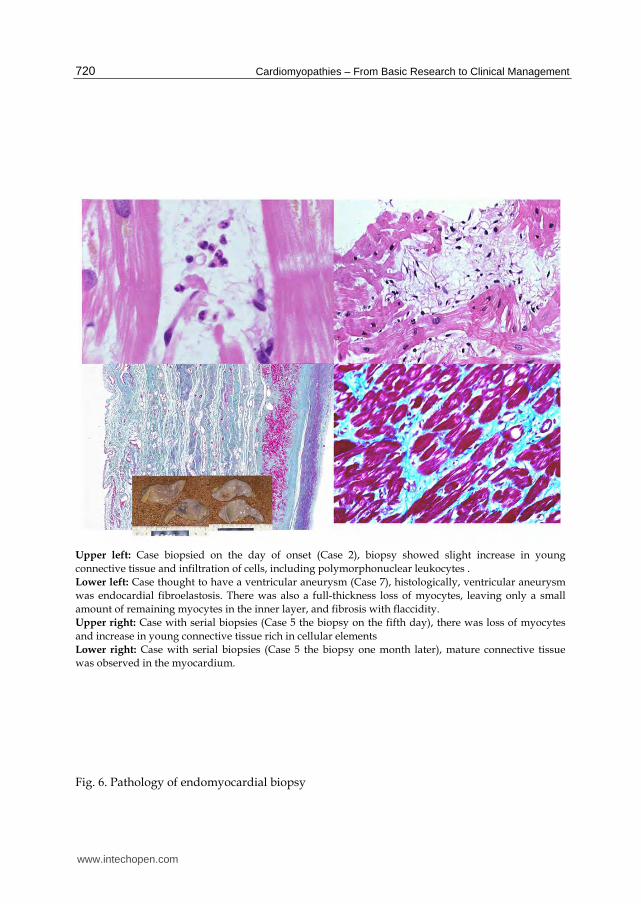
infiltration of cells, including polymorphonuclear leukocytes (Figure 6, upper left).
9. Case thought to have a ventricular aneurysm (Case 7)
The patient was a 69-year-old man with no contributory medical history. He became lost in the mountains while gathering edible wild plants and was rescued 7 hours later. He experienced shortness of breath thereafter and had pleural effusion 18 days later. Coronary angiography revealed 90% occlusion in #11 and complete occlusion in peripheral #12. There was also unexplainable severe left ventricular asynergy and an apical mural thrombus. Biopsy showed cell-rich fibrosis. One week later, his condition improved except in the circumflex branch and apical regions. He underwent resection of a ventricular aneurysm 2.5 months later. Histologically, ventricular aneurysm was endocardial fibroelastosis. There was also a full-thickness loss of myocytes, leaving only a small amount of remaining myocytes in the inner layer, and fibrosis with flaccidity (Figure 6, lower left).
www.intechopen.com
Pathology of Takotsubo Cardiomyopathy
719
This table shows 15 cases received biopsy or myocardial resection. Table was listed according to the day of biopsy; from 1st day to 28th day. Case 5 and case 7 had two samples. They received serial biopsy or aneurysmectomy. Six patients had no trigger event, and seven patients’ symptoms were not chest pain. Cases in early phase, from 1st day to 4th day biopsy, showed cell infiltration, myocardial depletion, and myocardial injury. Cases in late phase that is 12th day or later, showed focal fibrosis.
Table 4. Endomyocardial biopsy cases
www.intechopen.com
Cardiomyopathies – From Basic Research to Clinical Management
720
Upper left: Case biopsied on the day of onset (Case 2), biopsy showed slight increase in young
connective tissue and infiltration of cells, including polymorphonuclear leukocytes .
Lower left: Case thought to have a ventricular aneurysm (Case 7), histologically, ventricular aneurysm
was endocardial fibroelastosis. There was also a full-thickness loss of myocytes, leaving only a small
amount of remaining myocytes in the inner layer, and fibrosis with flaccidity.
Upper right: Case with serial biopsies (Case 5 the biopsy on the fifth day), there was loss of myocytes
and increase in young connective tissue rich in cellular elements
Lower right: Case with serial biopsies (Case 5 the biopsy one month later), mature connective tissue
was observed in the myocardium.
Fig. 6. Pathology of endomyocardial biopsy
www.intechopen.com
Pathology of Takotsubo Cardiomyopathy
721
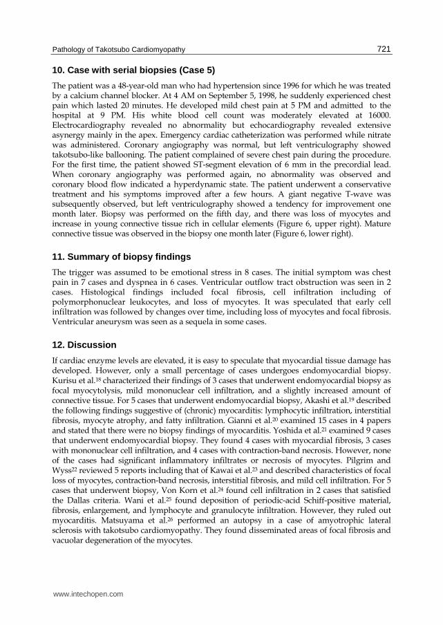
10. Case with serial biopsies (Case 5)
The patient was a 48-year-old man who had hypertension since 1996 for which he was treated by a calcium channel blocker. At 4 AM on September 5, 1998, he suddenly experienced chest pain which lasted 20 minutes. He developed mild chest pain at 5 PM and admitted to the hospital at 9 PM. His white blood cell count was moderately elevated at 16000. Electrocardiography revealed no abnormality but echocardiography revealed extensive asynergy mainly in the apex. Emergency cardiac catheterization was performed while nitrate was administered. Coronary angiography was normal, but left ventriculography showed takotsubo-like ballooning. The patient complained of severe chest pain during the procedure. For the first time, the patient showed ST-segment elevation of 6 mm in the precordial lead. When coronary angiography was performed again, no abnormality was observed and coronary blood flow indicated a hyperdynamic state. The patient underwent a conservative treatment and his symptoms improved after a few hours. A giant negative T-wave was subsequently observed, but left ventriculography showed a tendency for improvement one month later. Biopsy was performed on the fifth day, and there was loss of myocytes and increase in young connective tissue rich in cellular elements (Figure 6, upper right). Mature connective tissue was observed in the biopsy one month later (Figure 6, lower right).
11. Summary of biopsy findings
The trigger was assumed to be emotional stress in 8 cases. The initial symptom was chest pain in 7 cases and dyspnea in 6 cases. Ventricular outflow tract obstruction was seen in 2 cases. Histological findings included focal fibrosis, cell infiltration including of polymorphonuclear leukocytes, and loss of myocytes. It was speculated that early cell infiltration was followed by changes over time, including loss of myocytes and focal fibrosis. Ventricular aneurysm was seen as a sequela in some cases.
12. Discussion
If cardiac enzyme levels are elevated, it is easy to speculate that myocardial tissue damage has developed. However, only a small percentage of cases undergoes endomyocardial biopsy. Kurisu et al.18 characterized their findings of 3 cases that underwent endomyocardial biopsy as focal myocytolysis, mild mononuclear cell infiltration, and a slightly increased amount of connective tissue. For 5 cases that underwent endomyocardial biopsy, Akashi et al.19 described the following findings suggestive of (chronic) myocarditis: lymphocytic infiltration, interstitial fibrosis, myocyte atrophy, and fatty infiltration. Gianni et al.20 examined 15 cases in 4 papers and stated that there were no biopsy findings of myocarditis. Yoshida et al.21 examined 9 cases that underwent endomyocardial biopsy. They found 4 cases with myocardial fibrosis, 3 cases with mononuclear cell infiltration, and 4 cases with contraction-band necrosis. However, none of the cases had significant inflammatory infiltrates or necrosis of myocytes. Pilgrim and Wyss22 reviewed 5 reports including that of Kawai et al.23 and described characteristics of focal loss of myocytes, contraction-band necrosis, interstitial fibrosis, and mild cell infiltration. For 5 cases that underwent biopsy, Von Korn et al.24 found cell infiltration in 2 cases that satisfied the Dallas criteria. Wani et al.25 found deposition of periodic-acid Schiff-positive material, fibrosis, enlargement, and lymphocyte and granulocyte infiltration. However, they ruled out myocarditis. Matsuyama et al.26 performed an autopsy in a case of amyotrophic lateral sclerosis with takotsubo cardiomyopathy. They found disseminated areas of focal fibrosis and vacuolar degeneration of the myocytes.
www.intechopen.com
Cardiomyopathies – From Basic Research to Clinical Management
722
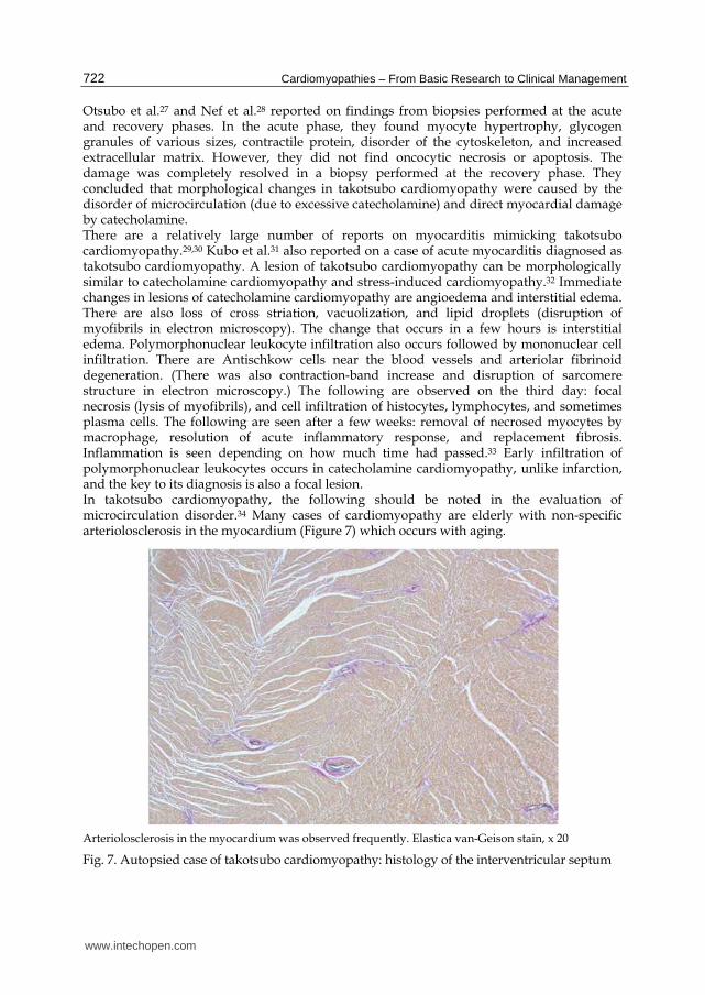
Otsubo et al.27 and Nef et al.28 reported on findings from biopsies performed at the acute and recovery phases. In the acute phase, they found myocyte hypertrophy, glycogen granules of various sizes, contractile protein, disorder of the cytoskeleton, and increased extracellular matrix. However, they did not find oncocytic necrosis or apoptosis. The damage was completely resolved in a biopsy performed at the recovery phase. They concluded that morphological changes in takotsubo cardiomyopathy were caused by the disorder of microcirculation (due to excessive catecholamine) and direct myocardial damage by catecholamine. There are a relatively large number of reports on myocarditis mimicking takotsubo cardiomyopathy.29,30 Kubo et al.31 also reported on a case of acute myocarditis diagnosed as takotsubo cardiomyopathy. A lesion of takotsubo cardiomyopathy can be morphologically similar to catecholamine cardiomyopathy and stress-induced cardiomyopathy.32 Immediate changes in lesions of catecholamine cardiomyopathy are angioedema and interstitial edema. There are also loss of cross striation, vacuolization, and lipid droplets (disruption of myofibrils in electron microscopy). The change that occurs in a few hours is interstitial edema. Polymorphonuclear leukocyte infiltration also occurs followed by mononuclear cell infiltration. There are Antischkow cells near the blood vessels and arteriolar fibrinoid degeneration. (There was also contraction-band increase and disruption of sarcomere structure in electron microscopy.) The following are observed on the third day: focal necrosis (lysis of myofibrils), and cell infiltration of histocytes, lymphocytes, and sometimes plasma cells. The following are seen after a few weeks: removal of necrosed myocytes by macrophage, resolution of acute inflammatory response, and replacement fibrosis. Inflammation is seen depending on how much time had passed.33 Early infiltration of polymorphonuclear leukocytes occurs in catecholamine cardiomyopathy, unlike infarction, and the key to its diagnosis is also a focal lesion. In takotsubo cardiomyopathy, the following should be noted in the evaluation of microcirculation disorder.34 Many cases of cardiomyopathy are elderly with non-specific arteriolosclerosis in the myocardium (Figure 7) which occurs with aging.
Arteriolosclerosis in the myocardium was observed frequently. Elastica van-Geison stain, x 20
Fig. 7. Autopsied case of takotsubo cardiomyopathy: histology of the interventricular septum
www.intechopen.com
Pathology of Takotsubo Cardiomyopathy
723
13. Conclusion
There was a clear myocardial lesion in takotsubo cardiomyopathy. The myocardial lesions are characterized mainly by individual myocytes or a group of myocytes with increased eosinophil staining, contraction band formation, necrosis, and rupture. The basis of the lesion is a damaged individual myocyte and aggregation of these damaged myocytes. It is speculated that early cell infiltration occurs followed by changes over time, including loss of myocytes and focal fibrosis.
14. Acknowledgements
I would like to express our appreciation to the following individuals for kindly supplying the tissues: Drs.Hiromasa Suzuki and Atsushi Yamada of Juntendo University School of Medicine, Dr. Koshi Mawatari of Kagoshima Seikyo Hospital, Dr.Toshio Shimada of Shimane Medical University (presently Shizuoka General Hospital), Drs. Hitoshi Sawada and Tadanori Aizawa of Cardiovascular Institute Hospital, Drs. Hironori Murakami and Nagisa Hanawa of Teine Keijinkai Hospital, Dr. Kasumi Takemura of the Kyoto Department of Cardiology at Prefectural Yosanoumi Hospital (Second Department of Medicine, Kyoto Prefectural University of Medicine), Dr. Osamu Yamanaka of International Goodwill Hospital (presently Poula Clinic), Dr. Nobuhiro Kotani of Shimane Medical University (presently Clinical Laboratory, Hamada Medical Center), Dr. Tsutomu Tamura of Itabashi Chuo Medical Center, Dr. Masafumi Watanabe of the Department of Cardiovascular Medicine at the University of Tokyo′Drs. Masahito Kawada and Tetsuya Nakamura of
National Hospital Organization Kobe Medical Center, and Dr. Hiroshi Matsushita of Toranomon Hospital.
15. References
[1] Kloner RA, Przyklen K, Patel B: Altered myocardial states. The stunned and hibernating
myocardium. Am J Med 1989; 86 (Suppl 1A):14-22.
[2] Kawai S, Suzuki H, Yamaguchi H, Kawada M, Nakamura T, Nishiyama S, Monomura S,
Matsushita H: [Autopsy findings of takotsubo cardiomyopathy.] Study report of
the study group of idiopathic cardiomyopathy sponsored by the Health and
Welfare Ministry. 106-109, March 2003. (in Japanese)
[3] Kawai S, Yamaguchi H, Suzuki H: [Pathology of takotsubo cardiomyopathy.] Heart
View 8(2):159-166, 2004. (in Japanese)
[4] Anzai H, Komiyama N, Kinoshita N, et al.: [Case of stunned myocardium caused by
overdose of epinephrine.] Kokyu to Junkan 44(2):199-204, 1996. (in Japanese)
[5] Kawada M, Okada T, Shimizu M, et al.: [An autopsy case of cardiac dysfunction of
unknown origin associated with severe pneumonia.] Heart 31(5):337-343, 1999. (in
Japanese)
[6] Kotani N, et al.: [Takotsubo-type fulminant myocarditis: a case report [abstract].] Jpn
Circ J 64(S2):826, 2000. (in Japanese)
[7] Batsakis JG : Degenerative lesions of the heart. In Gould SE ed. “Pathology of the Heart
and Blood Vessels, Third Edition.”, Charles C Thomas, Springfield, Illinois, 480-
521.
www.intechopen.com
Cardiomyopathies – From Basic Research to Clinical Management
724
[8] Kaneko N: [A study on the basic structure and specific function of microcirculatory
system in the human heart] Journal of Tokyo Women's Medical University
51(11):1574-92, 1981. (in Japanese)
[9] Rolf A, Nef HM, Möllmann H, Troidl C, Voss S, Conradi G, Rixe J, Steiger H, Beiring
K, Hamm CW, Dill T.:Immunohistological basis of the late gadolinium
enhancement phenomenon in tako-tsubo cardiomyopathy. Eur Heart J 30:1635-
164,2009.
[10] Tsuneoka T, et al.: [Myocardial infarction-like electrocardiographic pattern during the
course of malabsorption syndrome: an autopsy case [abstract].] Jpn Circ J 58(S2)
:651, 1994. (in Japanese)
[11] Hirata Y: [A case of stunned myocardium with takotsubo-like left ventricular asynergy
which developed cardiac rupture [abstract].] Jpn Circ J 63(S2):760, 1999. (in
Japanese)
[12] Sacha J, Maselko J, Wester A, Szudrowicz Z, Pluta W. Left ventricular apical rupture
caused by takotsubo cardiomyopathy –comprehensive pathological heart
investigation. Circ J 2007;71:982-985.
[13] Ohara Y, Hiasa Y, Hosokawa, S et al: Left Ventricular Free Wall Rupture in Transient
Left Ventricular Apical Ballooning. Circ J 65:621-623,2005
[14] Kuramoto K, Matsushita S, Murakami M: Acute reversible myocardial infarction after
blood transfusion in the aged. Jpn Heart J 18:191-201,1977.
[15] Sasa H, Tsuboi H, Sone T, et al.: [Clinical significance of transient myocardial infarction-
like electrocardiographic pattern in postoperative cases.] Heart 15:669-678, 1983. (in
Japanese)
[16] Tokioka M, Miura H, Masaoka Y, et al.: [Transient appearance of asynergy on the
echocardiogram and electrocardiographic changes simulating acute myocardial
infarction following non-cardiac surgery.] J Cardiography 15:639-653, 1983. (in
Japanese)
[17] Kawai S: [Takotsubo (ampulla, or amphora) cardiomyopathy.] Kokyu to Junkan
48(12):1237-1248,2000. (in Japanese)
[18] Kurisu S, Sato H, Kawagoe T, Ishihara M, Shimatani Y, Nishioka K, Kono Y, Umemura
T, Nakamura S: Tako-tsubo-like left ventricular dysfunction with ST-segment
elevation: a novel cardiac syndrome mimicking acute myocardial infarction. Am
Heart J. 2002;143:448-55.
[19] Akashi Y. J., Nakazawa K., Sakakibara M., Miyake F., Koike H., Sasaka K.: The clinical
features of takotsubo cardiomyopathy. Q J Med 2003; 96: 563-573.
[20] Gianni M, Dentali F, Grandi AM, Sumner G, Hiralal R, Lonn E.:Apical ballooning
syndrome or takotsubo cardiomyopathy: a systematic review. Eur Heart J.
2006;27(13):1523-9.
[21] Yoshida T, Hibino T, Kako N, Murai S, Oguri M, Kato K, Yajima K, Ohte N, Yokoi K ,
Kimura G. :A pathophysiologic study of tako-tsubo cardiomyopathy with F-18
fluorodeoxyglucose positron emission tomography. Eur Heart J (2007) 28:2598–
2604.
www.intechopen.com
Pathology of Takotsubo Cardiomyopathy
725
[22] Pilgrim TM, Wyss TR: Takotsubo cardiomyopathy or transient left ventricular
apical ballooning syndrome: a systematic review. Int J Cardiol 2007;124: 283-
292.
[23] Kawai S, Suzuki H, Yamaguchi H, Tanaka K, Sawada H, Aizawa T, Watanabe M,
Tamura T, Mawatari K, Kawada M, Nakamura T, Yamanaka O, Okada R.
Ampulla cardiomyopathy (Takotsubo cardiomyopathy) -reversible
left ventricular dysfunction with ST segment elevation- Jpn Circ J 2000;64:156-
159.
[24] von Korn H, Yu J, Lotze U, Ohlow MA, Huegl B, Schulte W, Haberl K, Wagner A,
Gruene S, Lauer B. :Tako-Tsubo-like cardiomyopathy: specific ECG findings,
characterization and clinical findings in a European single center. Cardiology.
2009;112 (1):42-8.
[25] Wani S, Glatz K, Suter Y, Jamshidi P, Erne P. “Apical ballooning”-what is the cause? J
Invasive Cardiol 2008;20:599-602.
[26] Matsuyama T, Sasagasako N, Koike A, Matsuura M, Koga T, Kawajiri M, Oyagi Y,
Iwaki T, Kira J: [An autopsy case of amyotrophic lateral sclerosis with ampulla
cardiomyopathy.] Clin Neurol 2008;48(4):249-54. (in Japanese)
[27] Ohtsubo M, Sakai H, Takano H, Kon H, Okamoto K, Yoshida N, Fujita M.[Atypical
takotsubo cardiomyopathy with preservation of apical contraction: a
case report including pathological findings.] J Cardiol 2005;46:237-242. (in
Japanese)
[28] Nef HM, Möllmann H, Kostin S, Troidl C, Voss S, Weber M, Dill T, Rolf A, Brandt R,
Hamm CW, Elsässer A. Tako-Tsubo cardiomyopathy: intraindividual structural
analysis in the acute phase and after functional recovery. Eur Heart J 2007;28:2456-
2464.
[29] Kawakami H, Matsuoka H, Koyama H, et al.: [A case of "Takotsubo" like
cardiomyopathy due to acute myocarditis.] Respiration & Circulation 46:913-917,
1998. (in Japanese)
[30] Caforio AL, Tona F, Vinci A, Calabrese F, Ramondo A, Cacciavillani L, Corbetti F,
Leoni L, Thiene G, Iliceto S, Angelini A.:Acute biopsy-proven lymphocytic
myocarditis mimicking Takotsubo cardiomyopathy. Eur J Heart Fail.
2009;11:428-31.
[31] Kubo K, Ishibashi I, Miyazaki Y, Sakai Y, Matsuno K, Namikawa S, Sano M, Yamaoka
T, Ono M, Kawana H: [An autopsy case of acute myocarditis that was diagnosed
antemortem as takotsubo cardiomyopathy.] Heart 41:1222-1226, 2009. (in
Japanese)
[32] Kassim TA, Clarke DD, Mai VQ, Clyde PW, Mohamed Shakir KM.: Catecholamine-
induced cardiomyopathy. Endocr Pract. 2008;14(9):1137-49.
[33] Kawai S, Hashimoto K: [Catecholamine myocarditis.] Japanese Journal of Clinical
Medicine 38 (fall special edition), Internal Medicine to Pathology 3680-3685, 1980.
(in Japanese)
www.intechopen.com
Cardiomyopathies – From Basic Research to Clinical Management
726
[34] Kume T, Akasaka T, Kawamoto T, Yoshitani H, Watanabe N, Neishi Y, Wada N,
Yoshida K.:Assessment of coronary microcirculation in patients with Takotsubo-
like left ventricular dysfunction¨Circ J 69:934-9,2005
www.intechopen.com
Cardiomyopathies - From Basic Research to Clinical ManagementEdited by Prof. Josef Veselka
ISBN 978-953-307-834-2Hard cover, 800 pagesPublisher InTechPublished online 15, February, 2012Published in print edition February, 2012
InTech EuropeUniversity Campus STeP Ri Slavka Krautzeka 83/A 51000 Rijeka, Croatia Phone: +385 (51) 770 447 Fax: +385 (51) 686 166www.intechopen.com
InTech ChinaUnit 405, Office Block, Hotel Equatorial Shanghai No.65, Yan An Road (West), Shanghai, 200040, China
Phone: +86-21-62489820 Fax: +86-21-62489821
Cardiomyopathy means "heart (cardio) muscle (myo) disease (pathy)". Currently, cardiomyopathies aredefined as myocardial disorders in which the heart muscle is structurally and/or functionally abnormal in theabsence of a coronary artery disease, hypertension, valvular heart disease or congenital heart diseasesufficient to cause the observed myocardial abnormalities. This book provides a comprehensive, state-of-the-art review of the current knowledge of cardiomyopathies. Instead of following the classic interdisciplinarydivision, the entire cardiovascular system is presented as a functional unity, and the contributors explorepathophysiological mechanisms from different perspectives, including genetics, molecular biology,electrophysiology, invasive and non-invasive cardiology, imaging methods and surgery. In order to provide abalanced medical view, this book was edited by a clinical cardiologist.
How to referenceIn order to correctly reference this scholarly work, feel free to copy and paste the following:
Sachio Kawai (2012). Pathology of Takotsubo (Ampulla) Cardiomyopathy, Cardiomyopathies - From BasicResearch to Clinical Management, Prof. Josef Veselka (Ed.), ISBN: 978-953-307-834-2, InTech, Availablefrom: http://www.intechopen.com/books/cardiomyopathies-from-basic-research-to-clinical-management/pathology-of-takotsubo-ampulla-cardiomyopahty

© 2012 The Author(s). Licensee IntechOpen. This is an open access articledistributed under the terms of the Creative Commons Attribution 3.0License, which permits unrestricted use, distribution, and reproduction inany medium, provided the original work is properly cited.


![Supported by FUJIOKA Sachio, Conductor ... · Supported by FUJIOKA Sachio, Conductor Twitter 77 "h @sacchiy0608 ©SHIN YAMAGISHI PROGRAM PA whole new world] NISHIKIORI Ken, Tenor](https://static.fdocuments.net/doc/165x107/5e80ca1f0a52461cfc02ad9f/supported-by-fujioka-sachio-conductor-supported-by-fujioka-sachio-conductor.jpg)
