Saccharide Characterization of Monoglycosyl Flavonoids By...
1
Mn 2+ O O OH HO OH OH O O O OH HO OH Mn 2+ O O OH HO OH OH O O HO O OH O H Mn 2+ O O OH HO OH OH O OH O O OH O H Saccharide Characterization of Monoglycosyl Flavonoids By LC-MS n with Post-Column Manganese Complexation Barry D. Davis, Jennifer S. Brodbelt The University of Texas at Austin, Department of Chemistry and Biochemistry, Austin TX 78712 OVERVIEW INTRODUCTION There is a long history of using mass spectrometry for flavonoid identification. 1,2 However, determining the identities and locations (see Figure 1 for standard numbering scheme) of attached saccharide moieties remains a considerable challenge. Only one mass spectrometric method for identifying isomeric monosaccharide moieties (i.e. glucose vs. galactose) of flavonoid glycosides has been published, but the method requires isolation and an overnight derivatization of the analytes prior to analysis. 3 In this work, a metal complexation strategy is successfully applied to on-line analysis of extracts from the peel of Fuji apples and the flesh of red onions via post-column addition of MnCl 2 . Dissociation of the manganese complexes provides information on the identities and locations of the saccharide moieties. 1. Cuyckens, F.; Claeys, M. J. Mass Spectrom. 2004, 39, 1-15. 2. Stobiecki, M. Phytochemistry 2000, 54, 237-256. 3. Cuyckens, F.; Shahat, A. A.; Pieters, L.; Claeys, M. J. Mass Spectrom. 2002, 37, 1272-1279. 4. Dick, A. J.; Redden, P. R.; Demarco, A. C.; Lidster, P. D.; Grindley, T. B. J. Agric. Food Chem. 1987, 35, 529-531. 5. Marotti, M.; Piccaglia, R. J. Food Sci. 2002, 67, 1229-1232. 6. Gaucher, S. P.; Leary, J. A. Anal. Chem. 1998, 70, 3009-3014. 7. Scheiber, A.; Keller, P.; Streker, P.; Klaiber, I.; Carle, R. Phytochem. Anal. 2002, 13, 87-94. 8. Sánchez-Rabaneda, F.; Jáuregui, O.; Lamuela-Raventós, R. M.; Viladomat, F.; Bastida, J.; Codina, C. Rapid Commun. Mass Spectrom. 2004, 18, 553-563. 9. Lommen, A.; Godejohann, M.; Venema, D. P.; Hollman, P. C. H.; Spraul, M. Anal. Chem. 2000, 72, 1793-1797. 10.Oleszek, W.; Lee, C. Y.; Jaworski, A. W.; Price, K. R. J. Agric. Food Chem. 1998, 36, 430-432. 11.Day, A. J.; Mellon, F.; Barron, D.; Sarrazin, G.; Morgan, M. R. A.; Williamson, G. Free Radical Res. 2001, 35, 941-952. 12. Park, Y. K.; Lee, C. Y. J. Agric. Food Chem. 1996, 44, 34-36. 13.Ferreres, F.; Gil, M. I.; Tomás-Barberán, F. A. Food Res. Int. 1996, 29, 389-395. 14. Tsushida, T.; Suzuki, M. J. Jpn. Soc. Food Sci. 1995, 42, 100-108. Results Characteristic fragments for glucosides, galactosides, arabinosides, and xylosides were found The position of glycosylation may be determined using Mn(II) complexation Flavonoid glycosides in the extracts were positively identified without the need for complementary or supplemental analytical methods Flavonoid glycosides in the extracts were positively identified without the need for further anaylsis Following the discovery of these diagnostic dissociation pathways, several monoglycosyl flavonoids were identified directly from extracts of Fuji apples and red onions by LC-MS n analysis, with the Mn(II) complexes formed on-line via post-column addition of MnCl 2 . Apples were a special target of interest because they are known to contain several monoglycosyl flavonoid isomers that differ only by the identity of the saccharide moieties. The flavonoids in the peel were extracted with methanol 4 and selectively eluted by solid phase extraction. The UV chromatogram (Figure 5) shows at least 8 flavonoid components in the extract, along with some early-eluting cinnamic acids. Negative ion mode tandem mass spectrometry was first employed to obtain the molecular weight of each compound, along with the aglycon and saccharide weights. Compounds 1-7 each lost a single saccharide moiety, leaving behind a deprotonated aglycon with mass 301. Further fragmentation proved that this aglycon was quercetin in all cases. ( 8 was later determined to be a member of the chalcone family, a minor flavonoid class that is outside the scope of the current study.) In order to probe the identities and locations of the saccharides, post-column Mn complexation was employed in the positive ion mode. The following compounds were identified based on the CAD data from their Mn complexes: 1 – quercetin-3-O-galactoside; 2 – quercetin-3- O-glucoside; 3 – quercetin-3-O-xyloside; 5 – quercetin-3-O-arabinofuranoside; 7 – quercetin-3-O- rhamnoside (based on mass of the saccharide). 4 and 6 did not give sufficiently abundant complexes for further analysis. A similar analysis was performed on an extract from red onion. The flavonoids were extracted with a methanol/water/acetic acid mixture 5 and selectively eluted by solid phase extraction. The UV chromatogram shows at least 7 flavonoid components (Figure 6). Negative ion mode proved that these were a mixture of monoglycosides and diglycosides, with aglycons of two different molecular weights. The aglycons were determined to be quercetin (m/z 301) and isorhamnetin (m/z 315) based on their fragment ions. Compound 15 was simply the aglycon quercetin. The following compounds were identified based on the CAD data from their Mn complexes: 12 – quercetin-3-O-glucoside; 13 – quercetin-4'-O-hexoside; 14 – isorhamnetin-4'-O- hexoside. CAD energies were used as confirmational evidence for differentiating 3-O and 4'-O glycosides. The diglycosides are outside the scope of this study, but at least one of the hexoses of 10 was determined to be at the 4' position on the quercetin molecule. Several methods were used to confirm the compound identifications made by Mn complexation. Retention time matching with standards confirmed the identities of 1, 2, 3, 5, 7, 12 and 15. The saccharide moiety of 13 was concluded to be glucose based on retention time matching. No standard for isorhamnetin-4'-O-glucoside was available for comparison to 14, but an isorhamnetin-3-O-glucoside standard eluted earlier than 14, which is consistent with our observation that 3-O-glycosides generally elute earlier than 4'-O-glycosides. The remaining compounds are all outside the scope of this study, and their identities were inferred from previous studies of apple and onion flavonoids. 4,5,7-14 This yields the following tentative identifications: 4 – quercetin-3-O-arabinopyranoside; 8 – phloretin-2'-O- xyloglucoside; 9 – quercetin-7,4'-di-O-glucoside; 10 – quercetin-3,4'-di-O- glucoside; 11 – isorhamnetin-3,4'-di-O-glucoside. 6 has been observed previously but has defied attempts to fully characterize it, including NMR spectroscopy. 9 REFERENCES MnCl 2 , 500 μM 20 μL/min C18 column 2.1 x 50 mm mixing tee HPLC H 2 O/ACN 0.33% formic acid 0.3 mL/min ESI interface +4.5 kV Quadrupole ion trap Full scan mass spectra Collisional activated dissociation (CAD) Ion optics O O OH HO OH Glc O O OH GlcO OH O O OH GlcO OH OH O O OH HO OGlc OH OH O O OH HO OH Glc O O OH GlcO OH O O OH HO OH OH OXyl O O OH HO OH OH OAra O O OH HO OGlc OH O O OH HO OH OGlc O O OH HO OH OH Glc O O OH HO OH OH Glc O O OH GlcO OH OH OH O O OH HO OH OH OGal O O OH HO OH OH OGlc O O OH HO OH OCH3 OGal OCH 3 O O OH HO OH OCH3 OGlc OCH3 O O OH HO OH OCH3 OGlc O flavonoid aglycon HO HO OH O OH flavonoid aglycon HO HO O H O flavonoid aglycon HO OH OH O flavonoid aglycon HO OH OH OH apigenin- 7-O-glucoside MW=432 flavone apigenin- 6-C-glucoside (isovitexin) MW=432 flavone apigenin- 8-C-glucoside (vitexin) MW=432 flavone quercetin-3-O- arabinofuranoside (avicularin) MW=434 flavonol quercetin- 3-O-xyloside (reynoutrin) MW=434 flavonol naringenin- 7-O-glucoside (prunin) MW=434 flavanone kaempferol- 3-O-glucoside (astragalin) MW=448 flavone luteolin- 6-C-glucoside (homoorientin) MW=448 flavone luteolin- 8-C-glucoside (orientin) MW=448 flavone quercetin- 4'-O-glucoside (spiraeoside) MW=464 flavonol luteolin- 4'-O-glucoside MW=448 flavone luteolin- 7-O-glucoside MW=448 flavone quercetin- 3-O-galactoside (hyperoside) MW=464 flavonol quercetin- 7-O-glucoside MW=464 flavonol quercetin- 3-O-glucoside MW=464 flavonol syringetin- 3-O-glucoside MW=508 flavonol syringetin- 3-O-galactoside MW=508 flavonol Glc=ß-D- glucopyranosyl Gal=ß-D- galactopyranosyl Ara=a-L- arabinofuranosyl Xyl=ß-D- xylopyranosyl Methods Formation of flavonoid glycoside/Mn(II) complexes for structural characterization using collisional activated dissociation (CAD) Analysis of apple and onion extracts by LC-MS n with post-column addition of MnCl 2 Purpose To find a simple LC-MS method for identifying saccharide moieties of monoglycosyl flavonoids isorhamnetin- 3-O-glucoside MW=478 flavonol ACKNOWLEDGEMENTS This material is based upon work supported under a National Science Foundation Graduate Research Fellowship to BDD. This work is also supported by the National Institutes of Health (NIH RO1 GM63512) and the Welch Foundation (F-1155). CONCLUSIONS CAD using flavonoid glycoside/Mn complexes of the form [Mn(II) (L) (L- H)] + and [Mn(II) (L) 2 (L-H)] + are useful in characterizing the saccharide portions of monoglycosyl flavonoids In the case of flavonoid hexosides, the most common glycosylation positions (3-O, 4'-O, 7-O, 6-C and 8-C) may be determined In the case of flavonoid-3-O-glycosides, an effective method for differentiating glucose and galactose was found. A xyloside and an arabinofuranoside have also been differentiated Mn complexation is easily applied to on-line LC-MS analysis; the method may therefore be applied directly to complex mixtures without first isolating individual components Supporting evidence for compound identifications may be obtained from retention time comparison with authentic standards, knowledge of the elution order associated with various structural features, and confirming the characteristic CAD energies needed to dissociate various complexes Compound identifications made by this method are in agreement with the literature METHODS For direct infusion studies, flavonoid standards (Figure 2) and MnCl 2 were dissolved in methanol, 10 μM each A Finnigan LCQ Duo quadrupole ion trap mass spectrometer with electrospray ionization (ESI) was used CAD was used to find fragmentation pathways characteristic of analyte structure Flavonoids were extracted 4,5 from Fuji apple peel and from red onions Extracts were separated by HPLC, with MnCl 2 added post-column, and the mixture was fed to the ion trap mass spectrometer for analysis (Figure 3) RESULTS Characteristic fragments for glucosides, galactosides, arabinosides, and xylosides were found Differentiation of flavonoid 3-O-hexosides (glucosides and galactosides) is observed by MS 3 fragmentation of the 2:1 flavonoid glycoside/Mn complex, [Mn(II) (L) (L-H)] + , where L is the flavonoid glycoside. When these complexes are subjected to CAD, the only fragment ion results from the loss of one hexose moiety, -162 u. However, performing a second stage of CAD on this key primary fragment ion leads to a clear differentiation of the 3-O-glucosides and -galactosides. All of the complexes exhibit losses of the second hexose moiety and of one aglycon unit, but the flavonoid galactoside complexes display the additional loss of an aglycon plus 102 u, distinguishing these compounds from the corresponding flavonoid glucosides (Figure 4). The loss of 102 u from flavonoid galactoside complexes is theorized based on a similar fragmentation observed by Gaucher and Leary from hexose/Zn(II)/dien complexes 6 (Scheme 1). A similar method employing the 3:1 complex, [Mn(II) (L) 2 (L-H)] + , was found to differentiate quercetin-3-O-arabinofuranoside and quercetin-3-O-xyloside. Using single-stage CAD, the arabinoside complex yields only one significant product ion stemming from the loss of one flavonoid glycoside. However the xyloside complex also yields an abundant fragment ion corresponding to the loss of a flavonoid glycoside plus a pentose moiety (Figure 4). The position of glycosylation may be determined using Mn(II) complexation In addition to allowing the confident identification of the saccharide moiety, the Mn(II) complexes provide a means to determine the glycosylation sites of the flavonoid hexosides. Unique and consistent fragmentation patterns are observed for complexes involving flavonoids glycosylated at five common positions: attachment through an oxygen atom at position 3, 4' or 7; or through a carbon atom at position 6 or 8. The MS/MS fragments yielded from the [Mn(II) (L) (L-H)] + complexes are summarized in Table 1. For each glycosylation site, a specific set of fragment ions from the complex is obtained. The loss of a hexose residue is indicative of O-glycosylation. The 3-O-hexoside complexes yield no other significant fragments, while the 4'-O-hexosides also show several additional fragments ions. The 7-O-hexoside complexes are the only ones to display both the loss of a hexose residue and a 0,2 cross-ring saccharide cleavage (-120 u). This cross-ring cleavage is the most abundant fragment ion for the C-glycoside complexes. 6-C and 8-C glycosides can be differentiated by other fragment ions. The CAD energies are also characteristic of the flavonoid glycosylation site. 15 0 5 10 20 25 min UV 280 intensity 1 2 3 4 5 6/7 8 1: 463 301 (aglycon) 2: 463 301 (aglycon) 3: 433 301 (aglycon) 4: 433 301 (aglycon) 5: 433 301 (aglycon) 6: 433 301 (aglycon) 7: 447 301 (aglycon) 8: 567 273 (aglycon) -162 -162 -132 -132 -132 -132 -146 -(132+162) 15 0 5 10 20 25 30 10 13 14 11 9 12 15 min UV 280 intensity 9: 625 463 301 (aglycon) 10: 625 463 301 (aglycon) 11: 639 477 315 (aglycon) 12: 463 301 (aglycon) 13: 463 301 (aglycon) 14: 477 315 (aglycon) 15: 301 (aglycon) -162 -162 -162 -162 -162 -162 -162 -162 -162 Figure 2. Flavonoid glycosides included in this study Figure 3. Instrumental set-up for LC-MS n with post-column complexation Figure 4. CAD spectra for selected flavonoid glycoside/Mn(II) complexes Scheme 1. Proposed mechanism for loss of 102 u from flavonoid galactoside complexes Table 1. Relative abundances of selected fragment ions from [Mn(II) (L) (L-H)] + complexes (N = 2-3) – – – – 100 100 100 100 100 100 100 100 100 100 100 100 -Hex – – – – 12 10 15 8 5 10 – – – – – – -2 Hex – – – – 2 2 3 2 8 7 – – – – – – -A 100 100 100 100 11 13 6 14 – – – – – – – – -120 2 3 40 40 3 2 – 2 – – – – – – – – -H 2 O – – 14 15 – – – – – – – – – – – – -(120 & H 2 O) 7 6 4 4 – – – – – – – – – – – – -90 448 432 448 432 464 448 434 432 464 448 508 508 478 464 464 448 MW 19.9 luteolin-8-Glc 20.3 apigenin-8-Glc 8-C 22.6 luteolin-6-Glc 23.0 apigenin-6-Glc 6-C 22.2 quercetin-7-Glc 24.3 luteolin-7-Glc 20.7 naringenin-7-Glc 23.9 apigenin-7-Glc 7-O 22.4 quercetin-4'-Glc 25.9 luteolin-4'-Glc 4'-O 18.0 syringetin-3-Gal 17.6 syringetin-3-Glc 17.9 isorhamnetin-3-Glc 18.3 quercetin-3-Gal 17.8 quercetin-3-Glc 18.8 kaempferol-3-Glc 3-O CAD % Flavonoid Glycoside Glycos. Site Figure 5. UV chromatogram of apple peel extract, with summary of saccharide losses Figure 6. UV chromatogram of onion extract, with summary of saccharide losses A: H 2 O, 0.33% formic acid B: ACN, 0.33% formic acid Isocratic @ 12% B, 0.3 mL/min 10 μL injection Negative mode MS/MS results Negative mode MS/MS results A: H 2 O, 0.33% formic acid B: ACN, 0.33% formic acid 10% B to 25% B in 10 min, 0.3 mL/min 10 μL injection, 10x dilution Abbreviations: Hex = hexose moiety, A = aglycon portion, 120 = 0,2 saccharide cleavage, 90 = 0,3 saccharide cleavage. CAD energies were selected so that the parent ion was reduced to 5-10% relative intensity. isorhamnetin-3-O- glucoside kaempferol-3-O- glucoside syringetin-3-O- galactoside syringetin-3-O- glucoside quercetin-3-O- galactoside quercetin-3-O- glucoside 746 -Hex 746 -Hex 562 -A 562 -A 518 -A 518 -A 658 -Hex 658 -Hex 626 -Hex 686 -Hex 532 -A 502 -A 460 -(A+102) 416 -(A+102) 500 300 400 600 700 800 900 820 * 820 * 908 * 908 * 788 * 848 * m/z 100 100 100 100 100 100 0 0 0 0 0 0 quercetin-3-O-xyloside 600 1400 800 1000 1200 922 -L 790 -(L+Pent) 1224 -Pent 1139 2+-L quercetin-3-O-arabinofuranoside 922 -L 1356 * 1356 * 100 100 0 0 m/z Spectra of flavonoid hexoside complexes are the MS 3 from [Mn(II) (L) (L-H)] + following the loss of one hexose moiety, with CAD energy 22- 23%. Spectra of flavonoid pentoside complexes are the MS/MS from [Mn(II) (L) 2 (L-H)] + with CAD energy 17%. The parent ions are denoted by an asterisk (*). Fragment ions are labeled as follows: -Hex (loss of a hexose moiety); -A (loss of an aglycon portion); -L (loss of a flavonoid glycoside); -Pent (loss of a pentose moiety); 2 + - L ([2 Mn(II) (L) 4 (L-H) 2 ] 2+ - L). O O 3 7 5 6 8 3' 4' 5' 6' 2' 4 2 1' Figure 1. Standard numbering scheme for carbon atoms of flavones and flavonols -102 u, C 4 H 6 O 3 - - -
Transcript of Saccharide Characterization of Monoglycosyl Flavonoids By...
Mn2+
O
OOH
HO
OHOH
OO
O
OHHO
OH
Mn2+
O
OOH
HO
OH
OH
OO
HO
O
OH O
H
Mn2+
O
OOH
HO
OH
OH
OOH
O
O
OH
O
H
Saccharide Characterization of Monoglycosyl Flavonoids By LC-MSn with Post-Column Manganese Complexation
Barry D. Davis, Jennifer S. BrodbeltThe University of Texas at Austin, Department of Chemistry and Biochemistry, Austin TX 78712
OVERVIEW
INTRODUCTION
There is a long history of using mass spectrometry for flavonoid identification.1,2 However, determining the identities and locations (see Figure 1 for standard numbering scheme) of attached saccharide moieties remains a considerable challenge. Only one mass spectrometric method for identifying isomeric monosaccharide moieties (i.e. glucose vs. galactose) of flavonoid glycosides has been published, but the method requires isolation and an overnight derivatization of the analytes prior to analysis.3 In this work, a metal complexation strategy is successfully applied to on-line analysis of extracts from the peel of Fuji apples and the flesh of red onions via post-column addition of MnCl2. Dissociation of the manganese complexes provides information on the identities and locations of the saccharide moieties.
1. Cuyckens, F.; Claeys, M. J. Mass Spectrom. 2004, 39, 1-15.2. Stobiecki, M. Phytochemistry 2000, 54, 237-256.3. Cuyckens, F.; Shahat, A. A.; Pieters, L.; Claeys, M. J. Mass Spectrom.
2002, 37, 1272-1279.4. Dick, A. J.; Redden, P. R.; Demarco, A. C.; Lidster, P. D.; Grindley, T. B.
J. Agric. Food Chem. 1987, 35, 529-531.5. Marotti, M.; Piccaglia, R. J. Food Sci. 2002, 67, 1229-1232.6. Gaucher, S. P.; Leary, J. A. Anal. Chem. 1998, 70, 3009-3014.7. Scheiber, A.; Keller, P.; Streker, P.; Klaiber, I.; Carle, R. Phytochem.
Anal. 2002, 13, 87-94.8. Sánchez-Rabaneda, F.; Jáuregui, O.; Lamuela-Raventós, R. M.;
Viladomat, F.; Bastida, J.; Codina, C. Rapid Commun. Mass Spectrom.2004, 18, 553-563.
9. Lommen, A.; Godejohann, M.; Venema, D. P.; Hollman, P. C. H.; Spraul, M. Anal. Chem. 2000, 72, 1793-1797.
10.Oleszek, W.; Lee, C. Y.; Jaworski, A. W.; Price, K. R. J. Agric. Food Chem. 1998, 36, 430-432.
11.Day, A. J.; Mellon, F.; Barron, D.; Sarrazin, G.; Morgan, M. R. A.; Williamson, G. Free Radical Res. 2001, 35, 941-952.
12.Park, Y. K.; Lee, C. Y. J. Agric. Food Chem. 1996, 44, 34-36.13.Ferreres, F.; Gil, M. I.; Tomás-Barberán, F. A. Food Res. Int. 1996, 29,
389-395.14.Tsushida, T.; Suzuki, M. J. Jpn. Soc. Food Sci. 1995, 42, 100-108.
ResultsØ Characteristic fragments for glucosides, galactosides, arabinosides, and xylosides were found
Ø The position of glycosylation may be determined using Mn(II) complexation
Ø Flavonoid glycosides in the extracts were positively identified without the need for complementary or supplemental analytical methods
Flavonoid glycosides in the extracts were positively identified without the need for further anaylsisFollowing the discovery of these diagnostic dissociation pathways, several monoglycosyl flavonoids were identified directly from extracts of Fuji apples and red onions by LC-MSn analysis, with the Mn(II) complexes formed on-line via post-column addition of MnCl2. Apples were a special target of interest because they are known to contain several monoglycosyl flavonoid isomers that differ only by the identity of the saccharide moieties. The flavonoids in the peel were extracted with methanol4 and selectively eluted by solid phase extraction. The UV chromatogram (Figure 5) shows at least 8 flavonoid components in the extract, along with some early-eluting cinnamic acids. Negative ion mode tandem mass spectrometry was first employed to obtain the molecular weight of each compound, along with the aglycon and saccharide weights. Compounds 1-7 each lost a single saccharide moiety, leaving behind a deprotonated aglycon with mass 301. Further fragmentation proved that this aglycon was quercetin in all cases. (8 was later determined to be a member of the chalcone family, a minor flavonoid class that is outside the scope of the current study.) In order to probe the identities and locations of the saccharides, post-column Mncomplexation was employed in the positive ion mode. The following compounds were identified based on the CAD data from their Mn complexes: 1 – quercetin-3-O-galactoside; 2 – quercetin-3-O-glucoside; 3 – quercetin-3-O-xyloside; 5 – quercetin-3-O-arabinofuranoside; 7 – quercetin-3-O-rhamnoside (based on mass of the saccharide). 4 and 6 did not give sufficiently abundant complexes for further analysis.
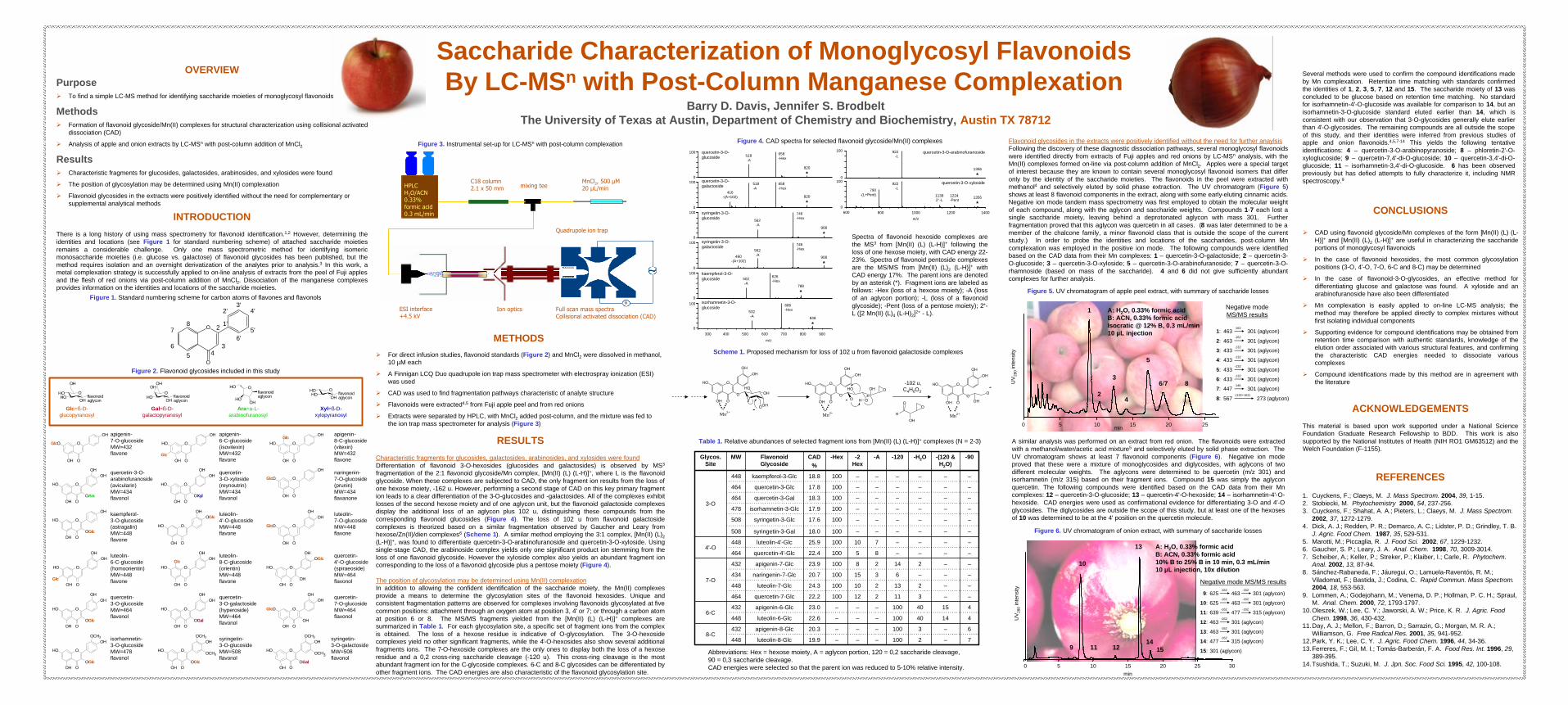
A similar analysis was performed on an extract from red onion. The flavonoids were extracted with a methanol/water/acetic acid mixture5 and selectively eluted by solid phase extraction. The UV chromatogram shows at least 7 flavonoid components (Figure 6). Negative ion mode proved that these were a mixture of monoglycosides and diglycosides, with aglycons of two different molecular weights. The aglycons were determined to be quercetin (m/z 301) and isorhamnetin (m/z 315) based on their fragment ions. Compound 15 was simply the aglyconquercetin. The following compounds were identified based on the CAD data from their Mncomplexes: 12 – quercetin-3-O-glucoside; 13 – quercetin-4'-O-hexoside; 14 – isorhamnetin-4'-O-hexoside. CAD energies were used as confirmational evidence for differentiating 3-O and 4'-O glycosides. The diglycosides are outside the scope of this study, but at least one of the hexosesof 10 was determined to be at the 4' position on the quercetin molecule.
Several methods were used to confirm the compound identifications made by Mn complexation. Retention time matching with standards confirmedthe identities of 1, 2, 3, 5, 7, 12 and 15. The saccharide moiety of 13 was concluded to be glucose based on retention time matching. No standard for isorhamnetin-4'-O-glucoside was available for comparison to 14, but an isorhamnetin-3-O-glucoside standard eluted earlier than 14, which is consistent with our observation that 3-O-glycosides generally elute earlier than 4'-O-glycosides. The remaining compounds are all outside the scope of this study, and their identities were inferred from previous studies of apple and onion flavonoids.4,5,7-14 This yields the following tentative identifications: 4 – quercetin-3-O-arabinopyranoside; 8 – phloretin-2'-O-xyloglucoside; 9 – quercetin-7,4'-di-O-glucoside; 10 – quercetin-3,4'-di-O-glucoside; 11 – isorhamnetin-3,4'-di-O-glucoside. 6 has been observed previously but has defied attempts to fully characterize it, including NMR spectroscopy.9
REFERENCES
MnCl2, 500 µM20 µL/min
C18 column 2.1 x 50 mm mixing teeHPLC
H2O/ACN0.33% formic acid0.3 mL/min
ESI interface+4.5 kV
Quadrupole ion trap
Full scan mass spectraCollisional activated dissociation (CAD)
Ion optics
O
OOH
HO
OHGlc
O
OOH
GlcO
OH
O
OOH
GlcO
OHOH
O
OOH
HO
OGlcOH
OH
O
OOH
HO
OH
Glc
O
OOH
GlcO
OH
O
OOH
HO
OHOH
OXyl
O
OOH
HO
OHOH
OAra
O
OOH
HO
OGlcOH
O
OOH
HO
OH
OGlc
O
OOH
HO
OHOH
GlcO
OOH
HO
OHOH
Glc
O
OOH
GlcO
OHOH
OH
O
OOH
HO
OHOH
OGal
O
OOH
HO
OHOH
OGlc
O
OOH
HO
OHOCH3
OGal
OCH3O
OOH
HO
OHOCH3
OGlc
OCH3O
OOH
HO
OHOCH3
OGlc
Oflavonoidaglycon
HOHO
OH
O
OH
flavonoidaglycon
HO
HO
OHO
flavonoidaglycon
HO
OH
OH
Oflavonoidaglycon
HOOH
OHOH
apigenin-7-O-glucosideMW=432flavone
apigenin-6-C-glucoside(isovitexin)MW=432 flavone
apigenin-8-C-glucoside(vitexin)MW=432flavone
quercetin-3-O-arabinofuranoside(avicularin)MW=434 flavonol
quercetin-3-O-xyloside(reynoutrin)MW=434 flavonol
naringenin-7-O-glucoside(prunin)MW=434 flavanone
kaempferol-3-O-glucoside(astragalin)MW=448 flavone
luteolin-6-C-glucoside(homoorientin)MW=448 flavone
luteolin-8-C-glucoside(orientin)MW=448flavone
quercetin-4'-O-glucoside(spiraeoside)MW=464 flavonol
luteolin-4'-O-glucosideMW=448 flavone
luteolin-7-O-glucosideMW=448 flavone
quercetin-3-O-galactoside(hyperoside)MW=464 flavonol
quercetin-7-O-glucosideMW=464 flavonol
quercetin-3-O-glucosideMW=464 flavonol
syringetin-3-O-glucosideMW=508 flavonol
syringetin-3-O-galactosideMW=508 flavonol
Glc=ß-D-glucopyranosyl
Gal=ß-D-galactopyranosyl
Ara=a-L-arabinofuranosyl
Xyl=ß-D-xylopyranosyl
MethodsØ Formation of flavonoid glycoside/Mn(II) complexes for structural characterization using collisional activated
dissociation (CAD)
Ø Analysis of apple and onion extracts by LC-MSn with post-column addition of MnCl2
PurposeØ To find a simple LC-MS method for identifying saccharide moieties of monoglycosyl flavonoids
isorhamnetin-3-O-glucosideMW=478 flavonol
ACKNOWLEDGEMENTS
This material is based upon work supported under a National Science Foundation Graduate Research Fellowship to BDD. This work is also supported by the National Institutes of Health (NIH RO1 GM63512) and the Welch Foundation (F-1155).
CONCLUSIONS
Ø CAD using flavonoid glycoside/Mn complexes of the form [Mn(II) (L) (L-H)]+ and [Mn(II) (L)2 (L-H)]+ are useful in characterizing the saccharide portions of monoglycosyl flavonoids
Ø In the case of flavonoid hexosides, the most common glycosylation positions (3-O, 4'-O, 7-O, 6-C and 8-C) may be determined
Ø In the case of flavonoid-3-O-glycosides, an effective method for differentiating glucose and galactose was found. A xyloside and an arabinofuranoside have also been differentiated
Ø Mn complexation is easily applied to on-line LC-MS analysis; the method may therefore be applied directly to complex mixtures without first isolating individual components
Ø Supporting evidence for compound identifications may be obtained from retention time comparison with authentic standards, knowledge of the elution order associated with various structural features, and confirming the characteristic CAD energies needed to dissociate various complexes
Ø Compound identifications made by this method are in agreement with the literature
METHODS
Ø For direct infusion studies, flavonoid standards (Figure 2) and MnCl2 were dissolved in methanol, 10 µM each
Ø A Finnigan LCQ Duo quadrupole ion trap mass spectrometer with electrospray ionization (ESI) was used
Ø CAD was used to find fragmentation pathways characteristic of analyte structure
Ø Flavonoids were extracted4,5 from Fuji apple peel and from red onions
Ø Extracts were separated by HPLC, with MnCl2 added post-column, and the mixture was fed to the ion trap mass spectrometer for analysis (Figure 3)
RESULTS
Characteristic fragments for glucosides, galactosides, arabinosides, and xylosides were foundDifferentiation of flavonoid 3-O-hexosides (glucosides and galactosides) is observed by MS3
fragmentation of the 2:1 flavonoid glycoside/Mn complex, [Mn(II) (L) (L-H)]+, where L is the flavonoid glycoside. When these complexes are subjected to CAD, the only fragment ion results from the loss of one hexose moiety, -162 u. However, performing a second stage of CAD on this key primary fragment ion leads to a clear differentiation of the 3-O-glucosides and -galactosides. All of the complexes exhibit losses of the second hexose moiety and of one aglycon unit, but the flavonoid galactoside complexes display the additional loss of an aglycon plus 102 u, distinguishing these compounds from the corresponding flavonoid glucosides (Figure 4). The loss of 102 u from flavonoid galactosidecomplexes is theorized based on a similar fragmentation observed by Gaucher and Leary from hexose/Zn(II)/dien complexes6 (Scheme 1). A similar method employing the 3:1 complex, [Mn(II) (L)2(L-H)]+, was found to differentiate quercetin-3-O-arabinofuranoside and quercetin-3-O-xyloside. Using single-stage CAD, the arabinoside complex yields only one significant product ion stemming from the loss of one flavonoid glycoside. However the xyloside complex also yields an abundant fragment ion corresponding to the loss of a flavonoid glycoside plus a pentose moiety (Figure 4).
The position of glycosylation may be determined using Mn(II) complexationIn addition to allowing the confident identification of the saccharide moiety, the Mn(II) complexes provide a means to determine the glycosylation sites of the flavonoid hexosides. Unique and consistent fragmentation patterns are observed for complexes involving flavonoids glycosylated at five common positions: attachment through an oxygen atom at position 3, 4' or 7; or through a carbon atom at position 6 or 8. The MS/MS fragments yielded from the [Mn(II) (L) (L-H)]+ complexes are summarized in Table 1. For each glycosylation site, a specific set of fragment ions from the complex is obtained. The loss of a hexose residue is indicative of O-glycosylation. The 3-O-hexoside complexes yield no other significant fragments, while the 4'-O-hexosides also show several additional fragments ions. The 7-O-hexoside complexes are the only ones to display both the loss of a hexoseresidue and a 0,2 cross-ring saccharide cleavage (-120 u). This cross-ring cleavage is the most abundant fragment ion for the C-glycoside complexes. 6-C and 8-C glycosides can be differentiated by other fragment ions. The CAD energies are also characteristic of the flavonoid glycosylation site.
150 5 10 20 25min
UV
280
inte
nsity
1
2
3
4
5
6/7 8
1: 463 301 (aglycon)
2: 463 301 (aglycon)
3: 433 301 (aglycon)
4: 433 301 (aglycon)
5: 433 301 (aglycon)
6: 433 301 (aglycon)
7: 447 301 (aglycon)
8: 567 273 (aglycon)
-162
-162
-132
-132
-132
-132
-146
-(132+162)
150 5 10 20 25 30
10
13
14119 12 15
min
UV
280
inte
nsity
9: 625 463 301 (aglycon)
10: 625 463 301 (aglycon)
11: 639 477 315 (aglycon)
12: 463 301 (aglycon)
13: 463 301 (aglycon)
14: 477 315 (aglycon)
15: 301 (aglycon)
-162
-162
-162
-162
-162
-162 -162
-162 -162
Figure 2. Flavonoid glycosides included in this study
Figure 3. Instrumental set-up for LC-MSn with post-column complexation Figure 4. CAD spectra for selected flavonoid glycoside/Mn(II) complexes
Scheme 1. Proposed mechanism for loss of 102 u from flavonoid galactoside complexes
Table 1. Relative abundances of selected fragment ions from [Mn(II) (L) (L-H)]+ complexes (N = 2-3)
–
–
–
–
100
100
100
100
100
100
100
100
100
100
100
100
-Hex
–
–
–
–
12
10
15
8
5
10
–
–
–
–
–
–
-2 Hex
–
–
–
–
2
2
3
2
8
7
–
–
–
–
–
–
-A
100
100
100
100
11
13
6
14
–
–
–
–
–
–
–
–
-120
2
3
40
40
3
2
–
2
–
–
–
–
–
–
–
–
-H2O
–
–
14
15
–
–
–
–
–
–
–
–
–
–
–
–
-(120 & H2O)
7
6
4
4
–
–
–
–
–
–
–
–
–
–
–
–
-90
448
432
448
432
464
448
434
432
464
448
508
508
478
464
464
448
MW
19.9luteolin-8-Glc
20.3apigenin-8-Glc8-C
22.6luteolin-6-Glc
23.0apigenin-6-Glc6-C
22.2quercetin-7-Glc
24.3luteolin-7-Glc
20.7naringenin-7-Glc
23.9apigenin-7-Glc
7-O
22.4quercetin-4'-Glc
25.9luteolin-4'-Glc4'-O
18.0syringetin-3-Gal
17.6syringetin-3-Glc
17.9isorhamnetin-3-Glc
18.3quercetin-3-Gal
17.8quercetin-3-Glc
18.8kaempferol-3-Glc
3-O
CAD%
Flavonoid Glycoside
Glycos. Site
Figure 5. UV chromatogram of apple peel extract, with summary of saccharide losses
Figure 6. UV chromatogram of onion extract, with summary of saccharide losses
A: H2O, 0.33% formic acidB: ACN, 0.33% formic acidIsocratic @ 12% B, 0.3 mL/min10 µL injection
Negative modeMS/MS results
Negative mode MS/MS results
A: H2O, 0.33% formic acidB: ACN, 0.33% formic acid10% B to 25% B in 10 min, 0.3 mL/min10 µL injection, 10x dilution
Abbreviations: Hex = hexose moiety, A = aglycon portion, 120 = 0,2 saccharide cleavage, 90 = 0,3 saccharide cleavage.CAD energies were selected so that the parent ion was reduced to 5-10% relative intensity.
isorhamnetin-3-O-glucoside
kaempferol-3-O-glucoside
syringetin-3-O-galactoside
syringetin-3-O-glucoside
quercetin-3-O-galactoside
quercetin-3-O-glucoside
746-Hex
746-Hex
562-A
562-A
518-A
518-A
658-Hex
658-Hex
626-Hex
686-Hex532
-A
502-A
460-(A+102)
416-(A+102)
500300 400 600 700 800 900
820
*
820
*
908
*
908
*
788
*
848
*
m/z
100
100
100
100
100
100
0
0
0
0
0
0
quercetin-3-O-xyloside
600 1400800 1000 1200
922-L790
-(L+Pent) 1224-Pent
11392+-L
quercetin-3-O-arabinofuranoside922-L
1356
*
1356
*
100
1000
0
m/z
Spectra of flavonoid hexoside complexes are the MS3 from [Mn(II) (L) (L-H)]+ following the loss of one hexose moiety, with CAD energy 22-23%. Spectra of flavonoid pentoside complexes are the MS/MS from [Mn(II) (L)2 (L-H)]+ with CAD energy 17%. The parent ions are denoted by an asterisk (*). Fragment ions are labeled as follows: -Hex (loss of a hexose moiety); -A (loss of an aglycon portion); -L (loss of a flavonoid glycoside); -Pent (loss of a pentose moiety); 2+-L ([2 Mn(II) (L)4 (L-H)2]2+ - L).
O
O
3
7
56
8
3'4'
5'6'
2'
4
2 1'
Figure 1. Standard numbering scheme for carbon atoms of flavones and flavonols
-102 u,C4H6O3
--
-