S -Methylmethionine Plays a Major Role in Phloem Sulfur ... · The Plant Cell, Vol. 11, 1485–1497...
14
The Plant Cell, Vol. 11, 1485–1497, August 1999, www.plantcell.org © 1999 American Society of Plant Physiologists S-Methylmethionine Plays a Major Role in Phloem Sulfur Transport and Is Synthesized by a Novel Type of Methyltransferase Fabienne Bourgis, a Sanja Roje, a Michael L. Nuccio, a Donald B. Fisher, b Mitchell C. Tarczynski, c Changjiang Li, c Cornelia Herschbach, d Heinz Rennenberg, d Maria Joao Pimenta, e Tun-Li Shen, f Douglas A. Gage, f and Andrew D. Hanson a,1 a Horticultural Sciences Department, University of Florida, Gainesville, Florida 32611-0690 b Botany Department, Washington State University, Pullman, Washington 99164-4238 c Pioneer Hi-Bred International, 7300 N.W. 62nd Avenue, Johnston, Iowa 50131-1004 d Institut für Forstbotanik und Baumphysiologie, Albert-Ludwigs-Universität, D-79085 Freiburg, Germany e Frontier Research Program, Institute of Physical and Chemical Research (RIKEN), 2-1 Hirosawa, Wako-shi, Saitama 351-0198, Japan f Biochemistry Department, Michigan State University, East Lansing, Michigan 48824-1319 All flowering plants produce S-methylmethionine (SMM) from Met and have a separate mechanism to convert SMM back to Met. The functions of SMM and the reasons for its interconversion with Met are not known. In this study, by using the aphid stylet collection method together with mass spectral and radiolabeling analyses, we established that L-SMM is a major constituent of the phloem sap moving to wheat ears. The SMM level in the phloem ( z2% of free amino acids) was 1.5-fold that of glutathione, indicating that SMM could contribute approximately half the sulfur needed for grain protein synthesis. Similarly, L-SMM was a prominently labeled product in phloem exudates obtained by EDTA treatment of detached leaves from plants of the Poaceae, Fabaceae, Asteraceae, Brassicaceae, and Cucurbita- ceae that were given L– 35 S-Met. cDNA clones for the enzyme that catalyzes SMM synthesis (S-adenosylMet:Met S-meth- yltransferase; EC 2.1.1.12) were isolated from Wollastonia biflora, maize, and Arabidopsis. The deduced amino acid sequences revealed the expected methyltransferase domain ( z300 residues at the N terminus), plus an 800-residue C-terminal region sharing significant similarity with aminotransferases and other pyridoxal 5 9-phosphate–dependent enzymes. These results indicate that SMM has a previously unrecognized but often major role in sulfur transport in flowering plants and that evolution of SMM synthesis in this group involved a gene fusion event. The resulting bipartite enzyme is unlike any other known methyltransferase. INTRODUCTION Plant Met metabolism differs from that in other organisms by involving S-methylmethionine (SMM). SMM is a ubiquitous constituent of the free amino acid pool in flowering plants, occurring in leaves, roots, and other organs at levels that typically range from 0.5 to 3 mmol g 21 dry weight, a concen- tration that is often higher than those of Met or S-adenosyl- methionine (AdoMet) (Giovanelli et al., 1980; Mudd and Datko, 1990; Bezzubov and Gessler, 1992). SMM also has been detected as a metabolite of radiolabeled L-Met in all flowering plants tested ( .50 species from .20 families; Paquet et al., 1995). As shown in Figure 1, SMM is formed from L-Met via the action of AdoMet:Met S-methyltrans- ferase (MMT; EC 2.1.1.12) and can be reconverted to Met by donating a methyl group to L-homocysteine (Hcy) in a reac- tion catalyzed by Hcy S-methyltransferase (HMT; EC 2.1.1.10; Giovanelli et al., 1980; Mudd and Datko, 1990). The tandem action of MMT and HMT, together with S-adenosyl- L-Hcy hydrolase, constitutes the SMM cycle, which is ap- parently futile (Mudd and Datko, 1990). As expected from the universality of SMM, MMT activity has been found in many flowering plants (Giovanelli et al., 1980; Mudd and Datko, 1990). It has been purified from leaves of Wollastonia biflora (James et al., 1995a) and from germinating barley (Pimenta et al., 1998), and it is known to have subunits of z115 kD. Because this is approximately three times larger than any other small-molecule methyl- transferase (Fujioka, 1992; Joshi and Chiang, 1998), it sug- gests that the MMT polypeptide could have more than one 1 To whom correspondence should be addressed. E-mail adha@ gnv.ifas.ufl.edu; fax 352-392-6479.
Transcript of S -Methylmethionine Plays a Major Role in Phloem Sulfur ... · The Plant Cell, Vol. 11, 1485–1497...
The Plant Cell, Vol. 11, 1485–1497, August 1999, www.plantcell.org © 1999 American Society of Plant Physiologists
S
-Methylmethionine Plays a Major Role in Phloem Sulfur Transport and Is Synthesized by a Novel Typeof Methyltransferase
Fabienne Bourgis,
a
Sanja Roje,
a
Michael L. Nuccio,
a
Donald B. Fisher,
b
Mitchell C. Tarczynski,
c
Changjiang Li,
c
Cornelia Herschbach,
d
Heinz Rennenberg,
d
Maria Joao Pimenta,
e
Tun-Li Shen,
f
Douglas A. Gage,
f
and Andrew D. Hanson
a,1
a
Horticultural Sciences Department, University of Florida, Gainesville, Florida 32611-0690
b
Botany Department, Washington State University, Pullman, Washington 99164-4238
c
Pioneer Hi-Bred International, 7300 N.W. 62nd Avenue, Johnston, Iowa 50131-1004
d
Institut für Forstbotanik und Baumphysiologie, Albert-Ludwigs-Universität, D-79085 Freiburg, Germany
e
Frontier Research Program, Institute of Physical and Chemical Research (RIKEN), 2-1 Hirosawa, Wako-shi,Saitama 351-0198, Japan
f
Biochemistry Department, Michigan State University, East Lansing, Michigan 48824-1319
All flowering plants produce
S
-methylmethionine (SMM) from Met and have a separate mechanism to convert SMMback to Met. The functions of SMM and the reasons for its interconversion with Met are not known. In this study, byusing the aphid stylet collection method together with mass spectral and radiolabeling analyses, we established that
L
-SMM is a major constituent of the phloem sap moving to wheat ears. The SMM level in the phloem (
z
2% of freeamino acids) was 1.5-fold that of glutathione, indicating that SMM could contribute approximately half the sulfurneeded for grain protein synthesis. Similarly,
L
-SMM was a prominently labeled product in phloem exudates obtained byEDTA treatment of detached leaves from plants of the Poaceae, Fabaceae, Asteraceae, Brassicaceae, and Cucurbita-ceae that were given
L
–
35
S-Met. cDNA clones for the enzyme that catalyzes SMM synthesis (
S
-adenosylMet:Met
S
-meth-yltransferase; EC 2.1.1.12) were isolated from
Wollastonia biflora
, maize, and Arabidopsis. The deduced amino acidsequences revealed the expected methyltransferase domain (
z
300 residues at the N terminus), plus an 800-residueC-terminal region sharing significant similarity with aminotransferases and other pyridoxal 5
9
-phosphate–dependentenzymes. These results indicate that SMM has a previously unrecognized but often major role in sulfur transport inflowering plants and that evolution of SMM synthesis in this group involved a gene fusion event. The resulting bipartiteenzyme is unlike any other known methyltransferase.
INTRODUCTION
Plant Met metabolism differs from that in other organismsby involving
S
-methylmethionine (SMM). SMM is a ubiquitousconstituent of the free amino acid pool in flowering plants,occurring in leaves, roots, and other organs at levels thattypically range from 0.5 to 3
m
mol g
2
1
dry weight, a concen-tration that is often higher than those of Met or
S
-adenosyl-methionine (AdoMet) (Giovanelli et al., 1980; Mudd andDatko, 1990; Bezzubov and Gessler, 1992). SMM also hasbeen detected as a metabolite of radiolabeled
L
-Met in allflowering plants tested (
.
50 species from
.
20 families;Paquet et al., 1995). As shown in Figure 1, SMM is formedfrom
L
-Met via the action of AdoMet:Met
S
-methyltrans-
ferase (MMT; EC 2.1.1.12) and can be reconverted to Met bydonating a methyl group to
L
-homocysteine (Hcy) in a reac-tion catalyzed by Hcy
S
-methyltransferase (HMT; EC2.1.1.10; Giovanelli et al., 1980; Mudd and Datko, 1990). Thetandem action of MMT and HMT, together with
S
-adenosyl-
L
-Hcy hydrolase, constitutes the SMM cycle, which is ap-parently futile (Mudd and Datko, 1990).
As expected from the universality of SMM, MMT activityhas been found in many flowering plants (Giovanelli et al.,1980; Mudd and Datko, 1990). It has been purified fromleaves of
Wollastonia biflora
(James et al., 1995a) and fromgerminating barley (Pimenta et al., 1998), and it is known tohave subunits of
z
115 kD. Because this is approximatelythree times larger than any other small-molecule methyl-transferase (Fujioka, 1992; Joshi and Chiang, 1998), it sug-gests that the MMT polypeptide could have more than one
1
To whom correspondence should be addressed. E-mail [email protected]; fax 352-392-6479.

1486 The Plant Cell
enzymatic domain. HMT has been less investigated, but ittoo has been found in diverse plants, especially in seeds(Giovanelli et al., 1980).
Although it is clear how plants synthesize SMM and re-convert it to Met, the physiological roles of SMM and its cy-cle remain undefined, except for the few species that useSMM as the precursor for synthesis of the osmoprotectant3-dimethylsulfoniopropionate (Trossat et al., 1996; Kocsis etal., 1998). Proposed general roles for SMM and its cycle in-clude Met storage, methyl donation, and regulation of theAdoMet/Met ratio, all of which are reasonable but unsup-ported by experimental evidence (Giovanelli et al., 1980;Mudd and Datko, 1990). In all of these proposed roles, SMMwould exert its functions without exiting the cells that pro-duce it.
Data reported in a study of sieve tube protein turnover byFisher et al. (1992) led us to suspect that SMM might have aquite different role—in phloem sulfur transport. Fisher et al.(1992) found that when wheat flag leaves were given
35
S-Met, most of the sulfur-35 in the phloem sap moving to theear was in an unidentified metabolite with low mobility inthin-layer chromatography (TLC). We noted that this producthad the mobility characteristic of SMM. In this study, we re-peated the experiment of Fisher et al. (1992) and demon-strated that the unidentified phloem-mobile metabolite wasindeed
35
S-SMM. This prompted us to quantify the SMMpresent in wheat phloem by mass spectral methods and todetermine whether SMM occurs in the phloem sap of otherflowering plants. Because the data indicated that SMM is amajor and common phloem constituent, we proceeded toisolate and characterize cDNAs for MMT, the SMM-synthe-sizing enzyme, from three diverse plants. The deduced MMTamino acid sequences define a novel type of methyltrans-ferase.
RESULTS
Wheat Leaves Supplied with
35
S-Met Export
35
S-SMM in the Phloem
Using the same procedures as Fisher et al. (1992), we gavea flag leaf from a wheat plant in the middle part of the grain-filling period a pulse of tracer
L
–
35
S-Met and collectedphloem exudate from the peduncle by using severed aphidstylets (Figure 2A). Analysis of the soluble labeled com-pounds from both exudate and leaf showed that SMM is aprominent metabolite, accounting for
z
80% of the total sul-fur-35 in the exudates and 60% in the leaves (Figure 2B).The identity of
35
S-SMM was established by three criteria:comigration with authentic SMM in three separation sys-tems; complete destruction by hot 1 M NaOH; and conver-sion to the corresponding
a
-hydroxy acid by nitrous acidtreatment (Figure 2C). The proportion (80%) of the total sul-fur-35 in the exudate that was accounted for by
35
S-SMMmatched that reported for the unidentified
35
S-Met metabo-lite (i.e., SMM) by Fisher et al. (1992).
Mass Spectral Evidence That SMM Is a Major Form of Sulfur in Wheat Phloem Sap
To reinforce the radiolabeling evidence for the presence ofSMM in wheat phloem, we analyzed exudates by matrix-assisted laser desorption ionization mass spectrometry(MALDI-MS). There was a strong signal at a mass-to-chargeratio (
m/z
) of 164, corresponding to SMM (Figure 3A). Con-firmation that this peak represented SMM was obtained byMALDI postsource decay experiments; these showed theexpected fragment at an
m/z
ratio of 102, formed from theprecursor ion (
m/z
of 164) by neutral loss of (CH
3
)
2
S (Figure3B). MALDI-MS also was used to quantify SMM in wheatphloem exudates by using an internal standard of methyl-
2
H
6
-SMM; the free
a
-amino acid contents of same sampleswere determined using ninhydrin. As shown in Table 1, SMMmade up 1.8
6
0.1 mol% (mean
6
SE
) of free amino acids.This value suggested that SMM import could be a majoritem in the sulfur budget of the grain, given that the sulfuramino acid content of wheat grain protein is
z
4 mol% (Khanand Eggum, 1978). The SMM level in flag leaf blades wasmeasured for comparison with the phloem data; it was 170
6
20 nmol g
2
1
fresh weight (mean
6
SE
,
n
5
3) or
z
85 nmolper blade.
SMM Levels in Wheat Phloem Sap Are Similar to Glutathione Levels
Because glutathione (GSH) is known to be a major form ofreduced sulfur in the phloem of many species (Rennenberg,1982; Brunold and Rennenberg, 1997), we compared the
Figure 1. The SMM Cycle of Flowering Plants.
The reaction mediated by HMT generates two molecules of Met.AdoHcy, S-adenosyl-L-homocysteine.
SMM Transport and MMT Characterization 1487
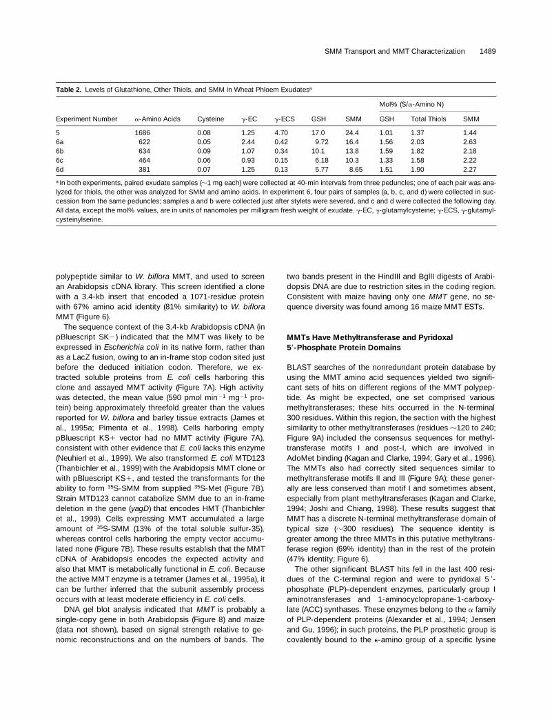
levels of GSH and related thiols to those of SMM. For theseexperiments, the phloem samples were collected at 40-minintervals and immediately frozen because tests indicatedthat the thiols in exudates underwent slow degradation in airat room temperature (
z
50% loss in 14 hr). Table 2 showsthat the GSH levels in wheat phloem were 66
6
3% (mean
6
SE
) of SMM levels. The GSH precursor
g
-glutamylcysteinewas present in lesser amounts, as was a compound with thechromatographic properties of
g
-glutamylcysteinylserine.This homolog of GSH has been found in wheat leaves at lev-els ranging from three- to 15-fold lower than those of GSHitself (Klapheck et al., 1992; McKee et al., 1997), and its levelalso varied markedly among the phloem exudate samples.As reported previously for wheat phloem (Fisher and Macnicol,1986), free Cys was virtually absent. The total thiol level wassimilar to the SMM level (Table 2).
Leaves of Diverse Flowering Plants Export SMM inthe Phloem
To determine whether SMM is a constituent of phloem sapin other species, we used EDTA to enhance phloem exuda-tion from cut leaf bases or petioles. EDTA chelates the Ca
2
1
required for callose formation and thereby blocks the seal-ing of cut sieve tubes (King and Zeevaart, 1974). The sapobtained by the EDTA technique is comparable in composi-tion to that from severed stylets (Weibull et al., 1990; Valle etal., 1998). A tracer dose of
35
S-Met was applied to the tips ofleaves from species representing five diverse families, andthe corresponding phloem exudates were analyzed for
35
S-SMM (Figure 4A). Amino acid exudation was measured frommatching unlabeled leaves, plus or minus EDTA, as a checkon the technique. EDTA enhanced amino acid exudation byan average of ninefold (Figure 4B), which is consistent withthe exudates coming mainly from cut sieve tubes (King andZeevaart, 1974).
35
S-SMM was detected in the exudatesfrom all 11 species tested, and in five of them it accountedfor
>
35% of the total label (Figure 4A). The proportion of
35
S-SMM in the exudate from wheat (70%) agreed well withthat seen in stylet-derived exudate (Figure 2B), which further
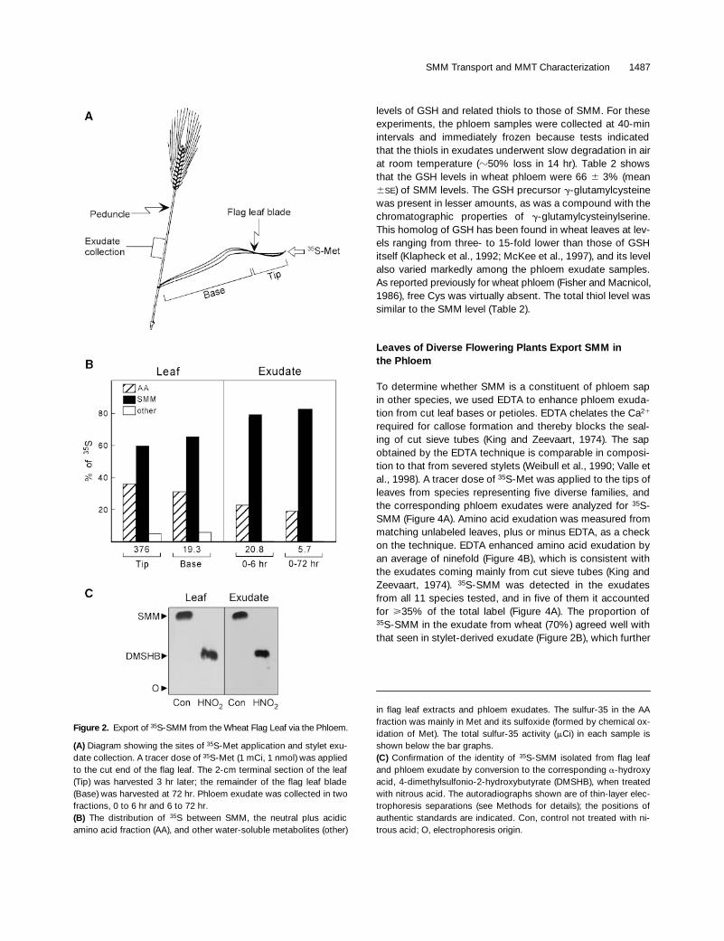
Figure 2.
Export of
35
S-SMM from the Wheat Flag Leaf via the Phloem.
(A)
Diagram showing the sites of
35
S-Met application and stylet exu-date collection. A tracer dose of
35
S-Met (1 mCi, 1 nmol) was appliedto the cut end of the flag leaf. The 2-cm terminal section of the leaf(Tip) was harvested 3 hr later; the remainder of the flag leaf blade(Base) was harvested at 72 hr. Phloem exudate was collected in twofractions, 0 to 6 hr and 6 to 72 hr.
(B)
The distribution of
35
S between SMM, the neutral plus acidicamino acid fraction (AA), and other water-soluble metabolites (other)
in flag leaf extracts and phloem exudates. The sulfur-35 in the AAfraction was mainly in Met and its sulfoxide (formed by chemical ox-idation of Met). The total sulfur-35 activity (
m
Ci) in each sample isshown below the bar graphs.
(C)
Confirmation of the identity of
35
S-SMM isolated from flag leafand phloem exudate by conversion to the corresponding
a
-hydroxyacid, 4-dimethylsulfonio-2-hydroxybutyrate (DMSHB), when treatedwith nitrous acid. The autoradiographs shown are of thin-layer elec-trophoresis separations (see Methods for details); the positions ofauthentic standards are indicated. Con, control not treated with ni-trous acid; O, electrophoresis origin.

1488 The Plant Cell
validates this application of the EDTA technique. In all oursulfur-35 exudation experiments, the applied tracer
35
S-Metmay have entered the phloem directly, whereas sulfur-35could only have reached SMM after isotope dilution by en-dogenous Met and SMM pools. These pools are far largerthan the applied
35
S-Met doses and vary in size with speciesand leaf age (Gessler et al., 1991). Thus, the low proportionsof
35
S-SMM in the exudates of some species (Figure 4A) byno means necessarily connote low chemical levels of SMM.
The SMM Present in Leaves and Phloem Sap Is Exclusively the
L
-Enantiomer
The enzyme that catalyzes SMM formation, MMT, is knownto be specific for
L
-Met (James et al., 1995a), but the config-uration of SMM itself has not been determined. We cor-rected this deficiency by analyzing
35
S-SMM isolated fromleaves and phloem exudates of a monocot (wheat) and a di-cot (Arabidopsis). We exploited the stereoselectivity of
L
- and
D
-amino acid oxidases, which convert SMM to an
a
-keto
acid that decomposes rapidly to yield dimethylsulfide (DMS;Rhodes et al., 1997). The relative amounts of
35
S-DMS re-leased by the
L
- and
D
-specific enzymes showed that the
35
S-SMM from leaves and phloem of both species was es-sentially all (
>
97%) the
L
-enantiomer (Figure 5). This resultestablishes that SMM is not enzymatically racemized.
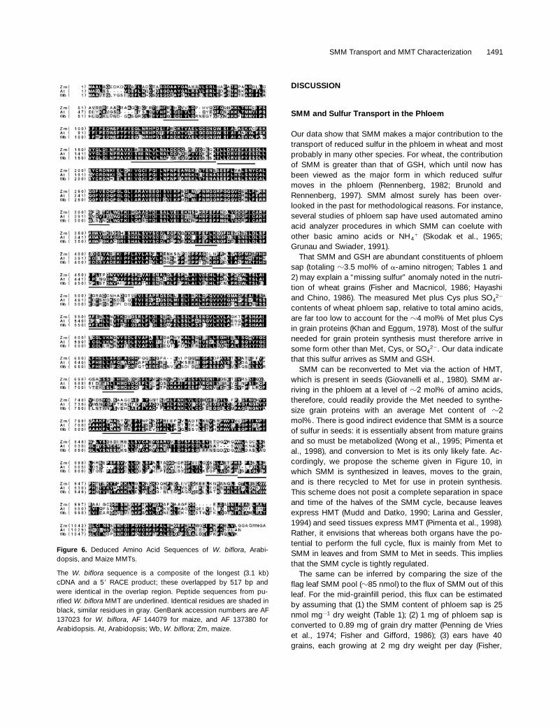
cDNA Cloning, Sequence Analysis, and Functional Expression of MMTs
The widespread occurrence of SMM in phloem sap led us toisolate cDNAs for the SMM-synthesizing enzyme, MMT,starting with
W. biflora
. Purified
W. biflora
MMT was di-gested to obtain peptides, 14 of which were sequenced. De-generate polymerase chain reaction (PCR) primers matchingthe ends of a 19-residue peptide (see Methods) were usedto amplify the corresponding 56-bp DNA sequence, with a
W. biflora
cDNA library as template. A primer specific for thecentral part of this 56-bp sequence, together with one fromthe vector, enabled PCR amplification of a 1.1-kb fragmentcomprising the 3
9
terminal region of MMT. This fragmentwas used to screen the library, which led to isolation of a3.1-kb MMT cDNA that lacked the 5
9
region. The missing re-gion was obtained by rapid amplification of cDNA ends(RACE). The complete cDNA encodes a 1088-residue pro-tein of calculated mass of 121.6 kD (Figure 6). The deducedamino acid sequence includes all the MMT peptides thatwere sequenced, establishing its authenticity (Figure 6). NoN-terminal signal sequence was recognizable, consistentwith the exclusively cytosolic localization of
W. biflora
MMT(Trossat et al., 1996).
Searches of expressed sequence tag (EST) databases us-ing the W. biflora nucleotide and amino acid sequences re-vealed matches with 16 maize and two Arabidopsis ESTs.The longest maize EST (3.4 kb) was sequenced and found toencode a 1091-residue protein with 62% amino acid identity(77% similarity) to W. biflora MMT (Figure 6). One Arabidop-sis EST (1.2 kb) was sequenced, confirmed to encode a
Figure 3. Mass Spectral Evidence for the Presence of SMM inWheat Phloem Exudate.
(A) MALDI-MS spectrum of the BioRex-70 fraction. The major peakat m/z of 164 corresponds to SMM; the small peaks at m/z of 165and 166 are due to natural-abundance C, S, and O isotopes.(B) MALDI postsource decay experiment showing the predictedfragment ion at m/z of 102.
Table 1. Levels of SMM and Total a-Amino Acids in Wheat Phloem Exudatesa
ExperimentNumber
Weightb
(mg)SMM(nmol)
a-Amino Acids(nmol)
SMM Level
Mol% nmol mg21
1 8.8 187 8750 2.14 21.32 10.8 215 12490 1.72 19.93 9.3 247 14270 1.73 26.64 32.0 383 25140 1.52 12.0
a In each experiment, phloem exudate was collected from one pe-duncle for 14 hr (experiments 1 to 3) or 72 hr (experiment 4).b Fresh weight. The dry weight of exudates was z80% of the freshweight.
SMM Transport and MMT Characterization 1489
polypeptide similar to W. biflora MMT, and used to screenan Arabidopsis cDNA library. This screen identified a clonewith a 3.4-kb insert that encoded a 1071-residue proteinwith 67% amino acid identity (81% similarity) to W. bifloraMMT (Figure 6).
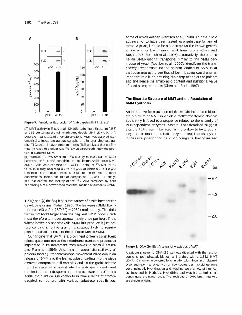
The sequence context of the 3.4-kb Arabidopsis cDNA (inpBluescript SK2) indicated that the MMT was likely to beexpressed in Escherichia coli in its native form, rather thanas a LacZ fusion, owing to an in-frame stop codon sited justbefore the deduced initiation codon. Therefore, we ex-tracted soluble proteins from E. coli cells harboring thisclone and assayed MMT activity (Figure 7A). High activitywas detected, the mean value (590 pmol min 21 mg21 pro-tein) being approximately threefold greater than the valuesreported for W. biflora and barley tissue extracts (James etal., 1995a; Pimenta et al., 1998). Cells harboring emptypBluescript KS1 vector had no MMT activity (Figure 7A),consistent with other evidence that E. coli lacks this enzyme(Neuhierl et al., 1999). We also transformed E. coli MTD123(Thanbichler et al., 1999) with the Arabidopsis MMT clone orwith pBluescript KS1, and tested the transformants for theability to form 35S-SMM from supplied 35S-Met (Figure 7B).Strain MTD123 cannot catabolize SMM due to an in-framedeletion in the gene (yagD) that encodes HMT (Thanbichleret al., 1999). Cells expressing MMT accumulated a largeamount of 35S-SMM (13% of the total soluble sulfur-35),whereas control cells harboring the empty vector accumu-lated none (Figure 7B). These results establish that the MMTcDNA of Arabidopsis encodes the expected activity andalso that MMT is metabolically functional in E. coli. Becausethe active MMT enzyme is a tetramer (James et al., 1995a), itcan be further inferred that the subunit assembly processoccurs with at least moderate efficiency in E. coli cells.
DNA gel blot analysis indicated that MMT is probably asingle-copy gene in both Arabidopsis (Figure 8) and maize(data not shown), based on signal strength relative to ge-nomic reconstructions and on the numbers of bands. The
two bands present in the HindIII and BglII digests of Arabi-dopsis DNA are due to restriction sites in the coding region.Consistent with maize having only one MMT gene, no se-quence diversity was found among 16 maize MMT ESTs.
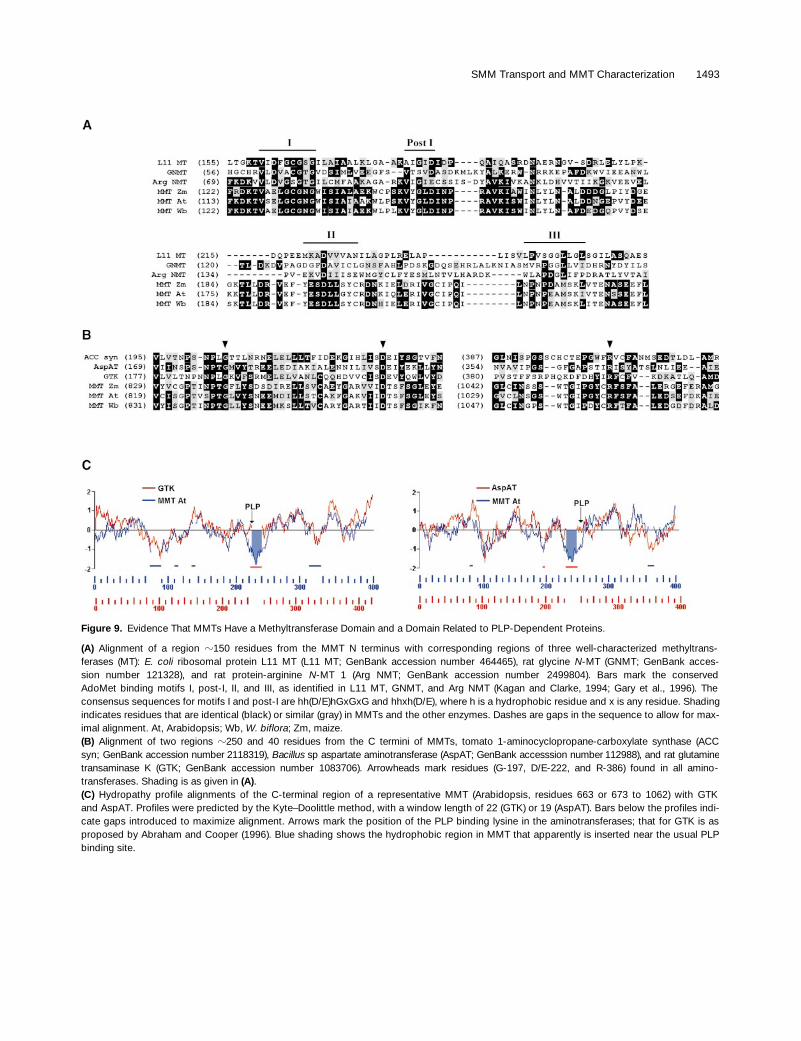
MMTs Have Methyltransferase and Pyridoxal 59-Phosphate Protein Domains
BLAST searches of the nonredundant protein database byusing the MMT amino acid sequences yielded two signifi-cant sets of hits on different regions of the MMT polypep-tide. As might be expected, one set comprised variousmethyltransferases; these hits occurred in the N-terminal300 residues. Within this region, the section with the highestsimilarity to other methyltransferases (residues z120 to 240;Figure 9A) included the consensus sequences for methyl-transferase motifs I and post-I, which are involved inAdoMet binding (Kagan and Clarke, 1994; Gary et al., 1996).The MMTs also had correctly sited sequences similar tomethyltransferase motifs II and III (Figure 9A); these gener-ally are less conserved than motif I and sometimes absent,especially from plant methyltransferases (Kagan and Clarke,1994; Joshi and Chiang, 1998). These results suggest thatMMT has a discrete N-terminal methyltransferase domain oftypical size (z300 residues). The sequence identity isgreater among the three MMTs in this putative methyltrans-ferase region (69% identity) than in the rest of the protein(47% identity; Figure 6).
The other significant BLAST hits fell in the last 400 resi-dues of the C-terminal region and were to pyridoxal 5 9-phosphate (PLP)–dependent enzymes, particularly group Iaminotransferases and 1-aminocyclopropane-1-carboxy-late (ACC) synthases. These enzymes belong to the a familyof PLP-dependent proteins (Alexander et al., 1994; Jensenand Gu, 1996); in such proteins, the PLP prosthetic group iscovalently bound to the e-amino group of a specific lysine
Table 2. Levels of Glutathione, Other Thiols, and SMM in Wheat Phloem Exudatesa
Mol% (S/a-Amino N)
Experiment Number a-Amino Acids Cysteine g-EC g-ECS GSH SMM GSH Total Thiols SMM
5 1686 0.08 1.25 4.70 17.0 24.4 1.01 1.37 1.446a 622 0.05 2.44 0.42 9.72 16.4 1.56 2.03 2.636b 634 0.09 1.07 0.34 10.1 13.8 1.59 1.82 2.186c 464 0.06 0.93 0.15 6.18 10.3 1.33 1.58 2.226d 381 0.07 1.25 0.13 5.77 8.65 1.51 1.90 2.27
a In both experiments, paired exudate samples (z1 mg each) were collected at 40-min intervals from three peduncles; one of each pair was ana-lyzed for thiols, the other was analyzed for SMM and amino acids. In experiment 6, four pairs of samples (a, b, c, and d) were collected in suc-cession from the same peduncles; samples a and b were collected just after stylets were severed, and c and d were collected the following day.All data, except the mol% values, are in units of nanomoles per milligram fresh weight of exudate. g-EC, g-glutamylcysteine; g-ECS, g-glutamyl-cysteinylserine.

1490 The Plant Cell
residue. Alignments indicated that MMTs have three of thefour invariant residues that characterize all aminotrans-ferases, Gly-197, Asp/Glu-222, and Arg-386, yet lack thefourth and most crucial one, the PLP binding Lys-258 (Fig-ure 9B; numbering is according to the sequence of the pro-totype, porcine cytosolic aspartate aminotransferase; Mehtaet al., 1993). Other residues found in group I aminotrans-ferases, Asn-194, Pro-195, Tyr-225, and Gly-268, are alsopresent in MMTs or are conservatively replaced. Moreover,MMTs show 18 to 23% overall amino acid identity (36 to42% similarity) to the a family enzymes in Figure 9B and
have similar hydropathy profiles, with the major exception ofan z20-residue hydrophobic insert that comprises the re-gion in which the PLP binding lysine would be expected toreside (Figure 9C).
The C-terminal section of MMT thus has enough of theprotein scaffold shared by aminotransferases and other a
family proteins to suggest that it is descended from them,but it does not bind PLP in the usual site, if at all. BecauseMMTs have several conserved lysines in the C-terminal re-gion (Figure 6), a highly displaced PLP binding lysine cannotbe ruled out, although the conserved three-dimensionalstructure of a family enzymes makes this seem improbable(John, 1998). It is nevertheless noteworthy that the con-served lysine nearest the C terminus is set in a 40-residuesequence that is 23 to 35% identical (33 to 43% similar) tothose around the PLP binding lysine in glucan phosphory-lases, a group unrelated to the a family (John, 1998). An-other possibility is that PLP is bound noncovalently to theMMT polypeptide, as in various engineered enzymes (John,1998), but no natural instances of this are known.
Figure 4. Evidence That SMM Occurs in Phloem Exudates fromDiverse Plants.
(A) The 35S-SMM contents of exudates, as a percentage of totalsulfur-35 exuded. Attached leaves were supplied with 20 mCi (17pmol) of L–35S-Met, severed after 2 hr, and placed with their cutends in 5 mM Na2EDTA, pH 7.0, for 20 hr in darkness. Total sul-fur-35 exudation ranged from 7.6 nCi (maize) to 917 nCi (Arabi-dopsis). Inset is an autoradiograph of a TLC separation of theBioRex-70 fraction of exudates from representative species:wheat (W), canola (C), and soybean (S).(B) The levels of amino acids exuded with (1) or without (2) 5mM Na2EDTA in the medium. The leaves used were matched insize and age to those supplied with 35S-Met. The values for zuc-chini have been multiplied by 0.5 to fit the scale used for otherspecies.
Figure 5. Determination of the Configuration of SMM from Leavesand Phloem.
35S-SMM isolated from leaves or phloem exudates was incubatedwith L- or D-amino acid oxidase preparations of equivalent activity,and the 35S-DMS reaction product was quantified. The relativeamounts of 35S-DMS released indicate in every case that 97 to 98%of the SMM was the L-enantiomer. Slight racemization during sam-ple processing may account for traces of the D-form. Phloem exu-dates were obtained by the EDTA technique, except for one wheatsample from stylets. Data are means 6SE of duplicate determina-tions on two independent samples. A. th., Arabidopsis thaliana; D,incubated with D-amino acid oxidase; L, incubated with L-aminoacid oxidase.
SMM Transport and MMT Characterization 1491
DISCUSSION
SMM and Sulfur Transport in the Phloem
Our data show that SMM makes a major contribution to thetransport of reduced sulfur in the phloem in wheat and mostprobably in many other species. For wheat, the contributionof SMM is greater than that of GSH, which until now hasbeen viewed as the major form in which reduced sulfurmoves in the phloem (Rennenberg, 1982; Brunold andRennenberg, 1997). SMM almost surely has been over-looked in the past for methodological reasons. For instance,several studies of phloem sap have used automated aminoacid analyzer procedures in which SMM can coelute withother basic amino acids or NH4
1 (Skodak et al., 1965;Grunau and Swiader, 1991).
That SMM and GSH are abundant constituents of phloemsap (totaling z3.5 mol% of a-amino nitrogen; Tables 1 and2) may explain a “missing sulfur” anomaly noted in the nutri-tion of wheat grains (Fisher and Macnicol, 1986; Hayashiand Chino, 1986). The measured Met plus Cys plus SO4
22
contents of wheat phloem sap, relative to total amino acids,are far too low to account for the z4 mol% of Met plus Cysin grain proteins (Khan and Eggum, 1978). Most of the sulfurneeded for grain protein synthesis must therefore arrive insome form other than Met, Cys, or SO4
22. Our data indicatethat this sulfur arrives as SMM and GSH.
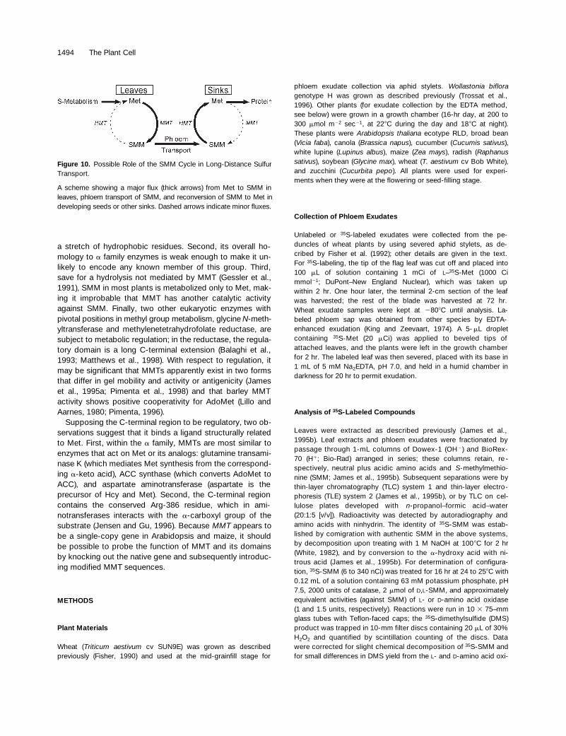
SMM can be reconverted to Met via the action of HMT,which is present in seeds (Giovanelli et al., 1980). SMM ar-riving in the phloem at a level of z2 mol% of amino acids,therefore, could readily provide the Met needed to synthe-size grain proteins with an average Met content of z2mol%. There is good indirect evidence that SMM is a sourceof sulfur in seeds: it is essentially absent from mature grainsand so must be metabolized (Wong et al., 1995; Pimenta etal., 1998), and conversion to Met is its only likely fate. Ac-cordingly, we propose the scheme given in Figure 10, inwhich SMM is synthesized in leaves, moves to the grain,and is there recycled to Met for use in protein synthesis.This scheme does not posit a complete separation in spaceand time of the halves of the SMM cycle, because leavesexpress HMT (Mudd and Datko, 1990; Larina and Gessler,1994) and seed tissues express MMT (Pimenta et al., 1998).Rather, it envisions that whereas both organs have the po-tential to perform the full cycle, flux is mainly from Met toSMM in leaves and from SMM to Met in seeds. This impliesthat the SMM cycle is tightly regulated.
The same can be inferred by comparing the size of theflag leaf SMM pool (z85 nmol) to the flux of SMM out of thisleaf. For the mid-grainfill period, this flux can be estimatedby assuming that (1) the SMM content of phloem sap is 25nmol mg21 dry weight (Table 1); (2) 1 mg of phloem sap isconverted to 0.89 mg of grain dry matter (Penning de Vrieset al., 1974; Fisher and Gifford, 1986); (3) ears have 40grains, each growing at 2 mg dry weight per day (Fisher,
Figure 6. Deduced Amino Acid Sequences of W. biflora, Arabi-dopsis, and Maize MMTs.
The W. biflora sequence is a composite of the longest (3.1 kb)cDNA and a 59 RACE product; these overlapped by 517 bp andwere identical in the overlap region. Peptide sequences from pu-rified W. biflora MMT are underlined. Identical residues are shaded inblack, similar residues in gray. GenBank accession numbers are AF137023 for W. biflora, AF 144079 for maize, and AF 137380 forArabidopsis. At, Arabidopsis; Wb, W. biflora; Zm, maize.
1492 The Plant Cell
1990); and (4) the flag leaf is the source of assimilates for thedeveloping grains (Fisher, 1990). The leaf–grain SMM flux istherefore (40 3 2 3 25/0.89) 5 2250 nmol per day. This dailyflux is .20-fold larger than the flag leaf SMM pool, whichmust therefore turn over approximately once per hour. Thus,wheat leaves do not stockpile SMM but produce it just be-fore sending it to the grains—a strategy likely to requireclose metabolic control of the flux from Met to SMM.
Our finding that SMM is a prominent phloem constituentraises questions about the membrane transport processesimplicated in its movement from leaves to sinks (Rentschand Frommer, 1996). Assuming an apoplastic pathway ofphloem loading, transmembrane movement must occur onrelease of SMM into the leaf apoplast, loading into the sieveelement–companion cell complex and, in the grain, releasefrom the maternal symplast into the endosperm cavity anduptake into the endosperm and embryo. Transport of aminoacids into plant cells is known to involve a range of proton-coupled symporters with various substrate specificities,
some of which overlap (Rentsch et al., 1998). To date, SMMappears not to have been tested as a substrate for any ofthese. A priori, it could be a substrate for the known generalamino acid or basic amino acid transporters (Chen andBush, 1997; Rentsch et al., 1998); alternatively, there couldbe an SMM-specific transporter similar to the SMM per-mease of yeast (Rouillon et al., 1999). Identifying the trans-porter(s) responsible for the phloem loading of SMM is ofparticular interest, given that phloem loading could play animportant role in determining the composition of the phloemsap and hence the amino acid content and nutritional valueof seed storage proteins (Chen and Bush, 1997).
The Bipartite Structure of MMT and the Regulation of SMM Synthesis
An imperative for regulation might explain the unique bipar-tite structure of MMT in which a methyltransferase domainapparently is fused to a sequence related to the a family ofPLP-dependent enzymes. Several considerations suggestthat the PLP protein-like region is more likely to be a regula-tory domain than a metabolic enzyme. First, it lacks a lysinein the usual position for the PLP binding site, having instead
Figure 7. Functional Expression of Arabidopsis MMT in E. coli.
(A) MMT activity in E. coli strain DH10B harboring pBluescript (pBS)or pBS containing the full-length Arabidopsis MMT cDNA (A. th.).Data are means 6SE of three observations. MMT was assayed radi-ometrically. Insets are autoradiographs of thin-layer chromatogra-phy (TLC) and thin-layer electrophoresis (TLE) analyses that confirmthat the reaction product was 35S-SMM; arrowheads mark the posi-tion of authentic SMM.(B) Formation of 35S-SMM from 35S-Met by E. coli strain MTD123harboring pBS or pBS containing the full-length Arabidopsis MMTcDNA. Cells were exposed to 9 mCi (18 nmol) of 35S-Met for 65to 75 min; they absorbed 3.7 to 4.4 mCi, of which 0.8 to 1.0 mCiremained in the soluble fraction. Data are means 6SE of threeobservations. Insets are autoradiographs of TLC and TLE analy-ses that confirm the identity of the 35S-SMM produced by cellsexpressing MMT. Arrowheads mark the position of authentic SMM.
Figure 8. DNA Gel Blot Analysis of Arabidopsis MMT.
Arabidopsis genomic DNA (2.5 mg) was digested with the restric-tion enzymes indicated, blotted, and probed with a 1.2-kb MMTcDNA. Genomic reconstructions made with linearized plasmidDNA equivalent to one, two, or five copies per haploid genomewere included. Hybridization and washing were at low stringency,as described in Methods. Hybridizing and washing at high strin-gency gave the same result. The positions of DNA length markersare shown at right.
SMM Transport and MMT Characterization 1493
Figure 9. Evidence That MMTs Have a Methyltransferase Domain and a Domain Related to PLP-Dependent Proteins.
(A) Alignment of a region z150 residues from the MMT N terminus with corresponding regions of three well-characterized methyltrans-ferases (MT): E. coli ribosomal protein L11 MT (L11 MT; GenBank accession number 464465), rat glycine N-MT (GNMT; GenBank acces-sion number 121328), and rat protein-arginine N-MT 1 (Arg NMT; GenBank accession number 2499804). Bars mark the conservedAdoMet binding motifs I, post-I, II, and III, as identified in L11 MT, GNMT, and Arg NMT (Kagan and Clarke, 1994; Gary et al., 1996). Theconsensus sequences for motifs I and post-I are hh(D/E)hGxGxG and hhxh(D/E), where h is a hydrophobic residue and x is any residue. Shadingindicates residues that are identical (black) or similar (gray) in MMTs and the other enzymes. Dashes are gaps in the sequence to allow for max-imal alignment. At, Arabidopsis; Wb, W. biflora; Zm, maize.(B) Alignment of two regions z250 and 40 residues from the C termini of MMTs, tomato 1-aminocyclopropane-carboxylate synthase (ACCsyn; GenBank accession number 2118319), Bacillus sp aspartate aminotransferase (AspAT; GenBank accesssion number 112988), and rat glutaminetransaminase K (GTK; GenBank accession number 1083706). Arrowheads mark residues (G-197, D/E-222, and R-386) found in all amino-transferases. Shading is as given in (A).(C) Hydropathy profile alignments of the C-terminal region of a representative MMT (Arabidopsis, residues 663 or 673 to 1062) with GTKand AspAT. Profiles were predicted by the Kyte–Doolittle method, with a window length of 22 (GTK) or 19 (AspAT). Bars below the profiles indi-cate gaps introduced to maximize alignment. Arrows mark the position of the PLP binding lysine in the aminotransferases; that for GTK is asproposed by Abraham and Cooper (1996). Blue shading shows the hydrophobic region in MMT that apparently is inserted near the usual PLPbinding site.
1494 The Plant Cell
a stretch of hydrophobic residues. Second, its overall ho-mology to a family enzymes is weak enough to make it un-likely to encode any known member of this group. Third,save for a hydrolysis not mediated by MMT (Gessler et al.,1991), SMM in most plants is metabolized only to Met, mak-ing it improbable that MMT has another catalytic activityagainst SMM. Finally, two other eukaryotic enzymes withpivotal positions in methyl group metabolism, glycine N-meth-yltransferase and methylenetetrahydrofolate reductase, aresubject to metabolic regulation; in the reductase, the regula-tory domain is a long C-terminal extension (Balaghi et al.,1993; Matthews et al., 1998). With respect to regulation, itmay be significant that MMTs apparently exist in two formsthat differ in gel mobility and activity or antigenicity (Jameset al., 1995a; Pimenta et al., 1998) and that barley MMTactivity shows positive cooperativity for AdoMet (Lillo andAarnes, 1980; Pimenta, 1996).
Supposing the C-terminal region to be regulatory, two ob-servations suggest that it binds a ligand structurally relatedto Met. First, within the a family, MMTs are most similar toenzymes that act on Met or its analogs: glutamine transami-nase K (which mediates Met synthesis from the correspond-ing a-keto acid), ACC synthase (which converts AdoMet toACC), and aspartate aminotransferase (aspartate is theprecursor of Hcy and Met). Second, the C-terminal regioncontains the conserved Arg-386 residue, which in ami-notransferases interacts with the a-carboxyl group of thesubstrate (Jensen and Gu, 1996). Because MMT appears tobe a single-copy gene in Arabidopsis and maize, it shouldbe possible to probe the function of MMT and its domainsby knocking out the native gene and subsequently introduc-ing modified MMT sequences.
METHODS
Plant Materials
Wheat (Triticum aestivum cv SUN9E) was grown as describedpreviously (Fisher, 1990) and used at the mid-grainfill stage for
phloem exudate collection via aphid stylets. Wollastonia bifloragenotype H was grown as described previously (Trossat et al.,1996). Other plants (for exudate collection by the EDTA method,see below) were grown in a growth chamber (16-hr day, at 200 to300 mmol m22 sec21, at 228C during the day and 188C at night).These plants were Arabidopsis thaliana ecotype RLD, broad bean(Vicia faba), canola (Brassica napus), cucumber (Cucumis sativus),white lupine (Lupinus albus), maize (Zea mays), radish (Raphanussativus), soybean (Glycine max), wheat (T. aestivum cv Bob White),and zucchini (Cucurbita pepo). All plants were used for experi-ments when they were at the flowering or seed-filling stage.
Collection of Phloem Exudates
Unlabeled or 35S-labeled exudates were collected from the pe-duncles of wheat plants by using severed aphid stylets, as de-cribed by Fisher et al. (1992); other details are given in the text.For 35S-labeling, the tip of the flag leaf was cut off and placed into100 mL of solution containing 1 mCi of L–35S-Met (1000 Cimmol21; DuPont–New England Nuclear), which was taken upwithin 2 hr. One hour later, the terminal 2-cm section of the leafwas harvested; the rest of the blade was harvested at 72 hr.Wheat exudate samples were kept at 2808C until analysis. La-beled phloem sap was obtained from other species by EDTA-enhanced exudation (King and Zeevaart, 1974). A 5-mL dropletcontaining 35S-Met (20 mCi) was applied to beveled tips ofattached leaves, and the plants were left in the growth chamberfor 2 hr. The labeled leaf was then severed, placed with its base in1 mL of 5 mM Na2EDTA, pH 7.0, and held in a humid chamber indarkness for 20 hr to permit exudation.
Analysis of 35S-Labeled Compounds
Leaves were extracted as described previously (James et al.,1995b). Leaf extracts and phloem exudates were fractionated bypassage through 1-mL columns of Dowex-1 (OH2) and BioRex-70 (H1; Bio-Rad) arranged in series; these columns retain, re -spectively, neutral plus acidic amino acids and S-methylmethio-nine (SMM; James et al., 1995b). Subsequent separations were bythin-layer chromatography (TLC) system 1 and thin-layer electro-phoresis (TLE) system 2 (James et al., 1995b), or by TLC on cel-lulose plates developed with n-propanol–formic acid–water(20:1:5 [v/v]). Radioactivity was detected by autoradiography andamino acids with ninhydrin. The identity of 35S-SMM was estab-lished by comigration with authentic SMM in the above systems,by decomposition upon treating with 1 M NaOH at 1008C for 2 hr(White, 1982), and by conversion to the a-hydroxy acid with ni-trous acid (James et al., 1995b). For determination of configura-tion, 35S-SMM (6 to 340 nCi) was treated for 16 hr at 24 to 258C with0.12 mL of a solution containing 63 mM potassium phosphate, pH7.5, 2000 units of catalase, 2 mmol of D,L-SMM, and approximatelyequivalent activities (against SMM) of L- or D-amino acid oxidase(1 and 1.5 units, respectively). Reactions were run in 10 3 75–mmglass tubes with Teflon-faced caps; the 35S-dimethylsulfide (DMS)product was trapped in 10-mm filter discs containing 20 mL of 30%H2O2 and quantified by scintillation counting of the discs. Datawere corrected for slight chemical decomposition of 35S-SMM andfor small differences in DMS yield from the L- and D-amino acid oxi-
Figure 10. Possible Role of the SMM Cycle in Long-Distance SulfurTransport.
A scheme showing a major flux (thick arrows) from Met to SMM inleaves, phloem transport of SMM, and reconversion of SMM to Met indeveloping seeds or other sinks. Dashed arrows indicate minor fluxes.

SMM Transport and MMT Characterization 1495
dase reactions (measured by gas chromatography; Rhodes et al.,1997).
Analysis of SMM, Thiols, and Amino Acids
SMM analyses were conducted on BioRex-70 fractions. SMM inphloem samples was analyzed without derivatization by matrix-assisted laser desorption ionization mass spectrometry (MALDI-MS)by using the instrumentation and procedures described by Trossat etal. (1998). SMM in leaf extracts was determined as described by Hansonet al. (1994). Thiols were analyzed by HPLC as their monobromobi-mane derivatives, as described by Herschbach et al. (1998); the g-glu-tamylcysteinylserine peak was identified based on its chromatographicbehavior (Klapheck et al., 1992) and quantified relative to a gluta-thione (GSH) standard. Amino acids were determined as describedby Trossat et al. (1998).
cDNA Cloning and Sequence Analysis
S-Adenosylmethionine:Met S-methyltransferase (MMT) was purifiedto homogeneity from W. biflora leaves as described by James et al.(1995a); Figures 2 and 3 of James et al. (1995a) document the purityof the product. Peptides were obtained using endoproteinase Lys Cor trypsin, separated by HPLC, and sequenced by Edman degrada-tion or by mass spectrometry at the Michigan State and Harvard Uni-versity facilities. mRNA was isolated from W. biflora leaves asdescribed (Rathinasabapathi et al., 1997) and used to construct acDNA library in the l Uni-Zap XR vector (Stratagene, La Jolla, CA).The degenerate polymerase chain reaction (PCR) primers 59-AAR-TTYTTRAAYGCIAAYATHATG-39 (Y 5 T or C; R 5 A or G; H 5 T or C)and 59-TTRAAICCIACYTCRGCYTC-39 (corresponding to the ends ofthe peptide KFLNANIMSIPTEAEVGFK) were used to amplify a 56-bpDNA fragment by using the cDNA library (2 3 108 plaque-formingunits per 50-mL reaction) as template. Sequencing confirmed thatthis fragment encoded the expected residues; a specific primer (59-ATCATGTCTATCCCTACAGA-39) from its center was then used withthe vector T7 primer to amplify the 1.1-kb 39 region of the MMTcDNA. The 1.1-kb fragment then was used to screen the library (3 3104 plaques); this yielded .100 MMT cDNAs, all truncated, of whichthe longest was 3.1 kb. The 59 terminus was obtained by rapid am-plification of cDNA ends (RACE), by using the Gibco BRL kit. ClonedRACE products from four independent reactions were sequenced; aclone with no errors (determined from the sequence consensus) wasused to construct the complete cDNA by fusion at the NheI site.
Arabidopsis expressed sequence tag (EST) 205D23T7 (GenBankaccession number H77211) was obtained from the Arabidopsis Bio-logical Resource Center and shown to have a 1.2-kb insert encodingthe C-terminal part of a protein homologous to W. biflora MMT. Theinsert then was used to isolate a full-length cDNA from an Arabidop-sis (ecotype Landsberg erecta) leaf library in l Uni-Zap XR (from T.L.Thomas, Texas A&M University, College Station). The maize EST wasrecovered from the collection at Pioneer Hi-Bred International Inc.,Johnston, IA. Both strands of cDNAs were sequenced by using theABI Prism dye terminator cycle sequencing Ready Reaction (PE Ap-plied Biosystems, Warrington, UK) and an ABI model 373 sequencer.Sequence alignments were made using Multalin (Corpet, 1988) orClustal W version 1.7 (Thompson et al., 1994) programs. Homologysearches were made using BLAST programs (Altschul et al., 1997).Hydropathy profile alignments were conducted via the Weizmann In-
stitute Bioinformatics server (http://bioinformatics.weizmann.ac.il),by using the Kyte–Doolittle calculation method.
Expression of MMT in Escherichia coli
For measurements of MMT activity in vitro, cultures of E. coli strainDH10B harboring the full-length Arabidopsis MMT cDNA or theempty pBluescript KS1 vector were grown to an OD600 nm of 0.6 to0.8 in of Luria–Bertani medium containing 100 mg mL21 ampicillinand 1 mM isopropyl b-D-thiogalactopyranoside (IPTG). Soluble pro-teins were isolated by sonicating cells in Tris–Mes–acetate buffer, pH7.2 (James et al., 1995a), containing 1 mM DTT and 10% (v/v) glyc-erol, and centrifuging for 15 min at 10,000g. MMT activity was as-sayed radiometrically (James et al., 1995a); protein concentrationswere determined according to Bradford (1976). To confirm that Ara-bidopsis MMT is active in vivo in E. coli, we grew cultures of thestrain MTD123 (DyagD DmetE DmetH; Thanbichler et al., 1999) har-boring the MMT cDNA or the pBluescript KS1 vector as above; cellsfrom a 2-mL aliquot were then transferred to 2 mL of M9 mediumcontaining 0.8% glucose, 1 mM IPTG, and 9 mCi (18 nmol) of 35S-Met. When 40 to 50% of the 35S-Met had been taken up (65 to 75min), the cells were harvested and extracted in boiling water for 10min. The soluble fraction was then analyzed for 35S-SMM by ion ex-change, TLC, and TLE as described above.
DNA Gel Blot Analysis
For Arabidopsis (ecotype RLD), genomic DNA was isolated fromleaves as described (Dellaporta, 1994); 2.5 mg of the isolated DNAwas digested, separated in 1% agarose gels, and transferred to Du-ralon-UV membranes (Stratagene). Blots were both hybridized andwashed at low or high stringency; the probe was the insert from EST205D23T7. Low stringency was hybridization at 508C and 1 M Na1,with a final wash at 228C and 40 mM Na1; high stringency was hy-bridization at 658C and 1 M Na1, with a final wash at 658C and 20 mMNa1 (Taylor et al., 1993). For maize (genotype Hi-II), genomic DNAwas prepared from leaves as described (Richards, 1997); 6 mg ofdigested DNA was separated as above and blotted to Hybond-N1 membranes (Amersham). Blots were hybridized at 60 or 658C(Sambrook et al., 1989) with a 0.5-kb maize MMT genomic fragmentcomprising part of the region between the 59 EcoRV and PstI sites,and washed at low or high stringency (final wash in 0.1 3 SSC [1 3SSC is 0.15 M NaCl and 0.015 M sodium citrate] and 0.1% SDS at 22or 658C, respectively). Probes were labeled with phosphorus-32 by arandom hexamer priming method.
ACKNOWLEDGMENTS
We thank Franck James, Claudine Trossat, Bala Rathinasabapathi,and Kurt Nolte for help with experiments and Terry Thomas for thegift of the Arabidopsis cDNA library. This work was supported in partby National Science Foundation Grants IBN-9514336 and IBN-9816075 to A.D.H., and IBN-9628750 and IBN-9904263 to D.A.G.,by an endowment from the C.V. Griffin, Sr., Foundation, and by theFlorida Agricultural Experiment Station, Journal Series No. R-06873.

1496 The Plant Cell
Received February 19, 1999; accepted May 3, 1999.
REFERENCES
Abraham, D.G., and Cooper, A.J.L. (1996). Cloning and expressionof a rat kidney cytosolic glutamine transaminase K that has strongsequence homology to kynurenine aminotransferase. Arch. Bio-chem. Biophys. 335, 311–320.
Alexander, F.W., Sandmeier, E., Mehta, P.K., and Christen, C.(1994). Evolutionary relationships among pyridoxal-59-phosphate-dependent enzymes: Regio-specific a, b and g families. Eur. J.Biochem. 25, 953–960.
Altschul, S.F., Madden, T.L., Schäffer, A.A., Zhang, J., Zhang, Z.,Miller, W., and Lipman, D.J. (1997). Gapped BLAST and PSI-BLAST: A new generation of protein database search programs.Nucleic Acids Res. 25, 3389–3402.
Balaghi, M., Horne, D.W., and Wagner, C. (1993). Hepatic one-car-bon metabolism in early folate deficiency in rats. Biochem. J. 291,145–149.
Bezzubov, A.A., and Gessler, N.N. (1992). Plant sources of S-meth-ylmethionine. Prikl. Biokhim. Mikrobiol. 28, 423–429.
Bradford, M.M. (1976). A rapid and sensitive method for the quanti-tation of microgram quantities of protein using the principle ofprotein–dye binding. Anal. Biochem. 72, 248–254.
Brunold, C., and Rennenberg, H. (1997). Regulation of sulfurmetabolism in plants: First molecular approaches. Prog. Bot. 58,164–186.
Chen, L., and Bush, D.R. (1997). LHT1, a lysine- and histidine-spe-cific amino acid transporter in Arabidopsis. Plant Physiol. 115,1127–1134.
Corpet, F. (1988). Multiple sequence alignment with hierarchicalclustering. Nucleic Acids Res. 16, 10881–10890.
Dellaporta, S. (1994). Plant DNA miniprep and microprep: Versions2.1-2.3. In The Maize Handbook, M. Freeling and G.O. Kirst, eds(New York: Springer-Verlag), pp. 522–525.
Fisher, D.B. (1990). Measurement of phloem transport rates by anisotope-dilution technique. Plant Physiol. 94, 455–462.
Fisher, D.B., and Gifford, R.M. (1986). Accumulation and conver-sion of sugars by developing wheat grains. Plant Physiol. 82,1024–1030.
Fisher, D.B., and Macnicol, P.K. (1986). Amino acid compositionalong the transport pathway during grain filling in wheat. PlantPhysiol. 82, 1019–1023.
Fisher, D.B., Wu, Y., and Ku, M.B. (1992). Turnover of soluble pro-teins in the wheat sieve tube. Plant Physiol. 100, 1433–1441.
Fujioka, M. (1992). Mammalian small molecule methyltransferases:Their structural and functional features. Int. J. Biochem. 24, 1917–1924.
Gary, J.D., Lin, W.-J., Yang, M.C., Herschman, H.R., and Clarke,S. (1996). The predominant protein-arginine methyltransferasefrom Saccharomyces cerevisiae. J. Biol. Chem. 271, 12585–12594.
Gessler, N.N., Bezzubov, A.A., Podlepa, E.M., and Bykhovskii,V.Y. (1991). S-Methylmethionine (vitamin U) metabolism in plants.Prikl. Biokhim. Mikrobiol. 27, 246–254.
Giovanelli, J., Mudd, S.H., and Datko, A.H. (1980). Sulfur aminoacids in plants. In The Biochemistry of Plants, Vol. 5, B.J. Miflin,ed (New York: Academic Press), pp. 453–505.
Grunau, J.A., and Swiader, J.M. (1991). Chromatographic quantita-tion of free amino acids: S-Methylmethionine, methionine andlysine in corn. J. Plant Nutr. 14, 653–662.
Hanson, A.D., Rivoal, J., Paquet, L., and Gage, D.A. (1994). Bio-synthesis of 3-dimethylsulfoniopropionate in Wollastonia biflora(L.) DC. Evidence that S-methylmethionine is an intermediate.Plant Physiol. 105, 103–110.
Hayashi, H., and Chino, M. (1986). Collection of pure phloem sapfrom wheat and its chemical composition. Plant Cell Physiol. 27,1387–1393.
Herschbach, C., Jouanin, L., and Rennenberg, H. (1998). Overex-pression of g-glutamylcysteine synthetase, but not glutathionesynthetase, elevates glutathione allocation in the phloem of trans-genic poplar trees. Plant Cell Physiol. 39, 447–451.
James, F., Nolte, K.D., and Hanson, A.D. (1995a). Purification andproperties of S-adenosyl-L-methionine:L-methionine S-methyl-transferase from Wollastonia biflora leaves. J. Biol. Chem. 270,22344–22350.
James, F., Paquet, L., Sparace, S.A., Gage, D.A., and Hanson,A.D. (1995b). Evidence implicating dimethylsulfoniopropionalde-hyde as an intermediate in dimethylsulfoniopropionate biosynthe-sis. Plant Physiol. 108, 1439–1448.
Jensen, R.A., and Gu, W. (1996). Evolutionary recruitment of bio-chemically specialized subdivisions of family I within the proteinsuperfamily of aminotransferases. J. Bacteriol. 178, 2161–2171.
John, R.A. (1998). Pyridoxal phosphate–dependent enzymes. InComprehensive Biological Catalysis, M. Sinnott, ed (New York:Academic Press), pp. 173–200.
Joshi, C.P., and Chiang, V.L. (1998). Conserved sequence motifs inplant S-adenosyl-L-methionine–dependent methyltransferases.Plant Mol. Biol. 37, 663–674.
Kagan, R.M., and Clarke, S. (1994). Widespread occurrence ofthree sequence motifs in diverse S-adenosylmethionin–depen-dent methyltransferases suggests a common structure for theseenzymes. Arch. Biochem. Biophys. 310, 417–427.
Khan, M.A., and Eggum, B.O. (1978). Effect of baking on the nutri-tive value of Pakistani bread. J. Sci. Food Agric. 29, 1069–1075.
King, R.W., and Zeevaart, J.A.D. (1974). Enhancement of phloemexudation from cut petioles by chelating agents. Plant Physiol. 53,96–103.
Klapheck, S., Chrost, B., Starke, J., and Zimmermann, H. (1992).g-Glutamylcysteinylserine—A new homologue of glutathione inthe family of Poaceae. Bot. Acta 105, 174–179.
Kocsis, M.G., Nolte, K.D., Rhodes, D., Shen, T.-L., Gage, D.A.,and Hanson, A.D. (1998). Dimethylsulfoniopropionate biosynthe-sis in Spartina alterniflora. Evidence that S-methylmethionine anddimethylsulfoniopropylamine are intermediates. Plant Physiol.117, 273–281.
Larina, T.V., and Gessler, N.N. (1994). On the physiological role ofS-methylmethionine in cabbage heads during storage. Russ. J.Plant Physiol. 41, 358–363.
Lillo, C., and Aarnes, H. (1980). Biosynthesis of the methyl donorsS-adenosylmethionine and S-methylmethionine in barley seedlings(Hordeum vulgare L.). Biochem. Physiol. Pflanzen 175, 104–122.
SMM Transport and MMT Characterization 1497
Matthews, R.G., Sheppard, C., and Goulding, C. (1998). Methyl-enetetrahydrofolate reductase and methionine synthase: Bio-chemistry and molecular biology. Eur. J. Pediatr. 157, S54–S59.
McKee, I.F., Eiblmeier, M., and Polle, A. (1997). Enhanced ozone-tolerance in wheat grown at an elevated CO 2 concentration:Ozone exclusion and detoxification. New Phytol. 137, 275–284.
Mehta, P.K., Hale, T.I., and Cristen, P. (1993). Aminotransferases:Demonstration of homology and division into evolutionary sub-groups. Eur. J. Biochem. 214, 549–561.
Mudd, S.H., and Datko, A.H. (1990). The S-methylmethionine cyclein Lemna paucicostata. Plant Physiol. 93, 623–630.
Neuhierl, B., Thanbichler, M., Lottspeich, F., and Böck, A. (1999).A family of S-methylmethionine–dependent thiol/selenol methyl-transferases. J. Biol. Chem. 274, 5407–5414.
Paquet, L., Lafontaine, P.J., Saini, H.S., James, F., and Hanson,A.D. (1995). Évidence en faveur de la présence du 3-diméthylsul-foniopropionate chez une large gamme d’Angiospermes. Can. J.Bot. 73, 1889–1896.
Penning de Vries, F.W.T., Brunsting, A.H.M., and van Laar, H.H.(1974). Products, requirements and efficiency of biosynthesis: Aquantitative approach. J. Theor. Biol. 45, 339–377.
Pimenta, M.J. (1996). Study of S-Adenosyl-L-Methionine:L-Methio-nine S-Methyltransferase from Green Barley Malt. PhD Disserta-tion (Louvain, Belgium: Université Catholique de Louvain).
Pimenta, M.J., Kaneta, T., Larondelle, Y., Dohmae, N., andKamiya, Y. (1998). S-Adenosyl-L-methionine:L-methionine S-meth-yltransferase from germinating barley: Purification and localiza-tion. Plant Physiol. 118, 431–438.
Rathinasabapathi, B., Burnet, M., Russell, B.L., Gage, D.A., Liao,P.-C., Nye, G.J., Scott, P., Golbeck, J.H., and Hanson, A.D.(1997). Choline monooxygenase, an unusual iron-sulfur enzymecatalyzing the first step of glycine betaine synthesis in plants:Prosthetic group characterization and molecular cloning. Proc.Natl. Acad. Sci. USA 94, 3454–3458.
Rennenberg, H. (1982). Glutathione metabolism and possible bio-logical roles in higher plants. Phytochemistry 21, 2771–2781.
Rentsch, D., and Frommer, W.B. (1996). Molecular approachestoward an understanding of loading and unloading of assimilatesin higher plants. J. Exp. Bot. 47, 1199–1204.
Rentsch, D., Boorer, K.J., and Frommer, W.B. (1998). Structureand function of plasma membrane amino acid, oligopeptide andsucrose transporters from higher plants. J. Membr. Biol. 162,177–190.
Rhodes, D., Gage, D.A., Cooper, A.J.L., and Hanson, A.D. (1997).S-Methylmethionine conversion to dimethylsulfoniopropionate:Evidence for an unusual transamination reaction. Plant Physiol.115, 1541–1548.
Richards, E.J. (1997). Preparation of plant DNA using CTAB. InShort Protocols in Molecular Biology, 3rd ed, F. Ausubel, R. Brent,R.E. Kingston, D.D. Moore, J.G. Seidman, J.A. Smith, and K.Struhl, eds (New York: John Wiley), pp. 2.10–2.11.
Rouillon, A., Surdin-Kerjan, Y., and Thomas, D. (1999). Transportof sulfonium compounds: Characterization of the S-adenosylme-thionine and S-methylmethionine permeases from the yeast Sac-charomyces cerevisiae. J. Biol. Chem., in press.
Sambrook, J., Fritsch, E.F., and Maniatis, T. (1989). MolecularCloning: A Laboratory Manual, 2nd ed. (Cold Spring Harbor, NY:Cold Spring Harbor Laboratory Press).
Skodak, F.I., Wong, F.F., and White, L.M. (1965). Determination ofS-methylmethionine ion in plant materials by automated aminoacid analysis. Anal. Biochem. 13, 568–571.
Taylor, B.H., Manhart, J.R., and Amasino, R.M. (1993). Isolationand characterization of plant DNAs. In Methods in Plant MolecularBiology and Biotechnology, B.R. Glick and J.E. Thompson, eds(Boca Raton, FL: CRC Press), pp. 37–47.
Thanbichler, M., Neuhierl, B., and Böck, A. (1999). S-Methylmethi-onine metabolism in Escherichia coli. J. Bacteriol. 181, 662–665.
Thompson, J.D., Higgins, G.D., and Gibson, T.J. (1994). CLUSTALW: Improving the sensitivity of progressive multiple sequencealignment through sequence weighting, position-specific gappenalties and weight matrix choice. Nucleic Acids Res. 22, 4673–4680.
Trossat, C., Nolte, K.D., and Hanson, A.D. (1996). Evidence that thepathway of dimethylsulfoniopropionate biosynthesis begins in thecytosol and ends in the chloroplast. Plant Physiol. 111, 965–973.
Trossat, C., Rathinasabapathi, B., Weretilnyk, E.A., Shen, T.-L.,Huang, Z.-H., Gage, D.A., and Hanson, A.D. (1998). Salinity pro-motes accumulation of 3-dimethylsulfoniopropionate and its pre-cursor S-methylmethionine in chloroplasts. Plant Physiol. 116,165–171.
Valle, E.M., Boggio, S.B., and Heldt, H.W. (1998). Free amino acidcomposition of phloem sap and growing fruit of Lycopersiconesculentum. Plant Cell Physiol. 39, 458–461.
Weibull, J., Ronquist, F., and Brishammar, S. (1990). Free aminoacid composition of leaf exudates and phloem sap. A comparativestudy in oats and barley. Plant Physiol. 92, 222–226.
White, R.H. (1982). Analysis of dimethyl sulfonium compounds inalgae. J. Mar. Res. 40, 529–536.
Wong, A.D., Swiader, J.M., and Juvick, J.A. (1995). Nitrogen and sul-fur fertilization influences aromatic flavor components in Shrunken2sweet maize kernels. J. Am. Soc. Hort. Sci. 120, 771–777
DOI 10.1105/tpc.11.8.1485 1999;11;1485-1497Plant Cell
Andrew D. HansonLi, Cornelia Herschbach, Heinz Rennenberg, Maria Joao Pimenta, Tun-Li Shen, Douglas A. Gage and
Fabienne Bourgis, Sanja Roje, Michael L. Nuccio, Donald B. Fisher, Mitchell C. Tarczynski, ChangjiangNovel Type of Methyltransferase
-Methylmethionine Plays a Major Role in Phloem Sulfur Transport and Is Synthesized by aS
This information is current as of July 5, 2018
References /content/11/8/1485.full.html#ref-list-1
This article cites 50 articles, 22 of which can be accessed free at:
Permissions https://www.copyright.com/ccc/openurl.do?sid=pd_hw1532298X&issn=1532298X&WT.mc_id=pd_hw1532298X
eTOCs http://www.plantcell.org/cgi/alerts/ctmain
Sign up for eTOCs at:
CiteTrack Alerts http://www.plantcell.org/cgi/alerts/ctmain
Sign up for CiteTrack Alerts at:
Subscription Information http://www.aspb.org/publications/subscriptions.cfm
is available at:Plant Physiology and The Plant CellSubscription Information for
ADVANCING THE SCIENCE OF PLANT BIOLOGY © American Society of Plant Biologists