RVC OPEN ACCESS REPOSITORY COPYRIGHT NOTICE · Comparative myology and evolution of marsupials and...
53
RVC OPEN ACCESS REPOSITORY – COPYRIGHT NOTICE This is the peer reviewed version of: Diogo, R., Bello-Hellegouarch, G., Kohlsdorf, T., Esteve-Altava, B. and Molnar, J. L. (2016), Comparative Myology and Evolution of Marsupials and Other Vertebrates, With Notes on Complexity, Bauplan, and “Scala Naturae”. Anat. Rec., 299: 1224–1255. doi:10.1002/ar.23390 which has been published in final form at http://dx.doi.org/10.1002/ar.23390. This article may be used for non-commercial purposes in accordance with Wiley Terms and Conditions for Self-Archiving. The full details of the published version of the article are as follows: TITLE: Comparative Myology and Evolution of Marsupials and Other Vertebrates, With Notes on Complexity, Bauplan, and "Scala Naturae" AUTHORS: Diogo, R., Bello-Hellegouarch, G., Kohlsdorf, T., Esteve-Altava, B. and Molnar, J. L. JOURNAL TITLE: Anatomical Record: Advances in Integrative Anatomy and Evolutionary Biology PUBLISHER: Wiley PUBLICATION DATE: September 2016 DOI: 10.1002/ar.23390
Transcript of RVC OPEN ACCESS REPOSITORY COPYRIGHT NOTICE · Comparative myology and evolution of marsupials and...
RVC OPEN ACCESS REPOSITORY – COPYRIGHT NOTICE
This is the peer reviewed version of:
Diogo, R., Bello-Hellegouarch, G., Kohlsdorf, T., Esteve-Altava, B. and Molnar, J. L. (2016),
Comparative Myology and Evolution of Marsupials and Other Vertebrates, With Notes on
Complexity, Bauplan, and “Scala Naturae”. Anat. Rec., 299: 1224–1255. doi:10.1002/ar.23390
which has been published in final form at http://dx.doi.org/10.1002/ar.23390.
This article may be used for non-commercial purposes in accordance with Wiley Terms and
Conditions for Self-Archiving.
The full details of the published version of the article are as follows:
TITLE: Comparative Myology and Evolution of Marsupials and Other Vertebrates, With Notes on
Complexity, Bauplan, and "Scala Naturae"
AUTHORS: Diogo, R., Bello-Hellegouarch, G., Kohlsdorf, T., Esteve-Altava, B. and Molnar, J.
L.
JOURNAL TITLE: Anatomical Record: Advances in Integrative Anatomy and Evolutionary
Biology
PUBLISHER: Wiley
PUBLICATION DATE: September 2016
DOI: 10.1002/ar.23390
Comparative myology and evolution of marsupials and other vertebrates, with notes on complexity, Bauplan,
and ‘scala naturae’
Rui Diogo1, Gaelle Bello-Hellegouarch2, Tiana Kohlsdorf2, Borja Esteve-Altava1,3, Julia L. Molnar1
1 Department of Anatomy, Howard University College of Medicine, Numa Adams Building, 520 W St. NW,
Washington, DC 20059, US. 2 Department of Biology, FFCLRP. University of São Paulo. Avenida Bandeirantes, 3900. Bairro Monte 7
Alegre. Ribeirão Preto, SP. Brazil.3 Structure & Motion Laboratory, Department of Comparative Biomedical Sciences, Royal Veterinary
College, Hawkshead Lane, Hatfield, Hertfordshire AL9 7TA, United Kingdom.
Corresponding author: Rui Diogo. Department of Anatomy, Howard University College of Medicine,
Numa Adams Building, 520 W St. NW, Washington, DC 20059, US. contact: [email protected].
Running title: Comparative myology of marsupials and other vertebrates
Grant sponsor (Grant number)
RD: Howard University College of Medicine Start-Up Package; NSF grant (1516557)
GB-H, TK: Brazilian Postdoctoral National Program (PNPD/CAPES 2015) fellowship to Programa de
Pós-graduação em Biologia Comparada-USP
BE-A: EU Horizon 2020 Marie Sklodowska-Curie Action (654155).
JLM: Postdoctoral grant of American Association of Anatomists
Abstract
Opossums are frequent subjects of developmental studies because marsupials share developmental features
not seen in placentals and because Didelphimorpha is the sister-group of other extant Marsupialia. But is the
adult marsupial muscular system markedly different from that of placentals or is it, like the skeletal system,
very similar? We provide, for the first time, a brief description of all head and limb muscles of Didelphis
virginiana based on our dissections and using a unifying nomenclature by integrating the data gathered in our
long-term project on the development, homologies and evolution of the muscles of all major vertebrate taxa.
Our data indicate that there were many more muscle synapomorphic changes from the last common ancestor
(LCA) of amniotes to the mammalian LCA (63) and from this LCA to the LCA of extant therians (48) than
from this latter LCA to the LCA of extant placentals (10 or 11). Importantly, Didelphis is anatomically more
plesiomorphic (only 14 changes from LCA of extant therians) than are rats (37 changes) and humans (63
changes), but its musculature is more complex (193 muscles) than that of humans (only 180 muscles). Of the
194 muscles of Didelphis, 172 (89%) are present in rats, meaning that their adult muscle anatomy is indeed
very similar. This similarity supports the existence of a common, easy recognizable therian Bauplan, but one
that is caused by developmental constraints and by evolutionary change driven by the needs of the
embryos/neonates, rather than by a ‘goal’ towards a specific adult plan/‘archetype,’ as the name Bauplan
suggests.
Keywords: muscles, homology, evolution, comparative anatomy, marsupials, placentals, monotremes,
mammals, complexity, scala naturae, Bauplan
Introduction
Marsupial opossums (order Didelphimorpha) are frequently the focus of evolutionary developmental studies
because this order is the sister-group of a clade including all other extant marsupials and is therefore a good
model to study the origin and early evolution of marsupials as a whole (e.g. Horovitz & Sánchez-Villagra
2003). Moreover, opossums are a very useful model to investigate the diversity of mammalian development
because marsupials share very peculiar developmental features that are not seen in placentals, many of
which are related to remarkable heterochronic and heterotopic changes in marsupials (Smith, 1994, 2001,
2006; Sánchez-Villagra et al., 2002; Vaglia & Smith, 2003; Keyte & Smith, 2010; Sears, 2004; Kelly &
Sears, 2011; Moustakas et al., 2011; Goswami et al., 2012; Hübler et al., 2013; Wakamatsu et al., 2014;
Chew et al., 2014). Interestingly, despite these marked developmental differences, the skeletal structures of
adult marsupials such as opossums and placentals such as mice are, in general, quite similar (e.g. Smith
2006; Goswami et al., 2012).
With the exception of very few studies (e.g., Cheng 1955, Smith 1994, Keyte & Smith 2010),
works on the developmental biology of opossums do not refer to muscles. This lack of muscular
developmental studies limits our understanding of mammalian evolution because the scarce data available
indicates that muscle development might also differ considerably between marsupials and placentals (e.g.
Keyte & Smith, 2010). For instance, in most tetrapods the skeletal and muscular systems develop over the
same period, but in marsupials, because so many skeletal elements are delayed in development, some
muscles appear far in advance of the skeletal elements that will form their attachment points (Smith, 2006).
However, we do not know whether the adult muscular phenotype of marsupials such as opossums is very
different from that found in most placentals or is instead fairly similar (as is the skeletal system). The latter
case would be a further example of different developmental patterns leading to a similar adult
configuration.
In fact, a major weakness of most of the studies on the adult muscles of the Virginia opossum (D.
virginiana) and other marsupials is that they do not provide comparisons with placentals or other tetrapods.
For instance, numerous authors have studied and discussed the hand muscles of marsupials, including D. virginiana
(e.g. Brandell, 1935; Campbell, 1939; McMurrich, 1903ab; Lewis, 1989; Jouffroy, 1971; Stein, 1981; Coues, 1872;
Brooks, 1886a; Young, 1880). However, their lack of comparative context and common nomenclature rendered them
confusing or simply “useless”, as stated by Lewis (1989). Moreover, most studies on the muscles of adult
opossums mainly refer to a specific region of the head or limbs (e.g. Young, 1880; Huber 1930a, 1930b,
1931; Langworthy, 1932; Haines, 1939; Lightoller, 1940a, 1940b, 1942; Straus, 1941a, 1941b, 1942;
Shrivastava, 1962; Brandell, 1965; Hiiemae & Jenkins, 1969; Minkoff et al.,’s, 1979; Grant et al., 2013).
Even those studies that focus on both the head and limbs (e.g. Coues 1870, 1872) omit some muscles. For
instance, Coues’ (1872) description of the facial and pharyngeal muscles is very incomplete.
In some cases such omissions were deliberate, but in most cases they were due to a strong historical
bias regarding marsupials as a perfect intermediate taxon within the ‘scala naturae’ leading to placentals,
and then to humans. The notion of a scala naturae, which dates back to thinkers such as Aristotle, represents
an evolutionary trend in complexity from “lower” to “higher” taxa, with Homo sapiens as the end stage
(discussed in Diogo et al., 2015a). For example, many authors described only a few undifferentiated facial
muscles in marsupials, more numerous muscles in placentals such as rats, and ‘most complex’ facial
musculature in humans (e.g. Huber 1930a, 1930b, 1931; Lightoller, 1940a, 1940b, 1942). Such notions of
‘scala naturae’, i.e. of progress towards greater complexity leading to humans, are found not only in works
from the 19th and early 20th centuries, but even those from the late 20th century. For instance, Minkoff et
al.,’s (1979) study of the facial muscles of D. virginiana describes 21 muscles of facial expression in this
species, including extrinsic ear muscles, i.e., only about 2/3 of the 31 facial muscles found in humans (25 +
6 extrinsic ear muscles: see Table 2). In contrast, we found exactly the same number of facial muscles in D.
virginiana as in placentals such as rats, which is very similar to the number found in humans, as we will
explain below (Table 2).
In our paper (Diogo et al., 2015a) we also discussed related notions, such as that of Bauplan, or
"body plan", which was originally related to a pre-evolutionary notion of “archetype” in the sense that it
refers to a “plan”. We also argued that the (justified) refutation of old notions such as scala naturae does not
mean that we should abandon terms such as “phylogenetically basal” and particularly “anatomically
plesiomorphic” to refer to groups such as the urodeles within the Tetrapoda, or lemurs within the Primates.
Here we investigate whether the term "anatomically plesiomorphic" might also apply to marsupials such as
the opossum, within the Mammalia, by providing, for the first time, a rigorous comparative framework
between the adult myology of marsupials, placentals, monotremes, and other tetrapods, for both the head
and limbs. Specifically, we provide a list and brief description of all the D. virginiana head and limb
muscles using an updated, unifying vertebrate myological nomenclature to allow more straightforward
comparison between marsupials and other taxa. We combine the new anatomical data obtained from our
dissections of D. virginiana with the myological information obtained in our previous works, and a detailed
literature review of works done by others on the myology of marsupials and other mammals. The present
work is therefore the culmination this 20-year project because we include marsupials and also Gambaryan
et al.'s (2002) detailed data on monotreme hindlimb musculature in the evolutionary/homology tables
(Tables 1-10) elaborated in our previous works (e.g. Diogo, 2007, 2008, 2009; Diogo & Abdala, 2007,
2010; Diogo et al., 2008, 2009a, 2009b, 2013a, 2013b, 2014, 2015a, 2015b, 2015c, 2015d; Diogo & Wood,
2012; Diogo & Molnar, 2014; Diogo & Tanaka, 2014; Diogo & Ziermann, 2014).
Materials and Methods
Five adult specimens of D. virginiana (three males and two females) were dissected by RD (RuiDiogoLab
specimen #HUDV1-5, preserved in alcohol) for the present work, and compared with one male specimen of
D. albiventris dissected by GBH (Department of Biology, FFCLRP specimen #XDB1, frozen) and with
descriptions of marsupials and other vertebrates from the literature (see Introduction). We found no major
differences between the D. virginiana specimens we dissected. Differences between our observations and
those of other authors are described in the Results/Discussion below. The homology/evolutionary
hypotheses (Tables 1-10) integrate data from our dissections, our previous studies using techniques such as
histological sectioning and fluorescent labeling, and the literature. A detailed description of the
methodology used to compile these tables, including the hypotheses about the presence/absence of muscles
in the last common ancestor (LCA) of specific clades, was provided in Diogo et al., (2015a, 2015d).
Importantly, in the present paper when we refer to the LCA of a certain clade (e.g. placentals), we always
refer to the LCA of the extant members of that clade. Therefore, we use the information available on the
extant members of the other clades to make assumptions about the presence/absence of muscles. For
instance, if a muscle is present in all extant monotremes and all extant marsupials and missing in all extant
placentals, we make the more parsimonious assumption that it was secondarily lost in placentals (1
evolutionary step) rather than independently acquired in monotremes and marsupials (2 evolutionary steps).
As we use the updated, unifying muscle nomenclature for vertebrates developed in our previous
works the names commonly used by other authors in the past are given in the Results section, to facilitate
comparisons. A list of the other non-primate vertebrate specimens studied by us in our long-term project is
provided in Diogo & Abdala (2010), Diogo et al., (2010, 2013a, 2013b, 2014), Diogo & Molnar (2014),
Diogo & Ziermann (2014), and Diogo & Tanaka (2014); a list of the studied primates is provided in Diogo
& Wood (2012).
Results
The order followed in the descriptions below basically follows Tables 1-10, in which muscles are grouped
based on their developmental origins (for more details see Diogo & Abdala, 2010, and Discussion below).
Mandibular muscles (1st branchial arch) (Table 1; Fig. 1)
All muscles in this paragraph insert on the mandible unless otherwise noted. The mylohyoideus connects the
hemimandibles and is fused to its counterpart at the midline. It has no central raphe or direct attachment onto the hyoid
apparatus, but only an indirect attachment via fascia, as described by Coues (1872). The digastricus anterior
originates on and is continuous with the digastricus posterior and has no well-defined intermediate tendon. The
digastricus anterior muscles also attach indirectly through the ‘tendinous arcade’ to the hyoid apparatus. The digastricus
anterior is not blended with its counterpart at the midline, although according to Coues (1872) the two muscles might
contact each other. The masseter (masseter + external adductor of Hiiemae & Jenkins 1969) originates from the
squamosal, presphenoid, maxillary, jugal, frontal, parietal, and occipital bones and is blended with the temporalis and
the pterygoideus medialis. It is subdivided into an inferior/anterior bundle, a superficial bundle, a deep bundle, and a
zygomatico-mandibular bundle. The latter bundle is sometimes considered to be an independent muscle, but it is in fact
mixed with the other masseter bundles and partially with the temporalis as well. The temporalis (posterior adductor +
internal adductor of Hiiemae & Jenkins 1969) originates from the squamosal, presphenoid, parietal, frontal and
occipital. It is subdivided into superficial/anterior and deep/posterior bundles that mainly insert on the lateral and
medial surfaces of the coronoid process, respectively, and a pars suprazygomatica that is mainly fused with the
deep/posterior bundle. The pterygoideus lateralis originates from the presphenoid. It is a very thin muscle, but still
slightly differentiated into an inferior head inserting onto the condylar process and a superior head inserting onto the
capsule of the squamodentary joint. The much thicker pterygoideus medialis originates from the palatine and
presphenoid bones. Lastly, the tensor tympani and tensor veli palatini originate from the temporal and sphenoid
bones, respectively. The tensor tympani inserts onto the malleolus, and the tensor veli palatini inserts onto the soft
palate.
Hyoid muscles (2nd branchial arch) (Table 2; Fig. 1)
Reiss (2001) coded the stylohyoideus as absent in Didelphis, but it is actually present, as described by e.g. Coues
(1872) and Minkoff et al. (1979). The stylohyoideus and digastricus posterior originate from the paroccipital process
of the occipital bone. The former inserts on the basihyal, passing deep to - but not being blended with - the latter. The
stapedius runs from the squamosal and/or surrounding regions to the stapes. The remaining hyoid muscles are
muscles of facial expression, and all except the mandibulo-auricularis and extrinsic ear muscles are blended with their
counterparts on the opposite side of the body. The platysma cervicale and platysma myoides (both part of platysma
of Minkoff et al., 1979) are fused with each other and with the orbicularis oris. The former muscle originates from a
raphe anchored to the ligamentum nuchae and the latter from skin extending as far posteriorly as the acromion of the
scapula, and both insert onto the corner of the mouth and the lower lip. The occipitalis connects the occipital bone and
ligamentum nuchae to the frontalis muscle anteriorly and to the ear and scutiform cartilage anterolaterally and is
blended with the auricularis posterior. As is the case in many placentals (e.g. rats), the occipitalis has a medial portion
(occipitalis of Lightoller, 1934, and Minkoff et al., 1979) that extends anteriorly to blend with the frontalis and a lateral
portion (cervico-auriculo-occipitalis of Lightoller, 1934; cervicoauricularis superficialis + cervicoscutularis of Minkoff
et al., 1979) that runs anteroventrolaterally to attach onto the posterior surface of the ear and to the scutiform cartilage.
These two portions are fused posteriorly, attaching to the dorsal region of the neck just anterior to the posterior
attachment of the auricularis posterior (which mainly corresponds to the cervicoauricularis medius of Minkoff et al.,
1979). The auricularis posterior runs anterolaterally from the ligamentum nuchae to the ear. It mainly corresponds to
the cervicoauricularis medius (which includes the ‘transversus nuchae’, ‘interparietoscutularis’, ‘cervicoauricularis
posterior profundus’ and ‘interparietoauricularis’) of Minkoff et al., (1979). Examples of extrinsic (facial) muscles of
the ear present in marsupials are the obliquus auriculae, transversus auriculae, helicis, tragicus, depressor helicis
and/or antitragicus. These muscles, with the exception of the depressor helicis (‘auricularis inferior’) seem to be
blended with each other in D. virginiana, forming the ‘auricularis externus’ muscle complex of Minkoff et al., (1979).
The mandibulo-auricularis connects the external ear to the mandible.
The sphincter colli profundus (sphincter colli preauricularis + part of sphincter colli superficialis of Minkoff
et al., 1979) is mainly deep to the platysma myoides and platysma cervicale. It seems to have no bony attachments and
is mingled with the zygomaticus minor of the same side of the body and the sphincter colli profundus of the opposite
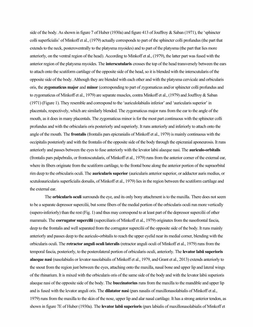
side of the body. As shown in figure 7 of Huber (1930a) and figure 413 of Jouffroy & Saban (1971), the ‘sphincter
colli superficialis’ of Minkoff et al., (1979) actually corresponds to part of the sphincter colli profundus (the part that
extends to the neck, posteroventrally to the platysma myoides) and to part of the platysma (the part that lies more
anteriorly, on the ventral region of the head). According to Minkoff et al., (1979), the latter part was fused with the
anterior region of the platysma myoides. The interscutularis crosses the top of the head transversely between the ears
to attach onto the scutiform cartilage of the opposite side of the head, so it is blended with the interscutularis of the
opposite side of the body. Although they are blended with each other and with the platysma cervicale and orbicularis
oris, the zygomaticus major and minor (corresponding to part of zygomaticus and/or sphincter colli profundus and
to zygomaticus of Minkoff et al., 1979) are separate muscles, contra Minkoff et al., (1979) and Jouffroy & Saban
(1971) (Figure 1). They resemble and correspond to the ‘auriculolabialis inferior’ and ‘auricularis superior’ in
placentals, respectively, which are similarly blended. The zygomaticus major runs from the ear to the angle of the
mouth, as it does in many placentals. The zygomaticus minor is for the most part continuous with the sphincter colli
profundus and with the orbicularis oris posteriorly and superiorly. It runs anteriorly and inferiorly to attach onto the
angle of the mouth. The frontalis (frontalis pars epicranialis of Minkoff et al., 1979) is mainly continuous with the
occipitalis posteriorly and with the frontalis of the opposite side of the body through the epicranial aponeurosis. It runs
anteriorly and passes between the eyes to fuse anteriorly with the levator labii alaeque nasi. The auriculo-orbitalis
(frontalis pars palpebralis, or frontoscutularis, of Minkoff et al., 1979) runs from the anterior corner of the external ear,
where its fibers originate from the scutiform cartilage, to the frontal bone along the anterior portion of the supraorbital
rim deep to the orbicularis oculi. The auricularis superior (auricularis anterior superior, or adductor auris medius, or
scutuloauricularis superficialis dorsalis, of Minkoff et al., 1979) lies in the region between the scutiform cartilage and
the external ear.
The orbicularis oculi surrounds the eye, and its only bony attachment is to the maxilla. There does not seem
to be a separate depressor supercilii, but some fibers of the medial portion of the orbicularis oculi run more vertically
(supero-inferiorly) than the rest (Fig. 1) and thus may correspond to at least part of the depressor supercilii of other
mammals. The corrugator supercilii (superciliaris of Minkoff et al., 1979) originates from the nasofrontal fascia,
deep to the frontalis and well separated from the corrugator supercilii of the opposite side of the body. It runs mainly
anteriorly and passes deep to the auriculo-orbitalis to reach the upper eyelid near its medial corner, blending with the
orbicularis oculi. The retractor anguli oculi lateralis (retractor anguli oculi of Minkoff et al., 1979) runs from the
temporal fascia, posteriorly, to the posterolateral portion of orbicularis oculi, anteriorly. The levator labii superioris
alaeque nasi (nasolabialis or levator nasolabialis of Minkoff et al., 1979, and Grant et al., 2013) extends anteriorly to
the snout from the region just between the eyes, attaching onto the maxilla, nasal bone and upper lip and lateral wings
of the rhinarium. It is mixed with the orbicularis oris of the same side of the body and with the levator labii superioris
alaeque nasi of the opposite side of the body. The buccinatorius runs from the maxilla to the mandible and upper lip
and is fused with the levator anguli oris. The dilatator nasi (pars nasalis of maxillonasolabialis of Minkoff et al.,
1979) runs from the maxilla to the skin of the nose, upper lip and alar nasal cartilage. It has a strong anterior tendon, as
shown in figure 7E of Huber (1930a). The levator labii superioris (pars labialis of maxillonasolabialis of Minkoff et
al., 1979; part or totality of maxillolabialis of Grant et al., 2013) connects the maxilla to the upper lip and is blended
posteriorly with the dilatator nasi. The nasalis (nasolabialis profundus of Grant et al., 2013) lies on the anterior snout
region and is associated with vibrissae. The depressor septi nasi may be present as part of the ‘nasolabialis
profundus’ of Grant et al., (2013), but neither they nor other authors describe it as a separate muscle, and we
did not find it either. In addition to the extrinsic muscles described above, Grant et al., (2013) described
various intrinsic muscles of the snout/vibrissae in marsupials such as Monodelphis domestica. Therefore,
it is very likely that such muscles are also present in D. virginiana but are too small to be observed in gross
dissections. The levator anguli oris facialis (pars anterior of maxillonasolabialis of Minkoff et al., 1979) runs from
connective tissue of the upper lip and from the buccinatorius to the region between the upper lip and the anterior
portion of the snout, passing deep to the levator labii superioris. It is less differentiated from the surrounding muscles
than is the same muscle in placental mammals such as mice and rats. The orbicularis oris surrounds the mouth and is
blended with the orbicularis oris of the opposite side of the body. Minkoff et al., (1979) states that the mentalis is not
differentiated, or is poorly differentiated, in D. virginiana. We did not find a differentiated mentalis in our dissections,
nor do other authors describe it in this species, but Jouffroy & Saban (1971) stated that the mentalis is differentiated in
at least some other marsupials.
Branchial muscles (posterior branchial arches) (Table 3)
As noted by Jouffroy & Saban (1971), marsupials often have a broad stylopharyngeus running from the paroccipital
process of the occipital to the pharynx and thyroid cartilage. The pharyngeal portion (pars pharyngea) is said to be a
unique, derived feature of marsupials and to replace functionally the superior constrictor of placentals because it is a
circular constrictor (Smith, 1994). The thyroid portion (pars thyroidea) thus corresponds to the entire stylopharyngeus of
placentals because it is a longitudinal constrictor. The ceratohyoideus runs from the hypohyal to the ceratohyal.
Although the trapezius and sternomastoideus complexes are often described as part of the forelimb musculature, they
are in fact branchial muscles, derived from the posterior branchial arches (Diogo & Abdala, 2010; Diogo et al., 2015c).
The acromiotrapezius (anterior trapezius of Stein, 1981) connects the vertebrae, ligamentum nuchae and occipitalis to
the spine and acromion of scapula and to the lateral half of the clavicle. The spinotrapezius (posterior trapezius of
Stein, 1981) runs from vertebrae to the spine of the scapula and is mixed distally with the acromiotrapezius and the
deltoideus scapularis. The cleido-occipitalis extends from the occipital region to the clavicle. It is blended with the
cleidomastoideus but clearly is a separate muscle. The cleidomastoideus and sternomastoideus connect the mastoid
process of the squamosal to the clavicle and sternum, respectively.
The pharyngeal muscles constrictor pharyngis medius and inferior both originate on the pharyngeal raphe
and are blended with each other and with their counterparts on the opposite side of the body. The former muscle inserts
on the hyoid apparatus and the latter on the cricothyroid cartilage. The cricothyroideus is secondarily absent in adult
marsupials (present in at least some marsupial embryos), probably as a result of the fusion between the cricoid and
thyroid cartilages (see, e.g. Jouffroy & Saban, 1971). The palatoglossus runs from the soft palate to the tongue, where it
blends with the hyoglossus. The pterygopharyngeus and palatopharyngeus are blended, both extending from the
pterygoid region of the presphenoid (the former muscle also from the tympanic region and the latter from the soft palate) to
the pharyngeal musculature. The musculus uvulae (medialis veli palatini of Jouffroy & Saban, 1971) lies on the infero-
medial region of the soft palate.
The laryngeal muscles thyroarytenoideus and cricoarytenoideus lateralis connect the unpaired
cricothyroid cartilage to the arytenoid cartilage of the respective side. The latter muscle is fused with the unpaired
arytenoideus muscle and with the thyroarytenoideus muscle of the same side of the body. The arytenoideus attaches to
the left and right arytenoid cartilages and also to the unpaired interarytenoid cartilage. As noted by authors such as
Symington (1898) and Jouffroy & Saban (1971), the portion going to the interarytenoid cartilage (often designated the
procricoid cartilage) is sometimes named ‘ary-procricoideus.’ Lastly, the cricoarytenoideus posterior originates from
the unpaired cricothyroid cartilage. As described by Symington (1898), it is differentiated into a ‘cricoarytenoideus
posticus internus’ bundle attaching onto the arytenoid cartilage of the same side of the body and the unpaired
interarytenoid cartilage and a ‘cerato-crico-arytenoideus posticus’ bundle attaching onto the arytenoid cartilages. The
medial margin of this muscle approaches but does not reach its counterpart.
Hypobranchial muscles (somitic tongue and neck musculature) (Table 4)
The geniohyoideus and genioglossus are blended, both originating from the mandible and inserting on the unpaired
basihyal. The latter muscle also inserts on the tongue and is blended with the hyoglossus, which connects the hyoid
apparatus to the tongue. Edgeworth (1935) and Jouffroy & Saban (1971) suggested that marsupials such as Didelphis
have a ‘primitive hyoglossus’ that is not separated into the hyoglossus and styloglossus muscles found in most
placental mammals. However, Smith (1994) refers to a styloglossus in Monodelphis, and this muscle was present in
our D. virginiana specimens, running from the paraoccipital process of the occipital to the tongue but being mixed
with the hyoglossus. Osgood (1921) also described a styloglossus in other marsupials. As stated by Saban (1968), the
intrinsic muscles of the tongue of marsupials are similar to those of placentals in that they include the muscles
longitudinalis superior, longitudinalis inferior, transversus linguae and/or verticalis linguae. The sternohyoideus and
sternothyroideus, which do not have clear intersections, are blended with each other and with their counterparts on
the opposite side of the body. They originate from the sternum and insert onto the hyoid apparatus and cricothyroid
cartilage, respectively. The omohyoideus connects the scapula to the hyoid apparatus and has no defined intermediate
tendon, only a short tendinous intersection. Lastly, the thyrohyoideus runs from the cricothyroid cartilage to the hyoid
apparatus and is blended with the sternothyroideus.
Proximal forelimb muscles (pectoral girdle and arm) (Table 5; Fig. 3)
Six muscles connect the axial skeleton to the pectoral girdle. The serratus anterior (part of serratus magnus of
Coues, 1872; part of serratus ventralis of Jenkins & Weijs, 1979; serratus magnus of Stein, 1981) and levator
scapulae (part of serratus magnus of Coues, 1872; levator anguli scapulae of Stein, 1981) are fused to form a single,
continuous structure that inserts on the scapula. The former originates from ribs 1-7, 1-8, or 1-9 and the latter from C3-
C7 or C4-C7 vertebrae. The levator claviculae (levator scapulae ventralis of Jouffroy, 1971; atlanto-acromialis of
Stein, 1981, and Coues, 1872) and atlantoscapularis posticus (atlanto-scapularis of Stein, 1981, and Coues, 1872)
connect the transverse process of the atlas to the lateral and medial third of the scapular spine, respectively. The
subclavius runs from the first rib to the clavicle, acromion and cleidoacromial ligament and is blended with the
cleidoacromialis.
Regarding the pectoral musculature and its derivatives, the pectoralis major (ectopectoralis of Lander,
1918; pectoralis superficialis of Langworthy, 1932; part of pectoralis of Coues, 1872, and Jenkins & Weijs,
1979) has a clavicular head originating from the sternum and sometimes also from a small portion of the
clavicle, a sternocostal head originating mainly from the sternum and ribs, and an abdominal head
originating from the sternum. All insert on the humerus, the clavicular head being superficial to and
inserting more distally than the sternocostal head. These three heads are shown as part of the “pectoralis
superficialis” of Langworthy (1932: see his figs. 2 and 3). The pectoralis major contacts the pectoralis
major of the opposite side of the body at the midline. The pectoralis minor (entopectoralis of Lander 1918;
pectoralis abdominalis + pectoralis profundus of Langworthy, 1932; part of pectoralis of Coues, 1872, and
Jenkins & Weijs, 1979) has three heads running from the xiphoid process and abdominal muscles to the
humerus only. Langworthy (1932) stated that the pectoralis minor inserts on the coracoid process of the
scapula, but other authors confirmed that it does not (e.g. Coues, 1872, and Jenkins & Weijs, 1979). Lander
(1918) stated that the entopectoralis is absent in Didelphis. On the contrary, the configuration seen in D. virginiana is
very similar to that seen in rats: in both species, the entopectoralis has three heads. Because the “pectoralis
abdominalis” of Langworthy (1932) is deep to the other two heads (of the “pectoralis profundus” of
Langworthy, 1932), it cannot correspond to the abdominal head of the pectoralis major seen in, e.g.,
humans, which is never deep to the pectoralis minor. This supports the idea of Jouffroy (1971) that the
“pectoralis abdominalis” of different authors in different taxa probably corresponds to different structures.
That is, when the structure is superficial to the pectoralis minor and adjacent to the main body of the
pectoralis major, it corresponds to the abdominal head seen in, e.g., humans, while when the structure is
deep to the pectoralis minor and adjacent to the main body of the pectoralis minor, it corresponds to part of
the pectoralis minor instead, as seen in, e.g., Didelphis and rats. The “fourth pectoral layer” of Langworthy
(1932) actually corresponds to the muscle sternalis, not to part of the pectoralis major or pectoralis minor.
The pectoralis minor contacts the pectoralis minor of the opposite side of the body at the midline. The
panniculus carnosus runs from soft tissues such as the abdominal muscles and fascia, its only bony attachment
being onto the humerus. It contacts its counterpart at the midline and is blended with the pectoralis minor and
the latissimus dorsi of the same side of the body.
The posterior shoulder muscles infraspinatus, supraspinatus, teres minor, subscapularis, and teres
major connect the scapula to the proximal humerus. Coues (1872) stated he could not find a teres minor, but as
stated by Stein (1981) and Jenkins & Weijs (1979), this muscle is present. The cleidoacromialis (probably
corresponds to scapulo-clavicularis of Wood, 1870) runs from the distal end of the clavicle to the acromion
and lateral third of the scapular spine and also to the superficial aponeurosis of the supraspinatus, as
described by Jenkins & Weijs (1979). It is partially blended with the subclavius. The deltoideus scapularis
(part of deltoid of Coues, 1872) runs from the scapula to the humerus. Stein (1981) describes only a ‘clavodeltoid’ and
a ‘spinodeltoid’ in D. virginiana. The ‘clavodeltoid’ is only partially differentiated into a pars acromialis (from
acromion) and a pars clavicularis (from clavicle), which are blended with each other and thus constitute the deltoideus
acromialis et clavicularis of the present work. The ‘spinodeltoid’ corresponds to the deltoideus scapularis of the
present work. The deltoideus acromialis et clavicularis (part of deltoid of Coues, 1872) extends from the acromion
of the scapula and the clavicle to the humerus and is blended with the pectoralis major. The latissimus dorsi runs
from T4 or T5 to L3 or L4 vertebrae to the humerus and is blended with the dorsoepitrochlearis muscle.
Lastly, regarding the arm muscles, as described by Jenkins & Weijs (1979), Coues (1872) and Stein (1981)
and as in humans, the triceps brachii has scapular, medial and lateral heads, as in humans. It connects the scapula and
humerus to the olecranon process of the ulna and is also blended with the dorsoepitrochlearis. As described by these
authors, the dorsoepitrochlearis (tensor fasciae antebrachii of Stein, 1981; omo-anconeus of Coues, 1872) runs from
the latissimus dorsi to the olecranon process of the ulna and adjacent fascia. The brachialis (brachialis anticus of
Coues, 1872) connects the humerus to the ulna. Coues (1872) suggested that the two heads of biceps brachii of D.
virginiana may both correspond to the short head of other mammals because they mainly originate from the coracoid
process and surrounding regions rather than from the capsular ligament of the shoulder joint or the supraglenoid
region. However, we agree with Jouffroy (1971) that these heads correspond to the long and short heads of other
mammals. Another peculiarity of the biceps is that it runs from the scapula to both the ulna and radius (without a well-
defined bicipital aponeurosis). Lastly, the coracobrachialis has a single head, which could in theory correspond to the
coracobrachialis medius/proprius of other mammals. However, it is very short, going from the coracoid process to the
proximal humerus, just distal to the lesser tubercle. Moreover, it is mainly deep to, and not fused with, the biceps
brachii. Jouffroy agreed with this interpretation, designating this head ‘coracobrachialis brevis/profundus’ (Jouffroy
1971, Fig. 93).
Distal forelimb muscles (forearm and hand) (Table 6; Fig. 3)
The flexor (ventral) forearm muscle pronator quadratus connects the distal ulna to the distal radius. The flexor
digitorum profundus is fused with the flexor digitorum superficialis, running from the humerus, radius and ulna to
the distal phalanges of digits 1, 2, 3, 4 and 5. The flexor digitorum superficialis connects the humerus to the middle
phalanges of digits 2, 3, 4 and 5, although in a few specimens of this species the muscle seems to go only to digits 2, 3
and 4, as a variant (e.g. Coues, 1872; Stein, 1981). The palmaris longus (palmaris longus externus of Jouffroy 1971)
and palmaris longus internus (of Jouffroy 1971) are fused with each other and also with the flexor digitorum longus
and flexor carpi ulnaris, respectively. Both run mainly from the humerus to the palmar aponeurosis. The flexor carpi
ulnaris connects the humerus and ulna to the pisiform bone and surrounding aponeurosis. This seems to be the
common condition for D. virginiana, as it is also described by Coues (1872) and Stein (1981), although authors such
as Straus (1942) refer to an attachment to both the pisiform and metacarpal 5 in Didelphis. The epitrochleoanconeus
(anconeus internus of Stein, 1981) runs from the medial epicondyle of the humerus to the olecranon process of the
ulna. Straus (1942) suggested that the epitrochleoanconeus of opossums does not correspond to the ‘anconeus
internus’ of other mammals, but the ‘anconeus internus’ of authors such as Stein (1981) clearly does correspond to the
epitrochleoanconeus of other mammalian taxa. The flexor carpi radialis connects the humerus to metacarpal 2. This
seems to be the common condition for D. virginiana, as it was also reported by Coues (1872) and Stein (1981), but
Straus (1942) reported an attachment to both metacarpals 1 and 2 in Didelphis. The pronator teres runs from the
humerus to the middle third or distal two thirds of the radius. A palmaris brevis was not present in the specimens
dissected by us or by Coues (1872) and Stein (1981), but Jouffroy & Saban (1971) explain that this muscle is present
in some other marsupials.
Among the numerous studies that referred to the hand muscles of D. virginiana (see Introduction), Brooks
(1886ab) is the most complete and accurate. Brooks describes four lumbricales, an abductor pollicis brevis (ab2),
flexor pollicis brevis (superficial head of human anatomy: f2r), adductor pollicis (a1), contrahentes to digits 2 (a2), 4
(a4) and 5 (a5, plus a somewhat distinct bundle, a5a1), an abductor digiti minimi (abd5), a flexor digiti minimi with
two heads (a5a and f5u), a flexor brevis digitorum manus to the middle phalanx of digit 5, the usual eight flexores
breves profundi plus an adductor pollicis accessorius, and the usual four intermetacarpales. Of the two heads of the
flexor digiti minimi brevis, the first (a5a) mainly corresponds to the flexor digiti minimi of humans, while the deeper
f5u probably derives from the same anlage that gives rise to the opponens digiti minimi of humans because it is deeper
to the main body of the flexor digiti minimi brevis. The adductor pollicis accessorius constitutes a ‘third’ short flexor
of digit 1 but seems to derive instead from the adductor pollicis (Brooks 1886ab). Hopefully, by providing one-to-one
comparisons with other mammals and tetrapods (Table 6), the present paper will help to solve once and for all the
controversy and misunderstandings concerning these muscles. The flexor brevis digitorum manus connects the
hamate to the middle phalanx of digit 5. The four lumbricales run from tendons of the flexor digitorum
profundus to the ventral side of the proximal phalanges of digits 2, 3, 4 and 5. They also attach indirectly
onto the dorsal side of the middle and distal phalanges of these digits via the extensor expansions. There are
three contrahentes digitorum other than the adductor pollicis: the contrahens of digit 2 runs from metacarpal 3 to the
proximal phalanx of digit 2; contrahens of digit 4 runs from metacarpal 3 to the proximal phalanx of digit 4; and the
contrahens of digit 5 runs from metacarpal 3 and capitate to the proximal phalanx of digit 5, having a small, somewhat
separate head. The adductor pollicis (contrahens of digit 1) connects metacarpal 3 and the capitate to the proximal
phalanx of digit 1. The adductor pollicis accessorius (‘volaris primus of Henle’) connects the trapezoid and
metacarpal 1 to the proximal phalanx of digit 1. All ten flexores breves profundi are present, but number 1
corresponds to the flexor pollicis brevis and number 10 corresponds to the flexor digiti minimi brevis, both of which
are described below. The other eight flexores breves profundi are: number 2 from trapezium to ulnar side of proximal
phalanx of digit 1; numbers 3, 4, 5, 6, 7 and 8 from metacarpals 3, 4, and 5 to the radial and ulnar sides of the
respective digit; and number 9 from metacarpal 5 to the radial side of proximal phalanx of digit 5. The flexor pollicis
brevis connects the trapezium to the radial side of proximal phalanx of thumb, corresponding to the ‘superficial head
of the flexor pollicis brevis’ of human anatomy. Although Figure 9.8A of Lewis (1989) suggests that at least some
marsupials have an opponens pollicis, the consensus is that this muscle is not present in D. virginiana, as noted above.
The flexor digiti minimi brevis runs from the pisiform to the proximal phalanx of digit 5 and has two heads, one
superficial and one deep, which correspond topologically to the flexor digiti minimi brevis and to the opponens digiti
minimi of humans, respectively. However, the latter head does not seem to be directly homologous to the opponens
digiti minimi of humans because this muscle was acquired only later in evolution, within placental mammals. The
condition seen marsupials reflects only a rough evolutionary parallelism because the deeper head attaches onto the
proximal phalanx of digit 5 rather than onto the metacarpal 5 as does the opponens digiti minimi of, e.g., humans (see
Table 6 and below). The abductor pollicis brevis connects the trapezium to the radial side of the proximal phalanx of
digit 1. The abductor digiti minimi runs from the pisiform to the ulnar side of the proximal phalanx of digit 5. Lastly,
there are four intermetacarpales: as described by Young (1880), number 1 runs from metacarpal 1 to radial side of
proximal phalanx of digit 2; number 2 runs from metacarpals 2 and 3 to ulnar side of proximal phalanx of digit 2 and
radial side of proximal phalanx of digit 3; number 3 runs from metacarpals 3 and 4 to ulnar side of proximal phalanx
of digit 3 and radial side of proximal phalanx of digit 4; and number 4 connects metacarpals 4 and 5 to the ulnar side
of proximal phalanx of digit 4 and radial side of proximal phalanx of digit 5.
There are ten muscles in the extensor layer of the forearm; all originate on the humerus except as otherwise
noted. The extensor carpi radialis longus and brevis insert on metacarpals 2 and 3, respectively (the latter may
attach to both, as a variant: see Straus, 1941). The brachioradialis (supinator longus of Coues 1872) inserts on the
scaphoid, lunate and triquetrum, as described by Haines (1939) and Straus (1941), and contra Coues (1872) and Stein
(1981). The supinator (supinator brevis of Coues 1872) inserts on the proximal radius. It is a well-defined, separated
muscle, as described by Stein (1981), and contra Coues (1872). The extensor carpi ulnaris runs from the humerus
and ulna to metacarpal 5. The anconeus (anconeus externus of Stein, 1981) runs from the lateral epicondyle of the
humerus to the olecranon process of the ulna. The extensor digitorum (extensor digitorum communis of Coues,
1872) inserts on the distal phalanges of digits 2, 3, 4 and 5 (as a variant the tendon to digit 5 might be missing:
e.g. Coues, 1872; Stein, 1981). The extensor digiti minimi (extensor digitorum lateralis of Stein, 1981; extensor
digitorum ulnaris of Haines, 1939) inserts on the distal phalanges of digits 4 and 5 (and 3, as a variant: e.g. Haines,
1939). The extensor indicis (extensor digitorum profundus of Straus, 1941) runs from the ulna to the distal phalanges
of digits 1, 2 and 3, as described by Straus (1941) and Haines (1939) (or 1 and 2 only, as a variant: Coues, 1872; Stein
1981). The abductor pollicis longus (extensor ossis metacarpi pollicis of Coues, 1872) runs from the ulna and radius
to metacarpal 1.
Proximal hindlimb muscles (pelvic girdle and thigh) (Table 7; Fig. 4)
Regarding the pelvic girdle muscles, the quadratus lumborum seems to mainly connect the ribs and
vertebrae to the pelvic girdle. The psoas minor is not present as a separate muscle. The gluteus maximus,
which originates from two sacral and first three caudal vertebrae, and the femorococcygeus, which
originates from the third and fourth caudal vertebrae, are usually blended, as stated by Coues (1872), and
insert together on the femur. The gluteus medius connects the ilium to the proximal femur, as does the
gluteus minimus, and is blended with the gluteus minimus, gluteus maximus, and piriformis. The tensor
fasciae latae is not present as a distinct structure. As will be seen below, it is probably a derived feature of
placentals because it does not seem to be present in monotremes or marsupials (e.g. Bardeen, 1906; Gregory
& Camp, 1918; Appleton, 1928). The scansorius (iliofemoralis of Coues, 1872, and Stein, 1981) is a small,
very slender fasciculus from the acetabular region to the lesser trochanter of the femur, lying in the same
position as the iliofemoral ligament of humans. Stein (1981) could not find this muscle in D. virginiana, but
other authors have also found it (e.g. Coues, 1872). The obturator internus and externus connect the
ischium and pubis to the proximal femur. The former and is blended distally with the gemellus superior
and gemellus inferior, which connect the ischium to the proximal femur, as does the quadratus femoris.
The piriformis runs from the second sacral and first caudal vertebrae to the proximal femur. Finally, the
iliacus runs from the ilium to the femur and is blended with the psoas major, which connects the last three
lumbar and first two sacral vertebrae to the femur.
As noted by Coues (1872) and Stein (1981) there is no distinct vastus intermedius (‘crureus’) in D.
virginiana or in other marsupials (Warburton 2003; see also, e.g. Thompson & Hillier, 1905, and Osgood,
1921). Instead, this muscle is mingled with the other parts of the ‘quadriceps femoris,’ making it a triceps
complex. The rectus femoris originates from the ilium, the vastus lateralis and vastus medialis originate
from the humerus, and all three are blended distally and insert together on the patella and tibia. The
sartorius connects the ilium to the patella and tibia. The pectineus seems to connect the femur to the
ischium, as stated by Stein (1981), but Coues (1872) states that it actually originates from the epipubic bone
(‘marsupial bone’: see Fig. 2), not from the ischium. The adductor brevis (adductor parvus of Coues, 1872,
and Stein, 1981) runs from the pubis to the femur. The adductor longus and adductor magnus run from
the pubis and ischium to the femur (adductor minimus not present as separate muscle). The gracilis runs
from the pubis to the tibia. The biceps femoris has a single head from the ischium to the fibula and is
blended with the semitendinosus. Therefore, the short head of the biceps femoris seems to be absent in D.
virginiana, as stated by Coues (1872), but the tenuissimus (‘bicipiti accessorius’) is present in some other
marsupials, such as marsupial moles (e.g. Warburton 2003). The semimembranosus runs from the ischium
to the tibia. The presemimembranosus is not present in D. virginiana or, seemingly, in most other
marsupials (see, e.g. Osgood 1921), with a few exceptions such as marsupial moles (Thompson & Hillier
1905, Warburton 2003).
Appleton (1928) stated that in marsupials the ‘caudofemoralis’ often inserts onto the proximal
femur and thus designated it ‘piriformis.’ However, as will be explained in the Discussion, the ‘piriformis’
muscle described in the opossum by authors such as Coues (1872) and Stein (1981) seems to correspond to
the piriformis of placental mammals. Lastly, the semitendinosus has an anterior/dorsal head and a
posterior/ventral head that correspond respectively to the ‘crurococcygeus’ and ‘semitendinosus’ of Coues
(1872) and Stein (1981). As recognized by these authors, even if the two heads are clearly differentiated in
D. virginiana, they are still blended proximally, forming a single muscle. The posterior/ventral head
connects the ischium to the tibia as described by Coues (1872) (not the femur, as stated by Stein, 1981),
while the anterior/dorsal head connects the third coccygeal vertebra to the tibia as described by Coues
(1872) (not the tibia + fibula as described by Stein, 1981).
Distal hindlimb muscles (leg and foot) (Tables 8, 9, 10; Fig. 4)
Concerning the flexor layer of the leg, the gastrocnemius internus connects the femur to the calcaneus.
The gastrocnemius externus (gastrocnemius externus et soleus of Coues, 1872, and Stein, 1981) runs from
the femur (part corresponding to lateral head of gastrocnemius of placental mammals) and fibula (part
corresponding to soleus of gastrocnemius of placental mammals) to the calcaneus and is well separated
from the gastrocnemius internus. The plantaris (tensor fasciae plantaris of Coues, 1872) connects the fibula
to the calcaneus and is blended with the gastrocnemius externus. The flexor digitorum longus (flexor
hallucis longus + flexor digitorum longus of Coues, 1872, and Stein, 1981) connects the fibula and tibia to
the distal phalanges of digits 1, 2, 3, 4 and 5. It is mixed with the quadratus plantae, interosseus cruris,
tibialis anterior, flexor digitorum brevis, and the four lumbricales. Coues (1872) and Stein (1981)
erroneously stated that the ‘flexor hallucis longus’ part of the flexor digitorum longus of the present work
does not attach onto digit 1. They considered the muscle that sends a tendon to the distal phalanx of digit 1
to be the ‘flexor brevis hallucis obliquus’ (of Stein, 1981) or ‘flexor brevis pollicis obliquus’ (of Coues,
1872). However, as stated by McMurrich (1903ab) and Lewis (1989), this muscle actually corresponds to
the quadratus plantae (flexor accesorius of McMurrich, 1905; flexor brevis pollicis obliquus of Coues,
1872; flexor hallucis obliquus of Stein, 1981; accessorius of Lewis, 1989), which originates from the
calcaneus and inserts on the tendon of flexor digitorum longus. The interosseus cruris (pronator tibiae of
McMurrich, 1905; rotator fibulae or popliteus of Lewis, 1989, and Coues, 1872) connects most of the
proximodistal length of the tibia and fibula and includes the popliteus, as stated by Stein (1981). A similar
condition is found in many non-mammalian tetrapods and in other marsupials as well (e.g. Bardeen, 1906).
Lastly, the tibialis posterior connects the tibia and fibula to the navicular bone.
All ten flexores breves profundi are present in the foot, as they are in the hand (see above).
Numbers 1 and 10 correspond to the flexor hallucis brevis (‘superficial or medial head of the flexor hallucis
brevis’ of authors such as Coues, 1872) and flexor digiti minimi brevis, respectively, and are described
below. The other eight are: number 2 (‘lateral head of flexor hallucis brevis’) from medial cuneiform to
proximal phalanx of digit 1; numbers 3 and 4 (‘flexor brevis indicis’) from intermediate cuneiform to
proximal phalanx of digit 2; numbers 5 and 6 (‘flexor brevis medii’) from lateral cuneiform to proximal
phalanx of digit 3; numbers 7 and 8 (‘flexor brevis annularis’) from cuboid to proximal phalanx of digit 4;
and number 9 from metatarsal 5 and cuboid to proximal phalanx of digit 5. There are four
intermetatarsales: number 1 from metatarsal 1 to the medial side of the proximal phalanx of digit 2;
number 2 from metatarsals 2 and 3 to the medial side of the proximal phalanx of digit 3; number 3 from
metatarsals 3 and 4 to the lateral side of the proximal phalanx of digit 3 and to the medial side of the
proximal phalanx of digit 4; and number 4 from metatarsals 4 and 5 to the lateral side of the proximal

phalanx of digit 4 and to the medial side of the proximal phalanx of digit 5 (see, e.g. figure 16.1 of Lewis,
1989). The abductor digiti minimi connects the calcaneus to the proximal phalanx of digit 5. Some authors
refer to two heads of the abductor digiti minimi in marsupials (see also figure 16.1 of Lewis, 1989).
However, their ‘superficial head’ is similar to the abductor digiti minimi of, e.g., humans, while their ‘deep
head’ (‘calcaneo-metatarsales’ of Coues, 1872, and Stein, 1981) is similar to the opponens digiti minimi of,
e.g., apes because it goes to metatarsal 5, not to the proximal phalanx of digit 5. Therefore, the ‘deep head’
might well correspond instead to a deeper bundle of the flexor brevis profundus 10 (i.e. flexor digiti minimi
brevis) that resembles (homoplastically) the opponens digiti minimi, as seems to be the case in the hand of
D. virginiana (see above). The abductor hallucis connects the navicular bone to the proximal phalanx of
digit 1. The flexor digitorum brevis inserts on the middle phalanges of digits 2, 3, 4 and 5, as it does in
placentals such as humans, but it originates from the tendon of flexor digitorum longus rather than from the
calcaneus. Therefore, like their corresponding muscles in the upper limb (flexor digitorum profundus and
flexor digitorum superficialis), the flexor digitorum brevis and longus in D. virginiana are more blended
with each other than they are in humans. The four lumbricales originate from the tendons of the flexor
digitorum longus and seem to insert onto the middle phalanges of digits 2-5. The four contrahentes pedis
all originate from metatarsal 3 and insert onto the proximal phalanges of digit 1, 2, 4 and 5, respectively.
The flexor digiti minimi brevis includes the ‘calcaneo-metatarsales’ of Coues (1872) and Stein (1981) and
the ‘abductor ossis metatarsi quinti digiti’ of McMurrich (1906). It connects the calcaneus and cuboid to the
base of metatarsal 5 and to the proximal phalanx of digit 5 (see also flexores breves profundi above).
Finally, the flexor hallucis brevis runs from the medial cuneiform to the proximal phalanx of digit 1. This
muscle is the medial head of flexor hallucis brevis of Stein (1981) and thus corresponds to the flexor brevis
profundus 1 of the present work (see above).
The last group of muscles to be described here is the extensor layer of the leg. Three blended
muscles originate from the fibula: extensor digitorum longus (to distal phalanges of digits 2, 3, 4 and 5)
and fibularis brevis and longus to metatarsals 5 and 1, respectively. The extensor hallucis longus runs
from the fibula to the distal phalanx of digit 1 and is blended with the extensor digitorum brevis. The
tibialis anterior (flexor tarsi tibialis of Coues 1872) runs from the tibia to the medial cuneiform. The
fibularis digiti quarti is not present as a distinct muscle in D. virginiana, confirming that it is simply a short
extensor to digit 4, because in this taxon the short extensor to digit 4 is fused with the main body of the
extensor digitorum brevis, the latter muscle thus going to digits 2, 3 and 4. In fact, Stein (1981) uses the
name extensor digitorum brevis in Didelphis, while he uses the names ‘fibularis digiti quinti’ and ‘fibularis
digiti quarti’ in Chironectes (also a marsupial), alternately. The fibularis digiti quinti (peroneus tertius of
Coues, 1872, and Stein, 1981) runs from the fibula to the distal phalanx of digit 5, and thus corresponds to
the short extensor of digit 5 rather than to the fibularis tertius. While the short extensor of digit 5 is also
seen in placentals such as rats and mice and always attaches onto the phalanges, the fibularis tertius is a
derived structure found only in a few placentals such as humans and often attaches instead onto metatarsal
5. The extensor digitorum brevis runs from the fibula to the middle phalanges of digits 2, 3 and 4 (as a
variation the tendon to digit 4 can send a thin branch to digit 5 as well: e.g. Coues, 1872), as is often the
case in other marsupials (e.g. Osgood 1921). Lastly, the extensor hallucis brevis (extensor brevis pollicis
of Coues, 1872) connects the lateral malleolus of the fibula to the distal phalanx of digit 1 (and 2, as a
variant: e.g. Coues, 1872; Stein, 1981).
Discussion
On the evolution, development, and homologies of the head muscles
In Tables 1-10 we mark the major differences from Diogo & Abdala (2010) using a bold red font.
Regarding the hyoid facial musculature, after comparing marsupials and other mammals, including primates
(e.g. Diogo & Wood, 2012), and taking into account new observations by others (e.g. Grant et al., 2013, on
opossums; Haidarliu et al., 2012, on mice), we concluded that the nasolabialis gave rise to the levator labii
superioris alaeque nasi (not to the levator labii superioris: Table 2). The ‘maxillo-naso-labialis’ mainly
corresponds to the levator labii superioris (not to the nasalis/depressor septi nasi), and the ‘naso-labialis
profundus’ to the nasalis (which subsequently gave rise to the depressor septi nasi). Intrinsic muscles of the
snout are present in rats and mice (e.g. the depressor and levator rhinari: Haidarliu et al., 2012), and similar
muscles are also found in marsupials such as opossums (Grant et al., 2013), so they were likely already
present in the LCA of placentals and marsupials.
The branchial laryngeal muscle cricoarytenoideus posterior is divided into two bundles in opossums
(see Results) and frequently in other marsupials as well, which are often seen as separate muscles (Jouffroy
& Saban, 1971). Because the cricoarytenoideus posterior is also divided into two bundles in some placentals
(Jouffroy & Saban 1971), it may have existed as such in the LCA of monotremes and therians. Regarding
the branchial pharyngeal muscles, the detailed study of Maier et al. (1996) on Monodelphis showed that fibers of
the palatopharyngeus and of the pterygopharyngeus are somewhat differentiated into parts that seem to correspond to
the superior constrictor of placentals (e.g. the part of the Didelphis musculature named ‘superior constrictor’ in
Jouffroy & Saban’s (1971) figure 300). However, there is no true, separated superior constrictor muscle with a median
raphe as there is in placentals. Instead, in marsupials the ‘pars pharyngea’ of the stylopharyngeus and parts of the
palatopharyngeus and pterygopharyngeus seem to partially fulfil the function of the placental superior constrictor.
Therefore, the pterygopharyngeus and superior constrictor of therians most likely derive from the anlage
that gives rise to the monotreme palatopharyngeus rather than from the anlage that gives rise to the
constrictor pharyngis of monotremes (Table 3). House (1953) and Smith (1992) suggested that the
pterygopharyngeus of, e.g., rats and mice probably corresponds to part of the constrictor pharyngis superior of modern
humans. Recent molecular developmental studies also support the idea that the superior constrictor is developmentally
closely related to the palatopharyngeus (e.g. Grimaldi et al., 2015). The levator veli palatini is not present as a well-
developed, well-differentiated muscle in marsupials. Maier et al.,’s (1996) study of Monodelphis showed that fibers of
the palatopharyngeus/pterygopharyngeus seem to correspond to a very poorly differentiated levator veli palatini, but
again there is no true, separated muscle like that seen in placentals. The medialis veli palatini - the precursor of the
human musculus uvulae (Saban, 1968) - is present in marsupials and therefore was probably present in the
LCA of placentals + marsupials (Table 3).
In Diogo & Abdala (2010) we stated that the palatoglossus was most likely derived from the
hypobranchial musculature, specifically from the styloglossus, as proposed by Edgeworth (1935) based on
his developmental studies and statements by others that the palatoglossus is usually innervated by the
hypoglossal nerve (CN XII), including in humans. However, most human atlases state that the palatoglossus
is innervated by the vagus nerve (CN X), grouping it with the true pharyngeal muscles rather than with the
hypobranchial tongue muscles. In support of Edgeworth’s hypothesis, several studies have suggested that in
at least some mammals, including non-human primates, the palatoglossus is innervated by the hypoglossal
nerve (Sokoloff & Deacon, 1992). However, in their careful study of Macaca fascicularis, Sokoloff &
Deacon (1992) did not find a pattern of innervation truly similar to that found in other tongue muscles such
as the styloglossus. Based on their data and also on developmental data in mice, Sokoloff & Deacon (1992)
stated that a palatal or a tongue (or both) origin of the palatoglossus were plausible hypotheses. House
(1953) suggested that the palatoglossus derives specifically from the glossopharyngeal part of the superior
constrictor of the pharynx, i.e. to the part that inserts onto the tongue, through an anterior migration of the
origin of the muscle from the pharyngeal wall/medial raphe to the soft palate/lateral wall of the oropharynx.
As noted above, developmental studies also support the idea that the palatoglossus is derived from the
palatopharyngeus/superior constrictor musculature (Schaeffer, 1929; Cohen et al., 1993, 1994). In
particular, studies of human (e.g. Cohen et al., 1993) and mouse (Grimaldi et al., 2015) development
strongly support the idea that the palatoglossus is a pharyngeal muscle and is more closely related,
developmentally, to the palatopharyngeus, levator veli palatini and uvulae than to the superior pharyngeal
constrictor. Our results and comparisons support this idea because they indicate that the palatoglossus is a
well-developed muscle in marsupials, implying that it was already differentiated in the LCA of placentals +
marsupials, while the superior constrictor only became differentiated in placentals (Table 3). In summary,
although more data are needed to settle the origin of the palatoglossus once and for all, the weight of
evidence strongly supports a pharyngeal origin of the palatoglossus, specifically from the primordia that
also give rise to the levator veli palatini, palatopharyngeus and musculus uvulae (Table 3).
On the evolution, development, and homologies of the forelimb muscles
By combining our new data with developmental studies (e.g. Cheng 1955), we were able to test the
homology/evolutionary hypotheses proposed by Cheng (1955) and Diogo & Abdala (2010), which agree on
most points. A major exception is that Diogo and Abdala (2010) considered it more likely that the
dorsoepitrochlearis derives from the triceps brachii than from the latissimus dorsi, as suggested by Cheng
(1955) (see Table 5). In their recent detailed study on monotreme forelimb musculature, Gambaryan et al.,
(2015) stated that the dorsoepitrochlearis is continuous with the distal portion of a bundle of the latissimus
dorsi in Zaglossus and Tachyglossus and that this condition is probably plesiomorphic for mammals. They
thus proposed that the dorsoepitrochlearis derives from the latissimus dorsi, as did Cheng (1955). However,
they recognized that the platypus (Ornithorhynchus) condition is similar to that found in therians; i.e. the
dorsoepitrochlearis is not completely continuous with the latissimus dorsi. Therefore, it is more
parsimonious to accept that the Zaglossus and Tachyglossus condition is derived (one step in branch leading
to family Tachyglossidae, including these two genera) than it is to accept that the conditions seen in
platypus and therians are homoplastic (2 steps). That is, the Tachyglossidae, while interesting, do not
provide new phylogenetic information to challenge the hypothesis that the dorsoepitrochlearis derived from
the triceps brachii. This hypothesis is moreover supported by the fact that the dorsoepitrochlearis is usually
innervated by the radial nerve (which usually innervates the triceps brachii), and not by the subscapular
nerves (which usually innervate the latissimus dorsi; for other pieces of evidence, see Diogo & Abdala
2010).
Regarding the mammalian teres minor (Table 5), the results of our re-analysis contradict Diogo &
Abdala (2010) and support Cheng (1955). Diogo & Abdala (2010) stated that the teres minor seems to correspond to
part of the deltoideus scapularis of non-mammalian tetrapods and might be directly homologous to the reptilian
scapulo-humeralis posterior. This statement was based in part upon Jouffroy’s (1971) criticism of the supposed
homology between the mammalian teres minor and the scapulo-humeralis anterior proposed by, e.g., Romer (1944)
and Cheng (1955). Jouffroy (1971) pointed out two main problems: 1) both the scapulo-humeralis anterior and teres
minor are present in monotremes; 2) in reptiles such as lizards the scapulo-humeralis anterior is innervated by a branch
of the radial nerve rather than of the axillary nerve, which usually innervates the teres minor in mammals (and the
deltoideus scapularis in mammals and reptiles). However, the second piece of evidence is not very strong because in
placentals such as humans both the radial and axillary nerves are branches of the posterior cord of the brachial plexus,
meaning that the two nerves are closely related to each other. Moreover, the scapulo-humeralis anterior is very likely
derived from the procoracohumeralis, which also gave rise to the deltoideus acromialis et clavicularis, and the latter
muscle is innervated by the axillary nerve. Regarding Jouffroy’s (1971) first piece of evidence, Gambaryan et al.,
(2015) stated that the structure that is often designated ‘teres minor’ in monotremes (in addition to the true scapulo-
humeralis anterior) corresponds in fact to the infraspinatus, because it is innervated by the supracoracoid nerve. The
authors therefore concluded that monotremes have three muscles derived from the ancestral supracoracoideus - i.e. the
infraspinatus, supraspinatus, and the remnant of the original supracoracoideus – and that the latter muscle was lost in
eutherians, which have only a supraspinatus and an infraspinatus. Thus, the ‘teres minor’, ‘scapulo-humeralis anterior’
and ‘infraspinatus’ of Diogo & Abdala (2010) correspond to the infraspinatus, teres minor and supracoracoideus of the
present work, respectively (Table 5). This updated scenario better combines all the available data on the development
(in marsupials, placentals, reptiles and amphibians) and innervation (in these groups and in monotremes) of the
shoulder muscles, as pointed out by Gambaryan et al., (2015).
The results of our dissections and comparisons agree with studies such as Jouffroy’s (1971) and
Warburton’s (2003) that the cleidoacromialis is not derived from the subclavius, but instead is an
appendicular muscle derived from the supracoracoid group (i.e., the group that gives rise to the
supraspinatus and infraspinatus). This idea was also supported by Cheng’s (1955) developmental study of
Didelphis, which strongly suggested that the cleidoacromialis originated from the supraspinatus. The
cleidoacromialis seems to be often absent in placentals, but Wood (1870) stated that at least some rats, and
also humans as variants, have a ‘scapulo-clavicularis’ that might correspond to the cleidoacromialis.
Presence of the cleidoacromialis as a distinct muscle in some adult placentals would support the idea that
this muscle was present in the LCA of placentals + marsupials (Table 5).
Jouffroy (1971) stated that most (so, supposedly not all) marsupials have a single, undivided rhomboideus, as
seen in Didelphis. However, most authors agree that marsupials have a single rhomboid, as do monotremes, indicating
that this was probably the plesiomorphic condition for both mammals as a whole and for the LCA of marsupials and
placentals (Table 5). The presence of a “rhomboideus occipitalis” in anurans is therefore probably homoplastic; see
Diogo & Ziermann, (2014).
Our dissections of Didelphis revealed a single deltoideus acromialis et clavicularis (rather than two
separate deltoid muscles). After dissecting more mice and rats (unpublished observations, Diogo) and
reviewing our notes on Tupaia, Cynocephalus and other mammals, we conclude that this was also the most
common condition in mammals and, therefore, very likely the condition in the LCA of placentals and
marsupials (Table 5). Also, we conclude that the atlantoscapularis posticus of placentals corresponds to the
‘atlanto-scapularis’ of marsupials and was thus very likely present in the LCA of placentals and marsupials
(Table 5). As shown by Cheng (1955), the atlantoscapularis posticus seems to be derived developmentally
from the levator scapulae, not from the levator claviculae. In fact, the levator claviculae itself is derived
from the levator scapulae. Our dissections of lizards, particularly Timon and Lacerta (unpublished
observations, Diogo), convinced us that there is a levator claviculae in Timon and many lepidosaurs,
meaning that it is probably a very ancient muscle (Table 5). Consequently, in the present paper we made a
slight change in nomenclature: in Diogo & Abdala (2010) we stated that the levator claviculae gave rise to
the atlantoscapularis posticus and anticus of, e.g., Tupaia, but here for taxa such as Tupaia we simply use
the names levator claviculae, levator scapulae, and atlantoscapularis posticus (Table 5). Because the levator
claviculae does not seem to have changed much from the LCA of placentals + marsupials to Tupaia, we
decided not to use the term ‘atlantoscapularis anticus’ for the levator claviculae of Tupaia, as we did in
2010.
Straus (1942) stated that the ‘palmaris longus’ muscles of placentals + marsupials may be derived from the
flexor carpi radialis (as an exception), from the flexor digitorum superficialis (most frequently) and/or from the
flexor carpi ulnaris (somewhat frequently). In certain mammals, such as some marsupials and some Carnivora,
there are in fact two ‘palmaris longus’ muscles, probably derived from the flexor carpi ulnaris and/or flexor
digitorum superficialis. Windle & Parsons (1897) refer to a ‘palmaris longus externus’ and a ‘palmaris longus
internus,’ stating that both are present in D. virginiana and that the ‘palmaris longus externus’ is more commonly
found in mammals. McMurrich (1903a) states that the palmaris longus of mammals corresponds to part of
the flexor digitorum longus of reptiles. Jouffroy (1971) reviewed the literature and stated that, although
some authors refer to three different ‘palmaris longus’ muscles (‘radial’ derived from flexor carpi radialis,
‘intermedius’ from flexor digitorum superficialis and ‘ulnaris’ from flexor carpi ulnaris), the ‘intermedius’
and ‘radialis’ never seem to coexist. Therefore, Jouffroy preferred to refer simply to the ‘palmaris longus
externus,’ derived from the flexor digitorum superficialis and often innervated by median nerve, and the
‘palmaris longus internus,’ derived from the flexor carpi ulnaris and often innervated by the ulnar nerve.
Our dissections and comparisons support the idea that the palmaris longus internus and externus are both
present in D. virginiana, and Jouffroy's idea that the presence of a palmaris longus internus (in addition to
the palmaris longus) in some marsupials and some placentals is probably due to homoplasy (Table 6). In
contrast, the palmaris longus (‘externus’) very likely was present in the LCA of marsupials and placentals
because it is also present in other tetrapods (Table 6).
The presence/absence and homologies of some hand muscles in various tetrapods are controversial.
Diogo & Abdala (2010) stated that the contrahentium caput longum of amphibians may correspond to part of the
flexor digitorum longus of reptiles and monotremes. However, based on the evidence available at the time, they
considered it more likely that the former muscle is completely absent in amniotes. On the contrary, the detailed
study of the forelimb musculature of monotremes by Gambaryan et al. (2015) revealed that one of the heads of the
flexor digitorum longus of monotremes is extremely similar to the contrahentium caput longum of amphibians,
lying between structures that clearly seem to correspond to the flexor accesorius lateralis and flexor accesorius
medialis of amphibians. Also, while some authors consider the intermetacarpales, contrahentes digitorum and
dorsometacarpales to be absent in monotremes (e.g. Howell, 1937; Jouffroy & Lessertisseur, 1971) - an idea
followed by Diogo & Abdala (2010) -, Gambaryan et al. (2015) recently described intermetacarpales in the
platypus (they did not find them in other extant monotremes, however) (see Table 6). Similarly, following
Jouffroy (1971), Diogo & Abdala (2010) stated that monotremes appear to have an adductor pollicis, but in
Gambaryan et al.,’s (2015) detailed study no such muscle was described, supporting Howell’s (1937)
suggestion that this muscle is probably absent in monotremes.
A new insight on the evolution, development, and homologies of the hindlimb muscles
The hindlimb muscles present in placentals such as rats/mice and humans are also, with very few
exceptions, present in marsupials such as opossums (Tables 7-10). This conclusion runs contrary to
previous ideas of ‘scala naturae’ from marsupials to placentals, culminating in humans (see Diogo et al.
2015a), and it also means that the homologies between marsupials and placentals are not particularly
difficult to accept. Instead, the main difficulty lies in comparing the muscles of these two groups with those
of monotremes, and such comparisons will thus be the main focus of this section. One of the few major
controversies about the homologies between the hindlimb muscles of marsupials and placentals concerns
the piriformis. Appleton (1928) stated that the muscle often designated ‘piriformis’ in marsupials
corresponds to the caudofemoralis, and not to the piriformis, of other mammals, but Stein (1981) considered
the marsupial ‘piriformis’ to be homologous with the placental piriformis. Our dissections and comparisons
show that the attachments of the marsupial ‘piriformis’ are similar to those of the placental piriformis (see
Results). Considering that the ‘crurococcygeus’ of opossums corresponds to the dorsal/anterior head of the
semitendinosus rather than to a true caudofemoralis, it seems that the caudofemoralis was secondarily lost
in the opossum (Table 7; N.B., the caudofemoralis does not correspond to the femorococcygeus, as both
these muscles are present in other marsupials and in early developmental stages of placentals such as rats).
Furthermore, some marsupials have a piriformis, a caudofemoralis, and a ‘crurococcygeus’ (e.g. Osgood
1921, who also stated that the caudofemoralis is not present in Didelphis species).
Regarding the origin of and homologies between the hindlimb muscles in monotremes, therians,
and other tetrapods, we agree with the homologies proposed by Gambaryan et al., (2002), with the
following five major exceptions. First, the sartorius was not described in the platypus by Gambaryan et al.,
(2002), but it is usually considered to be present in extant monotreme species. It runs from the pubis (and
not the ilium as is usually the case in therians) to the tibia (Table 7; see, e.g. Gregory & Camp, 1918;
Pearson, 1926; and Jouffroy & Lessertisseur, 1971).
Second, the ‘obturator internus’ is present in monotremes according to Appleton (1928) and in
Ornithorhynchus but not in Echidna according to Gregory & Camp (1918). However, as stated by Jouffroy
& Lessertisseur (1971), the structure designated ‘obturator internus’ or ‘gemellus’ (e.g. Gambaryan et al.,
2015) in monotremes seems to correspond simply to an undifferentiated ischiotrochantericus, meaning that
only one muscle derived from this group is present in the adult platypus (Table 7).
Third, according to Gambaryan et al., (2002), the ‘plantaris’ described in monotremes by authors
such as Coues (1870) does not correspond to the therian plantaris, but instead to the flexor hallucis longus
(‘flexor digitorum tibialis’). According to Gambaryan et al., (2002), in the platypus the ‘plantaris’ runs from
the fibula to the plantar fascia and then to a sesamoid lying on the tibial side of the tarsal region, not the
Achilles tendon, and it is broader than the plantaris of many therians. However, the authors did not employ
a rigorous comparative anatomical framework, and in particular they did not draw comparisons with
marsupials. Coues (1870, 1872), who did compare the opossum with the platypus, designated the platypus
muscle ‘plantaris’ because it closely resembles the plantaris of opossums in, e.g., its origin from the fibula
and topological relationship with the gastrocnemius externus. Moreover, unlike the plantaris, which was
clearly present in the LCA of placentals and marsupials, the flexor hallucis longus is consistently present as
a well-separated muscle only in some placental clades (Table 8). Further (as recognized by Gambaryan et
al., 2002), in the platypus the flexor digitorum longus goes to digits 1-5. Therefore, it is extremely unlikely
that platypus would also have a separate flexor hallucis longus because this muscle is derived from the part
of the flexor digitorum longus going to digit 1 (Table 8). Finally, the distal attachment of the plantaris onto
the plantar aponeurosis is not surprising because the name ‘plantaris’ refers precisely to the fact that this
muscle is the ‘topological equivalent’ of the palmaris longus in the forelimb, which usually inserts mainly
on the palmar aponeurosis.
Fourth, Gambaryan et al., (2002) did not describe a flexor digitorum brevis in monotremes, but this
muscle is clearly present in these mammals (e.g. Coues 1870) (see Table 8). Instead, they divide the flexor
digitorum brevis into two muscles: the part attaching to digits 2 and 3 and the part attaching to digit 4. They
explain that the first part (‘flexor digitorum intermedii’) originates from the tendon of the flexor digitorum
longus rather than from the calcaneus, which is the origin of the flexor digitorum brevis in most therians.
Accurately identifying it as one of the short the short flexors of the foot, they named the second part, which
does originate from the calcaneus, ‘flexor digiti quarti brevis.’ However, had the authors compared the
platypus with other marsupials such as D. virginiana, they would have found that entire flexor digitorum
brevis originates from the tendon of the flexor digitorum longus. Therefore, the condition seen in the
platypus, in which the muscle originates partly from the calcaneus and partly from the tendons of the flexor
digitorum longus, is intermediate between the condition seen in opossums and that seen in many placentals.
Fifth, Gambaryan et al., (2002) misinterpreted the homologies of various foot muscles. Once again,
their errors resulted from lack of comparison with other mammals, particularly marsupials. For example,
they stated that the flexor digiti minimi brevis is absent in the platypus, but they described an ‘abductor
metatarsi V’ and an ‘abductor hallucis brevis’ in this taxon. Their description of the flexores breves
profundi and of the intermetacarpales was extremely confusing, and they erroneously used the names
‘interossei plantares’ and ‘interossei dorsales,’ respectively, for these muscles. However, the homologies of
these foot muscles had already been resolved prior to Gambaryan et al’s 2002 study. Earlier homology
hypotheses (e.g., Coues 1870, Jouffroy & Lessertisseur 1971) were very simple and are well supported by
recent work, including the present study: the foot muscles found in the platypus are essentially the same as
those found in D. virginiana, the only exception being that the platypus lacks a distinct abductor hallucis
brevis (Table 9).
Despite their lack of an in-depth comparative anatomical framework, the work by Gambaryan et al.
(2002) is extremely valuable because it describes all the hindlimb muscles of monotremes and resolves
some points of controversy. For instance, enormous confusion has surrounded the homologies of the
extensor muscles of the leg in monotremes. However, taking into account the configuration of muscles,
particularly their distal attachments, in phylogenetically basal (see Diogo et al., 2015a) therians such as
opossums, it is clear that the patterns in monotremes are actually very similar to those found in many
therians. Our homology hypotheses largely agree with those proposed by Gambaryan et al., (2015); their
‘extensor digitorum superficialis’ and ‘extensor digitorum profundus’ correspond to the extensor digitorum
longus and extensor digitorum brevis of the present work (Table 10). Thus, the muscle that inserts onto the
distal phalanx of digit 5 corresponds to the fibularis digiti quinti of therians, which is very likely a short
extensor to digit 5. That is why in therians the extensor digitorum brevis, formed by the short extensors,
normally does not include a tendon to digit 5; this condition is e.g. seen in the opossum. It is not clear
whether the extensor digitorum brevis in the platypus sends a tendon to digit 5. Gambaryan et al., (2002)
state on their page 18 that it does, but then on page 19 they state that it only sends tendons to digits 1-4 and
that the individual tendon to digit 5 is the tendon of the fibularis digiti quinti, as is the case in many
therians. If the extensor digitorum brevis does send a tendon to digit 5, part of the short extensor to digit 5
might be included in this muscle in the platypus, or, alternatively, the short extensor to digit 4 might send a
tendon to digit 5, as such evolutionary changes are not uncommon in tetrapods.
Several pieces of evidence suggest that the ‘superficial extensor’ of the platypus corresponds to the
extensor digitorum longus of other mammals. For one thing, this muscle does not go to digit 1; the portion
going to digit 1 is differentiated into an extensor hallucis longus, as is the case in other mammals. Also, the
tendons of both this ‘superficial extensor’ to digits 2-5 and of the extensor hallucis longus to digit 1 pass
superficial to the tendons of the ‘extensor profundus’ in the platypus. Likewise, in other mammals the
tendons of the extensor digitorum longus are superficial to the tendons of the short extensors (which form
the extensor digitorum brevis and extensor hallucis brevis). In fact, the overall number of extensor muscles
and the pattern of their distal attachments in the platypus is almost the same as that found in D. virginiana,
with only two differences (Table 10): 1) the extensor hallucis brevis is not differentiated as a separate
muscle in monotremes; 2) the proximal attachment of the short extensors (i.e. of the extensor digitorum
brevis) migrated proximally, all the way to the proximal leg. The latter configuration is also found in the
hindlimb of therians (fibularis digiti quinti and fibularis digiti quarti) and in the short forelimb flexors of
therians (flexores breves superficiales), which are integrated into the long forearm muscle flexor digitorum
superficialis (see Table 6).
Muscle synapomorphies of the head, forelimb and hindlimb of mammals, therians and placentals
Our results, summarized in Tables 1-10, allow us to provide a very detailed list of muscle synapomorphies
shared by extant marsupials, extant therians, and extant mammals as a whole. Based on this comparison,
extant mammals share 63 muscle synapomorphies, for the head (34 in total), forelimb (11 in total) and
hindlimb (18 in total). These numbers illustrate the utility of studying muscles to characterize certain clades,
and pave the way for paleontological, developmental and functional works that investigate the specific
evolutionary time of origin/loss and developmental mechanisms that led to the characteristic muscle
anatomy of each clade and their functional implications.
There are ten synapomorphies of the mandibular muscles of extant mammals: differentiation of
mylohyoideus, digastricus anterior, masseter, temporalis, pterygoideus lateralis, pterygoideus medialis,
tensor tympani, and tensor veli palatini; loss of adductor mandibulae A2-PVM and of dorsal mandibular
muscles (Table 1). There are 12 for the hyoid muscles: differentiation of styloideus, stapedius, platysma
cervicale, platysma myoides, extrinsic ear muscles, sphincter colli superficialis, sphincter colli profundus,
orbicularis oculi, naso-labialis/levator labii superioris alaeque nasi, buccinatorius, orbicularis oris, and
mentalis (Table 2). There are 11 for the branchial muscles: differentiation of acromiotrapezius,
spinotrapezius, dorsocutaneous, cleidomastoideus, sternomastoideus, constrictor pharyngis, cricothyroideus,
palatopharyngeus, thyrocricoarytenoideus, and arytenoideus; loss of constrictor laryngis (Table 3). There is
only one for the hypobranchial muscles: differentiation of sternothyroideus (Table 4). Therefore, in the
transitions that led to the LCA of extant mammals, all major groups of head muscles experienced drastic
changes with the exception of the much more conserved hypobranchial muscles of somitic origin, which
experienced a single synapomorphic change.
Regarding the proximal (girdle/arm) forelimb muscles, during these transitions there were eight
synapomorphic changes: differentiation of pectoralis major, pectoralis minor, panniculus carnosus,
infraspinatus, supraspinatus, subscapularis, teres major, and dorsoepitrochlearis (Table 5). In contrast, there
were only three synapomorphic changes within the distal (forearm/hand) forelimb muscles: loss of palmaris
profundus 1, of flexores breves superficiales as a group, and of dorsometacarpales (Table 6). These
differences between fore- and hindlimb are probably related to major changes in posture and locomotion
from stem amniotes to basal mammals, including the major rotations/torsions of the proximal region of the
limbs that produced the characteristic mammalian posture (see, e.g. Kardong, 2011). While most non-
mammalian tetrapods use a sprawling gait in which the legs are splayed out to the side and the spine
undulates mediolaterally, most mammals use a parasagittal gait in which the legs are held beneath the body
and the spine bends dorsoventrally (e.g. Schilling, 2011). Most of the synapomorphic changes in the
hindlimb leading to the LCA of extant mammals occurred in the proximal region, supporting this
hypothesis (16 synapomorphic changes in the proximal (girdle/thigh) hindlimb region - differentiation of
psoas minor, gluteus medius, gluteus minimus, gluteus maximus, rectus femoris, vastus lateralis, vastus
medialis, iliopsoas, pectineus, adductor magnus, adductor longus, obturator externus, semimembranosus,
biceps femoris, semitendinosus, and quadratus femoris (Table 7) -, as opposed to only two changes in the
distal (leg/foot) hindlimb muscles - differentiation of plantaris and extensor hallucis longus (Tables 8-10)).
However, there were fewer total changes in the two limbs (=29) than in the head (=34, or 54% (34/63) of all
changes). In addition to describing the drastic changes that occurred in both the head and limbs during the
transitions leading to extant mammals, our results also show mosaic evolution because some subregions of
these three major regions experienced many more changes than others. For example, the hypobranchial and
distal limb muscles changed less than those of other subregions.
There were 48 synapomorphic changes from the LCA of extant mammals to the LCA of extant
therians: 28 in the head, 11 in the forelimb, and nine in the lower limb. There were no changes within the
mandibular muscles (Table 1), 18 within the hyoid muscles (differentiation of stylohyoideus, digastricus
posterior, occipitalis, auricularis posterior, mandibulo-auricularis, interscutularis, zygomaticus major,
zygomaticus minor, frontalis, auriculo-orbitalis, auricularis superior, corrugator supercilii, retractor anguli
oculi lateralis, dilatator nasi, levator labii superioris, nasalis, and levator anguli oris facialis; loss of
remaining of original interhyoideus: Table 2), eight within the branchial muscles (differentiation of cleido-
occipitalis, constrictor pharyngis medius, constrictor pharyngis inferior, palatoglossus, pterygopharyngeus,

musculus uvulae, thyroarytenoideus, and cricoarytenoideus lateralis: Table 4) and only two within the
hypobranchial muscles (differentiation of styloglossus and thyrohyoideus: Table 4). These results indicate
that the origin of therians was particularly marked by evolutionary changes in facial muscles, pharyngeal
muscles, and laryngeal muscles. These changes were probably related to specializations in facial and vocal
communication through movements of both the larynx and pharynx and new ways of feeding, including
mastication and suckling.
Among the therian limb synapomorphies, four relate to the proximal (girdle/arm) forelimb muscles
(differentiation of atlantoscapularis posticus and cleidoacromialis; loss of costocoracoideus and of
remaining of original supracoracoideus: Table 5), seven to the distal (forearm/hand) forelimb muscles
(differentiation of flexor digitorum superficialis, palmaris longus, palmaris brevis, flexor brevis digitorum
manus, extensor carpi radialis longus, and extensor carpi radialis brevis: Table 6), nine to the proximal
(girdle/thigh) hindlimb muscles (differentiation of piriformis, scansorius, femorococcygeus, tenuissimus,
psoas major, iliacus, obturator internus, gemellus superior, and gemellus inferior: Table 7), and none to the
distal (leg/foot) hindlimb muscles (Tables 8-10). Therefore, unlike the transitions to mammals, those
leading to therians were more dramatic in the distal forelimb, consistent with the idea that the former were
mainly related to posture and locomotion. However, in the hindlimb all synapomorphic changes occurred in
the proximal region. There are fewer therian than mammalian synapomorphies - about 4/5, or 77% (i.e.
48/63) -, but still there were substantial changes in the head (almost as many as seen in the transitions from
stem amniotes to mammals: 28 vs. 34), forelimb (as many as seen in the transitions to mammals: 11 vs. 11),
and hindlimb. However, there were only half as many changes in the hindlimb in the transitions leading to
therians as in those leading to mammals (nine vs. 18). Overall, changes in the head accounted for a slightly
greater percentage of total changes in the transitions to therians 58% (28/48) than in the transitions to
mammals (53%, 34/63).
There are ten or 11 synapomorphies of extant placentals: three or four in the head, three in the
forelimb, and four in the hindlimb. None concerns the mandibular and hypobranchial muscles (Tables 1, 4),
none or just one concern the hyoid muscles (possibly the differentiation of depressor septi nasi: Table 2),
and three concern the branchial muscles (differentiation of constrictor pharyngis superior, levator veli
palatini, and salpingopharyngeus: Table 3). Thus, except for the pharyngeal muscles, the head muscles
changed very little from the LCA of extant therians to the LCA of placentals. These changes probably
related to further specializations of the movements of the larynx (moved by the pharyngeal muscle
salpingopharyngeus) and pharynx for vocal communication and/or feeding mechanisms. There were no
synapomorphic changes in the distal (forearm/hand) forelimb either, and only three in the proximal
(girdle/arm) forelimb (differentiation of rhomboideus major, rhomboideus minor, and rhomboideus
occipitalis), two in the proximal (girdle/thigh) hindlimb (differentiation of tensor fasciae latae and vastus
intermedius) and two in the distal (leg/foot) hindlimb (differentiation of soleus; loss of interosseus cruris).
Therefore, the few synapomorphic changes from the LCA of extant therians to the LCA of extant placentals
are distributed more or less equally among the three major anatomical regions (head three or four, forelimb
three, hindlimb four). These numbers provide empirical support for a well-defined therian body plan, which
can still be easily recognized in most extant placentals and marsupials except very specialized taxa such as
bats and whales. Interestingly, over the transitions from the LCA of extant therians to the LCA of extant
placentals, changes in the head account for only about 1/3 of the total (three or four within ten or 11), i.e. a
much smaller ratio than that seen in the transitions to mammals (53%) and the transition to therians (58%).
General comments: mammalian evolution, complexity, scala naturae, body plans, and developmental constraints
In order to discuss morphological complexity and make myological comparisons such as those listed in
Table 1-10, we must be able to measure complexity. For the present work we will do so using the concept
of gross morphological complexity, or “pure complexity” of McShea & Brandon (2010).
However, we are of course aware that there are other ways to measure complexity. For instance,
one of the more notable features that distinguish mammals from other tetrapods is the reorganization of the
motor nuclei of the brainstem, and in particular the development of discrete motor nuclei to special
branchiomeric muscles (Smith, 1992). In other tetrapods these motor nerves are arranged in a broadly
overlapping column with a variety of functional components, while in mammals the motor nucleus of the
facial nerve and especially the nucleus ambiguus are spatially distinct nuclei with a large series of
branchiomeric muscles as their efferent targets. Therefore, it is likely that general functional changes in the
organization of motor systems occurred during the transition to and evolutionary history of mammals, and
that in at least some cases fewer musculoskeletal structures have finer motor control due to a more complex
neural organization. Also, in recent years we have been working to develop alternative ways of measuring
morphological complexity in the organization of body parts, such as anatomical network analyses that take
into account not only the number and presence/absence of anatomical structures but also their topological
connectivity (e.g. Esteve-Altava, 2015ab; Diogo et al., 2015b). We plan to apply such tools to the data
obtained in the present paper in future works, in order to compare “pure complexity” from comparative
studies with the organizational complexity of systems biology data obtained from anatomical network
analyses.
However, for the specific purposes of the present paper, which already includes discussion on a
wide range of various different topics, we will analyze complexity by using the simplest and most objective
way to do so in view of the anatomical data obtained here. That is, we use a morphological definition of
complexity of Eble (2004, 2005), which corresponds to “pure complexity” as defined by McShea &
Brandon (2010). These authors argued that “pure complexity” - i.e. the number of part types (e.g. different
muscles) within a body - is a much more appropriate, and practical, measure of complexity than is
“colloquial complexity,” which refers not only to part types but also their functionality, sophistication and
integration, among other characteristics. According to them, “colloquial complexity” is so vague,
subjective, and/or difficult to measure that it has become the source of many problems in biology, including
the paucity of empirical treatments of complexity in the biological literature. Therefore, here we refer to
“pure complexity” by taking into account the number of muscles found in Didelphis, rats/mice and humans
with the number most likely found in the LCA of these three taxa (i.e., of marsupials + placentals), and the
number of evolutionary changes that probably occurred between the LCA of marsupials + placentals and
each of those three taxa. This method is particularly valuable because 1) the number of muscles is an
objective measure (e.g., most researchers/anatomical atlases describe the same number of muscles in the
human body); and 2) it combines macroevolutionary and developmental definitions of complexity,
including the notion that each muscle is the result of parcellation, i.e. of innovation through differentiation
leading to a morphogenetic semi-independence of the muscle (e.g. Bonner 1988, Wagner & Altenberg,
1996, Vermeij, 1973).
There are nine mandibular muscles in the LCA of therians, eight in Didelphis, nine in rats, and eight
in Homo. Didelphis and Homo acquired a single change from the LCA (lost intermandibularis anterior), and rats
acquired none (Table 1). There are 27 hyoid muscles in the LCA of therians, 25 in Didelphis and rats, and 27 in
Homo. From the LCA of extant therians there were two changes to Didelphis (loss of sphincter colli
superficialis and mentalis), seven to rats (gain of sternofacialis, frontalis + auriculo-orbitalis + auricularis
superior formed orbito-temporo-auricularis, loss of corrugator superioris, loss of retractor anguli oculi
lateralis, gain of procerus, gain of depressor septi nasi, loss of mentalis), and 14 to humans (loss of platysma
cervicale, loss of mandibulo-auricularis, gain of risorius, loss of sphincter colli profundus, loss of sphincter
colli superficialis, loss of interscutularis, gain of temporoparietalis, gain of depressor supercilii, loss of
retractor anguli oculi lateralis, gain of procerus, loss of dilatator nasi, gain of depressor septi nasi, gain of
depressor labii inferioris, gain of depressor anguli oris) (Table 2). There are 19 branchial muscles in the
LCA of extant therians, 17 in Didelphis and Homo, and 21 in Rattus. From this LCA there were two
changes to Didelphis (loss of dorsocutaneous, loss of cricothyroideus), 6 to rats (loss of dorsocutaneous,
gain of superior constrictor, loss of palatoglossus, gain of levator veli palatini, gain of salpingopharyngeus,
gain of cricoarytenoideus alaris), and ten to humans (loss of ceratohyoideus, acromiotrapezius and
spinotrapezius formed trapezius, loss of dorsocutaneous, loss of cleido-occipitalis, sternocleidomastoideus
and cleidomastoideus formed sternocleidomastoideus, gain of superior constrictor, loss of
pterygopharyngeus, gain of levator veli palatini, gain of salpingopharyngeus, and gain of arytenoideus
obliquus) (Table 3). Lastly, there were eight hypobranchial muscles in the LCA of extant therians and in
Didelphis, Homo and rats, with no change from the LCA to any of the latter three taxa (Table 4).
Regarding the forelimb muscles, there are 23 proximal (pectoral girdle/arm) muscles in the LCA of
extant therians, 23 in Didelphis and rats, and 18 in humans. There were no changes from LCA to Didelphis,
four to rats (rhomboids minor differentiated, rhomboid occipitalis differentiated, atlantoscapularis posticus
lost, cleidoacromialis lost), and seven to humans (rhomboid minor differentiated, levator claviculae lost,
atlantoscapularis posticus lost, panniculus carnosus lost, cleidoacromialis lost, two deltoids fused into one,
dorsoepitrochlearis lost) (Table 5). The number of distal (forearm/hand) muscles was 48 in the LCA of
extant therians, 45 in Didelphis, 44 in Rattus, and 41 in Homo. Didelphis has four changes since the LCA of
extant therians (gain of palmaris longus internus, loss of palmaris brevis, fusion of extensor digiti minimi
and extensor digiti quarti into extensor digiti minimi, fusion of extensor digit 3 proprius, extensor indicis
and extensor pollicis longus into extensor indicis), rats have six (loss of flexor brevis digitorum manus, loss
of one contrahens, loss of adductor pollicis accessorius, gain of opponens digiti minimi, loss of
brachioradialis, undifferentiated extensor digiti 3 proprius), and humans have 12 (gain of flexor pollicis
longus, loss of epitrochleoanconeus, loss of flexor brevis digitorum manus, loss of contrahens to digit 2,
loss of contrahens to digit 4, loss of contrahens to digit 5, intermetacarpales integrated into interossei, gain
of opponens pollicis, gain of opponens digiti minimi, undifferentiated extensor digiti quarti, undifferentiated
extensor digiti 3 proprius, gain of extensor pollicis brevis) (Table 6).
Regarding the hindlimb muscles, the number of proximal (pelvic girdle/thigh) muscles is 30 in the
LCA of extant therians, 27 in Didelphis and rats, and 26 in humans. There were three changes from the
LCA of extant therians to Didelphis (lost tenuissimus, presemimembranosus and caudofemoralis), seven
changes to rats (loss of scansorius in adults, gain of tensor fasciae latae, loss of femorococcygeus in adults,
loss of tenuissimus in adults, differentiation of vastus intermedius, loss of sartorius in adults, loss of
presemimembranosus in adults), and eight changes to Homo (loss of psoas minor in most/many individuals,
loss of scansorius, gain of tensor fasciae latae, loss of femorococcygeus, loss of tenuissimus, differentiation
of vastus intermedius, loss of presemimembranosus, loss of caudofemoralis) (Table 7). Finally, the number
of distal (leg/foot) muscles was 42 in the LCA of extant therians, 40 in Didelphis, 37 in rats, and 35 in
Homo. There were two changes from LCA of extant therians to Didelphis (popliteus not differentiated,
fibularis digiti quinti not differentiated), seven changes to rats (single gastrocnemius muscle, separate
soleus muscle, interosseus cruris not differentiated, abductor hallucis not differentiated, contrahens to digit
1 lost, contrahens to digit 4 lost, extensor hallucis brevis not differentiated), and 11 changes to humans
(single gastrocnemius muscle, separate soleus muscle, flexor hallucis longus differentiated, interosseus
cruris not differentiated, contrahens to digit 2 lost, contrahens to digit 4 lost, contrahens to digit 5 lost,
intermetacarpales incorporated into dorsal interossei, fibularis brevis differentiated, fibularis digiti quinti
lost, fibularis digiti quarti not differentiated) (Tables 8-10).
The total numbers of head and limb muscles are 206 for the LCA of extant therians, 193 for
Didelphis, 194 for rats, and 180 for humans. Therefore, our results show no support for a scala naturae or
increase in pure morphological complexity of McShea & Brandon (2010) between the LCA of therians and
humans. In fact, if a trend exists it is toward fewer muscles and less pure complexity. Specifically, serial
muscles such as the contrahentes, intermetacarpales, and flexores breves profundi were lost or fused,
leading toward anisomerism (see Diogo et al., 2015d). Humans have 26 fewer muscles than the LCA of
extant eutherians, 14 fewer muscles then placentals such as rats and 13 fewer muscles than marsupials such
as Didelphis. These results contradict previous reports, which suggested that humans have many more
muscles than opossums (e.g. Coues, 1872; Huber, 1930a, 1930b, 1931; Minkoff et al., 1979: see
Introduction). Similarly, Grant et al., (2013) showed that, contrary to previous assumptions, all major
muscles present in the snout region of placentals such as mice are actually present in opossums. From the
LCA of extant eutherians there were 14 changes to Didelphis, 37 to rats, and 63 to humans. Although even
more muscles were lost during the evolution of humans than during that of opossums or rats, some muscles
also became differentiated, for a total of 63 changes but only 26 fewer muscles than the LCA.
These numbers are important, because they show that being anatomically plesiomorphic and being
morphologically less complex are two very different things. This distinction is often neglected or
downplayed by evolutionary biologists, partly because of the continuing profound effect of scala naturae in
our field (Diogo et al., 2015a). Anatomical changes do not always produce greater complexity, particularly
in phylogenetically derived animals such as mammals. Didelphis provides an illustrative example, because
it is anatomically more plesiomorphic (only 14 changes from LCA of extant therians) than are rats (37
changes) and humans (63 changes), but in terms of pure morphology its musculature is more complex than
that of humans. Thus, Didelphis is a very good model for the LCA of extant therians. Similarly, our
previous study showed that monotremes provide a very good model for the head and forelimb musculature
of basal mammals (Diogo et al., 2015a). Another recent study independently reached the same conclusion:
“among living tetrapods, one cannot find a better model for reconstruction of the locomotorium of the
mammal-like reptiles than monotremes” (Gambaryan et al., 2015: p. 54).
Of the 194 muscles of Didelphis, 172 (89%) are present, with exactly the same one-to-one
identity/homology, in rats. Therefore, the adult anatomy of placentals and marsupials is very similar despite
their very different early developmental patterns, which include not only marked heterochronies but also
differences in gene upregulation and expression between the two taxa (e.g. Hübler et al., 2013). As
explained in the introduction, this pattern is also seen in the skeletal system. For instance, marsupials have a
transient shoulder arch that results from the fusion of several elements of the shoulder girdle complex,
including the scapula, coracoid, and manubrium. This structure provides support and important muscle
attachment sites for the crawl to the teat (reviewed in e.g. Hübler et al., 2013). The early morphology of the
marsupial shoulder arch is thus similar to that seen in embryonic and adult monotremes. Immediately before
or shortly after parturition, the coracoid often detaches from the sternum, leading to the adult marsupial
shoulder girdle morphology. This morphology is shared with placentals, despite the fact that placentals
never form a shoulder arch during their ontogeny (Hübler et al., 2013). A similar pattern is seen in the
nervous system. Spinal nerve outgrowth into the opossum forelimb bud is expanded, including cervical
nerves 3-8 and thoracic nerves 1-2. This outgrowth is probably related to the expansion of the opossum
forelimb early in development (Keyte & Smith, 2010). The contribution from the third cervical nerve is
then reduced later in ontogeny, resulting in an adult phenotype like that of adult placentals such as mice in
which there is no connection between C3 and the brachial plexus (Keyte & Smith, 2010). This result led
Keyte & Smith (2010: 4290) to point out that “although the anterior expansion of the forelimb bud seems to
have impacted early spinal nerve growth into the limb, this is corrected as development proceeds so that in
the adult a typical mammalian pattern is observed.”
Smith (2001:129) also briefly referred to this topic, using marsupials and placentals to suggest “that
development, even at its earliest stages, is highly plastic; the observation of significant early plasticity, even
in animals in which the adults are quite similar, is important for understanding the ways in which
development and evolution interact.” This is because the changes observed in marsupials “occur
immediately before, during and after the phylotypic stage and include shifts in some of the major patterning
events in the body; these observations thus confirm recent studies that suggest that the degree of
conservation at a phylotypic stage in vertebrates has been overestimated” (Smith, 2001:129). Such
examples actually reinforce the notion of a Bauplan (body plan), although this interpretation is something of
a paradox because the “phylotypic stage” is precisely the stage at which earlier authors might have thought
that a Bauplan would be most obvious. Smith’s (2001) case study shows that many evolutionary changes
are driven by the needs of the embryos/neonates rather than by a ‘goal’ towards a specific adult plan or
‘archetype.’ That is, within a clade like Theria, even drastic changes in early development do not produce,
except in very rare cases (e.g. whales and bats), major changes in the adult body plan. This new Evo-Devo
interpretation of Bauplan is not adult-driven, as suggested by its name, related to a pre-evolutionary notion
of “archetype” and “plan". It is instead development-driven in that it emphasizes the importance of
developmental constraints. These constraints are so prevalent and interdependent (e.g., through well-defined
genomic networks) that even drastic developmental changes, such as those seen in early marsupial
ontogeny, do not break the whole “spider web” created by the constraints and lead to completely new
phenotypes (see, e.g. Wagner, 2014). Therefore, the ancient term “Bauplan” with its modern Evo-Devo
interpretation does reinforce both the importance of developmental constraints and the existence of a
common, easily identifiable phenotype shared by some clades, but not by others.
Such developmental constraints may explain why the adult musculature of marsupials is similar to
that of other mammals even though their development is very different. For example, consider the
specificity of fate of, e.g. muscle insertions through neural crest cells of the first and second arches. Muscles
derived from each of these arches tend to attach to skeletal elements of the same arch via connective tissues
derived from crest cells from the same arch, as seen e.g. in chickens and mice (reviewed in Smith, 2001).
The neural crest cells from the midbrain and first rhombomere are found throughout the first arch, while
those from the second rhombomere are localized in the posterior margin of the first arch. The cells from the
midbrain produce distal elements (e.g. Meckel’s cartilage), while the cells from the first two rhombomeres
produce proximal elements (e.g. articular, quadrate, squamosal). In opossums, because of the marked
heterochrony characteristic of marsupial early development, the bulk of migration occurs before there is any
hint of morphological differentiation of these regions of the brain. As noted by Smith (2001), one possible
scenario is that these regions exhibit genetic differentiation early relative to morphological differentiation: if
genetic differentiation was advanced then, even though the midbrain, forebrain and first two rhombomeres
are not morphologically distinguishable at the time of neural crest migration, they would possess region
specific genetic identity.
An alternative scenario would be that in marsupials the populations of neural crest do not exhibit
the region-specific identity seen in chicken and mice. When we compare the first arch muscles of mice and
humans, as well as the two second arch muscles that normally insert either directly or indirectly onto the
hyoid bone in these taxa (stylohyoideus and digastricus posterior, i.e. all except the muscles of facial
expression) with those of opossums, it is clear that the specificity fate constraint is not at all broken in
marsupials despite the characteristic peculiarities of early marsupial development. This is because all
mandibular muscles of D. virginiana still attach onto hard/soft tissues derived from the first arch, and the
stylohyoideus and digastricus posterior are still deeply associated with the hyoid apparatus, either directly
or indirectly (the latter muscle is attached via aponeurotic/connective tissue, as is also the case in mice and
humans: see Results). Therefore, the adult phenotypic insertions of all these muscles are identical in the
three taxa because of the strong specificity of fate constraints, despite the differences in early ontogeny seen
in placentals and marsupials, and also the more than 150 million years that have passed since these two
major groups split (Luo et al., 2011).
Of course, even among therians that conform to the characteristic therian Bauplan (i.e., not
including highly specialized forms such as bats or whales), there are minor differences in adult phenotype,
particularly between taxa from different higher clades such as placentals vs. marsupials. For instance, the
larynx in marsupials is clearly derived: the cricoid and thyroid cartilages are fused, leading to absence of the
cricothyroid muscles and an articulation between the two arytenoid cartilages (N.B. the articulation between
these cartilages and an interarytenoid cartilage seems to be plesiomorphic for mammals). Even so, many of
these specific, “minor” differences among adults of different taxa seem to be related to needs of the
embryos and/or neonates. For instance, Symington (1898) explained that these differences in larynx
morphology might be related to the fact that marsupials remain in the pouch for a long time attached to the
teat and thus need to, for instance, have safer ways to drink and breathe simultaneously. This requirement
might also explain the expansion of the palatopharyngeus muscle/connective tissue, and perhaps the
expansion of the pars pharyngea of the stylopharyngeus, which are also derived characters within
marsupials. As noted above, we plan to apply tools such as anatomical network analysis to the data obtained
in the present paper in future works, in order to compare different ways of discussing complexity.
Therefore, it is hoped that our long-term project will contribute toward the multidisciplinary data needed for
an integrative synthesis of the anatomical macroevolution of vertebrates and for future functional and
developmental comparative studies.
Acknowledgments
We particularly thank the numerous colleagues involved in the various parts of this long-term project on the
development, evolution and homologies of the muscles of vertebrates.
References
Appleton AB. 1928. The muscles and nerves of the post-axial region of the tetrapod thigh. J Anat 62: 364-
438.
Bardeen CR. 1906. Development and variation of the nerves and the musculature of the inferior extremity
and of the neighboring regions of the trunk in man. Am J Anat 6: 259-390.
Bonner JT. 1988. The evolution of complexity by means of natural selection. Princeton: Princeton
University Press.
Brandell B. 1965. Innervation of the hand muscles of Didelphis marsupialis virginiana, Kerr. J Morphol
116: 133-139.
Brooks HSJ. 1886a. On the morphology of the intrinsic muscles of the little finger, with some observations
on the ulnar head of the short flexor of the thumb. J Anat Physiol 20: 644-661.
Brooks HSJ. 1886b. Variations in the nerve supply of the flexor brevis pollicis muscle. J Anat Physiol 20,:
641-644.
Campbell B. 1939. The comparative anatomy of the dorsal interosseous muscles. Anat Rec 73: 115-125.
Cheng C-C. 1955. The development of the shoulder region of the opossum, Didelphis virginiana, with
special reference to musculature. J Morphol 97: 415-471.
Chew KY, Shaw G, Yu H, Pask AJ, Renfree MB. 2014. Heterochrony in the regulation of the developing
marsupial limb. Dev Dyn 243: 324-338.
Cohen SR, Chen L, Trotman CA, Burdi AR. 1993. Soft-palate myogenesis: a developmental field
paradigm. Cleft Palate Craniofac J 30: 441-446.
Cohen SR, Chen LL, Burdi AR, Trotman CA. 1994. Patterns of abnormal myogenesis in human cleft
palates. Cleft Palate Craniofac J 31: 345-50.
Coues E. 1870. On the myology of the Ornithorhynchus. Proc Essex Inst 6: 127-173.
Coues E. 1872. On the osteology and myology of Didelphys virginiana. Mem Boston Soc Nat Hist 2: 41-
154.
Diogo R. 2007. On the origin and evolution of higher-clades: osteology, myology, phylogeny and
macroevolution of bony fishes and the rise of tetrapods. Enfield: Science Publishers.
Diogo R. 2008. Comparative anatomy, homologies and evolution of the mandibular, hyoid and
hypobranchial muscles of bony fish and tetrapods: a new insight. Anim Biol 58: 123-172.
Diogo R. 2009. The head musculature of the Philippine colugo (Dermoptera: Cynocephalus volans), with a
comparison to tree-shrews, primates and other mammals. J Morphol 270: 14-51.
Diogo R, Abdala V. 2007. Comparative anatomy, homologies and evolution of the pectoral muscles of bony
fish and tetrapods: a new insight. J Morphol 268: 504-517.
Diogo R, Abdala V. 2010. Muscles of vertebrates – comparative anatomy, evolution, homologies and
development. Oxford: Taylor and Francis.
Diogo R, Abdala V, Aziz MA, Lonergan N, Wood BA. 2009a. From fish to modern humans - comparative
anatomy, homologies and evolution of the pectoral and forelimb musculature. J Anat 214: 694-716.
Diogo R, Abdala V, Lonergan N, Wood BA. 2008. From fish to modern humans - comparative anatomy,
homologies and evolution of the head and neck musculature. J Anat 213: 391-424.
Diogo R, Esteve-Altava B, Smith C, Boughner JC, Rasskin-Gutman D. 2015b. Anatomical network
comparison of human upper and lower, newborn and adult, and normal and abnormal limbs, with
notes on development, pathology and limb serial homology vs. homoplasy. PLoS ONE 10:
e0140030.
Diogo R, Kelly R, Christiaen L, Levine M, Ziermann JM, Molnar J, Noden D, Tzahor E .2015c. A new
heart for a new head in vertebrate cardiopharyngeal evolution. Nature 520: 466-473.
Diogo R, Linde-Medina M, Abdala V, Ashley–Ross MA. 2013a. New, puzzling insights from comparative
myological studies on the old and unsolved forelimb/hindlimb enigma. Biol Rev 88: 196-214.
Diogo R, Molnar JL. 2014. Comparative anatomy, evolution and homologies of the tetrapod hindlimb
muscles, comparisons with forelimb muscles, and deconstruction of the forelimb-hindlimb serial
homology hypothesis. Anat Rec 297: 1047-1075.
Diogo R, Murawala P, Tanaka EM. 2013b. Is salamander hindlimb regeneration similar to that of the
forelimb? Anatomical and morphogenetic analysis of hindlimb muscle regeneration in GFP
transgenic axolotls as a basis for regenerative and developmental studies. J Anat 10, 459-68.
Diogo R, Nacu E, Tanaka EM. 2014. Is salamander limb regeneration really perfect? Anatomical and
morphogenetic analysis of forelimb muscle regeneration in GFP-transgenic axolotls as a basis for
regenerative, developmental and evolutionary studies. Anat Rec 297: 1076-1089.
Diogo R, Tanaka EM. 2014. Development of fore- and hindlimb muscles in GFP-transgenic axolotls:
Morphogenesis, the tetrapod bauplan, and new insights on the forelimb-hindlimb Enigma. J Exp
Zool B (Mol Dev Evol) 322: 106-127.
Diogo R, Tanaka EM. 2014. Development of fore- and hindlimb muscles in GFP-Transgenic axolotls:
morphogenesis, the tetrapod Bauplan, and new insights on the forelimb-hindlimb enigma. J Exp
Zool B (Mol Dev Evol) 322: 106-127.
Diogo R, Wood B. 2012. Comparative anatomy and phylogeny of primate muscles and human evolution.
Oxford: Taylor and Francis.
Diogo R, Wood BA, Aziz MA, Burrows A. 2009b. On the origin, homologies and evolution of primate
facial muscles, with a particular focus on hominoids and a suggested unifying nomenclature for the
facial muscles of the Mammalia. J Anat 215: 300-319.
Diogo R, Ziermann JM. 2014. Development of fore- and hindlimb muscles in frogs: morphogenesis,
homeotic transformations, digit reduction, and the forelimb-hindlimb enigma. J Exp Zool B (Mol
Dev Evol) 322: 86-105.
Diogo R, Ziermann JM. 2014. Development of fore- and hindlimb muscles in frogs: morphogenesis,
homeotic transformations, digit reduction, and the forelimb-hindlimb enigma. J Exp Zool B (Mol
Dev Evol) 322: 86-105.
Diogo R, Ziermann JM, Linde-Medina M. 2015a. Is evolutionary biology becoming too politically correct?
A reflection on the scala naturae, phylogenetically basal clades, anatomically plesiomorphic taxa,
and ‘lower’ animals. Biol Rev 90: 502-521.
Diogo R, Ziermann JM, Linde-Medina M. 2015d. Specialize or risk disappearance - empirical evidence of
anisomerism based on comparative and developmental studies of gnathostome head and limb
musculature. Biol Rev 90: 964-978.
Eble GJ. 2004. The macroevolution of phenotypic integration. In: Phenotypic integration. Studying the
ecology and evolution of complex phenotypes (eds. Pigliucci M, Preston), pp. 253-272. Oxford:
Oxford University Press.
Eble GJ. 2005. Morphological modularity and macroevolution: conceptual and empirical aspects. In:
Modularity. Understanding the development and evolution of natural complex systems (eds.
Callebaut W, Rasskin-Gutman D), pp. 221-238. Cambridge: The MIT Press.
Edgeworth FH. 1935. The cranial muscles of vertebrates. Cambridge: Cambridge University Press.
Esteve-Altava B, Boughner JC, Diogo R, Villmoare B, Rasskin-Gutman. 2015a. Anatomical network
analysis shows decoupling of modular lability and complexity in the evolution of the primate skull.
PLOS One 10: e0127653.
Esteve-Altava B, Diogo R, Smith C, Boughner JC, Rasskin-Gutman D. 2015b. Anatomical networks reveal
the musculoskeletal modularity of the human head. Sci Rep 5: 8298.
Gambaryan PP, Aristov AA, Dixon JM, Zubtsova GY. 2002. Peculiarities of the hind limb musculature in
monotremes: an anatomical description and functional approach. Russian J Theriol 1: 1-36.
Gambaryan PP, Kuznetsov AN, Panyutina AA, Gerasimov SV. 2015. Shoulder girdle and forelimb
myology of extant Monotremata. Russian J Theriol 14: 1-56.
Goswami A, Binder WJ, Meachen J, O’Keefe FR. 2015. The fossil record of phenotypic integration and
modularity: A deep-time perspective on developmental and evolutionary dynamics. Proc Nat Acad
Sci 112: 4891-4896.
Goswami A, Polly PD, Mock OB, Sánchez-Villagra MR. 2012. Shape, variance and integration during
craniogenesis: contrasting marsupial and placental mammals. J Evol Biology.
Grant RA, Haidarliu S, Kennerley NJ, Prescott TJ. 2013. The evolution of active vibrissal sensing in
mammals: evidence from vibrissal musculature and function in the marsupial opossum
Monodelphis domestica. J Exp Biol 216: 3483-3494.
Gregory WK, Camp CL. 1918. Studies in comparative myology and osteology. Bull Am Mus Nat Hist 38:
447-564.
Grimaldi A, Parada C, Chai Y. 2015. A comprehensive study of soft palate development in mice. PLoS
ONE 10: e0145018.
Haidarliu S, Golomb D, Kleinfeld D, Ahissar E. 2012. Dorsorostral snout muscles in the rat subserve
coordinated movement for whisking and sniffing. Anat Rec 295: 1181–1191.
Haines RW. 1939. A revision of the extensor muscles of the forearm in tetrapods. J Anat 73: 211-233.
Hiiemae K, Jenkins F. 1969. The anatomy and internal architecture of the muscles of mastication in
Didelphis marsupialis. Postilla 140: 1-49.
Horovitz I, Sánchez-Villagra MR. 2003. A morphological analysis of marsupial mammal higher-level
phylogenetic relationships. Cladistics 19: 181-212.
House EL. 1953. A myology of the pharyngeal region of the albino rat. Anat Rec 116: 363-381.
Howell AB. 1937. The musculature of antebrachium and manus in the platypus. Amer J Anat 59: 425-432.
Huber E. 1930a. Evolution of facial musculature and cutaneous field of trigeminus - Part I. Quart Rev Biol
5: 133-188.
Huber E. 1930b. Evolution of facial musculature and cutaneous field of trigeminus - Part II. Quart Rev Biol
5: 389-437.
Huber E. 1931. Evolution of facial musculature and expression. Baltimore: The Johns Hopkins University
Press.
Hübler M, Molineaux AC, Keyte A, Schecker T, Sears KE. 2013. Development of the marsupial shoulder
girdle complex: a case study in Monodelphis domestica. Evol Dev 15: 18-27.
Jenkins F, Weijs W. 1979. The functional anatomy of the shoulder in the Virginia opossum (Didelphis
virginiana). J Zool 188: 379-410.
Jouffroy FK. 1971. Musculature des membres. In: Traité de Zoologie, XVI: 3 (Mammifères) (ed. Grassé
PP), pp. 1-475. Paris: Masson et Cie.
Jouffroy FK, Lessertisseur J. 1971. Particularités musculaires des Monotrémes - musculature post-
craniénne. In: Traité de Zoologie, XVI: 3 (Mammifères) (ed. Grassé PP), pp. 679-836. Paris:
Masson et Cie.
Jouffroy FK, Saban R. 1971. Musculature peaucière. In: Traité de Zoologie, XVI: 3 (Mammifères) (ed.
Grassé PP), pp. 477-611. Paris: Masson et Cie.
Kardong KV. 2011. Vertebrates: comparative anatomy, function, evolution, 7th ed. Boston: Mcgraw-Hill.
Kelly EM, Sears KE. 2011. Reduced phenotypic covariation in marsupial limbs and the implications for
mammalian evolution. Biol J Linn Soc 102: 22-36.
Keyte AL, Smith KK. 2010. Developmental origins of precocial forelimbs in marsupial neonates.
Development 137: 4283-4294.
Lander KF. 1918. The pectoralis minor: a morphological study. J Anat 52: 292-318.
Langworthy OR. 1932. The panniculus carnosus and pouch musculature of the opossum, a marsupial. J
Mammal 13: 241-251.
Lewis OJ. 1989. Functional morphology of the evolving hand and foot. Oxford: Clarendon Press.
Lightoller GS. 1940a. The comparative morphology of the platysma: a comparative study of the sphincter
colli profundus and the trachelo-platysma. J Anat 74: 390-396.
Lightoller GS. 1940b. The comparative morphology of the m. Caninus. J Anat 74: 397-402.
Lightoller GS. 1942. Matrices of the facialis musculature: homologization of the musculature in
monotremes with that of marsupials and placentals. J Anat 76: 258-269.
Luo Z-X, Yuan C-X, Meng Q-J, Ji Q. 2011. A Jurassic eutherian mammal and divergence of marsupials
and placentals. Nature 476: 442-445.
Maier W, van den Heever J, Durand F. 1996. New therapsid specimens and the origin of the secondary hard
and soft palate of mammals. J Zool Syst Evol Res 34: 9-19.
Mcmurrich JP. 1903a. The phylogeny of the forearm flexors. Amer J Anat 2: 177-209.
Mcmurrich JP. 1903b. The phylogeny of the palmar musculature. Amer J Anat 2: 463-500
Mcmurrich JP. 1905. The phylogeny of the crural flexors. Am J Anat 4: 33-76.
Mcmurrich JP. 1906. The phylogeny of the plantar musculature. Am J Anat 6: 407–437.
McShea DW, Brandon RN. 2010. Biology’s first law: the tendency for diversity and complexity to increase
in evolutionary systems. Chicago: University of Chicago Press.
Minkoff EC, Mikkelsen P, Cunningham WA, Taylor KW. 1979. The facial musculature of the opossum
(Didelphis virginiana). J Mammal 60: 46-57.
Moustakas JE, Smith KK, Hlusko LJ. 2011. Evolution and development of the mammalian dentition:
insights from the marsupial Monodelphis domesticus. Dev Dyn 240: 232-239.
Osgood WH. 1921. A monographic study of the American marsupial Caenolestes. Field Mus Nat Hist Zool
Ser 16: 1-162.
Pearson JS. 1926. Pelvic and thigh muscles of Ornithorhynchus. J Anat 60: 152-163.
Reiss KZ. 2001. Using phylogenies to study convergence: the case of the ant-eating mammals. Am Zool 41:
507-525.
Romer AS. 1944. The development of tetrapod limb musculature - the shoulder region of Lacerta. J
Morphol 74: 1-41.
Saban R. 1968. Musculature de la tête. In: Traité de Zoologie XVI: 3. Mammifères. (ed. Grassé PP). pp.
229-472. Paris: Masson et Cie.
Sánchez-Villagra MR, Gemballa S, Nummela S, Smith KK, Maier W. 2002. Ontogenetic and phylogenetic
transformations of the ear ossicles in marsupial mammals. J Morphol 251: 219-238.
Schaeffer JP. 1929. Some problems in genesis and development with special reference to the human palate.
Int J Orthod 15: 291-310.
Schilling N. 2011. Evolution of the axial system in craniates: morphology and function of the perivertebral
musculature. Front Zool 8: 1-19.
Sears KE. 2004. Constraints on the morphological evolution of marsupial shoulder girdles. Evolution 58:
2353-2370.
Shrivastava RK. 1962. The deltoid musculature of the Marsupialia. Amer Midland Nat 67: 305-320.
Smith KK. 1992. The evolution of the mammalian pharynx. Zool J Linn Soc 104: 313-349.
Smith KK. 1994. Development of craniofacial musculature in Monodelphis domestica (Marsupialia:
Didelphidae). J Morphol 222: 149-173.
Smith KK. 2001. Early development of the neural plate: neural crest and facial region in marsupials. J Anat
199: 121-131.
Smith KK. 2006. Craniofacial development in marsupial mammals: developmental origins of evolutionary
change. Dev Dyn 235: 1181-1193.
Smith KK. 1994. Development of craniofacial musculature in Monodelphis domestica (Marsupialia:
Didelphidae). J Morphol 222: 149-173.
Sokoloff AJ, Deacon TW. 1992. Musculotopic organization of the hypoglossal nucleus in the cynomolgus
monkey (Macaca fascicularis). J Comp Neurol 324: 81-93.
Straus WL. 1941a. The phylogeny of the human forearm extensors. Hum Biol 13: 23-50.
Straus WL. 1941b. The phylogeny of the human forearm extensors (concluded). Hum Biol 13: 203-238.
Straus WL. 1942. The homologies of the forearm flexors: urodeles, lizards, mammals. Am J Anat 70: 281-
316.
Symington J. 1898. The marsupial larynx. J Anat Physiol 33: 31-49.
Thompson P, Hillier WT. 1905. The myology of the hind limb of the marsupial mole. Notoryctes typhlops. .
J Anat Physiol 39: 308-331.
Vaglia J, Smith KK. 2003. Early development of cranial neural crest in the marsupial (Monodelphis
domestica). Evol Dev 5: 121-135.
Vermeij GJ. 1973. Biological versatility and earth history. Proc Nat Acad Sci 70: 1936-1938.
Wagner G, Altenberg L. 1996. Perspective: complex adaptations and the evolution of evolvability.
Evolution 50: 967-976.
Wagner GP. 2014. Homology, genes, and evolutionary innovation. New Jersey, Princeton University Press.
Wakamatsu Y, Nomura T, Osumi N, Suzuki K. 2014. Comparative gene expression analyses reveal
heterochrony for Sox9 expression in the cranial neural crest during marsupial development. Evol
Dev 16: 197-206.
Warburton NM. 2003. Functional morphology and evolution of marsupial moles (Marsupialia:
Notoryctemorphia). PhD Thesis, The University of Western Australia.
Windle BCA, Parsons FG. 1897. On the myology of the terrestrial carnivora, muscles of the head, neck, and
fore-limb. Proc. Zool. Soc. Lon. 65: 370-409.
Wood J. 1870. On a group of varieties of the muscles of the human neck, shoulder, and chest, with their
transitional forms and homologies in the Mammalia. Phi Trans R Soc Lond 160: 83-116.

Young AH. 1880. Intrinsic muscles of the marsupial hand. J Anat Physiol 14: 149–165.
Ziermann JM, Diogo R. 2013. Cranial muscle development in the model organism Ambystoma mexicanum:
implications for tetrapod and vertebrate comparative and evolutionary morphology and notes on
ontogeny and phylogeny. Anat Rec 296: 1031-1048.
Ziermann JM, Diogo R. 2014. Cranial muscle development in frogs with different developmental modes:
direct development vs. biphasic development. J Morphol 275: 398-413.
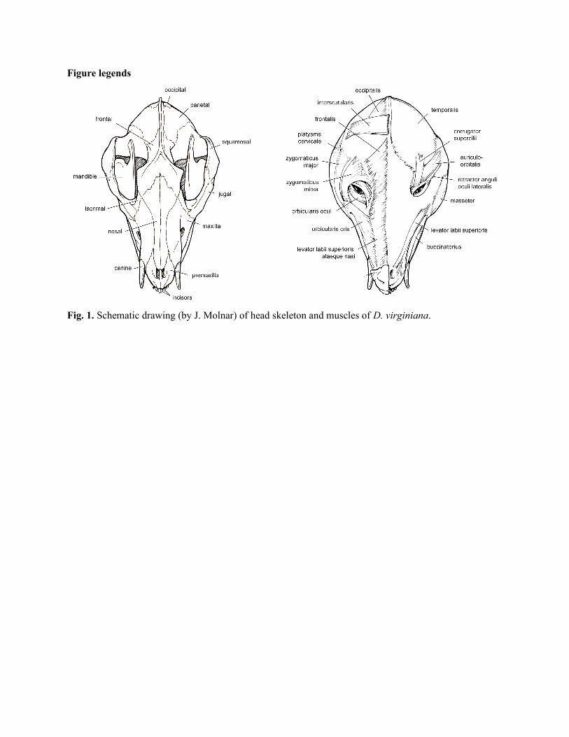
Figure legends
Fig. 1. Schematic drawing (by J. Molnar) of head skeleton and muscles of D. virginiana.
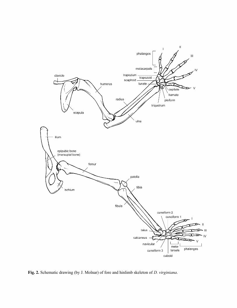
Fig. 2. Schematic drawing (by J. Molnar) of fore and hinlimb skeleton of D. virginiana.
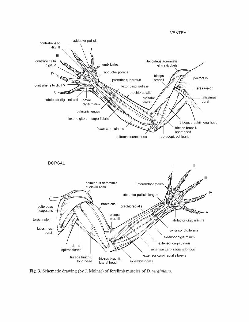
Fig. 3. Schematic drawing (by J. Molnar) of forelimb muscles of D. virginiana.
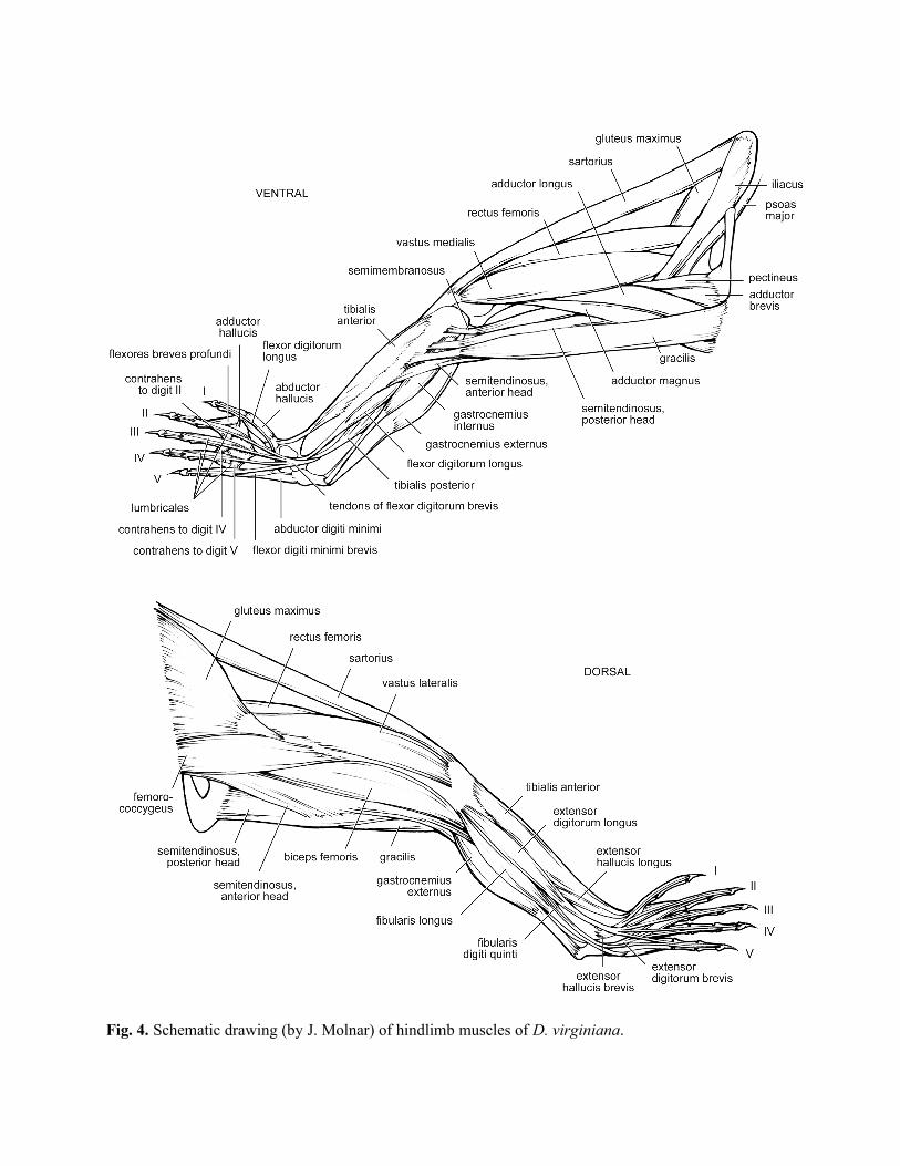
Fig. 4. Schematic drawing (by J. Molnar) of hindlimb muscles of D. virginiana.
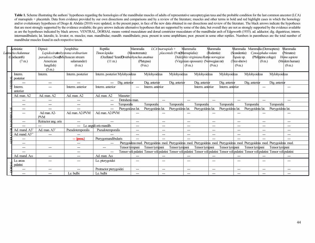
Table 1. Scheme illustrating the authors’ hypotheses regarding the homologies of the mandibular muscles of adults of representative sarcopterygian taxa and the probable condition for the last common ancestor (LCA)of marsupials + placentals. Data from evidence provided by our own dissections and comparisons and by a review of the literature; muscles and other terms in bold and red highlight cases in which the homologyand/or evolutionary hypotheses of Diogo & Abdala (2010) were updated, in the present paper, in face of the new data obtained in our dissections and review of the literature. The black arrows indicate the hypothesesthat are most strongly supported by the evidence available; the grey arrows indicate alternative hypotheses that are supported by some of the data, but overall they are not as strongly supported by the evidence availableas are the hypotheses indicated by black arrows. VENTRAL, DORSAL means ventral musculature and dorsal constrictor musculature of the mandibular arch of Edgeworth (1935). ad. adductor; dig. digastricus; interm.intermandibularis; lat. lateralis; le. levator; m. muscles; man. mandibulae; mandib. mandibularis; psoa. present in some amphibians; psor. present in some other reptiles. Numbers in parentheses are the total number ofmandibular muscles found in each respective taxon.
44
Actinistia:Latimeria chalumnae
(Coelacanth)(7 m.)
Dipnoi:Lepidosiren
paradoxa (SouthAmericanlungfish)
(5 m.)
Amphibia:Ambystoma ordinarium
(Michoacan streamsalamander)
(6 m.)
Reptilia:Timon lepidus
(Ocellated ‘lizard’)(11 m.)
Mammalia(Monotremata):
Ornithorhynchus anatinus(Platypus)
(9 m.)
LCA marsupials +placentals (9 m.)
Mammalia(Marsupialia):
Didelphis virginiana(Virginian opossum)
(8 m.)
Mammalia(Rodentia):
Rattus norvegicus(Norwegian rat)
(9 m.)
Mammalia(Scandentia):Tupaia sp.
(Tree-shrew)(9 m.)
Mammalia (Dermoptera):Cynocephalus volans(Philippine colugo)
(8 m.)
Mammalia(Primates):
Homo sapiens(Modern human)
(8 m.)
VEN
TRA
L
Interm. posterior
Interm. Interm. posterior Interm. posterior Mylohyoideus Mylohyoideus Mylohyoideus Mylohyoideus Mylohyoideus Mylohyoideus Mylohyoideus
--- --- --- --- Dig. anterior Dig. anterior Dig. anterior Dig. anterior Dig. anterior Dig. anterior Dig. anteriorInterm. anterior
--- Interm. anterior Interm. anterior --- Interm. anterior Interm. anterior Interm. anterior --- ---
AD
DU
CT
OR
MA
ND
IBU
LAE
Ad. man. A2 Ad. man. A2 Ad. man. A2 Ad. man. A2 Masseter--- --- --- --- Detrahens man. --- --- --- --- --- ------ --- --- --- Temporalis Temporalis Temporalis Temporalis Temporalis Temporalis Temporalis--- --- --- --- Pterygoideus lat. Pterygoideus lat. Pterygoideus lat. Pterygoideus lat. Pterygoideus lat. Pterygoideus lat. Pterygoideus lat.--- Ad. man. A2-
PVMAd. man. A2-PVM Ad. man. A2-PVM --- --- --- --- --- --- ---
--- Retractor ang. oris --- --- --- --- --- --- --- --- ------ --- --- Le. anguli oris mandib. --- --- --- --- --- --- ---
Ad. mand. A3’ Ad. man. A3’ Pseudotemporalis Pseudotemporalis --- --- --- --- --- --- ---Ad. mand. A3’’ --- --- --- --- --- --- --- --- --- ---
--- --- [psoa.] Pterygomandibularis --- --- --- --- --- --- ------ --- --- Pterygoideus med. Pterygoideus med. Pterygoideus med. Pterygoideus med. Pterygoideus med. Pterygoideus med. Pterygoideus med.--- --- --- --- Tensor tympani Tensor tympani Tensor tympani Tensor tympani Tensor tympani Tensor tympani Tensor tympani--- --- --- Tensor veli palatini Tensor veli palatini Tensor veli palatini Tensor veli palatini Tensor veli palatini Tensor veli palatini Tensor veli palatini
Ad. mand. Aω --- --- Ad. man. Aω --- --- --- --- --- --- ---
DO
RSA
L Le. arcus palatini
--- --- Le. pterygoidei --- --- --- --- --- --- ---
--- --- --- Protractor pterygoidei --- --- --- --- --- --- ------ --- Le. bulbi Le. bulbi --- --- --- --- --- --- ---
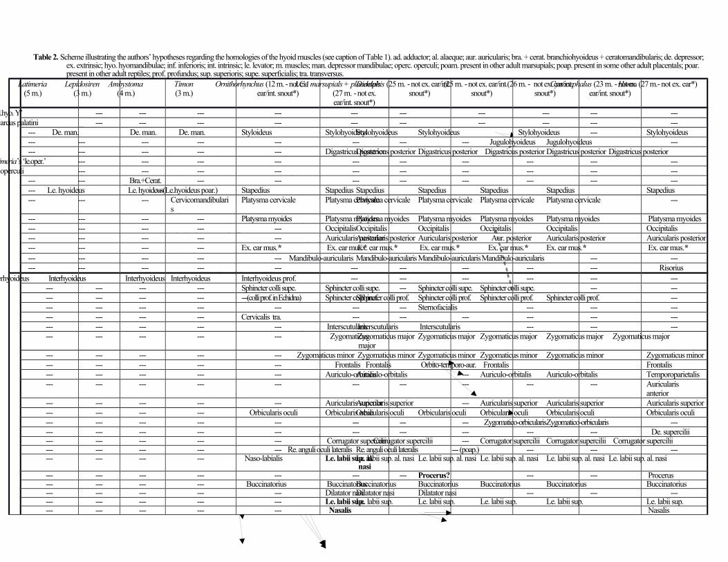
Table 2. Scheme illustrating the authors’ hypotheses regarding the homologies of the hyoid muscles (see caption of Table 1). ad. adductor; al. alaeque; aur. auricularis; bra. + cerat. branchiohyoideus + ceratomandibularis; de. depressor; ex. extrinsic; hyo. hyomandibulae; inf. inferioris; int. intrinsic; le. levator; m. muscles; man. depressor mandibulae; operc. operculi; poam. present in other adult marsupials; poap. present in some other adult placentals; poar. present in other adult reptiles; prof. profundus; sup. superioris; supe. superficialis; tra. transversus.
Latimeria(5 m.)
Lepidosiren(3 m.)
Ambystoma(4 m.)
Timon(3 m.)
Ornithorhynchus (12 m. - not ex.ear/int. snout*)
LCA marsupials + placentals(27 m. - not ex.ear/int. snout*)
Didelphis (25 m. - not ex. ear/int.snout*)
(25 m. - not ex. ear/int.snout*)
(26 m. - not ex. ear/int.snout*)
Cynocephalus (23 m. - not ex.ear/int. snout*)
Homo (27 m.- not ex. ear*)
DO
RSO
-MED
IAL
HY
OID
MU
S.
‘Ad.hyo. Y’ --- --- --- --- --- --- --- --- --- ---Ad. arcus palatini --- --- --- --- --- --- --- --- --- ---
--- De. man. De. man. De. man. Styloideus StylohyoideusStylohyoideus Stylohyoideus Stylohyoideus --- Stylohyoideus--- --- --- --- --- --- --- --- Jugulohyoideus Jugulohyoideus ------ --- --- --- --- Digastricus posteriorDigastricus posterior Digastricus posterior Digastricus posterior Digastricus posterior Digastricus posterior
Latimeria’s ‘le.oper.’ --- --- --- --- --- --- --- --- --- ---Ad. operculi --- --- --- --- --- --- --- --- --- ---
--- --- Bra.+Cerat. --- --- --- --- --- --- --- ------ Le. hyoideus Le. hyoideus---(Le.hyoideus poar.) Stapedius Stapedius Stapedius Stapedius Stapedius Stapedius Stapedius--- --- --- Cervicomandibulari
sPlatysma cervicale Platysma cervicalePlatysma cervicale Platysma cervicale Platysma cervicale Platysma cervicale ---
--- --- --- --- Platysma myoides Platysma myoidesPlatysma myoides Platysma myoides Platysma myoides Platysma myoides Platysma myoides--- --- --- --- --- OccipitalisOccipitalis Occipitalis Occipitalis Occipitalis Occipitalis--- --- --- --- --- Auricularis posteriorAuricularis posterior Auricularis posterior Aur. posterior Auricularis posterior Auricularis posterior--- --- --- --- Ex. ear mus.* Ex. ear mus.* Ex. ear mus.* Ex. ear mus.* Ex. ear mus.* Ex. ear mus.* Ex. ear mus.*--- --- --- --- --- Mandibulo-auricularis Mandibulo-auricularisMandibulo-auricularis Mandibulo-auricularis --- ------ --- --- --- --- --- --- --- --- --- Risorius
VE
NT
RA
L H
YO
ID M
US.
Interhyoideus Interhyoideus Interhyoideus Interhyoideus Interhyoideus prof. --- --- --- --- --- ------ --- --- --- Sphincter colli supe. Sphincter colli supe. --- Sphincter colli supe. Sphincter colli supe. --- ------ --- --- --- ---(colli prof. in Echidna) Sphincter colli prof.Sphincter colli prof. Sphincter colli prof. Sphincter colli prof. Sphincter colli prof. ------ --- --- --- --- --- --- Sternofacialis --- --- ------ --- --- --- Cervicalis tra. --- --- --- --- --- ------ --- --- --- --- Interscutularis Interscutularis Interscutularis --- --- ------ --- --- --- --- Zygomaticus
major Zygomaticus major Zygomaticus major Zygomaticus major Zygomaticus major Zygomaticus major
--- --- --- --- --- Zygomaticus minor Zygomaticus minor Zygomaticus minor Zygomaticus minor Zygomaticus minor Zygomaticus minor--- --- --- --- --- Frontalis Frontalis Orbito-temporo-aur. Frontalis Frontalis--- --- --- --- --- Auriculo-orbitalisAuriculo-orbitalis --- Auriculo-orbitalis Auriculo-orbitalis Temporoparietalis--- --- --- --- --- --- --- --- --- --- Auricularis
anterior--- --- --- --- --- Auricularis superiorAuricularis superior --- Auricularis superior Auricularis superior Auricularis superior--- --- --- --- Orbicularis oculi Orbicularis oculiOrbicularis oculi Orbicularis oculi Orbicularis oculi Orbicularis oculi Orbicularis oculi--- --- --- --- --- --- --- --- Zygomatico-orbicularisZygomatico-orbicularis ------ --- --- --- --- --- --- --- --- --- De. supercilii--- --- --- --- --- Corrugator superciliiCorrugator supercilii --- Corrugator supercilii Corrugator supercilii Corrugator supercilii--- --- --- --- --- Re. anguli oculi lateralis Re. anguli oculi lateralis --- (poap.) --- --- ------ --- --- --- Naso-labialis Le. labii sup. al.
nasiLe. labii sup. al. nasi Le. labii sup. al. nasi Le. labii sup. al. nasi Le. labii sup. al. nasi Le. labii sup. al. nasi
--- --- --- --- --- --- --- Procerus? --- --- Procerus--- --- --- --- Buccinatorius BuccinatoriusBuccinatorius Buccinatorius Buccinatorius Buccinatorius Buccinatorius--- --- --- --- --- Dilatator nasiDilatator nasi Dilatator nasi --- --- ------ --- --- --- --- Le. labii sup.Le. labii sup. Le. labii sup. Le. labii sup. Le. labii sup. Le. labii sup.--- --- --- --- --- Nasalis Nasalis
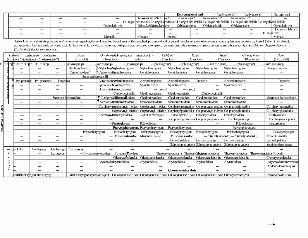
--- --- --- --- --- --- --- Depressor septi nasi --- (really absent?) --- (really absent?) De. septi nasi--- --- --- --- --- In. snout mus.*In. snout mus.* In. snout mus.* In. snout mus.* In. snout mus.* ------ --- --- --- --- Le. anguli oris facialis Le. anguli oris facialisLe. anguli oris facialis Le. anguli oris facialis Le. anguli oris facialis Le. anguli oris facialis--- --- --- --- Orbicularis oris Orbicularis orisOrbicularis oris Orbicularis oris Orbicularis oris Orbicularis oris Orbicularis oris--- --- --- --- --- --- --- --- --- --- Depressor labii inf.--- --- --- --- --- --- --- --- --- --- De. anguli oris--- --- ---- --- Mentalis Mentalis --- (poam.) --- Mentalis
Table 3. Scheme illustrating the authors’ hypotheses regarding the evolution and homologies of the branchial, pharyngeal and laryngeal muscles of adults of representative sarcopterygian taxa (see caption of Table 1). ab. absent; ap. apparatus; br. branchial; co. constrictor; fu. functional; le. levator; m. muscles; post. posterior; pro. protractor; psom. present some other marsupials; psop. present some other placentals; see DA, see Diogo & Abdala (2010); st. of stricto; sup. superior.
Latimeria(4 m.-not st.*)
Lepidosiren(3 mus.-not st.*)
Ambystoma(4 m.-not str.*)
Timon(6 m. total)
Ornithorhynchus(14 m. total)
LCA marsupials + placentals (19m.total)
Didelphis(17 m. total)
Rattus(21 m. total)
Tupaia(17 m. total)
Cynocephalus(19 m. total)
Homo(17 m. total)
‘TR
UE’
BR
AN
CH
IAL
MU
S.
STR
ICTO
*Fu.m.br.ap.* Fu.m.br.ap.* Fu.m.br.ap.. --- (ab. as a group) --- (ab. as group) --- (ab. as a group) --- (ab. as a group) --- (ab. as a group) --- (ab. as a group) --- (abs. as a group) --- (abs. as a group)--- --- --- Hyobranchialis StylopharyngeusStylopharyngeus Stylopharyngeus Stylopharyngeus Stylopharyngeus Stylopharyngeus Stylopharyngeus--- --- --- ‘Ceratohyoideus’ CeratohyoideusCeratohyoideus Ceratohyoideus Ceratohyoideus Ceratohyoideus Ceratohyoideus ------ --- --- ---Subarcualis rectus III --- --- --- --- --- ---
OT
HE
R ---Pro. pectoralis Pro. pectoralis Trapezius AcromiotrapeziusAcromiotrapezius Acromiotrapezius Acromiotrapezius Trapezius Acromiotrapezius Trapezius--- --- --- --- SpinotrapeziusSpinotrapezius Spinotrapezius Spinotrapezius --- Spinotrapezius ------ --- --- --- DorsocutaneousDorsocutaneous --- (psom.) --- (psop.) --- --- ------ --- --- --- --- Cleido-occipitalis Cleido-occipitalis Cleido-occipitalis Cleido-occipitalis --- ------ --- ---Sternocleidomastoideus Cleidomastoideus Cleidomastoideus Cleidomastoideus Cleidomastoideus Cleidomastoideus Cleidomastoideus Sternocleidomastoideus--- --- --- --- SternomastoideusSternomastoideus Sternomastoideus Sternomastoideus Sternomastoideus Sternomastoideus ---
PHA
RY
NG
EAL
MU
S. --- --- --- --- Co. pharyngisCo.pharyngis medius Co.pharyngis medius Co.pharyngis medius Co. pharyngis medius Co. pharyngis medius Co. pharyngis medius---- --- --- --- ---Co.pharyngis inferior Co.pharyngis inferior Co.pharyngis inferior Co. pharyngis inferior Co. pharyngis inferior Co. pharyngis inferior---- --- --- --- CricothyroideusCricothyroideus ---(lost in marsupials) Cricothyroideus Cricothyroideus Cricothyroideus Cricothyroideus--- --- --- --- --- --- --- Co. pharyngis superior Co. pharyngis superior Co. pharyngis sup. Co. pharyngis superior--- --- --- --- ---Palatoglossus Palatoglossus --- --- Palatoglossus Palatoglossus---- --- --- --- ---Pterygopharyngeus Pterygopharyngeus Pterygopharyngeus --- Pterygopharyngeus ------- --- --- --- Palatopharyngeus Palatopharyngeus Palatopharyngeus Palatopharyngeus Palatopharyngeus Palatopharyngeus Palatopharyngeus--- --- --- --- --- Musculus uvulae Musculus uvulae Musculus uvulae --- ? (really absent?) --- ? (really absent?) Musculus uvulae---- --- --- --- --- --- --- Le. veli palatini Le. veli palatini Le. veli palatini Le. veli palatini---- --- --- --- --- --- --- Salpingopharyngeus Salpingopharyngeus Salpingopharyngeus Salpingopharyngeus
LA
RY
NG
EA
L M
US.---? (see DA) Co. laryngis Co. laryngis Co. laryngis --- --- --- --- --- --- ---
--- --- Laryngeus --- Thyrocricoarytenoideus Thyroarytenoideus Thyroarytenoideus ThyroarytenoideusThyroarytenoideus Thyroarytenoideus Thyroarytenoideus (+vocalis)--- --- --- --- --- Cricoarytenoideus lat. Cricoarytenoideus lat. Cricoarytenoideus lat. Cricoarytenoideus lat. Cricoarytenoideus lat. Cricoarytenoideus lat.--- --- --- --- ArytenoideusArytenoideus Arytenoideus Arytenoideus Arytenoideus Arytenoideus Arytenoideus transversus--- --- --- --- --- --- --- --- --- --- Arytenoideus obliquus--- --- --- --- --- --- --- Cricoarytenoideus alaris --- --- ---
---? (see DA)Dilator laryngisDilator laryngis Dilator laryngisCricoarytenoideus post. Cricoarytenoideus post. Cricoarytenoideus post. Cricoarytenoideus post. Cricoarytenoideus post. Cricoarytenoideus post. Cricoarytenoideus post.
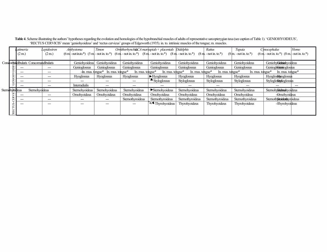
Table 4. Scheme illustrating the authors’ hypotheses regarding the evolution and homologies of the hypobranchial muscles of adults of representative sarcopterygian taxa (see caption of Table 1). ‘GENIOHYOIDEUS’, ‘RECTUS CERVICIS’ mean ‘geniohyoideus’ and ‘rectus cervicus’ groups of Edgeworth (1935). in. to. intrinsic muscles of the tongue; m. muscles.
Latimeria(2 m.)
Lepidosiren(2 m.)
Ambystoma(6 m. - not in.to.*)
Timon(5 m. - not in. to.*)
Ornithorhynchus(6 m. - not in. to.*)
LCA marsupials + placentals(8 m. - not in. to.*)
Didelphis(8 m. - not in. to.*)
Rattus(8 m. - not in. to.*)
Tupaia(8 m. - not in. to.*)
Cynocephalus(6 m. - not in. to.*)
Homo(8 m. - not in. to.*)
‘GE
NIO
HY
OID
EU
S’
Coracomandibularis Coracomandibularis Geniohyoideus Geniohyoideus Geniohyoideus Geniohyoideus Geniohyoideus Geniohyoideus Geniohyoideus GeniohyoideusGeniohyoideus--- --- Genioglossus Genioglossus Genioglossus Genioglossus Genioglossus Genioglossus Genioglossus GenioglossusGenioglossus--- --- --- In. mus. tongue* In. mus. tongue* In. mus. tongue* In. mus. tongue* In. mus. tongue* In. mus. tongue* In. mus. tongue* In. mus. tongue*--- --- Hyoglossus Hyoglossus Hyoglossus Hyoglossus Hyoglossus Hyoglossus Hyoglossus HyoglossusHyoglossus--- --- --- --- --- Styloglossus Styloglossus Styloglossus Styloglossus StyloglossusStyloglossus--- --- Interradialis --- --- --- --- --- --- --- ---
‘RE
CT
US
-CE
RV
ICIS
’
Sternohyoideus Sternohyoideus Sternohyoideus Sternohyoideus Sternohyoideus Sternohyoideus Sternohyoideus Sternohyoideus Sternohyoideus SternohyoideusSternohyoideus--- --- Omohyoideus Omohyoideus Omohyoideus Omohyoideus Omohyoideus Omohyoideus Omohyoideus ---Omohyoideus--- --- --- --- Sternothyroideus Sternothyroideus Sternothyroideus Sternothyroideus Sternothyroideus SternothyroideusSternothyroideus--- --- --- --- --- Thyrohyoideus Thyrohyoideus Thyrohyoideus Thyrohyoideus ---Thyrohyoideus
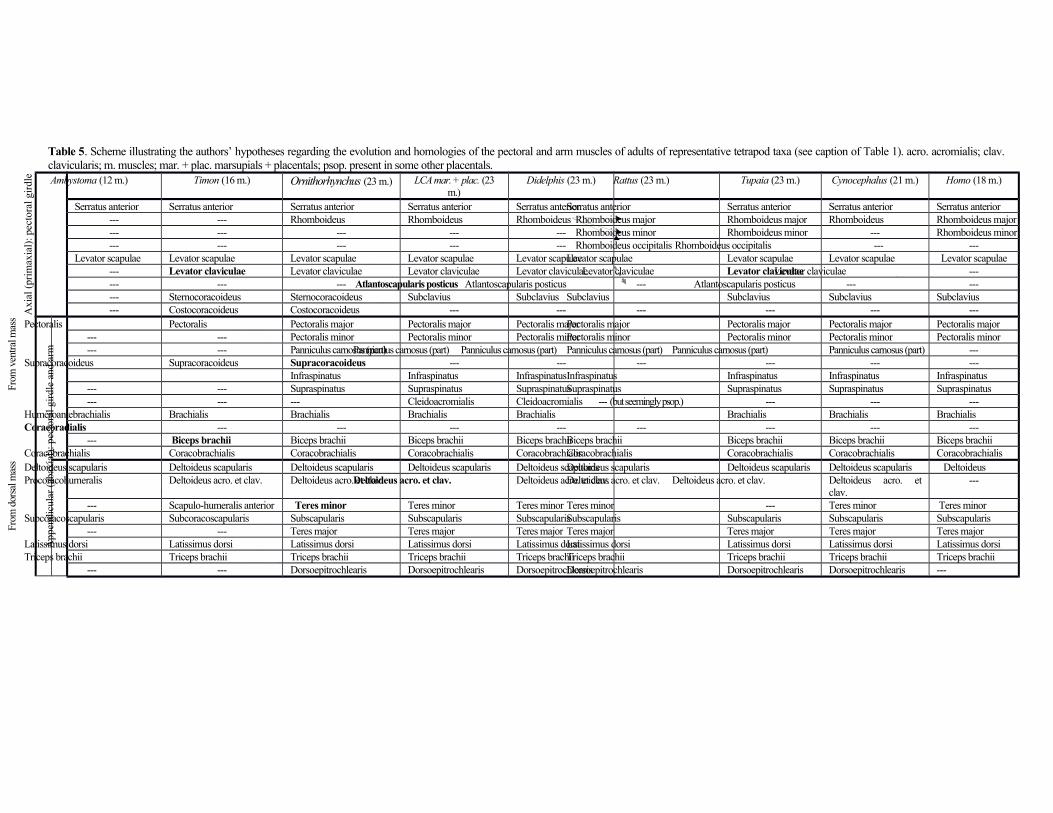
Table 5. Scheme illustrating the authors’ hypotheses regarding the evolution and homologies of the pectoral and arm muscles of adults of representative tetrapod taxa (see caption of Table 1). acro. acromialis; clav.clavicularis; m. muscles; mar. + plac. marsupials + placentals; psop. present in some other placentals.
Axi
al (
prim
axia
l): p
ecto
ral g
irdl
e Ambystoma (12 m.) Timon (16 m.) Ornithorhynchus (23 m.) LCA mar. + plac. (23m.)
Didelphis (23 m.) Rattus (23 m.) Tupaia (23 m.) Cynocephalus (21 m.) Homo (18 m.)
Serratus anterior Serratus anterior Serratus anterior Serratus anterior Serratus anteriorSerratus anterior Serratus anterior Serratus anterior Serratus anterior--- --- Rhomboideus Rhomboideus Rhomboideus Rhomboideus major Rhomboideus major Rhomboideus Rhomboideus major--- --- --- --- --- Rhomboideus minor Rhomboideus minor --- Rhomboideus minor--- --- --- --- --- Rhomboideus occipitalis Rhomboideus occipitalis --- ---
Levator scapulae Levator scapulae Levator scapulae Levator scapulae Levator scapulaeLevator scapulae Levator scapulae Levator scapulae Levator scapulae--- Levator claviculae Levator claviculae Levator claviculae Levator claviculae Levator claviculae Levator claviculae Levator claviculae ------ --- --- Atlantoscapularis posticus Atlantoscapularis posticus --- Atlantoscapularis posticus --- ------ Sternocoracoideus Sternocoracoideus Subclavius Subclavius Subclavius Subclavius Subclavius Subclavius--- Costocoracoideus Costocoracoideus --- --- --- --- --- ---
App
endi
cula
r (a
baxi
al):
pec
tora
l gir
dle
and
arm
From
ven
tral m
ass Pectoralis Pectoralis Pectoralis major Pectoralis major Pectoralis majorPectoralis major Pectoralis major Pectoralis major Pectoralis major
--- --- Pectoralis minor Pectoralis minor Pectoralis minorPectoralis minor Pectoralis minor Pectoralis minor Pectoralis minor--- --- Panniculus carnosus (part)Panniculus carnosus (part) Panniculus carnosus (part) Panniculus carnosus (part) Panniculus carnosus (part) Panniculus carnosus (part) ---
Supracoracoideus Supracoracoideus Supracoracoideus --- --- --- --- --- ---Infraspinatus Infraspinatus InfraspinatusInfraspinatus Infraspinatus Infraspinatus Infraspinatus
--- --- Supraspinatus Supraspinatus SupraspinatusSupraspinatus Supraspinatus Supraspinatus Supraspinatus--- --- --- Cleidoacromialis Cleidoacromialis --- (but seemingly psop.) --- --- ---
Humeroantebrachialis Brachialis Brachialis Brachialis Brachialis Brachialis Brachialis BrachialisCoracoradialis --- --- --- --- --- --- --- ---
--- Biceps brachii Biceps brachii Biceps brachii Biceps brachiiBiceps brachii Biceps brachii Biceps brachii Biceps brachiiCoracobrachialis Coracobrachialis Coracobrachialis Coracobrachialis Coracobrachialis Coracobrachialis Coracobrachialis Coracobrachialis Coracobrachialis
From
dor
sal m
ass Deltoideus scapularis Deltoideus scapularis Deltoideus scapularis Deltoideus scapularis Deltoideus scapularisDeltoideus scapularis Deltoideus scapularis Deltoideus scapularis Deltoideus
Procoracohumeralis Deltoideus acro. et clav. Deltoideus acro. et clav.Deltoideus acro. et clav. Deltoideus acro. et clav.Deltoideus acro. et clav. Deltoideus acro. et clav. Deltoideus acro. etclav.
---
--- Scapulo-humeralis anterior Teres minor Teres minor Teres minor Teres minor --- Teres minor Teres minorSubcoracoscapularis Subcoracoscapularis Subscapularis Subscapularis SubscapularisSubscapularis Subscapularis Subscapularis Subscapularis
--- --- Teres major Teres major Teres major Teres major Teres major Teres major Teres majorLatissimus dorsi Latissimus dorsi Latissimus dorsi Latissimus dorsi Latissimus dorsiLatissimus dorsi Latissimus dorsi Latissimus dorsi Latissimus dorsiTriceps brachii Triceps brachii Triceps brachii Triceps brachii Triceps brachiiTriceps brachii Triceps brachii Triceps brachii Triceps brachii
--- --- Dorsoepitrochlearis Dorsoepitrochlearis DorsoepitrochlearisDorsoepitrochlearis Dorsoepitrochlearis Dorsoepitrochlearis ---
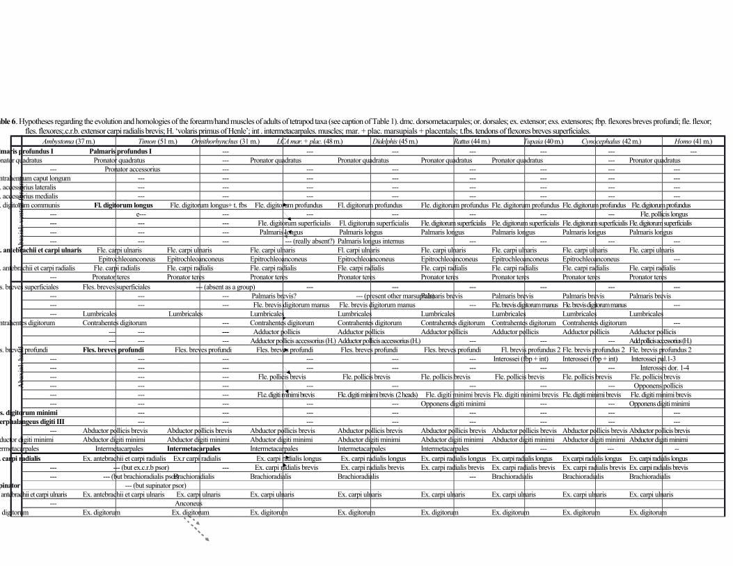
Table 6. Hypotheses regarding the evolution and homologies of the forearm/hand muscles of adults of tetrapod taxa (see caption of Table 1). dmc. dorsometacarpales; or. dorsales; ex. extensor; exs. extensores; fbp. flexores breves profundi; fle. flexor;fles. flexores;.c.r.b. extensor carpi radialis brevis; H. ‘volaris primus of Henle’; int . intermetacarpales. muscles; mar. + plac. marsupials + placentals; t.fbs. tendons of flexores breves superficiales.
Ambystoma (37 m.) Timon (51 m.) Ornithorhynchus (31 m.) LCA mar. + plac. (48 m.) Didelphis (45 m.) Rattus (44 m.) Tupaia (40 m.) Cynocephalus (42 m.) Homo (41 m.)
Aba
xial
: ven
tral
for
earm
Palmaris profundus I Palmaris profundus I --- --- --- --- --- --- ---Pronator quadratus Pronator quadratus --- Pronator quadratus Pronator quadratus Pronator quadratus Pronator quadratus --- Pronator quadratus
--- Pronator accessorius --- --- --- --- --- --- ---Contrahentium caput longum --- --- --- --- --- --- --- ---Fle. accessorius lateralis --- --- --- --- --- --- --- ---Fle. accessorius medialis --- --- --- --- --- --- --- ---Fle. digitorum communis Fl. digitorum longus Fle. digitorum longus+ t. fbs Fle. digitorum profundus Fl. digitorum profundus Fle. digitorum profundus Fle. digitorum profundus Fle. digitorum profundus Fle. digitorum profundus
--- e--- --- --- --- --- --- --- Fle. pollicis longus--- --- --- Fle. digitorum superficialis Fl. digitorum superficialis Fle. digitorum superficialis Fle. digitorum superficialis Fle. digitorum superficialis Fle. digitorum superficialis--- --- --- Palmaris longus Palmaris longus Palmaris longus Palmaris longus Palmaris longus Palmaris longus--- --- --- --- (really absent?) Palmaris longus internus --- --- --- ---
Fle. antebrachii et carpi ulnaris Fle. carpi ulnaris Fle. carpi ulnaris Fle. carpi ulnaris Fl. carpi ulnaris Fle. carpi ulnaris Fle. carpi ulnaris Fle. carpi ulnaris Fle. carpi ulnaris Epitrochleoanconeus Epitrochleoanconeus Epitrochleoanconeus Epitrochleoanconeus Epitrochleoanconeus Epitrochleoanconeus Epitrochleoanconeus ---
Fle. antebrachii et carpi radialis Fle. carpi radialis Fle. carpi radialis Fle. carpi radialis Fle. carpi radialis Fle. carpi radialis Fle. carpi radialis Fle. carpi radialis Fle. carpi radialis--- Pronator teres Pronator teres Pronator teres Pronator teres Pronator teres Pronator teres Pronator teres Pronator teres
Aba
xial
: han
d
Fles. breves superficiales Fles. breves superficiales --- (absent as a group) --- --- --- --- --- ------ --- --- Palmaris brevis? --- (present other marsupials)Palmaris brevis Palmaris brevis Palmaris brevis Palmaris brevis --- --- --- Fle. brevis digitorum manus Fle. brevis digitorum manus --- Fle. brevis digitorum manus Fle. brevis digitorum manus ------ Lumbricales Lumbricales Lumbricales Lumbricales Lumbricales Lumbricales Lumbricales Lumbricales
Contrahentes digitorum Contrahentes digitorum --- Contrahentes digitorum Contrahentes digitorum Contrahentes digitorum Contrahentes digitorum Contrahentes digitorum ------ --- --- Adductor pollicis Adductor pollicis Adductor pollicis Adductor pollicis Adductor pollicis Adductor pollicis--- --- --- Adductor pollicis accessorius (H.) Adductor pollicis accessorius (H.) --- --- --- Add pollicis accessorius (H.)
Fles. breves profundi Fles. breves profundi Fles. breves profundi Fles. breves profundi Fles. breves profundi Fles. breves profundi Fl. brevis profundus 2 Fle. brevis profundus 2 Fle. brevis profundus 2--- --- --- --- --- --- Interossei (fbp + int) Interossei (fbp + int) Interossei pal.1-3--- --- --- --- --- --- --- --- Interossei dor. 1-4--- --- --- Fle. pollicis brevis Fle. pollicis brevis Fle. pollicis brevis Fle. pollicis brevis Fle. pollicis brevis Fle. pollicis brevis--- --- --- --- --- --- --- --- Opponens pollicis--- --- --- Fl.e. digiti minimi brevis Fle. digiti minimi brevis (2 heads) Fle. digiti minimi brevis Fle. digiti minimi brevis Fle. digiti minimi brevis Fle. digiti minimi brevis--- --- --- --- --- Opponens digiti minimi --- --- Opponens digiti minimi
Fles. digitorum minimi --- --- --- --- --- --- --- ---Interphalangeus digiti III --- --- --- --- --- --- --- ---
--- Abductor pollicis brevis Abductor pollicis brevis Abductor pollicis brevis Abductor pollicis brevis Abductor pollicis brevis Abductor pollicis brevis Abductor pollicis brevis Abductor pollicis brevisAbductor digiti minimi Abductor digiti minimi Abductor digiti minimi Abductor digiti minimi Abductor digiti minimi Abductor digiti minimi Abductor digiti minimi Abductor digiti minimi Abductor digiti minimiIntermetacarpales Intermetacarpales Intermetacarpales Intermetacarpales Intermetacarpales Intermetacarpales --- --- --Ex. carpi radialis Ex. antebrachii et carpi radialis Ex.r carpi radialis Ex. carpi radialis longus Ex. carpi radialis longus Ex. carpi radialis longus Ex. carpi radialis longus Ex carpi radialis longus Ex. carpi radialis longus
--- --- (but ex.c.r.b psor) --- Ex. carpi radialis brevis Ex. carpi radialis brevis Ex. carpi radialis brevis Ex. carpi radialis brevis Ex. carpi radialis brevis Ex. carpi radialis brevis--- --- (but brachioradialis psor) Brachioradialis Brachioradialis Brachioradialis --- Brachioradialis Brachioradialis Brachioradialis
Supinator --- (but supinator psor)Ex. antebrachii et carpi ulnaris Ex. antebrachii et carpi ulnaris Ex. carpi ulnaris Ex. carpi ulnaris Ex. carpi ulnaris Ex. carpi ulnaris Ex. carpi ulnaris Ex. carpi ulnaris Ex. carpi ulnaris
--- AnconeusEx. digitorum Ex. digitorum Ex. digitorum Ex. digitorum Ex. digitorum Ex. digitorum Ex. digitorum Ex. digitorum Ex. digitorum
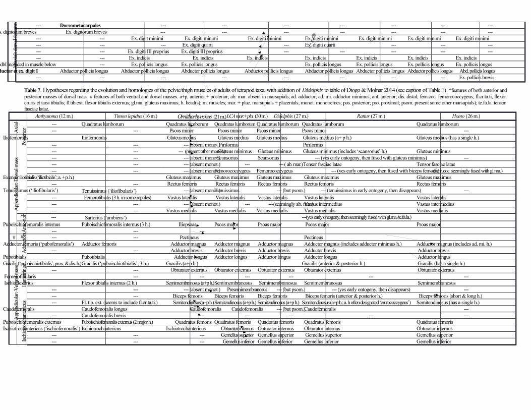
Aba
xial
: dor
sal f
orea
rm --- Dorsometacarpales --- --- --- --- --- --- ---Exs. digitorum breves Ex. digitorum breves --- --- --- --- --- --- ---
--- --- Ex. digit minimi Ex. digiti minimi Ex. digiti minimi Ex. digiti minimi Ex. digiti minimi Ex. digiti minimi Ex. digiti minimi--- --- --- Ex. digiti quarti --- Ex. digiti quarti --- --- ------ --- Ex. digiti III proprius Ex. digiti III proprius --- --- --- --- ------ --- Ex. indicis Ex. indicis Ex. indicis Ex. indicis Ex. indicis Ex. indicis Ex. indicis
---(edbI included in muscle below --- Ex. pollicis longus Ex. pollicis longus Ex. pollicis longus Ex. pollicis longus Ex. pollicis longus Ex. pollicis longusAbductor et ex. digit I Abductor pollicis longus Abductor pollicis longus Abductor pollicis longus Abductor pollicis longus Abductor pollicis longus Abductor pollicis longus Abductor pollicis longus Abd. pollicis longus
--- --- --- --- --- --- --- --- Ex. pollicis brevis
Table 7. Hypotheses regarding the evolution and homologies of the pelvic/thigh muscles of adults of tetrapod taxa, with addition of Didelphis to table of Diogo & Molnar 2014 (see caption of Table 1). *features of both anterior andposterior masses of dorsal mass; # features of both ventral and dorsal masses. a+p, anterior + posterior; ab. mar. absent in marsupials; ad. adductor; ad. mi. adductor minimus; ant. anterior; dis. distal; fem.coc. femorococcygeus; fl.cr.ta.ti, flexorcruris et tarsi tibialis; fl.tib.ext. flexor tibialis externus; gl.ma. gluteus maximus; h. head(s); m. muscles; mar. + plac. marsupials + placentals; monot. monotremes; pos. posterior; pro. proximal; psom. present some other marsupials); te.fa.la. tensorfasciae latae.
Ambystoma (12 m.) Timon lepidus (16 m.) Ornithorhynchus (21 m.)LCA mar.+pla. (30 m.) Didelphis (27 m.) Rattus (27 m.) Homo (26 m.)
Axi
al --- Quadratus lumborum Quadratus lumborum Quadratus lumborum Quadratus lumborum Quadratus lumborum Quadratus lumborum--- --- Psoas minor Psoas minor Psoas minor Psoas minor ---
App
endi
cula
r: D
orsa
l mas
s
Post
erio
r
Iliofemoralis Iliofemoralis Gluteus medius Gluteus medius Gluteus medius Gluteus medius (a+ p h.) Gluteus medius (has a single h.)--- --- --- (absent monot.) Piriformis Piriformis--- --- --- (present other monot.) Gluteus minimus Gluteus minimus Gluteus minimus (includes ‘scansorius’ h.) Gluteus minimus--- --- --- (absent monot.) Scansorius Scansorius --- (yes early ontogeny, then fused with gluteus minimus) ------ --- --- (absent monot.) --- --- ( ab. mar.) Tensor fasciae latae Tensor fasciae latae--- --- --- (absent monot.)Femorococcygeus Femorococcygeus --- (yes early ontogeny, then fused with biceps femoris) ---(fem.coc. seemingly fused with gl.ma.)
Extensor iliotibialis (‘iliotibialis’; a. + p. h.) Gluteus maximus Gluteus maximus Gluteus maximus Gluteus maximus Gluteus maximus--- --- Rectus femoris Rectus femoris Rectus femoris Rectus femoris Rectus femoris
Tenuissimus (‘iliofibularis’) Tenuissimus (‘iliofibularis’) --- (absent monot.)Tenuissimus --- (but psom.) --- (tenuissimus in early ontogeny, then disappears) ------ Femorotibialis (3 h. in some reptiles) Vastus lateralis Vastus lateralis Vastus lateralis Vastus lateralis Vastus lateralis--- --- --- (absent monot.) --- ---(seemingly ab. mar.) Vastus intermedius Vastus intermedius--- --- Vastus medialis Vastus medialis Vastus medialis Vastus medialis Vastus medialis
* --- Sartorius (‘ambiens’) ---(yes early ontogeny, then seemingly fused with gl.ma./te.fa.la.)
Ant
e-rio
r
Puboischiofemoralis internus Puboischiofemoralis internus (3 h.) Iliopsoas Psoas major Psoas major Psoas major Psoas major--- --- ---
# # --- --- Pectineus Pectineus
App
endi
cula
r: V
entra
l mas
s
Add
ucto
rs
Adductor femoris (‘pubofemoralis’) Adductor femoris Adductor magnus Adductor magnus Adductor magnus Adductor magnus (includes adductor minimus h.) Adductor magnus (includes ad. mi. h.)--- --- Adductor brevis Adductor brevis Adductor brevis Adductor brevis Adductor brevis
Pubotibialis Pubotibialis Adductor longus Adductor longus Adductor longus Adductor longus Adductor longusGracilis (‘puboischiotibialis’, prox. & dis. h.)Gracilis (‘puboischiotibialis’; 3 h.) Gracilis (a+p h.) Gracilis (anterior & posterior h.) Gracilis (has a single h.)
--- --- Obturator externus Obturator externus Obturator externus Obturator externus Obturator externus
Ham
strin
gs
Femorofibularis --- --- --- --- --- --- Ischioflexorius Flexor tibialis internus (2 h.) Semimembranosus (a+p h.)Semimembranosus Semimembranosus Semimembranosus Semimembranosus
--- --- --- (absent monot.) Presemimembranosus --- (but psom.) --- (yes early ontogeny, then disappears) ------ --- Biceps femoris Biceps femoris Biceps femoris Biceps femoris (anterior & posterior h.) Biceps femoris (short & long h.)--- Fl. tib. ext. (seems to include fl.cr.ta.ti.) Semitendinosus(a+p h.)Semitendinosus (a+p h.) Semitendinosus (a+p h.) Semitendinosus (a+p h.; a. h often designated ‘crurococcygeus’) Semitendinosus (has a single h.)
Caudofemoralis Caudofemoralis longus Caudofemoralis Caudofemoralis --- (but psom.) Caudofemoralis ------ Caudofemoralis brevis --- --- --- --- ---
Isch
iotro
-cha
nter
icus
Puboischiofemoralis externus Puboischiofemoralis externus (2 major h.) Quadratus femoris Quadratus femoris Quadratus femoris Quadratus femoris Quadratus femorisIschiotrochantericus (‘ischiofemoralis’) Ischiotrochantericus Ischiotrochantericus Obturator internus Obturator internus Obturator internus Obturator internus
--- --- --- Gemellus superior Gemellus superior Gemellus superior Gemellus superior--- --- --- Gemellus inferior Gemellus inferior Gemellus inferior Gemellus inferior
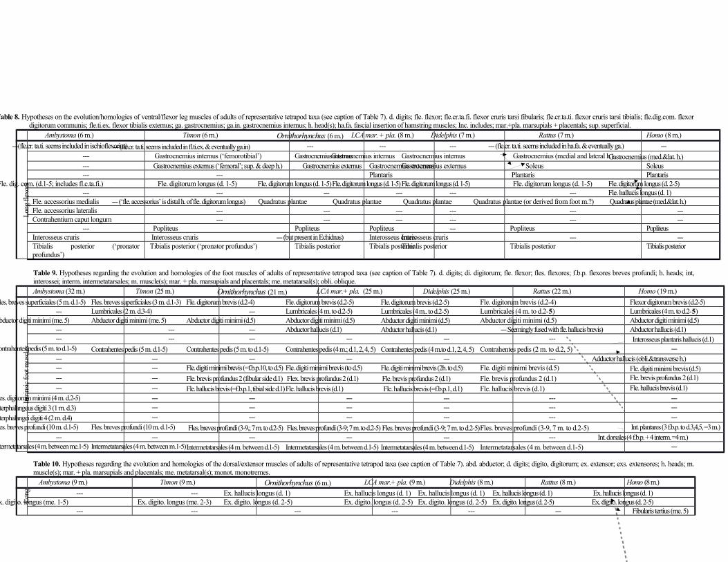
Table 8. Hypotheses on the evolution/homologies of ventral/flexor leg muscles of adults of representative tetrapod taxa (see caption of Table 7). d. digits; fle. flexor; fle.cr.ta.fi. flexor cruris tarsi fibularis; fle.cr.ta.ti. flexor cruris tarsi tibialis; fle.dig.com. flexordigitorum communis; fle.ti.ex. flexor tibialis externus; ga. gastrocnemius; ga.in. gastrocnemius internus; h. head(s); ha.fa. fascial insertion of hamstring muscles; Inc. includes; mar.+pla. marsupials + placentals; sup. superficial.
Ambystoma (6 m.) Timon (6 m.) Ornithorhynchus (6 m.) LCA mar. + pla. (8 m.) Didelphis (7 m.) Rattus (7 m.) Homo (8 m.)
Lon
g fle
xors
---(fle.cr. ta.ti. seems included in ischioflexorius) --- (fle.cr. ta.ti. seems included in fl.ti.ex. & eventually ga.in) --- --- --- --- (fle.cr. ta.ti. seems included in ha.fa. & eventually ga.) ------ Gastrocnemius internus (‘femorotibial’) Gastrocnemius internusGastrocnemius internus Gastrocnemius internus Gastrocnemius (medial and lateral h.) Gastrocnemius (med.&lat. h.)
--- Gastrocnemius externus (‘femoral’; sup. & deep h.) Gastrocnemius externus Gastrocnemius externusGastrocnemius externus Soleus Soleus --- --- Plantaris Plantaris Plantaris
Fle. dig. com. (d.1-5; includes fl.c.ta.fi.) Fle. digitorum longus (d. 1-5) Fle. digitorum longus (d. 1-5)Fle. digitorum longus (d. 1-5)Fle. digitorum longus (d. 1-5) Fle. digitorum longus (d. 1-5) Fle. digitorum longus (d. 2-5) --- --- --- --- --- --- Fle. hallucis longus (d. 1)
Fle. accessorius medialis --- (‘fle. accessorius’ is distal h. of fle. digitorum longus) Quadratus plantae Quadratus plantae Quadratus plantae Quadratus plantae (or derived from foot m.?) Quadratus plantae (med.&lat. h.)Fle. accessorius lateralis --- --- --- --- --- ---Contrahentium caput longum --- --- --- --- --- ---
--- Popliteus Popliteus Popliteus --- Popliteus Popliteus Interosseus cruris Interosseus cruris --- (but present in Echidnas) Interosseus crurisInterosseus cruris --- ---Tibialis posterior (‘pronatorprofundus’)
Tibialis posterior (‘pronator profundus’) Tibialis posterior Tibialis posteriorTibialis posterior Tibialis posterior Tibialis posterior
Table 9. Hypotheses regarding the evolution and homologies of the foot muscles of adults of representative tetrapod taxa (see caption of Table 7). d. digits; di. digitorum; fle. flexor; fles. flexores; f.b.p. flexores breves profundi; h. heads; int,interossei; interm. intermetatarsales; m. muscle(s); mar. + pla. marsupials and placentals; me. metatarsal(s); obli. oblique.Ambystoma (32 m.) Timon (25 m.) Ornithorhynchus (21 m.) LCA mar.+ pla. (25 m.) Didelphis (25 m.) Rattus (22 m.) Homo (19 m.)
Intri
nsic
foot
mus
cles
Fles. breves superficiales (5 m. d.1-5) Fles. breves superficiales (3 m. d.1-3) Fle. digitorum brevis (d.2-4) Fle. digitorum brevis (d.2-5) Fle. digitorum brevis (d.2-5) Fle. digitorum brevis (d.2-4) Flexor digitorum brevis (d.2-5)--- Lumbricales (2 m. d.3-4) --- Lumbricales (4 m. to d.2-5) Lumbricales (4 m.. to d.2-5) Lumbricales (4 m. to d.2-5) Lumbricales (4 m. to d.2-5)
Abductor digiti minimi (me. 5) Abductor digiti minimi (me. 5) Abductor digiti minimi (d.5) Abductor digiti minimi (d.5) Abductor digiti minimi (d.5) Abductor digiti minimi (d.5) Abductor digiti minimi (d.5)--- --- --- Abductor hallucis (d.1) Abductor hallucis (d.1) --- Seemingly fused with fle. hallucis brevis) Abductor hallucis (d.1)--- --- --- --- --- --- Interosseus plantaris hallucis (d.1)
Contrahentes pedis (5 m. to d.1-5) Contrahentes pedis (5 m. d.1-5) Contrahentes pedis (5 m. to d.1-5) Contrahentes pedis (4 m.; d.1, 2, 4, 5) Contrahentes pedis (4 m.to d.1, 2, 4, 5) Contrahentes pedis (2 m. to d.2, 5) ---
--- --- --- --- --- --- Adductor hallucis (obli.&transverse h.)--- --- Fle. digiti minimi brevis (=f.b.p.10, to d.5) Fle. digiti minimi brevis (to d.5) Fle. digiti minimi brevis (2h. to d.5) Fle. digiti minimi brevis (d.5) Fle. digiti minimi brevis (d.5)
--- --- Fle. brevis profundus 2 (fibular side d.1) Flex. brevis profundus 2 (d.1) Fle. brevis profundus 2 (d.1) Fle. brevis profundus 2 (d.1) Fle. brevis profundus 2 (d.1)
--- --- Fle. hallucis brevis (=f.b.p.1, tibial side d.1) Fle. hallucis brevis (d.1) Fle. hallucis brevis (=f.b.p.1, d.1) Fle. hallucis brevis (d.1) Fle. hallucis brevis (d.1)Fles. digitorum minimi (4 m. d.2-5) --- --- --- --- --- ---Interphalangeus digiti 3 (1 m. d.3) --- --- --- --- --- ---
Interphalangei digiti 4 (2 m. d.4) --- --- --- --- --- ---Fles. breves profundi (10 m. d.1-5) Fles. breves profundi (10 m. d.1-5) Fles. breves profundi (3-9,; 7 m. to d.2-5) Fles. breves profundi (3-9; 7 m. to d.2-5) Fles. breves profundi (3-9; 7 m. to d.2-5) Fles. breves profundi (3-9, 7 m. to d.2-5) Int. plantares (3 f.b.p. to d.3,4,5, =3 m.)
--- --- --- --- --- --- Int. dorsales (4 f.b.p. + 4 interm. =4 m.)Intermetatarsales (4 m. between me.1-5) Intermetatarsales (4 m. between m.1-5)Intermetatarsales (4 m. between d.1-5) Intermetatarsales (4 m. between d.1-5) Intermetatarsales (4 m. between d.1-5) Intermetatarsales (4 m. between d.1-5) ---
Table 10. Hypotheses regarding the evolution and homologies of the dorsal/extensor muscles of adults of representative tetrapod taxa (see caption of Table 7). abd. abductor; d. digits; digito, digitorum; ex. extensor; exs. extensores; h. heads; m.muscle(s); mar. + pla. marsupials and placentals; me. metatarsal(s); monot. monotremes.
Ambystoma (9 m.) Timon (9 m.) Ornithorhynchus (6 m.) LCA mar.+ pla. (9 m.) Didelphis (8 m.) Rattus (8 m.) Homo (8 m.)
Lon
g --- --- Ex. hallucis longus (d. 1) Ex. hallucis longus (d. 1) Ex. hallucis longus (d. 1) Ex. hallucis longus (d. 1) Ex. hallucis longus (d. 1)Ex. digito. longus (me. 1-5) Ex. digito. longus (me. 2-3) Ex. digito. longus (d. 2-5) Ex. digito. longus (d. 2-5) Ex. digito. longus (d. 2-5) Ex. digito. longus (d. 2-5) Ex. digito. longus (d. 2-5)
--- --- --- --- --- --- Fibularis tertius (me. 5)
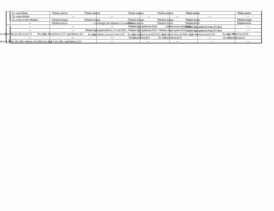
exte
nsor
s Ex. tarsi tibialis Tibialis anterior Tibialis anterior Tibialis anterior Tibialis anterior Tibialis anterior Tibialis anteriorEx. cruris tibialis --- --- --- --- --- ---Ex. cruris et tarsi fibularis Fibularis longus Fibularis longus Fibularis longus Fibularis longus Fibularis longus Fibularis longus
--- Fibularis brevis --- (seemingly not separate m. in monot.)Fibularis brevis Fibularis brevis Fibularis brevis Fibularis brevis
Shor
t ext
enso
rs --- --- --- Fibularis digiti quarti (me.4/d.4) --- (distinct some marsupials)Fibularis digiti quarti (me.4 rats; d.4 mice) ------ --- Fibularis digiti quinti (short ex. d. 5; me.5/d.5) Fibularis digiti quinti (me.5/d.5) Fibularis digiti quinti (d.5) Fibularis digiti quinti (me.4 rats; d.5 mice) ---
Exs. digito. breves (4m. to d.2-5) Exs. digito. breves (4 m. d. 2-5 + part short ex. d.1) Ex. digito. brevis (1 m. to d. 1-4 or 1-5?) Ex. digito. brevis (1 m. to d.2-3) Ex. digito. brevis (1m.; d.2-4) Ex. digito. brevis (1 m. to d. 2-3) Ex. digito. brevis (1 m. d.2-4)--- --- --- Ex.hallucis brevis (d.1) Ex. hallucis brevis (d.1) --- Ex. hallucis brevis (d.1)
Abd. et ex. digiti 1 (d.1; abd.+ short ex. d.1)Abd. et ex. digiti 1 (d.1; abd. + part short ex. d.1) --- --- --- --- ---