Running head: ARF-GAPs and root hair growth … · the formation of root hairs with multiple ......
46
1 Running head: ARF-GAPs and root hair growth Correspondence: Elison B. Blancaflor Plant Biology Division The Samuel Roberts Noble Foundation 2510 Sam Noble Parkway Ardmore, OK 73401 USA Tel (580) 224-6687 Fax (580) 224-6692 e-mail: [email protected] Journal Research Area: Focus Issue on Membrane Trafficking – Cell Biology Plant Physiology Preview. Published on June 4, 2008, as DOI:10.1104/pp.108.119529 Copyright 2008 by the American Society of Plant Biologists www.plantphysiol.org on June 16, 2018 - Published by Downloaded from Copyright © 2008 American Society of Plant Biologists. All rights reserved.
Transcript of Running head: ARF-GAPs and root hair growth … · the formation of root hairs with multiple ......

1
Running head: ARF-GAPs and root hair growth
Correspondence:
Elison B. Blancaflor Plant Biology Division The Samuel Roberts Noble Foundation 2510 Sam Noble Parkway Ardmore, OK 73401 USA Tel (580) 224-6687 Fax (580) 224-6692 e-mail: [email protected] Journal Research Area: Focus Issue on Membrane Trafficking – Cell Biology
Plant Physiology Preview. Published on June 4, 2008, as DOI:10.1104/pp.108.119529
Copyright 2008 by the American Society of Plant Biologists
www.plantphysiol.orgon June 16, 2018 - Published by Downloaded from Copyright © 2008 American Society of Plant Biologists. All rights reserved.
2
A class one ADP-ribosylation factor GTPase-activating protein is critical for
maintaining directional root hair growth in Arabidopsis thaliana1
Cheol-Min Yoo, Jiangqi Wen, Christy M. Motes, J. Alan Sparks and Elison B. Blancaflor*
Plant Biology Division, The Samuel Roberts Noble Foundation Inc., 2510 Sam Noble Parkway,
Ardmore, OK 73401, USA
www.plantphysiol.orgon June 16, 2018 - Published by Downloaded from Copyright © 2008 American Society of Plant Biologists. All rights reserved.

3
1 This work was supported by the Samuel Roberts Noble Foundation and in part by the
Oklahoma Center for the Advancement of Science and Technology (OCAST grant PSB08-003 to
E.B.B.). The confocal microscopes used in this study were acquired through Multi-User
Equipment and Major Research Instrumentation grants from the National Science Foundation
(DBI-0400580 and DBI-0722635 to E.B.B.).
* Corresponding author; e-mail [email protected]; fax 580-224-6692
The author responsible for distribution of materials integral to the findings presented in this
article in accordance with the policy described in the Instructions for Authors
(www.plantphysiol.org) is Elison B. Blancaflor ([email protected])
www.plantphysiol.orgon June 16, 2018 - Published by Downloaded from Copyright © 2008 American Society of Plant Biologists. All rights reserved.

4
Abstract
Membrane trafficking and cytoskeletal dynamics are important cellular processes that drive tip
growth in root hairs. These processes interact with multitude signaling pathways that allow for
the efficient transfer of information to specify the direction in which tip growth occurs. Here we
show that AGD1, a class one ADP ribosylation factor-GTPase activating protein (ARF-GAP), is
important for maintaining straight growth in Arabidopsis root hairs since mutations in the AGD1
gene resulted in wavy root hair growth. Live cell imaging of growing agd1 root hairs revealed
bundles of endoplasmic microtubules and actin filaments extending into the extreme tip. The
wavy phenotype and pattern of cytoskeletal distribution in root hairs of agd1 partially resembled
that of mutants in an armadillo-repeat containing kinesin (ARK1). Root hairs of double
agd1/ark1 mutants were more severely deformed compared to single mutants. Organelle
trafficking as revealed by a fluorescent Golgi marker was slightly inhibited and Golgi stacks
frequently protruded into the extreme root hair apex of agd1 mutants. Transient expression of
GFP-AGD1 in tobacco epidermal cells labeled punctate bodies that partially colocalized with the
endocytic marker, FM4-64, while ARK1-YFP associated with microtubules. Brefeldin A rescued
the phenotype of agd1 indicating that the altered activity of an AGD1-dependent ARF
contributes to the defective growth, organelle trafficking and cytoskeletal organization of agd1
root hairs. We propose that AGD1, a regulator of membrane trafficking and ARK1, a
microtubule motor, are components of converging signaling pathways that impact cytoskeletal
organization to specify growth orientation in Arabidopsis root hairs.
www.plantphysiol.orgon June 16, 2018 - Published by Downloaded from Copyright © 2008 American Society of Plant Biologists. All rights reserved.

5
Introduction
Cells that undergo tip growth provide an excellent experimental system to uncover fundamental
mechanisms underlying polarity establishment in plants. The most widely studied tip growing
cells in plants are root hairs and pollen tubes. In these cell types, expansion is restricted primarily
to the extreme apex of the cell leading to the formation of long tubular structures. Years of
research has established that tip growth is modulated by a number of cellular processes including
a dynamic cytoskeleton, tip-focused cytoplasmic calcium gradients and oscillations, and
vesicle/membrane trafficking events. These processes in turn are under tight regulation by an
elaborate network of signaling molecules such as reactive oxygen species (ROS), monomeric
guanine nucleotide binding proteins (small GTPases), mitogen-activated protein kinases
(MAPKs) and phosphoinositides (Cole and Fowler, 2006; Samaj et al., 2006; Campanoni and
Blatt, 2007; Kost, 2008).
An intriguing question in regard to studies on root hairs and pollen tubes is how the
directionality of tip growth is maintained. Whereas a change in growth direction in pollen tubes
occurs partly in response to chemotropic factors from the stigma (Kim et al., 2003), root hairs
maintain an endogenous polarity that guides the tip to grow straight and away from the primary
root body (Bibikova et al., 1997). In both cases, calcium appears to play an important role in
maintaining tip growth since artificially relocalizing the tip-focused cytoplasmic calcium
gradient using caged calcium ionophores resulted in a change in tip growth direction that often
followed the new calcium gradient (Malho and Trewavas, 1996; Bibikova et al., 1997).
In addition to cytoplasmic calcium, the cytoskeleton, which consists of microtubules,
actin filaments (F-actin) and several accessory proteins (Hussey et al., 2006; Blancaflor et al.,
2006), plays an essential role in regulating tip growth direction. The important role of the
cytoskeleton in this process is supported by the observation that exposure to microtubule
inhibitors or down-regulation of tubulin, the major protein subunit of microtubules, resulted in
the formation of root hairs with multiple tips and wavy growth (Bibikova et al., 1999; Bao et al.,
2001). Recently, root hairs of Arabidopsis knockouts to an armadillo-repeat containing kinesin,
displayed wavy and branched root hairs further establishing the link between microtubules and
the maintenance of root hair growth direction (Jones et al., 2006; Yang et. al., 2007; Sakai et al.
2008). The actin cytoskeleton also is essential for maintaining tip-focused growth. Actin
disrupting drugs inhibited or delocalized root hair growth (Bibikova et al., 1999; Ketelaar et al.,
www.plantphysiol.orgon June 16, 2018 - Published by Downloaded from Copyright © 2008 American Society of Plant Biologists. All rights reserved.

6
2003) consistent with reports showing that knockouts to the vegetative ACT2 gene in
Arabidopsis have distorted root hair morphology (Ringli et al., 2002). Moreover, altering the
expression of genes encoding proteins that impact actin turnover such as profilin, actin
depolymering factor, formin, actin interacting protein and cyclase associated protein disrupted
normal root hair development (Ramachandran et al., 2000; Dong et al., 2001; Yi et al., 2005;
Ketelaar et al., 2007; Deeks et.al., 2007).
In coordination with the cytoskeleton and calcium gradients, vesicle trafficking at the tip
contributes largely to membrane turnover and deposition of cell wall precursors necessary to
sustain the rapid growth of pollen tubes and root hairs. Several components of the vesicle
trafficking machinery have been implicated in tip growth including the Rab, Rho and ADP-
ribosylation factor (ARF) subfamilies of small GTPases (Cole and Fowler, 2006; Kost, 2008).
Small GTPases act as molecular switches that cycle between an active (GTP-bound) and inactive
(GDP-bound) conformation. In its active form, small GTPases interact with specific effector
proteins that modulate cytoskeletal organization, vesicle budding and docking, and protein
trafficking. Whereas conversion of the inactive to the active GTPase is facilitated by guanine
nucleotide exchange factors (GEFs), inactivation occurs through the action of GTPase activating
proteins (GAPs), which stimulate the hydrolysis of GTP to GDP (Bos et al., 2007).
The ROPs (for Rho of plants) and Rabs have been the most widely studied subfamilies of
small GTPases in regard to the control of tip growth in plants. ROPs and Rabs have been shown
to localize to the tips of root hairs and pollen tubes (Molendijk et al., 2001; Jones et al., 2002;
Preuss et al., 2004), and consistent with its proposed function in polarized growth, tip
localization of Rab4Ab was only observed in growing root hairs but not in mature root hairs that
have ceased growing or root hair mutants with defective tip growth (Preuss et al., 2004).
Furthermore, constitutively active or dominant negative ROPs altered tip growth resulting in the
formation of bulbous tips in pollen tubes (Kost et al., 1999; Li et al., 1999), and root hairs with
wavy growth, multiple tips and partially swollen tips (Molendijk et al., 2001; Jones et al., 2002;
Bloch et al., 2005). Disrupting ROP GTPase function by expression of constitutively active or
dominant negative ROP has been shown to impact apical F-actin dynamics, calcium signaling
and ROS production. In pollen tubes, ROPs appear to modulate F-actin dynamics and calcium
oscillations via effector proteins with CRIB (for Cdc42/Rac-interactive binding) motifs (Gu et
al., 2005). Moreover, Arabidopsis mutants in a ROP guanine dissociation inhibitor (GDI), a
www.plantphysiol.orgon June 16, 2018 - Published by Downloaded from Copyright © 2008 American Society of Plant Biologists. All rights reserved.

7
protein that prevents ROP activation by GEFs, have multiple tip growing sites in their root hairs.
This particular ROP GDI was demonstrated to function in root hair growth by restricting ROS
production to a single growing point (Carol et al., 2005).
Small GTPases also have been shown to coordinate tip growth via their interaction with
lipid signaling pathways. In Arabidopsis root hairs for example, RabA4b specifically interacts
with a phosphatidylinositol 4-OH kinase at the tip and this interaction has been proposed to be
relevant for maintaining the cytoplasmic calcium gradient that drives polar root hair growth
(Preuss et al., 2006). In this regard, inhibiting the production of lipid signaling molecules such as
phosphatidic acid (PA) disrupted tip growth, disorganized the actin cytoskeleton and dissipated
the tip-focused cytoplasmic calcium gradient (Potocky et al., 2003; Motes et al., 2005; Monteiro
et al., 2005). Also, a mutation in an Arabidopsis phosphatidylinositol transfer protein
compromised polarized growth in root hairs, which coincided with a disorganization of the
microtubule cytoskeleton (Vincent et al., 2005).
The ROP and Rab GTPases are themselves under tight regulation by other small GTPases
such as those belonging to the ARF subfamily. In Arabidopsis, the fungal macrolide Brefeldin A
(BFA), an inhibitor of ARF-GEFs, disrupted the polar localization of ROPs in root hairs
(Molendijk et al., 2001). Similarly, expression of the GTP-locked form of ARF1 induced the
formation of bulged root hairs, which occurred concomitantly with altered ROP localization (Xu
and Scheres, 2005). Recently, an Arabidopsis mutant in a class II ARF-GAP displayed deformed
root hairs and reduced growth of pollen tubes further implicating ARFs in tip growth
maintenance (Song et al., 2006).
It is clear that a number of interconnected signaling pathways and cellular processes are
required for the polarized delivery of vesicles and new cell wall material important for driving
tip growth in plants. It is likely, however, that additional molecular components of the tip growth
machinery remain to be identified. In an effort to uncover new players involved in maintaining
root hair growth directionality we conducted a forward genetic screen of T-DNA activation
tagged Arabidopsis seedlings to isolate root hair mutants with altered growth direction (Weigel
et al., 2000). Here we characterize two loss-of-function Arabidopsis mutants with wavy root hair
growth. We show that one mutant is disrupted in a gene that encodes a class I ARF-GAP (AGD1;
Vernoud et al., 2003) while the other mutant is a new allele of an armadillo repeat-containing
kinesin previously designated as MRH2 or ARK1 (Jones et al., 2006; Yang et. al., 2007; Sakai et
www.plantphysiol.orgon June 16, 2018 - Published by Downloaded from Copyright © 2008 American Society of Plant Biologists. All rights reserved.

8
al. 2008). AGD1 localized to punctate intracellular compartments that partially associated with
the endocytic marker, FM4-64, consistent with its function as a mediator of vesicle trafficking.
ARK1 on the other hand localized to microtubules consistent with its function as a microtubule
motor protein. Low concentrations of BFA that did not inhibit root hair growth reversed the
wavy root hair phenotype of agd1 mutants supporting the notion that AGD1 modulates root hair
growth direction, organelle trafficking and cytoskeletal organization through the activity of an
AGD1-dependent ARF substrate. The more severe root hair phenotype of double agd1/ark1
mutants compared to single mutants indicates that AGD1 and ARK1 are components of
independent but possibly intersecting signaling pathways that specify growth orientation in
Arabidopsis root hairs by impacting cytoskeletal dynamics at the tip.
RESULTS
Identification and Characterization of Arabidopsis Mutants with Altered Root Hair
Growth Direction
When germinated in semi-solid media, wild-type Arabidopsis root hairs typically grow straight
and away from the primary root axis (Fig. 1A; Supplemental Movie S1). This highly predictable
growth of Arabidopsis root hairs allows for rapid genetic screening of mutants with altered root
hair morphology (Parker et. al., 2000). We isolated two mutants from a T-DNA activation tagged
population in the Columbia (Col-0) background that exhibited wavy root hair growth (Fig. 1B,
C). When the mutants were backcrossed to wild type, the F1 progeny did not exhibit the wavy
root hair phenotype. In the F2 generation, the root hair phenotype for both mutants segregated 3
(wild-type) to 1(wavy) indicating that both mutants were recessive in a single Mendelian locus
(data not shown). We initially designated these two mutants as wrh1 and wrh2 (for wavy root
hair) but renamed them agd1-1 and ark1-4 respectively upon subsequent molecular identification
of the disrupted genes (see below).
After backcrossing two times to wild-type to remove possible extraneous mutations, we
conducted a more detailed microscopic analysis of the root hairs of wrh1(agd1) and wrh2(ark1).
In contrast to wild-type root hairs, which typically maintained straight growth (Fig. 1D;
Supplemental Movie S1), root hairs of wrh1(agd1) redirected their growth at almost regular
intervals leading to the wavy phenotype (Fig. 1E; Supplemental Movie S2). Although the
phenotype of wrh1(agd1) generally appeared wavy, other time-lapse movies revealed that some
www.plantphysiol.orgon June 16, 2018 - Published by Downloaded from Copyright © 2008 American Society of Plant Biologists. All rights reserved.

9
root hairs grew in a spiral manner (data not shown). Also, roots of wrh1(agd1) occasionally
exhibited two hairs originating from a single initiation site (Fig. 1F). Whereas some root hairs of
wrh2(ark1) displayed similar wavy growth as wrh1(agd1), wrh2 (ark1) had a greater tendency to
form root hairs with multiple tips and branches (Fig. 1G). Time-lapse imaging of growing root
hairs of wrh2 (ark1) revealed occasional cessation of tip growth, which was followed by gradual
broadening of the tip. The broader tip apex eventually gave rise to two or more tips that either
grew simultaneously or displayed alternate pulses of growth (Supplemental Movie S3). We
measured the growth rate of root hairs from 3-4 d old wild type and wrh mutants located at 500–
700 µm from the primary root tip since this is the region where root hairs are actively elongating.
The average growth rate of wild-type root hairs was 1.15±0.25 µm/min while the growth rate of
wrh1 and wrh2 was significantly less with values at 0.77±0.29 µm/min and 0.74±0.23 µm/min
respectively (values are means of 50-60 root hairs± SD, Student’s t-test; p<0.001).
Molecular Identification of the Disrupted Genes in the Root Hair Mutants
Examination of root hairs of the F1 progeny resulting from a cross between wrh1 and wrh2
showed wild-type phenotypes indicating that WRH1 and WRH2 were not allelic. We then
conducted thermal asymmetric interlaced (TAIL)-PCR to identify flanking T-DNA insertions in
the wrh mutants (Liu et al., 1995). We found that wrh2 had a T-DNA insertion in the 16th exon
of the gene At3g54870, which encodes an armadillo repeat-containing kinesin motor protein
(Fig. 2A). Two independent groups had previously identified mutants in this gene (Jones et al.,
2006; Yang et al., 2007; Sakai et al., 2008). These mutants were designated mrh2 but were
recently renamed ark1 (for armadillo repeat kinesin; Sakai et al., 2008). Since root hairs of the
previously described ark1 mutants exhibited similar wavy and multiple tip phenotypes as our
wrh2 mutant, it is likely that wrh2 is another allele of ARK1. This was confirmed by our
independent identification of two homozygous SALK lines with T-DNA insertions in the ARK1
gene (SALK_081412 and SALK_035063; Alonso et al., 2003). We found that root hairs from
these insertion lines exhibited similar phenotypes as wrh2 (data not shown). We therefore
renamed wrh2 as ark1-4 since 3 other ark1 mutants that included the homozygous SALK lines
we identified here, have already been described previously (Fig. 2A; Jones et al., 2006; Sakai et
al., 2008).
www.plantphysiol.orgon June 16, 2018 - Published by Downloaded from Copyright © 2008 American Society of Plant Biologists. All rights reserved.

10
Since our attempts to identify the disrupted gene in wrh1 using TAIL-PCR were
unsuccessful we tested for the presence of the T-DNA insert in this line using BASTA
(glufosinate ammonium) and found that wrh1 was not resistant. We therefore performed map-
based cloning to identify the WRH1 gene. We found that wrh1 had a deletion of 46 base pairs
spanning the splice donor site of exon 6 of the At5g61980 gene (Fig. 2A). Comparison of wild-
type and mutant WRH1 cDNA revealed a 31 base pair deletion resulting from aberrant splicing
of intron 6 that caused a frame-shift mutation that introduced a TAA stop codon after 7 irrelevant
amino acid residues (Supplemental Figure S1).
The At5g61980 gene encodes a class I ARF-GAP, which encompasses 19 exons and
encodes a predicted 850 amino acid protein (Fig. 2A). An analysis of the Arabidopsis genome
identified 15 ARF-GAP domain (AGD) proteins belonging to four classes. In addition to a GAP
domain, class I ARF-GAP proteins, which consist of 4 members, contain Bin1-amphiphysin-
Rvs167p/Rvs161p (BAR) and Pleckstrin Homology (PH) domains, and two ankyrin repeats.
At5g61980 was designated as AGD1 for ARF-GAP Domain containing protein 1 (Vernoud et al.,
2003). Previously, a closely related class I ARF-GAP (AGD3) was shown to be involved in
auxin efflux and vein patterning in Arabidopsis (Koizumi et al., 2005; Sieburth et al., 2006).
Functional analyses of single mutants in the other class I ARF-GAPs including mutations in the
AGD1 gene, however, did not reveal any apparent phenotype (Sieburth et al., 2006). Therefore,
to verify whether disruption of the AGD1 gene is indeed the cause of the wavy root hair
phenotype of wrh1, we searched the publicly available SALK collection for T-DNA insertions in
the At5g61960 gene (Alonso et al., 2003). We genotyped the segregating progeny by PCR and
identified homozygous plants for one SALK line (SALK_036034), which was similar to the
agd1 mutant reported by Sieburth et al (2006) and one SAIL line (SAIL_819_C10), which had
an insertion in the 13th exon of AGD1 (Fig. 2A). Root hairs of these two T-DNA insertion
mutants exhibited similar wavy phenotypes as wrh1(data not shown), suggesting that these were
alleles of wrh1. We therefore renamed wrh1 as agd1-1 while SALK_036034 and
SAIL_819_C10 were designated as agd1-2 and agd1-3 respectively (Fig. 2A). All of the
subsequent cellular analyses reported here were conducted with agd1-1 mutants. RT-PCR
analysis showed that agd1-2, agd1-3 and ark1-4 had no detectable transcript using primers
downstream of the T-DNA insertion. As noted agd1-1 being a frame shift mutant still produced a
transcript but this transcript is likely non-functional (Fig. 2B; Supplemental Figure S1).
www.plantphysiol.orgon June 16, 2018 - Published by Downloaded from Copyright © 2008 American Society of Plant Biologists. All rights reserved.

11
Sequencing of three independently cloned, PCR-amplified wild-type AGD1 cDNA
fragments showed a discrepancy with the National Center for Biotechnology Information
(NCBI)-annotated At5g61980 gene. From our sequencing results we found that the cDNA
encodes a predicted 828 amino acid protein, which differs from NCBI database prediction due to
the use of a GC splice acceptor site in intron 9 between the BAR and PH domain rather than the
predicted GT splice site (Supplemental Figure S1). This result is consistent with a previous
report that 1% of Arabidopsis introns have GC at their 5’ ends (Brown et al., 1996).
Cytoskeletal Organization is Disrupted in Growing Root Hairs of agd1-1 and ark1-4
The wavy root hair phenotype of agd1-1 and ark1-4 was reminiscent of root hairs treated with
microtubule inhibitors suggesting that microtubule organization could be impacted in these
mutants (Bibikova et al., 1999). Indeed, Sakai et al. (2008) and Yang et al. (2007) recently
reported that microtubule organization was altered in root hairs of other ark1 alleles. We
therefore asked whether root hairs of agd1-1 also had disrupted cytoskeletal organization since
both agd1and ark1 often exhibited similar wavy root hair phenotypes. We crossed agd1-1 and
ark1-4 with Arabidopsis plants expressing a green fluorescent protein (GFP) construct that label
microtubules (GFP-MBD; Marc et al., 1998) and F-actin (GFP-ABD2-GFP; Wang et al., 2008),
and analyzed cytoskeletal organization in growing root hairs using confocal microscopy (Fig. 3-
5).
We observed that the extent of cytoskeletal disruption in the agd1-1 and ark1-4 mutants
was somewhat dependent on the severity of the root hair phenotype. A projection of several
optical sections from a wild-type root hair expressing GFP-MBD showed predominantly
longitudinal microtubules consistent with a previous report (Van Bruaene et al., 2004). On the
other hand root hairs of agd1-1 that displayed regular wavy growth accumulated thick
microtubule bundles along their length (Fig. 3A). In addition, root hairs of agd1-1 that formed
branches had thick bundles of microtubules close to the point of branching. Root hairs of agd1-1
that displayed a milder wavy phenotype, however, contained predominantly longitudinal
microtubules and a low incidence of thick microtubule bundles (Fig. 3B). Similar observations
were made in root hairs of the ark1-4 mutant. Thick microtubule bundles were observed at the
tips of root hairs that had changed their growth direction and those that developed a broad tip
(Fig. 3C, D). Differences in microtubule organization between wild-type and agd1-1 or ark1-4
www.plantphysiol.orgon June 16, 2018 - Published by Downloaded from Copyright © 2008 American Society of Plant Biologists. All rights reserved.

12
were most apparent in single optical sections and time-lapse confocal movies of endoplasmic
microtubules of growing root hairs. Whereas bundles of endoplasmic microtubules in wild-type
root hairs were generally less dense, root hairs of both agd1-1 and ark1-4 mutants displayed
thick bundles of endoplasmic microtubules at the tip (Fig. 3E; Supplemental Movies S4-6).
Since the actin cytoskeleton is also relevant for sustaining tip growth, we next examined
whether the organization of F-actin is affected in root hairs of agd1-1 and ark1-4 mutants. When
decorated with the GFP-ABD2-GFP reporter described in Wang et al. (2008), F-actin bundles in
growing wild-type root hairs are typically oriented parallel to the length of the root hair. These
thick bundles of longitudinal F-actin did not extend into the apical 10-15 µm of growing root
hairs. Within the apical region, labeling with the GFP-ABD2-GFP reporter was typically diffuse
(Wang et al., 2008; Fig. 4A; Supplemental Movie S7). In agd1-1 root hairs, F-actin organization
was clearly disorganized. The extent of actin disruption also was partly dependent on the severity
of the root hair phenotype. For example, in growing root hairs with regular waves, the apical 10-
15 µm of the tip was devoid of distinct F-actin bundles similar to wild-type root hairs. Highly
fluorescent longitudinal actin cables however, appeared to congregate at the center of the root
hair cell (Fig. 4B). In root hairs with more compressed waves and strong spiral growth, F-actin
bundles typically followed the contour of the cell, and distinct F-actin cables were observed to
protrude into the extreme tip (Fig. 4C, D; Supplemental Movie S8). Similar disruptions in F-
actin organization were detected in ark1-4 mutants such that distinct F-actin bundles often
extended into the extreme apex of swollen or branched root hairs (Fig. 4E, F).
To quantify differences in F-actin organization in wild-type and mutant root hairs, we
took the ratio of the average fluorescence intensity of the apical 20 µm of growing root hairs and
a 30 µm area basal to the tip region (inset in Fig. 5A). Compared to wild-type, the average
fluorescence ratio in agd1-1 and ark1-4 mutants was higher at all time points measured (Fig.
5A). Furthermore, measurements from an extremely wavy root hair of agd1-1 revealed strong
oscillations in fluorescence ratio compared to a wild-type root hair (Fig. 5B, C). Our tip to basal
fluorescence ratio measurements were consistent with the observation that distinct actin bundles
occasionally extended into the extreme root hair tips of agd1-1 and ark1-4 mutants.
Other evidence indicative of altered actin dynamics in agd1-1 and ark1-4 mutants was
obtained using spinning disk confocal microscopy. Previously, we were able to augment our
ability to image dynamic F-actin in Arabidopsis seedlings by a simple modification to the
www.plantphysiol.orgon June 16, 2018 - Published by Downloaded from Copyright © 2008 American Society of Plant Biologists. All rights reserved.

13
ABD2-GFP construct (Wang et al., 2008). We asked whether spinning disk confocal
microscopy, which significantly minimizes sample bleaching, combined with the enhanced
fluorescence brought about by our improved GFP F-actin reporter, would allow us to observe the
finer details of actin dynamics at the tip of growing wild-type and mutant root hairs. We obtained
time-lapse sequences of wild-type and mutant root hairs at 300 msec intervals. Despite the short
time interval between image collection we did not observe bleaching of GFP labeled F-actin in
the root hairs. In fact, the combination of spinning disk confocal microscopy and the enhanced
fluorescence of our improved GFP-ABD2-GFP construct allowed us to resolve highly dynamic
fine F-actin at the tip of growing wild-type root hairs, which was typically more difficult to
resolve with conventional point scanning confocal microscopy (Supplemental Movie S9). In
agd1-1 root hairs that exhibited extreme wavy growth, actin bundles at the tip although still
highly dynamic, were generally thicker than the fine F-actin arrays characteristic of wild-type
root hairs (Supplemental Movie S10). Similar observations were made in wavy and branched
root hair tips of ark1-4 mutants (data not shown).
Organelle Motility in agd1 Root Hairs
As mutants in the ARK1 gene have already been described recently (Yang et al., 2007; Sakai et
al., 2008), we focused our efforts on further characterizing the cellular phenotypes of agd1 root
hairs. Since root hairs of agd1-1 had obvious defects in cytoskeletal dynamics, we asked whether
other cellular processes that are dependent on normal cytoskeletal function were impacted. One
important process regulated by the cytoskeleton is organelle transport. The movement of Golgi
stacks in root hairs for example, is regulated by class XI myosin motors that facilitate their
transport along F-actin (Peremyslov et al., 2008). To evaluate whether the AGD1 mutation
affected organelle trafficking in root hairs, we crossed agd1-1mutants with plants expressing a
yellow fluorescent protein (YFP) fused to the rat sialyl-transferase transmembrane domain (ST-
YFP), which is used to fluorescently mark Golgi stacks in plants (Saint-Jore et al., 2002). There
were no obvious gross morphological differences in the appearance of Golgi stacks between
growing wild-type and agd1-1 root hairs (Fig. 6A). Furthermore, spinning disk confocal
microscopy of growing wild-type and agd1-1 root hairs expressing ST-YFP revealed that Golgi
stacks were highly dynamic along the length of the root hair (Supplemental Movies S11,12).
Using Volocity classification software (Peremyslov et al., 2008), we tracked individual Golgi
www.plantphysiol.orgon June 16, 2018 - Published by Downloaded from Copyright © 2008 American Society of Plant Biologists. All rights reserved.

14
stacks from the apical 20 µm and the basal region of the root hair as indicated by the white boxes
in Fig. 6A to obtain Golgi velocity measurements. In both wild-type and agd1-1, the average
velocity of Golgi stacks differed significantly between the apical and basal regions of the root
hair with mean velocity in the tip region being about 2 fold less than Golgi stacks at the basal
region (Fig. 6B). There were no significant differences in the velocity of Golgi stacks at the tip
region between wild-type and agd1-1. In the basal region however, the average velocity of Golgi
stacks in agd1-1 was slightly reduced compared to wild-type and this difference was statistically
significant (Fig. 6B; Student’s t-test; p<0.005). More importantly, time-lapse spinning disk
confocal microscopy revealed that Golgi stacks in root hairs of agd1-1 occasionally extended
into the root hair tip whereas Golgi stacks in wild-type root hairs maintained approximately a 10
µm distance from the extreme apex (Fig. 6A; Compare Supplemental Movies S11 and S12).
Brefeldin A (BFA) Rescues the Wavy Root Hair Phenotype and Cytoskeletal Defects of
agd1
Given that AGD1 encodes a class I ARF-GAP, we hypothesized that root hair growth in agd1
might be differentially affected by inhibitors of vesicle trafficking. One compound that has been
used extensively for such studies is the fungal macrolide, BFA. BFA is known to inhibit the
activity of ARF-GEFs in mammalian cells and there is evidence that similar targets for BFA
exists in plants (Nebenfuhr et al., 2002). We found that 100 nM BFA did not inhibit root hair
growth of wild-type seedlings but BFA concentrations above 200 nM did. Strikingly, we found
that average root hair length of agd1-1 increased significantly when germinated in 100 nM BFA
(Fig. 7A). More importantly, the increase in average root hair length of agd1-1 mutants upon
exposure to 100 nM BFA coincided with root hairs reverting to the straight growth pattern
characteristic of wild-type root hairs (Fig. 7B, C). We quantified the effect of BFA on root hair
waviness by measuring the angle of tip deviation from the main axis of the root hair (inset in Fig.
7B). The average angle of tip deviation in root hairs of agd1-1 decreased from 19 to 3 degrees
when grown on 100-200 nM BFA, which was similar to that of untreated and BFA-treated wild-
type root hairs (Fig. 7B). Furthermore, confocal microscopy showed that the abundant bundles
of endoplasmic microtubules and F-actin in the tips of agd1-1 root hairs were reduced by
incubation in BFA (Fig. 7F; Supplemental Movie S13). When BFA was washed out, agd1-1 root
hairs reverted back to the wavy growth pattern (data not shown).
www.plantphysiol.orgon June 16, 2018 - Published by Downloaded from Copyright © 2008 American Society of Plant Biologists. All rights reserved.

15
To quantify the effect of BFA on cytoskeletal organization we focused on studying
further the bundling of endoplasmic microtubules at the root hair tip since means to evaluate this
population of microtubules have been established recently (Sakai et al., 2008). We projected Z-
stacks obtained with the spinning disk microscope using Volocity classification software to
generate cross-sections at a region 5 µm from the root hair tip (Fig. 7H). Mean fluorescence
intensity ratios of endoplasmic and cortical microtubules from these computer generated cross-
sections were obtained following the methods of Sakai et al. (2008). The ratio of endoplasmic to
cortical microtubule fluorescence in agd1-1 root hairs was higher than that of wild-type (Fig. 7I)
consistent with visual observations of abundant endoplasmic microtubules at the root hair tip of
agd1-1 (Fig. 3E; 7H; Supplemental Movie S5). When grown on 100 nM BFA, the mean ratio of
endoplasmic to cortical microtubule fluorescence in agd1-1 root hairs decreased to a value
slightly less than that of wild-type (Fig. 7I). These measurements were consistent with visual
observations that endoplasmic microtubule organization in untreated agd1-1 root hairs reverted
to that of wild-type root hairs after BFA treatment (Fig. 7H; Supplemental Movie S13). 100-200
nM BFA did not affect the overall organization of Golgi bodies in wild-type root hairs (data not
shown). However, many agd1-1 root hairs treated with BFA displayed wild-type Golgi
distribution consistent with their reversal to straight growth (Supplemental Movie S14).
We then asked whether BFA had similar effects on the root hair phenotype of the ark1-4
mutant. In contrast to agd1-1, root hair length of ark1-4 did not increase in response to BFA
(Fig. 7A). Although the wavy phenotype of ark1-4 was somewhat dampened by BFA as evident
from a reduction in the frequency of waves along the root hair, ark1-4 root hairs, unlike agd1-1,
did not completely revert to the wild-type phenotype. When incubated in BFA many root hairs of
ark1-4 still contained branches and occasional kinks at the tip (Fig. 7D). The partial rescue of
ark1-4 root hairs by BFA was manifested as a reduction in the angle of root hair tip deviation
from 17 degrees to 9 degrees (Fig. 7B). Furthermore, in contrast to agd1-1, abundant F-actin and
endoplasmic microtubule bundles in root hair tips of ark1-4 persisted despite BFA treatment
(Fig. 7G- I).
Like single mutants, when agd1/ark1 double mutants were examined they exhibited no
other obvious plant phenotype (data not shown). However, we found that root hair growth of
agd1/ark1 double mutants was more strongly inhibited compared to single mutants. The average
root hair length of agd1/ark1 was only about 20 % of controls and 25% of single mutants (Fig.
www.plantphysiol.orgon June 16, 2018 - Published by Downloaded from Copyright © 2008 American Society of Plant Biologists. All rights reserved.
16
7A). Furthermore, root hairs of agd1/ark1 double mutants were more severely deformed as
evident from the formation of root hairs with short branches that ceased elongating soon after
initiation (Fig. 7E). The tips of these short root hairs also had the tendency to bend resulting in a
tip angle of deviation comparable to that of single agd1 or ark1 mutants (Fig. 7B). When
germinated in 100-200 nM BFA, average root hair length of agd1/ark1 double mutants increased
by about 50% (Fig. 7A, B) but root hairs were still characterized by swollen bases, kinks and
waves (Fig. 7B,E). The tip angle of deviation of agd1/ark1 double mutants particularly after
incubation in 200 nM BFA was similar to that of ark1-4 single mutants (Fig. 7B).
AGD1 Partly Localizes to the Endocytic Marker, FM4-64 and ARK1 Associates with
Microtubules
Co-sedimentation assays have shown that the N-terminal motor domain of ARK1 binds strongly
to polymerized tubulin and the C-terminal armadillo repeat binds weakly to polymerized actin
(Yang et al., 2007). The in vivo localization of ARK1 however, has not yet been reported. We
therefore created translation fusions of yellow fluorescent protein (YFP) to the C-terminus of full
length ARK1 under the control of the 35S cauliflower mosaic virus (35S) promoter (35S::ARK1-
YFP). This construct was biolistically bombarded into tobacco leaf epidermal cells and
transformed cells were imaged using confocal microscopy. The 35S::ARK1-YFP construct
localized to filamentous structures that were reminiscent of microtubules (Fig. 8A). To verify
that the 35S::ARK1-YFP-labeled filaments were microtubules, leaf cells were incubated in either
oryzalin or latrunculin B, which disrupt microtubules and F-actin respectively. Incubation in
oryzalin but not latrunculin B caused the dissipation of the filamentous structures in ARK1-YFP
expressing cells indicating that ARK1 indeed localized to microtubules (Fig. 8B and data not
shown).
We also created GFP translational fusions to the full length AGD1 protein under the
control of the 35S promoter. The 35S::GFP-AGD1 fusion transiently expressed in tobacco
epidermal cells decorated punctate structures of varying sizes that resembled Golgi stacks (Fig.
8C). To determine whether these fluorescent structures represented Golgi stacks, we co-
bombarded the GFP-AGD1 with mCherry-G-rk, which localizes to Golgi based on a fusion with
the transmembrane domain of α-1,2-mannosidase I (Nelson et al., 2007). We also incubated
leaves that were agroinfiltrated with GFP-AGD1 with the lipophilic styryl dye, FM-4-64, which
www.plantphysiol.orgon June 16, 2018 - Published by Downloaded from Copyright © 2008 American Society of Plant Biologists. All rights reserved.
17
labels diverse components of the endocytic pathway (Müller et. al., 2008). We found that the
punctate GFP-AGD1 structures partially overlapped with FM4-64-labeled vesicles but not Golgi
stacks (Fig. 8C, D).
DISCUSSION
A forward genetics approach enabled us to identify two Arabidopsis mutants with wavy root hair
growth but with no other obvious phenotype. The phenotype of these mutants was reminiscent of
root hairs treated with microtubule disrupting drugs (Bibikova et al., 1999) prompting us to
hypothesize that the genetic lesions in these mutants might encode for proteins that are important
for cytoskeletal regulation. Indeed TAIL-PCR revealed that one of our mutants had a T-DNA
insertion in ARK1, which encodes a kinesin microtubule motor protein. At the time we were
characterizing our mutants, two groups had independently isolated mutants in the ARK1 gene,
which showed similar defects in root hair morphology as our mutants (Yang et al., 2007; Sakai et
al., 2008). Although in vitro biochemical studies demonstrated that the N-terminal motor domain
of ARK1 binds to microtubules (Yang et al., 2007), our transient localization of ARK1-YFP to
oryzalin-sensitive filaments provides supporting evidence that ARK1 is a microtubule associated
protein in vivo (Fig. 8A, B).
Map based cloning of our other wavy root hair mutant revealed a disruption in AGD1, a
gene encoding an ARF-GAP (Vernoud et al., 2003). ARF-GAPs are negative regulators of
ARFs, small GTP binding proteins that mediate the formation of membrane trafficking
intermediates in the cell. In the classical model of ARF function, the active ARF bound GTP
recruits coat proteins to membranes to form protein coated vesicles. Hydrolysis of GTP by ARF-
GAP inactivates ARF triggering the dissociation of coat proteins, which then allows vesicles to
fuse with acceptor membranes (Nie and Randazzo, 2007). In Arabidopsis, ARF-GAPs belong to
a 15 member family that is further subdivided into four classes (Vernoud et al., 2003). AGD1
encodes a class I ARF-GAP, which is closely related to the multi-domain containing mammalian
AZAP-type ARF-GAPs (Vernoud et al., 2003; Inoue and Randazzo, 2007). Among the
Arabidopsis class I ARF-GAPs, only AGD3 has been demonstrated to function in plant
development by impacting auxin dependent vascular differentiation (Koizumi et al., 2005;
Sieburth et al., 2006). Also, the overexpression of a rice ARF-GAP (OsAGAP) hampered root
development by interfering with the normal trafficking of auxin influx carriers (Zhuang et al.,
www.plantphysiol.orgon June 16, 2018 - Published by Downloaded from Copyright © 2008 American Society of Plant Biologists. All rights reserved.

18
2006). Here we show that an additional function for class I ARF-GAPs in plants is in the
maintenance of root hair growth directionality. The identification of two independent SALK T-
DNA insertion alleles for AGD1 that exhibited wavy root hair phenotypes indicates that altered
AGD1 expression is the cause of the root hair defect in our mutant and not due to secondary
mutations elsewhere in the genome.
The function of AGD1 in tip growth is likely specific to root hairs since we did not
observe any defects in pollen germination or pollen tube growth in agd1 mutants (data not
shown). In a study of expression patterns of the four class I ARF-GAPs using real time RT-PCR,
AGD1 was not detected in hypocotyls and cotyledons, and had very low expression in roots,
siliques and leaves (Sieburth et al., 2006). The low expression of AGD1 is also apparent from an
in silico analysis of AGD1 from the Genevestigator database, which collates data from
Arabidopsis affymetrix gene chip experiments (Zimmerman et al., 2004; data not shown). As
speculated by Sieburth et al. (2006), the low abundance of AGD1 likely reflects its function in
only a few cell types and the phenotype we observed in agd1 mutants strongly indicates that
AGD1 might operate specifically in root hairs. It was shown recently that AGD10, a class II
ARF-GAP also is important for tip growth in Arabidopsis root hairs. The root hair phenotype of
agd10, however, was different from that of agd1 in that agd10 had very short bulbous root hairs
and also exhibited defects in pollen germination (Song et al., 2006). This indicates that some of
the mechanisms in which AGD10 regulates tip growth in plants might be distinct from that of
AGD1. AGD10 is more closely related to the mammalian ARF-GAP1, which has a simpler
domain organization than the AZAP class of ARF-GAPs to which AGD1 belongs. AGD10 and
AGD7, another class II ARF-GAP, only contain a GAP domain, and localize to Golgi stacks
(Vernoud et al., 2003; Song et al., 2006; Min et al., 2007) in agreement with mammalian ARF-
GAP1 localization (Liu et al., 2005). AGD1 and AGD3 on the other hand partially localized to
FM4-64 labeled endocytic vesicles and the trans-Golgi network (Fig. 8C; Koizumi et al., 2005).
The partial localization of AGD1 to FM4-64 labeled bodies that we observed here is consistent
with the endosomal localization of the closely related animal AZAPs (Jackson et al., 2000; Nie et
al., 2002). The differences in localization, domain organization and possibly ARF substrate
preference between AGD1 and AGD10 could likely explain the different nature of tip growth
defects caused by their respective mutants. Whereas AGD10 activates the GTPase hydrolyzing
www.plantphysiol.orgon June 16, 2018 - Published by Downloaded from Copyright © 2008 American Society of Plant Biologists. All rights reserved.

19
activity of plant ARF1 (Song et al., 2006), the precise plant ARF proteins that are substrates of
the class I ARF-GAPs remain to be determined.
AGD3, which shares similar domain organization to AGD1, has been shown to activate
the GTPase hydrolyzing activity of yeast ARF1p (Koizumi et al., 2005). Although we have yet to
assay for AGD1 GAP activity, the possibility that the root hair phenotype of agd1 results from
modified endogenous ARF function is evident from our results with BFA. Low concentrations of
BFA not only caused a statistically significant increase in root hair length but also caused wavy
root hairs of agd1 to grow straight and revert to wild-type cytoskeletal organization (Fig. 7). A
likely explanation for this observation is that loss of AGD1 function leads to an increase in the
active GTP bound form of an AGD1-dependent ARF in the cell. Since BFA is known to inhibit
endogenous ARF-GEF activity, which activates ARF by facilitating the exchange reaction of
GDP for GTP (Bos et al., 2007), BFA treatment could conceivably cause the build-up of GDP
bound (inactive) ARF in the cell. Therefore, despite the absence of AGD1 activity, agd1 root
hairs treated with BFA reverted to straight growth because the opposing biochemical reaction
triggered by a yet to be identified BFA sensitive ARF-GEF in the cell was inhibited. In this
regard, it is worth noting that AGD3 knockouts, which display discontinuous venation patterns
(Koizumi et al., 2005; Sieburth et. al., 2006) have roughly opposite phenotypes to mutants of the
ARF-GEF, GNOM, which display extensive venation (Geldner et al., 2003). If the effect of BFA
on agd1 root hairs is due to inhibited ARF-GEF activity counteracting the loss of AGD1, one
would predict that overexpression of a constitutively active AGD1-dependent ARF will lead to
similar wavy root hair phenotypes as agd1. Arabidopsis has a number of ARF and ARF-like
proteins, some of which are expressed in roots (Genevestigator, Zimmerman et al., 2004). Future
studies will attempt to identify which among these ARF or ARF-like proteins are substrates of
AGD1.
Since the overexpression of dominant negative ROP2 induced the formation of wavy root
hairs similar to that of agd1 mutants (Jones et al., 2002) and modified ARF1 activity disrupted
normal root hair development by inhibiting the polar localization of ROP2 (Xu and Scheres,
2005), the root hair phenotype of agd1 could partly be explained by altered ROP signaling. Also,
RabA4b, whose root tip localization is actin-dependent (Preuss et al., 2004), might be
misdirected in agd1. Interestingly, ROP2-induced root hair defects were accompanied by
disrupted apical F-actin organization similar to that observed in agd1 mutants (Fig. 4,5; Jones et
www.plantphysiol.orgon June 16, 2018 - Published by Downloaded from Copyright © 2008 American Society of Plant Biologists. All rights reserved.
20
al., 2002). Collectively, these observations reinforce the concept of cross-talk between the
different small GTPases in defining the highly polarized growth of root hairs by impinging on
cytoskeletal function (Xu and Scheres, 2005; Yang et al., 2007). Such cross-talk with other
signaling molecules is likely facilitated by the other protein domains of AGD1. For instance, the
PH domain of class I ARF-GAPs as demonstrated in the closely related AGD3, binds to
phosphatidylinositol-4,5-bisphosphate (Koizumi et al., 2005). In mammalian systems, GAP
activity of the AZAP type ARF-GAPs is stimulated by phosphoinositide binding to the PH
domain (Jackson et al., 2000). In this regard, over-expression or down regulation of a type B
phosphatidylinositol-4-phosphate 5-kinase 3, which regulates endogenous phosphatidylinositol-
4,5-bisphosphate levels have deformed root hairs (Stenzel et al., 2008; Kusano et. al., 2008).
Further support for the interdependence of lipid signaling and small GTPases in root hair growth
comes from the observation that RabA4b recruits phosphatidyl 4-OH kinase to the growing root
hair tip and knockouts of this gene induced the formation of wavy root hairs similar to agd1
(Preuss et al., 2006). More recently, the root hair defective 4 (rhd4) mutant, which has short,
branched and bulged root hairs, was shown to be disrupted in a gene encoding a
phosphoinositide phosphatase that regulates the levels of phosphatidylinositol-4-phosphate
(Thole et al., 2008). One possible effect of such changes in steady state levels of
phosphoinositides is a modification in the activity of AGD1. For technical reasons, we have been
unsuccessful in our initial attempts to express recombinant AGD1 in a heterologous system to
assay for lipid binding. Identification of the type and specificity of phosphoinositide binding to
AGD1 will shed light on how AGD1 interacts with lipid signaling to specify polarized root hair
growth. We are currently crossing our agd1 mutants with fluorescent markers for RabA4b and
ROPs to determine whether targeting of these tip growth effectors are impacted in the wavy root
hairs of agd1.
In animal cells, AZAP-type ARF-GAPs by virtue of their effects on membrane
trafficking induce a remodeling of the actin cytoskeleton (Nie et al., 2002; Randazzo and Hirsch,
2004; Randazzo et al., 2007). Modification in membrane structure by AZAP has been shown to
be mediated in part by the BAR domain at the N-terminus of the protein (Nie et al., 2006) and
proposed to facilitate binding of other classes of small GTPases (Habermann, 2004). Here, live
cell imaging allowed us to gain additional insight into the underlying cytoskeletal defects that led
to the root hair phenotype of agd1. Extensive bundles of F-actin periodically extended into the
www.plantphysiol.orgon June 16, 2018 - Published by Downloaded from Copyright © 2008 American Society of Plant Biologists. All rights reserved.

21
extreme apex of growing agd1 root hairs (Fig. 4, 5; Supplemental Movie S8, S10). This pattern
of cytoskeletal reorganization was in contrast to that of growing wild-type root hairs, which
typically display a region at the tip with diffuse or fine arrays of F-actin (Wang et al., 2008;
Supplemental Movie S7, S9). The abnormally high levels of F-actin bundles have a number of
consequences that could explain the root hair phenotype of agd1. One possibility is the
disruption of organelle trafficking in the mutant root hair. Indeed, agd1 displayed Golgi stacks
extending into the very tip of growing root hairs and slightly reduced Golgi stack velocity at the
root hair base (Fig. 6). Recently, knockouts to a class XI myosin, an actin based molecular
motor, resulted in short root hairs and reduced Golgi motility (Peremyslov et al., 2008). The
impact of the AGD1 mutation on Golgi trafficking (i.e. the protrusion of Golgi stacks into the
extreme root hair apex) was similar to that observed in depolarized pollen tubes expressing a
catalytically inactive phospholipase C (Dowd et al. 2006). Movement of organelles to regions of
growing root hairs where they are typically excluded could impact targeted delivery and
localized recycling of vesicles leading to differential tip growth (Ovecka et. al., 2005).
Alternatively, disrupted F-actin dynamics could interfere with the tip recruitment of lipid
signaling components that bind to actin such as phosphatidylinositol phosphate kinase 1(Davis et
al., 2007) or trafficking of RHD2, the ROS generating NADPH oxidase, which has recently been
shown to be dependent on actin for proper root hair tip localization (Takeda et al., 2008). As
alterations in the metabolism of phosphoinositide lipid mediators have been shown to impact
root hair growth (Bohme et al., 2004; Vincent et al., 2005; Thole et al., 2008; Stenzel et al.,
2008; Kusano et. al., 2008), the disruption of apical F-actin dynamics at the root hair tip whether
or not it is a direct result of loss of AGD1 function, continue to point to an elaborate system of
feedback regulation between lipids, small GTPases and the cytoskeleton in the maintenance of
tip growth (Nibau et al., 2006; Kost, 2008).
Another consequence of the loss of AGD1 function was a modification in microtubule
organization as evident from thick bundles of endoplasmic microtubules at the root hair tip of
agd1. Intriguingly, the pattern of microtubule distribution in agd1 was similar to that of ark1 root
hairs confirming previous observations in other ark1 alleles (Fig. 3; Supplemental Movie S6;
Sakai et al., 2008). In Arabidopsis root hairs, endoplasmic microtubules have been proposed to
function as microtubule nucleation complexes (Van Bruaene et al., 2004). Thus, ARK1 and
AGD1 could conceivably be part of a signaling pathway that regulates microtubule nucleation
www.plantphysiol.orgon June 16, 2018 - Published by Downloaded from Copyright © 2008 American Society of Plant Biologists. All rights reserved.

22
and their altered activity results in abnormally high levels of polymerized tubulin at the root hair
tip leading to unstable growth. The extensive bundles of endoplasmic microtubules of agd1
mutants could also explain the abnormal Golgi movement at the root hair tip. In this regard, it
was shown that Golgi stacks can interact with both microtubules and actin, and that certain
kinesins facilitate the dispersal of Golgi stacks in plant cells (Lu et al., 2005). Since ark1 root
hairs exhibited bundled arrays of F-actin at the tip similar to agd1, both AGD1 and ARK1 may
have common downstream targets that regulate cytoskeletal dynamics. The more severe root hair
phenotype of double agd1/ark1 mutants, however, indicate that ARK1 and AGD1 are
components of separate but possibly convergent signaling pathways that impinge on cytoskeletal
function to specify root hair growth orientation. The complete reversal of the wavy root hair
phenotype of agd1 by low doses of BFA suggests that AGD1 impacts tip growth and the
cytoskeleton via a BFA-sensitive pathway involving unknown ARFs. On the other hand since
BFA did not completely reverse the root hair and cytoskeletal phenotypes of the ark1, the
pathway in which ARK1 modulates tip growth and cytoskeletal organization occurs in part via a
BFA independent process. It is possible that AGD1 by virtue of its impact on membrane and
actin remodeling indirectly modulates ARK1 trafficking since ARK1 was shown to bind F-actin
in vitro (Yang et al. 2007). Such an effect on ARK1 trafficking in agd1 could in turn affect
microtubule polymerization explaining the very similar nature of endoplasmic microtubule
bundling at the root hair tips of both agd1 and ark1 mutants. Additional studies using functional
ARK1 or AGD1 fluorescent protein fusions will be needed to determine the manner in which
ARK1 and AGD1 interact to specify root hair orientation. In the future it will be important to
compare cytoskeletal organization in root hairs treated with cytoskeletal disrupting drugs to that
of agd1 and ark1 root hairs to determine whether the cytoskeletal defects in the mutant root hairs
are the direct result of loss of AGD1 or ARK1 function.
In summary, we present new evidence showing that AGD1, a class I ARF-GAP protein,
is essential for maintaining straight root hair growth in Arabidopsis. One function of AGD1 is in
the maintenance of normal cytoskeletal turnover in the root hair and this process is facilitated by
a BFA-dependent pathway involving ARF and ARF-GEF proteins. We propose that the multi-
domain structure of AGD1 play similar roles in plants as their animal AZAP type ARF-GAP
counterparts, by mediating cross-talk and feedback regulation between phosphoinositides, small
GTPases and the cytoskeleton in polarized root hair growth.
www.plantphysiol.orgon June 16, 2018 - Published by Downloaded from Copyright © 2008 American Society of Plant Biologists. All rights reserved.

23
MATERIALS AND METHODS
Isolation of Root Hair Mutants with Altered Growth Direction and Molecular
Identification of the Disrupted Genes
An Arabidopsis thaliana Columbia (Col-0) T-DNA mutant seed stock, CS31100, from the
Arabidopsis Biological Resource Center (ABRC, Columbus, Ohio) was used to screen root hairs
with altered growth direction. Upon confirmation of the root hair phenotype in the progeny,
mutants were backcrossed to Col-0 at least two times. The gene of the T-DNA insertion in
wrh2(ark1) was identified by TAIL PCR essentially as described in Liu et al. (1995).
For map-based cloning, homozygous wrh1(agd1) was crossed to Ler ecotype. wrh1
(agd1) mutant plants from the resulting F2 seedlings were phenotyped based on the occurrence of
wavy root hairs. Publicly available Simple Sequence Length Polymorphisms (SSLP) and
Cleaved Amplified Polymorphic Sequences (CAPS) markers were used to map the wrh1(agd1)
loci (Lukowitz et al. 2000). We mapped the WRH1 locus to a 95kb region on chromosome 5.
Candidate gene sequencing revealed a 46 bp deletion in the annotated gene At5g61980
(Supplemental Figure S1). There were no other mutations in this region. Additional T-DNA
knockout lines namely agd1-2 (SALK_036034), agd1-3 (SAIL_819_C10), ark1-1
(SALK_081412) and ark1-2 (SALK_035063) were obtained from the ABRC.
Complementary DNA Isolation and Semi-quantitative RT-PCR Analysis
Total RNA was extracted from roots of 2-week-old wild type Arabidopsis (Col-0) and mutant
agd1 seedlings using the RNeasy plant mini-kit (Qiagen). Reverse transcription was carried out
using 500 ng of total RNA with the Omniscript RT kit (Qiagen) following the manufacturer’s
protocol to get a pool of cDNAs. The full length wild type AGD1 and mutant agd1 cDNA was
amplified using gene-specific primers (see below), and sequenced completely.
For semi-quantitative RT-PCR, root cDNA was prepared from the wild-type and mutants
as described above. Primers used for AGD1 were AGD1-RT-f, 5’-
AAGGTTGCAGAAAATACAC-3’ and AGD1-RT-r, 5’-TGATCCTGTGCATTCTCTGC-3’, to
generate a 1,135-bp product. For ARK1 the primers used were ARK1-RT-f, 5’-
AGGATCAGCAGAATCTGGAGCTC-3’ and ARK1-RT-r, 5’-
TCAGCGGCTAGATTAGCAAGGAC-3’, to generate a 195-bp product. Arabidopsis translation
www.plantphysiol.orgon June 16, 2018 - Published by Downloaded from Copyright © 2008 American Society of Plant Biologists. All rights reserved.

24
initiation factor EIF4A2 forward 5’-GAATCTTCTTAGGGGTATCTATGC-3’ and reverse 5’-
CTATGACATATTCCAGCTTCTCCC-3’ primers were used as a control.
Microscopic Analysis of Root Hair Growth
Seeds of mutants were germinated on 48 x 64 mm coverslips layered with 0.5% phyta-agar
supplemented with half-strength MS media as described in Bibikova et al., (1999). After 4 days,
coverslips containing the seedlings were transferred directly onto the stage of an inverted Nikon
TE300 compound microscope equipped with differential interference contrast (DIC) optics.
Movies and still images of growing root hairs were acquired using a Hamamatsu C2400-75i
camera running on Metamorph 6.3 image acquisition software (Molecular Devices,
Downingtown, PA, USA).
Imaging of Cytoskeletal Organization and Golgi Stack Motility
agd1-1 and ark1-4 mutants were crossed with wild-type Arabidopsis plants harboring GFP
constructs that label microtubules and F-actin (Marc et al., 1998; Wang et al., 2008). Seeds of
homozygous lines expressing the cytoskeletal reporters were germinated in the coverslip system
described above. Root hairs of 3-4 day-old seedlings were imaged using a Leica TCS SP2 AOBS
confocal laser scanning microscope (Leica Microsystems, Exton, PA) or a Perkin Elmer
UltraView ERS spinning disk confocal microscope (Perkin Elmer Life and Analytical Sciences,
Waltham, MA) equipped with 63x water immersion objectives. GFP was excited using the 488
nm line of the argon laser and emission detected at 510 nm. For quantification of F-actin
organization, the average fluorescence from the apical 20 µm and 30 µm basal region
immediately adjacent to the tip was obtained using the rectangular selection marquee of Image J
software (Fig. 5A). Measurements were collected from individual movie frames of growing root
hairs that were each separated by a 2 min interval.
For studies of Golgi trafficking, agd1-1 was crossed with plants expressing ST-YFP
(Saint-Jore et al., 2002). Time-lapse sequences of growing mutant and wild-type root hairs were
obtained with a spinning disk confocal microscope at 1 sec intervals and 60 sec duration.
Individual Golgi stacks were tracked using Volocity 3.70 Classification software (Improvision,
Lexington, MA). Velocity measurements were obtained from about 200 individual Golgi stacks
www.plantphysiol.orgon June 16, 2018 - Published by Downloaded from Copyright © 2008 American Society of Plant Biologists. All rights reserved.

25
from growing root hairs of 5-10 independent seedlings. Statistical analyses of Golgi velocity
were conducted using SPSS 15.0 (SPSS Inc, Chicago, IL, USA).
Brefeldin A Assays
Wild-type and mutant seedlings were germinated in the layered agar-coverslip system described
above but this time the media was supplemented with 100-500 nM BFA or with the equivalent
volume of solvent control solution. After 4~5 days of germination, digital images of the root
hairs were taken using a Nikon SMZ1500 stereomicroscope equipped with a DXM1200 camera
(Nikon Inc., Melville, NY, USA). The length and angle of tip deviation of the root hairs from the
digital images were measured using Image J 1.36b software (Wayne Rasband, NIH, USA).
For quantification of endoplasmic microtubules, wild-type and mutant seedlings
expressing GFP-MBD were germinated on BFA for 4 days. Z-stacks from 25-30 growing root
hairs from 8-10 independent seedlings were acquired using the Perkin Elmer spinning disk
confocal microscope and computer reconstructed transverse sections of the root hair tip were
generated using Volocity classification software. Laser power and camera gain setting were kept
constant for each treatment. Average fluorescence from the cortical region and endoplasmic
region were measured using Image J software as defined by Sakai et al. (2008). One-way
Analysis of Variance (ANOVA) was used to test statistical significance and Tukey’s honestly
significant difference test was used for multiple comparison of means. Statistical analyses were
conducted using SPSS software.
Generation of Expression Constructs
The plant expression vector pCAMBIA (CAMBIA, Canberra, Australia) was used for expression
assays. All the constructs were driven by the Cauliflower mosaic virus 35S promoter.
The full length of the AGD1 cDNA was amplified using the wild type cDNA pool as described
above and primers AGD1-F-HindIII, 5’-CTAAGCTTCATTTCGCCAAGCTCGATGATTCTC-
3’ and AGD1-R-XhoI, 5’-GACTCGAGTCATCTTTTGGAGTCTGTTAATAAAGC-3’. The
resulting product was fused to the C terminus of GFP to generate the 35S::GFP-AGD1. For the
full length ARK1, ARK1-F-SalI, 5’-ATGTCGACATGAGTTCGTCAAATTCCTCCTCC-3’ and
ARK1-R-BamHI, 5’- CTGGATCCTCGCTTGAGAAGTAAGGGTTTG -3’ primers were used to
www.plantphysiol.orgon June 16, 2018 - Published by Downloaded from Copyright © 2008 American Society of Plant Biologists. All rights reserved.

26
amplify the cDNA and this product was fused to the N-terminus of YFP to generate the
35S::ARK1-YFP.
Transient Localization of AGD1 and ARK1 by Particle Bombardment and Direct
Agrobacterium Infiltration
0.5 µg of plasmid DNA containing the 35S:: ARK1-YFP fusion construct was mixed with 25 uL
of an aqueous suspension containing 1.6 µm gold particles. The gold–DNA suspension was
mixed with moderate vortexing in the presence of CaCl2 and spermidine. After a brief
centrifugation, the plasmid-coated gold particles were washed and resuspended in ethanol. The
gold was spread onto plastic carrier discs for biolistic bombardment of tobacco epidermal cells
using a Bio-Rad 1000/HE particle delivery system. For co-bombardment of GFP-AGD1 and
mCherry-G-rk (Nelson et al., 2007) both plasmids were mixed in the same gold suspension.
After 24h, epidermal cells expressing the constructs were selected using a fluorescence stereo
microscope and images were acquired using a Leica confocal microscope.
For localization of 35S:: GFP-AGD1 via leaf infiltration, the constructs were transformed
in Agrobacterium tumefaciens strain C58C1. Overnight 5 ml cultures of each construct were
used to inoculate 40ml LB culture plus antibiotics, 10mM MES and 20 µM acetosyringone. The
overnight bacterial culture was centrifuged and the pellet was resuspended in media consisting of
10 mM MES, 10mM MgCl2 and 150 uM acetosyringone to OD 600 ~0.3. After 2 h incubation at
room temperature, the bacterial solution was infiltrated into the abaxial side of expanded three
week old Nicotiana leaves using a 5 ml syringe. The leaves remained attached to the plants for
48 hrs. The infiltrated leaves were removed and counterstained with FM4-64 prior to imaging
with a confocal microscope.
ACKNOWLEDGMENTS
We thank Dr. Jeremy Murray for critical reading of the manuscript and Dr. Ian Moore (Oxford
University, UK) for seeds of ST-YFP.
www.plantphysiol.orgon June 16, 2018 - Published by Downloaded from Copyright © 2008 American Society of Plant Biologists. All rights reserved.

27
Literature Cited
Alonso JM, Stepanova AN, Leisse TJ, Kim CJ, Chen H, Shinn P, Stevenson DK,
Zimmerman J, Barajas P, Cheuk R, Gadrinab C, Heller C, Jeske A, Koesema E,
Meyers CC, Parker H, Prednis L, Ansari Y, Choy N, Deen H, Geralt M, Hazari N,
Hom E, Karnes M, Mulholland C, Ndubaku R, Schmidt I, Guzman P, Aguilar-
Henonin L, Schmid M, Weigel D, Carter DE, Marchand T, Risseeuw E, Brogden D,
Zeko A, Crosby WL, Berry CC, Ecker JR (2003) Genome-wide insertional
mutagenesis of Arabidopsis thaliana. Science 301: 653-657
Bao Y, Kost B, Chua NH (2001) Reduced expression of α-tubulin genes in Arabidopsis
thaliana specifically affects root growth and morphology, root hair development and root
gravitropism. Plant J 28: 145–157
Bibikova TN, Zhigilei A, Gilroy S (1997) Root hair growth in Arabidopsis thaliana is directed
by calcium and an endogenous polarity. Planta 203:495-505
Bibikova TN, Blancaflor EB, Gilroy S (1999) Microtubules regulate tip growth and orientation
in root hairs of Arabidopsis thaliana. Plant J 17: 657-665
Blancaflor EB, Wang Y-S, Motes CM (2006). Organization and function of the actin
cytoskeleton in developing root cells. Int Rev Cytol 252: 153-198
Bloch D, Lavy M, Efrat Y, Efroni I, Bracha-Drori K, Abu-Abied M, Sadot E, Yalovsky S
(2005) Ectopic expression of an activated RAC in Arabidopsis disrupts membrane
cycling. Mol Biol Cell 16: 1913-1927
Bohme K, Li Y, Charlot F, Grierson C, Marrocco K, Okada K, Laloue M, Nogué F (2004)
The Arabidopsis COW1 gene encodes a phosphatidylinositol transfer protein essential for
root hair tip growth. Plant J 40: 686-698
Bos JL, Rehmann H, Wittinghofer A (2007) GEFs and GAPs: Critical elements in the control
of small G proteins. Cell 129: 865-877
Brown JW, Smith P, Simpson CG (1996) Arabidopsis consensus intron sequences. Plant Mol
Biol 312: 531-535
Campanoni P, Blatt MR (2007) Membrane trafficking and polar growth in root hairs and pollen
tubes. J Exp Bot 58: 65-74
www.plantphysiol.orgon June 16, 2018 - Published by Downloaded from Copyright © 2008 American Society of Plant Biologists. All rights reserved.

28
Carol RJ, Takeda S, Linstead P, Durrant MC, Kakesova H, Derbyshire P, Drea S, Zarsky
V, Dolan L (2005) A RhoGDP dissociation inhibitor spatially regulates growth in root
hair cells. Nature 438: 1013-1016
Cole RA, Fowler JE (2006) Polarized growth: maintaining focus on the tip. Curr Opin Plant
Biol 9: 579-588
Davis AJ, Im YJ, Dubin JS, Tomer KB, Boss WF (2007) Arabidopsis phosphatidylinositol
phosphate kinase 1 binds F-actin and recruits phosphatidylinositol 4-kinase beta1 to the
actin cytoskeleton. J Biol Chem 282:14121-14131
Deeks MJ, Rodrigues C, Dimmock S, Ketelaar T, Maciver SK, Malhó R, Hussey PJ (2007)
Arabidopsis CAP1 - a key regulator of actin organisation and development J Cell Sci
120: 2609-2618
Dong C-H, Xia G-X, Hong Y, Ramachandran S, Kost B, Chua N-H (2001) ADF proteins are
involved in the control of flowering and regulate F-actin organization, cell expansion, and
organ growth in Arabidopsis. Plant Cell 13: 1333-1346
Dowd PE, Coursol S, Skirpan AL, Kao TH, Gilroy S (2006) Petunia phospholipase c1 is
involved in pollen tube growth. Plant Cell 18: 1438-1453
Geldner N, Anders N, Wolters H, Keicher J, Kornberger W, Muller P, Delbarre A, Ueda T,
Nakano A, Jürgens G (2003) The Arabidopsis GNOM ARF-GEF mediates endosomal
recycling, auxin transport, and auxin-dependent plant growth. Cell 112: 219-230
Gu Y, Fu Y, Dowd P, Li S, Vernoud V, Gilroy S, Yang Z (2005) A Rho family GTPase
controls actin dynamics and tip growth via two counteracting downstream pathways in
pollen tubes. J Cell Biol 169: 127-138
Habermann B (2004) The BAR-domain family of proteins: a case of bending and binding.
EMBO rep 5: 250-255
Hussey PJ, Ketelaar T, Deeks MJ (2006) Control of actin cytoskeleton in plant cell growth.
Annu Rev Plant Biol 57: 109-125
Inoue H, Randazzo PA (2007) Arf GAPs and their interacting proteins. Traffic 8: 1465-1475
Jackson TR, Brown FD, Nie Z, Miura K, Foroni L, Sun J, Hsu VW, Donaldson JG,
Randazzo PA (2000) ACAPs are arf6 GTPase-activating proteins that function in the
cell periphery. J Cell Biol 151: 627-38
www.plantphysiol.orgon June 16, 2018 - Published by Downloaded from Copyright © 2008 American Society of Plant Biologists. All rights reserved.

29
Jones MA, Shen J-J, Fu Y, Li H, Yang Z, Grierson CS (2002) The Arabidopsis Rop2 GTPase
is a positive regulator of both root hair initiation and tip growth. Plant Cell 14: 763-776
Jones MA, Raymond MJ, Smirnoff N (2006) Analysis of the root-hair morphogenesis
transcriptome reveals the molecular identify of six genes with roles in root-hair
development in Arabidopsis. Plant J 45: 83–100
Ketelaar T, de Ruijter NC, Emons AM (2003) Unstable F-actin specifies the area and
microtubule direction of cell expansion in Arabidopsis root hairs. Plant Cell 15: 285–292
Ketelaar T, Allwood EG, Hussey PJ (2007) Actin organization and root hair development are
disrupted by ethanol-induced overexpression of Arabidopsis actin interacting protein 1
(AIP1). New Phytol 174: 57-62
Kim S, Mollet J-C, Dong J, Zhang K, Park S-Y, Lord EM (2003) Chemocyanin, a small basic
protein from the lily stigma, induces pollen tube chemotropism. Proc Natl Acad Sci USA
100: 16125-16130
Koizumi K, Naramoto S, Sawa S, Yahara N, Ueda T, Nakano A, Sugiyama M, Fukuda H
(2005) VAN3 ARF-GAP mediated vesicle transport is ivolved in leaf vascular network
formation. Development 132: 1699-1711
Kost B, Lemichez E, Spielhofer P, Hong Y, Tolias K, Carpenter C, Chua NH (1999) Rac
homologues and compartmentalized phosphatidylinositol 4, 5-bisphosphate act in a
common pathway to regulate polar pollen tube growth. J Cell Biol 145: 317-30
Kost B (2008) Spatial control of Rho(Rac-Rop) signaling in tip-growing plant cells. Trends Cell
Biol 18: 119-127
Kusano H, Testerink C, Vermeer JEM, Tsuge T, Shimada H, Oka A, Munnik T, Aoyama T
(2008) The Arabidopsis phosphatidylinositol phosphate 5-kinase PIP5K3 is a key
regulator of root hair tip growth. Plant Cell 20: 367-380
Li H, Lin Y, Heath RM, Zhu MX, Yang Z (1999) Control of pollen tube tip growth by a Rop
GTPase-dependent pathway that leads to tip-localized calcium influx. Plant Cell 11:
1731-42
Liu YG, Mitsukawa N, Oosumi T, Whittier RF (1995) Efficient isolation and mapping of
Arabidopsis thaliana T-DNA insert junctions by thermal asymmetric interlaced PCR.
Plant J 8: 457-463
www.plantphysiol.orgon June 16, 2018 - Published by Downloaded from Copyright © 2008 American Society of Plant Biologists. All rights reserved.

30
Liu W, Duden R, Phair RD, Lippincott-Schwartz J (2005) ArfGAP1 dynamics and its role in
COPI coat assembly on Golgi membranes of living cells. J Cell Biol 168: 1053-1063
Lu L, Lee Y-RJ, Pan R, Maloof JN, Liu B (2005) An internal motor kinesin in associated with
the Golgi apparatus and plays a role in trichome morphogenesis in Arabidopsis. Mol Biol
Cell 16: 811-823
Lukowitz W, Gillmor CS, Scheible WR (2000) Positional cloning in Arabidopsis. Why it feels
good to have a genome initiative working for you. Plant Physiol 123:795-805
Malho R, Trewavas AJ (1996) Localized apical increases of cytosolic free calcium control
pollen tube orientation. Plant Cell 8: 1935-1949
Marc J, Granger CL, Brincat J, Fisher DD, Kao TH, McCubbin AG,Cyr RJ (1998) A GFP-
MAP4 reporter gene for visualizing cortical microtubule rearrangements in living
epidermal cells. Plant Cell 10: 1927-40
Molendijk A, Bischoff F, Rajendrakumar SV, Friml J, Braun M, Gilroy S, Palme K (2001)
Arabidopsis thaliana Rop GTPases are localized to tips of root hairs and control polar
growth . EMBO J 20: 2779-2788
Monteiro D, Liu Q, Lisboa S, Scherer GE, Quader H, Malhó R (2005) Phosphoinositides and
phosphatidic acid regulate pollen tube growth and reorientation through modulation of
[Ca2+]c and membrane secretion. J Exp Bot 56:1665-1674
Motes CM, Pechter P, Yoo C-M, Wang Y-S, Chapman KD, Blancaflor EB (2005).
Differential effects of two phospholipase D inhibitors, 1-butanol and N-
acylethanolamine, on in vivo cytoskeletal organization and Arabidopsis seedling growth.
Protoplasma 226: 109-123
Min MK, Kim SJ, Miao Y, Shin J, Jiang L, Hwang I (2007) Overexpression of Arabidopsis
AGD7 causes relocation of Golgi-localized proteins to the endoplasmic reticulum and
inhibits protein trafficking in plant cells. Plant Physiol 143: 1601-1614
Müller J, Mettbach U, Menzel D, Šamaj J (2008) Molecular dissection of endosomal
compartments in plants. Plant Physiol 145: 293-304
Nelson BK, Cai X, Nebenfuhr A (2007) A multicolored set of in vivo organelle markers for co-
localization studies in Arabidopsis and other plants. Plant J 51: 1126-1136
Nibau C, Wu H-m, Cheung AY (2006) RAC/ROP GTPases: ‘hubs’ for signal integration and
diversification in plants. Trends Plant Sci 11: 309-315
www.plantphysiol.orgon June 16, 2018 - Published by Downloaded from Copyright © 2008 American Society of Plant Biologists. All rights reserved.

31
Nebenfuhr A, Ritzenthaler C, Robinson DG (2002) Brefeldin A: Deciphering an enigmatic
inhibitor of secretion. Plant Physiol. 130: 1102-1108
Nie Z, Stanley KT, Stauffer S, Jacques KM, Hirsch DS, Taketi J, Randazzo PA (2002)
AGAP1, an endosome-associated, phosphoinositide-dependent ADP-ribosylation factor
GTPase-activating protein that affects actin cytoskeleton. J Bio Chem 277: 48965-48975
Nie Z, Hirsch DS, Luo R, Jian X, Stauffer S, Cremesti A, Andrade J, Lebowitz J, Marino
M, Ahvazi B, Hinshaw JE, Randazzo PA (2006) A BAR domain in the N terminus of
the Arf GAP ASAP1 affects membrane structure and trafficking of epidermal growth
factor receptor. Curr Biol 16:130-9
Nie Z, Randazzo PA (2007) Arf GAPs and membrane traffic. J Cell Sci 119: 1203-1211
Ovecka M, Lang I, Baluska F, Ismail A, Illes P, Lichtscheidl IK (2005) Endocytosis and
vesicle trafficking during tip growth of root hairs. Protoplasma 226: 39-54
Parker JS, Cavell AC, Dolan L, Roberts K, Grierson CS (2000) Genetic interactions during
root hair morphogenesis in Arabidopsis. Plant Cell 12: 1961-1974
Peremyslov VV, Prokhnevsky AI, Avisar D, Dolja VV (2008) Two class XI myosins function
in organelle trafficking and root hair development in Arabidopsis thaliana. Plant Physiol
146: 1109-1116
Potocký M, Eliás M, Profotová B, Novotná Z, Valentová O, Zárský V (2003) Phosphatidic
acid produced by phospholipase D is required for tobacco pollen tube growth. Planta 217:
122-30
Preuss ML, Serna J, Falbel TG, Bednarek SY, Nielsen E (2004) The Arabidopsis Rab
GTPase RabA4b localizes to the tips of growing root hair cells. Plant Cell 16: 1589-1603
Preuss ML, Schmitz AJ, Thole JM, Bonner HK, Otegui MS, Nielsen E (2006) A role for the
RabA4b effector protein PI-4Kbeta1 in polarized expansion of root hair cells in
Arabidopsis thaliana. J Cell Biol 172: 991-8
Ramachandran S, Christensen HEM, Ishimaru Y, Dong C-H, Chao-Ming W, Cleary AL,
Chua N -H (2000). Profilin plays a role in cell elongation, cell shape maintenance, and
flowering in Arabidopsis. Plant Physiol 124: 1637-1647
Randazzo PA, Hirsch DS (2004) Arf GAPs: multifunctional proteins that regulate membrane
traffic and actin remodeling. Cell Signal 16: 401-413
www.plantphysiol.orgon June 16, 2018 - Published by Downloaded from Copyright © 2008 American Society of Plant Biologists. All rights reserved.

32
Randazzo PA, Inoue H, Bharti S (2007) ArfGAPs as regulators of the actin cytoskeleton. Biol
Cell 99: 583-600
Ringli C, Baumberger N, Diet A, Frey B, Keller B (2002). ACTIN2 is essential for bulge site
selection and tip growth during root hair development of Arabidopsis. Plant Physiol 129:
1464-1472
Saint-Jore CM, Evins J, Batoko H, Brandizzi F, Moore I, Hawes C (2002) Redistribution of
membrane proteins between the Golgi apparatus and endoplasmic reticulum in plants is
reversible and not dependent on cytoskeletal networks. Plant J 29: 661-78
Sakai T, Honing H, Nishioka M, Uehara Y, Takahashi M, Fujisawa N, Saji K, Seki M,
Shinozaki K, Jones MA, Smirnoff N, Okada K, Wasteneys GO (2008) Armadillo
repeat-containing kinesins and a NIMA-related kinase are required for epidermal-cell
morphogenesis in Arabidopsis. Plant J 53:157-71
Samaj J, Muller J, Beck M, Bohm N, Menzel D (2006) Vesicular trafficking, cytoskeleton and
signaling in root hairs and pollen tubes. Trends Plant Sci 11: 594-600
Sieburth LE, Muday GK, King EJ, Benton G, Kim S, Metcalf KE, Meyers L, Seamen E,
Van Norman JM (2006) SCARFACE encodes an ARF-GAP that is required for normal
auxin efflux and vein patterning in Arabidopsis. Plant Cell 18: 1396-1411
Song XF, Yang CY, Liu J, Yang WC. (2006) RPA, a class II ARFGAP protein, activates
ARF1 and U5 and plays a role in root hair development in Arabidopsis. Plant Physiol
141: 966-76
Stenzel I, Ischebeck T, König S, Holubowska A, Sporysz M, Hause B, Heilmann I (2008)
The Type B Phosphatidylinositol-4-Phosphate 5-Kinase 3 Is Essential for Root Hair
Formation in Arabidopsis thaliana. Plant Cell 20: 124-141
Takeda S, Gapper C, Kaya H, Bell E, Kuchitsu K, Dolan L (2008) Local positive feedback
regulation determines cell shape in root hair cells. Science 319: 1241-1244
Thole JM, Vermeer JEM, Zhang Y, Gadella TWJ, Nielsen E (2008) ROOT HAIR
DEFECTIVE4 encodes a phosphatidylinositol-4-phosphate phophatase required for
proper root hair development in Arabidopsis thaliana. Plant Cell 20: 381-395
Van Bruaene N, Joss G, Van Oostveldt P (2004) Reorganization and in vivo dynamics of
microtubules during Arabidopsis root hair development. Plant Physiol 136: 3905-3919
www.plantphysiol.orgon June 16, 2018 - Published by Downloaded from Copyright © 2008 American Society of Plant Biologists. All rights reserved.

33
Vernoud V, Horton AC, Yang Z, Nielsen E (2003) Analysis of the small GTPase gene
superfamily of Arabidopsis. Plant Physiol 131: 1191-1208
Vincent P, Chua M, Nogue F, Fairbrother A, Mekeel H, Xu Y, Allen N, Bibikova TN,
Gilroy S, Bankaitis VA (2005) A Sec14p-nodulin domain phosphatidylinositol transfer
protein polarizes membrane growth of Arabidopsis thaliana root hairs. J Cell Biol 168:
801-812
Wang Y-S, Yoo C-M, Blancaflor EB (2008) Improved imaging of actin filaments in transgenic
Arabidopsis plants expressing a green fluorescent protein fusion to the C and N termini of
the fimbrin actin binding domain 2. New Phytol 177: 525-536
Weigel D, Ahn JH, Blázquez MA, Borevitz JO, Christensen SK, Fankhauser C, Ferrándiz
C, Kardailsky I, Malancharuvil EJ, Neff MM, Nguyen JT, Sato S, Wang Z-Y, Xia
Y, Dixon RA, Harrison MJ, Lam CJ, Yanofsky MF, Chory J (2000) Activation
tagging in Arabidopsis. Plant Physiol 122: 1003-1013
Xu J, Scheres B (2005) Dissection of Arabidopsis ADP-ribosylation factor-1 function in
epidermal cell polarity. Plant Cell 17: 525-536.
Yang G, Gao P, Zhang H, Huang S, Zheng Z-L (2007) A mutation in MRH2 kinesin enhances
the root hair tip growth defect caused by constitutively activated ROP2 small GTPase in
Arabidopsis. PLOS one 10: e1074
Yi K, Guo C, Chen D, Zhao B, Yang B, Ren H (2005) Cloning and functional characterization
of a formin-like protein (AtFH8) from Arabidopsis. Plant Physiol 138: 1071-82
Zhuang X, Jiang J, Li J, Ma Q, Xu Y, Xue Y, Xu Z, Chong K (2006) Over-expression of
OsAGAP, an ARF-GAP, interferes with auxin influx, vesicle trafficking and root
development. Plant J 48: 581-91
Zimmermann P, Hirsch-Hoffmann M, Hennig L, Gruissem W (2004) GENEVESTIGATOR.
Arabidopsis microarray database and analysis toolbox. Plant Physiol 136: 2621-32
www.plantphysiol.orgon June 16, 2018 - Published by Downloaded from Copyright © 2008 American Society of Plant Biologists. All rights reserved.
34
Figure Legends
Figure 1. Root hair morphology of wild-type, wrh1(agd1) and wrh2(ark1) mutants. Whereas
wild-type root hairs grow straight (A), some root hairs of wrh1(agd1) and wrh2(ark1) display
wavy growth (B, C, arrows). High magnification image of a straight-growing wild-type root hair
(D) and a wrh1(agd1) root hair with a distinct wavy growth pattern (E). Roots of wrh1(agd1)
would sometimes have two root hairs emerging from a common initiation point (F; arrows). Root
hairs of wrh2(ark1) occasionally had root hairs with multiple tips (G, arrows). Bars: 50 µm (A-
C); 20 µm (D-G).
Figure 2. Mutant alleles of agd1 and ark1. A, Schematic diagram of the genome organization of
ARK1 and AGD1. Boxes indicate exons and lines indicate introns. The deletion site (agd1-1) and
T-DNA insertions sites for each of the mutants are indicated. The ark1-3 is a point mutation
described in Sakai et al. (2008). B, Semi-quantitative RT-PCR of AGD1 and ARK1 in wild-type
(WT), agd1-1, agd1-2, agd1-3 and ark1-4. Total RNA was prepared from root tissue and arrows
in A indicate the position of the primers used for RT-PCR. EIF4A2 primers were used as a
control.
Figure 3. Microtubule organization in living wild-type, agd1-1 and ark1-4 as revealed by GFP-
MBD. A, Microtubules in a straight-growing wild-type root hair are mostly longitudinal in
orientation. The apical 5 µm of the tip appears devoid of distinct microtubules (arrowhead).
Thick bundles of microtubules are observed throughout the length of a wavy root hair in agd1-1
mutants (arrows). B, In a root hair of agd1-1 that shows mild waving, the pattern of microtubules
resembles that of wild type (arrow). In a root hair with two tips, thick bundles of microtubules
are abundant along the tips and regions of root hair branching (arrowheads). A root hair of ark1-
4 with a wavy growth (C) and a root hair that has developed a broad tip (D). Like agd1-1,
bundles of microtubules are abundant along the length of the root hair (arrows in D) and at the
extreme tip (arrowheads in C and D) of ark1-4. E, Single optical sections of root hair tips
showing thick bundles of endoplasmic microtubules in agd1-1 and ark1-4 (arrows) but not in
wild type. Images in A-D are projections of 30 optical sections taken at 0.5 µm intervals. Bars =
20 µm (A-D); 10 µm (E).
www.plantphysiol.orgon June 16, 2018 - Published by Downloaded from Copyright © 2008 American Society of Plant Biologists. All rights reserved.
35
Figure 4. F-actin organization in living wild-type, agd1-1 and ark1-4 as revealed by the GFP-
ABD2-GFP reporter. A, Straight-growing wild-type root hairs show longitudinal actin cables
(arrowheads) and diffuse fluorescence at the apical 10-20 µm (arrows). B, A wavy root hair of
agd1-1 shows diffuse labeling at the tip (arrowhead) and thick longitudinal bundles along the
center of the cell (arrow). C, A root hair of agd1-1 showing extreme spiral growth. In this root
hair, distinct F-actin bundles follow the outline of the cell (arrowheads) and extend to the very
tip of the root hair (arrows). D, A root hair of agd1-1 showing extreme wavy growth. In this cell,
longitudinal F-actin bundles extend very close to the tip (arrows). E, A wavy and swollen root
hair of ark1-4 showing F-actin bundles at the extreme tip (arrows). F, A root hair of ark1-4 with
three tips also exhibit distinct F-actin bundles that entirely occupy the tip regions of the cell
(arrows). Images are projections of 30-40 optical sections taken at 0.5 µm intervals. Bars = 20
µm.
Figure 5. Quantification of F-actin organization in growing root hairs of agd1-1 and ark1-4. A,
The average fluorescence intensity from the apex and base of 20 root hair movie sequences was
acquired using Image J as indicated by the white boxes. The ratio of apical and basal
fluorescence was plotted at 2 min intervals for 30 min. The higher apex to base fluorescence
ratio in agd1-1 and ark1-4 mutants is indicative of fluorescent F-actin bundles that occasionally
extend into the extreme root hair apex. Values are means from 20 root hairs±SD. B, Apical to
basal fluorescence ratio from a single wild-type and agd1-1 root hair. Note the greater
fluctuations in apical to basal fluorescence ratio in the root hair of agd1-1 compared to wild-type.
C, Heat map of growing root hairs of wild-type and agd1-1 expressing GFP-ABD2-GFP. Intense
patches of fluorescence as indicated by red-white color (arrows) extend into the very tip of agd1-
1 root hairs but not in wild-type root hairs. Blue-purple color indicates regions of lowest
fluorescence.
Figure 6. Golgi motility in living root hairs of wild-type and agd1-1 mutants. A, Representative
images of wild-type and agd1-1 root hairs expressing the Golgi marker, ST-YFP. Whereas Golgi
stacks are excluded from a 10 µm region at the tip of growing wild-type root hairs (arrowhead),
Golgi stacks extend closer to the apical region in agd1-1 root hairs (arrow). B, Mean velocity of
www.plantphysiol.orgon June 16, 2018 - Published by Downloaded from Copyright © 2008 American Society of Plant Biologists. All rights reserved.

36
individual Golgi stacks from the apical 20 µm (tip) and basal region of wild-type and agd1-1
root hairs. Velocity measurements were taken from the areas delineated by the white boxes in A.
Values are means +SD of about 200 individual Golgi stacks from root hairs of 5-10 independent
seedlings (Student’s t-test; p<0.005). Bar = 10 µm.
Figure 7. Effect of BFA on root hair morphology and cytoskeletal organization of agd1-1 and
ark1-4 mutants. A, Average root hair length of seedlings germinated and maintained in BFA for
4 days. Note that incubation in 100 nM BFA results in a statistically significant increase in mean
root hair length of single agd1-1 and agd1/ark1 double mutants but not single ark1-4 mutants
and wild type. B, Average angle of root hair tip deviation as measured by white lines in the
inset. Note that the angle of tip deviation of agd1-1 but not ark1-4 or agd1/ark1 double mutants
was restored to wild-type values. Values are means of 100-200 root hairs from 15-20
independent seedlings for each genotype. Error bars indicate standard deviation. Means with
different letters are significantly different ( p<0.005; Tukey’s test). C-E, Representative images
of root hairs from 4-day old agd1-1, ark1-4 and agd1/ark1 seedlings germinated in MS media
with or without 100 nM BFA. Root hairs of agd1-1 have completely straightened when grown on
BFA (arrows, C). Root hairs of ark1-4 are only partially reversed by incubation in BFA as
evident from the persistence of kinks and bends in the cell (arrowheads, D). Root hairs of double
agd1/ark1 are more deformed than single agd1 or ark1 mutants (arrowheads; E). Incubation of
double agd1/ark1 mutants in BFA results in an increase in average root hair length (A) but
deformed root hairs are still apparent (arrows; E). F-G, Representative single optical confocal
sections showing F-actin in root hair tips of agd1-1 (F) and ark1-4 (G). Note that root hairs
incubated in BFA diminishes the bundling of F-actin in apex of agd1 root hairs (arrow in F) but
not in ark1 (G). H, Computer generated cross-sections 5 µm from the root hair apex. Note the
more abundant endoplasmic microtubules in agd1and ark1 in root hair tips of seedlings grown in
the control solution and ark1 mutants exposed to BFA. I, Ratio of fluorescence of endoplasmic
to cortical microtubules. BFA treatment reduces the fluorescence ratio of agd1 more strongly
than ark1. Data are means from 25-30 root hairs + SD. Means with different letters are
significantly different ( p<0.005; Tukey’s test). Bar = 50 µm (for C-E); 20 µm (F-G).
www.plantphysiol.orgon June 16, 2018 - Published by Downloaded from Copyright © 2008 American Society of Plant Biologists. All rights reserved.
37
Figure 8. Transient localization of ARK1 and AGD1 in tobacco epidermal cells. A, ARK1-YFP
decorates filamentous structures in the cell. B, Incubating cells in the microtubule
depolymerizing drug, oryzalin, disrupts the filamentous labeling pattern of ARK1-YFP. C,
Tobacco leaves inoculated with 35S::GFP-AGD1 and counter stained with FM4-64. GFP-AGD1
localizes to punctate bodies of varying sizes that partly overlap with internalized FM4-64
vesicles (arrows). D, Tobacco leaf epidermal cells transiently expressing GFP-AGD1 and
mCherry-rk. GFP-AGD1 bodies (arrows) do not colocalize with Golgi stacks (arrowheads).
Bars= 50 µm (for A, B); 20 µm (C, D).
Supplemental Material
Supplemental Figure S1 A, The nature of the mutation in AGD1. The dashed line and solid line
indicate the deleted genomic (gDNA) and cDNA sequences at the junction of exon 6-intron 6.
Note that the mRNA has been alternatively spliced because of the deletion resulting in a frame-
shift mutation. B, Correction for the 9th exon-intron NCBI- annotated AGD1 gene. The solid line
indicates an extension of actual intron sequence in the wild type gene. The amino acid sequence
that has to be deleted is indicated by the italicized letters.
Supplemental Movie Legends
Supplemental Movie S1. Time-lapse imaging of wild-type root hairs. Movie shows images
taken every 1 minute. Total elapsed time is 60 min.
Supplemental Movie S2. Time-lapse imaging of agd1-1 root hairs. Movie shows images taken
every 1 minute. Total elapsed time is 60 min.
Supplemental Movie S3. Time-lapse imaging of ark1-4 root hairs. Movie shows images taken
every 1 minute. Total elapsed time is 150 min.
Supplemental Movies S4. Confocal time-lapse imaging of a wild-type root hair expressing a
microtubule marker (GFP-MBD). Movie shows images taken every 2 minutes. Total
elapsed time is 60 min.
Supplemental Movie S5. Confocal time-lapse imaging of an agd1-1 root hair expressing GFP-
MBD. Movie shows images taken every 2 minutes. Total elapsed time is 82 min.
www.plantphysiol.orgon June 16, 2018 - Published by Downloaded from Copyright © 2008 American Society of Plant Biologists. All rights reserved.

38
Supplemental Movie S6. Confocal time-lapse imaging of an ark1-4 root hair expressing GFP-
MBD. Movie shows images taken every 2 minutes. Total elapsed time is 60 min.
Supplemental Movie S7. Confocal time-lapse imaging of a wild-type root hair expressing a
GFP-ABD2-GFP. Movie shows images taken every 2 minutes. Total elapsed time is 84
min.
Supplemental Movie S8. Confocal time-lapse imaging of an agd1-1 root hair expressing a GFP-
ABD2-GFP. Movie shows images taken every 2 minutes. Total elapsed time is 106 min.
Supplemental Movie S9. Spinning disk confocal microscopy of a wild-type root hair expressing
GFP-ABD2-GFP to label F-actin. Movie shows images taken every 300 msec. Total
elapsed time is 180 sec.
Supplemental Movie S10. Spinning disk confocal microscopy of an agd1-1 root hair expressing
GFP-ABD2-GFP. Movie shows images taken every 300 msec. Total elapsed time is 180
sec.
Supplemental Movie S11. Spinning disk confocal microscopy of a wild-type root hair
expressing ST-YFP to label Golgi stacks. Movie shows images taken every 1 sec. Total
elapsed time is 60 sec
Supplemental Movie S12. Spinning disk confocal microscopy of an agd1-1 root hair expressing
ST-YFP to label Golgi stacks. Movie shows images taken every 1 sec. Total elapsed
time is 60 sec.
Supplemental Movie S13. Confocal time-lapse imaging of an agd1-1 root hair expressing GFP-
MBD and germinated in 100 nM BFA. Movie shows images taken every 2 min. Total
elapsed time is 60 min.
Supplemental Movie S14. Spinning disk confocal microscopy of BFA-treated agd1-1 root hair
expressing ST-YFP to label Golgi stacks. Movie shows images taken every 1 sec. Total
elapsed time is 60 sec.
www.plantphysiol.orgon June 16, 2018 - Published by Downloaded from Copyright © 2008 American Society of Plant Biologists. All rights reserved.

www.plantphysiol.orgon June 16, 2018 - Published by Downloaded from Copyright © 2008 American Society of Plant Biologists. All rights reserved.

www.plantphysiol.orgon June 16, 2018 - Published by Downloaded from Copyright © 2008 American Society of Plant Biologists. All rights reserved.

www.plantphysiol.orgon June 16, 2018 - Published by Downloaded from Copyright © 2008 American Society of Plant Biologists. All rights reserved.

www.plantphysiol.orgon June 16, 2018 - Published by Downloaded from Copyright © 2008 American Society of Plant Biologists. All rights reserved.
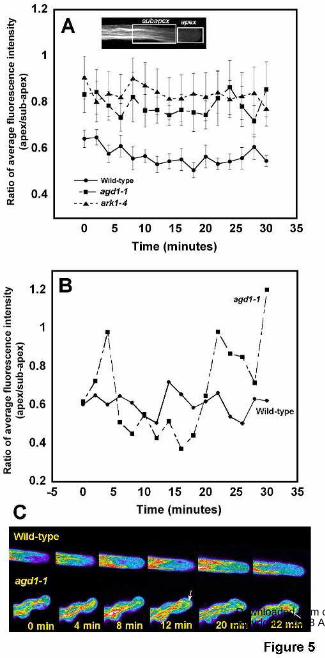
www.plantphysiol.orgon June 16, 2018 - Published by Downloaded from Copyright © 2008 American Society of Plant Biologists. All rights reserved.

www.plantphysiol.orgon June 16, 2018 - Published by Downloaded from Copyright © 2008 American Society of Plant Biologists. All rights reserved.
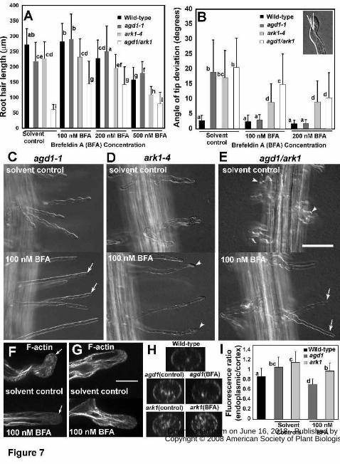
www.plantphysiol.orgon June 16, 2018 - Published by Downloaded from Copyright © 2008 American Society of Plant Biologists. All rights reserved.
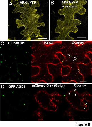
www.plantphysiol.orgon June 16, 2018 - Published by Downloaded from Copyright © 2008 American Society of Plant Biologists. All rights reserved.