RSD PUZZLE #1 WHAT IS RSD? WHAT IS SYMPATHETIC ANYWAY? · 3 CAN RSD BE MORE CLEARLY DEFINED? RSD is...
53
1 RSD PUZZLE #1 WHAT IS RSD? WHAT IS SYMPATHETIC ANYWAY? REFLEX SYMPATHETIC DYSTROPHY (RSD) To define Reflex Sympathetic Dystrophy (RSD) one should understand the terminology of sympathetic and parasympathetic nervous systems. There are two different types of nervous systems controlling the body. One is the so-called "somatic" nervous system which has clear-cut anatomical structures and is controlled by the cerebral cortex (cerebral hemispheres) in a relatively volitional manner. We see, hear, touch, taste or smell something, and volitionally and knowingly respond positively or negatively towards the stimulus. We can influence our response through the judgment of higher centers of the brain (cerebral hemispheres). This is the "somatic" system which is strongly influenced and controlled by our conscious mind. The other system is the so-called autonomic system. The name implies that it is autonomous (kind of having a mind of its own). It is almost autonomous but it can be influenced to a certain extent by conscious brain as well. This system is quite primitive and old (from evolutional standpoint). Even a worm has an autonomous nervous system. If one does an experiment by warming up one end of a fish tank water and cooling the other end of the; the worm will go from one extreme to the other and eventually will reside halfway between the two extremes of temperature in the mid portion of the fish tank. The worm does not need a brain to decide where to retire. The autonomic system does the job for it. The autonomic nervous system concerns itself with preservation and protection of the "Internal Environment". For example, in warm blooded animals the autonomic nervous system keeps the temperature inside the body around 99º Fahrenheit (37 ºC). To protect the internal environment, the autonomic nervous system has two main components. 1. The sympathetic system. 2.The parasympathetic system. The sympathetic system is a fight component of the "Fight and Flight" reflexes of the autonomic nervous system. On the fight end part of it the sympathetic system
Transcript of RSD PUZZLE #1 WHAT IS RSD? WHAT IS SYMPATHETIC ANYWAY? · 3 CAN RSD BE MORE CLEARLY DEFINED? RSD is...
1
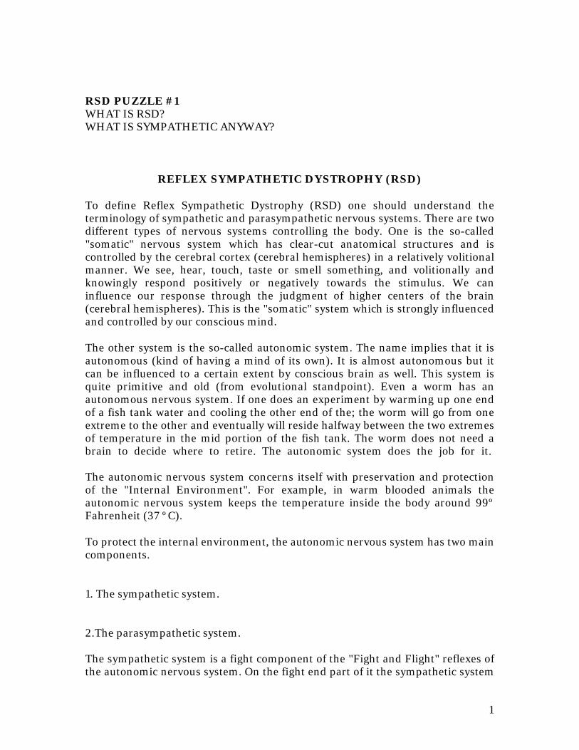
RSD PUZZLE #1 WHAT IS RSD? WHAT IS SYMPATHETIC ANYWAY?
REFLEX SYMPATHETIC DYSTROPHY (RSD)
To define Reflex Sympathetic Dystrophy (RSD) one should understand the terminology of sympathetic and parasympathetic nervous systems. There are two different types of nervous systems controlling the body. One is the so-called "somatic" nervous system which has clear-cut anatomical structures and is controlled by the cerebral cortex (cerebral hemispheres) in a relatively volitional manner. We see, hear, touch, taste or smell something, and volitionally and knowingly respond positively or negatively towards the stimulus. We can influence our response through the judgment of higher centers of the brain (cerebral hemispheres). This is the "somatic" system which is strongly influenced and controlled by our conscious mind.
The other system is the so-called autonomic system. The name implies that it is autonomous (kind of having a mind of its own). It is almost autonomous but it can be influenced to a certain extent by conscious brain as well. This system is quite primitive and old (from evolutional standpoint). Even a worm has an autonomous nervous system. If one does an experiment by warming up one end of a fish tank water and cooling the other end of the; the worm will go from one extreme to the other and eventually will reside halfway between the two extremes of temperature in the mid portion of the fish tank. The worm does not need a brain to decide where to retire. The autonomic system does the job for it. The autonomic nervous system concerns itself with preservation and protection of the "Internal Environment". For example, in warm blooded animals the autonomic nervous system keeps the temperature inside the body around 99º Fahrenheit (37 ºC).
To protect the internal environment, the autonomic nervous system has two main components.
1. The sympathetic system.
2.The parasympathetic system. The sympathetic system is a fight component of the "Fight and Flight" reflexes of the autonomic nervous system. On the fight end part of it the sympathetic system
2
increases the internal temperature, raises the blood pressure, strengthens the protective function of the skin, makes the skin cold so that there would be no waste of temperature, makes the skin sweat excessively (so that there would be no extreme increase of the internal temperature); and increases muscle metabolism, bone circulation, circulation of the brain and guts. The end result is the animal is ready to fight. On the other hand, the parasympathetic system that is the balancer of the other end of the autonomic system (the flight system), drops the blood pressure, slows down the pulse, relaxes the muscles, and preserves energy by cutting down the calories burned in the body. As such, the parasympathetic system works the opposite of the sympathetic system. The two systems are totally independent and one cannot give a patient parasympathetic enhancing medication in the hope of cooling the sympathetic and vice-versa.
THEN WHAT IS RSD?
RSD is one form of disturbance of the function of the autonomic nervous system. Simply having a hyperactive sympathetic nervous system does not make RSD. The disturbance of the autonomic system comprises several diseases, some acquired, some genetic, some metabolic, some traumatic, etc. Some examples of dysautonomias "disturbances of the autonomic nervous system" are attacks of hypotension (low blood pressure), congenital absence of sweating, and neuropathic pain syndrome.
The latter category of chronic neuropathic pain syndrome refers to the conditions that are not exactly necessarily reflex sympathetic dystrophy, but have an abnormal sympathetic component to them. They share sympathetically maintained pain (SMP) with RSD but they are not RSD.
Examples of such chronic neuropathic pain are postherpectic neuralgia (pain accompanying and following shingles), neuropathic diabetic neuropathy, acute neuropathic pain accompanying bee stings, snake bites or spider venom stings as well as involvement of the sympathetic nerves due to the systemic AIDS infection. The neuropathic pains are not at all synonymous with RSD. They do not even in any way resemble RSD. In some cases, however, they can end up with RSD. The chronic neuropathic pain syndrome is far more common than the true clinical picture of RSD.
3
CAN RSD BE MORE CLEARLY DEFINED?
RSD is a definitive chronic pain syndrome called by several different names such as reflex sympathetic dysfunction, (stage I), reflex sympathetic dystrophy (stage II), reflex sympathetic atrophy (stage III), late stage RSD complicated by disturbance of immune system, suicidal and fetal tendency, cancer, heart attack or stroke (stage IV), complex regional pain syndrome (CRPS) which encompasses all different forms of RSD as well as the causalgic RSD, causalgia (burning, stabbing, constant pain, acting more like an epileptic seizure), shoulder-hand syndrome, sudeck's atrophy (circa 1900), traumatic vasospasm of Lehman (1934), mimocausalgia (1973), minor causalgia (1940), and half a dozen other names.
In medicine there is a trend. When a disease becomes confusing, the physicians become desperate and give it new names. Each of the above almost dozen names reflect some features of RSD.
Merskey and Bogduk in January of 1994 defined the syndrome as follows (IASP Press, Seattle classification of chronic pain 2nd edition)[1]: CRPS TYPE I is a syndrome that usually develops after an initiating noxious event, is not limited to the distribution of a single peripheral nerve, and is apparently disproportionate to the inciting event. It is associated at some point with evidence of edema, changes in skin blood-flow, abnormal sudomotor activity (sweating) in the region of the pain, or allodynia or hyperalgesia. They also clarify in the main features of CRPS (RSD). "The symptoms and signs may spread proximally or involve other extremities. Impairment of motor function is frequently seen". They clarify associated symptoms and signs and specify "atrophy of the skin, nails, and other tissues, alterations in hair growth, and loss of joint mobility may develop. Impairment of motor function can include weakness, tremor, and in rare instances, dystonia. Symptoms and signs fluctuate at times. Sympathetically maintained pain may be present and may be demonstrated with pharmacological blocking or provocation techniques. Affective symptoms of disorders occur secondary to the pain and disability. Guarding of the affected part is usually observed". This is a long but relatively comprehensive definition of RSD. Building on the basis of this comprehensive definition of RSD, one can come to the conclusion that RSD is a syndrome with multiple manifestations which require the following minimal symptoms and signs for the condition to be called RSD (CRPS).
1. Pain: constant, burning pain, and in some forms at times during the course of the disease, stabbing type of pain (causalgic). The pain is relentless and is invariably accompanied by allodynia (even simple touch or breeze aggravating
4
the pain) and hyperpathia (marked painful response to even a simple stimulation).
2. Spasms in the blood vessels of the skin and muscles of the extremities. The spasms in the blood vessels result in a cold extremity. The muscle spasms result in tremor, movement disorders such as dystonia, flexion spasm, weakness and clumsiness of the extremities, and tendency to fall.
3. RSD is accompanied by a certain degree of inflammation in practically all cases. This inflammation may be in the form of swelling (edema), skin rash (neurodermatitis), inflammatory changes of the skin color (mottled or purplish, bluish or reddish or pale discolorations), tendency for bleeding in the skin, skin becoming easily bruised, inflammation and swelling around the joints as well as in the joints (such as wrists, shoulders, knee, etc.) which can be identified on MRI in later stages, and secondary freezing of the joints.
4.The fourth component and pre-requisite of diagnosis of RSD is insomnia and emotional disturbance. The fact that the sympathetic sensory nerve fibers carrying the sympathetic pain and impulse up to the brain terminate in the part of the brain called "limbic system". This limbic (marginal) system which is positioned between the old brain (brainstem) and the new brain (cerebral hemispheres) is mainly located over the temporal and frontal lobes of the brain. The disturbance of function of these parts of the brain results in insomnia, agitation, depression, irritability, and disturbance of judgment. Insomnia is an integral part of an untreated RSD. So are problems of depression, irritability and agitation.
So the clinical diagnosis of RSD is based on the above four principles rather than simply excluding RSD and finding some other cause for the patient's pain.
5
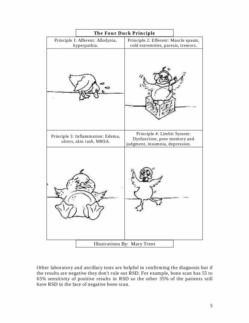
The Four Duck Principle
Principle 1: Afferent: Allodynia, hyperpathia.
Principle 2: Efferent: Muscle spasm, cold extremities, paresis, tremors.
Principle 3: Inflammation: Edema, ulcers, skin rash, MRSA.
Principle 4: Limbic System: Dysfunction, poor memory and
judgment, insomnia, depression.
Illustrations By: Mary Trent
Other laboratory and ancillary tests are helpful in confirming the diagnosis but if the results are negative they don't rule out RSD. For example, bone scan has 55 to 65% sensitivity of positive results in RSD so the other 35% of the patients still have RSD in the face of negative bone scan.
6
The thermography may be positive and helpful in confirming the diagnosis of RSD but at least 10% of RSD patients do not have the positive thermographic test. This is because of symmetrical involvement of RSD in both extremities. Thermography also has the handicap of tests such as MRI in that it can show false-positive results showing temperature changes in the absence of the symptoms and signs of RSD.
Phentolamine IV nerve block test is probably the most sensitive test to confirm RSD in the earlier stages (stages I and II within the first two to three years of the disease). However, as the disease becomes chronic, the longstanding constriction of the blood vessels causes disturbance of the circulation in the peripheral somatic nerves with resultant involvement of the somatic sensory nerves as well. As a result, the patient does not show a pure sympathetically mediated pain (SMP) but can show sympathetically independent pain (SIP) with no relief from Phentolamine in late stages of RSD.
In conclusion, RSD is a clinical bedside diagnosis. Not every hyperpathic pain is RSD. Not every SMP is RSD. SMP can be due to a simple post-herpetic neuralgia or diabetic neuropathy but that does not make RSD.
"Now that RSD has been diagnosed by the above criteria, what is the nature of the illness, manifestations, and treatment of RSD?"
RSD, as defined above, usually develops after a minor trauma. There are precipitating factors that enhance the development of RSD. These consist of immobilization of the extremity with cast or brace, application of ice, and inactivity due to strong addictive narcotics and tranquilizers.
The application of ice plays a major role. In experiments regarding the conductibility of the nerve impulse to the nerves, cold has shown to play a major role. If the temperature of the extremity drops from 37ºcentigrade to 10º centigrade, then the larger somatic sensory nerves stop conducting electricity through the nerve fibers. It takes the temperature to drop to 0º centigrade (freezing temperature) for the sympathetic nerves which are small thin fibers to stop conducting. The reason is the smaller the nerve fiber the less fatty sheath of myelin (insulator) surrounds the nerve. The fat in the myelin freezes more readily with the drop of temperature and stops the conduction of the nerve impulse. What the above implies is the fact that once the extremity is cooled down with ice to 10º temperature or lower, the normal conduction of the somatic nerves (touch, vibration, and position sense) is blocked off and the extremities left with purely the sympathetic nerve fibers function (constant burning pain).
7
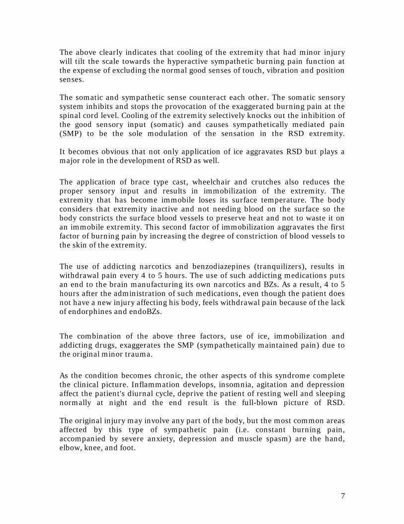
The above clearly indicates that cooling of the extremity that had minor injury will tilt the scale towards the hyperactive sympathetic burning pain function at the expense of excluding the normal good senses of touch, vibration and position senses. The somatic and sympathetic sense counteract each other. The somatic sensory system inhibits and stops the provocation of the exaggerated burning pain at the spinal cord level. Cooling of the extremity selectively knocks out the inhibition of the good sensory input (somatic) and causes sympathetically mediated pain (SMP) to be the sole modulation of the sensation in the RSD extremity. It becomes obvious that not only application of ice aggravates RSD but plays a major role in the development of RSD as well.
The application of brace type cast, wheelchair and crutches also reduces the proper sensory input and results in immobilization of the extremity. The extremity that has become immobile loses its surface temperature. The body considers that extremity inactive and not needing blood on the surface so the body constricts the surface blood vessels to preserve heat and not to waste it on an immobile extremity. This second factor of immobilization aggravates the first factor of burning pain by increasing the degree of constriction of blood vessels to the skin of the extremity.
The use of addicting narcotics and benzodiazepines (tranquilizers), results in withdrawal pain every 4 to 5 hours. The use of such addicting medications puts an end to the brain manufacturing its own narcotics and BZs. As a result, 4 to 5 hours after the administration of such medications, even though the patient does not have a new injury affecting his body, feels withdrawal pain because of the lack of endorphines and endoBZs.
The combination of the above three factors, use of ice, immobilization and addicting drugs, exaggerates the SMP (sympathetically maintained pain) due to the original minor trauma.
As the condition becomes chronic, the other aspects of this syndrome complete the clinical picture. Inflammation develops, insomnia, agitation and depression affect the patient's diurnal cycle, deprive the patient of resting well and sleeping normally at night and the end result is the full-blown picture of RSD. The original injury may involve any part of the body, but the most common areas affected by this type of sympathetic pain (i.e. constant burning pain, accompanied by severe anxiety, depression and muscle spasm) are the hand, elbow, knee, and foot.
8
In the United States, over five million patient suffer from this extremely painful and disabling illness called by many names, including reflex sympathetic dystrophy (RSD), sympathetic dysfunction syndrome (SDS), and causalgia, to name a few. This unusual, but severe painful condition is caused by disturbance of the function of the sympathetic nervous system (SNS).
Normally, the sense of pain is perceived through two separate channels in the body. The most common type of pain is transmitted through the nerves that end up in the cortex of the brain, over the vertex of the head (parietal lobe). This somatic pain is temporary, clear-cut and focalized. As the area of nerve damage is healed, the pain disappears.
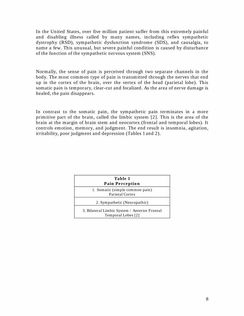
In contrast to the somatic pain, the sympathetic pain terminates in a more primitive part of the brain, called the limbic system [2]. This is the area of the brain at the margin of brain stem and neocortex (frontal and temporal lobes). It controls emotion, memory, and judgment. The end result is insomnia, agitation, irritability, poor judgment and depression (Tables 1 and 2).
Table 1 Pain Perception
1. Somatic (simple common pain) Parietal Cortex
2. Sympathetic (Neuropathic)
3. Bilateral Limbic System / Anterior Frontal Temporal Lobes [2]
9
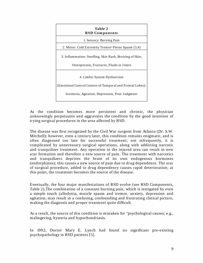
Table 2 RSD Components
1. Sensory: Burning Pain
2. Motor: Cold Extremity Tremor/Flexor Spasm [3,4]
3. Inflammation: Swelling, Skin Rash, Bruising of Skin,
Osteoporosis, Fractures, Fluids in Joints
4. Limbic System Dysfunction
(Emotional Control Centers of Temporal and Frontal Lobes):
Insomnia, Agitation, Depression, Poor Judgment
As the condition becomes more persistent and chronic, the physician unknowingly perpetuates and aggravates the condition by the good intention of trying surgical procedures in the area affected by RSD.
The disease was first recognized by the Civil War surgeon from Atlanta (Dr. S.W. Mitchell); however, even a century later, this condition remains enigmatic, and is often diagnosed too late for successful treatment; not infrequently, it is complicated by unnecessary surgical operations, along with addicting narcotic and tranquilizer treatment. Any operation in the injured area can result in new scar formation and therefore a new source of pain. The treatment with narcotics and tranquilizers deprives the brain of its own endogenous hormones (endorphines); this causes a new source of pain due to drug dependence. The scar of surgical procedure, added to drug dependency causes rapid deterioration; at this point, the treatment becomes the source of the disease.
Eventually, the four major manifestations of RSD evolve (see RSD Components, Table 2).The combination of a constant burning pain, which is instigated by even a simple touch (allodynia, muscle spasm and tremor, anxiety, depression and agitation, may result in a confusing, confounding and frustrating clinical picture, making the diagnosis and proper treatment quite difficult.
As a result, the source of this condition is mistaken for "psychological causes; e.g., malingering, hysteria and hypochondriasis.
In 1992, Doctor Mary E. Lynch had found no significant pre-existing psychopathology in RSD patients [5].
10
As the disease becomes chronic, it goes through four stages; these stages are not distinctive or clear-cut, but develop in an overlapping fashion. Stages I and II are the earliest and easiest to treat. If the disease is diagnosed and properly treated in the first six months, then stages III and IV shall be aborted. Any attempt at proper treatment, in the form of physical therapy or sympathetic block, will prevent serious complications seen in stage III and IV. (Table 3) As a result, because the classical advanced complications are not seen in partially treated patients, the clinician does not arrive at the proper diagnosis of RSD. The staging of RSD is not as critically done on the basis of dysfunction (stage I) meaning abnormal function of the sympathetic system in the extremity, dystrophy (stage II , meaning trophic and inflammatory changes, skin changes and other signs of inflammation), atrophy (stage III, meaning usually minimal degree of atrophy in the involved muscles), or stage IV (meaning disturbance of the immune system, suicidal attempts, stroke, heart attacks, intractable hypertension and chest pain, and in some cases development of cancer). The prognosis is more related to the temporal development of the above four stages.
11
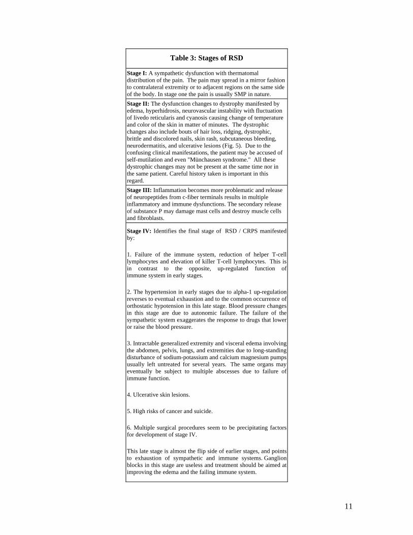
Table 3: Stages of RSD
Stage I: A sympathetic dysfunction with thermatomal distribution of the pain. The pain may spread in a mirror fashion to contralateral extremity or to adjacent regions on the same side of the body. In stage one the pain is usually SMP in nature.
Stage II: The dysfunction changes to dystrophy manifested by edema, hyperhidrosis, neurovascular instability with fluctuation of livedo reticularis and cyanosis causing change of temperature and color of the skin in matter of minutes. The dystrophic changes also include bouts of hair loss, ridging, dystrophic, brittle and discolored nails, skin rash, subcutaneous bleeding, neurodermatitis, and ulcerative lesions (Fig. 5). Due to the confusing clinical manifestations, the patient may be accused of self-mutilation and even "Münchausen syndrome." All these dystrophic changes may not be present at the same time nor in the same patient. Careful history taken is important in this regard.
Stage III: Inflammation becomes more problematic and release of neuropeptides from c-fiber terminals results in multiple inflammatory and immune dysfunctions. The secondary release of substance P may damage mast cells and destroy muscle cells and fibroblasts.
Stage IV: Identifies the final stage of RSD / CRPS manifested by:
1. Failure of the immune system, reduction of helper T-cell lymphocytes and elevation of killer T-cell lymphocytes. This is in contrast to the opposite, up-regulated function of immune system in early stages.
2. The hypertension in early stages due to alpha-1 up-regulation reverses to eventual exhaustion and to the common occurrence of orthostatic hypotension in this late stage. Blood pressure changes in this stage are due to autonomic failure. The failure of the sympathetic system exaggerates the response to drugs that lower or raise the blood pressure.
3. Intractable generalized extremity and visceral edema involving the abdomen, pelvis, lungs, and extremities due to long-standing disturbance of sodium-potassium and calcium magnesium pumps usually left untreated for several years. The same organs may eventually be subject to multiple abscesses due to failure of immune function.
4. Ulcerative skin lesions.
5. High risks of cancer and suicide.
6. Multiple surgical procedures seem to be precipitating factors for development of stage IV.
This late stage is almost the flip side of earlier stages, and points to exhaustion of sympathetic and immune systems. Ganglion blocks in this stage are useless and treatment should be aimed at improving the edema and the failing immune system.
12
The use of addicting narcotics and benzodiazepines (tranquilizers), results in withdrawal pain every 4 to 5 hours. The use of such addicting medications puts an end to the brain manufacturing its own narcotics and BZs.
As a result, 4 to 5 hours after the administration of such medications, even though the patient does not have a new injury affecting his body, feels withdrawal pain because of the lack of endorphines and endoBZs.
The combination of the above three factors, use of ice, immobilization and addicting drugs, exaggerates the SMP (sympathetically maintained pain) due to the original minor trauma.
As the condition becomes chronic, the other aspects of this syndrome complete the clinical picture. Inflammation develops, insomnia, agitation and depression affect the patient's diurnal cycle, deprive the patient of resting well and sleeping normally at night and the end result is the full-blown picture of RSD.
The one extreme in the case of major causalgia with significant damage to the sympathetic nerve, the status 1 through 3 can develop in a matter of weeks or a few months.
On the other extreme, for example in the case of heart attack or stroke causing RSD, the stages evolve in a very slow fashion and it may take a few years before stages III and IV are seen.
The faster the stages develop, the more severe the RSD and the treatment should be applied in a more aggressive fashion.
On the other hand, even a partial or minimal treatment of chronic stages of RSD can change the clinical picture of stage III (atrophy) back to stage II or stage I. That is a good sign, but that does not mean that the patient's condition is mild just because the patient is in stage I. The more chronic the disease the more likely the persistence of the symptoms and complications even though the bed side examination shows reversal to stages I and II from stages III and IV. If properly treated with extensive physical therapy, heat, mobilization, exercise and sympathetic nerve blocks, the success rate for full recovery in the first six months (stages I and II) is better than 80% [6].
13
SPREAD OF THE DISEASE
Even in the early stages, laboratory tests such as triphasic bone scanning or thermography show a spread of the disease from one side to the other. The headache and facial pain becomes bilateral the facial pain is complicated by dizziness, tinnitus (buzzing in the ear). In the later stages of the disease, the spread is both horizontal and vertical (e.g., from right arm to left, down the legs). The reason for this spread is the anatomical structure of the sympathetic nervous system. The majority of the Sympathetic nerve fibers do not allow the standard somatic nerve fibers, but follow the wall of the blood vessels and end up in the chain of clumps of the nerve cells, called sympathetic ganglia", which are present on each side of the spine. Input of pain into any part of these chains of nerves causes the impulse to spread both vertically and horizontally [7]. This is the main reason sympathectomy or removal of the ganglia is fraught with an extremely high percentage of failure.
This complex clinical picture of the spread of this disease has played a major role in confusing and delaying the proper diagnosis and treatment of RSD. to begin with, the injury causes such a small scar that may be barely visible. This is followed by a constant burning pain, severe neck pain and headache, spread of pain to the opposite side, followed by dizziness, fatigue, insomnia, agitation and irritability. On this background, the patient may develop pain in the arm, tremor of the hand, may have trouble walking, spasms in the legs, and may end up in a wheelchair. It is obvious that such a patient may be viewed as a neurotic, depressed, and a hypochondriac. The condition is further compounded by the fact that the patient has normal MRI, CAT scans, and x-rays. The pain is a physiological phenomenon, due to the disturbance of small sympathetic nerve fibers; CAT scan and MRI will not show such an abnormality. An individual, who suffers a heart attack and goes to the emergency room, has a normal CAT scan and MRI in the face of a potentially fatal disease. By the time MRI is abnormal, showing fluid in the involved joints or damage to the bone due to increased circulation in the deep structures, the disease is quite advanced and easy to diagnose.
Eventually, the spasm may spread to the truncal muscles; as a result, an accordion-like jamming effect of the vertebrae evolves, with resultant bulging of the lower lumbar discs noted on MRI. This may result in unnecessary surgical operation. The RSD causes constriction of blood vessels to the hands; in this clinical picture, the patient is often mis-diagnosed as carpal tunnel syndrome, tardy ulnar nerve palsy, or thoracic outlet syndrome.
14
Not infrequently, such patients undergo multiple operations over the arms and cervical and lumbar spine regions, with rapid acceleration and deterioration of the RSD.
The worst risk factor, and the cardinal sin, is amputation of a limb. If the patient has any sympathetic mediated pain, amputation is going to multiply the disease by several times, due to the fact that the stump of the Amputation causes scar formation at the cut ending of the nerves with marked exacerbation of RSD in the most severe form, called "Causalgia".
Even without application, surgical procedure over the inflamed area of involvement of the RSD (such as ankle, knee or wrist), is a major aggravator of RSD which overnight changes as stage I RSD to stage III or later. The Three areas that are most commonly and unfortunately operated on are: 1.Wrist according to the diagnosis of carpal tunnel syndrome. This has already discussed in the RSD quiz related to carpal tunnel syndrome. Simply put, carpal tunnel syndrome rarely causes RSD but in rare cases of RSD the inflammation of the soft tissue at the wrist results in a clinical picture identical to carpal tunnel syndrome (CTS). This form of carpal tunnel syndrome is the effect not the cause. It is the result of RSD rather than being the cause of RSD. In this type of CTS, the entire hand, wrist and forearm are quite hypopathic, allodynic and sensitive to touch in contrast to the true somatic carpal syndrome. In this form of RSD, treatment with nerve blocks and anti-inflammatory medications Baclofen, moist heat and Epsom salt clears up the symptoms and signs of CTS. On the other hand, surgery ends up with disastrous results.
2. The same is true in the case of so-called tarsal tunnel syndrome which is over-diagnosed and over-treated universally by the podiatrists and surgery over the tarsal tunnel on the ankle rapidly deteriorates the already existing RSD. 3.The same is true with a lot of minor injuries to the knee with full-blown clinical picture of cold extremity, flexion, weakness and atrophy of the muscles around the knee, difficulty with weight bearing, severe constant burning pain, all of which prompt the surgeon to explore the knee with obvious disastrous results. Opposite to the prevalent notion of "treat the source" the proper principle should be treat the RSD and then see what happens to the so-called source.
Even nerve blocks should not be done in the area of inflammation of RSD (over the wrist, ankle, and dorsum of the foot, dorsum of the hand or the knee). Instead the nerve blocks which are very effective in treatment of RSD should be done in axial area (over the spine) where the nerves enter and exit from the spinal cord to the involved part of the extremity.
15
Simply said, do not needle, amputate, or operate the area of inflammation of the RSD.
DIAGNOSIS
The diagnosis is achieved by the physician's familiarity with the triad of RSD, as mentioned above. Confirmatory tests, such as beneficial effect from sympathetic nerve block, as well as the use of bone scan or thermography are quite helpful. None of the above tests yield 100% positive diagnostic proof. Even sympathetic nerve block is positive and relieves the pain in early stages of the disease. In the later stages, there has been enough damage - due to vasoconstriction - in the involved area, that non-sympathetic pain contaminates the clinical picture and the patient does not receive 100% relief from the nerve block.
TREATMENT
Early diagnosis in the first six months to maximal two years is the key to successful treatment [6]. Surgical procedures have no place in treatment of RSD. Sympathectomy or removal of a part of the chain of sympathetic ganglia on the side of the spine) has an extremely high rate of failure. It has been reported to help the war type of RSD, which is quite different from civil type. In the war type, the soldier is a young teenager who responds favorably to treatment, regardless of the mode of treatment. The war time RSD is due to high velocity damage to the nerves in the proximal parts of the extremities and sympathectomy, even in these cases, has a high rate of failure in the long run. The civil type of RSD is due to a small damage of the sympathetic nerves in the central or peripheral nervous system. If the patient lives longer than five years, the rate of failure from sympathectomy is over 80%. The scar of the surgical procedure becomes a new source of RSD. Removal of a part of sympathetic ganglia does not prevent spread of the disease in the areas of the body where the sympathetic nerves have been removed. This is due to the fact that the adjacent sympathetic nerves eventually compensate for the lack of sympathetic function due to surgery.
16
SPINAL STIMULATORS
Insertion of epidural spinal stimulators has been quite vogues. In our comparison of 41 RSD patients treated with spinal stimulators vs. 40 non-RSD chronic pain patients who received the same treatment, the RSD group of patients had only maximum three and one half to four months relief of pain. Afterwards, the stimulator acted as a foreign body and became a new source of RSD. The reason may be due to the fact that the stimulators are digital in nature with predicted rhythmic stimulation, whereas the pain of RSD is practically constant and analog (variable) in nature.
No single physician is smart and potent enough to treat RSD. Successful treatment requires a teamwork of physical medicine, anesthesiology, and neuropharmacology physicians. The keys to successful treatment are early diagnosis, early mobilization and extensive physical therapy, and early detoxification of the patient from addicting narcotics, alcohol and addicting tranquilizers.
The anesthesiologist should interrupt the sympathetic hyperactivity by doing repetitive, successive (six) nerve blocks, combined in the same day with physical therapy and exercise. Discontinuation of ice and all other assistive devices, such as wheelchair, brace, cast, walker, etc., is essential.
The addicting drugs should be replaced with the treatment of choice for chronic pain in the form of newer-generation antidepressants, Paxil, Zoloft, and Trazodone, which are not trycyclic antidepressants. Trazodone is the treatment of choice to replace the tranquilizers and sleeping pills. It provides normal sleep, as well as prevention of chronic pain. The patient can be detoxified quickly and easily by discontinuation of the narcotics and replacement with nonaddicting ones, such as Stadol. The use of muscle relaxant, Baclofen, which selectively works on the spinal cord, counteracts the spasm, clumsiness and tremor.
INFUSION PUMP
In severe brain or spinal cord injuries that result in severe crippling spasticity, the infusion pump can be very effective in the treatment of spasticity. Unfortunately, not enough infusion pumps are being used for treatment of severe head and spinal cord injuries. With the use of Baclofen in the pump, such victims of severe crippling spasticity can become mobilized and can be spared from life-threatening inactivity and bed sores.
17
The infusion pump has been used for treatment of severe pain. In our studies of over 400 cases of advanced RSD, over three dozen patients have been treated with infusion pump, with close to 90% success rate. Through the pump, a drip irrigation form of pain medication is introduced to the spinal fluid. The pain medication given in one month is equivalent to the amount of medication given in two to three days by mouth or by IM injection.
Infusion pump should not be mistaken with other forms of narcotic administration. The infusion pump is totally different and practically opposite to administration of narcotics in the muscle (IM), IV, by skin patch, or simply in the epidural space.
The intrathecal infusion pump (administration of the medication directly to the spinal fluid that surrounds the brain and spinal cord), provides direct access to the brain and spinal cord bypassing liver, kidney and other organs. As a result, the patient requires only 1/20 monthly dose of the narcotic to provide complete relief of pain. In addition, when the pain is optimally achieved by use of as little as 1 to 7 mg a day (usually 3mg a day) of Morphine sulfate, if the patient for other reasons has pain (such as drinking alcohol or simultaneously taking other addicting narcotics by mouth), increasing the dosage of Morphine in the spinal fluid over and above 9mg per day, causes recurrence of severe pain. This is because the system is so flooded by such a strong dose of narcotics that the brain does not form its own endorphines, and the large doses of Morphine in the pump only causes drowsiness, causes the patient respiratory trouble, but does not completely control the pain.
The above phenomenon emphasizes the importance of optimal small dose of narcotic infusion in the spinal fluid which is obviously non-addicting. When distress is violated by increasing the dosage of medicine in the pump, then it becomes like any other form of addicting narcotic administration. The patient develops severe pain due to the fact that the brain cannot form its own endorphines. If the brain manufactures its own endorphine in the face of large doses of narcotic applied in the spinal fluid, then the patient faces the risk of dying from arrest of respiration. So it becomes obvious that the infusion pump is not just another form of giving addicting medications. It works because a very small amount (usually 1/20 to 1/30 dose) of pain medication is given in the form of drip irrigation directly in the spinal fluid with complete control of pain and complete relief of symptoms and signs of RSD.
18
DIET
The use of proper diet, with avoidance of chocolate (phenyletholamine), hot dog, liver and sausage, and alcohol is essential in management of RSD.
REFERENCES
1. Merskey H, Bogduk N.: Classification of Chronic Pain: Descriptions of Chronic Pain Syndromes and Definitions of Pain Terms. Second Edition. Task Force on Taxonomy of the International Association for the Study of Pain. Merskey H, Bogduk N, Editors. IASP Press. Seattle, WA 1994.
2. Benarroch EE: The central autonomic network: functional organization, dysfunction, and perspective. Mayo Clin Proc 68:988-1001 1993.
3. Schwartzman RJ, Kerrigan J: The movement disorder of reflex sympathetic dystrophy. Neurology 40:57-61 1990.
4. Yokota T, Furukawa T, Tsukagoshi H: Motor paresis improved by sympathetic block a motor form of reflex sympathetic dystrophy? Arch Neurol 46: 683-687 1989.
5. Lynch ME: Psychological aspects of reflex sympathetic dystrophy: a review of the adult and pediatric literature. Pain 49:337-47 1992.
6. Poplawski ZJ, Wiley AM, Murray JF: Post traumatic dystrophy of the extremities. J Bone Joint Surg [Am] 65:642-655, 1983.
7. Appenzeller O: The Autonomic Nervous System: An introduction to basic and clinical concepts, 4th rev, Elsevier 1990.

RSD PUZZLE #2 RSD As Related To Trauma Injuries
A serious misconception that needs to be corrected is as follows: The physician tells the patient that "Your injury was too mild. It could not cause RSD".
Trauma is not the exclusive cause of RSD. Even when trauma is the cause, it is usually in the form of a minor injury. Of all traumatic types of pain, less than 5% end up in RSD. The majority of traumatic cases are in more severe form, and result in simple somatic (non-sympathetic) pain. The reason is the fact that in non-sympathetic pain the larger sensory nerve fibers are affected and as a result they overshadow the small sensory C nerve fibers which are responsible for the development of RSD.
On the other hand, if a minor trauma affects selectively the small C fibers that transmit sympathetic sensory impulse there is more likelihood of the development of RSD. Usually RSD develops due to a minor trauma in certain specific parts of the body. The areas that are more susceptible to develop RSD are dorsum of the hand, dorsum of the foot, knee, elbow, ankle, elbow and shoulder. In these areas, there is the anatomical phenomenon called WATER-SHED ZONES. In these water-shed areas, there are multiple sensory nerve roots adjacent to each other. A minor trauma in such an area causes an electric short among these small C nerve fibers and as a result stimulates the multiple sympathetic centers in multiple levels of spinal cord with resultant augmentation of sympathetic input and a moderate RSD.
These water-shed zones are in contrast with other areas such as abdominal wall or chest wall. Even though the chest and abdomen frequently undergo accidental or surgical trauma, rarely cause RSD. The reason is that in the chest and abdominal wall the nerve roots are organized in certain parallel dermatomal fashion and they do not have such close proximity as over the dorsum of the foot, hand, or knee. Simple minor procedures such as arthroscopy or intravenous insertion in the areas of water-shed zones can end up in RSD if other risk factors are present (e.g., application of ice, inactivity, and long term use of narcotics). The only exception to the above rule is the major causalgia due to bullet wound. In this rare form of RSD (civil RSD is nine to one more common than war RSD); the vibration of the bullet selectively stimulates the small C fibers and ends up in severe RSD. Just because a minor injury causes RSD, that does not equate with a minor RSD. (For further information regarding water-shed zones and RSD please see the textbook "Chronic Pain: Reflex Sympathetic Dystrophy: Prevention and Management", H. Hooshmand, M.D., CRC Press, pages 62, 92, 94 and 104.)
H. Hooshmand, M.D.
RSD PUZZLE #3 RSD and TMJ Pain "Your injury was to the right hand causing RSD. This is in no way related to the right TMJ pain or the loss of teeth that you have developed since". As remote and unrelated as they may seem, RSD can and does result in cranio-cervical pain, muscle spasm, TMJ disease and deterioration of the teeth. It is quite common for patients who have suffered from foot or hand injury to develop low back pain, neck pain and headache. One common manifestation of RSD in response to pain is muscle spasm, and motor dysfunction. This can be in the form of flexion deformity of the extremity, difficulty with walking, flexor withdrawal of the muscles of the extremity, and lumbar and cervical paraspinal spasm. As a result, the patient develops muscle tension headaches, as well as spread of the muscle spasm to the facial muscles with resultant stress on the temporomandibular joint (TMJ) and severe pain and spasm around the TMJ. With passage of time, the same phenomenon results in chronic trauma to the TMJ as well as clinching of the teeth and trauma to the teeth. The patient develops severe pain in the distribution of trigeminal nerve (sensory nerve for the face) and develops moderate migrainous vascular headaches (trigeminal vascular headaches). In later stages of RSD the immune system becomes disturbed, and the patient develops poor oral hygiene and dental deterioration. Long-standing unilateral (one sided) spasm of cervical paraspinal muscles causes increased input of pain into the upper portion of the cervical spinal cord. As a result, a referred pain develops with resultant facial pain and secondary muscle spasm around the TMJ and the jaw. The same referred pain causes migraine headaches, TMJ pain and chronic stress on the teeth with dental deterioration. H. Hooshmand, M.D.
1
RSD PUZZLE #4 RSD and the use of assistive devices "If the patient is in constant, severe pain, has trouble with walking, has spasms in the muscles of the lower extremities, and has flexion deformity, why not use a wheelchair?" Unfortunately, the use of assistive devices (braces, crutches, canes, walker, or wheelchair) results in inactivity and lack of use of the extremity. This, in turn, leaves the hyperactive sympathetic system unopposed and uninhibited. Normally weight bearing, walking, and the use of hands and feet stimulate the position sense (the larger sensory nerve fibers) which in turn inhibit the antero-lateral horn cells of the spinal cord. The antero-lateral horn cells of the spinal cord are the sympathetic nerves that cause constriction of the blood vessels in the extremity, cold extremity, and poor oxygenation to the small c nerve fibers (pain). Any kind of inactivity by any assistive device leaves the sympathetic nerves at the spinal cord level uninhibited and increases the firing of the sympathetic nerves with aggravation of RSD. After sympathetic nerve block is done, the sympathetic nerve fibers are temporarily blocked. This provides a window of opportunity of a few hours to a few days for the patient to have physical therapy and stimulation of the large sensory nerve fibers (position sense) so that the antero-lateral horn cells of the spinal cord are inhibited further and the beneficial effect of the sympathetic nerve block is prolonged and perpetuated. If after sympathetic nerve block the patient is left resting in bed without exercise of the extremities or if ice is applied to the extremities, then the sympathetic nerve block is counteracted and the patient rapidly returns back to the pre-nerve block state. The name of the game in the management of RSD is mobilization, exercise, heat, massage, electrical stimulation, and any other stimulation that increases the position sense (proprioception) to block and inhibit the hyperactivity of the sympathetic nerve fibers. The cardinal sins in the management of RSD are inactivity, use of ice, use of assistive devices, and amputation. To achieve mobilization and ability to exercise, two factors should be counteracted. One is pain and the other is tendency for spasm and tremor in the extremity involved with RSD.
2
The pain can be treated with non-addictive pain medications (such as Stadol and other non-addictive analgesics), and the muscle spasm and tremor are best counteracted by the use of Baclofen (Lioresol). Unfortunately, Soma transforms to Meprobamate after oral intake and has the potential of addiction. Flexeril, on the other hand, has the side effects of depression and tendency for sedation and inactivity. With the help of physical therapy, moist heat, use of enough pain medication and muscle relaxant, the patient should get rid of the wheelchair and other assistive devices. The wrist and hand braces, and shoulder and elbow slings, result in flexion deformity of the hand, flexion deformity of the elbow, and frozen shoulder which are going to cause serious complications in the long term care. The use of crutches, walkers and wheelchair result in avoidance of weight bearing with the serious side effects of cold skin and hot bone with rapid turn over of blood in the bone marrow, osteoporosis, and fracture of the bones which eventually may necessitate amputation. The amputation is the beginning of the end because the stump of the amputation will have hundreds of neuromas all in RSD mode. Even the patients who have fracture of the small bones in the foot due to lack of weight bearing can regain the function of the foot by weight bearing which will result in a spontaneous healing of the small fractures. The small bones will heal in an irregular fashion but will spare the patient from amputation. H. Hooshmand, M.D.
RSD PUZZLE #5 Why Not Use Ice For RSD Therapy? "Why not apply alternative ice and heat treatment to the extremity as it is done commonly in the Physical Therapy Departments?"
The practice of alternate ice and heat application only has its place in the neurophysiological experiments and diagnostic tests for the function of the sympathetic system. Application of ice results in stimulation of the sympathetic system and secondary constriction of the superficial sympathetic vasoconstrictors. On the other hand, the heat application results in the dilatation of superficial blood vessels and relaxation of the vasoconstrictor activity of the sympathetic nervous system. Obviously, in RSD, the goal is to warm up the extremity, to dilate the superficial blood vessels, and to slow down the simultaneous inflammation and increased circulation in the deep structures of the extremity (which result in osteoporosis and fracture of the bones). As the heat application helps the patient, the ice application negates the beneficial effects of the heat. The end result is a zero therapeutic effect. Ice application has its place in acute soft tissue injuries but has no place in treatment of complex chronic pain of RSD. H. Hooshmand, M.D.
RSD PUZZLE #6 Physical Therapy
"There have been references in the literature that physical therapy can aggravate pain and RSD. Yet in every outline of treatment for RSD, the use of physical therapy is emphasized. These two statements seem to be contradictory." Both statements are absolutely true. Excessive exercise and physical therapy that causes fatigue, pain, and distress to any part of the body, only flares-up and aggravates the inflammation and pain of RSD. On the other hand, the commonest aggravators of RSD are bed rest, inactivity, application of ice, and the use of assistive devices. In RSD, the best treatment is eustress not distress. Distress refers to the stress of prolonged bed rest and inactivity. Like any other machine, prolonged idling of the body is distressful and causes damage to the body. Especially in RSD, the prolonged bed rest results in aggravation of pain and insomnia. The RSD patients suffer from severe, chronic insomnia due to the constant allodynic pain as well as due to the aggravation of constriction of blood vessels secondary to inactivity. One of the earliest signs of RSD is a restless night with the patient constantly being fidgety and changing position all night as well as having to get up and walk to get some relief.
The second form of distress is too much exercise, prolonged physical therapy. The RSD patient has to learn that they will have pain with too much exercise, and the patient will have more pain without exercise. The patient will have to find a happy medium. The patient will have to rest and exercise frequently. Three days a week in the P.T. Department is not enough. The RSD patient should continue the instructions of the physical therapist from morning to night with equal periods of rest and exercise. The patient should learn from the human heart which beats approximately once a second for 80 to 90 years without taking a vacation. The reason is the heart beats half a second and rests half a second. The same principle should apply to physical therapy in RSD.
H. Hooshmand, M.D.
RSD PUZZLE #7 RSD AND PREGNANCY "I have RSD. Is it safe to become pregnant?"
There are dozens of references in the literature regarding RSD and pregnancy. There is no contraindication for an RSD patient to become pregnant. As a matter of fact, as is the case with some other neurologic illnesses such as multiple sclerosis and epilepsy, pregnancy may be beneficial in the management of the disease. On the other hand, RSD may manifest itself for the first time during pregnancy. This does not imply that pregnancy causes RSD. If the patient already has had some form of nerve damage in the extremity then in the late stages of pregnancy due to inactivity and prolonged bed rest, a sub-clinical SMP (sympathetically mediated pain) can change into a full-blown picture of RSD. The high risk area for this complication is the hip area. This is especially true if the pregnant patient has a tendency to rest practically consistently on one side of the body. The hip that bears the brunt of the patient's weight for long hours or days can manifest the spread of RSD from the knee or ankle area. The pregnant RSD patient has to remember to stay as active as possible especially in the last trimester. Exercise, heat massage and use of Epsom salt and hot water over the lower extremities especially the hips, are quite helpful. H. Hooshmand, M.D.
RSD PUZZLE #8 Possible Progression of RSD
"Your RSD cannot be in stage III if you have had an injury only 2 months ago". This is not at all true. The stages I, II and III are not the prerequisites for the diagnosis of RSD. RSD can stay in stages I and II for months or years and never develop in stage III. This is especially the case if the patient has had even the slightest treatment such as sympathetic nerve blocks, physical therapy, oral sympathetic blocks or physical therapy. This disease begs for proper treatment and responds nicely to such treatment especially in the early stages in the first 3 to 6 months. Anymore delay causes a higher percentage of failure. Even in the earliest stages (less than 3 months), if the patient is treated with invasive, elective surgical procedures, the failure rate will be very high. The disease becomes accelerated and progressively gets worse if improper treatments are applied. Stage I- RSD can rapidly, and in a matter of weeks, change to a stage III if the following improper treatments are applied.
1. Excessive inactivity, assistive devices, and excessive bed rest.
2. Use of a lot of ice application.
3. Injection of nerve blocks in the area of the scar of the injury. Whereas the nerve blocks are quite helpful when applied for proximal portions of the nerves away from the scar of the injury, sticking needles into the area of the scar of the RSD injury only aggravates the condition and deteriorates the disease quickly into stages III and IV. The scar area already has partially damaged sympathetic nerves and does not need sticking of needles into those damaged nerves. This should be a strong reminder for people who stick needles in the areas of neuroma of the foot or areas of scars around the ankles.
4. Another factor that not infrequently causes acceleration and deterioration of the RSD to stage III in a matter of a few weeks or months is the naive concept of trying to remove the areas of skin damage or scars of RSD with surgical procedures. Not infrequently the disease is not diagnosed properly, negative bone scan is taken for an absolute diagnostic test to rule out RSD, and then the inflamed area is operated on for diagnoses such as "carpal tunnel syndrome", especially "tarsal tunnel syndrome", or "MRSA", or "Morphea". In the above examples, especially in the example of causalgia, the RSD can deteriorate in a matter of weeks and cause atrophy of the muscles of the extremity (stage III) or cause abnormal hair and skin growth as usually seen in other forms of RSD in a span of months or years. The commonest misconception is the fact that the patient who has had partial treatment for RSD can stay in the stages I and II for years yet the diagnosis is denied on the basis of misconception that in a matter of months to years the patient should have developed atrophy, skin changes, hair and nail growth abnormalities, and in the absence of such gross deformities then the patient is denied of the proper diagnosis of RSD.
H. Hooshmand, M.D.
1
RSD PUZZLE #9 Hair Changes In RSD "Can RSD cause change of hair color, change of consistency of the hair, and change of skin color?"
The skin and the nervous system both originate from the same germinal cells in very early fetal stage. It is not surprising that nerve dysfunction commonly manifests itself in the form of skin diseases. Neurodermatitis is quite commonly seen in both somatic and sympathetic nerve dysfunctions. In the somatic form, the neurodermatitis is in the distribution of dermatomes whereas in sympathetic nerve dysfunction it is in the distribution of thermatomes. The skin changes are quite varied with several manifestations. The commonest form is swollen, somewhat shiny, pale, pink or mottled skin. The skin looks like it is too tight because of the swelling of the tissue under it. Other manifestations are in the form of breakdown of the skin, progressively enlarging ulcer, and skin lesions that look like infected and non-healing, irregular shaped, relatively deep ulcer. Other manifestations are in the form of skin rash, urticaria, eczema, xerosis (very dry and wood-appearing type of skin). The hair growth or lack of the same is quite common. The abnormal hair growth may show a mutation to a thicker, darker hair or may be in the form of a thin, fragile and fuzzy hair. The skin may become thin and fragile easily developing ulcers or may become quite thick with a venous and lymphatic inflammation developing in elephantiasis or resulting in what appears to be a superficial phlebitis. This has been in erroneously mistaken for the development of phlebitis in RSD patients. This is nothing but a superficial inflammation of the venous circulation due to the inflammatory nature of RSD but does not have anything in common with the standard forms of phlebitis that can cause remote blood clots. Treatment consists of standard treatment of RSD. Nerve blocks, especially sympathetic ganglion nerve blocks, epidural nerve blocks, skin patch such as Clonidine patch, and corticosteroid creams are quite effective.
2
The majority of skin changes become self-contained and self- controlled when the proper treatment of RSD is initiated. The unattractive change of skin or hair color should not be corrected with plastic surgery because it will result in disastrous complications. The large ulcers which become larger with surgical excisions are best treated by avoidance of surgery, and avoidance of bandages and tight dressings, and treatment with sympathetic blocks applied proximally as well as systemic sympathetic blocks. Application of epsom salt and warm water is quite effective. The hyperosmolar effect of the epsom salt and warm water markedly reduces the inflammation and enhances the growth of the normal skin. The use of hot bath along with the application of epsom salt is quite effective in treatment of such conditions. When the patient has extensive inflammation, swelling, itching, eczema and neurodermatitis, treatment with Atarax, as well as Seldane (which is an antihistaminic type of medicine) can be helpful. In patients who have severe spells of neurodermatitis during the night keeping them awake, combination of antidepressants such as Zoloft 50mg t.i.d., Paxil 20mg b.i.d., or Trazodone 150 to300mg at night along with Atarax may be quite helpful. Whereas corticosteroid type of creams are quite helpful in the treatment of the skin lesions, the use of Prednisone to cut down or Cortisone by mouth or systemically should be avoided. Instead, treatment with ACTH which is not a corticosteroid but a protein similar to endorphine can be quite helpful in stimulating the patient's own adrenal glands to secrete the patient's own cortisone without causing atrophy of the adrenal glands and causing additional stress. H. Hooshmand, M.D.
RSD PUZZLE #10 Can RSD Ever Be Completely Gone
"HOW CAN YOUR RSD BE COMPLETELY CURED AND CLEARED UP AND GONE? RSD never clears up. RSD stays with you for the rest of your life".
This is a serious misconception and wrong information. In a way it is a self-fulfilled prophecy.
When the RSD is diagnosed within the first 3 to 6 months, and specifically within the first three months, the disease can be cured in some of the patients provided that the patient is not treated with excessive bed rest, excessive exercise, ice application (Cryotherapy), elective surgery, sympathectomy, and multiple simultaneous strong narcotic treatments. This is specifically true in children and teenagers. RSD is just as common among children and teenagers as among adults. However, the teenagers have the blessing of a surge of hormones (growth hormone and other hormones) which provides them with excellent healing power. This is even truer in the case of children below ten years of age. RSD will leave significant residuals and will stay with the patient for the rest of their life under the following conditions:
1. Misdiagnosis.
2. Delayed diagnosis after two years and longer.
3. Additional trauma due to surgical procedures at the area involved with RSD (e.g., "tarsal tunnel", "carpal tunnel" surgery "rotator cuff tear" surgery).
4. Prolonged improper treatments such as ice application, inactivity, abuse of narcotics and benzodiazepans, etc.
5. Sympathectomy, amputation, or insertion of a needle in the area of scar of RSD for injections, blocks, or other purposes.
6. Prolonged litigation with resultant emotional aggravation and delay in treatment. RSD is a disease begging to be cured in the early stages. On the other hand, when treated improperly or in late stages, then the treatment benefits are usually partial. In such patients, when everything else fails, infusion pump treatment can contain the disease in practically 90% of even severe or late stage RSD patients.
7. A special form of RSD, major causalgia, usually shows a partial improvement with treatment and has more tendencies to leave permanent residuals.
8. Operations in the form of sympathectomy or amputation invariably leave residuals behind with fewer tendencies for complete recovery and cure. As a matter of fact, amputation invariably perpetuates the RSD.
H. Hooshmand, M.D.
1
RSD PUZZLE #11 RSD-Children, Treatment and Therapy
"My daughter is 13 years old. She had a fall with a mild twisting of the right knee. The x-ray, MRI, and arthroscopy have all been normal. She has had physical therapy, and ice application to her knee. Since physical therapy, she has ended up in a wheelchair. The surgeon wants to explore the knee. What should I do?"
This RSD puzzle brings up three important points.
1. RSD in children. RSD occurs as frequently in children as in adults. Thedifference is that the prognosis of RSD in the children is excellent. This is due to the surge of growth hormone, endorphins, sex hormone, and other hormones during adolescence provide the body with excellent power of healing as far as the sympathetic system is concerned. In this age group it is hard to force the child into stages III and IV and make them become crippled. Even sympathectomy, which practically universally fails in RSD, helps these patients. It does not mean that they need sympathectomy; it means that any form of treatment is forgiven in this group short of amputation and unnecessary surgery. The excellent outcome of any kind of treatment for RSD among the soldiers is on the same basis and principle. This is because the soldiers are usually 18-21 years of age.
2. The second point brought up in this case, is that the patient ended up in awheelchair after physical therapy. This is not at all unusual. Physical therapy is the treatment of choice and most imperative and critical treatment for RSD. However, application of ice to the RSD limb is unforgivable, and aggravates the condition. Fortunately, the patients become aware of this important lesson the hard way. The moment the ice is applied, the patient scream with pain and they stop the physical therapist from applying ice. On the other hand, the children are expected to be obedient and they follow any order.
Application of alternative ice and heat which is so frequently in use in the treatment of RSD makes no logical sense and is just as harmful and useless. It is not at all uncommon for physical therapy to push the patient into a wheelchair and deteriorate the patient's condition because of the ridiculous "no pain-no gain" principle, and because of the application of ice.
3. The third point is the surgical exploration of the target organ of RSD. Thisdesperate act is common place especially among podiatrists, orthopedists, and some hand surgeons.
2
Any invasive treatment be it insertion of needle for nerve blocks, surgical exploration, arthroscopy, or neurectomy if applied to the area of RSD inflammation (the target organ manifestation of the RSD) only aggravates and deteriorates he patient's condition, and changes a stage I RSD into a stage III in a matter of hours or days. This is also true regarding the cases that suffer from RSD due to lumbar spine injury, and an indwelling paravertebral catheter for sympathetic nerve block is inserted in the area of inflammation. In such cases, in a matter of less than 24 hours, the patient can become paralyzed and can develop urinary incontinence if the catheter is continuously in contact with the chain of ganglia. If such a catheter is inserted and it causes intractable pain, it should not be left inside over night and it should be removed immediately.
If the MRI of the joint (i.e., knee, wrist, ankle, hand, or foot) shows no torn ligament and mainly shows inflammation, fluid infiltration, and early stages of bone necrosis (osteoporosis and areas of decreased density of the bone), then it becomes obvious that the cause of the pain is simply RSD. In this situation, operative procedure in such an area ends up with disastrous results.
CONCLUSION: In the case of the above mentioned teenager in the wheelchair, the patient was started on Baclofen, moist heat, physical therapy, exercise. The patient quickly graduated from a wheelchair to a walker and after two weeks she did not need any assistive device. Epidural blocks and paravertebral nerve blocks at L2-L3 level on the side of the knee injury cleared the pain and after two months, the child was active and pain free. After 4 months, she returned back to normal life without any need for any treatment.
This is not an exception to the rule, but the rule of thumb. Hydrotherapy along with the use of Epsom salt accelerated the healing, mobilization, and recovery.
H. Hooshmand, M.D.

1
RSD PUZZLE #12 Misdiagnosing Carpal Tunnel In Place Of RSD
"My doctor has diagnosed me as suffering from RSD due to right carpal tunnel syndrome. Because my job is am assembly line worker using scissors all working day long, and because my hand is getting weaker, EMG and nerve conduction times were done and I was diagnosed as suffering from carpal tunnel syndrome on the right side. My doctor says without getting rid of the source of the RSD (right carpal tunnel syndrome), there is no hope for me."
This puzzle is quite a common occurrence among RSD patients. This is especially true in "over use" lines of professions such as assembly line workers, court reporters, transcribers, employees who work with computer all day long, workers who do a lot of drilling metals (e.g., in aerospace industry drilling titanium against steel). Frequently one encounters such a puzzle outlined above. Carpal tunnel syndrome is the most frequent entrapment neuropathy noted in the extremities in a neurologic practice. However, it is rarely accompanied by RSD (1.5%).
When carpal tunnel syndrome (CTS) is accompanied by RSD, it is usually the effect rather than the cause. This is especially true among the professionals mentioned above.
Because RSD is a disease of constant burning pain accompanied by constriction of blood vessels, muscle contraction, weakness of the muscles, insomnia, and inflammation of the soft tissues, it can easily cause carpal tunnel syndrome due to the inflammatory process at the carpal tunnel (wrist) as well as poor circulation to the nerve due to constriction of the blood vessels.
It is not at all difficult to diagnose the CTS cases that are caused by RSD. The following are the clues that differentiate the true CTS from RSD CTS.
1. The patient's profession (over use factor). In such a profession usually thepatient is well adjusted with the repetitive use of the hand unless there is a rush in productivity (e.g., a court reporter having to transcribe a voluminous court proceeding over night) or, improper work station hygiene (position of the hand and wrist in relation to the computer keyboard) complicate the assembly job work.
2
2. In true somatic, run of the mill, CTS, only the point of entrance of the mediannerve to the hand is tender and sore (the so-called Tinel's Sign). On the other hand in the CTS due to RSD, every part of the hand and wrist is sore, tender, and allodynic (hypersensitive to touch).
3. The non-sympathetic CTS causes weakness and atrophy limited to the firstthree fingers. The CTS causes weakness and atrophy involving the entire hand and causes a tendency for flexion spasm and contraction of all the fingers.
4. In somatic CTS, MRI of the hand and wrist is normal. In contrast, insympathetic CTS the MRI may show fluid between the small bones of the wrist, osteoporosis, focal areas of loss of bone in the small bones of the wrist, and soft tissue inflammation around the wrist.
5. In the operating room, the surgeon notices swelling of the soft tissues at thewrist, dark bluish or blackish dead tissue, and brittle and fragile bones at the wrist in the case of sympathetic CTS. On the other hand in the case of RSD CTS, the surgeon notices simple pressure on the median nerve by the carpal tunnel ligament at the wrist.
6. In RSD CTS, the injection of steroids at the wrist causes aggravation and flare-up of RSD. In the somatic CTS, such injections improve the patient's condition.
7. In sympathetic CTS, the patient's symptoms and signs are quite dramatic andsevere whereas the nerve conduction delay at the wrist is quite minimal. The reverse is true for the somatic CTS.
Of all the points of differentiation mentioned above, the most critical one is marked intolerance to simple touch over the entire hand and wrist in the sympathetic CTS patients. If this sign is present, surgical treatment is going to end up with disastrous results.
H. Hooshmand, M.D.
1
RSD PUZZLE #13 Choices Of Blocks For RSD
"They have tried stellate ganglion block on me, but because of complications they could not do it. Is there any hope for any other blocks?"
Stellate ganglion blocks, even in the hand of the best experts, have a failure rate of around 25% [1]. The stellate ganglion which is the main relay station of the sympathetic nerve connections from the head, face, neck, and upper extremities, is in a hazardous location in the neck, and the attempt for the block is fraught with significant complications.
There are other types of blocks available and practical. These consist of:
A. Non-invasive chemical blocks.
B. Epidural and paravertebral nerve blocks.
In our experience, and the experience of Doctor Schwartzman, the epidural blocks are safe, and have a high success rate in the treatment of RSD patients. In the past four years, the number of patients that we have referred to the anesthesiologists for paravertebral ganglion blocks, stellate blocks, and Bier blocks has dropped precipitously. We have spared close to 40% of the patients from ending up having invasive sympathetic ganglion blocks.
The epidural and paravertebral nerve blocks are done under local anesthesia, and the area of nerve block is identified with a careful examination of the cervical and lumbar spine regions in the RSD patients. One clue that is quite helpful is the presence of "jump sign" and the presence of "red reflex". The "jump sign" refers to the fact that pressure in certain area in the cervical or lumbar spine results in sudden muscle contraction of the paravertebral muscles and reproduction of the patient's pain in the form of headache, neck pain, or low back pain.
The "red reflex" refers to identification of the area of nerve irritation by touching and applying pressure to the paravertebral muscles in the neck and back. The equal pressure applied to the two sides of the spine identifies one or more than one areas of reddish discoloration of the skin along with pain and tenderness over the same areas. These areas of reddish discoloration have been found to be due to secretion of substance P which is a painful and irritating chemical substance.
2
After identification of this "red reflex", the area is injected with local anesthetic and if necessary anti-inflammatory medication.
The paravertebral and epidural nerve blocks have been quite helpful in the management of RSD. The patient is immediately referred to physical therapy department after the nerve blocks for heat, massage, ultrasound, exercise, and other proper treatments, but NO ICE.
The second form of nerve block, the systemic chemical nerve block, is usually achieved by the use of alpha blockers such as Clonidine (Clonidine Patch), Dibenzyline, Hytrin, an alpha II blocker by the name of Yohimbine. The Clonidine Patch has been quite helpful and successful in controlling the pain behind the cervical or lumbar spine regions and has been very well tolerated by the patients.
Such nerve blocks (epidural or paravertebral) are quite instrumental in better management of RSD patients.
H. Hooshmand, M.D.
Reference:
1. Bonica JJ: Causalgia and other reflex sympathetic dystrophies. Post Grad Med1973; 53:143-148
RSD PUZZLE #14 Necessity Of Early Diagnosis And Treatment
"You have no dystrophic changes and no atrophy in the extremity. You are in stage-I of RSD. Your condition is mild, and you have had the RSD for five years. There is nothing that can be done for you and being in stage-I RSD you should be able to go back to normal life."
The chronicity of RSD is far more important than the stage the patient is in.
The accurate predictor in regards to the patient's treatment is not presence or lack of atrophy in the muscles of the extremity.
What is more important is the length of time the patient has suffered from the illness. In the first six months, the disease is far more amenable to successful treatment. The success rate in the first six months, if the RSD is treated properly, is over 80-90%. Between six months to a year, it drops to 60-80% and after two years, there is a risk of over 40% failure and with the passage of each year, the disease becomes more established and more difficult to treat. The other accurate indicator is the patient's age. Up to 22 years of age, the patient has excellent recovery power (please see RSD Puzzle #11 regarding Children).
These entire indicators mean nothing if the patient undergoes treatment with ice, addicting narcotics, unnecessary operations such as sympathectomy, spinal stimulator, amputation, or surgery in the form of exploration in the area of inflammation of the RSD. Such dangerous treatments render a far lower rate of success in the long run independent of the stage of RSD. The above mentioned risky and dangerous treatments would be replaced with treatment with non-addicting narcotic pain medications (e.g., Ultram or Stadol). Some examples are antidepressants that are treatment of choice for chronic pain, such as SSRI antidepressants that are analgesic pain medication of choice for chronic pain; Ultram, and other non-addicting pain medications. The patient also needs non-addicting muscle relaxants. Soma is extremely addictive because it changes to Meprobamate in the body which is an addicting tranquilizer. Robaxin is too weak to do anything for RSD. The ideal muscle relaxant is Baclofen which has direct effect on the anterior lateral horn cells of the spinal cord and relaxes the muscles as well as taking away the flexion spasms and enables the patient to get around. If the patient needs to have an anticonvulsant for the sharp, stabbing, electric short type of pain (such as causalgia), addicting anticonvulsants such as barbiturates should be avoided. The treatment of choice in these cases would be Tegretol (non-generic) and/or Neurontin. The patient with RSD should not suffer from pain. Eventually, in late stages when everything has failed, then the patient should be treated with an infusion pump (Please see RSD Puzzle #17 regarding infusion pumps).
H. Hooshmand, M.D.

1
RSD PUZZLE #15 The Importance Of Controlling RSD Pain
"I am in constant pain. I need pain medications. Why shouldn't I take strong pain medicines?"
RSD cannot be brought under control unless the pain is brought under control. RSD is defined as a state of constant burning and pain which is severe, incapacitating, and is aggravated by even a breeze or a simple touch to the involved area (allodynia). This pain is accompanied by swelling, inflammation, disturbance of the immune system function, movement disorder (flexion spasm, tremor, etc.) and emotional disturbance in the form of insomnia, depression, agitation, and irritability.
Of the four major manifestations of:
1. PAIN
2. INFLAMMATION
3. MOVEMENT DISORDER
4. DISTURBANCE OF FUNCTION OF THE LIMBIC SYSTEM (insomnia,agitation, irritability, and depression)
None is as critical and important to control than the pain itself.
The form of pain control should aim at a long standing control of the pain rather than a short term, few hours of relief.
There are two distinct and opposite types of pain.
1. Acute pain that is experienced immediately after an injury to the body. Acuteheart attacks, blood clot in the lung, car accident, surgical procedure, or on going cancer with damage to the organs in the body are examples of acute pain.
The treatment of choice for acute pain is strong narcotics. Without the use of medications such as Morphine, Dilaudid, Demerol, Lortab, Percodan, Percocet, and other types of strong narcotics, the patient's life is in danger because of the severe type of pain causing shock to the system and aggravation of the acute
2
illness which requires the pain medication to begin with. In this regard, the narcotics are life-savers in the treatment of acute pain.
2. Chronic pain is opposite in every regard and has only the pain experience incommon with the acute pain. In chronic pain, the original injury has been several weeks, months, or years old, and the pain has become persistent due to the scar formation, nerve entrapment, or other factors such as abuse of pain medications. Examples of chronic pain are arthritis, neck and back injuries, and RSD (complex chronic pain).
The chronic pain can be as severe, harmful, and damaging as the acute pain. On the other hand, in at least one forth of the patients, the chronic pain is perpetuated due to the fact that the patient is continuing to take addicting narcotics and/or tranquilizers (especially Lortab, Percodan, Duramorph, MS Contin, Valium, Xanax, Tranxene, Ativan, and Halcion). Contin, Valium, Xanax,
Such patients are identical to an experimental poor dog that has been perfectly healthy before it was given injections of Morphine. After a few days, as the extraneous Morphine destroys the dog's brain endorphines, the dog becomes totally dependent on the Morphine injections to be pain free. The same dog that had no pain before the injections of Morphine were given, every 4-5 hours after the injections, starts barking and screaming because of the pain. This is not because the dog is deranged, sick, or "a druggy". This is because the Morphine injections have blocked any secretion of the dog's cerebral endorphines. Once the endorphines are gone, the brain is in a stressful state, and gives the warning signal of withdrawal pain to let the animal know that there are no endorphines.
Endorphines are completely different from man made narcotics. The only thing they have in common with the narcotics is the fact that the endorphines are 200-300 times stronger than the strongest narcotic in regard to pain relief. That is where the similarity stops. The rest of the side effects of endorphines and the extraneous pain medications are opposite to each other. The endorphines enhance the formation of sex hormones, growth hormones, and hormones necessary for the secretion of corticosteroids (Cortisone). The man made analgesics, addictive narcotics have the opposite effect by suppressing the secretion of sex hormones, growth hormones, etc. The endorphines are essential for survival and the arrest in the formation of endorphines results in severe distress, stress, severe pain, and depression.
TREATMENT OF CHOICE FOR CHRONIC PAIN IS ANTIDEPRESSANTS.
The antidepressants are analgesic of choice for treatment of persistent and long standing pain for any cause. They are the treatment of choice not because the patient is depressed. A lot of patients suffering from pain are not depressed. THE ANTIDEPRESSANTS PROVIDE ANALGESIA (RELIEF OF PAIN), REST AND SLEEP, as well as counteracting depression. This is a similar principle as is the
3
case with aspirin. Aspirin is an analgesic, arthritis medicine, and prevents heart attack and stroke.
Whereas in acute pain, it is vital for the patient to take strong narcotics, in chronic pain it is vital for the patient to use antidepressants as analgesics of choice.
However, antidepressant alone at the beginning is not enough. The patient has to be weaned off the addictive narcotics, and in the transition the patient needs to have strong pain medications which do not suppress the formation of endorphines.
One group of such strong pain medications that are not addicting are the so-called Morphine agonists antagonists medications. One example is Stadol which is not even a controlled drug because it can be withdrawn at any time, "cold turkey" without causing any withdrawal or complications of addiction. It is true that psychologically the patients will develop an affinity to it and can abuse it from a psychological standpoint, but physiologically the Stadol does not interfere with the formation of endorphines and as such does not cause the three pillars of addiction (tolerance, withdrawal pain, and withdrawal physiological crisis of shaking, agitation, insomnia such as seen with withdrawal from addicting narcotic).
Another example of such a group of medications is Ultram which is also non-addicting and can be discontinued at any time without any problem.
It is going to take a lot of education for sufferers of chronic pain and their caregivers to understand that they have to take antidepressants in enough dosage (guided by blood level of antidepressant) and long enough time to allow their own brain to make its miraculous endorphines.
In RSD, the chronic pain is not because of the abuse of drugs, the chronic pain is because of delay in diagnosis and delay in treatment. The other factor that causes the chronic pain in RSD is inflammation which is part and parcel of RSD. Anti-inflammatory medications also are helpful in the treatment of RSD.
With the help of the combination of the proper antidepressants (e.g., Zoloft, Paxil, Trazodone, and Prozac) as well as the use of strong non-addicting pain medications as outlined above, the patient will become pain free which is the key to the treatment of RSD.
Unfortunately, it is going to take quite a while for everybody to become educated about this. We have seen all forms of treatment of RSD some of which are quite nonsensical. Some patients are treated with a combination of strong addictive narcotics in unlimited dosage. Some are given Duragesic patch, MS Contin, and other forms of strong addicting narcotics. In such patients, the disease progresses rapidly from stages I to II all the way to stage IV (disturbance of immune system,
4
suicidal attempts or successful suicide, heart attack, stroke, and severe hypertension). The problem becomes more complicated due the simultaneous use of strong addicting tranquilizers such as Valium, Xanax, Ativan, Halcion, and a slew of other addicting benzodiazepams (BZS). Such addicting tranquilizers should also be discontinued with the help of tapering the patient off the addicting tranquilizer, simultaneously replacing it with no addicting benzodiazepams (e.g., Klonopin) until the patient is complete detoxified after several weeks or months.
In treatment of RSD pain "less is more". The combination of proper antidepressants (not antidepressants that cause chronic fatigue and weight gain such as Elavil or Tofranil) and non-narcotic pain medications is essential.
Finally, when the patient is too far advanced, too fat gone for control of the condition with any of these forms of medications, the only other treatment that is left to be applied to the patient is infusion pump (please see RSD Puzzle #17 regarding Infusion Pumps).
One form of treatment that recently has been noted applied among RSD patients is the use of Naloxon. Naloxon is a chemical that blocks the analgesic effect of Morphine and that is how in experimental animals the presence of endorphine was originally identified. It makes no sense to use Naloxon to get rid of the beneficial effects of endorphines just because the Naloxon is also given in detoxifying the patients who are narcotic abusers. It does make sense to enhance the detoxification with Naloxon, but then morphine agonist-antagonists (e.g., Stadol) are partially neutralized, too.
H. Hooshmand, M.D.
1
RSD PUZZLE #16 FATIGUE, WEAKNESS, AND "IDIOPATHIC PARALYSIS"
1. Chronic fatigue is a common symptom of RSD only preceded by pain,depression, insomnia, and muscle spasms or cramps. The chronic fatigue syndrome is a common neurologic condition which is caused by a large list of chronic neurologic illnesses such as multiple sclerosis, chronic pain, systemic immune diseases, head injuries, and any other systemic or brain dysfunction that gets in the way of the normal daily cerebral activity and productivity.
2. Weakness is actually an independent symptom of RSD that may or may not beaccompanied by chronic fatigue. The weakness in the muscles of RSD patients is not simply because of fatigue, but it is due to the fact that the anterior horn cells and anterior lateral horn cells of the spinal cord are not functioning in coordination and getting in each others way. In RSD, the anterior lateral horn cells of the spinal cord are contributing to the secretion of alpha adinergic chemicals causing vasoconstriction, muscle spasm, and movement disorder. The movement disorder may be in the form of weakness in the extremity, muscle spasm, flexor spasm, tremor, dystonia, clumsiness, flexion of the elbow and knee with resultant inability to move around smoothly, and difficulty with coordination of rapid or repetitive movement of the extremity. The end result is weakness of the extremity.
The long standing disturbance of nerve and muscle function as mentioned above also results in gradual disuse atrophy of the extremity with the RSD being pushed into stage III with atrophy and weakness of the extremity.
3. "Idiopathic paraplegia". If the RSD is not treated aggressively or properly, andspecifically if the patient starts using assistive devices such as a cane, brace, crutches, and wheelchair, the RSD weakness deteriorates rapidly resulting in the patient's inability to even stand up. This co-called "idiopathic paralysis" is usually gradual and takes weeks or months to develop.
The exception in regard to gradual development of idiopathic paralysis is the so-called "acute idiopathic paralysis" of RSD. This acute, practically sudden onset of paralysis in the lower extremities is the result of two forms of treatments.
A. Insertion of spinal stimulators.
B. Insertion of indwelling catheter over the sympathetic chain of ganglia in the paraspinal region in the lumbar spine region.
2
The commonest form of "acute idiopathic paralysis" is seen in patients receiving a spinal stimulator. This is seen in around 1/4 of the patients receiving spinal stimulator implantation in the spinal canal. If the stimulator is inserted in the area of inflammation of RSD, then the foreign body effect of the stimulator causes an acute exacerbation and flare-up of the muscle weakness and clumsiness as outlined above with resultant development of paralysis in the lower extremities. If such a phenomenon occurs with the insertion of spinal stimulators in the chronic pain patient, one should realize that the condition is a relatively severe RSD, and the stimulator should be removed immediately.
The second less common form that we are starting to see with more frequency in the past two years is due to the insertion of the catheters into the epidural space, or the area of chain of sympathetic ganglia to prevent repeated needling of the patient for sympathetic nerve block. This occurrence is far less frequent among the spinal stimulator patients. However, when such a paralysis develops, it is essential to remove the catheter immediately and not to leave it in overnight. In the patients that the catheter is left and is not removed for a few days, the patient even develops incontinence of urine and stool.
Examination of such patients shows a sensory loss around the rectal (perineal) area which is unique in the form of "onion peeling" unilateral sensory loss which is typical of vascular involvement of the spinal cord on one side. This is simply explained on the basis of practically all the other central nervous system acute complications of RSD due to vascular spasm causing disturbance of function.
Unfortunately, such patients are accused of suffering, having "hysterical paralysis" without the examiner bothering to check the sensory function around the perineal region which is diagnostic and demands the removal of the catheter. The sooner the catheter is removed, the less likely the long term side effects.
H. Hooshmand, M.D.
1
RSD PUZZLE #17 Use Of The Infusion Pump, Why And Why Not
"I don't want infusion pump because I am afraid of addiction."
Infusion pump (in the form of Morphine, Dilaulid, Marcaine, or Baclofen) works on the principle of the application of a very small dose of medication infused in a continuous fashion ("drip irrigation") providing relief. The medication bypasses other organs such as liver, kidney, etc., and has direct access to the spinal cord and brain. It should be applied to the patients who have documented objective cause of chronic pain that has failed every other form of treatment.
The daily dosage of the treatment is 1-6mg Morphine Sulfate given in continuous drip irrigation. This dosage is on the average 1/20 the dose of the same pain medication given in other forms (by mouth, MS Contin, skin patch, IM, or IV).
There is an optimal dosage for control of the pain which usually is somewhere around 3-7 mg per day for Dilaudid and 5-18 mg per day for Morphine. Once the optimal analgesic dose is established, then there is no need to increase the dosage. This is because of the fact that the continuous drip irrigation prevents withdrawal effect, and the amount of analgesic is so small that it does not cause tolerance to the medication.
Interestingly, if the patient tries to additionally use alcohol or narcotic pills, then the pain becomes worse. Once the optimal dosage is increased by additional narcotic pain medication, the patient experiences more severe pain, and excessive drowsiness, and edema (swelling) of ankles. These phenomena and symptoms of abuse safeguard repetition of further abuse of the medications.
The fact is that the pain becomes worse (if the optimal dose is exceeded). This is suggestive of the large dose of narcotic blocking the formation of natural endorphines.
Addiction has three main principles:
1. Tampering with the brain's natural essential hormones and biogenic amines (inthe case of pain blocking the formation of endorphine).
2. Withdrawal effect. Even a normal person using narcotics for anxiety ortemporary tension headache develops severe pain 4-5 hours after the use of a strong narcotic.
2
3. Tolerance. This is the case of addiction with Lortab, Heroin, Alcohol,Chocolate, or Cocaine. In such addictions the brain requires more and more dosage of the drug to achieve the same pleasure.
The infusion pump bypasses all the three principles and pillars of habituation and dependence:
1. Infusion pump provides such a small dose of medication that does noteffectively block the brain formation of endorphine. The best proof of it is when too much of analgesic is applied through the infusion pump, and then the pain becomes worse rather than better suggesting the blockade of the cerebral endrophine.
2. In the infusion pump the medication is continuously administered so there isno tendency for withdrawal.
3. The infusion pump, in optimal dose, provides pain relief without the patientrealizing that he or she needs to takes any medication. The patient cannot self inflict tolerance because he has no control over the adjustment of the dosage of the infusate (the medicine given in the pump).
In our nine years experience of application of infusion pump in late advanced stages of RSD, the success rate of two years and longer has been around 80%. The other 20% failure rates have been due to the following factors:
1. Simultaneous oral intake of opioids, resulting in high levels of opioids (such asOxycontin or Methadone) in plasma and saturation of opioid receptors. As the result, the brain stops the formation of endorphines. This phenomenon results in lack of formation of endorphine, leading to withdrawal pain and dependence (tolerance) to strong narcotics.
2. In, advanced (stage IV) stages of CRPS/RSD, the immune system is sodysfunctional that the patient has a tendency towards intolerance of the foreign body leading to development of inflamation, scar formation and infection around the pump.
3. In rare cases, the patient cannot tolerate any type of infusate applied directlyinto the spinal fluid.
4. The patient's poor judgment and dependence on alcohol is apt to mess up thefunction of the infusion pump. The alcoholism is incompatible with any form of CRPS/RSD treatment including infusion pump if the patient has any tendency for alcohol abuse, then the pump should not be considered for that matter no treatment will do any good.
3
Fortunately, it takes a few weeks for the surgeon or anesthesiologist to decide if the patient is tolerating the pump. Usually a few weeks trial of the pump is done which weeds out most of the ones that are not candidates for it. One such example is the patient who has hysterical pain. Unbeknownst to the patient, the patient gets relief from pain with even normal saline or sugar and water inserted into the spinal fluid. This also sorts out the patients who are not the candidates. However, this specific placebo test should be done on every infusion pump patient as long as one takes into consideration that if the patient has been given narcotic in the infusion pump, it cannot be immediately discontinued and placebo tried because of the fact that the narcotic effect in the spinal fluid can last even 4-5 days after infusion of the medication.
H. Hooshmand, M.D.
1
RSD PUZZLE #18 Bilateral Involvement Of Complex Regional Pain Syndrome (CRPS)
INTRODUCTION
S. Weir Mitchell, M.D., the father of CRPS (RSD) diagnosis, in his book "Injuries of Nerves and Their Consequences (1872) [1]" made the following statement on the spread of RSD.
"Of the special cause which provokes it, we know nothing, except that it has sometimes followed the transfer of pathological changes from a wounded nerve to unwounded nerves, and has then been felt in their distribution, so that we do not need a direct wound to bring it about."
"Your Complex Regional Pain Syndrome (CRPS) has caused involvement of the brachial plexus and the hand on the right side. There is no way you can develop the same thing on the left side."
Two principle questions are brought up in this puzzle.
1. Involvement of the brachial plexus. Even though in occasional cases, brachialplexus injury can result in CRPS, practically any pathology that causes CRPS involving the hand, wrist, shoulder, or elbow area, causes constriction of blood vessels in the distribution of brachial artery branches. As a result, the patient invariably develops poor circulation to the nerves that get the supply of the blood from the brachial artery. The main nerve trunk is the brachial plexus with resultant poor oxygenation and as a result, the patient shows a clinical picture of weakness, pain, and spasm in the distribution of the brachial plexus. This is part and parcel of CRPS involving the upper extremity.
2. CRPS temperature changes are practically invariably bilateral in nature, byvirtue of the fact that the temperature regulation at the spinal cord level is modulated at the central gray matter of the spinal cord. In addition, the chains of sympathetic ganglia on each side of the spine (Figure 1) are connected vertically as well as horizontally through the sympathetic plexi in the anterior aspect of the spine (such as cardiac plexus, mesenteric plexus, etc.) [2]. However, clinically it becomes obvious in about 1/5 of patients usually the manifestation is far more prominent on one side. This phenomenon [3-16] has been noted in animal experiments when the animal sustains injury to the right front paw and then the left front paw, the right and left back paw also manifest inflammation and the pathologic changes typical of CRPS.
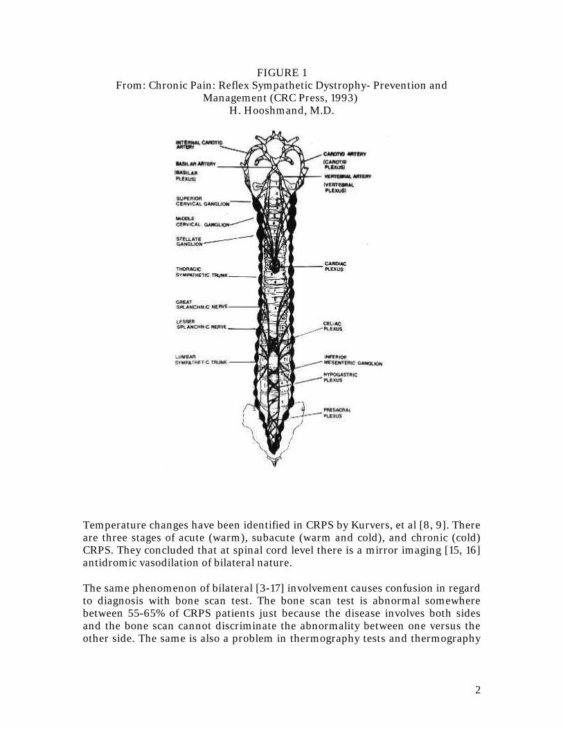
2
FIGURE 1 From: Chronic Pain: Reflex Sympathetic Dystrophy- Prevention and
Management (CRC Press, 1993) H. Hooshmand, M.D.
Temperature changes have been identified in CRPS by Kurvers, et al [8, 9]. There are three stages of acute (warm), subacute (warm and cold), and chronic (cold) CRPS. They concluded that at spinal cord level there is a mirror imaging [15, 16] antidromic vasodilation of bilateral nature.
The same phenomenon of bilateral [3-17] involvement causes confusion in regard to diagnosis with bone scan test. The bone scan test is abnormal somewhere between 55-65% of CRPS patients just because the disease involves both sides and the bone scan cannot discriminate the abnormality between one versus the other side. The same is also a problem in thermography tests and thermography
3
tests frequently show the CRPS hypothermia being on both sides rather than on one side.
The main reason for the CRPS becoming bilateral and spreading to other extremities is because in contrast to the somatic nervous system, the sympathetic nervous system has bilateral innervation. In the somatic nervous system (usual sensation and motor function) the abnormalities in dermatome in a specific nerve root distribution, whereas in CRPS the abnormality is distributed among the blood vessels, distribution of nerves (thermatomes) and to the sympathetic ganglia and their across the midline collections, the condition reflects itself on both sides rather than one side of the body. This bilateral manifestation through the sympathetic plexi across the midline explains the patient's problem with headache, dizziness, tinnitus, chest pain, and abdominal manifestations of CRPS (gastritis, diarrhea, cramps) and spread of CRPS to other extremities.
In treating CRPS, even if the opposite extremity looks normal, the treatment should be given to both extremities because of this principle of bilateral innervation. The enclosed graph shows the bilateral innervation of sympathetic nervous system.
H. Hooshmand, M.D.
References:
1. Mitchell, S. W.: Injuries or Nerves and Their Consequences. Philadelphia, J B.Lippincott and Co., 1872.
2. Appenzeller, O : The Autonomic Nervous System: An introduction to basic andclinical concepts. 4th rev., Elsevier 1990; 148.
3. Block P, Driessen M: The use of 99Tcm-HSA dynamic vascular examination inthe staging and therapy monitoring of reflex sympathetic dystrophy. Nuclear Med Commun. 1991; 12 (8): 725-731.
4. Gibbon JH, Landis EM: Vasodilation in the lower extremities in response toimmersing the forearms in warm water. J Clin Invest 1982; 2: 1019-1036.
5. Hooshmand H, Hashmi H: Complex regional pain syndrome (CRPS, RSDS)diagnosis and therapy. A review of 824 patients. Pain Digest. 1999; 9: 1-24.
6. Kozin F, McCarty DJ, Sims J, et al: The reflex sympathetic dystrophy syndromeI. Clinical and histologic studies: evidence of bilaterality, response to corticosteroids and articular involvement. Am J Med 1976; 60:321- 331.
4
7. Kurvers HA, Jacobs MJ, Beuk RJ, et al: Reflex sympathetic dystrophy:evolution of micro circulatory disturbance in time. Pain 1995; 60:333-340.
8. Kurvers HA, Jacobs MJ, Beuk RJ, et al: The spinal component to skin bloodflow abnormalities in Reflex sympathetic dystrophy. Arch Neurol 1997; 53 :58-65.
9. Munoz-Gomez J, et al: Reflex sympathetic dystrophy syndrome of the lowerlimb in renal transplant patients treated with cyclosporin A. Arth. & Rheum 1991;34 (5) : 625-630.
10. Pickering GW, Hess W: Vasodilation in the hands and feet in response towarming the body. Clin Sci 1993; 213-223.
11. Pulst SM, Haller P: Thermographic assessment of impaired sympatheticfunction and reproducibility. J. Neurosurg 1981; 226: 35-42.
12. Radt P: Bilateral reflex neurovascular dystrophy following a neurosurgicalprocedure. Clinical picture and therapeutic problems of the syndrome. Confinia. Neurl. 1968; 30 (5): 341-348.
13. Schwartzman RJ, Mclellan TL: Reflex Sympathetic Dystrophy. A review. ArchNeurol 1987; 44: 555-561.
14. Schwartzman RJ, Kerrigan J: The movement disorder of reflex sympatheticdystrophy. Neurology 1990; 40:57-61.
15. Schwartzman RJ: Reflex sympathetic dystrophy. Handbook of ClinicalNeurology. Spinal Cord Trauma, H.L.Frankel, editor. Elsevier Science Publisher B.V. 1992; 17: 121-136.
16. Schwartzman RJ: Patterns of spread of complex regional pain syndrome, typeI (reflex sympathetic dystrophy). Pain 2000; 88: 259-266.
17. Uematsu S, Edwin DH, Jankel WR, et al: Quantification of thermalasymmetry, Part 1: Normal values and reproducibility. J Neurosurg 1988; 69:552-5.
1
RSD PUZZLE #19 RSD and Other Organs
"Can RSD affect other internal organs?"
RSD is not usually limited to one part of an extremity or one extremity. Usually, the pathological sympathetic function spreads to adjacent areas.
The first areas becoming involved are the pathway of the sympathetic nerves between the end organ (e.g., foot or hand) and the spinal cord. This results in an inflammation and irritation of the nerves all the way from the end organ to the spinal cord. This is manifested by muscle spasm in the cervical and lumbar spine region, secondary back and neck pain, headache, dizziness, and tinnitus (buzzing in the ears).
This provides excellent treatment opportunities. In the path of the areas of inflammation, the posterior sensory nerve branches corresponding to the level of the involved nerves and secretes substance P (a painful substance half the molecule of endorphine and practically identical to jalapeño pepper extract). This secretion of substance P under the skin in the paraspinal regions can be identified by exerting equal pressure on the two sides of the vertebra, and observing the so-called "red reflex". Pressure on the normal areas causes no reddish discoloration. On the other hand, pressure on the areas of sensory nerve irritation causes a reddish discoloration of the skin which is accompanied by Travaill's Jump Sign. This area of reddish discoloration can be easily blocked and dissipated by injection of local anesthetic such as Marcaine and if the condition is chronic and severe, one can add a small amount of Celestone or Depo - Medrol® to it. This nerve block provides excellent relief of pain and reversal of constriction of the blood vessels.
The next area of involvement of RSD is the spinal cord itself. This is manifested by movement disorder, muscle spasm, weakness of the extremity, as well as urgency and frequency of urination and disturbance of erection.
Invasive procedures such as the insertion of a spinal stimulator can flare-up such an involvement of the spinal cord and it can cause "idiopathic paralysis" due to flare-up and constriction of blood vessels to the spinal cord. The same can be noted in rare cases of insertion of a catheter for sympathetic nerve blocks in the paravertebral or epidural regions.
The next is the visceral involvement with RSD. In RSD the skin is usually cold and the deep structures are hot and have an exaggerated blood circulation. This results in osteoporosis, fracture of the bones, areas of swelling and fluid
2
formation between the bones and joints identified on MRI, and severe pain as well as weakness in the deep structures. This causes a high risk of amputation for the patient. Amputation is totally unnecessary and should never be performed. Just simple weight bearing under the effect of a strong analgesic such as Stadol along with the use of moist heat and epsom salt, exercise and massage for the extremity to reverse the vasoconstriction on the surface and to increase the circulation in the deep structures corrects this situation without the need for amputation. Amputation in RSD is a slow, painful, gradual suicide.
The next structures being involved in some cases of RSD are the blood vessels to the kidney with resultant episodes of sudden brief and temporary bleeding through the kidney accompanied by a marked elevation of blood pressure. The same principle can cause attacks of nose bleeds, severe headache, dizziness, passing out spells as well. Application of Clonodine Patch in the area of the kidney in the flank (in the back) usually results in good relief of such spasm and inflammation of the blood vessels. The patient should be treated with Dibenzyline or Hytrin which are life saving in such patients.
The involvement of other sympathetic midline connections and plexi such as celiac (abdominal pain, peptic ulcer, nausea, vomiting, and weight loss), superior and inferior mesenteric plexi (diarrhea, abdominal cramps, and weight loss), and cardiac plexus (chest pain, abnormal heart beat, tachycardia, and heart attack), and carotid and vertebral plexi (severe vascular headaches, dizziness, tinnitus, attacks of falling spells, and syncopal attacks), should be identified as such and should be treated with the help of Clonodine Patch, Hytrin, or Dibenzyline as well as proper treatment applied to the source of RSD (definitely avoiding ice, but encouraging exercise, moist heat, epsom salt and hot water, and newer antidepressants as the best analgesics of choice for RSD).
The involvement of the same midline plexi (see the enclosed diagram) explains the reason for the involvement of other organs symmetrical on the opposite side such as the opposite hand or opposite foot or opposite side of the head in regard to headache and face pain or involvement of the removed areas such as involvement of right hand because of left knee injury).
Because of the above complex phenomenon and because of the fact that in RSD the sympathetic nerves follow the path of the blood vessels rather than somatic nerve roots resulting thermotomal rather than dermatomal sensory nerve distribution (mistaken for hysterical sensory loss) may cause a complex clinical picture that baffles the clinician and forces the clinician to blame the patient as being hysterical, hypochondriac, and blaming the serious warning signs of RSD as "functional and not organic". The end result is the deadly phrase "it is all in your head" which practically almost all RSD patients have had to put up with in the course of their treatment. Then the patient is sent to the psychiatrist who tries to shut the patient up with strong tranquilizers, benzodiazepams, Haldol, Valium, Xanax, Halcion, Ativan, Tranxene, etc., with further disastrous results by aggravating RSD due to inactivity, and due to the stress of strong addicting
3
benzodiazipams affecting the formation of brain's own endobenzodiazepams and endorphines.
It is about time to learn and understand that sympathetic system is complex, bilateral and diffuse. Its job is alerting mechanism to alert the entire body against stress and its manifestations are complex and multifaceted.
Just because the bone scan is negative (at best 55-65% of RSD patients have positive bone scans) [1] it does not mean that the patient's symptoms and signs are a figment of their imagination (unfortunately approximately 80% of RSD patients are females and more likely discriminated against).
Yesterday, I ran into one of my old classmates at a medical meeting and he said "I hate RSD." I told him so do I and all the patients do too, but we have to understand and manage it
H. Hooshmand, M.D.
Reference:
1. Lee GW, Weeks PM: The role of bone scintigraphy in diagnosing reflexsympathetic dystrophy. J Hand Surg [Am]. 1995; 20: 458-463.
RSD PUZZLE #20 Unexplained Temperature Changes
"I have had RSD for the past 6 years. In the past 1 year, I have been suffering from bouts of fever and skin rash. They don't necessarily happen in the same time. Every time I have a menstrual period, my temperature jumps to 100º to 101º. At other times, in the absence of any kind of infection, my temperature jumps up to 104º to 105º. The blood cultures have been negative and antibiotics do not help. The high temperature lasts one to two days and then it clears up".
Attacks of fever are not unusual in late stage (stages III and IV) of RSD. One of the major functions of the sympathetic system has been influencing and stimulating the immune system.
Years of inflammation of soft tissues due to RSD result in disturbance of function of the immune system. As a result the patient may develop a precipitous drop in platelet cell count, a rise in the white blood cell count, bouts of low or high-grade fever.
Whereas in the first few years the RSD patients have less tendency to suffer from viral infections and upper respiratory tract infections due to contamination from the other members of the family, in stages III and IV of RSD, the reverse is true. The late stage RSD patients are more likely to develop viral or upper respiratory tract infections.
The phenomenon of "aseptic fever" due to the disturbance of the immune system in absence of any type of infection is nothing but the disturbance of the function of the immune system and the manifestation of inflammation secondary to RSD. Such bouts of fever usually subside with effective treatment for RSD such as administration of infusion pump.
In the late stages of RSD, the sympathetic ganglion nerve blocks usually are not effective but the treatment with oral or skin patch alpha blockers can help control the bouts of aseptic fever.
H. Hooshmand, M.D.