‘Rota-Trach™ - Foremost Healthcareforemosthealthcare.net/wp-content/uploads/Rota-Trach-1.pdf ·...
55
‘Rota-Trach™’ Double Lumen Tracheostomy Tube VITALTEC
Transcript of ‘Rota-Trach™ - Foremost Healthcareforemosthealthcare.net/wp-content/uploads/Rota-Trach-1.pdf ·...

‘Rota-Trach™’Double Lumen Tracheostomy Tube
VITALTEC

Contents
➢ INTRODUCTION ➢HISTORY REVIEW ➢ANATOMY & PHYSIOLOGY ➢OPERATIVE PROCEDURE ➢THE RANGE OF TRACHEOSTOMY TUBES ➢ROTA-TRACH TRACHEOSTOMY TUBE

INTRODUCTION

INTRODUCTION➢The most comfortable, safe & convenient double
lumen Tracheostomy Tube
➢Inner Canula is made of Polyurethane (PU)- provides good flexibility, no toxicity & good compatibility with human body
➢‘ROTA-TRACH’ Tube Connection mechanism is (push & clip) to connect unlike other International brands which has either twist system or stop ring system which are very inconvenient

➢Being 100% metal free makes ‘ROTA-TRACH’ the only International brand to be MRI compatible
➢Each unit of ‘ROTA-TRACH’ comes with an individual disconnect wedge to avoid cross infection

History Review
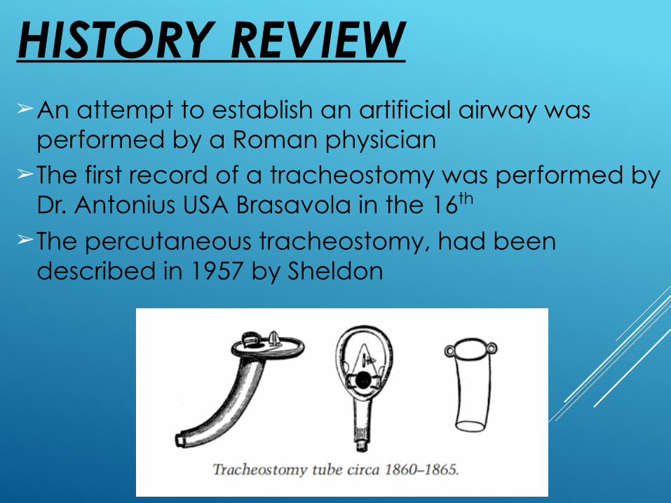
➢An attempt to establish an artificial airway was performed by a Roman physician
➢The first record of a tracheostomy was performed by Dr. Antonius USA Brasavola in the 16th
➢The percutaneous tracheostomy, had been described in 1957 by Sheldon
HISTORY REVIEW

ANATOMY &
PHYSIOLOGY

Anatomy and Physiology
➢The trachea is a tubular structure 10–15 cm in length in an adult
➢It is constructed of 15 to 20 C-shaped cartilaginous rings separated by fibrous muscular tissue which form the supporting framework. Each cartilage is incomplete dorsally where it is adjacent to the esophagus
➢The tracheal structure consist of four layers: mucosa, submucosa, cartilage, and adventitia

Trachea
➢The trachea is between 9~15mm in diameter in the adult
➢The adult male trachea will generally easily accept an 8.0~8.5mm inner diameter (ID) tracheostomy tube

➢The adult female may prefer a 7.5mm ID tube
➢Pediatric airway anatomy varies from adult anatomy
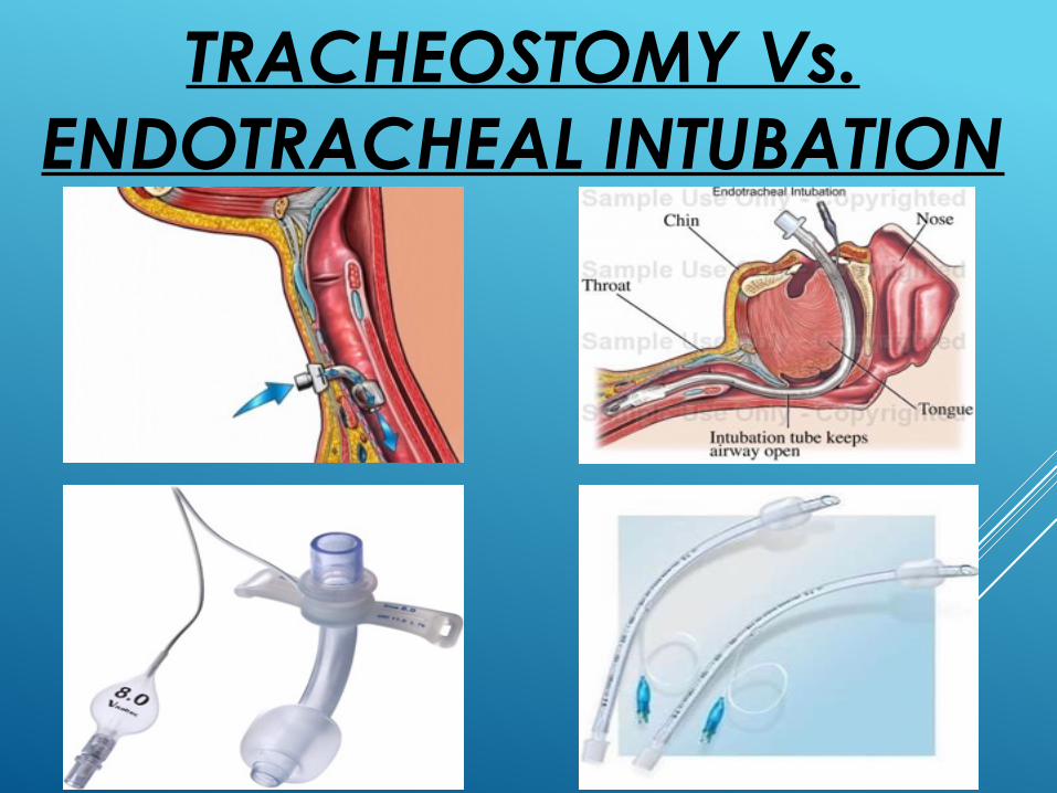
TRACHEOSTOMY Vs. ENDOTRACHEAL INTUBATION

➢Reduce need for sedation
➢Reduce damage to glottis ➢Reduce work of breathing by reducing low dead
space
➢Long term ventilation support

Indications for Tracheostomy
➢Prolonged or expected prolonged intubation ➢Inability of patient to manage secretions ➢Facilitation of ventilation support ➢Inability to intubate ➢Adjunct to manage head and neck surgery ➢Adjunct to manage significant head and neck trauma Defined by the American Academy of Otolaryngology –
Head and Neck Surgery in the Clinical Indicators Compendium

OPERATIVE PROCEDURE

Surgical Technique for Tracheostomy
➢Patient positioning with neck hyperextended to expose the laryngotracheal landmark
➢An incision is made through the 2nd, 3rd, or 4th tracheal cartilage
➢Removing tracheal ring to form window ➢A tracheostomy tube of proper size and
length is inserted through the anterior wall ➢The tracheostomy tube is gently positioned
and ventilation is confirmed through the tube

y

Complications of Tracheostomy
Immediate surgical complications ➢Bleeding from the operative site ➢Subcutaneous emphysema, mediastinal
emphysema ➢Aspiration of blood in the airway ➢Cardiac arrest secondary to hypoxia or
acidosis

Post-Tracheostomy Complication
➢Hemorrhage ➢Granuloma formation ➢Tracheo-esophageal fistula ➢Difficult decannulation ➢Tracheocutaneous fistula ➢Laryngotracheal stenosis ➢Tracheostomy scar

Complications caused by inappropriate tracheostomy tubes
➢Tracheal ulceration and necrosis ➢Tracheal stenosis (narrowing) ➢Tracheo-oesophageal fistula ➢Tracheo-onimate artery fistula and hemorrhage ➢Stoma ulceration and breakdown ➢Overgranulation tissue ➢Tracheal irritation and coughing ➢Discomfort

Tracheostomy Tube Dimensions
➢The tubes are sized according to the functional internal diameter (ID) at the narrowest point, the ID of the outer cannula for the case of single cannula tubes, and the ID of the inner cannula for double cannula tubes
➢The selection is a comprise between a desire to maximize the functional internal diameter (reduce airway resistance), and a need to limit the OD to approximately ¾ of the internal diameter of the trachea
➢A tube that is too small may result in the need to over-inflate the cuff, increasing the risk of mucosal pressure necrosis, which in turn increases the risk of complications such as tracheal stenosis and trachea-esophageal fistula
➢Most tracheostomy tubes are sized by internal diameter in millimeters, but this may not take account of inner cannula in all cases
➢Some of the inner cannula products are still described according to the Chevalier Jackson sizing system originally developed for metal tubes (the appropriate information is available on the neck plate of the tube)


THE RANGE OF TRACHEOSTOMY
TUBES

Cuffed Tracheostomy Tube & Uncuffed Tracheostomy Tube
Cuffed tracheostomy tubes are developed for patients require positive pressure ventilator and airway protection
Uncuffed tube is used when the patient no longer needs positive pressure ventilation and has no significant aspiration risk. The tube is still for access to chest secretions or bypass an upper airway obstruction

Cuffed tracheostomy tubes
➢The cuff should be of a “high volume / low pressure” design, and effectively seal the trachea at a pressure of no more than 20 – 25 cm H2O in order to minimize the risk of
tracheal mucosal ischemia and subsequent tracheal stenosis

FENESTRATION TRACHEOSTOMY TUBES
➢ Cuffed FEN tube is particularly useful when a stable patient is weaning from tracheostomy
➢ Uncuffed FEN tubes are used for patients who are no longer dependent on a cuffed tube

TRACHESTOMY TUBE FOR PEDIATRIC AND NEONATAL
PATIENT➢ Children under the age of
12 years have a narrow trachea particularly around the cricoid ring, and therefore air leak is minimal. This enable an uncuffed tracheostomy tube to be used effectively

DOUBLE CANNULA (INNER CANNULA)

Inner Cannula Tracheostomy Tube
➢An inner cannula allows it to be cleaned or replaced at regular intervals
➢It allows the immediate relief of life-threatening airway obstruction
➢The principal disadvantage is that the inner cannula may significantly reduce the effective inner diameter of the tracheostomy tube and thereby increase the work of breathing and impair weaning

ROTA-TRACH™ TRACHEOSTOMY TUBE

STANDARD TRACHESOTOMY TUBE –
POLYVINYL CHLORIDE (PVC) The medical grade PVC is the most cost effective
material for the short term tube.
➢ It allows the tube to be flexible maintains the shape.
➢ It is thermo sensitive to adjust to the body temperature
➢ DEHP Free

15mm connector
Low pressure cuff
Pilot balloon
Multiple fenestration Inflation line
Soft neck flange
Free-angle connector
Tube angle 100°
STANDARD TRACHESTOMY TUBE

Low pressure cuff
Clear size indication on pilot balloon
multiple fenestration
Metal free balloon valve
– MRI compatible
Cap for protection of air leak

Inner Cannula Tracheostomy Tube – Polyurethane (PU)
➢ Polyurethane has the advantages of good flexibility, no toxicity, and good compatibility with the human body
➢ This material is more prone to the retention of bacteria

Inner cannula
ObturatorOuter cannula
Secure device
Free angle connector
INNER CANNULA TRACHESTOMY TUBE

Rota-Trach Tracoe Portex Shiley LPC DPC
Inner cannula/tube Reusable Reusable Disposabl
e Reusable Disposable
Oscillating Connector ˇ ˇ X X X
Tube connection Mechanism
Push and Clip to
connect
Twist to connect Stop ring Twist to
connect Clip to
connect
Disconnect Wedge
Standard package Optional Optional Optional Optional
Material for use PU PU PVC PVC PVC
MRI Valve Metal freeNon
metal free
Non metal free
Non metal free
Non metal free
COMPARISON CHART

Vitaltec Inner cannula TT
➢ Polyurethane (PU)
➢ Free angle connector
➢ Soft neck plate fit the neck curves
➢ Clustered small hole at fenestration, avoid inner tube to pass through outer tube and stimulate trachea
➢ Thinner tube wall bring bigger ID of tube can increase the ventilation
Shiley Inner cannula TT➢ Polyvinyl Chloride (PVC)
➢ Fixed connector
➢ Hard neck plate, make discomfort for patient
➢ Single big hole at fenestration, inner tube may pass through outer tube easily and stimulate trachea
➢ Thicker tube wall bring small ID of tube, lower the ventilation

REF Size I.D. O.D. Length Cuff Ø (mm) (mm) (mm) (mm)
Shiley LPC 4LPC 4 5.0 9.4 65 20 6LPC 6 6.4 10.8 76 24 8LPC 8 7.6 12.2 81 27 10LPC 10 8.9 13.8 81 29 Vitaltec VT550 5500700 7.0 7.0 10.6 77 23 5500800 8.0 8.0 11.7 79 26 5500900 9.0 9.0 12.8 81 30 5501000 10.0 10.0 13.8 83 33

Inner cannula integrates free angle connector
Lower pressure cuf
Clear size indication On plot balloon
Metal free balloon valve MRI compatible
Multiple fenestration

Free Angle Connector (Patent protected)
The patient friendly design allows the connector to swivel freely , absorbing pressure of any movement from connector, and significantly reducing patient discomfort

Disconnect Wedge
➢Allow the disconnection of the product with minimal discomfort to the patients
➢Back clip is designed to fix pilot balloon on patient cloth
➢Single wedge is included with each kit to avoid cross infection

Inner CannulA–Secure DeviceLock inner cannula to the outer tube quickly and securely

TRACHEOSTOMY CARE
➢Cuff management
➢Humidification
➢Suctioning
➢Wound management
and dressing

Cuff Management
➢ The cuff provides a seal to enable positive pressure ventilation and also provides some protection against aspiration of secretions
➢ It is good practice to document cuff pressure and inflating volume on a daily basis
➢ Cuff pressure should not exceed 25 cm H2O ➢ If an air leak occurs with the cuff pressure at
the maximum recommended, the tracheostomy may need to be displaced or require changing
Humidification
➢ Inadequate humidification may lead to life-threatening blockage of the tracheostomy with tenacious sputum, sputum retention, and impaired gas exchange etc.
➢HMEs may be used for patients who remain mechanically ventilated and offer the additional advantage of bacterial filtration
➢Patients with more tenacious sputum, or requiring high flow oxygen therapy will require additional saline nebulizers and may require heated water humidification

Suctioning ➢ Appropriate suction will stimulate the cough reflex and
prevent accumulation of secretions which can block the tracheostomy
➢ Suctioning may be painful and distressing for the patient, and can also be complicated by hypoxemia, tracheal mucosal damage, bleeding, and introduction of infection etc.
➢ The frequency of suction required depends on the individual patients need
➢ Maintaining a closed suction system decreases patient’s risk of infection, helps maintain positive end expiratory pressure (PEEP) and oxygen levels during suctioning, and protects the nursing staff from exposure to patient secretions

Wound Management and Dressing
➢The stoma site should be assessed at least daily
➢The skin and stoma is cleaned using aseptic technique
➢The self-cut dressings are avoided to prevent the risk of
loose fibers which can break off and enter the stoma
➢The Velcro neck strap is recommended for
tracheostomy use, as they are easy to apply and adjust,
are more comfortable for the patient and less abrasive
to the skin

Changing Tracheostomy Tubes
➢The tube should only be changed once a tract has formed between the trachea and the skin (after 48–72 hr)
➢It is recommended the tubes be changed depending on the manufactures guideline. Tubes without an inner cannula need to be changed more frequently to ensure a patent airway
➢It is recommend that single lumen tubes are changed every 10 -14 days
➢A European Economic Community Directive (1993) states that tracheostomy tubes with an inner cannula can remain in place for a maximum of thirty days

Guidelines for changing inner cannula
➢Position patient with neck slightly extended ➢Preoxygenate and suction as necessary ➢Using a sterile technique, remove or change inner
cannula. Disposable inner cannula should be discarded after used. If non disposable clean cannula with 0.9% sterile saline or water and dry thoroughly
➢Clear persistent secretions on cannula in line with manufacturer’s instructions, rinsing the inner cannula thoroughly with normal saline or water before re-insertion. Do not leave inner cannula to soak

➢The inner cannula must be removed, inspected and when necessary cleaned at regular intervals
➢The frequency of inspection being determined by the volume and tenacity of a patient’s secretions

Decannulation
Decannulation is the permanent removal of the tracheostomy tube. It should only be considered when the indication for insertion of the tracheostomy has resolved.

Indications to proceeding with weaning
➢Reason for the tracheostomy resolved ➢Patient alert, responsive and consenting ➢Patient tolerating cuff deflation for a minimum of 12 hr ➢Patient managing to protect their airway and have a
clear chest ➢Patient maintaining oxygen saturation ➢Patient tolerating the use of speaking valve and/or
digital occlusion ➢Patient able to expectorate around the tube into their
mouth ➢ Tracheostomy tube type and size is appropriate

The Choice of Neck Strap
Neck Strap with Velcro fastener
Consist of 2-pieces of neck strap provide secure positioning of tracheostomy tube
Neck Strap with Velcro fastener & elastic
Consist of 2-pieces, adjustable length with of neck strap with elastic