Histidine-rich glycoprotein-induced vascular normalization ...
JOURNAL OF BACTERIOLOGY, Sept. 2011, p. 4904–4913 Vol. 193, No. 180021-9193/11/$12.00 doi:10.1128/JB.05231-11Copyright © 2011, American Society for Microbiology. All Rights Reserved.
Roles of the Redox-Active Disulfide and Histidine Residues Forminga Catalytic Dyad in Reactions Catalyzed by 2-Ketopropyl
Coenzyme M Oxidoreductase/Carboxylase�
Melissa A. Kofoed,1 David A. Wampler,1† Arti S. Pandey,2‡ John W. Peters,2 and Scott A. Ensign1*Department of Chemistry and Biochemistry, Utah State University, Logan, Utah 84322,1 and Department of Chemistry and
Biochemistry and Astrobiology Biogeocatalysis Research Center, Montana State University, Bozeman, Montana 597172
Received 4 May 2011/Accepted 5 July 2011
NADPH:2-ketopropyl-coenzyme M oxidoreductase/carboxylase (2-KPCC), an atypical member of the disul-fide oxidoreductase (DSOR) family of enzymes, catalyzes the reductive cleavage and carboxylation of 2-keto-propyl-coenzyme M [2-(2-ketopropylthio)ethanesulfonate; 2-KPC] to form acetoacetate and coenzyme M(CoM) in the bacterial pathway of propylene metabolism. Structural studies of 2-KPCC from Xanthobacterautotrophicus strain Py2 have revealed a distinctive active-site architecture that includes a putative catalytictriad consisting of two histidine residues that are hydrogen bonded to an ordered water molecule proposed tostabilize enolacetone formed from dithiol-mediated 2-KPC thioether bond cleavage. Site-directed mutants of2-KPCC were constructed to test the tenets of the mechanism proposed from studies of the native enzyme.Mutagenesis of the interchange thiol of 2-KPCC (C82A) abolished all redox-dependent reactions of 2-KPCC(2-KPC carboxylation or protonation). The air-oxidized C82A mutant, as well as wild-type 2-KPCC, exhibitedthe characteristic charge transfer absorbance seen in site-directed variants of other DSOR enzymes but witha pKa value for C87 (8.8) four units higher (i.e., four orders of magnitude less acidic) than that for the flavinthiol of canonical DSOR enzymes. The same higher pKa value was observed in native 2-KPCC when theinterchange thiol was alkylated by the CoM analog 2-bromoethanesulfonate. Mutagenesis of the flavin thiol(C87A) also resulted in an inactive enzyme for steady-state redox-dependent reactions, but this variantcatalyzed a single-turnover reaction producing a 0.8:1 ratio of product to enzyme. Mutagenesis of the histidineproximal to the ordered water (H137A) led to nearly complete loss of redox-dependent 2-KPCC reactions, whilemutagenesis of the distal histidine (H84A) reduced these activities by 58 to 76%. A redox-independent reactionof 2-KPCC (acetoacetate decarboxylation) was not decreased for any of the aforementioned site-directedmutants. We interpreted and rationalized these results in terms of a mechanism of catalysis for 2-KPCCemploying a unique hydrophobic active-site architecture promoting thioether bond cleavage and enolacetoneformation not seen for other DSOR enzymes.
The bacterial metabolism of gaseous propylene by theproteobacterium Xanthobacter autotrophicus Py2 and the ac-tinomycete Rhodococcus rhodochrous B276 is initiated bythe insertion of a single oxygen atom into the olefin bond ofpropylene, forming (R)- and (S)-epoxypropane (19, 27, 35,39). These epoxypropane enantiomers are further metabo-lized by a three-step linear pathway that uses four enzymesand the atypical cofactor coenzyme M (CoM) (2-mercapto-ethanesulfonic acid) to catalyze the net carboxylation ofepoxypropane to form acetoacetate as shown in Fig. 1 (1, 2,4, 5, 18, 24). NADPH:2-ketopropyl-coenzyme M oxidoreductase/carboxylase (2-KPCC) is the CO2-fixing enzyme of this path-way, catalyzing the reductive cleavage and carboxylation of2-ketopropyl-CoM (2-KPC) with the stoichiometry shown inequation 1 (1, 17, 18):
2-KPC � CO2 � NADPH3 acetoacetate
� NADP� � CoM (1)
2-KPCC is the only known carboxylase that is a member of thedisulfide oxidoreductase (DSOR) family of enzymes and, ac-cordingly, employs a mechanistic strategy for organic substratecarboxylation not seen in any other known enzyme (10, 18). Allmembers of the DSOR family follow the general mechanisticstrategy shown in Fig. 2A for the steps leading to the reductionof a cysteine disulfide bond, where the cysteine residuesproximal and distal to flavin adenine dinucleotide (FAD)are termed the flavin and interchange thiols, respectively (29).For the DSOR enzymes catalyzing the reduction of an oxidizedsubstrate containing a disulfide bond such as glutathione re-ductase, the interchange thiol attacks one of the sulfur atomsof the disulfide, leading to disulfide bond cleavage, reductionof one thiol of the substrate, and formation of a mixed disulfidebetween the interchange thiol and the second substrate thiol(29) (Fig. 2B). Reformation of the oxidized cysteine pair leadsto the reduction and release of the second substrate thiol.
As shown in Fig. 2C, 2-KPCC catalyzes the reduction of athioether rather than a disulfide bond, a feature not seen in anyother known DSOR enzyme. Mechanistic (10) and structural(28, 30, 31) studies have provided evidence for a reaction
* Corresponding author. Mailing address: Department of Chemistryand Biochemistry, Utah State University, 0300 Old Main Hill, Logan,UT 84322. Phone: (435) 797-3969. Fax: (435) 797-3390. E-mail: [email protected].
† Present address: University of Texas Health Science Center at SanAntonio, San Antonio, TX 78229.
‡ Present address: Department of Biochemistry, Kathmandu Medi-cal College and Teaching Hospital, Duwakot, Bhaktapur, Nepal.
� Published ahead of print on 15 July 2011.
4904
Dow
nloa
ded
from
http
s://j
ourn
als.
asm
.org
/jour
nal/j
b on
09
Febr
uary
202
2 by
220
.82.
44.1
91.

mechanism where thioether bond cleavage results in the for-mation of a mixed disulfide between CoM and the interchangethiol with the formation of the enolacetone anion (Fig. 2C).The enolacetone anion then undergoes carboxylation to formthe product acetoacetate, while the formation of the oxidizedcysteine pair results in the release of free CoM (Fig. 2C).
It is of interest to determine the unique features of 2-KPCCrelative to other DSOR enzymes that allow the enzyme tocatalyze the unique reactions of thioether bond cleavage,enolacetone anion formation, stabilization, and carboxylation.Significant insights into these features have come from X-raycrystallographic structures of 2-KPCC determined for variousstates, including the substrate-free enzyme (28), a 2-KPC-bound form of the enzyme (28), a form of the enzyme wherethe mixed disulfide of CoM was trapped (31), and a form inwhich the substrate CO2 is bound (30). Collectively, thestructural characterization of 2-KPCC revealed an active-site architecture unique among DSOR enzymes, where sub-strate binding induces a conformational change that createsa hydrophobic pocket that encapsulates the substrate 2-KPC(22, 28). Of relevance to the question of how 2-KPCC stabilizesthe enolacetone anion for subsequent carboxylation is that ahydrogen-bonding network was identified in the 2-KPC-boundenzyme, consisting of an ordered water molecule hydrogenbonded to both the carbonyl oxygen of 2-KPC and two histi-dine residues (H137 and H84) (Fig. 3A). It has been proposedthat this hydrogen-bonding network is responsible for stabiliz-ing enolacetone formed from thioether bond cleavage, as the
interchange thiol attacks the thioether bond, as shown in Fig.3B (22, 28). An additional novel feature of the active site of2-KPCC is a pair of methionine residues (M140 and M361),which flank the substrate 2-KPC. These methionine residuesare not present in other DSOR enzymes and may contribute tothe unique environment that promotes enolacetone stabiliza-tion and attack on CO2.
The construction of site-directed substitutions of 2-KPCCwould allow the tenets of this proposed mechanism to be testeddirectly. To date, however, all attempts to express 2-KPCC inan active state in a heterologous system have failed, in spite ofthe relative ease with which the other enzymes of the epoxy-propane carboxylation pathway have been expressed and pu-rified (11, 24, 34). In the present paper, we describe an expres-sion system that solves the problems encountered previouslyand allows 2-KPCC to be expressed in a fully active and solublestate. Site-directed mutants were constructed in the redox-active disulfide, the histidine dyad, and one of the flankingmethionine residues, and the effects of these mutations onvarious reactions catalyzed by 2-KPCC were determined inorder to gain further insights into their catalytic roles.
MATERIALS AND METHODS
Materials. Commercially available compounds used were of analytical gradeand were purchased from either Sigma-Aldrich Chemicals or Fisher Scientific.2-KPC was synthesized as described previously (1). All oligonucleotides werepurchased from Integrated DNA Technologies.
FIG. 1. Pathway of aliphatic epoxide carboxylation in Xanthobacterautotrophicus Py2 and Rhodococcus rhodochrous B276.
FIG. 2. Reactions of members of the DSOR family. (A) Stepsresulting in the reduction of the redox-active cysteine disulfide for allDSOR enzymes; (B) steps resulting in reduction of a substrate withan oxidized disulfide bond, as illustrated for glutathione reductase;(C) steps resulting in reduction of the thioether bond of 2-KPC in2-KPCC, resulting in the production of enolacetone, which undergoescarboxylation to form acetoacetate. Note that the reduced flavin cys-teine is shown in the thiol rather than as the thiolate form for 2-KPCCfor reasons described in the paper.
VOL. 193, 2011 MECHANISM OF 2-KETOPROPYL-CoM CARBOXYLATION 4905
Dow
nloa
ded
from
http
s://j
ourn
als.
asm
.org
/jour
nal/j
b on
09
Febr
uary
202
2 by
220
.82.
44.1
91.

Purification of native 2-KPCC from Xanthobacter autotrophicus strain Py2.Native 2-KPCC was purified from propylene-grown X. autotrophicus Py2 asdescribed previously (3).
Plasmid construction of recombinant 2-KPCC. Total genomic DNA was iso-lated from propylene-grown cells of X. autotrophicus strain Py2 as describedpreviously (24). The gene encoding 2-KPCC (xecC) was amplified by PCR usingthe primers 5�-CACCGTGAAAGTCTGGAACGCCC-3� and 5�-TCACAGGCTCACCAGATTCT-3� according to the Failsafe PCR protocol (Epicentre Bio-technologies, Madison, WI). PCR products were then ligated into a pBADDirectional TOPO vector (Invitrogen) according to the manufacturer’s protocolto generate plasmid pDW1, which was then transformed into Escherichia coliOne Shot Top10 cells (Invitrogen). pDW1 was isolated and the insert sequencewas confirmed by DNA sequencing at the Center for Integrated BioSystems,Utah State University.
Site-directed mutagenesis. Site-directed mutagenesis of pDW1 was carried outby utilizing a QuikChange site-directed mutagenesis kit (Stratagene) accordingto the manufacturer’s protocols. The sequences of the primer pairs used to createthe desired mutations are as follows: C82A, 5�-TCC TGG GCG GCT CGG CCCCGC ACA ATG CGT-3� and 5�-AAG GAC CCG CCG AGC CGG GGC GTGTTA CGC-3�; C87A, 5�-GTG CCC GCA CAA TGC GGC CGT GCC GCACCA TAT GTT-3� and 5�-GAA CAG ATG GTG CGG CAC GGC CGC ATTGTG CGG GCA-3�; H137A, 5�-GAA GTT CAT GAT GCC GGC CGG GCCGTT GCG CC-3� and 5�-CGC AAC GGC CCG GCC GGC ATCATG AACTTC CA-3�; H84A, 5�-CAC GCA CGC ATT GGC CGG GCA CGA GCCGCC-3� and 5�-CCG CCG AGC ACG GGC CGG TTA CGC ACG CAC-3�; andM140A, 5�-CGG CCC GCA CGG CAT CGC GAA CTT CCA GTC CAAGG-3� and 5�-CCT TGG ACT GGA AGT TCG CGA TGC CGT GCG GGC
CG-3�. Mutations were confirmed by primer extension sequencing at SeqWrightDNA Technology Services (Houston, TX).
Growth media. E. coli Top10 cells were grown in Luria-Bertani rich (LB-Rich)broth containing ampicillin (100 �g/ml). The LB-Rich media contained thefollowing components per liter: 20 g of tryptone, 15 g of yeast extract, 2 g ofK2HPO4, 1 g of KH2PO4, and 8 g of NaCl.
Growth of bacteria. All bacteria were grown at 37°C unless otherwise stated.E. coli Top10 cells that had been transformed with the pDW1 or correspondingmutant plasmid were plated and grown overnight. A single colony from this platewas used to grow a 25-ml liquid culture to an A600 of 0.6 for the preparation of25% glycerol (vol/vol) stocks that were stored at �80°C until use. For use, cellsfrom a frozen stock were inoculated into 125 ml of LB-Rich medium and grownto an A600 between 0.6 and 1.0, as measured on a Shimadzu UV160U spectro-photometer. This culture was used as the inoculum for a 15-liter capacity Mi-croferm fermentor (New Brunswick Scientific) containing 12 liters of LB-Richmedia supplemented with riboflavin (15 mg/liter) and antifoam A (0.005% [vol/vol]). Cells were allowed to grow at 37°C with agitation at 400 rpm and forcedaeration to an A600 between 0.6 and 1.0. At this time, the temperature wasreduced to 30°C, arabinose was added to 0.02%, and the cells were allowed togrow at this temperature for 6 h. Cells were concentrated using a tangential flowfiltration system (Millipore) and were pelleted by centrifugation. Cell paste wasdrop frozen in liquid nitrogen and stored at �80°C.
Purification of recombinant 2-KPCC. Cell paste was resuspended in 3 volumesof buffer A (50 mM Tris, 1 mM dithiothreitol [DTT], 0.1 mM EDTA, and 5%glycerol [vol/vol], prepared and buffered to pH 7.4 at 4°C) with DNase I (0.03mg/ml) and lysozyme (0.03 mg/ml) and thawed at 30°C with shaking. All subse-quent treatments were performed either on ice or at 4°C. The cell suspension was
FIG. 3. Active-site architecture and proposed mechanism for 2-KPCC. (A) Structure of 2-KPC bound to 2-KPCC, highlighting active-siteresidues believed to be key to catalysis (Protein Data Bank ID, 1MO9). (B) Proposed mechanism of thioether bond cleavage, enolacetoneformation and stabilization, and carboxylation based on the structures solved for 2-KPCC and the results of the present work. The initialabstraction of a proton from C82 may be facilitated by a general base that has not yet been identified. The reduction of the mixed disulfide of CoMand Cys82 is not shown but will occur as for glutathione reductase (Fig. 2B).
4906 KOFOED ET AL. J. BACTERIOL.
Dow
nloa
ded
from
http
s://j
ourn
als.
asm
.org
/jour
nal/j
b on
09
Febr
uary
202
2 by
220
.82.
44.1
91.
passed three times through a French pressure cell at a pressure of 1.1 � 105 kPaand clarified by centrifugation (184,000 relative centrifugal force for 30 min at4°C). Clarified cell extract was applied to a 0.5- by 5.0-cm column of a Ni-NTASuperflow instrument (Pharmacia Biotech) at 7.0 ml/min. The column was thenwashed with 4-column volumes of buffer A, and the bound sample was elutedwith a 15-column volume gradient from 0 to 400 mM imidazole. Purification wasfollowed by SDS-PAGE. Appropriate fractions were pooled, and (NH4)2SO4 wasadded to 800 mM. The solution was incubated at 4°C with gentle stirring andthen applied to a 2.6- by 5.5-cm column of phenyl Sepharose that had beenpreequilibrated with buffer A plus 800 mM (NH4)2SO4 (buffer C). The columnwas then washed with 4-column volumes of buffer C, and bound protein waseluted with a 15-column volume gradient from 0 to 100% of buffer A followed byan additional 5-column volume of buffer A. Appropriate fractions were pooledand concentrated by ultrafiltration using a YM30 membrane (Amicon).
For removal of the histidine patch (HP) thioredoxin leader acquired from thepBAD Directional TOPO vector, protein samples prepared as above were incu-bated with enterokinase (0.1 U EKMax; Invitrogen) at 4°C for 24 h. The proteinwas then reloaded onto the Ni-NTA Superflow column. Appropriate fractionswere pooled, concentrated by ultrafiltration, and frozen drop-wise in liquidnitrogen for storage at �80°C.
Protein concentrations were determined using a modified biuret assay (9).2-KPCC concentrations were also determined by using the previously deter-mined extinction coefficient (ε450 of 11,828 M�1 � cm� 1) (3).
SDS-PAGE and immunoblotting procedures. SDS-PAGE (12% total acryl-amide) was performed according to the Laemmli procedure (25). Electropho-resed proteins were visualized by staining with Coomassie blue R-250. Theapparent molecular masses of polypeptides were determined by comparison withRf values of standard proteins. Immunoblot analysis was conducted by electro-phoretically transferring proteins from an SDS-polyacrylamide gel onto a poly-vinylidene difluoride membrane. The membrane was incubated with polyclonalantiserum raised against purified 2-KPCC from X. autotrophicus strain Py2.Cross-reacting proteins were visualized using horseradish peroxidase conjugatedto goat anti-rabbit immunoglobulin G (Promega).
Coupled spectrophotometric assay for 2-KPCC carboxylation activity. A con-tinuous spectrophotometric assay described previously (8) that couples acetoac-etate production by 2-KPCC to acetoacetate reduction and NADH oxidation by�-hydroxybutyrate dehydrogenase (�-HBDH) was utilized. This assay relies onthe fact that DTT can be used as an alternate reductant for 2-KPCC in place ofNADPH while NADH cannot, so that loss of absorption at A340 due to NADHoxidation by �-HBDH as acetoacetate is reduced can be measured according toequations 2 and 3 (8):
2-KPC � CO2 � DTTred3 acetoacetate � DTTox � CoM (2)
acetoacetate � NADH � H�3 �-hydroxybutyrate � NAD� (3)
where DTTred is reduced DTT and DTTox is oxidized DTT. By including a largeexcess of highly active �-HBDH (260 U/mg, where one unit is defined as 1�mol/min) in assays with a fixed concentration of NADH, steady-state rates andkinetic parameters can be measured that cannot be measured in assays whereNADPH is consumed directly by 2-KPCC in stoichiometric proportions to2-KPC.
Assays were conducted in 2-ml anaerobic quartz cuvettes that contained a totalreaction volume of 1 ml. Assays contained either 0.125 or 0.25 mg of 2-KPCC,0.345 mg (90 units) of �-HBDH, 10 mM DTT, 0.2 mM NADH, and 60 mMcarbonate species (added as 33.5 mM CO2 gas plus 26.5 mM KHCO3) in 100 mMTris buffer, with the pH adjusted to 7.4 at 30°C. Reactions were allowed toequilibrate to 30°C, and assays were initiated by the addition of 2.5 �mol of2-KPC.
Continuous spectrophotometric assay for 2-KPC formation (reverse reac-tion). As described previously (10), 2-KPCC catalyzes the reverse of the physi-ologically important forward reaction shown in equation 1, a reaction that isdependent on NADP� as the oxidant, as shown in equation 4:
acetoacetate � NADP � � CoM3 2-KPC � CO2 � NADPH (4)
Assays were conducted in 2-ml anaerobic quartz cuvettes with a total reactionvolume of 1 ml. Each assay contained 0.125 mg of 2-KPCC, 5 mM NADP�, 5mM CoM, and 100 mM acetoacetate in 100 mM Tris buffer [pH 7.4]. Reactionswere allowed to equilibrate to 30°C, and assays were initiated by the addition ofacetoacetate. Rates of reaction were measured by monitoring the increase inabsorbance (A340) associated with the production of NADPH in a ShimadzuUV160U spectrophotometer containing a water-jacketed cell holder for temper-ature control.
2-Ketopropyl-CoM protonation assays. In the absence of CO2, 2-KPCC cat-alyzes the cleavage and protonation of 2-KPC to form acetone, using eitherNADPH or DTT as the reductant, at rates about 25% of the rate of 2-KPCcarboxylation, as shown using DTT as the reductant in equation 4 (10):
2-KPC � H� � DTTred3 acetone � DTTox �CoM (5)
Assays for acetone formation were performed essentially as described previously(10). Assays were conducted in 9-ml sealed serum vials with a total reactionvolume of 1 ml. Each assay contained either 0.125 or 0.25 mg of 2-KPCC and 10mM DTT in 100 mM Tris buffer [pH 7.4]. Assay vials were incubated in a 30°Cshaking water bath, and assays were initiated by the addition of 2.5 �mol of2-KPC. Acetone formation was quantified as a function of time by removingsamples and analyzing acetone by gas chromatography as described previously(13).
Single-turnover protonation assay. Assays were performed in sealed 3-mlserum vials with a total reaction volume of 1 ml. Each assay contained 2-KPCC(0, 57, or 144 �M) in 100 mM Tris buffer [pH 7.4]. Assay vials were incubated ina 30°C shaking water bath (200 cycles min�1), and assays were initiated by theaddition of 2.5 mM 2-KPC. After 10 min., the vials were transferred to a 65°Cstatic water bath and incubated for 10 min to allow denaturation of the proteinand volatilization of the acetone released. Acetone was quantified by gas chro-matography as described above.
Acetoacetate decarboxylation assay. Assays were performed as described pre-viously (10). Briefly, assays were conducted in 9-ml sealed serum vials with a totalreaction volume of 1 ml. Vials were depleted of carbonate species by includinga KOH-containing trap as described previously (13). Each assay contained 250mM acetoacetate and either 0 or 5 mM CoM in 100 mM Tris buffer [pH 7.4].Assay vials were incubated in a 30°C shaking water bath, and assays wereinitiated by the addition of 0.125 mg 2-KPCC. Acetone was quantified by gaschromatography as described above.
Preincubation of 2-KPCC with 2-KPC. For assays that used 2-KPCC prein-cubated with 2-KPC, 2-KPCC and 2-KPC were incubated together for 5 min at30°C in GTP buffer (200 mM [each] glycine, Tris, and sodium phosphate) (pH10). Following incubation, 2-KPCC was desalted by loading it onto a 2.3- by 5-cmcolumn of Sephadex G-25 PD-10 that had been preequilibrated with 50 mM Trisbuffer (pH 7.4).
Cys87-flavin charge transfer. All spectra were obtained at room temperaturein a quartz ultramicro-sized (120-�l) cuvette with a 1-cm light path. AliquotedGTP buffer was adjusted to the desired pH and added to 2-KPCC for a finalvolume of 130 �l. Spectra were then obtained using a Shimadzu UV160UUV-Vis recording spectrophotometer interfaced with a personal computer run-ning PC160 Personal Spectroscopy software, version 1.4. The reference cellcontained protein-free GTP buffer. The actual pH of each solution used toobtain a spectrum was determined by mixing an appropriate amount of protein-free buffer A with each of the GTP buffers.
UV/visible spectral analysis of native 2-KPCC. Samples of native 2-KPCC (3.6mg/ml) were incubated anoxically in the presence of 10 mM DTT and 10 mM2-bromoethanesulfonate (BES) for 4 h as described previously (8). The sampleswere then desalted using prepacked columns of Sephadex G-25 (PD-10; Phar-macia) equilibrated in 50 mM Tris buffer (pH 7.4). Desalted samples were airoxidized for 30 min. Desalted 2-KPCC was mixed with GTP buffer as describedabove.
Data analysis. Kinetic constants (Km and Vmax) were calculated by fittinginitial rate data to the Michaelis-Menten equation using the methods describedby Cleland (14) using SigmaPlot software. pKa values were calculated byfitting A555 versus pH data to a four-parameter sigmoidal equation, also usingSigmaPlot.
RESULTS
Cloning, expression, purification, and characterization ofrecombinant 2-KPCC (r-2-KPCC). All previous attempts toexpress 2-KPCC in a recombinant form resulted in insolubleand consequently inactive protein. To increase the solubilityand translation efficiency of 2-KPCC, a vector was chosen thatincluded a thioredoxin (HP) fusion tag (26). Clarified cellextract from cultures of E. coli Top10 cells that had beentransformed with pDW1 and induced with an optimized con-centration of L-(�)-arabinose revealed highly soluble expres-sion of 2-KPCC when subjected to SDS-PAGE and immuno-
VOL. 193, 2011 MECHANISM OF 2-KETOPROPYL-CoM CARBOXYLATION 4907
Dow
nloa
ded
from
http
s://j
ourn
als.
asm
.org
/jour
nal/j
b on
09
Febr
uary
202
2 by
220
.82.
44.1
91.

blot analyses (Fig. 4). R-2-KPCC purified by two steps (Ni2�
affinity and phenyl Sepharose columns) was �90% homoge-neous and migrated with the expected molecular mass of 68kDa, (57 kDa for the enzyme plus 11 kDa for the fusion tag)(Fig. 4, lane 4). Cleavage of the HP tag using enterokinaseresulted in the expected shift in migration to 57 kDa for2-KPCC (Fig. 4, lanes 5 and 6). R-2-KPCC samples containingand lacking the HP tag had the characteristic UV/visible ab-sorption spectrum expected for members of the DSOR family.A comparison of the absorption spectra of native 2-KPCC andr-2-KPCC (both with and without the HP tag) revealed that allthree enzymes contained 1 mol of FAD/mol of protein mono-mer, demonstrating that the recombinant expression systemresults in incorporation of FAD into each active site.
Purified 2-KPCC containing the HP tag from different en-zyme preparations exhibited specific activities consistently inthe range of 50 to 55 nmol of acetone produced/min/mg whenassayed according to equation 4. This activity is in the samerange as that measured for acetone formation with prepara-tions of native 2-KPCC under identical assay conditions (notethat rates of 2-KPC carboxylation and protonation when usingDTT are 60 to 65% of the corresponding rates when NADPHis used as the reductant and that the rate of 2-KPC protonationis about 25% of the rate of 2-KPC carboxylation) (8, 10).Removal of the HP tag resulted in no change in 2-KPCCactivity. Since 2-KPCC preparations containing and lacking theHP tag were both fully active, contained a full complement ofFAD, and migrated on gel filtration columns at the expectedpositions (i.e., as dimers), subsequent experiments used theform of 2-KPCC retaining the HP tag to avoid the laborious,time-consuming, and expensive steps required to remove thetag for large preparations of enzyme.
Kinetic parameters for r-2-KPCC-catalyzed substrate car-boxylation were determined using the recently developed con-tinuous spectrophotometric assay described in Materials anMethods (equations 2 and 3) by using saturating concentrationsof CO2 and DTT and varying the concentration of 2-KPC, as waspreviously done for native 2-KPCC using a discontinuous assaythat relies on measuring the incorporation of 14CO2 into ace-toacetate (10). As shown in Fig. 5, the enzyme conformed toMichaelis-Menten kinetics, providing the following apparentkinetic parameters: Km � 0.42 0.08 mM 2-KPC and Vmax �
233 12 nmol acetoacetate formed � min�1 � mg�1, whichcorresponds to a kcat of 13 min�1. In comparison, the apparentKm and Vmax values for native 2-KPCC determined previouslyby the discontinuous assay were 0.63 mM and 412 nmol/min/mg, respectively (10). The difference in Vmax to that reportedhere can be attributed to the use of DTT as the reductant inthe discontinuous assay, which results in rates about 60% ofthe rates with the physiological reductant NADPH (8, 10).
Redox-dependent activities for mutants in the redox-activedisulfide (C87 and C82). Cys87 and Cys82 have been identifiedby sequence analysis (37) and structural characterization (28)as the flavin and interchange thiols, respectively, of 2-KPCC(Fig. 3). Mutation of either cysteine residue to alanine resultedin complete loss of steady-state 2-KPC carboxylation (equa-tions 1 and 2) and protonation (equation 5) activities, regard-less of whether DTT or NADPH was used as the reductant inthe assays (Table 1). Likewise, no activity was seen when theenzymes substituted for alanine were assayed in the reverse di-rection for 2-KPC production from acetoacetate (equation 4).
It is noteworthy that the C87A variant was inactive for theforward steady-state reactions where DTT was used as thereductant in place of the physiologically important NADPHsince, conceivably, this enzyme might be capable of reacting toform the mixed disulfide of C82 and CoM (Fig. 3B), which
FIG. 4. SDS-PAGE and Western blotting of recombinant 2-KPCC.(A) SDS-polyacrylamide gel. Lanes: 1, molecular mass standards; 2,cell extract (13.3 �g); 3, first Ni2� fraction (6.7 �g); 4, phenyl-Sephar-ose fraction (6.2 �g); 5, enterokinase digestion (15 �g); 6, the eluatefrom the second Ni2� affinity column (15 �g). (B) Immunoblot-pre-pared blot from an identical SDS-polyacrylamide gel using antibodiesraised to native 2-KPCC.
FIG. 5. Effect of 2-KPC concentration on the reductive cleavageand carboxylation of 2-KPC to acetoacetate and CoM. All assays wereperformed with 0.089 mg of 2-KPCC. The line through the data pointswas generated by nonlinear least-squares fitting to a rectangular hy-perbola. Inset, double-reciprocal plot of the data. The line through thedata points was also generated by nonlinear least-squares fitting of thedata.
TABLE 1. Effect of active-site mutations on the three redox-dependent activities of 2-KPCCa
Enzyme
Specific activityb (nmol/min/mg) for:
2-KPCprotonation
2-KPCcarboxylation
Formation of 2-KPCfrom acetoacetate
wt r-2-KPCC 53.8 6.4 221 16 47 4C87A r-2-KPCC ND ND NDC82A r-2-KPCC ND ND NDH84A r-2-KPCC 22.7 0.1 37.0 4.2 12.0 0.3H137A r-2-KPCC ND 18 1 1.0 0.1
a All assays were repeated in duplicate. 2-KPC protonation and carboxylationassays were performed using DTT as the reductant, while formation of 2-KPCfrom acetoacetate was performed using NADP� as the oxidant.
b ND, no detectable activity.
4908 KOFOED ET AL. J. BACTERIOL.
Dow
nloa
ded
from
http
s://j
ourn
als.
asm
.org
/jour
nal/j
b on
09
Febr
uary
202
2 by
220
.82.
44.1
91.
could then be reduced by DTT to allow further turnovers. Totest the possibility that the C87A variant can undergo a singleturnover, which would not be detected using the steady-stateactivity assays, we incubated a high concentration of the en-zyme with the substrate 2-KPC and in the absence of reductantfor a fixed time, and we then determined the amount of ace-tone that was produced. When 57 nmol or 144 nmol of 2-KPCwas incubated in this fashion, 87 and 220 nmol of acetone,respectively, were produced once equilibrium was established.Since 2-KPCC is a dimer, this corresponds to an average pro-duction of 0.76 mol of acetone per enzyme-active site for bothconcentrations of enzyme assayed in this fashion, suggestingthat C82 is active for a single turnover to cleave the thioetherbond of 2-KPC to release enolacetone, which undergoes pro-tonation to acetone. This reaction presumably occurs by for-mation of the C82-CoM mixed disulfide. As a control, thisexperiment was repeated with the C82A variant, and no ace-tone was observed within the detection limit of the assay. Theaddition of DTT to assays of the C87A variant did not increasethe amount of acetone production.
UV/visible spectral properties of r-2-KPCC variants. A dis-tinguishing feature of DSOR enzymes is an interaction be-tween the oxidized flavin and the deprotonated flavin thiolwhen the enzyme is in the two-electron reduced state (Fig. 2A,product of reaction iiiA) (38). This interaction results in acharge-transfer complex that alters the spectral features ofFAD through a characteristic increase in absorbance above525 nm and a decrease in absorbance at the maximal absor-bance of 450 nm (38). The charge transfer complex can beobserved in one of several ways for DSOR enzymes: (i) bytitration of the enzyme under anoxic conditions to the two-electron reduced state; (ii) by rapid-scanning spectrophoto-metric techniques where the development of the charge trans-fer can be observed transiently for single-turnover catalysis;(iii) by site-directed mutagenesis of the interchange cysteine, inwhich case no disulfide can form and the oxidized enzymeexhibits the characteristic charge transfer; and (iv) by specificalkylation of the interchange thiol, which has the same effect asmethod iii. Methods iii and iv have routinely been used to
determine the pKa values for the flavin thiol of DSOR enzymes(7, 33).
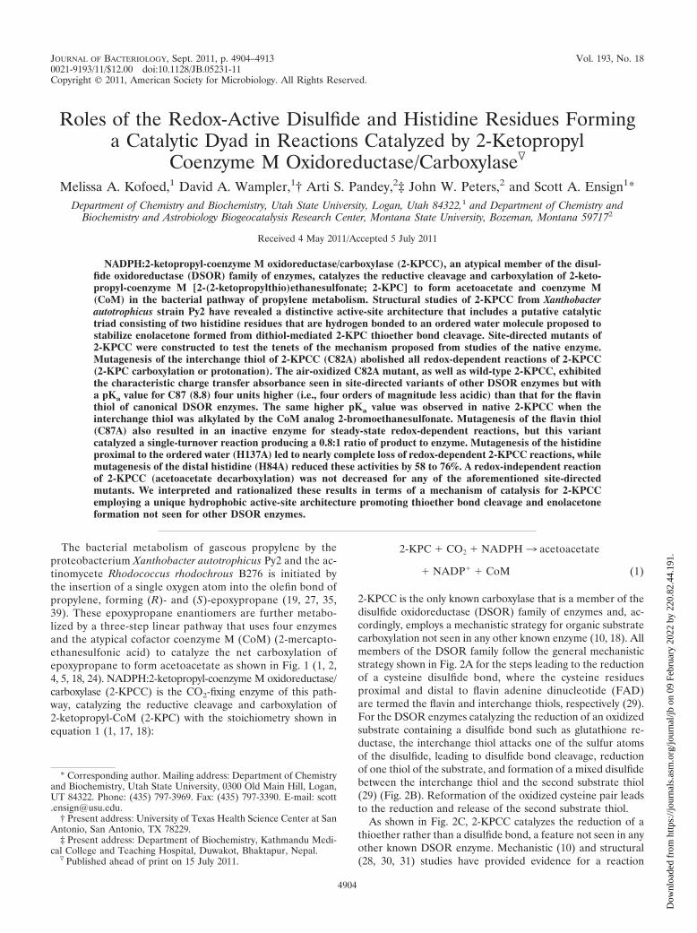
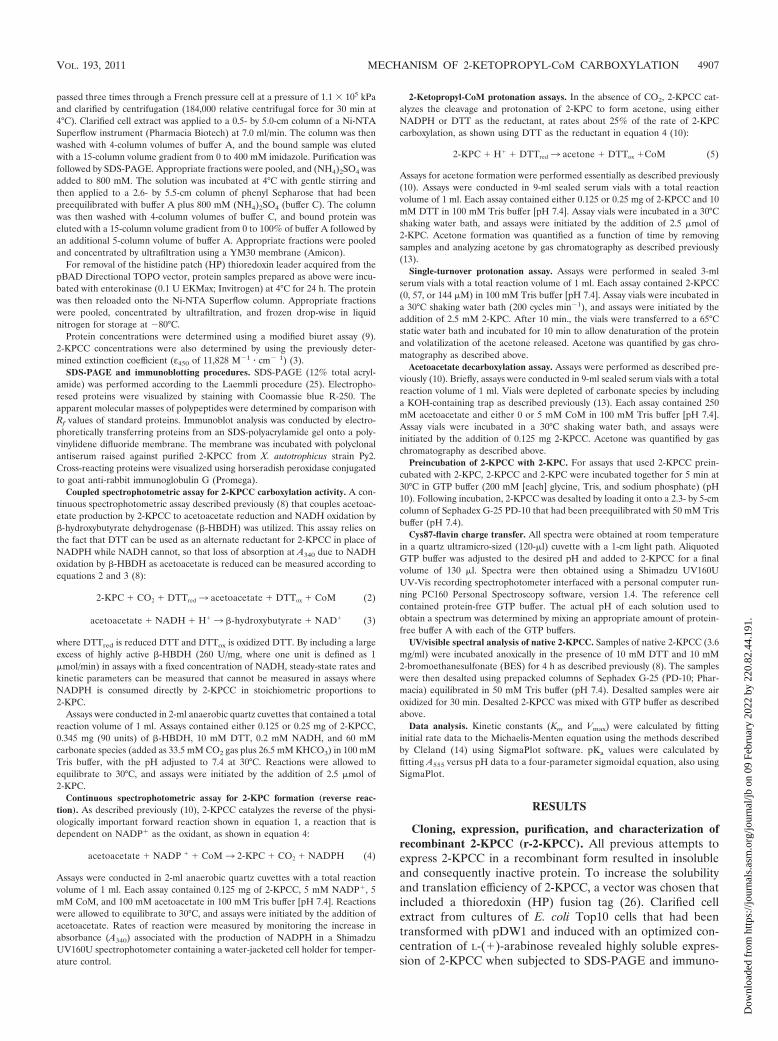
In order to characterize the interaction between C87 andFAD in 2-KPCC, we recorded UV/visible spectra for air-oxi-dized samples of native r-2-KPCC and the alanine substitu-tions in the interchange (C82) and flavin (C87) cysteine resi-dues over a range of pH values from 5 to 11. The spectra of thenative recombinant and C87A proteins were indistinguishablefrom that of the spectrum of oxidized native 2-KPCC, and nochange in the spectra were observed over the range of pHvalues from 5 to 11 (data not shown). This is as expected, sincefor air-oxidized native r-2-KPCC, C82 and C87 will be in thedisulfide form and C87A has no flavin thiol and hence nocharge transfer can develop. As shown in representative spec-tra for C82A at different pH values in Fig. 6, the characteristiccharge transfer due to interaction of the thiolate of C87 withFAD developed only at pH values substantially higher thanphysiological pH, reaching a maximum value at pH 11. This isin stark contrast to other DSOR enzymes where the flavincysteine is largely in the deprotonated form in the microenvi-ronment of the active sites of these enzymes at pH 7, since themicroscopic pKa values are perturbed from the value of 8.2 forfree cysteine to values around 5 (7, 15, 32, 33). The absorbanceat 555 nm was plotted versus pH for a range of pH values todetermine the pKa value for the flavin cysteine (C87) in C82A(Fig. 7, solid circles). A fit of this data provided a pKa of 8.76 0.09, which is fully four units higher (four orders of magnitudeless acidic) than that determined for the classical DSOR en-zymes glutathione reductase (7) and mercuric reductase (33).
Recently, the CoM analog 2-bromoethanesulfonate was shownto be a highly specific affinity label for 2-KPCC that specificallyalkylates the interchange thiol (C82) of 2-KPCC (8). As shownin Fig. 7 (open triangles), the charge transfer absorbance ofBES-modified 2-KPCC developed in a similar fashion to thatof C82A with increasing pH but to a lower maximal value thanfor the mutant. In spite of the lesser change in total absor-bance, a fit of the titration data for BES-modified 2-KPCCyielded a pKa value that is identical within experimental errorto that seen for the C82A mutant (8.74 0.13).
FIG. 6. UV/visible absorption spectra of the r-2-KPCC C82A mu-tant at various pH values. Spectra were obtained using 0.15 mg of2-KPCC diluted to a volume of 130 �l as described in Materials andMethods. The spectral noise at A420 and A535 is due to the ultra-micro-sized cuvette that was used. The spectra shown are for pH valuesof (i) 11.0, (ii) 9.0, (iii) 7.5, and (iv) 5.0.
FIG. 7. Determination of pKa values for the interchange thiol of2-KPCC. The fold increase in A555 due to formation of charge transferabsorbance between FAD and the thiolate of C87 is plotted versus pH.The lines were derived from the four-parameter sigmoidal curve fitused to calculate pKa values. Symbols: F, C82A mutant of r-2-KPCC;‚, native 2-KPCC alkylated on C82 with BES.
VOL. 193, 2011 MECHANISM OF 2-KETOPROPYL-CoM CARBOXYLATION 4909
Dow
nloa
ded
from
http
s://j
ourn
als.
asm
.org
/jour
nal/j
b on
09
Febr
uary
202
2 by
220
.82.
44.1
91.

Redox-dependent reactions of H84A and H137A 2-KPCC.The structures of 2-KPCC show the presence of a “catalyticdyad” of histidine molecules (H137 and H84) that are hydro-gen bonded to an ordered water molecule that is in turn withinhydrogen-bonding distance of the carbonyl oxygen of 2-KPC inthe 2-KPC-bound structure (Fig. 3) (28). This unique pair ofhistidine residues is not seen in any other member of theDSOR family and has been proposed to stabilize enolacetoneformed from 2-KPC thioether bond cleavage (28) (Fig. 3B).
In order to test the proposed roles of H137 and H84 inthe redox-dependent reactions of 2-KPCC, we individually mu-tated the residues to alanine residues and determined theeffects on specific activities for 2-KPC carboxylation (equation2), 2-KPC protonation (equation 5), and 2-KPC formationfrom acetoacetate (equation 4). As shown in Table 1, mutagen-esis of the histidine proximal to the ordered water (H137)resulted in drastically reduced activities (92 to 100% loss ofactivity) for the three redox-dependent activities of 2-KPCC.Mutagenesis of the distal histidine resulted in more moderatelosses of activity (58 to 76%).
Effect of mutations in the redox-active disulfide and cata-lytic dyad on acetoacetate decarboxylase activity of 2-KPCC.2-KPCC was previously shown to catalyze a reaction that doesnot involve redox chemistry: the decarboxylation of acetoace-tate to produce acetone and CO2 according to equation 6 (10):
acetoacetate � H�3 acetone � CO2 (6)
Acetoacetate decarboxylase activity was very low when cata-lyzed by 2-KPCC alone but was stimulated 77-fold upon theaddition of a saturating concentration (5 mM) of CoM (10).
The rate of CoM-dependent acetoacetate decarboxylationby native r-2-KPCC was found to be comparable to that pre-
viously reported for native 2-KPCC (10). As shown in Fig. 8,the rates of CoM-dependent acetoacetate decarboxylation byH137A and H84A were comparable to that of the wild-typeenzyme, while C82A and C87A exhibited rates that were some-what higher than that of the native enzyme. As for native2-KPCC, acetoacetate decarboxylase activity in each of thesite-directed variants was approximately 70- to 80-fold lowerwhen CoM was not included in the assays.
Kinetic characterization of a mutation in one of the methio-nines flanking 2-KPC. As shown in Fig. 3A, a distinguishingfeature of 2-KPCC is the presence of two methionine residues(M140 and M361) that flank the substrate 2-KPCC. An alaninesubstitution was successfully created in M140, but unfortu-nately, attempts to make and express a corresponding sub-stitution in M361 were unsuccessful. As shown in Table 2,the M140A substitution resulted in an enzyme with a 15-fold-higher Km for the substrate 2-KPC and a 3-fold-lowerkcat, resulting in an overall catalytic efficiency for 2-KPCcarboxylation that is 47-fold lower than that for the nativeenzyme.
DISCUSSION
In the present work, 2-KPCC has for the first time beenexpressed in a heterologous system. The biochemical and ki-netic properties of r-2-KPCC containing the HP tag show thatit is essentially identical to that of native 2-KPCC and thussuitable for construction of, and comparative studies with, site-directed variants. The characterization of five of these site-directed variants has allowed key tenets of the mechanism ofthioether bond cleavage and substrate carboxylation proposedfrom mechanistic (8, 10) and structural (28, 30, 31) studies tobe tested.
Catalytic roles and unique properties of the redox-activedisulfide of 2-KPCC. As expected, the redox-active cysteines of2-KPCC are crucial for all steady-state reactions requiringthioether bond cleavage or formation (equations 1, 2, 4, and 5),consistent with the key intermediacy of the Cys82-CoM mixeddisulfide in these reactions (Fig. 3B). The CoM-Cys82 mixeddisulfide has previously been captured for native 2-KPCC byincubating the enzyme in the presence of 20 mM CoM prior tocrystallization (31). The 2-KPCC with a C87A substitutioncatalyzed the single-turnover production of acetone from2-KPC, demonstrating that the interchange thiol (C82) is stillcapable of thioether bond cleavage and presumably CoM-Cys82 mixed disulfide formation in the absence of the flavinthiol. The fact that the addition of DTT did not lead to anincrease in acetone production in these assays indicates thatDTT is not capable of directly reducing the CoM-C82 mixeddisulfide, as by doing so C82 would be rereduced for additionalrounds of catalysis.
FIG. 8. Acetoacetate decarboxylase activity of wild-type and mu-tant r-2-KPCC proteins. Assays contained 0.125 mg of 2-KPC, 250 mMacetoacetate, and 5 mM CoM. Data points represent the averagesof duplicate experiments. Symbols: F, wild-type r-2-KPCC; E,H84A r-2-KPCC; �, H137A r-2-KPCC; ‚, C87A r-2-KPCC; f, C82Ar-2-KPCC.
TABLE 2. Kinetic parameters for the wild-type and M140A r-2-KPCC mutanta
Enzyme Km for 2-KPC(mM)
Change in Km(x-fold) kcat (min�1) Change in kcat
(x-fold)kcat/Km
(mM�1 � min�1)
wt r-2-KPCC 0.42 0.08 1 13.3 1 32M140A r-2-KPCC 6.37 0.82 15.3 4.3 0.32 0.68
a Apparent Km and kcat values for 2-KPC carboxylation were determined using the coupled spectrophotometric assay described in Materials and Methods.
4910 KOFOED ET AL. J. BACTERIOL.
Dow
nloa
ded
from
http
s://j
ourn
als.
asm
.org
/jour
nal/j
b on
09
Febr
uary
202
2 by
220
.82.
44.1
91.

These observations provide insights into how DTT medi-ates the redox-dependent reactions of 2-KPCC in place ofNADPH. As noted above, the rates of DTT-dependent2-KPC protonation or carboxylation are about 60% of the ratesobserved when NADPH is used as the reductant. It is pre-sumed that DTT reduces the oxidized disulfide of C82 and C87directly, i.e., without the intermediacy of FAD. Incubation ofair-oxidized 2-KPCC in the absence of substrates results inreduction of the redox-active disulfide, as evidenced by theability of the DTT-reduced enzyme to undergo stoichiometricalkylation on C82 by the CoM analog BES (8). Prolongedincubation of 2-KPCC with DTT results in bleaching of theflavin of 2-KPCC, but this occurs too slowly (on the order of 30min to an h) to be a relevant pathway for electron transferunder steady-state conditions. In any event, the results pre-sented herein show that DTT is a suitable reductant for2-KPCC when both redox-active thiols are present but notwhen C87 has been replaced with alanine. This suggests thatDTT does not directly reduce the mixed disulfide of C82 andCoM under steady-state turnover conditions but instead re-duces the redox-active cysteine pair.
As shown in Fig. 6 and 7, the flavin thiol (C87) is in the fullyprotonated state at neutral pH for both the alanine substitu-tion in the interchange thiol and for the native enzyme wherethe interchange thiol is alkylated by ethylsulfonate. C87 has apKa value fully 4 units higher (four orders of magnitude morebasic) than the perturbed value for the flavin thiols of othermembers of the DSOR family (7, 33). A key piece of structuralinformation that explains this difference is the replacement ofa histidine side chain that interacts with and lowers the pKa ofthe flavin thiol for all other DSOR enzymes (32) with a phe-nylalanine residue in 2-KPCC (28). It should be noted thatDSOR enzymes are dimers, with each active site being at theinterface between the two subunits and hence interacting withresidues from the second subunit. The histidine that lowers thepKa of the interchange thiol in conventional DSOR enzymesand that is replaced with phenylalanine in 2-KPCC resides onthe other subunit of the dimer (Phe501 of subunit 2 in thisinstance). All of the other active-site residues noted up untilnow reside within the primary subunit forming the active site of2-KPCC (with two such active sites present on each dimericenzyme). The replacement of histidine with phenylalanine inthe environment of the flavin thiol is thought to help maintainthe hydrophobicity of the enzyme-active site and to direct theimmediate product of thioether bond cleavage (enolacetone)toward carboxylation to form acetoacetate (the physiologicallyimportant reaction) rather than protonation to form acetone(a fortuitous side reaction that occurs when CO2 is not pres-ent) (10, 28). The prevention of protonation of enolacetone toform acetone, an essentially irreversible reaction, is of para-mount importance in 2-KPCC. In stark contrast, for glutathi-one reductase, a secondary (and major) role of the histidinethat lowers the pKa of the flavin thiol is to protonate the firstmolecule of glutathione formed upon disulfide bond cleavageto drive the reaction forward and prevent the reverse reactionfrom occurring (32).
The lack of the histidine general base that deprotonates theflavin thiol would be expected to slow the rate of disulfide bondformation by 2-KPCC relative to that of glutathione reductase(compare steps in Fig. 2B and C). However, it is likely that a
step other than disulfide bond formation or another step in-volving oxidation/reduction is rate limiting for 2-KPCC, whichturns over much slower than glutathione reductase. In thiscontext, it should be noted that 2-KPC carboxylation is approx-imately 40% faster when the physiological reductant NADPHis used instead of DTT, indicating that at least one redox stepis partially rate limiting.
It should be noted that the steady state kcat we report herefor 2-KPCC carboxylation (13.3 min�1) is fully 3,300 timeslower than the turnover number reported for glutathione re-duction by glutathione reductase from E. coli (44,000 min�1)(20). The large difference in steady-state turnover highlightsthe more difficult chemistry of thioether bond cleavage andsubstrate carboxylation relative to that of disulfide bond reduc-tion catalyzed by classical DSOR enzymes. The turnover num-bers for the other enzymes of the epoxypropane carboxylationpathway (Fig. 1) are significantly higher (6.5 s�1 for epoxyal-kane:CoM transferase and 25 to 50 s�1 for the R- and S-hydroxypropyl-CoM dehydrogenases [12, 23, 34]). As a reflec-tion of the low turnover of 2-KPCC, it is expressed at very highlevels (25% of soluble cell protein) in propylene-grown cellsof X. autotrophicus Py2 (3, 16). In spite of this high level ofexpression, 2-KPCC has the lowest specific activity of the en-zymes of the epoxide carboxylation pathway in cell extracts,with an activity that is identical to the specific activity of ep-oxypropane consumption by whole-cell suspensions of X. au-totrophicus Py2 growing with propylene as the carbon source(90 nmol of epoxypropane consumed/min/mg [36]). Clearly,the reaction catalyzed by 2-KPCC is the rate-limiting step ofthe epoxide carboxylation pathway shown in Fig. 1. Still, thevery high level of expression of 2-KPCC allows X. autotrophicusand R. rhodochrous to grow fairly well (doubling times in ex-ponential phase of about 8 h) with propylene as the carbonsource (4, 36).
Role of the catalytic histidine dyad in stabilizing enolacetone inreactions involving thioether bond cleavage or breakage. Site-directed mutagenesis of the histidines proximal (H137) anddistal (H84) to the ordered water that interacts with the car-bonyl oxygen of bound 2-KPC in the crystal structure (Fig. 3A)supports the hypothesis that these residues are critical in sta-bilizing enolacetone as an intermediate in reactions that makeor break a thioether bond (equations 1, 2, 4, and 5). As ex-pected, mutation of the proximal histidine resulted in muchlower levels of activity than for the distal histidine, indicatingthat the proximal histidine can still provide some stabilizationin the absence of the additional charge relay provided by H84.
Acetoacetate decarboxylase activity of 2-KPCC does not in-volve the redox-active disulfide or histidine dyad. The decar-boxylation of acetoacetate to acetone and CO2 by 2-KPCCmust of necessity occur by C-C bond cleavage to form enola-cetone, the same high-energy intermediate(s) formed in theredox-dependent reactions of 2-KPCC. The fact that theC82A, C87A, H84A, and H137A mutants all retain full (orslightly increased) CoM-dependent acetoacetate decarboxyl-ase activity demonstrates that acetoacetate decarboxylationmust occur by a fundamentally different mechanism than thatfor the reactions that involve redox chemistry and thioetherbond cleavage. At the same time, acetoacetate decarboxylaseactivity still requires CoM for optimal activity. These resultscan be rationalized and explained by examining the structural
VOL. 193, 2011 MECHANISM OF 2-KETOPROPYL-CoM CARBOXYLATION 4911
Dow
nloa
ded
from
http
s://j
ourn
als.
asm
.org
/jour
nal/j
b on
09
Febr
uary
202
2 by
220
.82.
44.1
91.

features of the form of 2-KPCC captured in the presence of themixed disulfide of CoM (31). This structure showed the pres-ence of acetone bound to a distinct binding pocket 4.5 Å fromthe thiol sulfur of CoM (31). Within this pocket, which consistsof residues from the second enzyme subunit, are Gln509, whichformed a hydrogen bond with the carbonyl O of acetone, andHis506. Based on this structure, it was proposed that thissecond pocket constitutes a binding pocket for the physiolog-ical product acetoacetate (31). By extrapolation, we proposenow that this second pocket is the active site for acetoacetatedecarboxylation. The stimulatory effect of CoM on acetoace-tate decarboxylase activity can be rationalized in one or both ofthe following ways: (i) the thiol of CoM, when bound to theCoM binding pocket of 2-KPCC, is the proton donor for for-mation of acetone from enolacetone, i.e., CoM serves as ageneral acid to promote decarboxylation; and/or (ii), bindingof CoM induces a conformational change similar to that in-duced by binding of the substrate 2-KPC that facilitates thebinding of acetoacetate within the second binding pocket suchthat it can undergo decarboxylation.
Methionine140 modulates Km and kcat for 2-KPC carboxy-lation. A distinctive feature of 2-KPCC relative to other DSORenzymes is the pair of methionine residues that flank the sub-strate 2-KPC (28) (Fig. 3A). The step prior to that catalyzed by2-KPCC in bacterial epoxide metabolism is catalyzed by a pairof stereoselective dehydrogenases that oxidize the R- and S-enantiomers of hydroxypropyl-CoM (HPC) to 2-KPC (1, 6).Interestingly, the crystal structure solved for R-hydroxypropyl-CoM dehydrogenase (R-HPCDH) in the presence of the prod-uct 2-KPC also shows a pair of methionine residues flanking2-KPC in a fashion similar to that seen in 2-KPCC (21). Ad-ditionally, a homology model for the other dehydrogenase,S-HPCDH, also shows flanking methionines (22). Thus, me-thionine residues seem to be a crucial feature in the binding ofCoM thioethers during the steps of bacterial epoxide carbox-ylation. The 2-KPCC mutant with an M140A substitution waskinetically characterized in the present work and found toexhibit both a marked increase in Km and a decrease in kcat
(Table 2). Unfortunately, all attempts to make an alanine sub-stitution in the other methionine (M361) were unsuccessful.Although the crystal structures of 2-KPCC and R-HPCDH donot indicate a catalytic role for the flanking methionines, thisinitial characterization shows that they appear to be crucial forsubstrate binding and catalysis.
Summary. 2-KPCC is distinct from all other known mem-bers of the DSOR family in catalyzing thioether bond cleavageand substrate carboxylation. The results of the present workestablish the essential role of a novel catalytic dyad that facil-itates enolacetone formation and stabilization and a uniqueactive-site environment for the redox-active cysteine pair thatis unlike that seen in any other DSOR enzyme. The combina-tion of structural biology and site-directed mutagenesis, to-gether with the characterization of redox-dependent and re-dox-independent reactions, provides a powerful complementfor elucidating mechanistic details for this novel carboxylase.
ACKNOWLEDGMENTS
This work was supported by National Institutes of Health grantGM51805 to S.A.E. and by Department of Energy grant DE-FG02-04ER15563 to J.W.P.
REFERENCES
1. Allen, J. R., D. D. Clark, J. G. Krum, and S. A. Ensign. 1999. A role forcoenzyme M (2-mercaptoethansulfonic acid) in a bacterial pathway of ali-phatic epoxide carboxylation. Proc. Natl. Acad. Sci. U. S. A. 96:8432–8437.
2. Allen, J. R., and S. A. Ensign. 1996. Carboxylation of epoxides to �-ketoacids in cell extracts of Xanthobacter strain Py2. J. Bacteriol. 178:1469–1472.
3. Allen, J. R., and S. A. Ensign. 1997. Characterization of three proteincomponents required for functional reconstitution of the epoxide carboxy-lase multienzyme complex from Xanthobacter strain Py2. J. Bacteriol. 179:3110–3115.
4. Allen, J. R., and S. A. Ensign. 1998. Identification and characterization ofepoxide carboxylase activity in cell extracts of Nocardia corallina strain B276.J. Bacteriol. 180:2072–2078.
5. Allen, J. R., and S. A. Ensign. 1997. Purification to homogeneity and recon-stitution of the individual components of the epoxide carboxylase multipro-tein enzyme complex from Xanthobacter strain Py2. J. Biol. Chem. 272:32121–32128.
6. Allen, J. R., and S. A. Ensign. 1999. Two short-chain dehydrogenases conferstereoselectivity for enantiomers of epoxypropane in the multiprotein epox-ide carboxylating systems of Xanthobacter strain Py2 and Nocardia corallinaB276. Biochemistry 38:247–256.
7. Arscott, L. D., C. Thorpe, and C. H. J. Williams. 1981. Glutathione reductasefrom yeast. Differential reactivity of the nascent thiols in two-electron re-duced enzyme and properties of a monoalkylated derivative. Biochemistry20:1513–1520.
8. Boyd, J. M., D. D. Clark, M. A. Kofoed, and S. A. Ensign. 2010. Mechanismof inhibition of aliphatic epoxide carboxylation by the coenzyme M analog2-bromoethanesulfonate. J. Biol. Chem. 285:25232–25242.
9. Chromy, V., J. Fischer, and V. Kulhanek. 1974. Re-evaluation of EDTA-chelated biuret reagent. Clin. Chem. 20:1362–1363.
10. Clark, D. D., J. R. Allen, and S. A. Ensign. 2000. Characterization of fivecatalytic activities associated with the NADPH:2-ketopropyl-coenzyme M[2-(2-ketopropylthio)ethanesulfonate] oxidoreductase/carboxylase of theXanthobacter strain Py2 epoxide carboxylase system. Biochemistry 39:1294–1304.
11. Clark, D. D., J. M. Boyd, and S. A. Ensign. 2004. The stereoselectivity andcatalytic properties of Xanthobacter autotrophicus 2-[(R)-2-hydroxypropylth-io]ethanesulfonate dehydrogenase are controlled by interactions betweenC-terminal arginine residues and the sulfonate of coenzyme M. Biochemistry43:6763–6771.
12. Clark, D. D., and S. A. Ensign. 2002. Characterization of the 2-[(R)-2-hydroxypropylthio]ethanesulfonate dehydrogenase from Xanthobacter strainPy2: product inhibition, pH dependence of kinetic parameters, site-directedmutagenesis, rapid equilibrium inhibition, and chemical modification. Bio-chemistry 41:2727–2740.
13. Clark, D. D., and S. A. Ensign. 1999. Evidence for an inducible nucleotide-dependent acetone carboxylase in Rhodococcus rhodochrous B276. J. Bacte-riol. 181:2752–2758.
14. Cleland, W. W. 1979. Statistical analysis of enzyme kinetic data. MethodsEnzymol. 63:103–138.
15. Distefano, M. D., K. G. Au, and C. T. Walsh. 1989. Mutagenesis of theredox-active disulfide in mercuric ion reductase: catalysis by mutant enzymesrestricted to flavin redox chemistry. Biochemistry 28:1168–1183.
16. Ensign, S. A. 1996. Aliphatic and chlorinated alkenes and epoxides as in-ducers of alkene monooxygenase and epoxidase activities in Xanthobacterstrain Py2. Appl. Environ. Microbiol. 62:61–66.
17. Ensign, S. A. 2001. Microbial metabolism of aliphatic alkenes. Biochemistry40:5845–5853.
18. Ensign, S. A., and J. R. Allen. 2003. Aliphatic epoxide carboxylation. Annu.Rev. Biochem. 72:55–76.
19. Gallagher, S. C., R. Cammack, and H. Dalton. 1997. Alkene monooxygenasefrom Nocardia corallina B-276 is a member of the class of dinuclear ironproteins capable of stereospecific epoxygenation reactions. Eur. J. Biochem.247:635–641.
20. Henderson, G. B., et al. 1991. Engineering the substrate specificity of gluta-thione reductase toward that of trypanothione reduction. Proc. Natl. Acad.Sci. U. S. A. 88:8769–8773.
21. Krishnakumar, A. M., B. P. Nocek, D. D. Clark, S. A. Ensign, and J. W.Peters. 2006. Structural basis for stereoselectivity in the (R)- and (S)-hy-droxypropylthioethanesulfonate dehydrogenases. Biochemistry 45:8831–8840.
22. Krishnakumar, A. M., et al. 2008. Getting a handle on the role of coenzymeM in alkene metabolism. Microbiol. Mol. Biol. Rev. 72:445–456.
23. Krum, J. G., H. Ellsworth, R. R. Sargeant, G. Rich, and S. A. Ensign. 2002.Kinetic and microcalorimetric analysis of substrate and cofactor interactionsin epoxyalkane:CoM transferase, a zinc-dependent epoxidase. Biochemistry41:5005–5014.
24. Krum, J. G., and S. A. Ensign. 2000. Heterologous expression of bacterialepoxyalkane:coenzyme M transferase and inducible coenzyme M biosynthe-sis in Xanthobacter strain Py2 and Rhodococcus rhodochrous B276. J. Bacte-riol. 182:2629–2634.
4912 KOFOED ET AL. J. BACTERIOL.
Dow
nloa
ded
from
http
s://j
ourn
als.
asm
.org
/jour
nal/j
b on
09
Febr
uary
202
2 by
220
.82.
44.1
91.

25. Laemmli, U. K. 1970. Cleavage of structural proteins during the assembly ofthe head of bacteriophage T4. Nature 227:680–685.
26. Lu, Z., et al. 1996. Histidine patch thioredoxins. Mutant forms of thioredoxinwith metal chelating affinity that provide for convenient purifications ofthioredoxin fusion proteins. J. Biol. Chem. 271:5059–5065.
27. Miura, A., and H. Dalton. 1995. Purification and characterization of thealkene monooxygenase from Nocardia corallina B-276. Biosci. Biotechnol.Biochem. 59:853–859.
28. Nocek, B., et al. 2002. Structural basis for CO2 fixation by a novel member ofthe disulfide oxidoreductase family of enzymes, 2-ketopropyl-coenzyme Moxidoreductase/carboxylase. Biochemistry 41:12907–12913.
29. Pai, E. F. 1991. Variations on a theme: the family of FAD-dependentNAD(P)H-(disulphide)-oxidoreductases. Curr. Opin. Struct. Biol. 1:796–803.
30. Pandey, A. S., D. W. Mulder, S. A. Ensign, and J. W. Peters. 2011. Structuralbasis for carbon dioxide binding by 2-ketopropyl coenzyme M oxidoreduc-tase/carboxylase. FEBS Lett. 585:459–464.
31. Pandey, A. S., B. Nocek, D. D. Clark, S. A. Ensign, and J. W. Peters. 2006.Mechanistic implications of the structure of the mixed-disulfide intermediateof the disulfide oxidoreductase, 2-ketopropyl-coenzyme M oxidoreductase/carboxylase. Biochemistry 45:113–120.
32. Rietveld, P., et al. 1994. Reductive and oxidative half-reactions of glutathi-one reductase from Escherichia coli. Biochemistry 33:13888–13895.
33. Schultz, P. G., K. G. Au, and C. T. Walsh. 1985. Directed mutagenesis of the
redox-active disulfide in the flavoenzyme mercuric ion reductase. Biochem-istry 24:6840–6848.
34. Sliwa, D. A., A. M. Krishnakumar, J. W. Peters, and S. A. Ensign. 2010.Molecular basis for enantioselectivity in the (R)- and (S)-hydroxypropylth-ioethanesulfonate dehydrogenases, a unique pair of stereoselective short-chain dehydrogenases/reductases involved in aliphatic epoxide carboxyla-tion. Biochemistry 49:3487–3498.
35. Small, F. J., and S. A. Ensign. 1997. Alkene monooxygenase from Xantho-bacter strain Py2. Purification and characterization of a four-componentsystem central to the bacterial metabolism of aliphatic alkenes. J. Biol.Chem. 272:24913–24920.
36. Small, F. J., and S. A. Ensign. 1995. Carbon dioxide fixation in the metab-olism of propylene and propylene oxide by Xanthobacter strain Py2. J. Bac-teriol. 177:6170–6175.
37. Swaving, J., C. A. Weijers, A. J. van Ooyen, and J. A. M. de Bont. 1995.Complementation of Xanthobacter Py2 mutants defective in epoxyalkanedegradation, and expression and nucleotide sequence of the complementingDNA fragment. Microbiology 141:477–484.
38. Walsh, C. 1979. Enzymatic reaction mechanisms. W. H. Freeman and Co.,New York, NY.
39. Zhou, N.-Y., C. K. Chan Kwo Chion, and D. J. Leak. 1996. Cloning andexpression of the genes encoding the propene monooxygenase from Xan-thobacter, Py2. Appl. Microbiol. Biotechnol. 44:582–588.
VOL. 193, 2011 MECHANISM OF 2-KETOPROPYL-CoM CARBOXYLATION 4913
Dow
nloa
ded
from
http
s://j
ourn
als.
asm
.org
/jour
nal/j
b on
09
Febr
uary
202
2 by
220
.82.
44.1
91.


![Research Article CrystalStructureofL-Histidinium2 ...chloride monohydrate [2], L-histidine tetrafluoroborate [3], L-histidine hydrochloride monohydrate [4], L-histidine hydrofluoride](https://static.fdocuments.net/doc/165x107/60b51c180636315681384205/research-article-crystalstructureofl-histidinium2-chloride-monohydrate-2.jpg)


